Embed Size (px)
Citation preview
Withoon Ungkitphaiboon
Assistant Professor, Department of Surgery,
Maha Chakri Sirindhorn Medical Center
Srinakharinwirot University
Present in “ One-day in Vascular Disease #11
4 Feb 2560
Troubleshooting Technique forHemodialysis Catheter Insertion
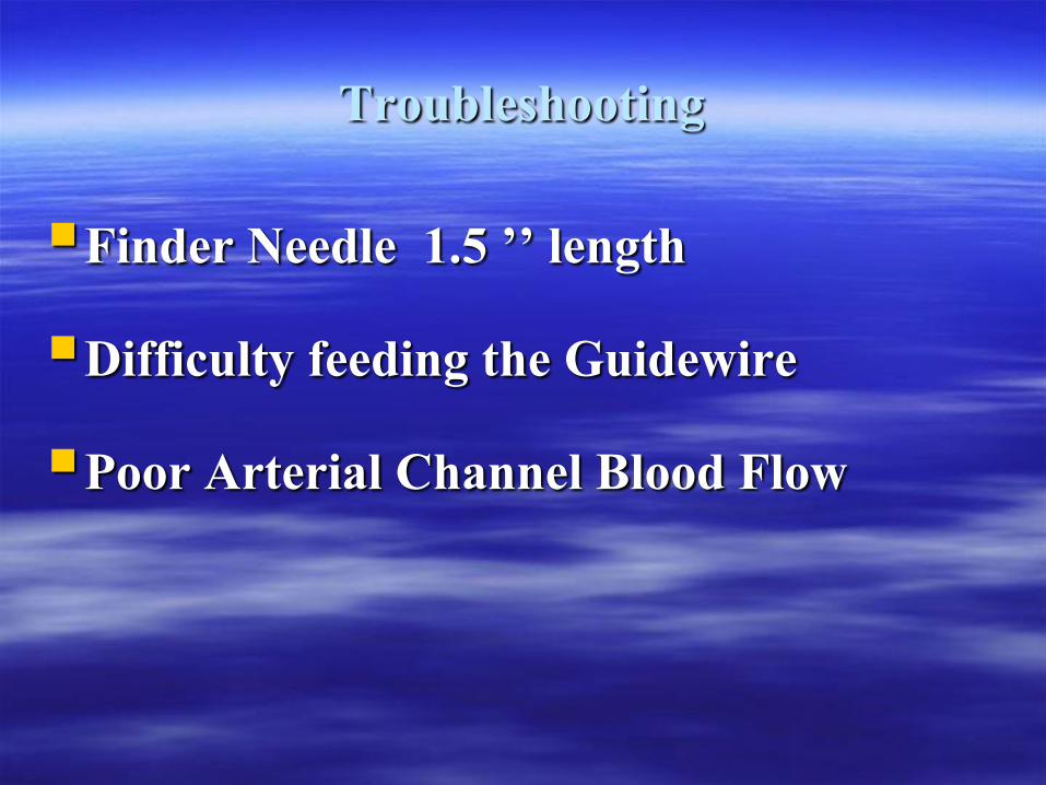
Troubleshooting
Finder Needle 1.5 ’’ length
Difficulty feeding the Guidewire
Poor Arterial Channel Blood Flow
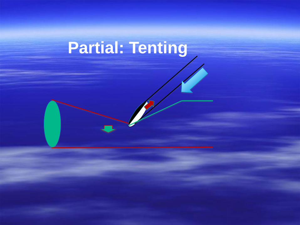
Partial: Tenting
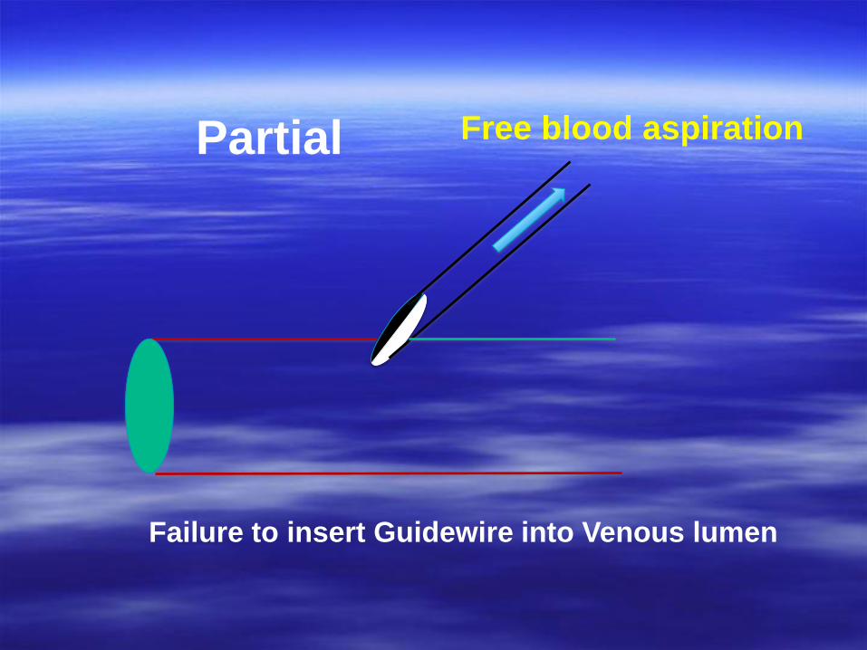
Partial
Failure to insert Guidewire into Venous lumen
Free blood aspiration
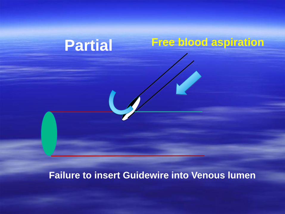
Partial
Failure to insert Guidewire into Venous lumen
Free blood aspiration
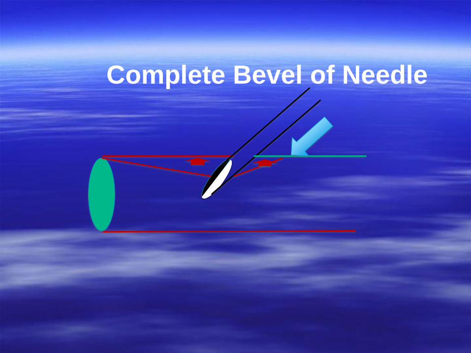
Complete Bevel of Needle
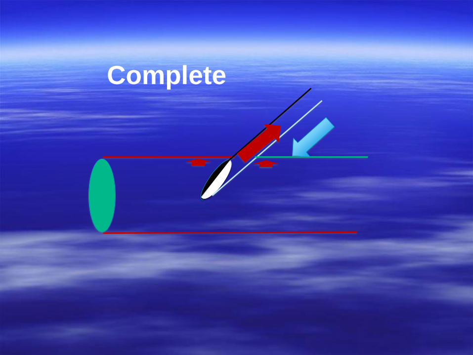
Complete
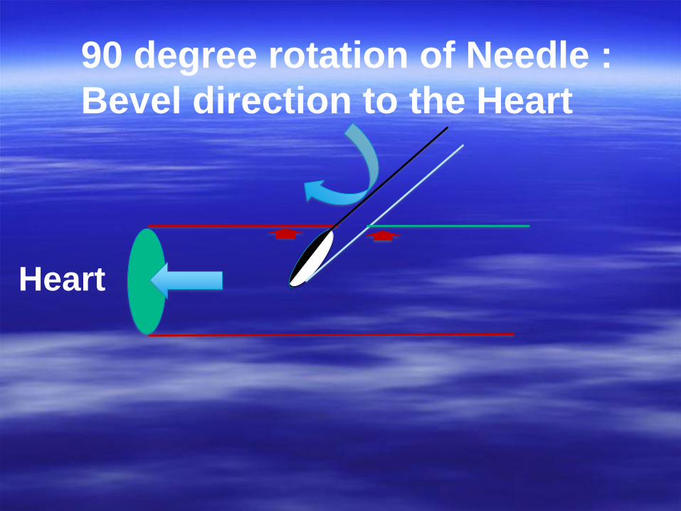
90 degree rotation of Needle :
Bevel direction to the Heart
Heart
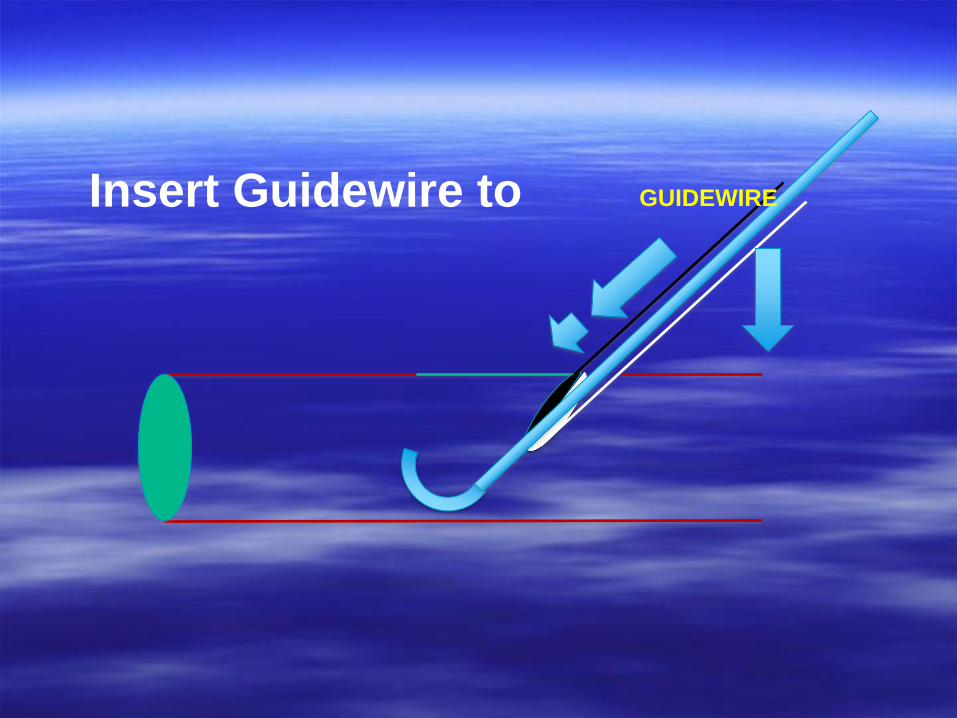
Insert Guidewire to GUIDEWIRE
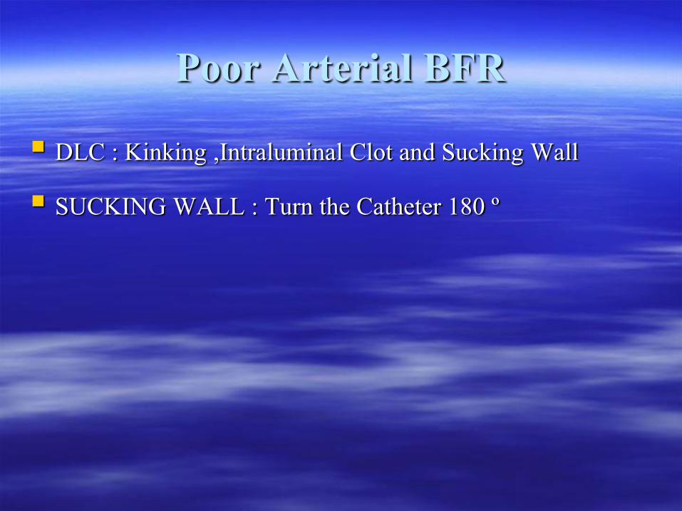
Poor Arterial BFR DLC : Kinking ,Intraluminal Clot and Sucking Wall SUCKING WALL : Turn the Catheter 180 º
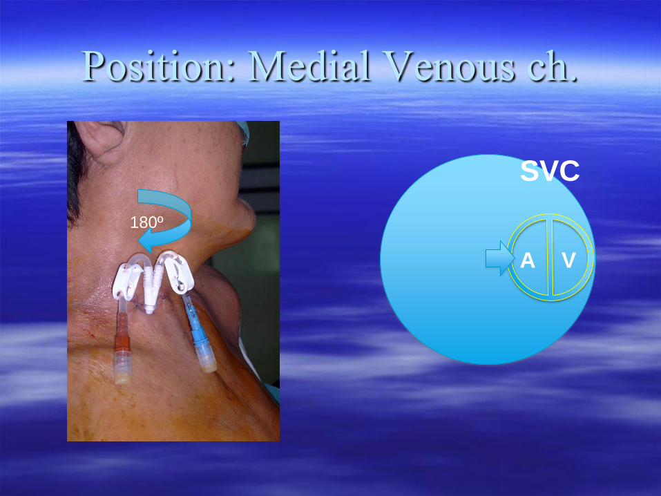
Position: Medial Venous ch.
A V
SVC
180º
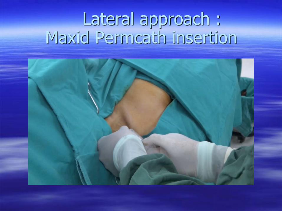
Lateral approach : Maxid Permcath insertion
How to get best result with Catheter insertion ?
Withoon Ungkitphaiboon
Assistant Professor, Department of Surgery,
Maha Chakri Sirindhorn Medical Center
Srinakharinwirot University
Present in “ One-day in Vascular Disease #11
4 Feb 2560
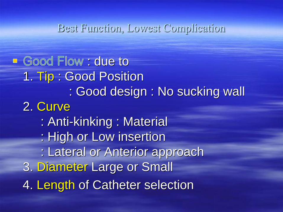
Best Function, Lowest Complication
: due to
1. Tip : Good Position
: Good design : No sucking wall
2. Curve
: Anti-kinking : Material
: High or Low insertion
: Lateral or Anterior approach
3. Diameter Large or Small
4. Length of Catheter selection
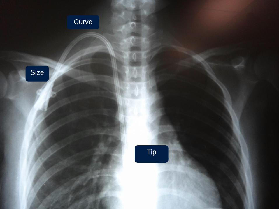
Curve
Tip
Size
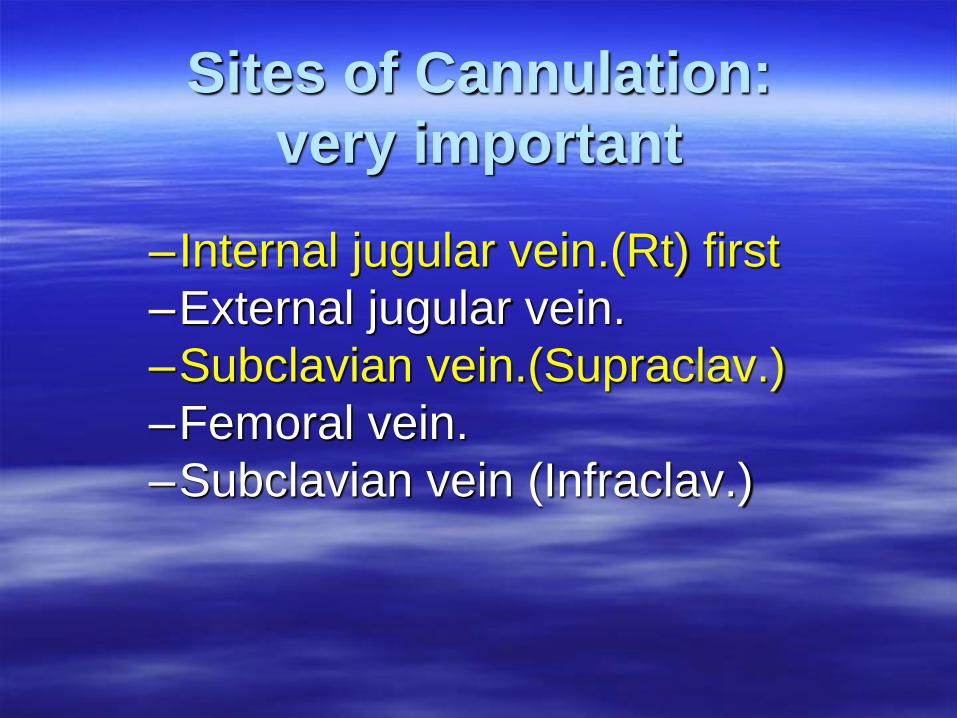
–Internal jugular vein.(Rt) first
–External jugular vein.
–Subclavian vein.(Supraclav.)
–Femoral vein.
–Subclavian vein (Infraclav.)
Sites of Cannulation:
very important
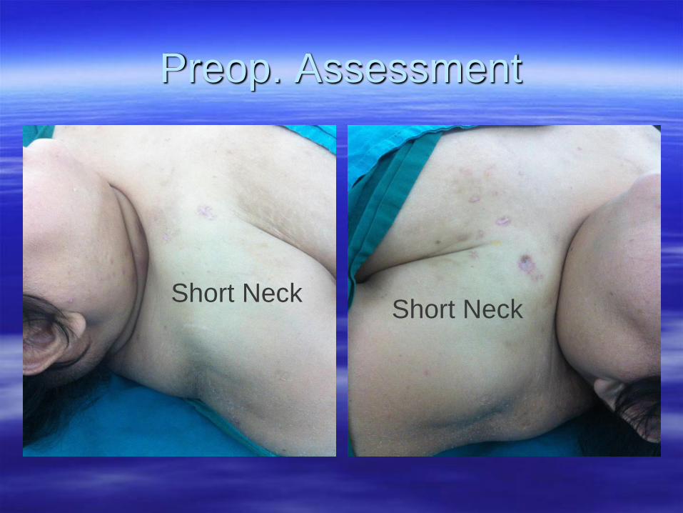
Preop. Assessment
Short NeckShort Neck
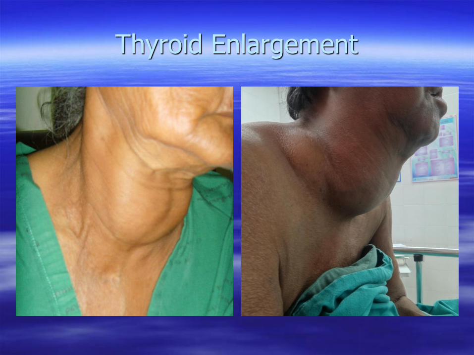
Thyroid Enlargement
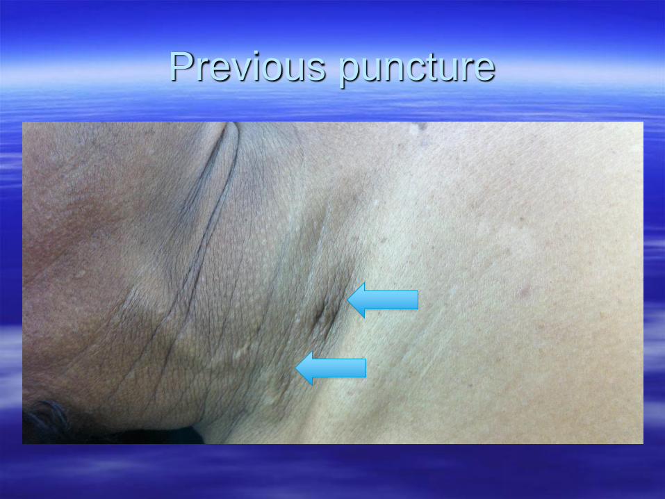
Previous puncture
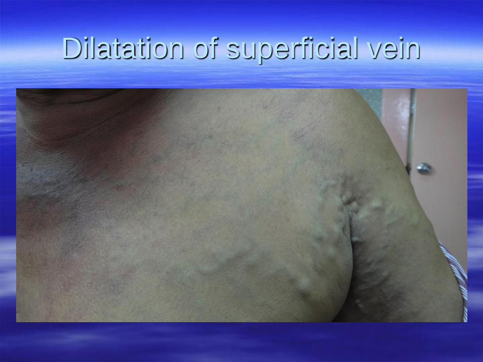
Dilatation of superficial vein
Central Vein stenosis
• Previous Central line or DLC insertion
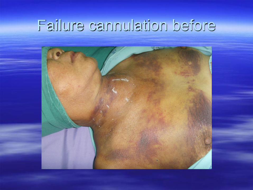
Failure cannulation before
Preop. Imaging: CTV & MRV
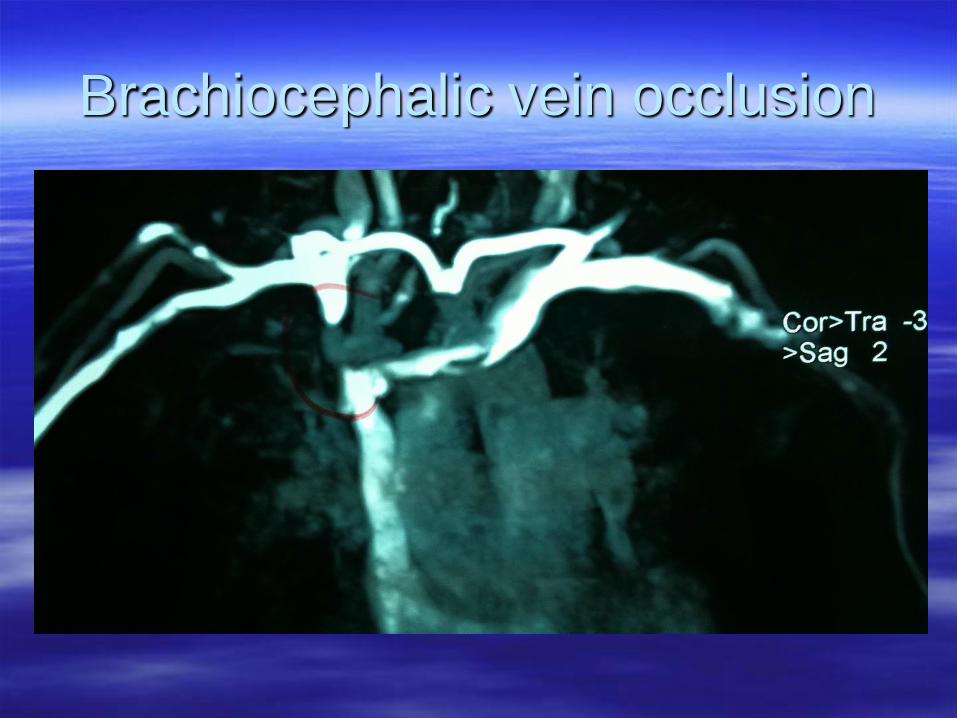
Brachiocephalic vein occlusion
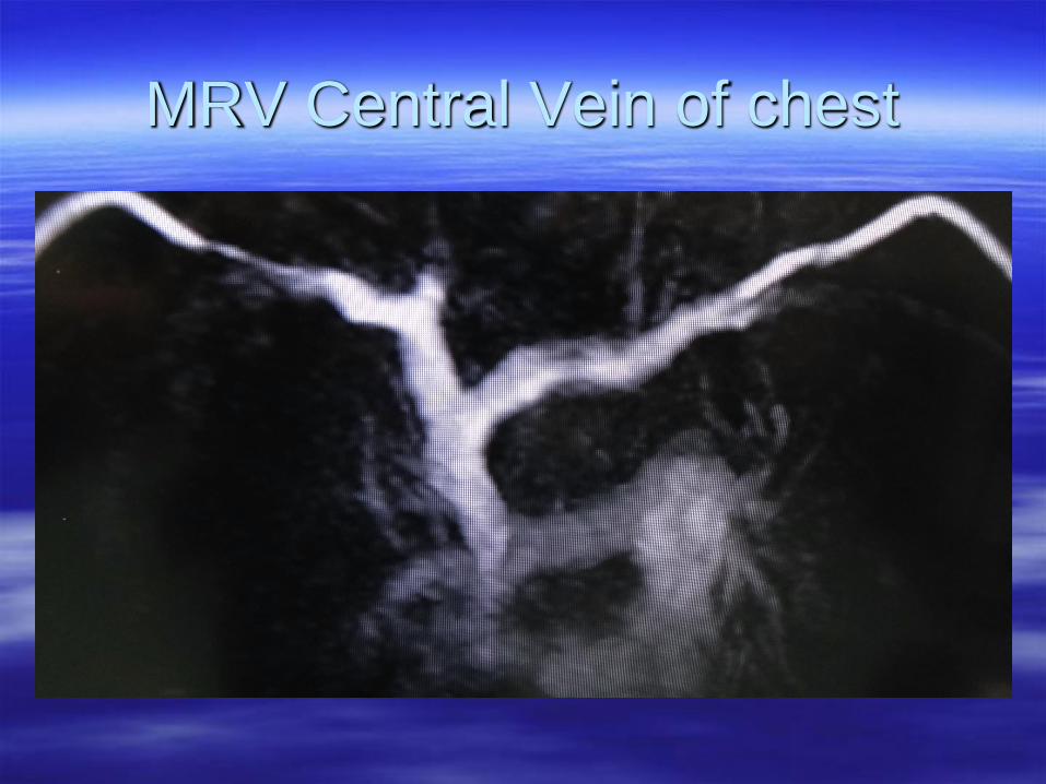
MRV Central Vein of chest
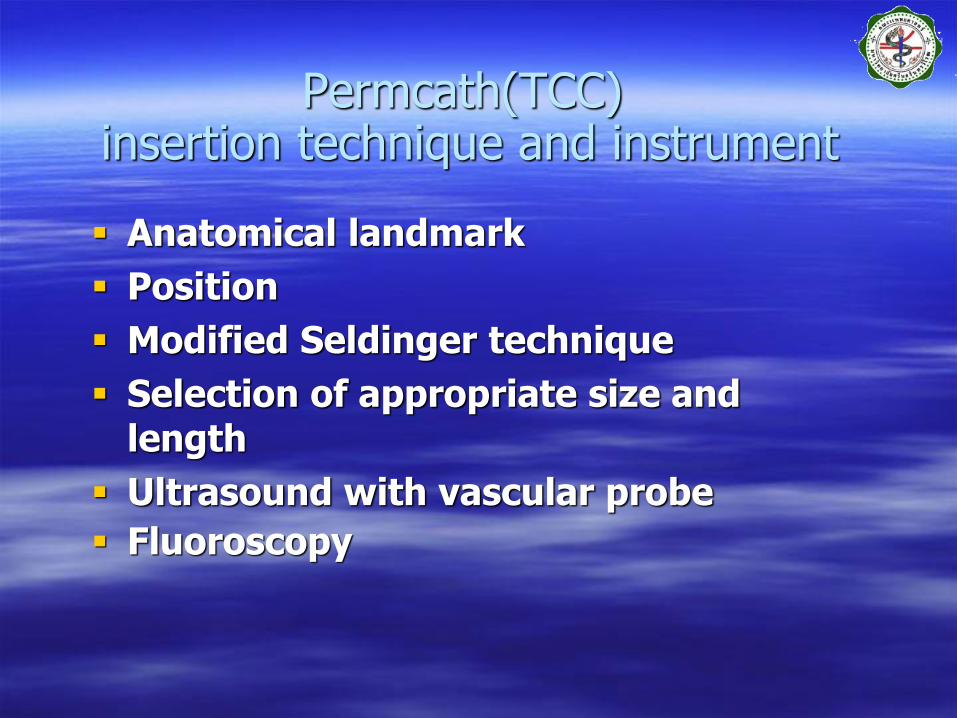
Permcath(TCC)insertion technique and instrument
Anatomical landmark
Position
Modified Seldinger technique
Selection of appropriate size and length
Ultrasound with vascular probe
Fluoroscopy
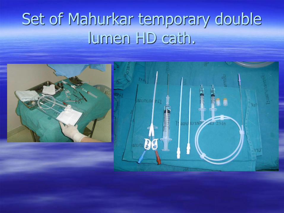
Set of Mahurkar temporary double lumen HD cath.
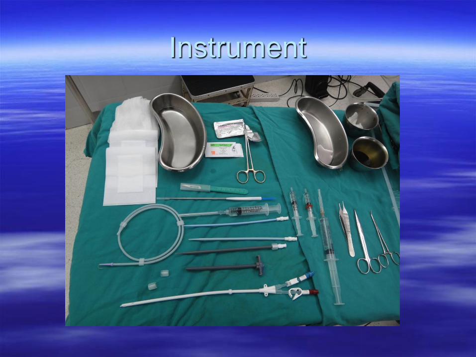
Instrument
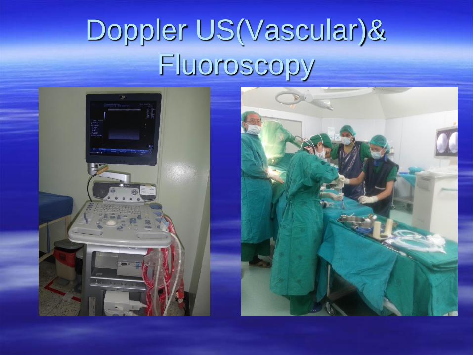
Doppler US(Vascular)&
Fluoroscopy
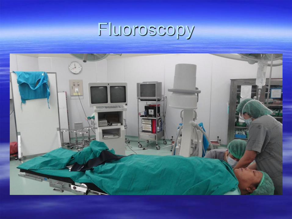
Fluoroscopy
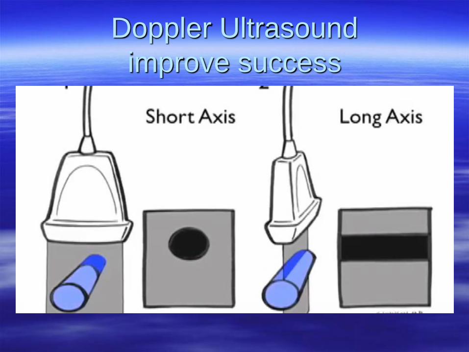
Doppler Ultrasound
improve success
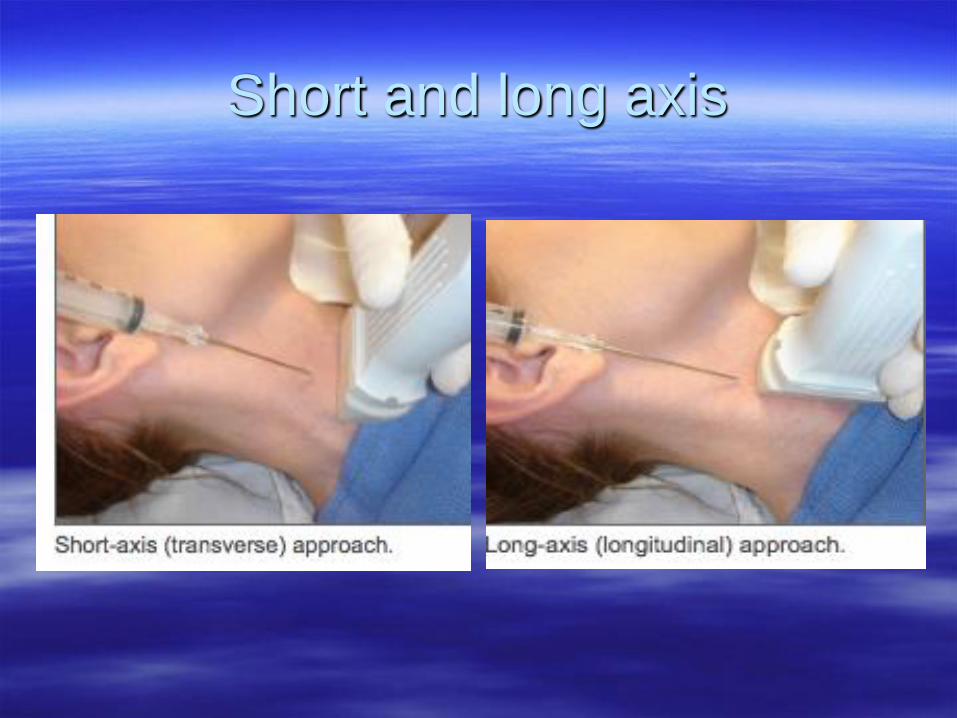
Short and long axis
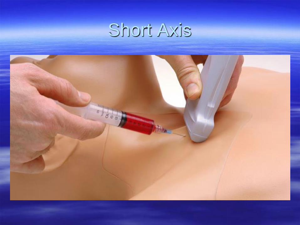
Short Axis
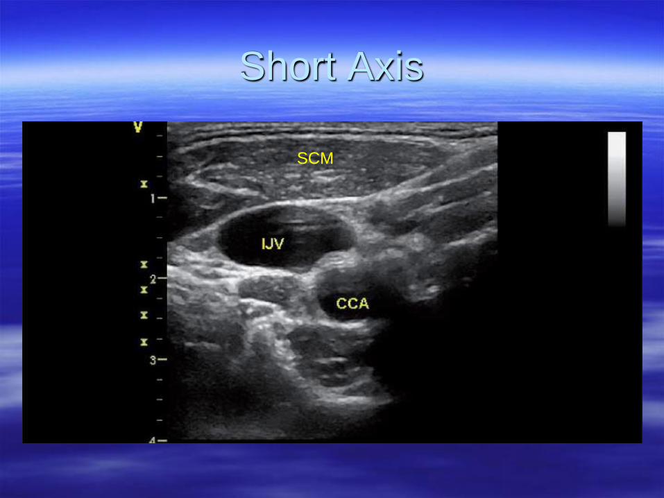
Short Axis
SCM
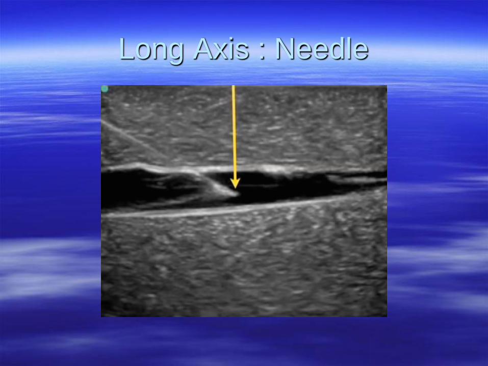
Long Axis : Needle
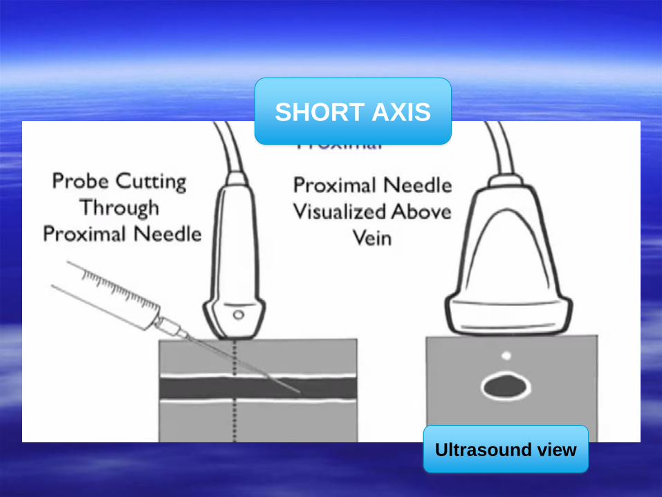
SHORT AXIS
Ultrasound view
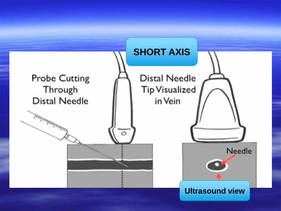
SHORT AXIS
Ultrasound view
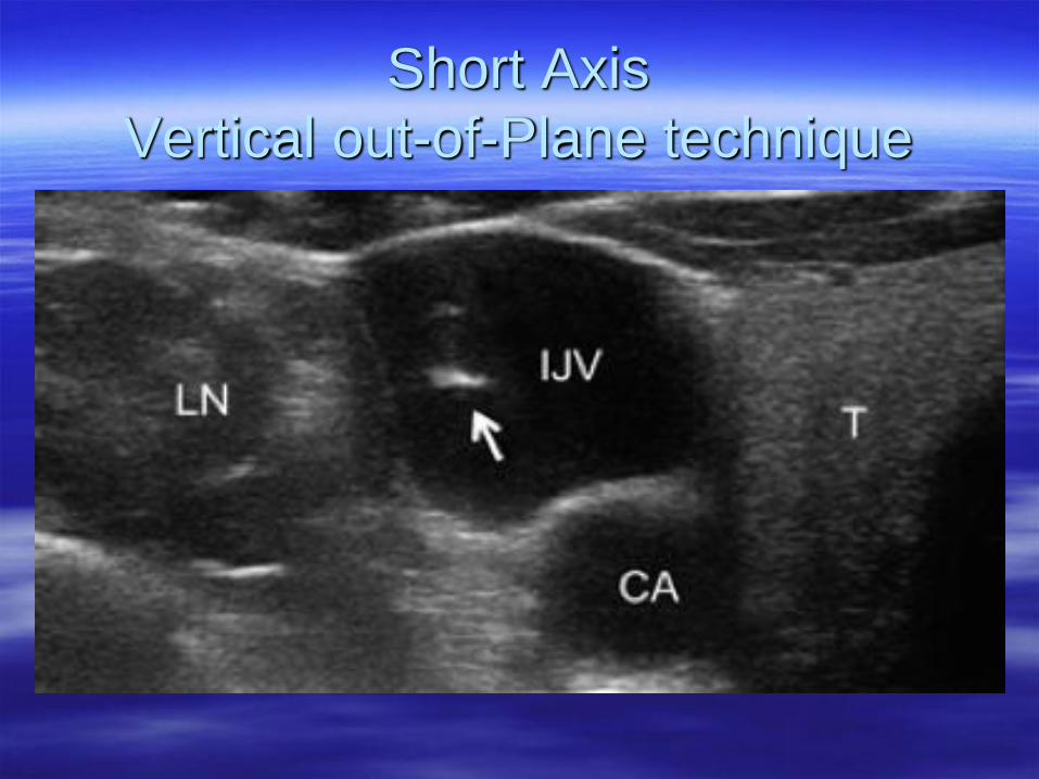
Short Axis
Vertical out-of-Plane technique
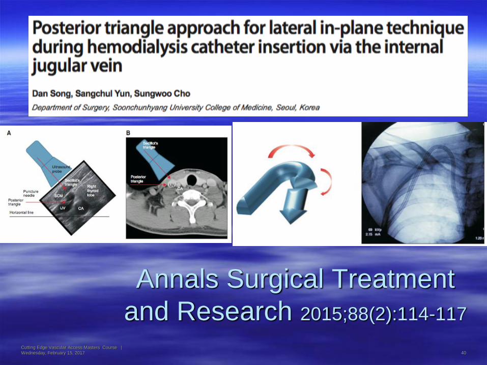
Annals Surgical Treatment
and Research 2015;88(2):114-117
Cutting Edge Vascular Access Masters Course |
Wednesday, February 15, 2017 40
Cutting Edge Vascular Access Masters Course |
Wednesday, February 15, 2017 41
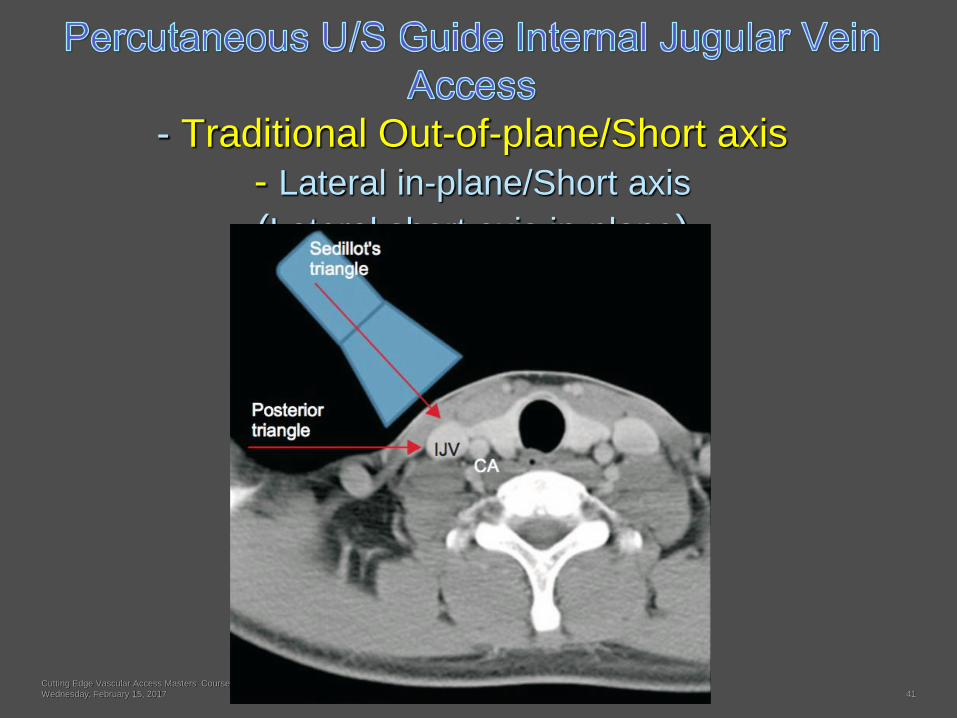
- Traditional Out-of-plane/Short axis
- Lateral in-plane/Short axis
(Lateral short axis in-plane)
Cutting Edge Vascular Access Masters Course |
Wednesday, February 15, 2017 42
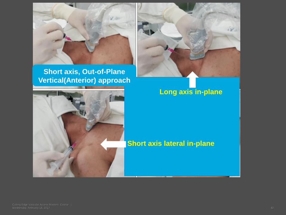
Long axis in-plane
Short axis lateral in-plane
Short axis, Out-of-Plane
Vertical(Anterior) approach
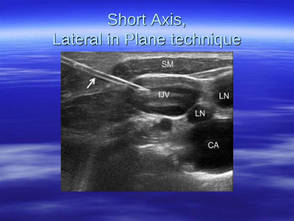
Short Axis,
Lateral in Plane technique
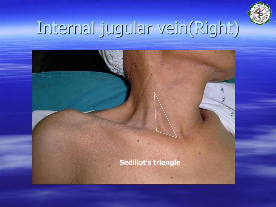
Internal jugular vein(Right)
Sedillot’s triangle
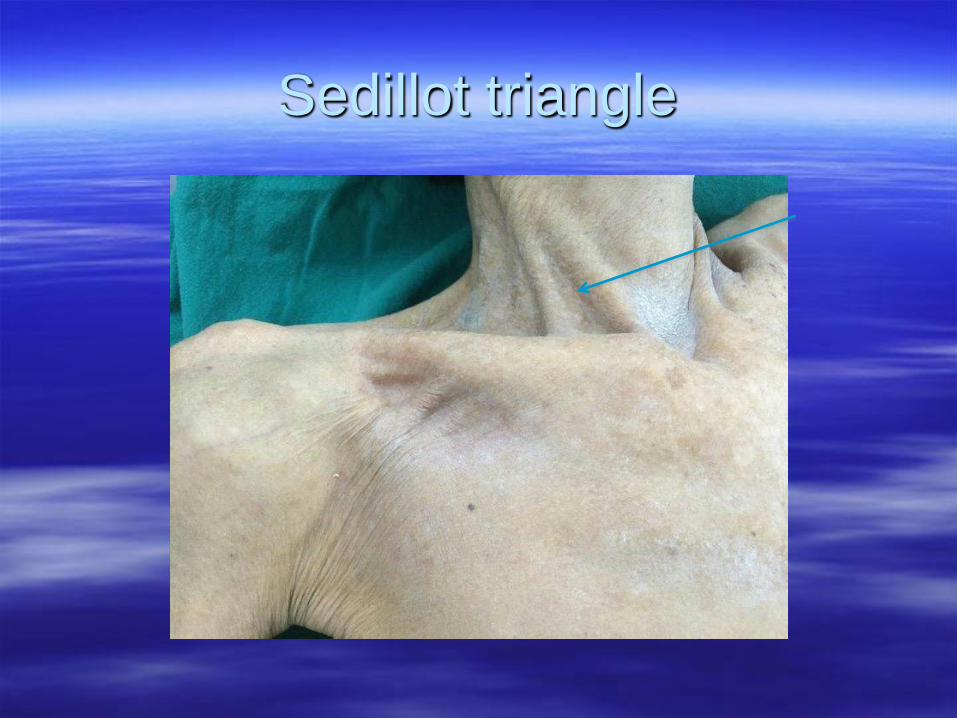
Sedillot triangle
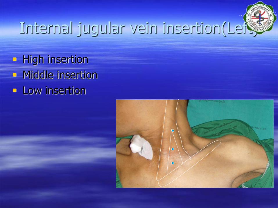
Internal jugular vein insertion(Left)
High insertion
Middle insertion
Low insertion
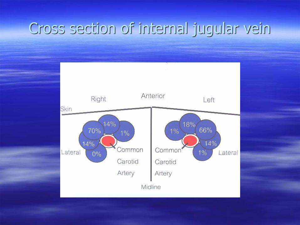
Cross section of internal jugular vein
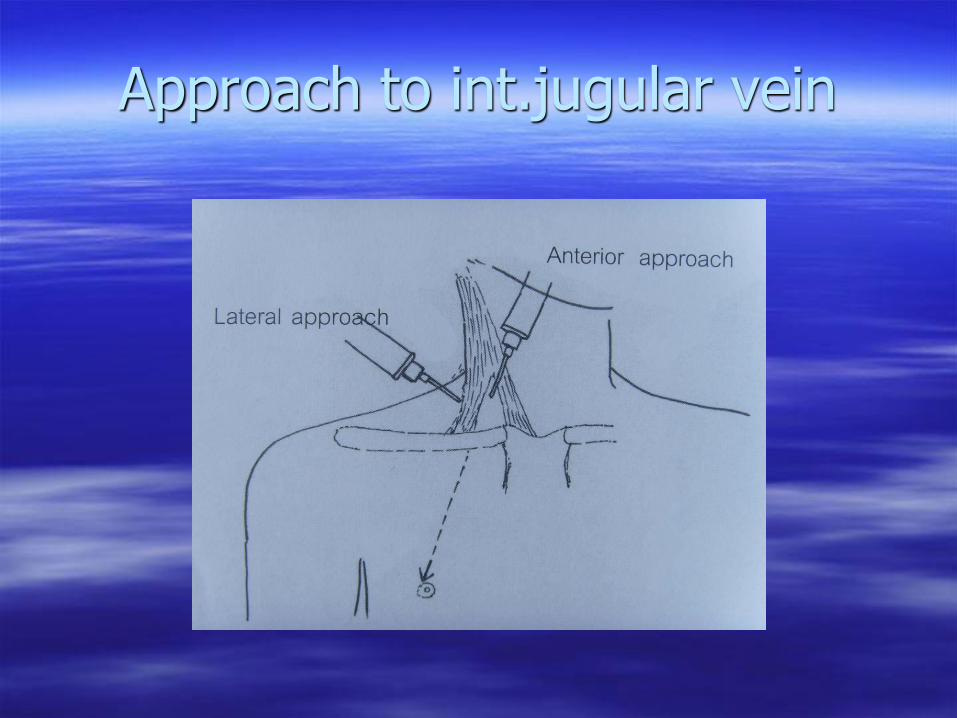
Approach to int.jugular vein
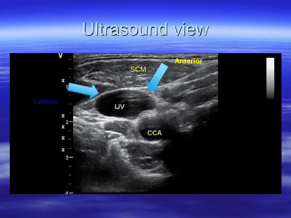
Ultrasound view
SCM
Lateral
Anterior
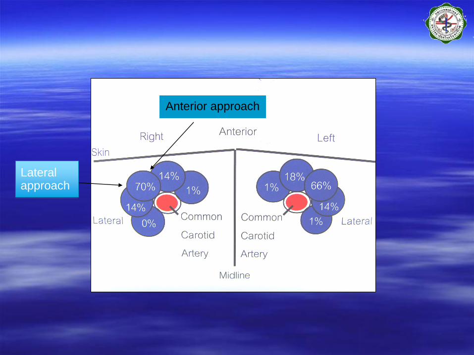
Anterior approach
Lateral approach
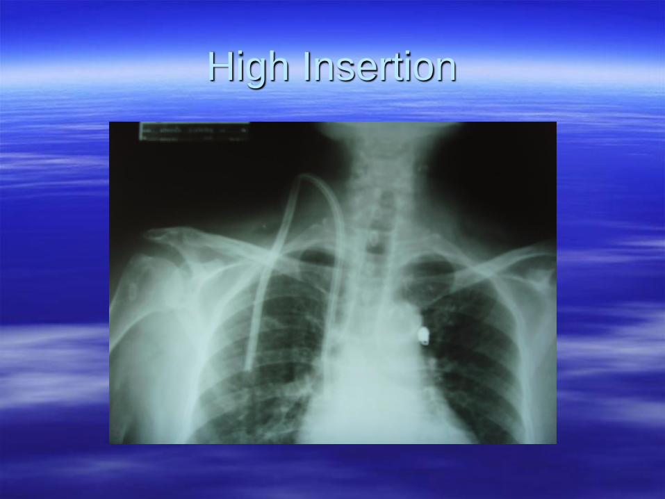
High Insertion
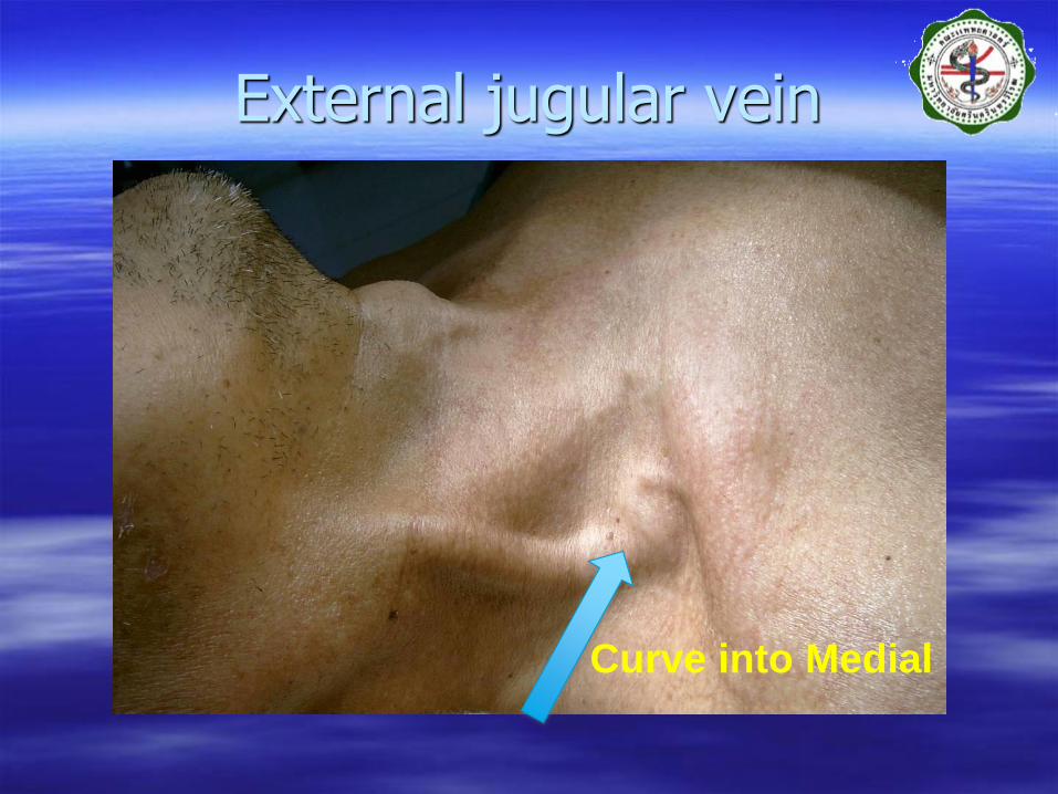
External jugular vein
Curve into Medial
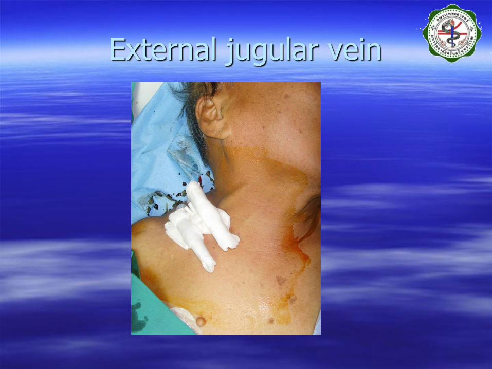
External jugular vein
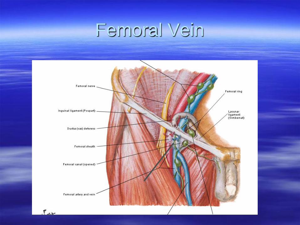
Femoral Vein

Common Site of Puncture
1.(Anterior) IJV.
2.(Infraclavicular)
Subclavian V.
1
2
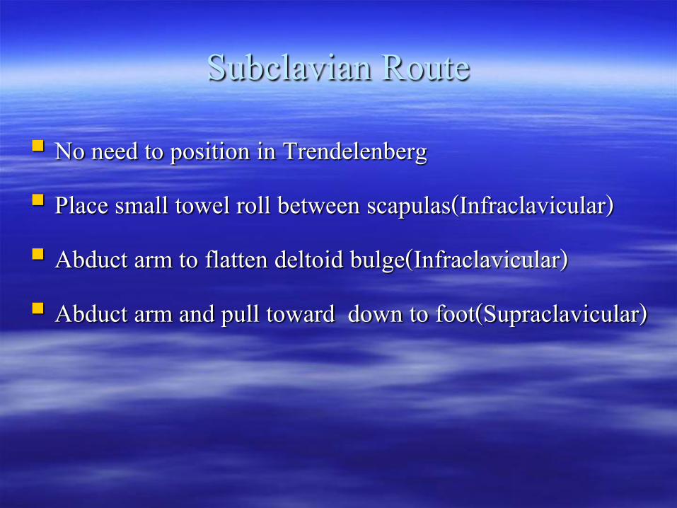
Subclavian Route No need to position in Trendelenberg Place small towel roll between scapulas(Infraclavicular) Abduct arm to flatten deltoid bulge(Infraclavicular) Abduct arm and pull toward down to foot(Supraclavicular)
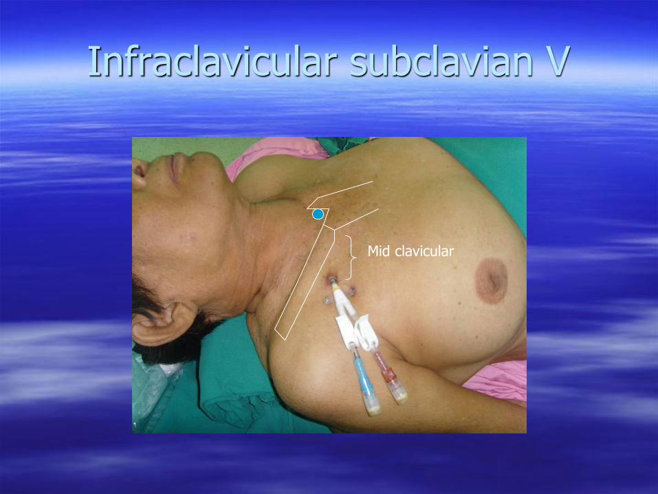
Infraclavicular subclavian V
Mid clavicular
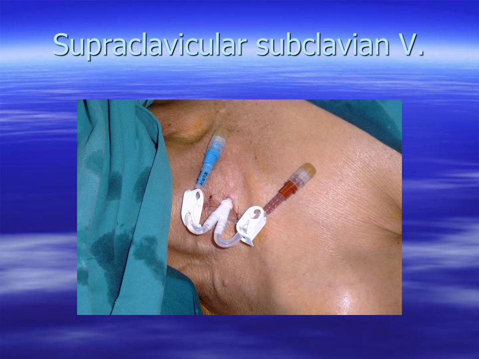
Supraclavicular subclavian V.
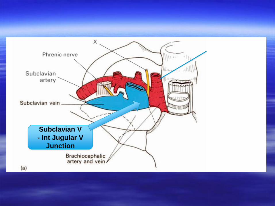
CCA
Subclavian V
- Int Jugular V
Junction
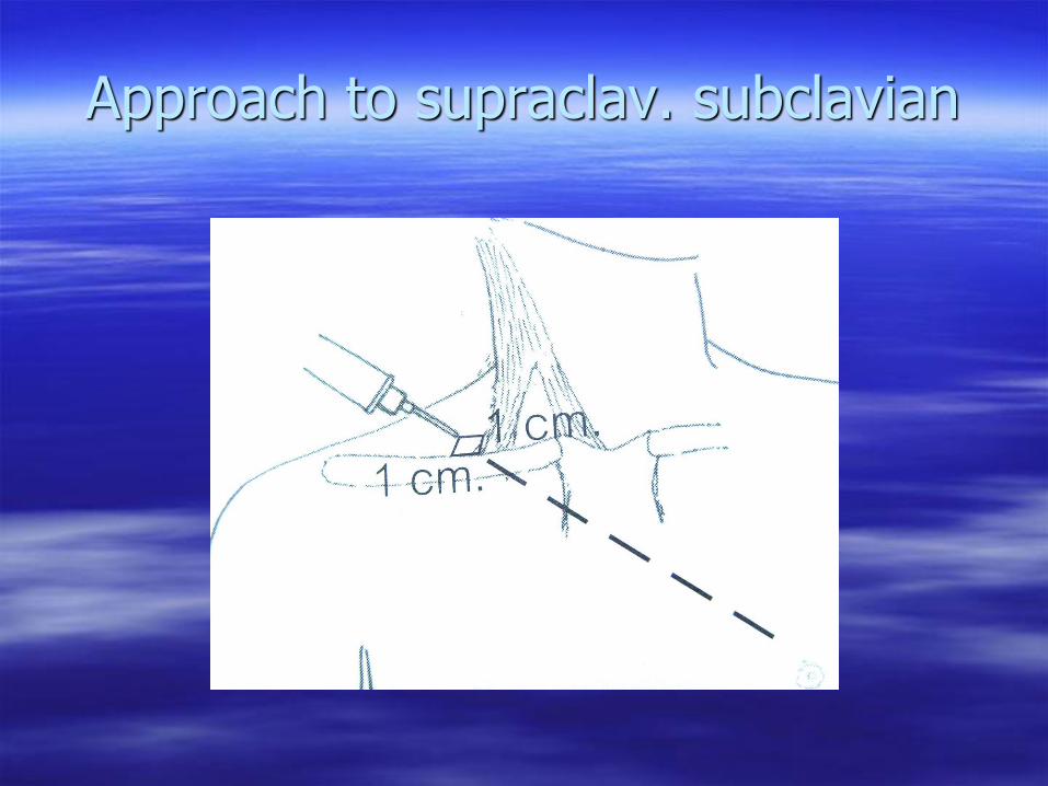
Approach to supraclav. subclavian
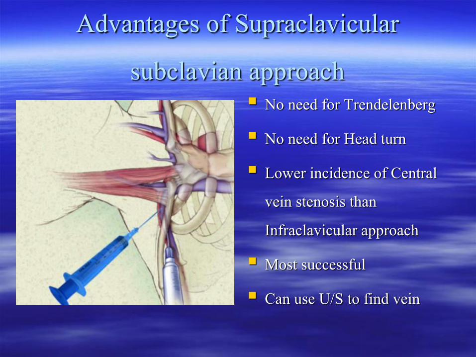
Advantages of Supraclavicular subclavian approach
No need for Trendelenberg No need for Head turn Lower incidence of Central
vein stenosis than Infraclavicular approach
Most successful Can use U/S to find vein


![Unusual Complication of Hemodialysis Cuffed Catheter Tunnel ... · 2019. 7. 30. · hemodialysis patients with vascular access central venous catheter [2, 5]. Infection is the second](https://img.pdfslide.us/doc/110x75/6112f543c4e8093a88485054/unusual-complication-of-hemodialysis-cuffed-catheter-tunnel-2019-7-30-hemodialysis.jpg)