Embed Size (px)
Citation preview

MIGUEL FILIPE TAVARES DA LUZ SOARES
TRINUCLEOTIDE REPEAT SCANNING IN PORTUGUESE
FAMILIAL AMYLOIDOTIC POLYNEUROPATHY KINDREDS
EXHIBITING GENETIC ANTICIPATION
( A
•'''«itaçî*'**̂ »- ' ;',
PORTO & NEW YORK
1997

MIGUEL FILIPE TAVARES DA LUZ SOARES
TRINUCLEOTIDE REPEAT SCANNING IN PORTUGUESE
FAMILIAL AMYLOIDOTIC POLYNEUROPATHY KINDREDS
EXHIBITING GENETIC ANTICIPATION
DISSERTAÇÃO DE CANDIDATURA AO GRAU DE MESTRE APRESENTADA À FACULDADE DE MEDICINA DA UNIVERSIDADE DO PORTO
ORIENTADOR: PROFESSORA MARIA JOÃO MASCARENHAS SARAIVA CO-OPIENTADOR: PROFESSOR JOEL N. BUXBAUM
PORTO & NEW YORK 1997

Abstract
Abstract
Familial Amyloidotic Polyneuropathy is a lethal hereditary autosomal dominant disorder
characterized by the deposition of a mutant form of the serum protein transthyretin (TTR) as
fibrillar material in several tissues and organs. The most common form, FAP Type I or
Portuguese, is characterized by a substitution of Met for Val at position 30 in the TTR molecule.
It exhibits genetic anticipation, with clinical symptoms developing at an earlier age in succeeding
generations.
Expansion of unstable trinucleotide repeats (TNRs) is the underlying mechanism in a
growing number of inherited human disorders exhibiting anticipation, such as Fragile X
Syndrome, Huntington's Disease, Myotonic Dystrophy, and Friedreich's Ataxia. The number of
trinucleotide repeats appears to be correlated with the age of onset, the severity of symptoms, or
both. Although the pathology of FAP is produced by the deposition of amyloid derived from a
mutant TTR, rather than the expansion of unstable TNRs, we view it possible that the
phenotypic variability manifested as anticipation, might be related to TNR expansion in a
modifier gene(s), rattier than TTR gene.
Using the Repeat Expansion Detection (RED) method we have scanned affected
members of FAP type I families from the Portuguese population to determine whether
anticipation within such families correlated with expansion of any of the ten possible
trinucleotide repeats. Eight generational pairs were tested; in 7 (6 parent/child and 1 uncle/niece)
the difference in age of onset was greater than 12 years. A mother/daughter pair, with identical
ages of onset, served as control.
Eight of the ten possible trinucleotide repeats were successfully assayed. No major
differences were found in the lengths of any of the repeats which could be analyzed. Expansion
of the two untested trinucleotides as not been associated with any prior instance of anticipation.
The observed distribution of the maximal triplet repeat sizes, as determined by the RED
method, was consistent with previously reported studies in healthy unrelated individuals from
several populations.
Our results are suggestive - within the sensitivity and limitations of the RED method -
that trinucleotide repeat expansions are unlikely to be responsible for genetic anticipation in FAP
kindreds in the Portuguese population.
3

Sumário
Sumário
A Polineuropatia Amiloidótica Familiar (PAF) é uma doença hereditária autossómica
dominante letal, que se caracteriza pela deposição de uma forma mutante da proteína plasmática
transtirretina (TTR) como material fibrilar, em vários tecidos e órgãos. A forma mais comum da
PAF - Tipo I ou Portuguesa - é caracterizada pela substituição de uma valina por uma metionina
na posição 30 da molécula da TTR. Este tipo de PAF apresenta antícipação genética,
desenvolvendo-se os sintomas clínicos em idades sucessivamente mais precoces nas gerações
seguintes.
A expansão de repetições instáveis de trinucleotídeos ("trinucleotide repeats - TNRs") é
um mecanismo associado a um número crescente de doenças humanas hereditárias que exibem
antícipação, tais como o Síndroma do X Frágil, a Doença de Huntington, a Distrofia Miotónica,
e a Ataxia de Friedreich. O número de repetições de trinucleotídeos parece estar correlacionado
com a idade de início, a severidade dos sintomas, ou ambos. Embora a patologia da PAF tenha
origem na deposição de TTR mutante como amilóide e não na expansão de "TNRs" instáveis,
consideramos a possibilidade de que a variação fenotípica manifesta no fenómeno de
antícipação possa eslar relacionada com a expansão de "TNRs" em um ou mais genes
modificadores.
Usando um método para a detecção de expansão de repetições ("Repeat Expansion
Detection (RED) method "), o DNA de vários membros de famílias com PAF da população
Portuguesa foi analisado, procurando determinar-se se existe uma correlação entre a antícipação
observada e a expansão de repetições de qualquer um dos dez possíveis trinucleotídeos. Foram
testados oito pares: em sete (6 progenitor/descendente e 1 tio/sobrinha) a diferença na idade de
início da doença era superior a 12 anos. Um par mãe/filha, com idades de início idênticas, foi
usado como controlo.
Oito das dez possíveis repetições de trinucleotídeos foram testadas com sucesso, não
tendo sido encontradas diferenças significativas nos comprimentos de nenhuma das repetições
analisadas. A expansão dos dois trinucleotídeos não testados não foi nunca associada a qualquer
circunstância de antícipação. Além disso, a distribuição dos tamanhos máximos das repetições
de trinucleotídeos determinada pelo método RED é consistente com estudos anteriores,
realizados com indivíduos saudáveis não relacionados entre si, de várias populações. Os
resultados deste trabalho sugerem, dentro da sensibilidade e limitações da técnica RED, que é
4

Sumário
improvável que a expansão de trinucleotídeos repetidos seja responsável pela anticipação
genética em famílias com PAF da população Portuguesa.
5

Acknowledgments
Acknowledgments
The research project presented in the this dissertation was only made possible by the
commitment, dedication and hard work of a number of people. I would like to express my warmest
appreciation to all that have been directly or indirectly involved in the experimental work and in
the publishing of the dissertation.
I would like to thank Professor Maria João Saraiva for giving me the privilege to be part of her
research group; for hta- trust, orientation, support and care. A very special thanking for the revision of
the dissertation manuscript and invaluable comments and suggestions.
I am especially grateful to Dr. Joel Buxbaum, co-supervisor of the project, for welcoming me
in his laboratory; for liis guidance, support and most valuable scientific advice; for his friendship, care,
understanding, good humor, and hospitality. Thank you also for the unconventional revision process of
the thesis manuscript.
I express my gratitude to the Coordination Committee of the Human Biology Master's
Programme, for having organized such a well structured graduate program. A special thank you to
Professor Conceição Magalhães for her constant presence and availability, for her care, concern, and
friendship.
To the New York University Medical Center for accepting me as a research scholar, and the
Veterans Affairs Medical Center for allowing me to perform the experimental work, a special thanking.
To Dr. Teresa Coelho and Dr. Alda Sousa, a very warm thanking for their collaboration and
commitment by selecting the pedigrees fulfilling the study requirements, and for their interest. I'm
particularly grateful to Dr. Teresa Coelho for her essential help in collecting the blood samples and
clinical evaluation of the studied subjects. I also thank Dr. Luísa Lobato and Dr. Sales Luis for their
interest and cooperation.
I am very grateful to Dr. Giorgio Sirugo for patiently teaching me the RED technique and
sharing years of experience, for very helpful technical advice and, above all, his friendship.
I express my gratitude to Dr. Kenneth Kidd for receiving me in his laboratory at the Genetics
Department of Yale Medical School, providing me with all the necessary means to learn the RED
technique.
My warmest thanks to everyone in the Molecular Pathogenesis laboratory of the N.Y. V.A.
6

Acknowledgments
Medical Center: Dr. Daniel Jacobson, Julia Chu, Dr. Anita Conte, Simson Hui, Beth Cohen, Dr. Carol
Bogdan, Dr. Clement Tagoe; for their technical assistance, companionship, and the awesome
environment at the lab. My special gratitude to Connie Kane and Dr. Mei Teng for so kindly having me
settled and working with all the comfort, for inestimable technical help, for their friendship,
encouragement and care.
I am also grateful to a whole lot of people of the other laboratories and administration services
of the 18th floor, whose help and care have been very important to my work.
To all that contributed to this work at the Centro de Estudos de Paramiloidose, a special
thanking. To Paul Moreira and Anabela Teixeira, for technical assistance; to Mónica, Isabel, Mário,
Susana, Rui, Helena, Lúcia, Teresa, Rosário, Isabel Alves, and Joana, for their help and sharing of a
fun working environment. A very special thanking to Isabel Friães and Laurinda for their essential help
in collecting blood samples throughout Northern Portugal, in whole-day journeys.
I am most grateful to everyone that has made my staying in New York not only bearable but also happy, during the past year:
To Connie Williams, for her friendship and care, and for the great fun we had together, and, of course, the rest of the group as well; to Allen, Weimin and Annie, my flatmates, for all the sharing, companionship and amusement; to the "guys" from Cold Spring for such great fun either in the Big Apple or Long Island, especially to Ofélia for her friendship, care and hospitality.
To Pedro, always close despite an ocean distance, for long years of true friendship...
To Clara, for giving me everything and keeping a smile in my face; for all her love, care and
wonderful friendship. For being present since I left to overseas, and for her invaluable help until the
printing of the last page of this dissertation.
To my parents, brothers and grandfather, a special thanking for everything.
I express my gratitude to the Episcopal Church of the United States for granting me financial
support for my staying in New York, especially to Canon Patrick Mauney for his assistance and care.
I also thank the Fundação Luso-Americana para o Desenvolvimento (FLAD) for the
concession of financial support for the research project.
This research project has been financed by Junta Nacional de Investigação Científica e Tecnológica (JNICT), through the Master's grant BM 6731/95 of the PRAXIS XXI Program.
7

Contents
Contents
Title page 2
Abstract -3
Sumário 4
Acknowledgments 6
Contents 8
List of Figures and Tables 11
Abbreviations 13
Chapter I. Introduction 15
1.1. General Introduction 16
1.2. Amyloid aid Transthyretin-related amyloidosis 17
1.2.1. TTR: Biochemistry and Molecular Biology 19
1.2.1.1. TTR. structure and physiological fonctions 19
1.2.1.2. Structure, expression and regulation of TTR gene 21
1.2.1.3. Molecular TTR variants 24
1.2.2. TTR Met30 and Familial Amyloidotic Polyneuropathy 27
1.2.2.1. Phenotypic variability in FAPMet30 29
a) Clinical presentations 29
b) Age of onset 29
1.2.3. Genetic anticipation in FAP 31
2. Dynamic mutations: trinucleotide repeats expansions (TREs) 32
2.1. Simple tandem repeats and dynamic mutation 32
8

Contents
2.2. Pathological! phenotypes 33
2.2.1.Type I disorders 34
2.2.2.Type II disorders 37
2.2.2.1. Fragile X syndrome 37
2.2.2.2. Myotonic dystrophy 39
2.2.2.3. Friedreich's ataxia 40
2.2.3. Other disorders 41
Chapter II. Experimental research 42
1. Objectives 43
2. Materials and methods 44
2.1. Generational pairs and controls 44
2.1.1. Lymphocyte immortalization 45
2.1.2. Genomic DNA extraction 47
2.2. RED Method 48
2.2.1. General principle 48
2.2.2. Oligonucleotide phosphorylation 48
2.2.3. Reaction conditions 49
2.3 Electrophoresis 50
2.4. Membrane transfer 51
2.5. Labeling of the probe 51
2.6. Hybridization 51
3. Results and discussion 53
9

3.1. CTG trinucleotide 53
3.2. AAG trinucleotide 54
3.3. TTA trinucleotide 60
3.4. AGT trinucleotide 65
3.5. CGT trinucleotide 65
3.6. CGG trinucleotide 70
3.7. TGG trinucleotide 74
3.8. ATG trinucleotide 74
3.9. GTT trinucleotide 77
3.10. CCT trinucleotide 77
Chapter in . General discussion and conclusions 82
Chapter IV. References 89
Chapter V. Appendices 109
10
List of Figures and Tables
List of Figures and Tables
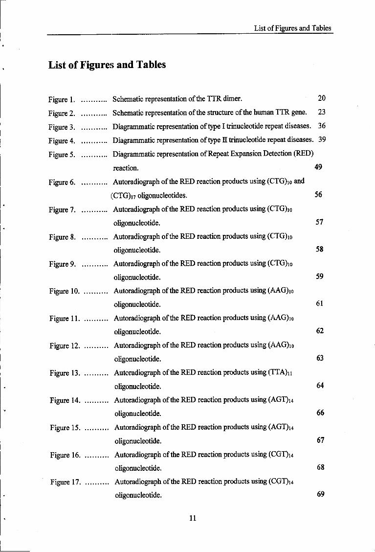
Figure 1 Schematic representation of the TTR dimer. 20
Figure 2 Schematic representation of the structure of the human TTR gene. 23
Figure 3 Diagrammatic representation of type I trinucleotide repeat diseases. 36
Figure 4 Diagrammatic representation of type II trinucleotide repeat diseases. 39
Figure 5 Diagrammatic representation of Repeat Expansion Detection (RED)
reaction. 49
Figure 6 Autoradiograph of the RED reaction products using (CTG)io and
(CTG)i7 oligonucleotides. 56
Figure 7 Autoradiograph of the RED reaction products using (CTG)io
oligonucleotide. 57
Figure 8 Autoradiograph of the RED reaction products using (CTG)io
oligonucleotide. 58
Figure 9 Autoradiograph of the RED reaction products using (CTG)io
oligonucleotide. 59
Figure 10 Autoradiograph of the RED reaction products using (AAG)io
oligonucleotide. 61
Figure 11 Autoradiograph of the RED reaction products using (AAG)io
oligonucleotide. 62
Figure 12 Autoradiograph of the RED reaction products using (AAG)io
oligonucleotide. 63
Figure 13. Autoradiograph of the RED reaction products using (TTA)i i
oligonucleotide. 64
Figure 14 Autoradiograph of the RED reaction products using (AGT)H
oligonucleotide. 66
Figure 15 Autoradiograph of the RED reaction products using (AGT)i4
oligonucleotide. 67
Figure 16 Autoradiograph of the RED reaction products using (CGT)i4
oligonucleotide. 68
Figure 17 Autoradiograph of the RED reaction products using (CGT)H
oligonucleotide. 69
11

List of'Figures and Tables
Figure 18 Autoradiograph of the RED reaction products using (CGG)i i
oligonucleotide. 71
Figure 19 Autoradiograph of the RED reaction products using (CGG)i i
oligonucleotide. 72
Figure 20 Autoradiograph of the RED reaction products using (CGG)i i
ohgonucleotide. 73
Figure 21 Autoradiograph of the RED reaction products using (TGG)i2
ohgonucleotide. 75
Figure 22 Autoradiograph of the RED reaction products using (TGG)i2
ohgonucleotide. 76
Figure 23 Autoradiograph of the RED reaction products using (ATG)i2
ohgonucleotide. 78
Figure 24 Autoradiograph of the RED reaction products using (ATG)i2
ohgonucleotide. 79
Figure 25 Autoradiograph of the RED reaction products using (GTT)i5
ohgonucleotide. 80
Figure I Maximal sizes of CTG/CAG repeat multimers in FAP kindred
pairs and unrelated controls as determined by RED. 5 5
Figure II Maximal sizes of AAG/CTT repeat multimers in FAP'kindred
pairs and unrelated controls as determined by RED. 55
Table 1 Amyloidosis diseases. 18
Table 2 Neuropathic TTR variants. 25
Table 3 Cardiopathie TTR variants. 26
Table 4 Other TTR variants. 26
Table 5 Non-amyloidogenic TTR variants. 26
Table 6 Type I trinucleotide repeat diseases. 35
Table 7 Type II trinucleotide repeat diseases. 37
12

Abbreviations
AP Amyloid p
APPP Amyloid P protein precursor
AA Amino acid
ACD Citric acid/trisodic citrate/dextrose
Apo AI Apolipoprotein AI
ApoE Apolipoprotein E
bp Base-pairs
BSA Bovine serum albumin
cAMP Cyclic adenosyne-mono-phosphate
cDNA Complementary Deoxyribonucleic acid
CNS Central nervous system
Da Dalton
dCTP Deoxycytidine triphosphate
DM Myotonic dystrophy
DMPK Myotonic dystrophy protein kinase
DMSO Dimethyl sulphoxide
DNA Deoxyribonucleic acid
dNTP Deoxynucleotide triphosphate
DRPLA Dentatorubral-pallidoluysian atrophy
EDTA Ethylenediamine-N',N' N',N'-tetra acetate
EPM1 Progressive myoclonus epylepsy type 1
FA Friedreich's ataxia
FAC Familial amyloidotic cardiomyopathy
FAP Familial amyloidotic polyneuropathy
FCS Foetal calf serum
FMR Fragile X mental retardation
FRAXA Fragile X syndrome
HAP-1 Huntingtin-associated protein-1
HD Huntington's disease
13

Abbreviations
kb Kilobase
MJD Machado-Joseph disease
mRNA Messenger ribonucleic acid
PBS Phosphate buffered saline
RBP Retinol-binding protein
RED Repeat Expansion Detection
RNA Ribonucleic acid
SAA Serum amyloid A
SBMA Spinal and bulbar muscular atrophy
SCA Spinocerebellar ataxia
SDS Sodium dodecyl sulphate
SSA Senile systemic amyloidosis
STR Short tandem repeat
T4 Thyroxine
TBE Tris buffer EDTA
TBG Thyroxine-binding globulin
TNR Trinucleotide repeat
TRE Trinucleotide repeat expansion
TTR Transthyretin
UTR Untranslated region
UV Ultra violet
14

Chapter I
Introduction
Introduction
Introduction
1.1. GENERAL INTRODUCTION
Familial Amyloidotic Polyneuropathy (FAP) is a hereditary autosomal dominant
disorder characterized by the deposition of a mutant form of the serum protein transthyretin
(TTR) as fibrillar material in several tissues and organs leading to death in 10 to 15 years. First
described by Andrade in 1952, FAP has its major focus in Northern Portugal with spread to
Japan, Sweden, Spain, and other countries.
FAP is one of most common and best studied of the genetically related amyloidoses, a
group of disorders characterized by the deposition of extracellular amyloid fibrils. It exhibits a
remarkable phenotypic heterogeneity expressed in the clinical symptomatology and the age of
onset. The variability of the disease onset in Portuguese kindreds accounts for the phenomenon
of genetic anticipation, with clinical symptoms developing at an earlier age in succeeding
generations. The molecular basis for anticipation in FAP remains obscure. The only known
molecular mechanism underlying anticipation is the expansion of unstable trinucleotide repeats
in responsible genes for a number of human hereditary dominant disorders. The identification of
such mechanism has provided a satisfactory explanation for anticipation at the level of genomic
DNA, and has opened the possibility to the speculate about the existence of similar repeat
instability in the responsible or accessory genes of other familial diseases with obvious
anticipation phenomena such as FAP.
The first section of this chapter will refer to the general aspects of the amyloidoses, with
particular incidence on TTR-related amyloidoses and more specifically, FAP. For a better
understanding of FAP pathology and the disease-involved mechanisms, the biochemistry and
molecular biology of transthyretin will be described. The phenotypic heterogeneity with special
reference to the age of onset of the disease and the phenomenon of genetic anticipation will also
be discussed.
The second section will introduce the characteristics of a recently emerged mechanism
of mutation - the unstable expansion of DNA-trinucleotide repeats. Some aspects of triplet
repeat expansion-associated disorders will be described in detail to illustrate the connection of
human gene mutation with pathology at the cellular level.
16

Introduction
1.2. AMYLOID,AND TRANSTHYRETLN-RELATEDAMYLOIDOSIS
Amyloidoses are diseases characterized by the aggregation of overproduced or abnormal
proteins into stable, insoluble fibrils. The component of the amyloid deposits, initially thought to
be a single substance;, revealed an unexpected chemical diversity when its identification was
made possible. Different precursor proteins precipitate to form amyloid fibrils giving rise to a
variety of clinical syndromes. The basis for the classification of the amyloidoses is presently the
chemical nature of the: deposited protein fibrils (Table I.).
A combination of physicochemical properties define all types of amyloid fibrils,
regardless the precursor protein involved. The fibrillar substance can be identified by Congo red
staining and exhibits a typical linear parallel organization on electron microscopy (Conhen et ai,
1959). It presents a (3-pleated sheet structure, as seen by X-ray diffraction, (Eanes et al, 1968)
and is highly insoluble; under physiological conditions.
The major fibrillar component is always associated with a minor glycoprotein designated
by P component which comprise about 5% of all amyloid deposits. Other extracellular
components such glicosaminoglycans, proteoglycans, apolipoproteins (E and J) and some serum
proteins are also part of the amyloid substance. It is not clear, however, if these molecules play
a role in the amyloidogenesis process by inducing conformational changes on the precursor
protein prior to its deposition, as suggested by Wisniewski & Frangione (1992), or simply
decorate the amyloid fibrils after they are formed.
The transthyretin (TTR) amyloidoses are the most prevalent type of genetically related
amyloidosis and are characterized by an autosomal dominant mode of inheritance.
The major biochemical defect underlying these diseases is the extracellular deposition of
mutant variants of the serum protein TTR predominantly in the peripheral nerves and in the
heart, with neuropathy and cardiomyopathy as major clinical manifestations.
Familial Amyloidotic Polyneuropathy (FAP) was the first disorder of this group to be
described and is characterized by the deposition of amyloid fibrils mainly in the peripheral
nervous system, starting by the nerves of the lower limbs (Andrade, C, 1952).
Since the description of the first cases, the number of identified kindreds with
transthyretin amyloidosis has steadily increased leading to the identification of different clinical
patterns of organ involvement. FAP was thus divided in four clinical types (I to IV) and the
17

Introduction
TABLE 1. Amyloidosis
AL
ALys APrP
ATTR
(3-protein precursor
Ap2M ^-microglobulin AA apo-SAA
AANF Atrial natriuretic factor AApoAI Apolipoprotein AI variants
ACal Calcitonin ACys Cystatin C variant
AFibA ochain fribrinogen variants AGel Gelsolin variants AH Immunoglobulin Gl
AIAPP Islet amyloid polypeptide
ATub
Immunoglobulin L-chain
Lysozyme variants Cellular prion protein
Prion variants
TTR TTR variants
Tubulin fragments
Alzeimer's disease Down syndrome
Hereditary cerebral hemorrhage with amyloidosis - Dutch type
Sporadic cerebral angiopathy Dialysis-associated amyloid Familial Mediterranean fever
Musckle-Wells syndrome Reactive amyloidosis
Isolated atrial amyloidosis Familial amyloidotic polyneuropathy - Iowa
type Familial nephropathy amyloidosis
Medulary carcinoma of thyroid Hereditary cerebral hemorrhage with
amyloidosis - Iceland type Hereditary renal amyloidosis
Familial amyloidosis - Finish type Heavy chain-associated amyloidosis
Diabetes mellitus type II Insulinoma
Idiopathic amyloidosis Myeloma-associated amyloidosis
Macroglobulinemia-associated amyloidosis Hereditary non-neuropathic amyloidosis
Creutzfeldt-Jacob disease (sporadic) Kuru
Creutzfeldt-Jacob disease (familial) Gertsmann-Strussler-Scheinker syndrome
Senile systemic amyloidosis Familial amyloidotic polyneuropathy Familial amyloidotic cardiomyopathy
Vitreous amyloidosis Meningocerebrovascular amyloidosis
Familial cerebral amyloid angiopathy-British
term Familial Amyloidotic Cardiomyopathy (FAC) was introduced to describe a Danish kindred
with extensive cardiac TTR deposition and very little nerve involvement (Nordlie, 1988).
Another type of cardiac TTR-related amyloidosis is Senile Systemic Amyloidosis (SSA),
distinguished from the other disorders by the deposition of normal TTR, rather than mutant
forms, as amyloid fibrils in the heart. This condition is diagnosed in about 25% of individuals
over age 80 (CornweU, 1983).
18

Introduction
The primary cause of TTR-amyloid formation is believed to be the presence of point
mutations within the TTR gene originating single amino acid substitutions in the mature protein.
An increasing number of TTR point mutations associated with TTR-amyloidoses has been
reported, accounting to a certain extent for the proliferation of FAP and FAC clinical subtypes.
Nonetheless, it appears that a single pathogenic process underlies the clinical and molecular
variations presented by the various forms of TTR-related amyloidoses.
1.2.1. TTR: BIOCHEMISTRY AND MOLECULAR BIOLOGY
The serum protein transthyretin was first isolated from human plasma in 1956 by
Shultze et al. (1956). It was identified as a band migrating ahead of serum albumin on
electrophoresis of whole plasma, and was originally named prealbumin. TTR interactions with
circulating thyroid hormones, retinol-binding protein and other physiologically important ligands
were progressively discovered. The innumerous studies on transthyretin make it one of the best
studied plasma proteins reflecting the physiological importance attributed this molecule.
1.2.1.1. TTR STRUCTURE AND PHYSIOLOGICAL FUNCTIONS
TTR is a soluble tetrameric protein synthesized predominantly by the liver. It is
composed of four identical subunits of 127 amino acids (Kanda et al., 1974) with a molecular
weight of 13,745 dalton. The total mass of the circulating tetramer is 54, 980 dalton (Smith et
al., 1979).
X-ray diffradion analysis has revealed the three dimensional structure of TTR as a
globular protein with substantial P-pleated sheet content and a small amount of a helix (Blake et
ah, 1971). The secondary structure of the polypeptide chains presents 8 p-strands (identified as
A to H) aligned in antiparallel fashion, forming two p-sheets composed of strands DAGH and
CBEF of each monomer. The p-sheets are linked by seven loops and a single a helix segment is
located at the end of fi-strand E (Figure 1).
The strong interactions between strands F and H of each monomer make the TTR
dimer, which is the teisic unit of the protein structure.
19

Introduction
Figure 1. Schematic representation of the TTR dimer.
The main physiological role of transthyretin is the transport of the thyroid hormone
thyroxine (T4) (Fergunson, et al., 1975), and retinol (vitamin A alcohol) through the formation of
a protein complex to retinol-binding protein (RBP) (Kanai at al., 1968).
TTR has two structurally identical binding sites for T» (Blake et ah, 1977) and is
responsible in humans, together with thyroxine binding globulin (TBG) and albumin, for the
distribution of the hormone throughout the body. Although in a lower concentration in plasma
than transthyretin, TBG carries about 70% of the circulating T4 due to a higher binding affinity
to the molecule. In turn, TTR is believed to play a particularly important role as a T4 carrier in
the central nervous systsm (CNS) since it is synthesized in high rate by the epithelial cells of the
choroid plexus, and is present in high concentration in the cerebrospinal fluid. (Dickson et al.,
1986). The synthesis of TTR in the choroid plexus early in development suggests a very
important function of the protein in the CNS presumably as a thyroxine hormone carrier. Recent
studies with TTR knockout mice have shown, however, that T4 transport: to the brain is not
impaired by the absence of transthyretin (Palha et al., 1997).
Transthyretin is an important component of the retinol transport complex. It has four
potential binding sites for RBP (van Jaarsveld et al, 1973) yet only two molecules of RBP bind
to the TTR tetramer (Monaco et al., 1995).
20

Introduction
The association with RBP protects the retinol transporter from renal catabolism and
glomerular filtration (Raz et al, 1970) and assures the delivery of retinol to the target cells. It
has been proposed that TTR controls the levels of circulating RBP thus regulating the
distribution of retinol to the tissues (Sivaprasadarao & Findlay, 1988). Nonetheless, as
demonstrated by studies with TTR null mouse strains, it is not the only mechanism for retinol
transport and delivery: both serum RBP and retinol levels were very low but the mice presented
a normal phenotype without any signs suggestive of retinol deprivation. (Episkopou et al,
1993).
In addition to RBP and T4, TTR interacts with diverse compounds including retinoic
acid (Smith et al, 1994), noradrenaline oxidation products (Boomsma et al, 1991), hemin and
hemoglobin (Martone and Herbert, 1993), pterins (Ernstrom et al, 1995), amyloid (3-peptide
(Schwarzman et al, 1994), among others. The meaning of most of these interactions is still very
obscure.
1.2.L2. STRUCTURE, EXPRESSION AND REGULATION OF TTR GENE
The gene encoding human TTR is a single copy gene located on the long arm of
chromosome 18 (Whitehead et al, 1984). Assigned to region 18qll.2-ql2.1 (Wallace et al,
1985), TTR gene contains four exons with approximately 200 bases each within about 7.0
kilobases. The first exon encodes a signal peptide of 20 amino acid residues and the first 3
amino acids of the mature protein; exon 2 encodes residues 4-47; exon 3, residues 48-92; and
exon 4, the last 35 residues, from 93 to 127. The introns, designated A, B and C, span 934 bp,
2090 bp and 3308 bp, respectively. Introns B and C contain two Alu sequences with opposite
polarity suggesting a hairpin formation in the precursor mRNA. The first and third introns
contain two open reading frames with respective regulating sequences. The meaning of these
sequences is not known but it has been suggested that they might encode gene expression
regulating proteins (Tsuzuki et al, 1985). Upstream the transcription initiation site, consensus
sequences include a TATA box at position -30, a G+C rich region of about 20 bp, a CAAT box
at position -101, and further up, at positions -224 and -212, two sequences homologous to
glucocorticoid responsive elements. The (CG)n dinucleotide is the only known repeated motif in
the entire TTR gene, including regulatory regions. Downstream the coding sequence, a
21

Introduction
polyadenilation site has been located at position 123 of the 3' untranslated region (Sasaki et al,
1985) (Figure 2).
Circulating TTR is synthesized predominantly by the adult liver. Normal plasma
concentration is 20-40 mg/dl (Gitlin and Gitlin, 1975) but a significant depression of this level
occurs when the liver is participating in the acute phase response to injury or in malnutrition
(Dickson et al, 1985; Fung et al, 1988; Murakami et al, 1988).
TTR is also s^thesized by the choroid plexus of the brain, as demonstrated by studies
on gene expression in rat and human (Soprano et al, 1985; Herbert et al, 1986), the synthesis
beginning very early in development at the eighth week of gestation (Jacobsson, 1989a). This
premature synthesis, together with a high transcriptional activity, imply a particularly important
role for TTR in the brain. The protein is also present in the pigmented epithelium of the retina
(Dwork et al, 1990) £ind the pineal gland (Martone, et al, 1993) in mammals.
The human, mouse and rat TTR genes demonstrate 80% sequence homology . Amino
acid sequence homology is even higher, of about 91% (Costa et al, 1986) with nearly complete
conservation of sequence in the regions involved in ligand binding (Blake et al, 1978;
Wakasugui et al, 1985). Studies on regulation and expression of mouse TTR gene were
performed in human hepatoma cells - HepG2 - (Costa et al, 1986; Costa et al, 1989) leading to
the identification of regulatory sequences in the human gene by comparative analysis.
The promoter region of the human gene contains elements for hepatocyte-specific
expression, including binding sites for HNF-1, C/EBP, HNF-3 and HNF-4. Comparative
analysis of human and mouse sequences showed that the binding sites for 1HNF-3 and HNF-4
are well conserved in the human TTR gene, but not those for C/EBP (Sakaki et al, 1989)
(Figure 2). Two other liver-specific nuclear protein potential binding sites were identified in the
human gene at positions -216 —221 and -199 ~ -204. The motif (TGG/AA/CCC/T) is
common to factors Tf-LFl, Tf-LF2 and LF-A1.
A region homologous to a tissue-specific enhancer of the mouse gene was found at - 3,4
kb, containing a binding site for factor HNF-4. This region seems to be important for TTR gene
expression in the choroid plexus. Another regulating segment, located at approximately
6 kb of the cap site, includes a sequence with homology to binding sites for nuclear factor APF1
(Sakaki et al, 1989).
TTR gene expression in transgenic mice has been shown to be dependent on the
22

Introduction
presence of about 600 bp upstream the transcription initiation site for hepatic and yolk sac
expression, and 6 kb for choroid plexus expression (Yan et al, 1990; Nagata et al, 1995).
(C/E) (C/E)
Á 1 Tf Tf H-4 H-1 CAAT TATA
U & ,1 Alu
■i- 3"
- T T -
I O
—■— 11 12 13
— I —
14 15 kb
Figure 2. Schematic representation of the structure of the human TTR gene and localization of the respective regulation regions. The exons (red boxes) are numbered from 1 to 4. Alu, LI and (CA)n: respective repetitive sequences. TATA: TATA box; CAAT: CAAT box. Hl, C/E, H3, H4: binding sites of hepatocyte nuclear factors HNF-1, C/EBP, HNF-3 and HNF-4, respectively. Tf: common motif to Tf-LF-1, Tf-LF-2, LF-A1. Enhancer: a region highly homologous to a tissue-specific enhancer of the mouse TTR gene. AFP: binding site for AFP-1 factor (from Sakaki et al, 1989).
23

Introduction
1.2.1.3. MOLECULAR TTR VARIANTS
Following the description of the first transthyretin variant in 1984 (TTR Met30)
(Saraiva et al, 1984), an increasing number of mutant forms were steadily identified. As
previously referred to, the vast majority is associated with neuropathic and cardiopathie
hereditary amyloidoses, however a number of non-amyloidogenic molecular variants have also
been recognized (Tables 2, 3, 4, and 5). Amyloidogenic TTR Met30 is the most frequent TTR
variant and it will be discussed later in detail. Other amyloidogenic variants occur with high
frequency like the cardiopathie variant TTR He 122 (0,02), as determined in the Afro-American
population by Jacobson (1992). Amyloidogenic mutant alleles are found almost exclusively in
heterozygosity but homozygotic individuals have been reported, namely for TTR Met30
(Holmgren et al, 1988), TTR His58 (Jacobson et al, 1994b), and TTR Ilel22 (Jacobson at al,
1991).
Non-amyloidogenic TTR variants are either associated with euthyroid
hyperthyroxinemia, like TTR Thrl09, and Vall09, or do not seem to have any pathological
effects, as observed for TTR Ser6, Asn90 and Metl 19, rather frequent in healthy carriers of the
normal population (Jacobson et al, 1995; Alves et al, 1997). TTR Ser6 is a common
polymorphism with an allele frequency of 0,06 in Caucasians (Jacobson et al, 1995) and the
absence of any association with amyloidosis suggests that it is a neutral polymorphism. In
screening studies of TTR variants in the Portuguese population (Alves et al, 1997) TTR
Meti 19 was found to be the most frequent variant in the analyzed 5,000 individuals, the
frequency (0,007) being very similar to that observed in other populations (Ii et al, 1992).
Initially thought to be associated with euthyroid hyperthyroxinemia, this variant was shown not
to have any pathogenic consequences in the Portuguese carriers. In fact, in addition to the
referred study, several other reports suggest that TTR Metl 19 carriers are not
hyperthyroxinemic. TTR Asn90, the second most frequent variant observed in the referred
study, occurred with a frequency of 0,0024 and does not seem to be pathogenic, as well.
Since the mentioned variants occur with high frequency, carriers of two different mutant
alleles have been detected (Saraiva et al, 1991; Izumoto et al, 1993; Alves et al, 1993;
Jacobson et al, 1994b aid Booth et al, 1994). Compound heterozygosity in the Portuguese
population has been reported in individuals carrying both Met30/Metl 19, Asn90/Metl 19 (Alves
et al, 1993), and Metî0/Asn90 (Saraivar al, 1991).
24

Introduction
TABLE 2. Neuropathic TFR variants
V T'ti'l1 Mutant Reference* remdue : . . " " " ■ ' ■
10 Arg Cys N, CTS, C,VO,NPH Uemichi et ai, 1992; Benson and Uemichi, 1996 18 Glu Asp N Booth et ai, 1996 24 Ser Pro N, CTS, C Uemichi et ai, 1995 30 Met Val N, C, VO Saraiva et ai, 1983; Tawara et ai, 1983 30 Leu Val N Nakazato et ai, 1992; Murakami et ai, 1992a 30 Ala Val N Jones et al, 1992 33 De Phe N,VO Nakazato et ai, 1984 33 Leu Phe N Hardling et ai, 1991; H et ai, 1992 33 Val Phe N Booth et al, 1996 34 Thr Arg N,C Patrosso et ai, 1996 35 Asn Lys N Reilly et ai, 1995 36 Pro .'Via N,VO Jones et al, 1991 42 Gly Glu N Ueno et ai, 1990a 47 Arg Gly N Murakami et ai, 1992b 47 Val Gly N, CTS, C Booth et al, 1994 47 Ala Gly N,C Ferlinieía/., 1994 49 Ala Thr N, C, VO Almeida et ai, 1992 50 Arg Ser N,C Ueno et ai, 1990a; Takahashi et ai, 1992 50 Ile Ser N Saeki era/, 1992 52 Pro Ser N,C,NPH Booth et al, 1994 54 Gly Glu N Booth étal, 1994 55 Pro l̂ eu N, C, VO Jacobson et al, 1992 58 His Leu N, CTS, C Nichols et al, 1989; Benson and Uemichi, 1996 58 Arg l̂ eu N, CTS, VO Saeki étal, 1991 60 Ala Thr N, CTS, C Wallace et al, 1986; Benson and Uemichi, 1996 61 Lys Glu N Shiomi étal, 1993 64 Leu Phe N,C H étal, 1991 70 Asn Lys N, CTS, C Izumoto et al, 1992 71 Ala Val N, CTS, C, VO Almeida et al, 1993; Benson H et al, 1993 77 Tyr Ser N Wallace et al, 1988 77 Phe Ser N Bordeneuve et al, 1996 84 Ser De N, CTS, C, VO Dwulet and Benson, 1986 89 Gin Glu CTS,N,C Almeida et al, 1992 91 Phe Ser N Bordeneuve et al, 1996 97 Gly Ala N,C Yasuda et al, 1994; Nakazato et al, 1994 107 Val He N.CTS Jacobson et al, 1994a; Uemichi et al, 1994a 112 He Ser N,C DeLucia étal, 1993 114 His Tyr CTS Murakami et al, 1994 114 Cys Tyr N,VO Ueno et al, 1990b 116 Tyr Ser N Bordeneuve et al, 1996 122 - Vai N,C Uemichi et al, 1996
N - Neuropathy; CTS - Csirpal Tunnel syndrome; C - Cardiomyopathy; VO - Vitreous opacitiesi; NPH - Nephropathy
25
Introduction
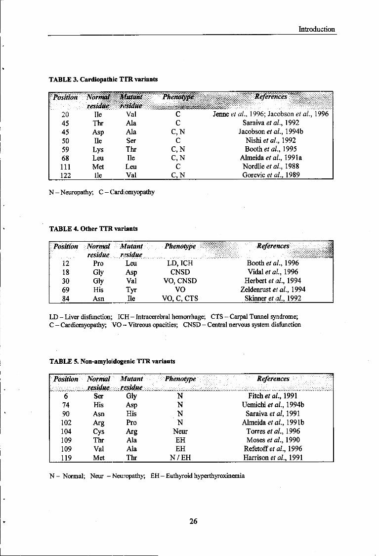
TABLE 3. Cardiopathie TIR. variants
Position Normal Mutant Phenotype References residue residue ■
::1P™:-:::
20 Ile Val C Jenneei al, 1996; Jacobson ef a/., 1996 45 Thr Ala C Saraiva et al, 1992 45 Asp Ala C,N Jacobson et al, 1994b 50 Ile Ser C Nishiefa/., 1992 59 Lys Thr C,N Booths ai., 1995 68 Leu Ile C,N Almeida et al, 1991a 111 Met Leu C Nordlie étal, 1988 122 Ile Val C,N Gorevic e/ a/., 1989
N - Neuropathy; C - Cardiomyopathy
TABLE 4. Other TTR variants
Position Normal Mutant Phenotype References 1 residue residue -■ '■■■': ■$;■:■ i
12 Pro Leu LD, ICH Booth et al, 1996 18 Gly Asp CNSD Vidal eia?., 1996 30 Gly Val VO, CNSD Herbert étal, 1994 69 His Tyr VO Zeldenrust et al, 1994 84 Asn Ile VO, C, CTS Skinner et al, 1992
LD - Liver disfunction; ICH - Intracerebral hemorrhage; CTS - Carpal Tunnel syndrome; C - Cardiomyopathy; VO - Vitreous opacities; CNSD - Central nervous system disfunction
TABLE 5. Non-amyloi dogenic TTR variants
Position Normal Mutant Phenotype References residue residue V~": '.::"! ■;' :':-;
:;:^
::C ̂ '..; :;■: :; . - ' . -:
:-_::- . . ::;- . -^J"? ->-:■; :■,-:;: ■;:
::::-:::--:;:. :. '.::
:;
:-.:;.-:.:.U.-:;
:;:::; ■.; ! -1 ^j ;;■: ■■ • ■ ■ . , , : , : . .
6 Ser Gly N Fitch etal, 1991 74 His Asp N Uemichi et al, 1994b 90 Asn His N Saraiva etal, 1991 102 Arg Pro N Almeida et al, 1991b 104 Cys Arg Neur Torres etal, 1996 109 Thr Ala EH Moses et al, 1990 109 Val Ala EH Refetoffeía/., 1996 119 Met Thr N / E H Harrison et al, 1991
N - Normal; Neur -Neuropathy; EH - Euthyroid hyperthyroxinemia
26

Introduction
In some cases, as in TTR Met30/Asn90 carriers, the presence of a second mutation does not
seem to affect the pathogenicity of Met30 (Saraiva et al, 1991). In others, as in TTR
Met30/Metl 19 carriers, the Metl 19 mutation appears to have a protective effect on the clinical
evolution of the disease, particularly referring to the age of onset which is later than for
heterozygous Met30 patients (Coelho et al, 1996).
The detection of double mutations in the same allele in two individuals - Ser6-Ile33
(Jacobson et al, 1994b) and Asn90-Gly42 (Skare et al, 1994) - has broaden even more the
known molecular variability of TTR. Very recently, Palha et al (1996) have reported the first
silent mutation in transthyretin gene resulting from a C for T substitution at codon 108.
The mutation» that originate a number of TTR variants, including TTR Met30, Ilel22,
Thrl09 and Metl 19, occur in a CpG dinucleotide, known as a sequence with high frequency of
mutation (Cooper and Youssoufian, 1988). The analysis of the human TTR gene demonstrates
the existence of nutny more CpG hot spots that could originate different amino acid
substitutions.
Polymorphisms in the non-coding region of the TTR gene have also been shown by the
existence of 5 different haplotypes defined by 7 intronic polymorphic substitutions (Yoshioka et
al, 1989). Analysis of these substitutions in several individuals carriers and non-carriers of TTR
Met30 mutation have demonstrated that none of them is related with FAP.
1.2.2. TTRMET30AND FAMILIAL AMYLOWOTICPOLYNEUROPATHY
In 1952, Corino de Andrade described for the first time a peculiar type of neuropathy.
The new clinical entity was characterized as a sensory-motor neuropathy starting in the lower
extremities, having associated a variety of symptoms in several organs which were attributed to
an autonomic neuropathy rather than to primary disease of the organs themselves (Andrade, C,
1952). The syndrome was latter designated by Familial Amyloidotic Polyneuropathy reflecting
the major clinical manifestation and the hereditary nature of the disease. The first symptoms
appear typically in the third decade of life with a slow progression to death in 10 to 15 years.
Following the discovery of transthyretin as the major component of the amyloid fibril
deposits by Costa et al (1978), the biochemical mechanisms involved in FAP pathology begun
to be unraveled. The deposited TTR was reported to be a variant of the normal protein with a
27
Introduction
substitution of a metMonine for a valine residue at position 30 of the polypetide chain (Saraiva et
al. 1983) and the associated mutation at the DNA level - a single base substitution - was soon
revealed after TTR gene cloning (Mita et al, 1984; Tsuzuki et al, 1985). TTR Met 30 was
shown to be a biochemical marker for FAP in the Portuguese population due to the complete
agreement between genotype and phenotype analysis in diseased individuals (Saraiva et al,
1985).
TTR Met30 is title most frequent mutation associated with FAP and accounts for the
most common type of the disease, called type I or Portuguese. To date more than 500 kindreds
with FAP have been reported in Northern Portugal, the largest focus in the world, with an
estimated prevalence rate in 1991 of 105xl0"5 (Sousa et al, 1995) and a frequency of gene
carriers calculated as 1 in 625. The Met30 variant is also present in other foci in Sweden, Japan,
Spain, Italy and Brazil. Some kindreds of French, English, Cypriot, Dutch and Turkish origins
also have this mutation (Benson et al, 1986; Saraiva et al, 1986; Holt et al, 1989; Munar-
Qués et al, 1990; Salvi et al, 1990; Ferlini et al; 1988; Hazenberg et al, 1990; Skinner et al,
1990). Recently, FAP type I has been associated with other TTR mutations (Saraiva at al,
1993), however, in Portugal, Brazil and Sweden, no pathogenic TTR mutations other then TTR
Met30 have been described so far.
Work by Yoshioka et al (1989) in Japanese FAP kindreds has shown that the Met30
mutation occurs in a CpG hot spot and was found associated with 3 different haplotypes
(defined by a set of 7 intronic polymorphic substitutions and classified as I, II and HI), as
previously referred to. This observation suggested that the Val-»Met mutation has probably
recurred in the human population to generate FAP families of independent origin. Almeida et al
(1995) have also shown two different haplotypes associated with the mutation in European
kindreds, substantiating ithe mutation independent origin hypothesis. Nevertheless, the possibility
of the one founder hypothesis in Portugal and Sweden cannot be exclude, since only the
haplotype I has been found in those areas.
28

Introduction
1.2.2.1. PHENOTYPIC VARIABILITY IN TTR Met30-ASSOCIATED FAP
The clinical phenotype first described by Andrade and thought to be "constant, repetitive
and monomorphic" was latter revealed as being very heterogeneous in respect to the patterns of
organ involvement and age of onset of the disease.
A) Clinical Presentations
Variability in. clinical manifestations secondary to variation in trie sites of amyloid
deposition have been observed in TTR Met30 associated FAP. The classical site of amyloid
deposition, as described by Andrade is the peripheral nervous system, giving rise to sensory and
motor neuropathy (the majority of cases in Portugal). Amyloid deposits are also frequently found
in the autonomic nervous system, in some cases producing the first symptoms of the disease.
Coutinho and coworkers (1980) reviewed 483 cases of FAP type I diseased individuals showing
a substantial variation of the first symptoms.
Deposition of amyloid fibrils can also take place in the vitreous of the eye and in the
kidney, producing vitreous opacities and renal disease, respectively. This phenotypes can appear
either associated wiib. neuropathy, or as single pathological manifestations. Although such
clinical features are not frequently observed among Portuguese patients carrying the Met30
mutation, they have been found to arise in some families, and in some but not all branches of the
same family.
B) Age of Onset
The most striiking feature of phenotypic heterogeneity in Portuguese FAP is by far the
age of onset of the disease. The onset of TTR Met30 amyloidosis ranges from an average of
33,5 years in the Portuguese population to an average of 56,6 years in the Swedish population
(Sousa et al, 1995). Japanese and Majorcan populations present intermediate mean ages-at-
onset of 34,2 and 49 years, respectively (Ikegawa et al., 1991; Munar-Qués et ah, 1996).
In the Portugese population, the age of onset varies from 17 to 78 years, with an
average of 33,5 and a standard deviation of 9,5 years. Eighty one percent of the patients exhibit
29

Introduction
the first symptoms before the age of 40 and 92% before the age of 50 years. The mean age of
onset is significantly Irigher in females (35,6 years, SD 9,0) than in males (31,9 years, SD 9,6)
(Sousa, A., 1995). It has also been shown that carriers of the TTR Met30 mutation could remain
disease free up to 93 years of age (Lobato et ai, 1988; Sousa et al., 1988; Coelho étal., 1994).
Furthermore, sporadic: cases have been reported. In a recent revision of 1,233 Portuguese FAP
cases, neither parents of 159 individuals had shown symptoms (Coelho et al, 1994). They form
a distinct group with an age of onset higher than the average (mean 45,1 years, SD 12,0), and
belong to families with a geographical origin slightly different from the areas of FAP major
prevalence. Nevertheless, this group of individuals is not significantly different from the general
group of patients in clinical presentation at onset and severity of symptoms.
Homozygosity of the Met30 allele has been reported in several individuals of Swedish
kindreds but neither the clinical symptoms nor the age of onset of the disease differ significantly
from the heterozygous individuals. Curiously, homozygous sporadic cases (in which none of the
progenitors had shown any symptoms) have been described in the Swedish population.
Interestingly, one of the: homozygous individuals was an asymptomatic carrier aged 62
(Holmgren et al., 1988). It is clear, therefore, that gene dosage is not the responsible mechanism
for the differences in the age of onset of the disease. Moreover, since the absence of normal
alleles does not cause the disease by itself, as observed in the case of the asymptomatic
homozygous Met30 carrier, the effect of isoalleles is also excluded as an explanation for the
disease late onset in FAP.
Several hypothesis have been raised to explain the existing variability of the age-at-onset
but the underlying mechanism of such phenomenon remains obscure. No evidence has ever been
found for the existence of a different mutation in the TTR gene segregating either with late- or
typical onset diseased individuals. If the late-onset FAP cases were derived from a TTR
mutation different from ttiat found in the typical forms of the disease, such mutation would have
to occur in the noncoding region of the TTR gene, in addition to the point mutation responsible
for the Val -» Met substitution at residue 30. Studies on TTR gene expression and structure in
late versus typical onset FAP Met30 pedigrees (Saraiva et al, 1986) have not revealed any
differences in TTR levels or in haplotype association. No correlation between levels of TTR
Met30 and age or duration of clinical symptoms has been found in those kindreds.
Although the role of environmental factors cannot be excluded, the existence of modifier
genes involved in the TTR amyloidogenesis process is still the most attractive hypothesis to
30

Introduction
explain the phenotypic variability in FAP.
1.2.3. GENETIC ANTICIPATION IN FAP
Genetic anticipation is a phenomenon characterized by a progressive reduction in the age
of onset and by an increase in severity of the disease in successive generations. The finding of
genetic anticipation was for many years attributed to ascertainment biases postulated by Penrose
in 1948. According to Penrose, anticipation was an artifact produced by the selection of
pedigrees, rather than a phenomenon with biological meaning. The biases were attributed to 1)
the preferential detection of progenitors with late-onset; 2) favored selection of descendants with
early onset (presenting more severe phenotypes); and 3) preferential detection of progenitors and
descendants with simultaneous onset. The arguments favoring the idea of a statistical illusion
begun to be questioned with the observation of different expressions of anticipation depending
on the parental transmission of the disease-causing allele (Ridley et al., 1988; Hõweler et al.,
1989). A few years ago the phenomenon of anticipation was dismissed as a statistical artifact
and a molecular mechanism explaining the perplexing features of genetic inheritance displayed
by a number of dominant disorders was identified.
Genetic anticipation regarding the age of onset has been for long recognized in FAP
Met30 kindreds of the Portuguese population (Coutinho et al, 1980; Bastos Lima A, Martins da
Silva A, 1980). Initially taken as the result of statistical artifacts, genetic anticipation in
Portuguese FAP was latter on proved to be a real phenomenon (Sousa et al, 1990a, 1991).
Anticipation of the age-at-onset was shown not to be the result of a simple effect of regression to
the mean (Sousa, A., 1995). Moreover the existence of the "complementary pairs" postulated by
Penrose (1948), i.e.., generational pairs in which the progenitors exhibit early onset and the
descendants late onset, has never been found in Portuguese FAP kindreds. Contrarily, the age of
onset was reported to be earlier in the second generation in the majority of cases, with a mean
anticipation of 6,6 years, in a study of 227 generational pairs of the Portuguese population
(Sousa, A., 1995). According to the same study, the anticipation range depends on the gender of
the diseased individual and the affected progenitor, as male descendants of affected mothers
have an earlier onset than female descendants of affected fathers. Sousa (1995) has also reported
a few cases of negative anticipation, i.e., descendants with an earlier onset than the progenitors,
31
Introduction
in average. These cases were mostly observed in descendants of male progenitors with early
onset (before 30 years).
The responsible mechanism for the phenomenon of anticipation in FAP is still unknown.
Dynamic mutation is an attractive model as the molecular underlying cause of this perplexing
mode of inheritance since it has been associated with other disorders exhibiting genetic
anticipation of the age-at-onset. Presently, at least a dozen human genetic diseases have been
shown to be caused by uinucleotide repeat expansions (TREs). Although there are 10 possible
trinucleotide repeat motifs, only 3 [(CAG)» / (CTG)„, (CGG)n, and (AAG)n] have been
associated with disease. The search for TREs in FAP kindreds exhibiting anticipation becomes
the first step to identify modifier genes, suggested to underlie phenotypic variability in FAP.
2. Dynamic mutations: trinucleotide repeat expansions (TREs)
2.1. SIMPLE TANDEM REPEATS AND DYNAMIC MUTATION
The human genome contains many nucleotide sequences that occur repeatedly. The
complexity of these repeated sequences vary from complete genes (such as the ribosomal RNA
genes) to simple sequences of one or a few base pairs. Dinucleotide repeats are among the
simplest and most common repeats, but other frequently occurring simple tandem repeats
(STRs) involve mono-, tri-, tetra, and pentnucleotide repeating units. The extensive
characterization of simple repeat sequences in the human genome came about as a consequence
of their use as highly informative genetic markers. Since their copy number is polymorphic in the
human population aid their distribution is relatively uniform throughout the genome, simple
tandem repeats have been used for the construction of genetic linkage maps, diagnosis by
linkage analysis and several other purposes. The general function of the STRs (if any) is still
unknown although sequence-specific DNA-binding proteins have been identified for di- and
trinucleotide repeats (Richards et al, 1993), and one of the repeats can act as a preferential site
of nucleosome assembly in vitro (Wang et al., 1994).
A general characteristic of STRs is the instability of the repeat which appears to be a
32

Introduction
function of the information content, i.e. the number of perfect repeating units. The greater the
repeat copy number without interruptions, the higher instability it reveals. Such behavior is very
evident in trinucleotide STRs, a feature of which is their ability to undergo dynamic mutation.
Dynamic mutation is a process that can occur over several generations and is distinguished from
conventional mutations by a number of properties: 1) the product of a dynamic mutation has a
different risk of undergoing further change than the original DNA sequence; 2) the probability of
mutation of a STR is a function of the number of perfect repeating units; 3) the mutation of a
given repeated sequence from a harmless copy-number polymorphism to a disease causing
unstable DNA sequence is not a single event, but a process involving multiple (sometimes
small) changes.
The unstable expansion of DNA-triplet repeats has recently emerged as a completely
new mechanism of mutation. A number of disorders displaying perplexing features of genetic
inheritance have been in recent years associated with expansion and intergenerational instability
of stretches of identical t̂rinucleotides that occur as normal shorter stable elements throughout
the human genome. Genetic anticipation, an intriguing phenomenon observed in a significant
number of human disorders exhibiting a dominant mode of inheritance has now been attributed
to the expansion of unstable trinucleotide repeats. The identification of a satisfactory explanation
for anticipation at the level of genomic DNA has led to a new series of challenges.
2.2. PATHOLOGICAL PHENOTYPES
Two different groups of conditions associated with triplet repeat expansion can be
considered. One group of disorders is characterized by a progressive neuronal loss. Designated
type I (Reddy and Housman, 1997) this group includes Huntington's disease (The Huntington's
Disease Collaborative Research Group, 1993), spinal and bulbar muscular atrophy (SBMA) (La
Spada et al, 1991), dentatorubral-pallidolusyan atrophy (DRPLA) (Koide et al, 1994;
Nagafuchi et al, 1994), Machado-Joseph disease (MJD) (Kawaguchi et al, 1994) and the
spinocerebellar ataxias (SCAs) (Orr et al, 1993; Imbert et al, 1996; Pulst et al, 1996; Sanpei
et al, 1996; and Zhuchenko et al, 1997). The pattern of neuronal loss is different for all theses
neurodegenerative disorders but it is, in all cases, a function of age and the size of the triplet
repeat expansion (Table 6).
33

Introduction
The other group of triplet repeat disorders includes myotonic dystrophy (DM) (Brook et
al, 1992), fragile X syndrome (Kremer et al, 1991; Verkerk et al, 1991; Gu et al, 1996) and
Friedreich's ataxia (Campuzano et al, 1996), designated type II diseases and involving a more
variable pathology (Table 7).
2.2.1. TYPE I DISORDERS
Type I diseases are caused by moderate expansions of CAG repeats within a coding
sequence, producing an extended stretch of polyglutamine in the corresponding protein. Several
of this disorders have been reported to display genetic anticipation, and CAG repeat length of
disease alleles typically correlates in an inverse manner with age of onset of the symptoms.
Surprisingly, and in contrast with type II disorders, an increase of copy number by as little of 5%
above a normal level can result in disease (Reddy and Housman, 1997). The number of triplets
never exceeds 150 which probably explains why anticipation in these diseases is rather limited,
although longer expansions in general result in earlier onset and more severe disease. In both
type I and type II disorders, the transmission of a triplet repeats in the range of 40 to 100 repeats
to the next generation, through either the male of female germline, exhibits mutation to a new
repeat number with a frequency approaching 100 %. This instability contrasts with the
transmission of repeat lengths in the order of 5-20 units were variations in size is almost never
observed.
Parental transmission of unstable repeats in the 40-100 repeat size range has been
observed to present a rather different instability pattern depending on the transmitting parent
(Brunner et al, 1993; Trottier et al, 1994). Paternal transmission gives rise to a distribution of
repeat sizes among the offspring with a much higher median value than the repeat length of the
father, whereas in maternal transmission the maternal mean in children remains normally
distributed. This phenomenon already referred to as "paternal anticipation" seems to be the
result of a significant increase in sperm TNR lengths compared to the repeat lengths of the
father, as observed in studies of individual sperm of males with HD (Leeflang et al, 1995).
These studies suggest that either the replication of DNA in primary male germ cells favors
repeat expansion, or there is a selection for germ cells or mature sperm with longer TREs.
While the disorder-related genes are widely expressed, the pathologic expression of the
diseases is late onset and limited to specific neurological tissue of the brain. They are
34

Introduction
characterized by ataxia, chorea, dementia, and in some cases psychosis.
Table 6. Type I trinucleotide repeat diseases (from Reddy and Housman, 1997)
Disease Repeat Clinical manic-station
Sites of neuropathology
Repeat Nam ber
Normal Disease Location of the repeats
Gene product
Gene function
Change in gene function
Huntington's sTslDanB
CAG Chorea, impairment of cognitive function,
emotional disturbances
Primarily in corpus stiatum, abo in cortex
in late stage
6-34 36-121 ORF HuntifltrttD Unknown Gam
Dentatorubral-pallidoluysian
•trophy (Smith's disease)
CAG Choreanthetosis, ataxia, dementia, myoclonus and
epilepsy
Globus pallidas, dentatorubral and
subthalamic nucleus
7-23 49-88 ORF Atrophia Unknown Gain
Spinobulbar muscular atrophy
(Kennedy's disease)
CAG Muscle weakness, atrophy and
fasciculations
Degeneration of anterior horn cells, bulbar neurons and dorsal root ganglia
11-34 40-62 ORF Androgen receptor
Transcription factor
Gain
SpinocerebellaT ataxia type 1
CAG Ataxia, dysarthria, dysmetria and
decreased vibration sense
Cerebellar cortex, dentate nucleus and
brainstem
6-39 41-81 ORF Ataxin-1 Unknown Gain
Spinocerebellar ataxia type 2
CAG Ataxia, dysarthria Cerebellum, pontine nuclei, substantia
nigra
15-29 35-59 ORF Ataxin-2 Unknown Gain
Spinocerebellar ataxia type 3
(Machado-Joseph disease)
CAG Ataxia, dystonia and ophtalmoplegia
Substantia nigra, globus pallidus, pontine nucleus, cerebellar cortex
13-36 68-79 ORF Ataxin-3 Unknown Gain
Spinocerebellar ataxia type 6
CAG Ataxia, dysarthria, nystagmus and
vibratory sensory loss
Cerebellar and mild brainstem atrophy
4-16 21-27 ORF Ataxin-6 Encodes calcium channel subunit (otu)
Gaia
The physiological function of CAG repeats translated into polyglutamine tracts is
unknown. Amplified CAG repeats might impair this activity (loss-of-function), or modify it
resulting in disease (gain-of-function). The finding of normal mRNA or protein levels in these
diseases (Strong et al, 1993; Hoogeveen et al, 1993; Banfi et al, 1994) and the absence of
SBMA and HD phenotype in patients with deletion of the androgen receptor (Quigley et al, 1992)
35

Introduction
or the HD locus (The HD Collaborative Research Group, 1993), respectively, argue against the
loss-of-fonction effect of the CAG triplet repeats. Also, the observation that diseases such as
HD are truly dominant with a similar phenotype in homozygotes for the disease allele (Wexler et
al, 1987), suggest a gain of fonction.
HD SCA1 DRPLA MJD
CAGm CAG CAG u CAGis CAGis
Affected CAG CAG
CAG CAG
CAG CAG
CAG CAG
Normal c CAG CAG C A G CAG CAG CAO
36
34
6
CAG CAG C A G CAO CAG CAO
4.1
39 6
CAG CAG 49 C A G CAG 2 5 CAG CAO n
CAG CAG 68 C A G CAG 3 6 CAG CAO 1 3
1_ 5'UTR '
Normal
Affected
: CAO CAG CAG C A G
CAG CAG CAG
15 29
35
ORr CM CAG CAG C A G
CAG CAG CAG
i l 34
40
J CAO 4 CAO . , CAG l b
C A G
CAG 1 1 CAG 27
3'UTR
CAG 59 CAG 6?
SCA2 SBMA SCA6
Figure 3. Diagrammatic representation of type I trinucleotide repeat diseaes, showing the number and location of the expanded repeats within a model gene. The repeats are represented by CAG, with increasing font size representing expanding number of repeats. The number of repeats are shown on the right of the trinucleotides. The number of repeats causing disease phenotype are underlined. The repeat expansions in each disease gene are shown at an approximate location within the model gene. ORF, open-reading frame; UTR, untranslated region (from Reddy and Housman, 1997).
36

Introduction
2.2.2. TYPE H DISORDERS
Type II disorders exhibit a much larger range of repeat expansion lengths which
correlates very clearly with the severity of the disease. Moreover, unlike (CAG)n-related
diseases, triplet repeat expansions are not found in coding regions of genes but in flanking
regions of coding sequences. Since the pathological phenotypes of fragile X syndrome, myotonic
dystrophy and Friedreich's ataxia are very different, and the disease-associated repeated motifs
are not a common feature as observed in type I disorders, these diseases will be discussed
separately.
TABLE 7. Type II trinucleotide repeat diseases (from Reddy and Housman, 1997).
Repeat Clinical
Manifestation
Repeat Number Ideation of the repeats
Gene product
Gene function Disease Repeat
Clinical Manifestation Normal Premutation Mutation
Ideation of the repeats
Gene product
Gene function
Change in gene function
Myotonic CTG Muscle weakness, 3-37 -50-180 ~200->2000 3'UTR Myotonin Encodes dystrophy wasting myotonia,
cataracts, mental retardation
protein kinase
Fragile X CGG Mental retaliation 6-52 -60-200 ~200->2000 J'UTR FMR-1 Encodes RNA- Loss syndrome (FRAXA)
FragileX GCC Mental retardation 6-23 43-200 syndrome (FRAXE)
Friedreich's GAA Progressive gait, 7-22 ataxia limb ataxia,
dysarthria
binding protein
>200
200->900 Intron Frataxin Unknown Loss
The question mark indicates an unknown location, gene product, gene function or change in gene function, and the dash denotes mat the premutation is not seen in Friedreich's ataxia
2.2.2.1. FRAGILEX SYNDROME
Fragile X syndrome was the first genetic disease to be associated with the dynamic
mutation of trinucleotide STRs (Yu et al, 1991; Oberlé et al., 1991). It is the most common
form of mental retardation and is associated with the presence of a folate-sensitive fragile site
{FRAXA) at chromossome Xq27.3 (Nelson D.L.,1995). Affected males have the fragile site on
their single X chromosome and present moderate to severe mental retardation, developmental
delay and autistic behavior. Female carriers have only mild cognitive defects and are more
difficult to identify by cytogenetics.
37

Introduction
The pattern of inheritance of fragile X shows a marked form of anticipation (Sherman el
al, 1984). The disorder is caused by an expanded CGG trinucleotide repeat located in the 5'
untranslated region of the FMR1 gene, which encodes a widely expressed product that has been
characterized as a RNA binding protein (Verkerk et al, 1991; Ashley et al, 1993). This repeat
is polymorphic in normal X chromosomes, having 6 to ~ 50 copies, typically interrupted by
AGG triplets (Kunst, et al, 1994; Eichler et al, 1994). These chromosomes are inherited
according to Mendelian patterns of inheritance, without expansion. At a length above ~ 50
copies, the repeat begins to display a certain degree of instability and can increase progressively
each time with a higher probability. Up to ~ 200 repeating units, the expansion-containing allele
is considered in the premutation range, since it is not associated with an abnormal phenotype,
although very unstable in intergenerational transmission. The lengths of normal and premutation
alleles may overlap but the different stability reflects internal differences in the presence or
location of AGG interruptions, the instability being a function of the length of the longest perfect
CGG stretch (Eichler et al, 1994). The full mutation comprises up to thousands of copies of the
repeated triplet originating the clinical expression of fragile X syndrome. Curiously, male
transmission of the premutation alleles invariably remains within the premutation size range,
while female transmitted alleles have an increasing probability of greater expansion into full
mutation.
Pieretti et al, 1991 have demonstrated that expansion of the CGG repeat up to hundreds
of copies causes loss of the FMR1 mRNA. The mechanism by which the transcription ceases is
the methylation of the CpG residues within the repeat (Oberlé et al, 1991; Wang et al, 1996).
Thus the mode of action of FRAXA mutation is to functionally inactivate the FMR1 gene. FMR1
protein seems to interact with other RNA-binding proteins, FXR1 and FXR2, and this complex
presumably binds to the 60S ribosomal subunit, suggesting a role of FMR1 in modulating
translation (Siomi et al, 1996). Whatever the role is played be this protein in the cell machinery
it must be an important one since the absence of FMR1 protein alone leads to clinical pathology
(De Boulle et al, 1993; Lugenbeel et al, 1995).
Two more fragile sites (FRAXE and FRAXF) have been revealed in families displaying
fragile X cytogenetic features but without CGG expansions (Nelson D.L., 1995). Both are
located distal to FRAXA and exhibit GCC triplet expansions behaving similarly to the CGG
repeat at FRAXA. FRAXE expanded alleles are associated with mild mental
retardation while FRAXF does not have any abnormal phenotype.
38
Introduction
Fragile X syndrome
Friedreich's ataxia
Myotonic dystrophy
Full mutation
Pre/protomutation
Normal
CGG C G G 200->2000 CGG CGG CGG
60-200
6-5?.
[
CGG C G G CGG COG COG
GAA G A A zoo- >*» GAA GAA GAA
5'UTR Exon
GAA GAA r,,\\ GAA
-I OAA
1-11
Intron Exon
CTG C T G 2o°->2000
CTG CTG CTG
C T G 30-180 CTG c™ 5-37 CTG J °'
Intron Exon 3'UTR
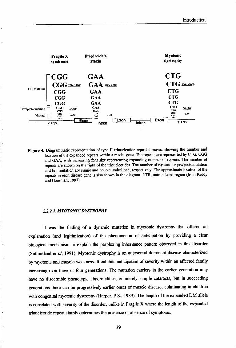
Figure 4. Diagrammatic representation of type II trinucleotide repeat diseases, showing the number and location of the expanded repeats within a model gene. The repeats are represented by CTG, CGG and GAA, with increasing font size representing expanding number of repeats. The number of repeats are shown on the right of the trinucleotides. The number of repeats for pre/protomutation and full mutation are single and double underlined, respectively. The approximate location of the repeats in each disease gene is also shown in the diagram. TJTR, untranslated region (from Reddy and Housman, 1997).
2.2.2.2. MYOTONIC DYSTROPHY
It was the finding of a dynamic mutation in myotonic dystrophy that offered an
explanation (and legitimization) of the phenomenon of anticipation by providing a clear
biological mechanism to explain the perplexing inheritance pattern observed in this disorder
(Suthertland et al, 1991). Myotonic dystrophy is an autosomal dominant disease characterized
by myotonia and muscle weakness. It exhibits anticipation of severity within an affected family
increasing over three or four generations. The mutation carriers in the earlier generation may
have no discernible phenotypic abnormalities, or merely simple cataracts, but in succeeding
generations there can be progressively earlier onset of muscle disease, culminating in children
with congenital myotonic dystrophy (Harper, P.S., 1989). The length of the expanded DM allele
is correlated with severity of the disorder, unlike in Fragile X where the length of the expanded
trinucleotide repeat simply determines the presence or absence of symptoms.
39
Introduction
The affected gene in myotonic dystrophy is located in 19ql3.3 and contains an expanded
CTG repeat within the 3' untranslated region. It is believed to encode, by sequence homology, a
cyclic adenosine monophosphate (cAMP)-dependent serine-threonine protein kinase designated
by myotonic dystrophy protein kinase (DMPK), but its in vivo substrate remains unknown
(Mahadevan et al, 1992; Fu et al, 1992; Brook et al, 1992).
The expanded CTG stretch is highly polymorphic in the normal population ranging from
5 to 30 repeats. The trinucleotide repeat can undergo either expansions or contractions, with a
strong bias in favor of expansion up to many hundred repeats. In females, the size of the repeat
can increase without apparent restrain from generation to generation reaching the point of
genetic lethality. On the other hand, in males the repeat reaches a maximum size of -1000
copies and then appears to decrease in size upon transmission (Lavedan et al, 1993; Mulley et
al, 1993). Although the smaller DM alleles are more unstable in male transmissions, the very
large alleles that transmit the most severe form of the disease are almost always transmitted by a
female. The different meiotic behavior of the repeat in the two sexes is also observed in fragile
X syndrome, where a female meiosis in an absolute requirement for the expression of the
disease. This behavior contrasts with the transmission pattern observed in type I disorders that
show much greater increases in size during paternal transmission - "the paternal anticipation"
already referred to.
The mechanism by which increased CTG copy number results in disease is not clear.
Reduction of DMPK mRNA levels has been reported (Fu et al, 1993), but the absence of point
mutations in the gene giving rise to a phenotype similar to DM, together with the dominant
nature of the disease, argue against the hypothesis of DMPK gene loss of function to be the
cause of DM pathology. Alternatively, Boucher et al (1995) have suggested that the CTG
repeat expansion has an effect on the neighboring genes, contributing in such manner to
myotonic dystrophy pathology.
2.2.2.3. FRIEDREICH'S ATAXIA
Friedreich's ataxia is the most recent disorder found to be associated with trinucleotide
repeat expansions (Campuzano et al, 1996). It has joined the type II disease group exhibiting
the expansion of a new disease-related triplet: GAA. As an autosomal recessive disease with
40

Introduction
little evidence for genetic anticipation, FA does not fit the mold of a dynamic mutation. Although
the age of onset may vary, most patients exhibit the symptoms in adolescence and the onset in
middle age or later is mostly uncommon, unlike the other triplet diseases with anticipation.
The GAA repeat is located within the first intron of frataxin gene which encodes a
protein of unknown function. The normal alleles include 10 to 21 copies of the repeat whereas
nearly 95% of FA alleles contain from ~ 200 to 900 copies - similar to the premutation alleles
found in FRAXA and DM. Recently, premutation alleles of around 40-60 GAAs have been
reported in FA (Cossée et al, in press). The disease-causing expansion appears in
heterozygosity in about 90% of the patients, but half of the remaining are compound
hétérozygotes carrying the repeat expansion in one allele and point mutations in the other.
The expanded GAA repeat in intron 1 appears to interfere in frataxin nuclear RNA
processing resulting in the absence of a mature message in the cytoplasm (Campuzano et al,
1996). Although such hypothesis has not been proved, the massive number of AG splice
acceptor sites formed by the expanded GAA repeating units, is likely to underlie the mechanism
by which the transcript levels are reduced in FA. Moreover, the GAA repeat cannot form
hydrogen-bonded hairpins, thus being unable to form the non-conventional DNA structures
characteristic of CG-rich repeats, which may be involved in DNA transcription impairment in
other TREs-associated diseases.
2.2.3. OTHER DISORDERS
Recent studies have shown that the expansion of a 12-bp repeat is responsible for a
monogenic form of epilepsy known has progressive myoclonus epilepsy (EPM1) (Lalioti et al.
1997; Lafrenière et al, 1997; Virtaneva et al, 1997). The gene encoding cystatin B, a
proteinase inhibitor, contains a (CCCCGCCCCGCG)3 in the 5' flanking region within the
promoter sequence which expands up to 600-900 bp in diseased alleles. EPM1 is the second
autosomal dominant disorder joining the growing number of genetic diseases that are associated
with unstable repeat expansions.
41
Chapter II
Experimental Research

Objectives
Experimental Research
1. Objectives
The molecular mechanisms underlying genetic anticipation in FAP remain one of the
major unsolved dilemmas concerning the phenotypic variability presented by this disorder. As a
neurological disease with an autosomal dominant mode of inheritance and evident anticipation,
FAP is a strong candidate for the involvement of trinucleotide repeat instability in the decreasing
age of onset over successive generations. The pathology of FAP is produced by point mutations
in the TTR gene, rather than unstable trinucleotide repeat expansions. Indeed, TTR gene
structure does not include repeated triplets neither within the coding regions nor in the
untranslated regulatory sequences (Sakaki et al, 1989). Since TTR amyloidogenesis is a
multistep process, modifier gene(s) are very likely to be involved in phenotypic variability
manifested as genetic anticipation. We have questioned whether anticipation in FAP might be
related to unstable trinucleotide repeat expansions in a modifier gene (or genes), rather than
TTR gene itself.
The main purposes of this project were 1) the determination of maximal trinucleotide
repeat lengths in generational pairs of Portuguese FAP kindreds, and 2) the detection of
intergenerational instability of any of the ten trinucleotide repeats in the same kindreds.
We have performed a complete genome scanning of trinucleotide repeat lengths in 8
generational pairs of FAP Portuguese families with a difference in age of onset greater than 12
years, using the Repeat Expansion Detection (RED) method. This technique does not require
flanking sequence information and allows a genome-wide search for potentially pathological
repeat expansions.
43

Materials and Methods
2. Materials and Methods
2.1. GENERATIONAL PAIRS AND CONTROLS
Eight generational pairs of Portuguese kindreds with FAP were scanned for trinucleotide
repeat expansions using the RED method. In seven of the tested pairs the differences in the age
of onset were greater than 12 years. One pair with identical ages of onset was used as a control.
The gender and age-at-onset of the individuals of each pair are described in the following table.
All affected individuals were confirmed carriers of the TTR Met30 mutation, and were
diagnosed as FAP patients based on the presence of characteristic clinical features of the
disorder. The two asymptomatic individuals did not exhibit any pathological symptoms,
although confirmed carriers of the TTR Met30 allele. None of the above individuals had the
TTR Metl 19 mutation.
As negative controls, nineteen unrelated individuals of the Portuguese population
without known genetic disease (11 females, 6 males and 2 individuals of unknown gender) were
used.
Blood samples were collected using vacutainer tubes with ACD (Becton Dickinson) for
lymphocyte immortalization and DNA extraction.
44

Materials and Methods
2.1.1. LYMPHOCYTE IMMORTALIZATION
A 3.0 ml whole blood aliquot was carefully laid onto 3.0 ml of Histopaque-1077
(Sigma) (previously brought to room temperature), on a 15 ml centrifuge Falcon tube. This
mixture was centrifuged at 1800 rpm, for 30 minutes, at room temperature.
After centrifugation, the upper layer to within 0.5 cm of the opaque interface, was
carefully aspirated using a Pasteur pipette and discarded. The opaque interface, containing
mononuclear cells, was then transferred with a Pasteur pipette into a clean centrifuge Falcon
tube. Ten ml of RPMI-1640 Medium (Sigma) were then added to the tube, and the contents
were mixed by gentle aspiration.
After a centrifugation at 1400 rpm, for 10 minutes, the supernatant was discarded and
the cell pellet was resuspended in 5.0 ml of RPMI-1640 medium. Another centrifugation
followed, at 1400 rpm, for 10 minutes, the supernatant was discarded and the cell pellet was
once again resuspended in RPMI-1640 medium and then centrifuged one last time at 1400 rpm,
for 10 minutes.
Using a Pasteur pipette, the resulting cell pellet was resuspended in 2.0 ml of RPMI-
1640 medium and the transformation step followed.
TRANSFORMATION BY EPSTEIN-BARR VIRUS (EBV)
A 2.0 ml volume of Transformation Medium was added to 1.0 ml of resuspended cell
pellet obtained by the procedure described above, and 1.5 ml aliquots of the mixture were
transferred to a couple of 25 cm3 culture flasks. 1.5 ml of EBV suspension (previously
incubated at 37°C for 3-4 hours) was added to each flask.
After 24 hours, 2.0 ml of Transformation Medium were added to the culture, and this
procedure was continued in each two days, for a week, after which the medium was transferred
to a clean centrifuge Falcon tube followed by a centrifugation at 1200 rpm, for 10 minutes. Half
of the supernatant volume was discarded and replaced with the same volume of Complete
Medium.
In each two days that followed the previous step, a given volume of Complete medium
(depending on the evolution of the culture) was added. After a week or two, the cells
45

Materials and Methods
were transformed and could by frozen for storage.
FREEZING OF LYMPHOCYTES
A 1.0 ml volume of Freezing Medium (Gibco), previous placed on ice, was added to 1.0
ml of resuspended cell pellet, very slowly and using a Pasteur pipette. During this step, the tube
containing the cells was kept on melting ice and was continuously shaken.
The mixture was then transferred to a Nunc Vial freezing tube and stored at -70°C for
at least 4 hours up to 24 hours, after which the tube was conserved in liquid nitrogen.
CULTURE OF FROZEN LYMPHOCYTES
One ampoule containing the frozen cells, was removed from the liquid nitrogen and
immediately thawed in a shaking water bath at 37°C. Before the disappearance of the last ice
crystals, the tube was placed on ice and the cell suspension was very carefully transferred to a
centrifuge Falcon tube, containing 5.0 ml of base Medium supplemented with 20% FCS, at 4°C.
After centrifugation at 1200 rpm, for 10 minutes, the supernatant was discarded, and the
cell pellet resuspended in 2.0 ml of Complete Medium (supplemented with 20% FCS). The
suspension was then transferred to a 25 cm2 flask and, following an incubation at 37°C, for 24
hours, 2.0 - 6.0 ml of Complete Medium were added.
Depending on the evolution of the culture, each two days, a given volume of Complete
medium was added, until the culture reached the desired point.
MEDIA
Base Medium
This medium contains 500.0 ml of RPMI-1640 with L-glutamine and Hepes (Gibco),
2.0 ml of penicillin/streptomycin (Gibco), 2.0 ml of kanamycin (25 mg/ml) and 5.0 ml of
fungizone (250ug/ml). After preparation was stored at 4°C.
46

Materials and Methods
Complete Medium
This medium contains 100.0 ml of Base Medium; 5.0 ml of foetal calf serum (FCS),
previously inactivated at 56°C, for 30 minutes and 1.5 ml Ultroser HY hydrated (Gibco). After
preparation this medium was stored at 4°C.
Transformation Medium
This medium contains 100.0 ml of Base Medium, 20.0 ml of FCS (previously
inactivated at 56°C, for 30 minutes) and 0.15 ml of ciclosporin (1 mg/ml in ethanol).
Freezing Medium
This medium contains 0.8 ml of FCS (previously inactivated at 56°C for 30 minutes)
and 0.2 ml of sterile DMSO. After preparation was stored at -20°C.
2.1.2. GENOMIC DNA EXTRACTION
Twelve ml of total blood were centrifuged at 2,000 rpm for 10 minutes. The plasma
layer was gently removed and the buffy coat - the leukocyte layer - was carefully placed in
eppendorf tubes. 1-2 volumes of a solution containing 72.0 mg of ammonium bicarbonate and
7.0 g of ammonium chloride in a liter of dd H2O were added to the cells for erythrocyte lysis.
The tubes were left on ice for 10 minutes, centrifuged at 10,000 rpm for 5 minutes, and the
supernatant discarded. This procedure was repeated a number of times in order to obtain a clear
white cell pellet.
A 5.0 ml volume of proteinase K buffer (Tris 0,05M pH 7,5; NaCl 1M; EDTA 0,001M;
and SDS 0,5%) was added to the cell pellet followed by 50.0 ul of proteinase K and incubated
overnight at 55 °C. Following the leukocyte lysis, 2.0 ml of an NaCl saturated solution and 2.0
ml of chloroform were added to the pellet and vortexed for 30 seconds. After a centrifugation at
9,000 rpm for 10 minutes, the supernatant was discarded and 2 volumes of 100% ethanol were
added to the tube which was inverted until DNA visualization. The precipitated DNA was
remove carefully and resuspended in an appropriate volume of TE buffer.
47

Materials and Methods
2.2. RED METHOD
Repeat Expansion Detection (RED) method is a generally applicable method which
provides a direct path to identify potentially pathological repeat expansions (Schalling et al,
1993). Single copies of large trinucleotide repeats can be detected based on the use genomic
DNA as a template for the annealing and ligation of repeat-specific oligonucleotides.
Consequently, it does not require flanking sequence information, or single copy probes. A
thermostable ligase is used in a cycling procedure which generates multimers of the
oligonucleotide used.
This method constitutes a unique way of studying triplet repeat expansion-associated
inherited disorders, particularly those characterized by anticipation.
2.2.1. GENERAL PRINCIPLE
The genomic DNA containing a long segment of repeated trinucleotides serves as a
template for annealing of oligonucleotides composed of several copies of the complementary
triplet (Figure 5). As two or more oligonucleotide molecules anneal to the genomic template in
adjacent positions, they are ligated by a thermostable ligase, forming a longer single stranded
molecule. This step is repeated several hundred times in a two-step cycling process using
adequate denaturation and annealing temperatures. The presence of long trinucleotide repeats
anywhere in the genome will give rise to the formation of large multimers of the original
oligonucleotide. The single stranded multimers are separated by size in denaturing conditions by
polyacrylamide gel electrophoresis, transferred to a nylon membrane and detected by
hybridization to a 32P-labeled complementary oligonucleotide. The radiolabeled probe
maximizes the detection of the RED reaction products.
2.2.2. OLIGONUCLEOTIDE PHOSPHORYLATION
For 20 RED reactions 100 ^1 of T4 polynucleotide kinase (Epicentre) mixture was
prepare by combining 73.0 |al of ddH20; 10.0 ^1 of ATP (10 mM); 10.0 [Ú of lOx T4
polynucleotide kinase buffer; 1.0 \x\ of the testing oligonucleotide (1 or 2 ug/ul depending on
48

Materials and Methods
the length of the oligo, being the higher concentration for oligonucleotides with about 17
repeated units); and 6.0 ul of T4 polynucleotide kinase (PNK). The reaction mix was incubated
at 37 °C for 50 minutes and used in the RED reaction immediately. Alternatively, the mixture
can be heated at 65 °C to inactivate PNK and stored at -80 °C for posterior use.
(CTG),o OLIGONUCLEOTIDES
ANNEALING
— i i i i i n i i i i i i i n i n CAG REPEAT
IBB i m in i imn imiH I I I 'P" 1 "—
LIGATION
GENOMIC DNA
* t ^ ^ i - iTTHiHi inmim i i i n i i i i m i i i i i j i i u i i j u i i j j i m i u i i i i i i i i i i =
1 DENATURATION
PRODUCTS = MULTIMERS OF (CTG)io OLIGONUCLEOTIDES
Figure 5. Diagram depicting amplification and detection of DNA-trinucleotide repeat using RED. Since only one strand of oligonucleotide multimer is produced per target molecule per cycle, a large amount of DNA is needed (10 ug). Products vary in length as multimers of the oligonucleotide used, and depend upon size of the target (from Schalling et ai, 1993).
2.2.3. REACTION CONDITIONS
Following the addition of 5.0 ul of phosphorylated oligonucleotide to -10.0 ul of DNA
template (~ 1 ug/ul), the reaction mixture was covered with 20.0 ul of mineral oil in eppendorf
49

Materials and Methods
tubes, heated at 98 °C for 10 minutes, to denature the DNA, and placed on ice (for CGG
oligonucleotide, 10.0 jul of 5M Betaine were added to the mixture). While keeping the samples
on ice, 10.0 ul of ligation reaction mix A containing: 2.0 ul of lOx Ampligase reaction buffer;
7.0 ul of ddH20; and 1.0 ul of Ampligase DNA ligase (5 Units) (Epicentre). The reaction
mixtures were quickly centrifuged for 30 seconds, and the tubes were placed into the
thermocycler (Perkin Elmer DNA Thermal Cycler) for cycling according to the following
parameters:
For (CTG)io/n-, (CGG)n- (TGG)i2-, and (CGT)i4-oligonucleotides - 100 cycles at 70 °C for
30 seconds and 95 °C for 10 seconds; for (CCT)i5-, (AAG)io-, (GTT)i5,- (AGT)H-, and
(ATG)i2- oligonucleotides - 100 cycles at 60 °C for 30 second and 95 °C for 10 seconds; for
(TAA)n -100 cycles at 50 °C for 30 seconds and 95 °C for 10 seconds.
After the first 100 cycles, the samples were placed on ice and 5.0 ul of ligation reaction
mix B containing: 3.5 ul of ddH20; 0.5 ul of lOx Ampligase reaction buffer; and 1.0 ul of
Ampligase DNA ligase (5 U), were added. The samples were quickly centrifuged for 30
seconds and the cycling was restarted for 300 additional cycles at the same conditions described
above. The RED reaction products were immediately used for electrophoretic separation or
stored at 4 °C.
2.3. ELECTROPHORESIS
The mnning gel consisting of 50.0 ml of a pre-warmed 6% acrylamide / 6M urea
solution, 500.0 ul of APS and 35.0 ul of TEMED was poured into a SequiGen II electrophoresis
apparatus (0.5 mm thick) (Biorad) and pre-run at 50 watts for 30 minutes. Meanwhile 20.0 ul of
the RED reaction products were mixed with 15 ul of gel loading dye, denatured for 5 minutes at
95 °C and placed on ice.
Following the gel pre-run (30 minutes), the wells were cleaned with a Pasteur pipette to
prevent urea deposits, the samples were loaded, and the gel was run in lx TBE for
approximately 1 hour and 10 minutes at 50 watts (the xylenocyanol dye should migrate to
middle of the plates). The intact gel apparatus was then put under cold tap water for 10 minutes.
50

Materials and Methods
2.4. MEMBRANE TRANSFER
A Hybond N+ membrane (Amersham) was quickly soaked in lx TBE and placed in
direct contact with the gel. The membrane was then covered with 3 layers of 3M paper and the
glass plate was placed on top. The RED products were left transferring by capillary action
overnight.
The membrane was removed, rinsed for 2 minutes in 2x SSC, dried at room
temperature for 1 hour and exposed to UV light to cross-link the DNA to the membrane.
Following this procedure, the membrane could be immediately hybridized or stored at room
temperature.
2.5. LABELING OF THE PROBE
The probe was radiolabeled by combining 1.5 ul of the probe (200 ng/ul); 6.5 ul of
ddH20; 4.0 ul of 5x terminal transferase buffer; 2.0 ul of CoCfe; 5.0 ul of P32-dCTP; and 1.0 ul
of terminal deoxynucleotidyl transferase (Boehringer Mannheim) in an Eppendorf tube.
Following the incubation for 60 minutes at 37 °C, 4.0 ul of glycogen solution (20 ug/ul) and
50.0 ul of 100% ethanol were added, followed by a centrifugation for 30 seconds. The ethanol
was carefully removed and the precipitated DNA was dried in the speedvac for at least 60
minutes. The radiolabeled DNA was finally resuspended in 100.0 ul of ddH20.
2.6. HYBRIDIZATION
The dried membranes were pre-hybridized in Rapid-Hyb buffer (Amersham) for at least
1 hour at the following temperatures: for (CTG)io/n-, (CGG)u-, (TGG)i2-, and (CGT)M-
oligonucleotides - at 65 °C; for (CCT)i5-, (AAG)io-, (GTT)i5,- (AGT)14-, and (ATG)i2-
oligonucleotides - at 60 °C; for (TAA)n - at 55 °C. The hybridization with the radiolabeled
probe was done overnight in the same buffer at the same temperature. After 2 washes in 2x SSC
(20 minutes each) at room temperature, followed by 2 washes in lx SSC (30 minutes each) at 5
°C below hybridization temperature, the membrane was exposed to Kodak X-AR
51
Materials and Methods
or Kodak Biomax MS films at -80 °C, using intensifying screens, for 1-15 days.
52

Results and Discussion
3. Results and Discussion
3.1. CTG/CAG TRINUCLEOTIDE
Eight inherited neurodegenerative disorders are known to involve expansions of CAG
trinucleotide repeats within susceptibility genes, resulting in an abnormally long stretch of
polyglutamine within the encoded proteins. CTG repeat expansions are associated with severe
phenotypes of congenital myotonic dystrophy as long segments of the repeated triplet in the 3'
untranslated region of the myotonin gene result in disease. These inherited diseases, in which the
length of the CTG or CAG repeat region is expanded, exhibit genetic anticipation.
We have tested the genomic DNA from 9 FAP kindred pairs and 8 negative controls
using (CTG)io and (CTG)n oligonucleotides to assess trinucleotide repeat lengths and to detect
the existence of possible expansions eventually responsible for genetic anticipation in FAP.
Some generational pair and control DNAs have been tested in duplicate or triplicate to confirm
the results obtained for this particular trinucleotide, and to define the reproducibility of RED in
our laboratory. As a positive control, the DNA of an individual exhibiting a very long stretch of
the repeated motif was used in all reactions. The RED reaction results are shown in the
autoradiographs of Figures 6, 7, 8, and 9.
All samples of seven of the eight pairs exhibited a maximal repeat size of 120 bp,
corresponding to 40 repeated triplets. Only pair IV has broken the consistency of the repeat
length distribution in FAP gene carriers by exhibiting a fragment of 210 bp corresponding to 70
copies of the trinucleotide (Fig. 6, lanes 11 and 12; Fig. 8, lanes 4 and 5; Fig. 9, lanes 8 and 9).
No differences in maximal repeat size have been found within the tested pairs with the exception
of an extra pair used only in CTG/CAG repeat detection experiments - pair Y. The descendant,
diseased with an onset at 40, presents a slightly longer repeat than the progenitor, asymptomatic
carrier, corresponding to the ligation of one more (CTG)io oligonucleotide referring to a 30 bp
(10 copies) difference (Fig. 6, lanes 1 and 2). The control samples have shown repeat lengths
ranging between 60 bp (20 repeats) and 270 bp (90 repeats). CTG/CAG repeat sizes, as
determined by RED, are compared in Figure I.
CTG/CAG repeats are highly polymorphic in length in the human genome but their
number usually does not exceed 40 repeats in normal individuals. Hofferbert et al. (1997) have
53
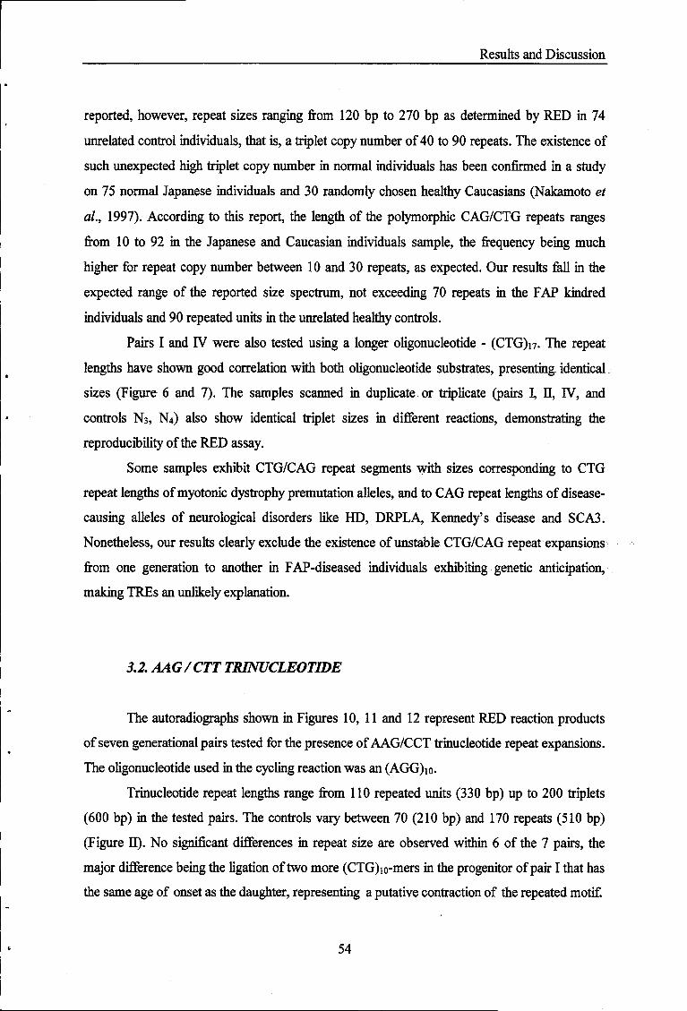
Results and Discussion
reported, however, repeat sizes ranging from 120 bp to 270 bp as determined by RED in 74
unrelated control individuals, that is, a triplet copy number of 40 to 90 repeats. The existence of
such unexpected high triplet copy number in normal individuals has been confirmed in a study
on 75 normal Japanese individuals and 30 randomly chosen healthy Caucasians (Nakamoto et
al., 1997). According to this report, the length of the polymorphic CAG/CTG repeats ranges
from 10 to 92 in the Japanese and Caucasian individuals sample, the frequency being much
higher for repeat copy number between 10 and 30 repeats, as expected. Our results fall in the
expected range of the reported size spectrum, not exceeding 70 repeats in the FAP kindred
individuals and 90 repeated units in the unrelated healthy controls.
Pairs I and IV were also tested using a longer oligonucleotide - (CTG)n. The repeat
lengths have shown good correlation with both oligonucleotide substrates, presenting identical
sizes (Figure 6 and 7). The samples scanned in duplicate or triplicate (pairs I, JJ, IV, and
controls N3, N4) also show identical triplet sizes in different reactions, demonstrating the
reproducibility of the RED assay.
Some samples exhibit CTG/CAG repeat segments with sizes corresponding to CTG
repeat lengths of myotonic dystrophy premutation alleles, and to CAG repeat lengths of disease-
causing alleles of neurological disorders like HD, DRPLA, Kennedy's disease and SCA3.
Nonetheless, our results clearly exclude the existence of unstable CTG/CAG repeat expansions
from one generation to another in FAP-diseased individuals exhibiting genetic anticipation,
making TREs an unlikely explanation.
3.2. AAG/CTT TRINUCLEOTIDE
The autoradiographs shown in Figures 10, 11 and 12 represent RED reaction products
of seven generational pairs tested for the presence of AAG/CCT trinucleotide repeat expansions.
The oligonucleotide used in the cycling reaction was an (AGG)io.
Trinucleotide repeat lengths range from 110 repeated units (330 bp) up to 200 triplets
(600 bp) in the tested pairs. The controls vary between 70 (210 bp) and 170 repeats (510 bp)
(Figure II). No significant differences in repeat size are observed within 6 of the 7 pairs, the
major difference being the ligation of two more (CTG)io-mers in the progenitor of pair I that has
the same age of onset as the daughter, representing a putative contraction of the repeated motif.
54
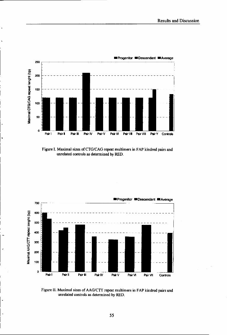
Results and Discussion
lProgenitor ■Descendant ■ Average 250
Pair I Pair Pair IV PairV Pair VI Pair VII Pair VIII Pair Y Controls
Figure I. Maximal sizes of CTG/CAG repeat multimers in FAP kindred pairs and unrelated controls as determined by RED.
700 ■ Progenitor ■ Descendant ■ Average
Pair III Pair IV PairV Pair VI Pair VII Controls
Figure II. Maximal sizes of AAG/CTT repeat multimers in FAP kindred pairs and unrelated controls as determined by RED.
55

Results and Discussion
(CTG)„ bp
120 360 110 330 100 300 90 270 80 240
70
60
50
30
210 —
180 —
150 *~
40 120 -
I N3 N4 I IV pc [ - • O O pc r rr
1 2 3 4 5 6 7 8 9 10 11 12 13 14
90 —
20 60
bp (CTG)»
357 119
306 102
255 85
-« 204
- 153
68
51
— 102 34
Figure 6. Autoradiograph of the RED reaction products using (CTG)i0 and (CTG)17 oligonucleotides (lanes 1-7 and 8-14, respectively). PC: positive control; lanes 4, 5 and 9-12: FAP generational pairs; lanes 6 and 7: control individuals. The remainder lanes refer to samples excluded from our study. Note the correlation of repeat sizes obtained with both oligonucleotide substrates. 10 ug of genomic DNA template were used in each reaction. Sizes of (CTG)„ multimers are indicated in bp.
56

Results and Discussion
Y n IV
bp (CTG)n
^ 210
— 180
- 150
70
60
50
120 40
— 90 30
— 60 20
Figure 7. Autoradiograph of the RED reaction products using (CTG)10. Lanes 1-6: FAP generational pairs. 10 ug of genomic DNA template were used in each reaction. Sizes of (CTG)n multimers are indicated in bp.
57

Results and Discussion
II IV V
PC •mi VIII N4 NONM
O O O
1 2 3 4 5 6 7 8 9 10 11 12 bp (CTG)n
- ^ 420 ^ j 390 — 360 -* 330 — 300 — 270 — 240 — 210
- 180
150
120
- 90
- 60
140 130 120 110 100 90 80 70
60
50
40
30
20
Figure 8. Autoradiograph of the RED reaction products using (CTG)i0. Lanes 2-9: FAP generational pairs; lanes 10-12: control individuals; PC: positive control. 10 ug of genomic DNA template were used in each reaction. Sizes of (CTG)n multimers are indicated in bp.
58

Results and Discussion
I II m IV N2N3N4N,7 pc • - ! #n • - ! B-iOOOD 11*11
1 2 3 4 5 6 7 8 9 1011 12 13 bp (CTG)n
330 300
110 100
270 90 240 80 210 70 180 60
-■ 150 50
-■ 120 40
-* 90
■* 60
30
20
Figure 9. Autoradiograph of the ÎŒD reaction products using (CTG)i0. Lanes 2-9: FAP generational pairs; lanes 10-13: control individuals; PC: positive control. 10 ug of genomic DNA template were used in each reaction. Sizes of (CTG)„ multimers are indicated in bp.
59
Results and Discussion
A difference in one ligation is also observed in pair II (Fig. 12, lanes 2 and 3). The accuracy of
band reading at the upper portion of the gel is slightly diminished by a progressive loss of
resolution which might explain the small differences found in pairs I and H.
The length of the AGG repeat could not be determined for the descendant of pair IV
(Fig. 12, lane 10) and for some controls (Fig. 11, lanes 4, 5; and Fig. 12, lanes 11, 13) due to a
weak cycling reaction which could not produce the necessary amount of (AAG)io multimers for
exact size detection.
Our results show the absence of AAG/CTT repeat expansions in the second generation
elements of the tested pairs, thus excluding the involvement of such mechanism as the molecular
basis of genetic anticipation in these pedigrees. The maximal number of copies is consistent with
previously reported studies in unrelated Caucasians (Hofferbert et al., 1997) where it ranges
from 252 bp to 540 bp.
AAG trinucleotide repeat expansions are associated with Friedreich's ataxia. Long
stretches of the AAG repeated motif are located within an intronic sequence of the frataxin gene
leading to the loss of function of the protein. The mutation-causing repeat number ranges
between 200 and > 900 triplets, while the results show a triplet repeat with similar length in one
patient - the progenitor of pair I - containing 200 copies, with a phenotype typical of FAP
classical onset. The length of the repeat is highly polymorphic and such high copy number is not
uncommon in normal individuals.
3.3. TTA/TAA TRINUCLEOTIDE
The products of the RED reaction of TTA/TAA triplet are shown in Figure 13. All
samples show a ligation product of 66 base pairs corresponding to a dimer of the (TTA)n
substrate. The samples refer to three FAP generational pairs (I, II and HI) and 7 control
individuals. RED reaction has produced low and unequal amounts of oligonucleotide dimers but
the single band is clearly visible for all samples in which the reaction has worked. No larger
multimers have been formed which is in accordance with the study by Hofferbert et al. (1997)
where a single ligation corresponding to 72 bp was expected as the maximal repeat length
determined for the 52 tested unrelated individuals. The discrepancy of size in base pairs is due
to the triplet copy number of the oligonucleotide used in both RED studies.
60

Results and Discussion
HI II Ns Ns III II N5 N6
#- i # - | 0 0 # - | # - i O O
1 2 3 4 5 6 7 8 9 101112 bp
180
150
— 120
- 90
- 60
(AAG)„
450 150 420 140 390 130 360 120 330 110 300 100 270 90 240 80 210 70
60
50
40
30
20
Figure 10. Autoradiograph of the RED reaction products using (AAG)]0. Lanes 1-4: FAP generational pairs; lanes 5 and 6: control individuals. The samples loaded on lanes 7 to 12 are the same but resulting from a different cycling reaction. 10 ug of genomic DNA template were used in each reaction. Sizes of (AAG)„ multimers are indicated in bp. Note the reproducibility of the RED assay.
61

Results and Discussion
N2N4NSN«N, OOOOD
1 2 3 4 5
VII III VI
1 1 1 6 7 8 9 10 11
' : '3
bp (AAG)„
480 450 420 390 360 330 300
— 270 — 240 -* 210
- 180
— 150
160 150 140 130 120 110 100 90 80
70
60
50
** 120 40
— 90 30
- 60 20
Figure 11. Autoradiograph of the RED reaction products using (AAG)i0. Lanes 1-5: control individuals; lanes 6-11: FAP generational pairs. 10 ug of genomic DNA were used in each reaction. Sizes of (AAG)n multimers are indicated in bp.
62

Results and Discussion
II IV Ni N3 N7
O O D
1 2 3 4 5 6 7 8 9 1011 1213
* #
Ni,
□
14
■
bp (AAG)„ 540 I 450 420 390 360 330
— 300 - - 270
4 — 240
d - 210
d -■ 180
#
180 170 160 150 140 130 120 110 100
90 80 70
60
* é - 150 50
-* 120 40
- 90 30
« < * T — 60 20
Figure 12. Autoradiograph of the RED reaction products using (AAG)i0. Lanes 1-10: FAP generational pairs; lanes 11 and 14: control individuals. 10 ug of genomic DNA template were used in each reaction. Sizes of (AAG)n multimers are indicated in bp.
63
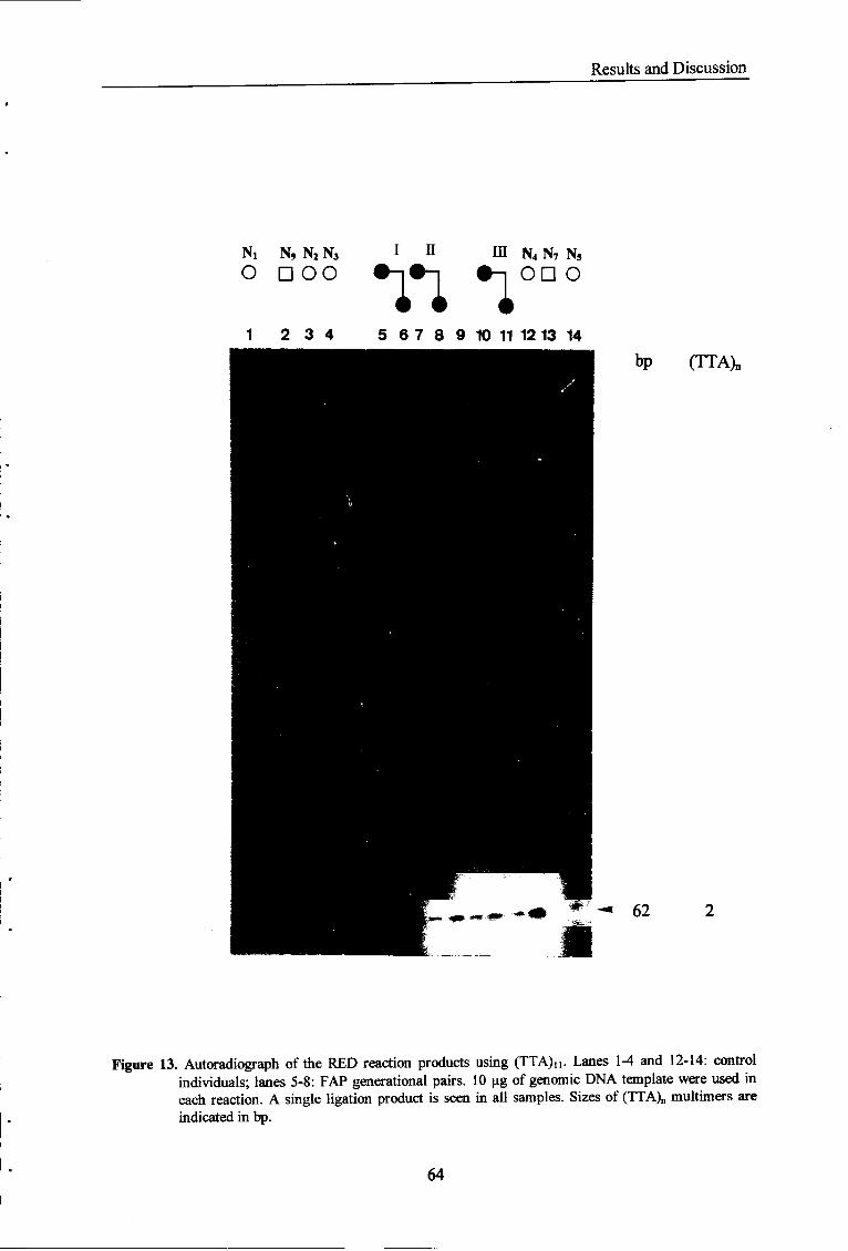
Results and Discussion
Ni N9 N2 N3
O D OO i n inn m N4 N7 N5
on o
1 2 3 4 5 6 7 8 9 10 11 12 13 14 bp (TTA)n
- 62
Figure 13. Autoradiograph of the RED reaction products using (TTA)U. Lanes 1-4 and 12-14: control individuals; lanes 5-8: FAP generational pairs. 10 ug of genomic DNA template were used in each reaction. A single ligation product is seen in all samples. Sizes of (TTA)n multimers are indicated in bp.
64

Results and Discussion
3.4. AGT/ACT TRINUCLEOTIDE
Genomic DNA of all FAP kindred pairs and 12 negative controls were used to
determine AGT/ACT repeat lengths. Autoradiographs of AGT triplet RED reaction products are
shown in Figures 14 and 15. The (AGT)i4 oligonucleotide was ligated only once in every
sample, giving rise to a single band of 84 bp (28 repeats). The resulting products are clearly
visible except in 2 control samples (Figure 14, lane 15; and Figure 15, lane 12) presenting feint
bands. The descendant of pair VII (lane 8 of Figure 14) exhibits a different pattern of migration,
observed in almost every reaction, characterized by a slower mobility throughout the gel which
would accelerate further down to end ahead of all other sample bands. This phenomenon is
probably due to the high viscosity of that particular DNA sample. The irregular bands observed
in the film of Figure 15 reflect irregularities of the polyacrylamide gel when the nylon membrane
was laid for DNA transfer, rather than different mobility patterns of the reaction products.
AGT/ACT trinucleotide repeats have not been associated with any triplet repeat
expansion disorder, and seem not to be polymorphic in size as observed in 57 unrelated
Caucasian individuals, with a constant size of about 72 bp (one single ligation), using the RED
method (Hofferbert et al., 1997). Our results support this view, although the size of the repeated
motif has been determined to be slightly different due to the oligonucleotide number of repeated
triplets.
3.5. CGT/ACG TRINUCLEOTIDE
The CGT/ACG repeat, tested with a (CGT)H oligonucleotide, was difficult to detect by
the RED method. Figures 16 and 17 illustrate the RED reaction results in seven FAP
generational pairs and 8 unrelated negative controls. The single oligonucleotide species appears
at the bottom of the gel as large blots overlapping with several bands corresponding to ligation
products of oligonucleotide fragments. The existence of such small products is indicative of
oligonucleotide impurity which may have been responsible for the formation of very few ligated
molecules during the cycling reaction. All samples show a faint band resulting from a single
ligation and corresponding to 28 repeated triplets (84 bp). Hofferbert et ai. (1997) have shown
65

Results and Discussion
IV VI VII VIII NiN3NuNi4NMNi7
ooooon 'ill 11 1 2 3 4 5 6 7 8 9 10111213 14 15 16
bp (AGT)n
28
Figure 14. Autoradiograph of the RED reaction products using (AGT)i4. Lanes 1-10: FAP generational pairs; lanes 11 and 16: control individuals. 10 ug of genomic DNA template were used in each reaction. A single ligation product is seen in all samples. Sizes of (AGT)„ multimers are indicated in bp.
66

Results and Discussion
NiiNiiNwNis O D D O
I—- n in N9N4 — I U 111
- 84 28
MM9-:
Figure 15. Autoradiograph of the RED reaction products using (AGT)i4. Lanes 1-4 and 12, 13: control individuals; lanes 5-11: FAP generational pairs. 10 ug of genomic DNA template were used in each reaction. A single ligation product is seen in all samples. Sizes of (AGT)n multimers are indicated in bp.
67

Results and Discussion
bp (CGTk
-■ 84 28
2 3 4 5 6 7 8 9 1011 12
Ni* VI W V H I N U N U N M
O Éé®~|#n® I O O O
Figure 16. Autoradiograph of the RED reaction products using (CGT)i4. Lanes 2-9: FAP generational pairs; lanes 1 and 9-12: control individuals. 10 ug of genomic DNA template were used in each reaction. A feint band corresponding to a single ligation product is visible in all samples and is indicated by the arrow. The other bands result from the ligation of oligonucleotide fragments. Sizes of (CGT)n multimers are indicated in bp.
68
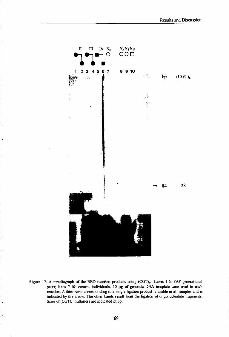
Results and Discussion
II III IV N2 N3N4N17 O OOD ™.
1 2 3 4 5 6 7 8 9 10 bp (CGT)„
- 84 28
Figure 17. Autoradiograph of the RED reaction products using (CGT)i4. Lanes 1-6: FAP generational pairs; lanes 7-10: control individuals. 10 ug of genomic DNA template were used in each reaction. A feint band corresponding to a single ligation product is visible in all samples and is indicated by the arrow. The other bands result from the ligation of oligonucleotide fragments. Sizes of (CGT)„ multimers are indicated in bp.
69

Results and Discussion
a maximal size for CGT trinucleotide repeat of approximately 60 bp in 54 unrelated individuals,
again the result of a single ligation of the oligonucleotide substrate [(CGTho]. Our results cannot
exclude the existence of longer segments of repeated CGT triplets in the analyzed DNA
samples, but the existing literature reports repeat lengths of less than 90 bp - 30 copies - of the
repeated motif (Lindblad et al, 1994; Hofferbert et al, 1997) . Moreover, only one band
resulting from a dimer formation has been consistently observed in all our experiments with this
particular oligonucleotide.
3.6. CGG/CCG TRINUCLEOTIDE
Expansion of CGG triplets is associated with the fragile X syndrome leading to the loss
of function of FMR1 protein (Pieretti et al, 1991). Disease-causing trinucleotide repeat lengths
range from 200 triplets up to thousands of copies of the repeated motif. Premutation alleles, i.e.,
unstable triplet repeat segment-containing alleles exhibit a range of ~ 50 to 200 trinucleotide
copies. The DNA from eight generational pairs and 8 normal individuals has been analysed for
the presence of CGG repeat expansions. The results presented in Figures 18, 19 and 20 show
the presence of a single band corresponding to a segment of 22 repeats (66 bp) in almost every
sample. The RED reaction has not produced detectable amounts of substrate oligonucleotide
dimers in 4 control individuals (Fig. 18, lane 12; Fig. 19, lanes 11, 12 and 13), in the second
generation element of pair V and the progenitor of pair VIE (Fig. 20, lanes 3 and 8). Some of
these samples exhibit a "smirr" throughout the gel, suggesting migration irregularities in addition
to the weak repeat detection reaction.
The absence of larger multimers of the (CGG)n oligonucleotide substrate is shown in
every film, excluding the presence of another of the "potential candidates" in the tested FAP
kindreds. Previous reports on CGG trinucleotide repeat lengths have revealed distinct fragments
of 60 bp in 36 unrelated Caucasians and putative larger fragments of 90 to 120 bp in some of
them (Hofferbert et al, 1997). This particular trinucleotide is difficult to detect by the RED
method presumably due to secondary structure formation (Pearson and Sindren, 1996) or other
processes that inhibit hybridization of the oligonucleotides. In order to resolve the former
problem, we have added betaine to all the reaction mixtures to prevent impairment of
oligonucleotide annealing resulting from hairpin or other complex DNA conformations.
70

Results and Discussion
i n m iv N2N3N4N17 OOOD •mi
1 2 3 4 5 6 7 8 9 1011 12 bp (CGG)„
- 66 22
Figure 18. Autoradiograph of the RED reaction products using (CGG)U. Lanes 1-8: FAP generational pairs; lanes 9-12: control individuals. 10 ng of genomic DNA template were used in each reaction. A single ligation product is seen in almost every sample. Sizes of (CGG)„ multimers are indicated in bp.
71

Results and Discussion
n m .—. N3N4N13 #-i #-, ÉêOOO
1 2 3 4 5 6 7 8 9
Ni4N16 NIT O O D
10 11 12 bp (CGG)„
- 66 22
Figure 19. Autoradiograph of the RED reaction products using (CGG)n. Lanes 1-6: FAP generational pairs; lanes 7-12: control individuals. 10 ug of genomic DNA template were used in each reaction. A single ligation product is seen in lanes 1-6 and 7; the remaining reactions did not work. Sizes of (CGG)„ multimers are indicated in bp.
72
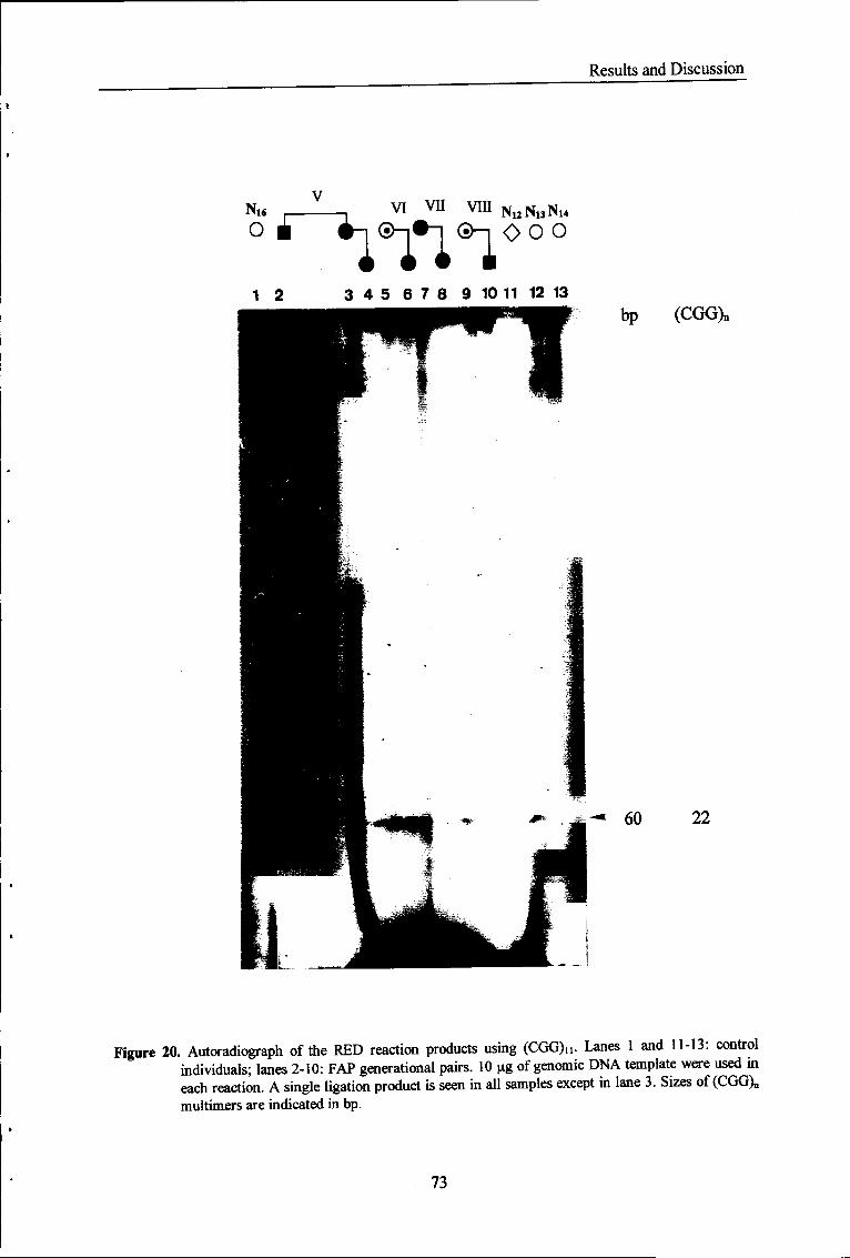
Results and Discussion
16
O à ^~i ̂ 1*1 ®~| vi vn vin N U N U N M
O O O
1 2 3 4 5 6 7 8 9 10 11 12 13 bp (CGG)„
- 60 22
Figure 20. Autoradiograph of the RED reaction products using (CGG)n. Lanes 1 and 11-13: control individuals; lanes 2-10: FAP generational pairs. 10 ug of genomic DNA template were used in each reaction. A single ligation product is seen in all samples except in lane 3. Sizes of (CGG)„ multimers are indicated in bp.
73
Results and Discussion
3.7. TGG/CCA TRINUCLEOTIDE
The maximal repeat size obtained for TGG/CAA trinucleotide ranged between 72 bp
(24 repeats) and 144 bp (48 repeats) corresponding to the formation of dimers to tetramers of
the oligonucleotide substrate, respectively (Figures 21 and 22). The exact repeat sizes were
difficult to assess in all samples since the RED reactions have generated weak signals.
All generational pairs and eleven control individuals were used for triplet repeat size
detection. Distinct fragments of 72 bp (24 repeated units) could be detected in all samples. In
pairs I to HI, fragments of 108 bp and 144 bp could also be seen (Figure 21, lanes 2-7). Pair IV
also presented the 108 bp band and a putative larger fragment of 144 bp (48 repeated triplets)
(lanes 7 and 8 of Figure 22). All the other pairs exhibit a weak signal and the presence of larger
multimers of (TGG)i2 cannot be excluded (data not shown). The control DNA samples were
found to have repeat lengths of 72 and 108 bp resulting from the ligation of 2 or 3 oligos,
respectively. The feint bands observed for the control samples and for those of pairs V to VIE
are suggestive of a low yield reaction, perhaps producing an incorrect estimate of the genomic
trinucleotide repeat length. In any case, our results do not show any difference in repeat size
within the FAP generational pairs, thus excluding the presence of TGG trinucleotide unstable
expansions from one generation to the other. Moreover, the estimated sizes ranges are in
accordance with previous studies where the repeat lengths vary between 120 and 210 bp
(Lindblad et al., 1995; Hofferbert et al., 1997).
3.8. ATG/CAT TRINUCLEOTIDE
The results of ATG/CAT trinucleotide repeat size detection are shown in Figures 23 and
24. Seven FAP kindred pairs and 10 control individuals present maximal repeat sizes ranging
between 24 repeated triplets (72 bp) and 48 repeats (144 bp). All pairs exhibit 3 bands
corresponding to the ligation of four (ATG)i2 oligonucleotides (144 bp), the third band being
very feint in pair VI due to a weaker reaction (Figure 24, lanes 5, 6). Repeat detection was not
successful above the 72 bp fragment for pair V and the progenitor of pair VIE, and the 108 bp
fragment for the descendent of pair VII (lanes 4, 5, 10 and 8 of Figure 24, respectively). It is
clear by comparison with the signals obtained for all the other samples that formation of
74

Results and Discussion
j — I II in N4 N Í N B N I S N H N I ,
Ó è - i # - i #-i O D O D D O
1 2 3 4 5 6 7 8 9 10 11 12 13 bp (TGG)n
— 144 48
- 108 36
— 72 24
Figure 21. Autoradiograph of the RED reaction products using (TGG)12. Lanes 1-7: FAP generational pairs; lanes 8-13: control individuals. 10 ug of genomic DNA template were used in each reaction. Note the reduced (TGG)I2 multimer formation in lanes 9 and 11-13. Sizes of (TGG)„ multimers are indicated in bp.
75

Results and Discussion
I II m rv N2N3N4N17
O O O D in 1 1 2 3 4 5 6 7 8 9 10 11 12
bp (TGG)n
- 108 36
-* 72 24
Figure 22. Autoradiograph of the RED reaction products using (TGG)i2. Lanes 1-8: FAP generational pairs; lanes 9-12: control individuals. 10 ug of genomic DNA template were used in each reaction. Sizes of (TGG)„ multimers are indicated in bp.
76

Results and Discussion
oligonucleotide multimers was much lower in the referred individuals, suggesting that the same
sizes would be detected if the cycling reaction efficiency had been the same. Identical results
were obtained with the control DNA samples where the reaction-generated (ATG)i2 multimers
are clearly insufficient for the correct genomic repeat length detection. Hofferbert et al. (1997)
have reported a distribution of maximal repeat sizes in 31 unrelated individuals ranging between
60 bp and 240 bp, with a high frequency of 120 bp repeat-containing positives. The repeat
lengths obtained in our experiments fall in the medium range of the reported size spectrum.
The triplet repeat lengths are conserved within each tested generational pair arguing
against the involvement of an ATG trinucleotide expansion as the responsible mechanism for
genetic anticipation in these FAP patients.
3.9. GTT/AAC TRINUCLEOTIDE
The GTT/AAC triplet repeat was difficult to detect by the RED method. The repeat
detection reactions have not produced detectable amounts of (GTT)i5 oligonucleotide multimers
in most of the experiments, resulting in the absence of ligation-corresponding bands. Figure 25
represents the only reaction in which a single band was obtained. It is not possible, however,, to
determine if it corresponds to the oligonucleotide itself or to the product of a single ligation
referring to a 90 bp fragment (30 repeats). The shape of the bands more resembles the product
of a dimer formation rather than a single oligonucleotide species, which would be in accordance
with the reported sizes for this triplet in 54 unrelated Caucasians where the maximal repeat
length was 90 bp (Hofferbert et a/., 1997). If that is the case, our results shown the absence of
any differences in size distribution among the members of four FAP generational pairs (V to
VIE) and 5 control individuals, excluding GTT/AAC repeat expansions as the molecular basis
for the phenomenon of anticipation.
3.10. CCT/AGG TRINUCLEOTIDE
The detection of CCT/AGG genomic repeat lengths by the RED method was a
technical Mure. Multimers of (CCT)i5 oligonucleotide substrate were never formed in any
77

Results and Discussion
I _ _ VI vn Vffl N« NMNi6
•-i è è©- | #-J ®-| o o o
1 2 3 4 5 6 7 8 9 10 11 12 13 bp (ATG)„
— 144 48
- 108 36
— 72 24
Figure 23. Autoradiograph of the RED reaction products using (ATG)12. Lanes 1-10: FAP generational pairs; lanes 11-13: control individuals. 10 ng of genomic DNA template were used in each reaction. Note the reduced (ATG)12 multimer formation in lanes 9 and 11-13. Sizes of (ATG),, multimers are indicated in bp.
78
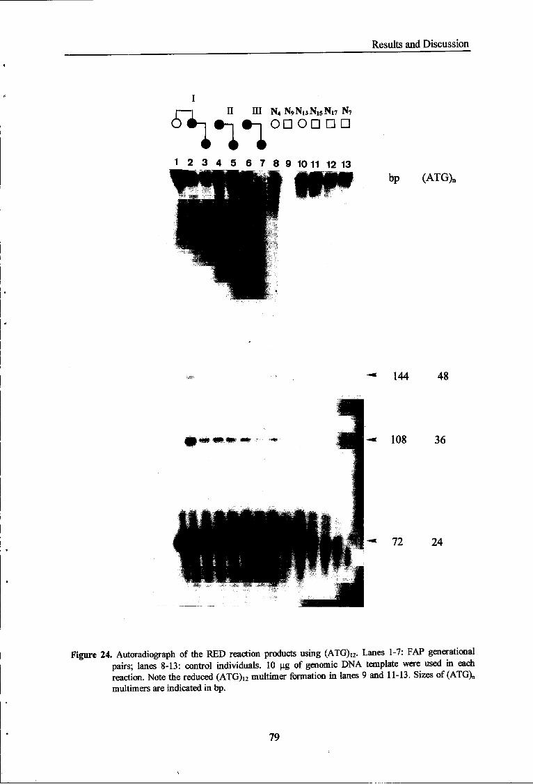
Results and Discussion
< ^ H ffl N4 N9Ni3NlsNi7 N7
on on a a -i 11 m
1 2 3 4 5 6 7 8 9 1011 12 13 bp (ATG)n
— 144 48
— 108 36
— 72 24
Figure 24. Autoradiograph of the RED reaction products using (ATG)12. Lanes 1-7: FAP generational pairs; lanes 8-13: control individuals. 10 \ig of genomic DNA template were used in each reaction. Note the reduced (ATG)i2 multimer formation in lanes 9 and 11-13. Sizes of (ATG)„ multimers are indicated in bp.
79

Results and Discussion
VI W Vffl N12NuNi4 N16N17
■ t®-i#-|®-i o o o o n
1 2 3 4 5 6 7 8 910 11 12 13 bp (GTT)n
-« 90 30
Figure 25. Autoradiograph of the RED reaction products using (GTT)i5. Lanes 1-8: FAP generational pairs; lanes 9-13: control individuals. 10 ng of genomic DNA template were used m each reaction. The band seen in all samples resembles the product of one ligation, but it can also correspond to the single oligonucleotide species. The size of the putative (GTT)15 dimer is indicated in bp.
80

Results and Discussion
experiment and only one band corresponding to the single oligonucleotide species was clearly
visible in all RED reactions (data not shown), ruling out radiolabeled probe hybridization
problems. The reasons why repeat detection has not been possible are not known, however it is
most likely that the problem lies on the cycling reaction itself where the oligonucleotide
annealing with the genomic DNA might have been impaired. Oligonucleotide impurity, for
example, could be the origin for the reaction failure.
The absence of any results prevent us to draw any conclusion about this trinucleotide
involvement in genetic anticipation in the tested kindreds, however the expected maximal repeat
sizes for this triplet, as determined by RED in 35 unrelated individuals, is 90 bp corresponding
to a trimer of the used oligonucleotide substrate (Hofferbert, 1997).
81

Chapter III
General Discussion and Conclusions

General Discussion and Conclusions
General Discussion and Conclusions
While the initially described disorders caused by trinucleotide repeat expansions (TREs)
were limited to CGG/CCG and CTG/CAG repeats, the recent discovery of an intronic GAA
repeat expansion as the cause of Friedreich's ataxia has shown that other TREs exist and have
disease-causing potential. The genetic instability of the expanded triplets results in increased
precocity, severity or penetrance in successive generations, a phenomenon known as genetic
anticipation. Indeed, eleven of the twelve triplet repeat-associated disorders exhibit a marked
anticipation of the age-at-onset, and/or increased severity of symptoms over generations. The
number of trinucleotide repeats appears to be correlated with the age of onset as increased
repeat copy number results in phenotypic anticipation. Anticipation cannot be seen in autosomal
recessive disorders, because they usually affect only a single generation, which makes FA the
sole disorder with little evidence for this phenomenon. Since dynamic mutations are the only
known molecular mechanism explaining genetic anticipation, other neurological or
neuropsychiatrie disorders with dominant inheritance and obvious anticipation are being
intensively investigated for associated dynamic mutations.
FAP is a potential candidate for a dynamic mutation-associated disorder, as a
neurological disease with autosomal dominant inheritance exhibiting genetic anticipation.
However, it is more than likely that such a mutation would occur in a modifier gene (or genes),
rather than the TTR gene. The TTR gene does not contain repeated motifs apart from two short
(CA)n stretches in the regulatory sequences of the 5' untranslated region (Sakaki, 1989).
Nonetheless, the existence of modifier genes underlying phenotypic heterogeneity in FAP,
particularly manifested as anticipation of the age-at-onset, is a possibility. To address this
question we have used the Repeat Expansion Detection (RED) technique that allows a genome-
wide analysis for TREs (Schalling et al, 1993; Sirugo et al, 1995). The RED assay has been
applied to search for triplet expansions in common neuropsychiatrie disorders with anticipation
(Lindblad et al, 1995; Morris et al, 1995), and to determine triplet repeat size polymorphisms
in individuals of several populations (Watkins et al, 1995; Hofferbert et al, 1997; Nakamoto et
al, 1997). For instance, Carrero-Valenzuela et al (1995) have excluded the association of
familial Parkinson's disease with CAG/CTG expansions, using the RED technique.
83

General Discussion and Conclusions
We have scanned eight generational pairs of Portuguese FAP kindreds exhibiting
marked anticipation of the age-at-onset (the differences within the pairs being greater than 12
years), and 19 unrelated healthy Portuguese individuals, for the existence of intergenerational
instability of the ten possible triplet repeats.
Our experiments have defined the triplet repeat sizes for 8 out of the 10 trinucleotides.
The determined repeat lengths fall in the range of the expected size spectrum when compared
with previously reported data (Lindblad et al, 1994 and 1995; Schalling et al, 1993; Hofferbert
et al, 1997; Nakamoto et al, 1997). The RED reactions testing CTG, CGG, and AAG triplets -
the known disease-causing motifs - have produced large amounts of oligonucleotide multimers
allowing good detection of the repeated triplet sizes in all screened individuals. Our analysis of
genomic DNA from 9 FAP generational pairs and 8 unrelated control individuals for CTG/CAG
triplet repeats (expanded in 8 neurodegenerative disorders), revealed a high frequency of 120 bp
repeat-containing positives, the repeat size being exactly the same within the FAP kindred pairs.
Only one pair was found to exceed this copy number, albeit without exceeding the expected
repeat length for normal alleles or exhibiting intergenerational expansion of the triplet repeat.
The use of a positive control DNA sample containing a very long segment of CTG repeats has
proved the detectability of expansion of a single copy allele under our experimental conditions.
For the CGG repeat, the cause of fragile X syndrome, a small repeat size corresponding
to 22 trinucleotide copies was detected in nearly all screened individuals. Difficulties in
detecting repeated CGG triplets were reported by Hofferbert et al (1997) being attributed to
secondary structure formation on template DNA with (C+G)-rich repeats (Person and Sinden,
1996). We circumvented that problem by using betaine, a reagent that weakens C-G annealing,
preventing the formation of secondary structures that could inhibit oligonucleotide hybridization
to the single stranded genomic template. The CGG repeat length determined in our experiments
is similar to previously reported data (Hofferbert et al, 1997) and clearly shows the absence of
any unstable expansion in the screened FAP individuals.
For the AAG repeat - expanded in FA - large maximal repeat sizes have been detected,
particularly in the screened FAP individuals. The (AAG)io oligonucleotide revealed repeat
lengths up to 600 bp (200 repeats). The presence of large multimers close to the upper
resolution of the gel was carefully assessed in order to determine the exact repeat sizes of the
samples. Moreover some samples were scanned in duplicate to ascertain the determined sizes.
Although the band readings are relatively difficult in that particular portion of the gel, no
84

General Discussion and Conclusions
differences in size were observed among the members of each pair, with the exception of pairs I
and H, presenting a difference of a single oligonucleotide ligation favoring a contraction and an
expansion, respectively. These results allow us to exclude the existence of unstable expansions
in second generation individuals of the FAP pairs.
TTA, AGT, and CGT trinucleotides have been shown to present monomorphic repeat
lengths, clearly ruling out anticipation-related intergenerational instability in the scanned FAP
kindred pairs. RED reaction experiments with the CGT oligonucleotide have produced a very
weak signal. The low yielding reactions are most certainly caused by oligonucleotide
contaminants, as previously mentioned, expressed by the detection of very small fragments of
the oligo substrate either alone or ligated, forming longer single stranded molecule species and
appearing as a ladder below the CGT dimer position. The faint band immediately over the 84 bp
fragment corresponding to the single ligation also results from the same contamination. As it will
be discussed later, RED technique is very sensitive to the purity of the oligonucleotides and
genomic DNA templates.
When compared to previously reported data, the frequency of trinucleotide repeats at or
above discrete lengths seemed to by similar for TGG and ATG. The determined triplet repeat
lengths fall in the polymorphic size range spectrum observed in healthy unrelated Caucasians
(Hofferbert et al, 1997), and are identical within each scanned FAP generational pair arguing
against the involvement of unstable expansions in these kindreds.
Our analysis of genomic DNA from the FAP kindred pairs and control individuals was
unsuccessful for GTT and CCT triplets and did not allow the determination of maximal triplet
repeat sizes. A doubt remains regarding GTT trinucleotide RED results since the single band
obtained in one experiment might correspond to the ligation of the (GTT) 15 substrate, rather than
to the oligonucleotide monomer.
The power and limitations of the RED assay have been evaluated by Hofferbert et al.
(1997). A dilution series of cloned trinucleotide repeats with known sizes has defined 10 \ig as
the required amount of DNA to detected the expansion of a TNR in one allele of a single copy
gene. Twenty diagnosed HD probands with previously determined sizes of the expanded HD
gene were tested with the RED method. The CTG repeat sizes, as determined by RED, were
never smaller than the actual repeat size, matching exactly the repeat lengths in 8 samples and
exceeding the previously determined sizes in 12 cases. The results obtained with the control
population suggested that those differences were due to CTG/CAG expansions in the genome
85

General Discussion and Conclusions
unrelated to the HD locus. CTG/CAG repeat lengths in normal alleles have been shown to
exceed the copy number expected for non-diseased individuals, i.e., falling in the premutation or
mutation size range spectrum of the TRE-associated diseases, suggesting the existence of other
expansions unrelated to the loci of those disorders (Nakamoto et ai, 1997).
In addition to the sensitivity of the RED technique, evaluated by Hofferbert and co
workers, we have assessed the reproducibility of the RED method, by scanning some samples in
duplicate or triplicate for CTG, CGG and AAG triplets. The maximal repeat sizes obtained were
identical in all cases. The autoradiograph shown in Figure 10 illustrates well the reproducibility
of the RED assay, exhibiting identical products from different cycling reactions using the same
samples. Our experiments have also shown that the RED reaction is very sensitive to small
variations in the cycling conditions and to the purity of the oligonucleotides and genomic DNA
template. The weak signals presented by some samples are consistently observed in most of the
RED experiments in which they have been used, suggesting a certain degree if impurity that
results in a low formation of oligonucleotide multimers. The hybridization of the oligonucleotide
substrate to the genomic template might be impaired resulting in decreased formation of ligated
products. The existence of contaminants in the oligonucleotide solution is also suggested by the
ladder of bands appearing below the oligo multimer products, as observed, for instance, in the
CGT triplet expansion reaction. The presence of contaminants is likely to slow down the
annealing and ligation processes, resulting in low multimer production. The radiolabeled probe
hybridization to the RED reaction products were not responsible for the faint bands obtained in
some autoradiographs since there were always samples with strong signals, arising from a
normal labeling procedure. In general, the RED assay is a reliable method for the detection of
triplet repeat expansions in the human genome and has been shown to be capable of detecting
even small expansions against a background of polymorphic larger repeated sizes as long as the
population is homogeneous (Hofferbert et al, 1997).
Our results show the absence of any size differences between the elements of the FAP
generational pairs, excluding the involvement of intergenerational unstable triplet expansions as
the underlying mechanism for genetic anticipation in these kindreds, for the eight successfully
tested trinucleotides. No conclusions can be drawn regarding GTT and CCT triplets since the
respective RED experiments have been unsuccessful. It should noticed, however, that expansion
of any of these two trinucleotides has not been associated with any prior instance of anticipation.
A limitation of our experiments was the absence of positive controls which would prove
86

General Discussion and Conclusions
the detectability of a single copy allele expansion under our experimental conditions. We have
used a positive control only for CTG/CAG trinucleotide repeat detection, showing that the
reaction conditions were perfectly adequate for genomic CTG repeat detection. Although all
other RED assays were performed without positive controls, the determined repeat lengths were
in accordance with previously reports, suggesting that the experimental conditions were suitable
for the exact repeat size detection.
The number of scanned FAP kindred generational pairs constitute a relatively small
sample of the Portuguese kindreds exhibiting anticipation. In a study with 227 generational pairs
of Portuguese FAP kindreds, Sousa (1995) has reported anticipation of the age-at-onset in
approximately 60% of the pairs. Considering that the Portuguese population is homogeneous
and relatively constrained in reproductive terms, we would expect to find an unstable
trinucleotide expansion in at least one pair if such mutation would indeed be responsible for
phenotypic anticipation in FAP. A statistical analysis of our results suggests that no more than
11% [(95% CI (0-37%)] of the Portuguese FAP kindreds exhibiting anticipation is likely to have
TREs as the underlying mechanism. The distribution of maximal triplet repeat sizes both for the
FAP individuals and the unrelated controls, as determined by RED, is rather uniform, implying
that the Portuguese population is genetically homogeneous with respect to genomic triplet repeat
sequence lengths, although our sample size is too small to make a definitive statement.
Our results are suggestive - within the sensitivity and limitations of the RED method -
that trinucleotide repeat expansions are unlikely to be responsible for genetic anticipation in FAP
kindreds in the Portuguese population. To further substantiate our conclusions, additional pairs
should be scanned for triplet repeat expansions.
This study has not excluded dynamic mutation as a potential molecular mechanism
responsible for genetic anticipation in FAP. Several other simple tandem repeats are known to
be unstable in the human genome and the cause of expansion-related disorders (Lalioti et al.
1997; Lafrenière et ai, 1997). The role of environmental factors in phenotypic anticipation in
FAP cannot be excluded but is very unlikely. The involvement of multiple genes in TTR
amyloidogenesis is strongly suggested, justifying a genome-wide scanning for modifier genes by
genetic linkage analysis and positional cloning. The detection of TREs systematically in a
significant proportion of FAP kindreds, would suggest that the gene bearing the expansion
responsible for anticipation might be one such modifier gene. Thus, the search for TREs in FAP
represents the first approach to assess the molecular basis for genetic anticipation and could
87

General Discussion and Conclusions
provide insight into the mechanisms of pathology at the cellular level, with immediate
applications on genetic counseling. Furthermore, the identification of protein products of the
TREs in amyloidogenesis, might help to elucidate the mechanism of amyloid deposition with
important consequences at the therapeutic level.
88

Chapter IV
References

References
References
Almeida MR, Hesse A, Steinmetz A, Maish B, Altland K, Linke RP, Gawinowicz MA, Saraiva MJM (1991a). Transthyretin Leu 68 in a form of cardiac amyloidosis. Basic Res. Cardiol. 86: 567-571.
Almeida MR, Altland K, Rauh S, Gawinowicz M, Moreira P, Costa PP, Saraiva MJM (1991b). Characterisation of a basic transthyretin variant -TTR Arg 102- in the German population. Biochem. Biophysic. Acta 1097:224-226.
Almeida M, Ferlini A, Forabosco A, Gawinowicz M, Costa PP, Salvi F, Plasmati R, Tassinari CA, Altland K, Saraiva MJM (1992). Two transthyretin variants (TTR Ala 49 and TTR Gin 89) in two Sicilian kindreds with hereditary amyloidosis. Hum. Mut. 1: 211-215.
Almeida MR, Andreu FL, Quês MM, Costa PP, Saraiva MJM (1993). Transthyretin Ala71: a new transthyretin variant in a Spanish family with familial amyloidotic polyneuropathy. Hum. Mut. 2: 420-421.
Almeida, M.R., Oishi, A., Sakaki, Y., Holmgren, G., Ulf, D., Ferlini, A, Salvi, F., Munar-Qués, M., Benson, M.D., Skinner, M., Costa, P.P. and Saraiva, M.J.M. (1995) Haplotype Analysis of Common Transthyretin Mutations. Human Genetics 96: 350-354.
Alves JE, Altland K, Almeida MR, Bêcher P, Costa PP, Saraiva MJM (1993). Screening of TTR variants in the Portuguese population by HIEF. J. Rheumatol. 20:185.
Alves IL, Jacobson DR, Torres MF, Holmgren G, Buxbaum JN, Saraiva MJM (1996) Transthyretin Ser6 as a neutral polymorphism in familial amyloidotic polyneuropathy. Amyloid: Int. J. Exp. Clin. Invest. 3: 242-244.
Alves JX, Altland K, Almeida MR, Winter P, Saraiva MJM (1997). Screening and biochemical characterisation of TTR variants in the Portuguese population. Human Mutation 9: 226-233.
Andrade C (1952). A peculiar form of peripheral neuropathy. Brain 75: 408-427
Ashley C Jr., Wilkinson KD, Reines D, Warren ST (1993). FMR1 protein: conserve RNP family domains and selective RNA binding. Science 262: 563-566.
Banfi S et al. (1994) Identification and characterization of the gene causing type-1 spinocerebellar ataxia. Nat Genet. 7: 513-519.
Bastos Lima A, Martins da Silva A (1980) Clinical evaluation of late-onset cases in Type I (Andrade) Amyloid Neuropathy. In: Amyloid and Amyloidosis, Glenner GG, Costa PP, Freitas AF (eds.), Amsterdam: Excerpta Medica, pp. 88-98.
Benson II MD, Turpin JC, Zeldenrust S, LeChevalier B, Benson MD (1993). A transthyretin variant (alanine 71) associated with familial amyloidotic polyneuropathy in a French family. J. Med. Gene. 30: 120-122.
90

References
Benson MD, Dwulet FE, Scheinberg MA, Greipp P (1986). Chemical classification of hereditary amyloidosis in Brazilian families and identification of the gene carriers. J. Rheumatol. 13: 927-931.
Benson MD, Kluve-Beckerman B, Liepnieks JJ, Murrell JR, Hanes D, Uemichi T (1996). Metabolism of amyloid proteins. In: The nature and origin of amyloid fibrils. Ciba Foundation, John Wiley & Sons Ltd, Chishester, pp 104-112.
Benson MD, Uemichi T (1996). Transthyretin amyloidosis. Amyloid: Int. J. Exp. Clin. Invest. 3: 44-56.
Blake CCF, Swan IDA (1971). An X-ray study of the subunit structure of prealbumin. J. Mol. Biol. 61:217-224.
Blake CCF, Oatley JS (1977). Protein-DNA and protein-hormone interactions in prealbumin: a model of the thyroid hormone nuclear receptor? Nature 268:115-120.
Blake CCF, Geisow MJ, Oatley SJ, Rerat B, Rerat C (1978). Structure of prealbumin: secondary, tertiary and quaternary interaction determined by Fourier refinement at 1.8 angstrom. J. Mol. Biol. 121: 339-356.
Boomsma F, Veld AJM, Schalekamp ADH (1991). Not norepinephrine but its oxidation products bind specifically to plasma proteins. J. Pharmac.Experiment.Therapeut. 259: 551-557.
Booth DR, Soutar AK, Hawkins PN, Pepys MB (1994). Three new amyloidogenic transthyretin gene mutations: advantages of direct sequencing. In: Amyloid and amyloidosis. Kisilevsky R, Benson MD, Frangione B, Gauldie J, Muckle T, Young I (eds), Parthenon Publishing, New York, pp 456-458.
Booth DR, Tan SY, Hawkins PN, Pepys MB, Frustaci A (1995). A novel variant of transthyretin, 59Thr-Lys, associated with autosomal dominant cardiac amyloidosis in an Italian family. Circulation 91: 962-967.
Booth DR, Booth SE, Persey MR, Tan SY, Madhoo S, Pepys MB, Hawkins PN (1996). Three new amyloidogenic TTR mutations: Prol2, Glul8 and Val33. Neuromusc. Disord. 6: 28.
Bordeneuve VP, Lalu T, Reilly M, Said G, Misrahi M (1996). Héterérogénéité des mutations ponctuelles du gène de la transthyrètine dans les amyloidoses familiales Françaises. Identification de 4 nouvelles mutations. Abstracts of the 6e colloque sur les maladies neuromusculaires, Versailles.
Boucher CA, King SK, Carey N, Krahe R, Winchester CL et al. (1995). A novel homeodomain-enconding gene is associated with a large CpG island interrupted by the myotonic dystrophy unstable (CTG)n repeat. Hum. Mol. Genet. 4:1919-1925.
Brook JD, McCurrach ME, Harley HG, Buckler AJ, Church D et al (1992). Molecular basis of myotonic dystrophy: expansion of a trinucleotide (CTG) repeat at the 3' end of a transcript encoding a protein kinase family member. Cell 68: 799-808.
91

References
Brunner HG, Bruggenwirth HT, Nillesen W, Jansen G, Hamel BC et al. (1993). Influence of sex of the transmitting parent as well as of parental allele size on the CTG expansion in myotonic dystrophy (DM). Am. J. Hum. Genet. 53:1016-1023.
Burright EN, Clark HB, Servadio A, Matilla T, Feddersen RM et al. (1995). SCA1 transgenic mice: a model for neurodegeneration caused by an expanded CAG trinucleotide repeat. Cell 82: 937-948.
Campuzano V, Monterniini L, Molto MD, Pianese L, Cossee M et al. (1996). Friedreich's ataxia: autosomal recessive disease caused by an intronic GAA triplet repeat expansion. Science 111: 1423-1427.
Carrero-Valenzuela R, Lindblad K, Payami H, Johnson W et al. (1995). No evidence for association of familial Parkinson's disease with CAG repeat expansion. Neurology 45: 1060-1063.
Cavallaro TC, Martone RL, Dwork AJ, Schon EA, Herbert J (1990). The retinal pigment epitelium is the unique site of transthyretin synthesis in the eye. Invest. Ophthal. Vis. Sci. 31: 497-501.
Coelho T, Sousa A, Lourenço E, Ramalheira J (1994). A study of 159 Portuguese patients with familial amyloidotic polyneuropathy (FAP) whose parents were both unaffected. J. Med. Genet. 31: 293-299.
Coelho T, Chorão R, Sousa A, Alves I, Torres MF, Saraiva MJM (1996). Compound hétérozygotes of transthyretin Met 30 and transthyretin Met 119 are protected from the devastating effects of familial amyloidotic polyneuropathy. Neuromusc. Disorders 6:27 (abstr.).
Cohen AS, Calkins E (1959). Electron microscope observation on a fibrous component in amyloid of diverse origins. Nature 183:1201-1203.
Cooper DN, Youssoufian H (1988). The CpG dinucleotide and human genetic disease. Hum. Genet. 78:151-155.
Cornwell m GG, Murdoch WL, Kyle RA, Wetermark P, Pitkanen P (1983). Frequency and distribution of senile cardiovascular amyloid. A clinicopathological correlation. Am. J. Med. 75: 618-623.
Cossée M et al. (in press) Evolution of the Friedreich's ataxia trinucleotide repeat expansion: Founder effect and premutations. Proc. Natl. Acad. Sci USA.
Costa PP, Figueira A, Bravo F (1978). Amyloid fibril protein related to prealbumin in familial amyloidotic polyneuropathy. Proc. Natl. Acad. Sci., 75: 4499-4503.
Costa PP, Saraiva MJM (1988). Familial amyloidotic polyneuropathy: screening of the population at risk in Portugal. In: Amyloid and Amyloidosis. Isobe T, Araki S, Ucbino F, Shozo K, Tsubura E (eds), Plenum press, New York, pp 425-428.
Costa RH, Lai E, Darnell Jr. (1986). Transcriptional control of the mouse prealbumin (transthyretin) gene: both promotor sequences and a distinct enhancer are cell specific. Molec. and Cell. Biol. 6: 4697-4708.
92
References
Costa RH, Grayson DR, Darnell Jr. (1989). Multiple hepatocyte-enriched nuclear factors function in the regulation of transthyretin and al-antitrypsin genes. Molec. Cell. Biol. 9:1415-1425.
Costa RH, Van Dike TA, Yan C, Kuo F, Darnell Jr. (1990). Similarities in transthyretin gene expression and differences in transcription factors: liver and yolk sac compared to choroid plexus. Proc. Natl. Acad. Sci. USA 87:6589-6593.
Coutinho P, Silva AM, Lima JL, Barbosa AR (1980). Forty years of experience with type I amyloid neuropathy. Review of 483 cases. In: Amyloid and Amyloidosis, Glenner GG, Costa PP, Freitas AF (eds), Excerpta Medica, Amsterdam-Oxford-Princeton, pp 88-98.
DeBoulle K, Verkerk AJ, Reyniers E, Vitz L, Hendrickx J et al. (1993). A point mutation in the FMR1 gene associated with fragile X mental retardation. Nat. Genet. 3: 31-35.
DeLucia R, Mauro A, Di Scapio A Buddo A, Mortara P, Orsi L, Schiffer D (1993). A new mutation on the transthyretin gene (Serll2-Ile) causes an amyloid neuropathy with severe cardiac impairment. Clin. Neuropathol. 12: S44.
Dickson PW, Howlett GJ, Schreiber G (1985). Rat transthyretin (prealbumin): molecular cloning, nucleotide sequence and gene expression in liver and brain. J. Biol. Chem. 260: 8214-8219.
Dickson PW, Schreiber G (1986). High levels of messenger RNA for transthyretin (prealbumin) in human choroid plexus. Neurosci. Lett., 66: 311-315.
DiFiglia M, Sapp E, Chase K, Schwarz C, Meloni A, Young C et al (1995). Huntingtin is a cytoplasmic protein associated with vesicles in human and rat brain neurons. Neuron 14: 1075-1081.
Dwork AJ, Cavallaro TC, Martone RL, Goodman D , Schon EA, Herbert J (1990). Distribution of transthyretin in the rat eye. Invest. Ophthal. Vis. Sci. 31: 489-496.
Dwulet FE, Benson MD (1986). Characterization of a transthyretin (prealbumin) variant associated with familial amyloidotic polyneuropathy type II (Indiana/Swiss). J. Clin. Invest. 78: 880-886.
Eanes ED, Glenner GG (1968). X-ray diffraction studies on amyloid filaments. J. Histochem. Cytochem. 16: 673-677.
Eichler EE, Holden JJA, Popovich BW et al. (1994). Length of uninterrupted CGG repeats determines instability in the FMR1 gene. Nat. Genet. 8: 88-94.
Eichler EE, Holden JJA, Popovich BW, Reiss AL, Snow K, Thibodeau SN, Richards CS, Ward PA, Nelson DL (1994) Length of uninterrupted CGG repeats determines instability in the FMR 1 gene. Nat. Genet. 8: 88-94.
Episkopou V, Maeda S, Nishiguchi S, Shimada K, Gaitanaris G, Gottesman ME, Robertson EJ (1993). Disruption of the transthyretin gene results in mice with depressed of plasma retinol and thyroid hormone. Proc. Natl. Acad. Sci. 90: 2375-2379.
93

References
Ernstrom U, Pettersson T, Jornvall H (1995). A yellow compound associated with human transthyretin has properties like a pterin derivative, 7,8-dihydropterin-6-carboxaldehyde. FEBS Lett. 360: 177-182.
Ferguson RN, Edelhoch H, Saroff HA, Robbins J, Cahnmann HJ (1975) Negative cooperativity in the binding of thyroxine to human serum prealbumin. Biochemistry 14:282-289.
Ferlini A, Romeo G, Tassinari CA, Saraiva MJM, Costa PP, Salvi F (1988). Discrimination of peripheral neuropathies caused by TTR variant or diabetes in the same pedigree through protein studies. Adv. Neurol. 48: 201-208.
Ferlini A, Patrosso MC, Repetto M, Frattini A, Villa A, Fini S, Salvi F, Vezzoni P, Forabosco A (1994). A new mutation (TTR Ala 47) in the transthyretin gene associated with hereditary amyloidosis. Hum. Mutat. 4: 61-64.
Ferlini A, Rapezzi C, Magnani B, Winter P, Altland K, Vezzoni P, Patrosso MC (1996). A compound heterozygous patient for two TTR mutations, affected with isolated restrictive amyloidotic cardiomyopathy. Neuromusc. Disord. 6:33.
Fitch NFS, Akbari MT, Ramsden DB (1991). An inherited non-amyloidogenic Transthyretin variant- Ser6 TTR- with increased thyroxine-binding affinity, characterized by DNA sequencing. J. Endoc. 129: 309-313.
Fu Y-H, Pizzuti A, Fenwick RG et al. (1992). An unstable triplet repeat in a gene related to myotonic muscular dystrophy. Science 255:1256-1258.
Fu YH, Friedman DL, Richards S, Pearlman JA, Gibbs RA et al. (1993). Decreased expression of ' myotonin-protein kinase messenger RNA and protein in adult form of myotonic dystrophy. Science 260:235-238.
Fung WP, Thomas T, Dickson PW, Aldred AR, Milland J, Dziadek M, Power B, Hudson P, Schreiber G (1988). Struture and expression of the rat transthyretion (prealbumin) gene. J. Biol. Chem. 263: 480-488.
Gacy AM, Goellner G, Juranic N, Macura S, McMurray CT (1995). Trinucleotide repeats that expand in human disease form hairpin structures in vitro. Cell 81: 533-540.
Gitlin D, Gitlin JD (1975). Fetal and neonatal development of human plasma proteins. In: The plasma proteins. Putman FW (ed), vol II, 2nd edition. Academic Press, New York, pp 264-371.
Gordenin DA, Kunkel TA, Resnick MA (1997). Repeat expansion - all in a flap? Nat. Genet. 16: 116-118.
Gorevic PD, Prelli FC, Wright J, Pras M, Frangione B (1989). Systemic senile amyloidosis. Identification of a new prealbumin (transthyretin) variant in cardiac tissue; immunologic and biochemical similarity to one form of familial amyloidotic polyneuropathy. J. Clin. Invest. 83: 836-843.
Gu Y, Shen SH, Gibbs RA, Nelson DL (1996). Identification of FMR2, a novel gene associated with the FRAXE CCG repeat and CpG island. Nat. Genet. 13:109-113.
94

References
Harding J, Skare J, Skinner M (1991). A second transthyretin mutation at position 33 (Leu/Phe) associated with familial amyloidotic polyneuropathy. Biochem. Byophys. Acta 1097: 183-186..
Harley H et al. (1991). Am. J. Hum. Genet. 49: 68-75.
Harper PS (1989). Myotonic dystrophy, vol 21 (ed 2). Philadelphia, PA, Saunders.
Harrison HH, Gordon ED, Nichols WC, Benson MD (1991). Biochemical and clinical characterization of prealbumin Chicago: an apparently benign variant of serum prealbumin (transthyretin) discovered with high-resolution two-dimensional electrophoresis. Am. J. Med. Genet. 39: 442-452.
Hazenberg BPC, Marrink J, Nakazato M, Jong R, Janssen S, van Rijswijk M (1990).Two Dutch families with FAP type I. In: Familial Amyloidotic Polyneuropathy and other Transthyretin related disorders, Costa PP, Freitas AF, Saraiva MJM (eds), Porto, Arquivos de Medicina 3 (Special Issue), pp 29-34.
Herbert J, Wilcox JN, Pham KC, Fremeau RT, Zeviani M, Dwork A, Soprano DR, Makover A, Goodman D , Zimmerman EA, Roberts JL, Schon EA (1986). Transthyretin: a choroid plexus specific transport protein in human brain. Neurology 36: 900-911.
Herbert J, Younger D, Latov N, Martone RL (1994). Clinical spectrum of familial amyloidotic polyneuropathy. In: Amyloid and Amyloidosis. Kisilevski R, Benson MD, Frangione B, Galuldie J, Muckle TJ, Young ID (eds). Parthenon Publisher, New York, pp 486-488.
Hofferbert S, Schanen NC, Chehab F, Francke Uta (1997). Trinucleotide repeats in the human genome: size distributions for all possible triplets and detection of expanded disease alleles in a group of Huntington disease individulas by the Repeat Expansion Detection method. Hum. Mol. Genet. 6 (1): 77-83.
Holmgren G, Haettner E, Nordenson I, Sandgren O, Steen L, Lundgren E (1988). Homozygosity for the transthyretin Met 30 gene in two Swedish sibs with familial amyloidotic polyneuropathy. Clin. Genet. 34: 333-338.
Holt IJ, Hardling AE, Middleton L, Chrysostomou G, Said G, King RHM, Thomas PK (1989). Molecular genetics of amyloid neuropathy in Europe. Lancet 1: 524-526.
Hoogeveen AT etal. (1993). Hum. Mol. Genet. 2: 2069-2073.
Housman D (1995). Gain of glutamines, gain of function? Nat. Genet. 10: 3-4.
Hõweler CJ, Busch HFM, Geraedts JPM, Niermeijer MF, Staal A (1989). Anticipation in Myotonic Dystrophy: fact or fiction? Brain 112: 779-797.
Ii S, Minnerath S, Ii K, Sommer SS (1991). Two tiered DNA based diagnosis of transthyretin amyloidosis reveals two novel point mutations. Neurology 41: 893-898.
Ii S, Sobell JL, Sommer SS (1992). From molecular variant to disease: initial steps in evaluating the association of transthyretin Met 119 with disease. Am. J. Hum. Genet. 50: 29-41.
95

References
Ikeda H, Yamaguchi M, Sugai S, Aze Y, Narumiya S, Kakizuka A (1996). Expanded polyglutamine in the Machado-Joseph disease protein induces cell death in vitro and in vivo. Nat. Genet. 13:196-202.
Ikegawa S, Yi S, Ando Y, Miyazaki A (1991). Réévaluation of 134 patients with familial amyloidotic polyneuropathy (FAP) in Japan, Kunamoto focus. In: Amyloid and Amyloidosis, Natvig JB, Forre O, Husby G, Husebekk A, Skogen B, Sletten K, Westermark P (eds.), Kluwer Publishing Dordrecht, pp. 675-678.
Imbert G, Saudou F, Yvert G, Devys D, Trottier Y, et al. (1996). Cloning of the gene for spinocerebellar ataxia 2 reveals a locus with high sensitivity to expanded CAG/glutamine repeats. Nat. Genet. 14: 285-291.
Izumoto S, Younger D, Hays AP, Martone RL, Smith RT, Herbert J (1992). Familial amyloidotic polyneuropathy presenting with carpal tunnel syndrome and a new transthyretin mutation, asparagine 70. Neurology 42: 2094-2102.
Izumoto S, Romberg J, Herbert J (1993). Two transthyretin variants associated with euthyroid hyperthyroxinemia. J. Rheumatol. 20:186.
Jacobson DR (1992). A specific test for transthyretin 122 (Val-Ile), based on PCR-primer-introduced restriction analysis (PCR-PIRA): Confirmation of the gene frequency in blacks. Am. J. Hum.Genet. 50:195-198.
Jacobson DR, Buxbaum JN (1991). Genetic Aspects of Amyloidosis. In: Advances in Human Genetics. Harris H, Hirschhorn K (eds.), Plenum Press, New York, pp. 69-123.
Jacobson DR, Buxbaum JN (1994b). A double variant transthyretin allele (Ser6, Ile33) in the Israeli patient "SKO" with familial amyloidotic polyneuropathy. Hum. Mut. 3: 254-260.
Jacobson DR, Reveille JD, Buxbaum J (1991). Frequency and genetic background of the position 122 (Val-Ile) transthyretin gene in the black population. Am. J. Hum. Genet. 49: 192-198.
Jacobson DR, Gertz MA, Buxbaum JN (1994a). Transthyretin Val 107, a new variant associated with familial cardiac and neuropathic amyloidosis. Hum. Mut., 3: 399-401.
Jacobson DR, Gertz MA Kane I, Buxbaum JN (1994b). Genetic analysis of 9 unrelated patients with transthyretin (TTR)-cardiac amyloidosis: correlation of clinical and genetic findings and description of 2 new TTR variants. In: Amyloid and amyloidosis. Kisilevsky R, Benson MD, Frangione B, Gauldie J, Muckle T, Young I (eds). Parthenon Publishing, New York, pp 474-476.
Jacobson DR, Alves IL, Saraiva MJM, Thibodeau SN, Buxbaum JN (1995). Transthyretin Ser 6 gene frequency in individuals without amyloidosis. Hum. Genet. 95: 308-312.
Jacobson DR, Kyle RA, Buxbaum JN (1996). TTR Ile20, a new transthyretin variant associated with cardiac amyloidosis. Neuromusc. Disord. 6: 23.
Jacobsson B (1989a). Localization of transthyretin mRNA and immunoreactive transthyretin in the human fetus. Virchows Arch. A. Pathol. Anat. 415: 259-263.
96

ï
References
Jacobsson B (1989b). In situ localization of transthyretin mRNA in the adult human liver, choroid plexus and pancreatic islets and in endocrine tumours of the pancreas and guts. Histochem. 91: 299-304.
Jaworsky A, Rosche WA, Gellibolian R, Kang S, Sliimizu M et al. (1995). Mismatch repair in Escherichia coli enhances instability of (CTG)n triplet repeat from human hereditary diseases. Procs. Natl. Acad. Sci USA 92:11019-11023.
Jenne DE, Denzel K, Blatzinger P, Winter P, Obermaier B, Linke RP, Altland K (1996). A highly conservative Ile20 substitution at the dimer-dimer interface selectively reduces stability of the tetrameric transthyretin complex and is associated with familial cardiomyopathy. Proc. Natl. Acad. Sci. 93: 6302-6307.
Jennings C (1995). How trinucleotide repeats may function. Nature 378:127.
Jones LA Skare JC, Harding JA Cohen AS, Milunsky A Skinner M (1991). Proline at position 36: a new transthyretin mutation associated with familial amyloidotic polyneuropathy. Am. J. Hum. Genet. 48: 979-982.
Jones LA, Skare jC, Cohen AS, Harding JA Milunsky A Skinner M (1992). Familial amyloidotic polyneuropathy- a new transthyretin position 30 mutations (alanine for valine) in a family of German descent. Clin. Genet. 41: 70-73.
Kahlem P, Terre C, Green H, Djian P (1996). Peptides containing glutamine repeats as substrates for transglutaminase-catalyzed cross-linking: relevance to diseases of the nervous system. Procs. Natl. Acad. Sci. USA 93:14580-14585.
Kanai M, Raz A Goodman DS (1968). Retinol-binding protein: the transport protein for vitamin A in human plasma. J. Clin. Invest. 47: 2025-2044.
Kanda Y, Goodman DS, Canfield RE, Morgan FJ (1974). The amino acid sequence of human plasma prealbumin. J. Biol. Chem. 249: 6796-6805.
Kang S, Jaworski A Ohshima K, Wells RD (1995). Expansion and deletion of CTG repeats from human disease genes are determined by the direction of replication in E. coli. Nat. Genet. 10: 213-218.
Kawaguchi Y, Okamoto T, Taniwaki M, Aizaya M, Inoue M, et al. (1994). CAG expansions in a novel gene for Machado-Joseph disease at chromosome 14q32.1. Nat. Genet. 8: 221-228.
Koide R, Dceuchi T, Onodera O, Tanaka H, Igarashi S, Endo K, Takahashi H, Kondo R, Ishikawa A Hayashi T et al (1994). Unstable expansion of CAG repeat in hereditary dentatorubral-pallidoluysian atrophy (DRPLA). Nat. Genet. 6: 9-13.
Korneluk RG, Narang MA (1997). Anticipating anticipation. Nat.Genet. 15:119-120.
Koshy B, Matilla T, Burright EN, Merry DE, Fischbeck KH, Orr HT, Zogbi HY (1996). Spinocerebellar ataxia type-1 and spinobulbar muscular atrophy gene products interact with glyceraldehyde-3-phosphate dehydrogenase. Hum. Mol. Genet. 5:1311-1318.
97

References
Kremer EJ, Pritchard M, Lynch M, Yu S, Holman K et al. (1991). Mapping of DNA instability at the fragile X to a trinucleotide repeat sequence p(CGG)n. Science 252:1711-1714.
Kunst CB, Warren ST (1994) Cryptic and polar variation of the Fragile-X repeat could result in predisposing normal alleles. Cell 77: 853-961.
Kuryavyi W , Jovin TM (1995). Triad-DNA: a model for trinucleotide repeats. Nat. Genet. 9: 339-341.
La Spada AR, Wilson EM, Lubahn DB, Harding AE, Fischbeck KH (1991). Androgen receptor gene mutations in X-linked spinal and bulbar muscular atrophy. Nature 352: 77-79.
Lafrenière RG, Rochefort DL, Chrétien N, Rommens JM, Cochius JI et al. (1997). Unstable insertion in the 5' flanking region of the cystatin B gene is the most common mutation in progressive myoclonus epilepsy type 1, EPMlAta. Genet. 15:298-302.
Lalioti MD, Scott HS, Buresi C, Rossier C, Botanni A, Morris MA, Malafosse A, Antonarakis SE (1997). Dodecamer repeat expansion in cystatin B gene in progressive myoclonus epilepsy Nature 386: 847-851.
Lavedan C, Hofmann-Radvanyi H, Shelbourne P, Rabes J-P, Duros C, Savoy D, Dehaupas I, Luce S, Johnson K, Junien C (1993) Myotomc-dystrophy-size-dependent and sex-dependent dynamics of CTG meiotic instability, and somatic mosaicism. Am. J. Hum. Genet. 52: 875-883.
Leeflang EP, Zhang L, Tavare S, Hubert R, Srinidhi J et al. (1995). Single sperm analysis of the trinucleotide repeats in the Huntington's disease gene: quantification of the mutation frequency spectrum. Hum. Mol. Genet. 4:1519-1526.
Li XJ, Li SH, Sharp AH, Nucifora F Jr., Schilling G, Lanahan A et al. (1995). A huntingtin-associated protein enriched in brain with implications for pathology. Nature 378: 398-402.
Lindblad K, Zander C, Schalling M, Hudson TJ (1994). Growing triplet repeats. Nat. Genet. 7: 124.
Lindblad K, Nylander P.-O, de Bruyn A, Sourey D, Zander C, et al. (1995) Detection of expanded CAG repeats in bipolar affective disorder using the repeat detection (RED) method. Neurobiol. Dis. 2: 55-62
.Lobato L, Teixeira F, Sousa A, Sequeiros J (1988). Genetic study of late-onset in hereditary amyloid neuropathy (HAN)-type I (Portuguese, Andrade). In: Amyloid and Amyloidosis, Isobe T, Araki A, Uchino F, Kito S, Tsubura E (eds.), Plenum press, New York, pp 441-446.
Lugenbeel K, Peier A, Carson N, Chudley A Nelson D (1995). Intragenic loss of funtion mutations demonstrate the primary role of FMR1 in fragile X syndrome. Nat. Genet. 10: 483-485.
MacDonald ME et al. (1992) The Huntington's-disease candidate region exhibits many different haplotypes. Nat. Genet. 1: 99-103.
98

References
Mahadevan M, Tsilfidis C, Sabourin L et al. (1992). Myotonic dystrophy mutation: an unstable CTG repeat in the 3' untranslated region of the gene. Science 255:1253-1255.
Mandei J-L (1997). Breaking the rule of three. Nature 386: 767-769.
Mangiarini L, Sathasivam K, Seller M, Cozens B, Harper A et al (1996). Exon 1 of the HD gene with an expanded CAG repeat is sufficient to cause a progressive neurological phenotype in transgenic mice. Cell 87: 493-506.
Martone R, Herbert J (1993). Transthyretin interacts with globin to form protein complexes with heme dependent solubility. J. Rheumatol. 20:176.
Martone RL, Herbert J, Dwork A, Schon EA (1988). Transthyretin in synthesized in the mammalian eye. Biochem. Biophys. Res. Commun. 151: 905-912.
Martone RL, Mizuno R, Herbert J (1993). The mammalian pineal gland is a synthetic site for TTR and RBP. J. Rheumatol. 20:175.
Mclnnis MG (1996). Anticipation: an old idea in new genes. Am. J. Hum. Genet. 59: 973-979.
McMurray CT (1996). Mechanisms of DNA expansion. Chromosoma 104: 2-13.
Mita S, Maeda S, Shimada K, Araki S (1984). Cloning and sequence analysis of cDNA for human prealbumin. Biochem. Biophys. Res. Commun. 124: 558-564.
Monaco HL, Maneia F, Rizzi M, Coda A (1995). Structure of a complex of two plasma proteins: transthyretin and retinol-binding protein. Science 268:1039-1041.
Morris AG, Gaitonde E, McKenna PJ, Mollon JD, Hunt DM (1995). CAG repeat expansions and schizophrenia: association with disease in females and with early age-at-onset. Hum. Mol. Genet. 4: 1957-1961.
Moses AC, Rose HN, Moller DE, Tsuzaki S, Haddow JE, Lawlor J, Liepnieks JJ, Nichols WC, Bensom MD (1990). A point mutation in transthyretin increases affinity for thyroxine and produces euthyroid hyperthyroxinemia. J. Clin. Invest. 86: 2025-2033.
Mulley JC, Staples A, Donnelly A, Gedeon AK, Hecht BK, Nicholson GA, Haan EA, Sutherland GR (1993) Explanation for exclusive maternal origin for congenital form of myotonic-dystrophy. Lancet 341: 236-237.
Munar-Qués M, Costa PP, Saraiva MJM (1990). The Majorcan focus of familial amyloidotic polyneuropathy type I. In: Familial Amyloidotic polyneuropathy and other transthyretin related disorders. Costa PP, Falcão de Freitas A, Saraiva MJM (eds). Arquivos de Medicina, Porto, pp 13-18.
Munar-Qués M. Costa PP, Saraiva MJM, Viadar-Farré C, Murn-Bernart C, Cifuents-Luna C, Forteza-Alberti JF (1996). Up-to-date report on the Majorcan focus of familial amyloidotic polyneuropathy. Neuromusc. Disord. 6: 64.
99
References
Murakami T, Ohnishi S, Nishigushi S, Maeda S, Araki S, Shimada K (1988). Acute phase response of mRNAs for serum amyloid P-component, C-reactive protein and prealbumin (transthyretin) in mouse liver. Biochem. Biophys. Res.Commun. 155: 554-560.
Murakami T, Atsumi T, Maeda S, Tanase S, Ishikawa K, Mita S, Kumamoto T, Araki S, Ando M (1992a). A novel transthytetin mutation at position 30 (Leu for Val) associated with familial amyloidotic polyneuropathy. Biochem. Biophys. Res. Commun. 187: 397-403.
Murakami T, Maeda S, Yi S, Ikegawa S, Kawashima E, Onodera S, Shimada K, Araki S (1992b). A novel transthyretin mutation associated with familial amyloidotic polyneuropathy. Biochem. Biophys. Res. Commun. 182: 520-526.
Murakami T, Tachibana S, Endo Y, Kawai R, Hara M, Tanase S, Ando M (1994). Familial carpal tunnel syndrome due to amyloidogenic transthyretin His 114 variant. Neurology 44: 315-318.
Nagafuchi S, Yanagisawa H, Sato K, Shirayama T, Ohsaki E, Bundo M, Takeda T, Tadokoro K, Kondo I, Murayama N et al. (1994) Dentatorubral and pallidoluysian atrophy expansion of an unstable CAG trinucleotide on chromosome 12p. Nat. Genet. 6:14-18.
Nagata Y, Tashiro F, Yi S, Murakami T, Maeda S, Takahashi K, Okamura H, Yamamura K (1995). A 6-kb upstream region of the human transthyretin gene can direct developmental, tissue-specific, and quantitatively normal expression in transgenic mouse. J. Biochem. 117: 169-175.
Nakamoto M, Takebayashi H, Kawaguchy Y, Narumya S, Taniwaki N et al. (1997). A CAG/CTG expansion in the normal population. Nat. Genet. 17: 385-386.
Nakazato M, Kangawa K, Minamino N, Tawara S, Matsuo H, Araki S (1984). Revised analysis of amino acid replacement in a prealbumin variant (SKO-IÏÏ) associated with familal amyloidotic polyneuropathy of Jewish origin. Biochem. Biophys. Res. Commun. 123: 921-928.
Nakazato M, Ikeda S, Shiomi K, Matsukura S, Yoshida K, Shimizu H, Atsumi T, Kangawa K, Matsuo H (1992). Identification of a novel transthyretin variant (Val30-Leu) associated with familial amyloidotic polyenuropathy. FEBS Lett. 306: 206-208.
Nakazato M, Shiomi K, Matsukura S (1994). Three transthyretin variants (Val30-Leu, Glu61-Lys, Ala97-Gly) cause familial amyloidotic polyneuropathy. In: Amyloid and Amyloidosis. Kisilevski R, Benson MD, Frangione B, Gauldie J, Muckle TJ, Young ID (eds), Parthenon Publisher, New York, pp 468-470.
Nelson DL (1995). The fragile X syndromes. Semin. Cell Biol. 6:5-11.
Nichols WC, Liepnieks JJ, McKusik VA Benson MD (1989). Direct sequencing of the gene for Maryland/German familial amyloidotic polyneuropathy type II and genotyping by allele-specific enzymatic amplification. Genomics 5: 535-540.
Nichols WC, Liepnieks JJ, Snyder EL, Benson MD (1991). Senile cardiac amyloidosis associated with homozygosity for a transthyretin variant (lie 122). J. Lab. Clin. Med. 117: 175-180.
100

References
Nishi H, Kimura A, Harada H, Hayashi Y, Nakamura M, Sasazuki T (1992). Novel variant transthyretin gene (Ser 50 to He) in familial cardiac amyloidosis. Biochem. Biophys. Res. Commun. 187: 460-466.
Nordlie M, Sletten K, Husby G, Ranlov PJ (1988). A new prealbumin variant in familial amyloid cardiomyopathy of Danish origin. Scand. J. Immunol. 27: 119-122.
Oberlé I, Rousseau F, Heitz D, Kretz C, Devys G, Hanauer A, Boue J, Bertheas MF, Mandei J-L (1991) Instability of a 550 base pair DNA segment and abnormal methylation in Fragile-X syndrome. Science 252:1097-1102.
Orr HT (1994). Unstable trinucleotide repeats and the diagnosis of neurodegenerative disease. Hum. Pathol. 25: 598-601.
Orr HT, Chung MY, Banfi S, Kwiatkowski TJ Jr, Servadio A et al. (1993). Expansion of an unstable trinucleotide CAG repeat in spinocerebellar ataxia type I. Nat. Genet. 4:221-226.
Palha JA, Episkopou V, Maeda S, Shimada K, Gottesman ME, Saraiva MJM (1994). Thyroid hormone metabolism in a transthyretin-null mouse strain. J. Biol. Chem. 269: 33135-33139.
Palha J A, Moreira P, Wisniewsky T, Frangione B, Saraiva MJM (1996). C for T substitution at codon 108: the first identified silent mutation in the transthyretin gene. Amyloid: Int. J. Clin. Invest. 4: 52-53.
Palha J A Hays MT, Escobar GM, Episkopou V, Gottseman ME, Saraiva MJM (1997). Transthyretin is not essencial for thyroxine to reach the brain and other tissues in transthyretin-null mice. Am. J. Physiol. Ill: E485-E493.
Paterson AD, Kennedy JL, Petronis A (1996). Evidence for genetic anticipation in Non-Mendelian diseases. Am. J. Hum. Genet. 59: 264-268.
Patrosso MC, Vezzoni P, Salvi F, Winter P, Altland K, Ferlini A (1996). Two new mutations in the transthyretin gene leading to polyneuropathy and cardiomyopathy in two unrelated Italian families. Neuromusc. Disord. 6: 34.
Pearson CE, Sinden RR (1996). Alternative structures in duplex DNA formed within trinucleotide repeats of the Myotonic Dystrophy and Fragile X loci. Biochemistry 35: 5041-5043.
Penrose LS (1948). The problem of anticipation in pedigrees of Dystrophia Myotonica. Ann. Eugen. 14:125-132
Perutz MF, Johnson T, Suzuki M, Finch JT (1994) Glutamine repeats as polar zippers-their possible role in inherited neurodegenerative diseases. Proc. Natl. Acad. Sci. USA 91: 5355-5358.
Pieretti M, Zhang FP, Fu YH, Warren ST, Oostra BA Caskey CT, Nelson DL (1991). Absence of expression of the FMR-1 gene in fragile X syndrome. Cell 66: 817-822.
101

References
Pulst SM, Nechiporuk A, Nechiporuk T, Gispert S, Chen XN et al. (1996). Moderate expansion of a normally biallelic trinucleotide repeat in spinocerebellar ataxia type 2. Nat. Genet. 14: 269-276.
Quigley CA et al. (1992). J. Clin. Endocrinol. Metabol. 74: 927-933.
Raz A, Shiratori T, Goodman DS (1970). Studies on the protein-protein interaction and protein-ligand interactions involved in retinol transport in plasma. J. Biol. Chem. 245:1903-1912.
Reddy PS, Housman, DE (1997). The complex pathology of trinucleotide repeats. Curr. Opin. Cell Biol. 9: 364-372.
Refetoff S, Marinov VSZ, Tunca H, Byrne, MM, Sunthornthepvarakul T, Weiss RE (1996). A new family with hyperthyroxinemia caused by transthyretin Val 109 misdiagnosed as thyrotoxicosis and resistance to thyroid hormone- a clinical research center study. J. Clin. Endoc. Metab. 81: 3335-3340.
Reilly MM, Adams D, Booth DR, Davis MB, Said G, Laubriat-Bianchin M, Pepys MB, Thomas PK, Harding AE (1995). Transthyretin gene analysis in European patients with suspected familial amyloidotic polyneuropathy. Brain, 118: 849-856.
Richards RI, Sutherland GR (1992) Dynamic mutations. A new class of mutations causing human-disease. Cell 70: 709-712.
Richards RI, Sutherland GR (1992) Heritable unstable DNA-sequences. Nat. Genet. 1: 7-9.
Richards RI, Sutherland GR (1996). Repeat offenders: simple repeat sequences and complex genetic problems. Human Mutation 8:1-7.
Richards RI, Holman K, Yu S, Sutherland GR (1993) Fragile-X syndrome unstable element, P (CCG)N, and other simple tandem repeat sequences are binding-sites for specific nuclear proteins. Hum. Mol. Genet. 2: 1429-1435.
Ridley RM, Frith CD, Crow TJ, Conneally PM (1988). Anticipation in Huntigton's disease is inherited through the male line but may originate in the female. J. Med. Genet. 25: 589-595.
Rosenberg RN (1996). DNA-triplet repeats and neurologic disease. The New England Journal of Medicine 335 (16): 1222-1224.
Saeki Y, Ueno S, Yorifuji S, Sugiyama Y, Ide Y, Matsuzawa Y (1991). New mutant gene (transthyretin Arg 58) in cases with hereditary polyneuropathy detected by non-isotope method of single-strand conformation polymorphism analysis. Biochem. Biophys. Res. Commun. 180: 380-385.
Saeki Y, Ueno S, Takahashi N, Soga F, Yanagihara T (1992). A novel mutant (transthyretin He 50) related to amyloid polyneuropathy. FEBS Letters, 308: 35-37.
Sakaki, Y. (1990) Further evidence for the mutation hot spot hypothesis of the Val-Met mutation most common to familial amyloidotic polyneuropathy type I. In: Familial Amyloidotic Polyneuropathy and other transthyretin related disorders. Arquivos de Medicina, Vol. 3 Spec. Issue, 1990, pp. 185-188.
102

References
Sakaki Y, Yoshioka K, Tanahashi H, Furaya H, Sasaki H (1989). Human transthyretin (prealbumin) gene and molecular genetics of familial amyloidotic polyneuropathy. Mol. Biol. Med. 6: 161-168.
Salvi F, Plasmati R, Rubboli G, Michelucci R, Forti A, Ferlini A, Saraiva MJM, Costa PP, Tassinari CA (1990). Familial amyloidotic polyneuropathy in Italy. Proc. 1st Int. Symp. Familial Amyloidotic Polyneuropathy 3:187.
Samadashwily GM, Raca G, Mirkin SM (1997). Trinucleotide repeats affect DNA replication in vivo. Nat. Genet. 17:298-304.
Sanpei K, Takano H, Igarashi S, Sato T, Oyake M et al. (1996). Identification of the spinocerebellar ataxia type 2 gene using a direct identification of repeat expansion and cloning technique, DIRECT. Nat. Genet. 14: 277-284.
Saraiva MJM (1995). Transthyretin mutations in health and disease. Human Mutation 5:191-196.
Saraiva MJM, Costa PP (1991). Molecular biology of amyloidogenesis in the transthyretin related amyloidosis. In: Amyloid and Amyloidosis, Natvig JB, Forre O, Husby G, Husebekk A, Skogen B, Sletten K, Westermark P (eds.), Kluwer publishing, Dordrecht, pp 569-574.
Saraiva MJM, Costa PP, Birken S, Goodman DS (1983). Presence of an abnormal transthyretin (prealbumin) in Portuguese patients with familial amyloidotic polyneuropathy. Trans. Ass. Amer. Phys. 96: 261-270..
Saraiva MJM, Birken S, Costa PP, Goodman D (1984). Amyloid fibril protein in familial amyloidotic polyneuropathy, Portuguese type. Definition of molecular abnormality in transthyretin (prealbumin). J. Clin. Invest. 74:104-109.
Saraiva MJM, Costa PP, Goodman DS (1985). Biochemical marker in familial amyloidotic polyneuropathy, Portuguese type. J. Clin. Invest. 76: 2171-2177.
Saraiva MJM, Costa PP, Goodman D (1986). Genetic expression of a transthyretin mutation in typical and late-onset Portuguese families with familial amyloidotic polyneuropathy. Neurology 36:1413-1417.
Saraiva MJM, Sherman W, Goodman DS (1986). Presence of a plasma transthyretin (prealbumin) variant in familial amyloidotic polyneuropathy in a kindred of Greek origin. J. Lab. Clin. Med. 106:17-22.
Saraiva MJM, Costa PP, Goodman DS (1988). Transthyretin (prealbumin) in familial amyloidotic polyneuropathy: genetic and functional aspects. In: Advances in Neurology. Didonato S, Mamoli A, DiMauro S, Rowland LP (eds) Vol 48. Raven Press, New York, pp 201-210.
Sasaki H, Nakasato M, Saraiva MJM, Matsuo H, Sakaki Y (1989). Activity of a methallothionein-transthyretin fusion gene in transgenic mice: possible effect of plasmid sequences on tissue-specific expression. Mol. Biol. Med. 6: 345-352.
103

References
Saraiva MJM, Sherman W, Marboe C, Figueira A, Costa PP, Freitas AF, Gawinowicz M (1990). Cardiac amyloidosis: report of a patient heterozygous for the transthyretin isoleucine 122 variant. Scand. J. Immunol. 32: 341-346.
Saraiva MJM, Almeida MR, Alves IL, Moreira P, Gawinowicz M, Costa PP, Raugh S, Banhzoff A Altland K (1991). Molecular analyses of an acidic transthyretin Asn 90 variant. Am. J. Hum. Genet. 48: 1004-1008.
Saraiva MJM, Almeida MR, Sherman W, Gawinowicz M, Costa P, Costa PP, Goodman D (1992). A new transthyretin mutation associated with amyloid cardiomyopathy. Am. J. Hum. Genet. 50:1027-1030.
Saraiva, M.J., Costa, P.P. and Goodman, D.S. (1993) Transthyretin and Familial Amyloidotic Polyneuropathy. In Rosenberg, R.N., Prusiner, S.B., Di Mauro, S. Barchi, R.L., Kuntel L.M (eds.): The Molecular and Genetic Basis of Neurological Disease. London: Butterwoths, pp. 889-894.
Sasaki H, Yoshioka K, Takagi Y, Sakaki Y (1985). Structure of the chromossomal gene for human serum prealbumin. Gene 37:191-197.
Sasaki H, Tanahashi H, Yoshioka K, Furuya H, Sakaki Y (1990). Structural and functional analysis of the upstream region of the human transthyretin gene. In: Costa PP, Freitas AF, Saraiva MJM (eds) Familial Amyloidotic Polyneuropathy and other Transthyretin Related Disorders. Porto: Arquivos de Medicina 3 (Special Issue): 171-176.
Schalling M, Hudson TJ, Buetow KH, Housman DE (1993). Direct detection of novel expanded trinucleotide repeats in the human genome. Nat. Genet. 4: 135-139.
Schultze HE, Schonenberg M, Schwick, HG (1956). Biochemistry 328: 267-284.
Schwarzman AL, Gregory L, Vitek MP, Lyubski S, Strittmatter W, Enghilde JJ, Bhasin R, Silverman J, Weisgraber KH, Coyle PK, Zagorski MG, Talafous J, Eisenberg M, Saunders AM, Roses AD, Goldgaber D (1994). Transthyretin sequesters amyloid p protein and prevents amyloid formation. Proc. Natl. Acad. Sci. 91: 8368-8372.
Sequeiros J and Saraiva MJM (1987). Onset in the seventh decade and lack of symptoms in hétérozygotes for the TTR Met 30 mutation in hereditary amyloid neuropathy type I (Portuguese, Andrade). Am. J. Med. Genet., 27: 345-357.
Servadio A, Koshy B, Armstrong D, Antalffy B, Orr HT, Zogbi HY (1995). Expression analysis of the ataxin-1 protein in tissues from normal and spinocerebellar ataxia type 1 individuals. Nat. Genet. 10: 94-98.
Sherman SL, Morton NE, Jacobs PA et al. (1984). The marker (X) syndrome: A cytogenetic and genetic analysis. Ann. Hum. Genet. 48: 21-37.
Shiomi K, Nakazato M, Matsukura S, Ohnishi A, Hatanaka H, Tsuji S, Murai Y, Kojima M, Kangawa K, Matsuo H (1993). A basic transthyretin variant (Glu61-Lys) causes familial amyloidotic polyneuropathy: protein and DNA sequencing and PCR-induced mutation restriction analysis. Biochem. Biophys. Res. Commun. 194:1090-1096.
104

References
Siomi, H, Siomi MC, Nussbaum RL, Dreyruss G (1993). The protein product of the fragile X gene, FMR1, has characteristics of an RNA-binding protein. Cell 74:291-298.
Siomi MC, Zhang Y, Siomi H, Dreyruss G (1996). Specific sequences in the fragile X syndrome protein FMR1 and the FXR proteins mediate their binding to 60S ribosomal subunits and the interactions among them. Mol. Cell. Biol. 16: 3825-3832.
Sirugo G, Kidd KK (1995). Repeat expansion detection using Ampligase Thermostable DNA Ligase. Epicentre Forum 2(3): 1-3.
Sivaprasadarao A, Findlay JB (1988). The interaction of retinol-binding protein with its plasma-membrane receptor. Biochem. J. 255: 561-569.
Skare JC, Jones LA, Myles N, Kane K, Milunsky A, Cohen A, Skinner M (1994). Two transthyretin mutations (glu42gly, his90asn) in an Italian family with amyloidosis. Clin. Genet. 45: 281-284.
Skinner M, Connors LH, Rubinow A, Libbey C, Sipe J, Cohen AS (1985). Lowered prealbumin levels in patients with familial amyloid polyneuropathy (FAP) and their non-affected but at risk relatives, ,4m. J. Med. Sci. 189:17-21.
Skinner M, Share JC, Yasici H, Erken E, Milunsky A, Cohen AS (1990). First Turkish FAP has homozygous Met 30 TTR. VIInt. Symp. Amyloidosis Progr. Abstr. 1990:122.
Skinner M, Hardings J, Skare IB, Jones LA Cohen A, Milunsky A, Skare J (1992). A new transthyretin mutation associated with amyloidotic vitreous opacities. Asparagine for isoleucine at position 84. Ophthalmology 99: 503-508.
Smith PPL, Hutchings GM, Moore GW, Humphrey RL (1979). Type and distribution of pulmonary parenchymal and vascular amyloid. Correlation with cardiac amyloidosis . Am. J. Med. 66: 96-104.
Smith TJ, Davis FB, Deziel MR, Davis PJ, Ramsden DB, Schoenl M (1994). Retinoic acid inhibition of thyroxine binding to human transthyretin. Biochim. Byophys. Acta 1199: 76-80.
Soprano DR, Herbert J, Soprano KJ, Schon EA Goodman D (1985). Demonstration of transthyretin mRNA in the brain and other extrahepatic tissues in the rat. J. Biol.Chem. 260:11793-11798.
Sousa A (1995). A variabilidade fenotípica da polineuropatia amiloidótica familiar: um estudo de genética quantitativa em Portugal e na Suécia. Dissertation for the Doctorate degree in biomedical sciences, genetic speciality, submitted to Instituto de Ciências Biomédicas de Abel Salazar, University of Porto.
Sousa A Lobato L, Sequeiros J (1988). Meio tardio na polineuropatia amiloidótica hereditária tipo I (Português, Andrade). Variação familiar e modelos genéticos. Boletim do Hospital 3: 63-69.
Sousa A, Coelho T, Lobato L, Sequeiros J (1990a). Anticipation of age of onset in familial amyloidotic polyneuropathy (Portuguese type). In: Amyloid and Amyloidosis, Natvig JB,
105

References
Forre O, Husby G, Husebekk A, Skogen B, Sletten K, Westermark P (eds), Kluwer Academic Publishers, Dordrecht, pp 694-697.
Sousa A, Coelho T, Sequeiros J (1990b). Parental transmission and age of onset in familial amyloidotic polyneuropathy (Portuguese type). In: Amyloid and Amyloidosis, Natvig JB, Forre O, Husby G, Husebekk A, Skogen B, Sletten K, Westermark P (eds), Kluwer Academic Publishers, Dordrecht, pp 691-693.
Sousa A, Coelho T, Lobato L, Sequeiros J (1991). Parental transmission and anticipation of age of onset in FAP-I. Am. J. Hum. Genet. (Suppl.) 49: A2757.
Sousa A, Andersson R, Drugge U, Holmgren G, Sandgren O (1993). Familial Amyloidotic Polyneuropathy in Sweden: geographical distribution, age of onset and prevalence. Hum. Hered. 43: 288-294.
Sousa A, Coelho T, Barros J, Sequeiros J (1995). Genetic epidemiology of familial amyloidotic polyneuropathy (FAP)-type I in Póvoa do Varzim and Vila do Conde (North of Portugal). Am. J. Med. Genet. (Neuropsych. Genet. ) 60: 512-521.
Strong TV et al. (1993) Widespread expression of the human and rat Huntington's-disease gene in brain and nonneural tissues. Nat. Genet. 5: 259-265.
Sutherland GR, Haan EA, Kremer E, Lynch M, Pritchard M, Yu S, Richards RI (1991) Hereditary unstable DNA - a new explanation for some old genetic questions. Lancet 358: 289-292.
Sutherland GR, Richards RI (1995). Simple tandem DNA repeats and human genetic disease. Proc. Natl. Acad. Sci. USA 92: 3636-3641.
Takahashi N, Ueno S, Fujimura H, Yorifuji S, Tarai S (1992). Amyloid polyneuropathy with transthyretin Arg50 in a Japanese case form Osaka. J. Neurol. Sci. 112: 58-64.
Tawara s, Nakazato M, Kangawa K, Matsuo M, Araki S (1983). Identification of amyloid prealbumin variant in familial amyloidotic polyneuropathy (Japanese type). Biochem. Biophys. Res. Commun. 116: 880-888.
The Huntington's Disease Collaborative Research Group (1993). A novel gene containing a trinucleotide repeat that is expanded and unstable on Huntington's disease chromosomes. Cell 72: 971-983.
Trottier Y, Biancalana V, Mandei JL (1994). Instability of CAG repeats in Huntington's disease: relation to parental transmission and age of onset. J. Med. Genet. 31: 377-382.
Tsuzuki T, Mita S, Maeda S, Araki S, Shimada K (1985). Structure of the human prealbumin gene. J. Biol. Chem. 260: 12224-12227.
Uemichi T, Murrell JR, Zeldenrust S, Benson MD (1992). A new mutant transthyretin (ArglO) associated with familial amyloidotic polyneuropathy. J. Med. Genet., 29: 888-891.
Uemichi T, Gertz MA, Benson MD (1994a). Amyloid polyneuropathy in two German-American families: a new transthyretin variant (Vall07). J. Med. Genet. 31:416-417.
106

References
Uemichi T, Liepnieks JJ, Altland K, Benson MD (1994b). Identification of a novel non-amyloidogenic transthyretin polymorphism (His 74) in the German population. Amyloid: Int. J. Exp. Clin. Invest. 1:149-153.
Uemichi T, Gertz MA, Benson MD (1995). A new transthyretin variant (Ser24) associated with familial amyloidotic polyneuropathy. J. Med. Genet. 32:279-281.
Uemichi T, Liepnieks JJ, Waits RP, Benson MD (1996). In frame deletion in the transthyretin gene (AVI 22) associated with amyloidotic polyneuropathy. Neuromusc. Disord. 6: 2.
Ueno S, Uemichi T, Yorifuji S, Tarui S (1990a). Two novel variants of transthyretin identified in Japanese cases with familial amyloidotic polyneuropathy: transthyretin (Glu42 to Gly) and transthyretin Ser50 to Arg). Biochem. Biophys. Res. Commun. 169: 1117-1121.
Ueno S, Uemichi T, Yorifuji S, Tarui S (1990b). A novel variant of transthyretin (Tyr 114 to Cys) deduced from the nucleotide sequences of gene fragments from familial amyloidotic polyneuropathy in Japanese sibling cases. Biochem. Biophys. Res. Commun. 169:143-147.
van Jaarsveld PP, Edelboch H, Goodman DS, Robbins J (1973). The interaction of human plasma retinol binding protein with prealbumin. J. Biol. Chem. 248: 4698-4705.
Verkerk AJ, Pieretti M, Sutcliffe JS, Fu YH, Kuhl DP, Pizzuti A, Reiner O et al. (1991). Identification of a gene (FMR-1) containig a CGG repeat coincident with a breakpoint cluster region exhibiting length variation in fragile-X syndrome. Cell 65: 905-914.
Vidal R, Garzuly F, Budka H, Lalowski M, Linke RP, Brittig F, Frangione B, Wisniewski T (1996). Meningocerebrovascular amyloidosis associated with a novel transthyretin mis-sence mutation at codon 18 (TTRD18G). Am. J. Pathol. 148: 361-366.
Virtaneva K et al. (1997) Unstable minisatellite expansion causing recessively inherited myoclonus epilepsy, EPM 1. Nat. Genet. 15: 393-396.
Wakasugi S, Maeda S, Shimada K, Nakashima H, Migita, S (1985). Structural comparisons between mouse ans human prealbumin. J. Biochem. 98:1707-1714.
Wallace MR, Naylor SL, Kluve-Beckerman B, Long GL, McDonald L, Shows T, Benson MD (1985). Localization of the human prealbumin gene to chromosome 18. Biochem. Biophys. Res. Commun. 129:753-758.
Wallace MR, Dwulet FE, Conneally PM, Benson MD (1986). Biochemical and molecular genetic characterization of a new variant prealbumin associated with hereditary amyloidosis. J. Clin. Invest. 78: 6-12.
Wallace MR, Dwulet FE, Williams E , Conneally P , Benson MD (1988). Identification of a new hereditary amyloidosis prealbumin variant, Tyr 77, and detection of the gene by DNA analysis./. Clin. Invest. 81:189-193.
Wang Y-H, Amirhaeri S, Kang S, Wells RD, Griffith JD (1994) Preferential nucleosome assembly at DNA triplet repeats from the myotonic-dystrophy gene. Science 265: 669-671.
107
References
Wang YH, Griffith J (1996). Methylation of expanded CCG triplet repeat DNA from fragile X syndrome patients enhances nucleosome exclusion. J. Biol. Chem. 271: 22937-22940.
Warren ST (1996). The expanding world of trinucleotide repeats. Science 111: 1374-1375.
Watkins WS, Bamshad M, Jorde LB (1995). Population genetics of trinucleotide repeat polymorphisms. Hum. Mol. Genet. 4(9): 1485-1491.
Wexler NS et al. (1987) Homozygotes for Huntington's disease. Nature 326:194.
Whitehead AS, Skinner M, Bruns GAP, Costello W, Edge MD, Cohen AS, Sipe JD (1984). Cloning of human prealbumin complementary DNA. Localization of the gene to chromosome 18 and detection of a variant prealbumin allele in a family with familial amyloid polyneuropathy. Mol. Biol. Med., 2: 411-423.
Willems, PJ (1994). Dynamic mutations hit double figures. Nat. Genet. 8: 213-215.
Wisniewski T, Frangione B (1992). Apolipoprotein E- a pathological chaperone protein in patients with cerebral and systemic amyloid. Neurosci. Lett. 135:235-238.
Yan C, Costa RH, Darnell Jr JE, Chen J, van Dyke T (1990). Distinct positive and negative elements control the limited hepatocyte and choroid plexus expression of transthyretin in transgenic mice. EMBO J. 9: 869-878.
Yasuda T, Sobue G, Doyu M, Nakazato M, Shiomi K, Yanagi T, Mitsuma T (1994). Familial amyloidotic polyneuropathy with late-onset and well preserved autonomic function- a Japanese kindred with novel mutant transthyretin (Ala 97 to Gly). J. Neurol. Sci. 121: 97-102.
Yoshioka K, Furuya H, Sasaki H, Saraiva MJM, Costa PP, Sakaki Y (1989). Haplotype analysis of familial amyloidotic polyneuropathy. Evidence for multiple origins if the Val-Met mutation most common to the disease. Hum. Genet. 82: 9-13.
Yu S, Pritchard M, Kremer E, Lynch M, Nancarrow J, Baker E, Holmen K, Mulley JC, Warren ST, Schlessinger D, Sutherland GR, Richards RI (1991) Fragile-X genotype characterized by an unstable region of DNA. Science 252:1179-1181.
Zeldenrust S, Skinner M, Harding J, Skare J, Benson MD (1994). A new transthyretin variant (His69) associated with vitreous amyloid in an FAP family. Amyloid: Int. J. Exp. Clin. Invest. 1: 17-22.
Zhuchenko O, Bailey J, Bonnen P, Ashizawa T, Stockton DW et al. (1997). Autosomal dominant cerebellar ataxia (SCA6) associated with small polyglutamine expansions in the alpha 1A-voltage-dependent calcium channel. Nat. Genet. 15:62-69.
108
V
f 1
Chapter V i
Appendices

Appendices
1. Abbreviations for Amino acids
AMINO ACIDS THREE-LETTER
ABBREVIATION
Alanine Ala
Arginine Arg
Asparagine Asn
Asparticacid Asp
Cysteine Cys
Glutamine Gin
Glutamic acid Glu
Glycine Gly
Histidine His
Isoleucine fle
Leucine Leu
Lysine Lys
Methionine Met
Phenylalanine Phe
Proline Pro
Serine Ser
Threonine Thr
Tryptophan Trp
Tyrosine Tyr
Valine Val
110

Appendices
2. The Genetic Code
Second position
U C A G
u UUU Phe UUC Phe UUA Leu UUG Leu
UCU Ser UCC Ser UCA Ser UCG Ser
UAU Tyr
UAC Tyr UCA Stop UCG Stop
UGU Cys UGC Cys UGA Stop UGGTry
U
C
A
G
c CUU Leu CUC Leu CUA Leu CUG Leu
CCU Pro CCC Pro CCA Pro CCG Pro
CAU His CAC His CAAGin CAG Gin
CGU Arg CGC Arg CGAArg CGG Arg
U
C
A
G
A AUU lie AUC He AU A He AUG He
ACU Thr ACC Thr ACAThr ACG Thr
AAUAsn AACAsn AAA Lys AAGLys
AGU Ser AGC Ser AGA Arg AGG Arg
U
C
A
G
G GUU Val GUC Val GUAVal GUG Val
GCU Ala GCC Ala GCAAIa GCG Ala
GAU Asp GAC Asp GAAGIu GAG Glu
GGU Gly GGC Gly GGA Gly GGG Gly
U
C
A
G
111