Embed Size (px)
Citation preview

Copyright © 2021 The Korean Neurosurgical Society 282
Clinical ArticleJ Korean Neurosurg Soc 64 (2) : 282-288, 2021https://doi.org/10.3340/jkns.2020.0179 pISSN 2005-3711 eISSN 1598-7876
Triggered Electrooculography for Identification of Oculomo-tor and Abducens Nerves during Skull Base Surgery
Ha-Neul Jeong,1,2 Sang-Il Ahn,1 Minkyun Na,3 Jihwan Yoo,3 Woohyun Kim,3 In-Ho Jung,3 Soobin Kang,1 Seung Min Kim,1 Ha Young Shin,1 Jong Hee Chang,3,4 Eui Hyun Kim3,4
Department of Neurology,1 Yonsei University College of Medicine, Seoul, Korea Department of Neurology,2 Myongji Hospital, Goyang, Korea Department of Neurosurgery,3 Yonsei University College of Medicine, Seoul, Korea Brain Tumor Center,4 Severance Hospital, Seoul, Korea
Objective : Electrooculography (EOG) records eyeball movements as changes in the potential difference between the negatively charged retina and the positively charged cornea. We aimed to investigate whether reliable EOG waveforms can be evoked by electrical stimulation of the oculomotor and abducens nerves during skull base surgery.Methods : We retrospectively reviewed the records of 18 patients who had undergone a skull base tumor surgery using EOG (11 craniotomies and seven endonasal endoscopic surgeries). Stimulation was performed at 5 Hz with a stimulus duration of 200 μs and an intensity of 0.1–5 mA using a concentric bipolar probe. Recording electrodes were placed on the upper (active) and lower (reference) eyelids, and on the outer corners of both eyes; the active electrode was placed on the contralateral side.Results : Reproducibly triggered EOG waveforms were observed in all cases. Electrical stimulation of cranial nerves (CNs) III and VI elicited positive waveforms and negative waveforms, respectively, in the horizontal recording. The median latencies were 3.1 and 0.5 ms for craniotomies and endonasal endoscopic surgeries, respectively (p=0.007). Additionally, the median amplitudes were 33.7 and 46.4 μV for craniotomies and endonasal endoscopic surgeries, respectively (p=0.40).Conclusion : This study showed reliably triggered EOG waveforms with stimulation of CNs III and VI during skull base surgery. The latency was different according to the point of stimulation and thus predictable. As EOG is noninvasive and relatively easy to perform, it can be used to identify the ocular motor nerves during surgeries as an alternative of electromyography.
Key Words : Abducens nerve · Electrooculography · Monitoring, Intraoperative · Oculomotor nerve.
• Received : June 22, 2020 • Revised : July 31, 2020 • Accepted : August 10, 2020• Address for reprints : Eui Hyun Kim
Department of Neurosurgery, Yonsei University College of Medicine, 50-1 Yonsei-ro, Seodaemun-gu, Seoul 03722, KoreaTel : +82-2-2228-2150, Fax : +82-2-393-9979, E-mail : [email protected], ORCID : https://orcid.org/0000-0002-2523-7122
This is an Open Access article distributed under the terms of the Creative Commons Attribution Non-Commercial License (http://creativecommons.org/licenses/by-nc/4.0) which permits unrestricted non-commercial use, distribution, and reproduction in any medium, provided the original work is properly cited.
INTRODUCTION
Intraoperative neurophysiologic monitoring (IONM) of ex-
traocular cranial nerves (CNs) is useful in surgeries involving
the skull base, brainstem, cavernous sinus, and orbital region.
In many previous studies, direct electromyography was used
to monitor the ocular motor nerve function to prevent post-
operative neurologic deficits3,8). Nevertheless, IONM of ocular
motor function is not commonly performed because of tech-
nical difficulties in placing the recording electrodes into the

Intraoperative Triggered Electrooculography | Jeong HN, et al.
283J Korean Neurosurg Soc 64 (2) : 282-288
extraocular muscles. As an alternative to this approach, elec-
trooculography (EOG) recording of eyeball movements was
used to detect changes in the potential difference between the
negatively charged retina and the positively charged cornea2).
Very few researchers2,7,11) suggested the usefulness of EOG for
monitoring ocular motor function, however, there is no con-
sensus on the use of a particular method to date. Thus, the
aim of this study was to investigate whether reliable EOG
waveforms can be evoked by electrical stimulation of the ocu-
lomotor and abducens nerves during skull base surgery.
MATERIALS AND METHODS
The study design was approved by the Appropriate Ethics
Review Board of Severance Hospital (4-2019-0500).
PatientsThe records of 18 patients who had undergone brain tumor
surgery were examined. The types of surgical approaches used
were endonasal endoscopic surgeries (seven patients) and cra-
niotomy (11 patients). In endonasal endoscopic cases, all tu-
mors were encroaching or adjoining the oculomotor and/or
abducens nerve as they were directly invading or apparently
compressing cavernous sinuses or orbits. Monitoring indica-
tions were skull base lesions of diverse origins : meningioma
(six patients), pituitary adenoma (five patients), schwannoma
(four patients), craniopharyngioma (one patient), hemangio-
pericytoma (one patient), and angioleiomyoma (one patient)
(Table 1). All patients received intravenous anesthesia with
propofol and remifentanil. Orotracheal intubation was facili-
tated by the use of rocuronium; thereafter, no additional mus-
cle relaxants were administered.
Intraoperative EOGThe Eclipse neuromonitoring system (Medtronic Xomed
Inc., Jacksonville, FL, USA) was used to trigger EOG wave-
forms. A 100-mm-long Medtronic concentric bipolar probe
was used. Stimulation was performed at 5 Hz with a stimulus
duration of 200 µs and an intensity of 0.1–5 mA until a re-
sponse was observed. The surgeon was able to identify and
stimulate the oculomotor and abducens nerves under direct
visualization during surgery. During endonasal endoscopic
surgery, the abducens and oculomotor nerves were indirectly
stimulated while tumors were not removed.
Two channels were recorded using surface electrodes. One
pair of electrodes was placed on the upper (active) and lower
(reference) eyelids of the examined eye for the vertical record-
ing. The other pair of electrodes was placed on the outer cor-
ners of both eyes for the horizontal recording; the active and
reference electrodes were placed on the contralateral and ipsi-
lateral sides, respectively (Fig. 1). The ground electrode was
placed on the proximal upper extremity. The electrical condi-
Table 1. Demographic and clinical information of patients
Patients with craniotomy
Patients with endonasal
endoscopic surgery
Total number 11 7
Sex, F : M 11 : 0 5 : 2
Age (years) 57.7±9.1 51.0±19.4
Pathologic diagnosis
Pituitary adenoma 3 2
Meningioma 6 -
Schwannoma 2 2
Craniopharyngioma – 1
Hemangiopericytoma – 1
Angioleiomyoma – 1
Clinical outcome
Preoperative CN III palsy – 1
Preoperative CN VI palsy 1 –
Postoperative CN III palsy 2 –
Postoperative CN VI palsy – –
Values are presented as mean±standard deviation or number unless otherwise indicated. F : female, M : male, CN : cranial nerve
Fig. 1. A schematic design showing the placement of electrodes for the intraoperative electrooculography. R : reference electrode, A : active electrode.

J Korean Neurosurg Soc 64 | March 2021
284 https://doi.org/10.3340/jkns.2020.0179
tions for measurements were the following : sensitivity, 50
µV/div; time base, 1 ms/div; low-frequency filter, 10 Hz; and
high-frequency filter, 1 kHz.
The responses were interpreted by neurophysiologic techni-
cians accompanied by a supervising neurophysiologist in the
operating room. The morphology, amplitude, and latency of
each triggered EOG waveform were analyzed. Amplitudes were
measured using the peak-to-peak method, and latencies were
measured as the distance to the initial peak. The difference in
latency and amplitude of EOG waveforms between craniotomy
and endoscopic endonasal surgery was evaluated by t-test. p-
value <0.05 was considered statistically significant.
RESULTS
Triggered EOG waveformsTriggered EOG waveforms were observed in all 18 cases.
CN III was examined in 12 patients and CN VI was examined
in 10 patients. Fourteen waveforms were obtained during the
craniotomies, and eight waveforms were obtained during the
endonasal endoscopic surgeries; eventually, 22 EOG wave-
Table 2. Characteristics of the triggered electrooculographic waveforms in 36 patients
Craniotomy Endoscopic endonasal surgery
Total number of observed waveforms 14 8
Numbers of cases with examined nerves
Cranial nerve III 7 5
Cranial nerve VI 7 3
Threshold of stimulation for triggered electrooculographic waveforms (mA)
0.1–0.4 4 –
0.5 3 5
1 5 3
2 2 –
Morphology of waveform
Biphasic 8 8
Monophasic 6 –
Amplitude of waveform (μV) 33.7 (26.0–71.8) 46.4 (32.1–55.7)
Latency of waveform (ms) 3.1 (0.5–3.4) 0.5 (0.4–0.5)
Values are presented as median (interquartile range) or number
A B C D
Fig. 2. T1-weighted coronal (A) and fluid attenuated inversion recovery axial (B) magnetic resonance images with enhancement showed left anterior clinoid pro-cess meningioma. The triggered electrooculographic responses of left eye in horizontal recording showed the biphasic positive waveforms with a latency of 0.5 ms and an amplitude of 50 μV when stimulating left oculomotor nerve at a stimulation intensity of 1 mA (C) and the biphasic negative waveforms with a latency of 0.5 ms and an amplitude of 75 μV when stimulating left abducens nerve at a stimulation intensity of 2 mA (D).

Intraoperative Triggered Electrooculography | Jeong HN, et al.
285J Korean Neurosurg Soc 64 (2) : 282-288
forms were analyzed (Table 2).
The responses in the vertical traces were either not observed
or inconsistent, regardless of the specific nerve stimulated,
and the responses in the horizontal traces were consistently
observed when each nerve was stimulated. Electrical stimula-
tion of CN III elicited positive peak, indicating movements of
the eyeball toward the active electrode. Electrical stimulation
of CN VI elicited negative peak, indicating movements of the
eyeball away from the active electrode (Fig. 2).
Reproducible EOG waveforms were triggered at an intensity
of less than 0.5 mA in 50% of cases (7/14) of craniotomies and
62.5% of cases (5/8) of endonasal endoscopic surgeries). Two
types of EOG waveforms were observed in the craniotomy
cases: One type had a latency of 3–4 ms, a long duration, and
a monophasic morphology (case 1), and the other had a laten-
cy of 0.5 ms, a short duration, and a biphasic morphology
(case 2). The waveforms observed in the endonasal endoscopic
surgeries were all similar, with a latency of 0.5 ms, a short du-
ration, and a biphasic morphology (case 3). The median am-
plitudes were 33.7 and 46.4 µV for craniotomies and endonasal
endoscopic surgeries, respectively (p=0.40). However, the me-
dian latencies were 3.1 and 0.5 ms for craniotomies and endo-
nasal endoscopic surgeries, respectively (p=0.007). The results
of all cases are presented in the supplementary tables (Supple-
mentary Tables 1 and 2).
Clinical outcomesOne patient who had undergone endonasal endoscopic sur-
gery for a craniopharyngioma had preoperative oculomotor
dysfunction prior to surgery, which resolved after surgery
(case 3). One patient who had undergone craniotomy for a tri-
geminal schwannoma had preoperative abducens dysfunc-
tion. Two patients who underwent craniotomy for pituitary
adenomas experienced postoperative oculomotor dysfunc-
tion. There were no specific differences in the waveforms of
these four patients with regard to ocular motor dysfunction.
Illustrative cases
Case 1A 69-year-old woman was admitted with a left petrous me-
ningioma and complained of a headache. She underwent a
craniotomy with tumor removal. The neurosurgeon identified
and stimulated the abducens nerve during the tumor resec-
tion. We observed negative monophasic waveforms with a la-
tency of 3.2 ms and an amplitude of 27 µV at a stimulation in-
tensity of 0.2 mA in the horizontal recording (Fig. 3). She had
no postoperative ocular motor dysfunction.
Case 2A 57-year-old woman was admitted with a left anterior cli-
noid process meningioma that had been incidentally found.
She underwent a craniotomy with tumor removal. The neuro-
surgeon identified and stimulated the oculomotor and abdu-
cens nerves during the tumor resection. We observed biphasic
waveforms with a latency of 0.5 ms in the horizontal record-
ing. Positive waveforms with an amplitude of 50 µV were ob-
served when the oculomotor nerve was stimulated at an in-
tensity of 1 mA, and negative waveforms with an amplitude of
75 µV were observed when the abducens nerve was stimulated
A B C D
Fig. 3. T1-weighted coronal (A) and axial (B) magnetic resonance images with enhancement showed left petrous meningioma. The triggered electrooculograph-ic responses of left eye in horizontal recording showed the monophasic negative waveforms with a latency of 3.2 ms and an amplitude of 27 μV (C) when stimulat-ing left abducens nerve at a stimulation intensity of 0.2 mA (D).
J Korean Neurosurg Soc 64 | March 2021
286 https://doi.org/10.3340/jkns.2020.0179
at an intensity of 2 mA (Fig. 2). She had no postoperative ocu-
lar motor dysfunction.
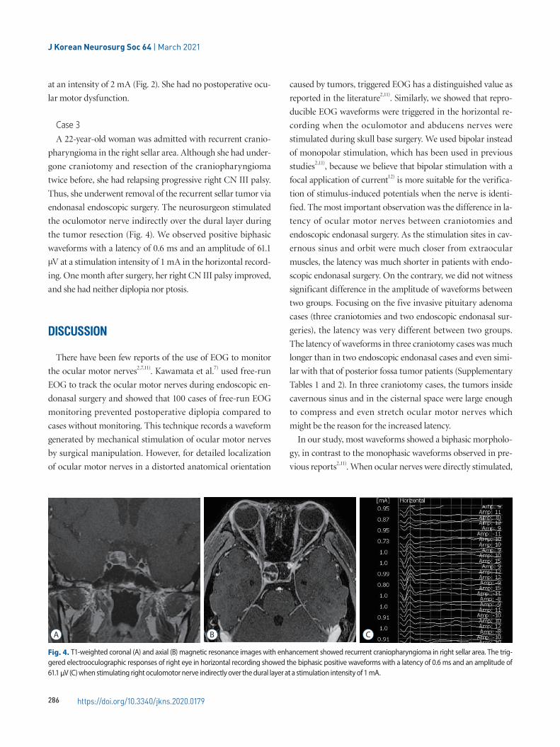
Case 3A 22-year-old woman was admitted with recurrent cranio-
pharyngioma in the right sellar area. Although she had under-
gone craniotomy and resection of the craniopharyngioma
twice before, she had relapsing progressive right CN III palsy.
Thus, she underwent removal of the recurrent sellar tumor via
endonasal endoscopic surgery. The neurosurgeon stimulated
the oculomotor nerve indirectly over the dural layer during
the tumor resection (Fig. 4). We observed positive biphasic
waveforms with a latency of 0.6 ms and an amplitude of 61.1
µV at a stimulation intensity of 1 mA in the horizontal record-
ing. One month after surgery, her right CN III palsy improved,
and she had neither diplopia nor ptosis.
DISCUSSION
There have been few reports of the use of EOG to monitor
the ocular motor nerves2,7,11). Kawamata et al.7) used free-run
EOG to track the ocular motor nerves during endoscopic en-
donasal surgery and showed that 100 cases of free-run EOG
monitoring prevented postoperative diplopia compared to
cases without monitoring. This technique records a waveform
generated by mechanical stimulation of ocular motor nerves
by surgical manipulation. However, for detailed localization
of ocular motor nerves in a distorted anatomical orientation
caused by tumors, triggered EOG has a distinguished value as
reported in the literature2,11). Similarly, we showed that repro-
ducible EOG waveforms were triggered in the horizontal re-
cording when the oculomotor and abducens nerves were
stimulated during skull base surgery. We used bipolar instead
of monopolar stimulation, which has been used in previous
studies2,11), because we believe that bipolar stimulation with a
focal application of current12) is more suitable for the verifica-
tion of stimulus-induced potentials when the nerve is identi-
fied. The most important observation was the difference in la-
tency of ocular motor nerves between craniotomies and
endoscopic endonasal surgery. As the stimulation sites in cav-
ernous sinus and orbit were much closer from extraocular
muscles, the latency was much shorter in patients with endo-
scopic endonasal surgery. On the contrary, we did not witness
significant difference in the amplitude of waveforms between
two groups. Focusing on the five invasive pituitary adenoma
cases (three craniotomies and two endoscopic endonasal sur-
geries), the latency was very different between two groups.
The latency of waveforms in three craniotomy cases was much
longer than in two endoscopic endonasal cases and even simi-
lar with that of posterior fossa tumor patients (Supplementary
Tables 1 and 2). In three craniotomy cases, the tumors inside
cavernous sinus and in the cisternal space were large enough
to compress and even stretch ocular motor nerves which
might be the reason for the increased latency.
In our study, most waveforms showed a biphasic morpholo-
gy, in contrast to the monophasic waveforms observed in pre-
vious reports2,11). When ocular nerves were directly stimulated,
A B C
Fig. 4. T1-weighted coronal (A) and axial (B) magnetic resonance images with enhancement showed recurrent craniopharyngioma in right sellar area. The trig-gered electrooculographic responses of right eye in horizontal recording showed the biphasic positive waveforms with a latency of 0.6 ms and an amplitude of 61.1 μV (C) when stimulating right oculomotor nerve indirectly over the dural layer at a stimulation intensity of 1 mA.
Intraoperative Triggered Electrooculography | Jeong HN, et al.
287J Korean Neurosurg Soc 64 (2) : 282-288
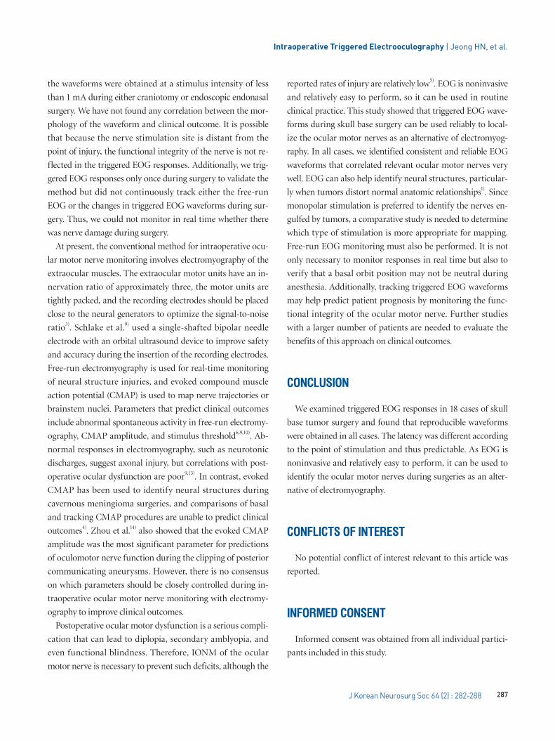
the waveforms were obtained at a stimulus intensity of less
than 1 mA during either craniotomy or endoscopic endonasal
surgery. We have not found any correlation between the mor-
phology of the waveform and clinical outcome. It is possible
that because the nerve stimulation site is distant from the
point of injury, the functional integrity of the nerve is not re-
flected in the triggered EOG responses. Additionally, we trig-
gered EOG responses only once during surgery to validate the
method but did not continuously track either the free-run
EOG or the changes in triggered EOG waveforms during sur-
gery. Thus, we could not monitor in real time whether there
was nerve damage during surgery.
At present, the conventional method for intraoperative ocu-
lar motor nerve monitoring involves electromyography of the
extraocular muscles. The extraocular motor units have an in-
nervation ratio of approximately three, the motor units are
tightly packed, and the recording electrodes should be placed
close to the neural generators to optimize the signal-to-noise
ratio3). Schlake et al.9) used a single-shafted bipolar needle
electrode with an orbital ultrasound device to improve safety
and accuracy during the insertion of the recording electrodes.
Free-run electromyography is used for real-time monitoring
of neural structure injuries, and evoked compound muscle
action potential (CMAP) is used to map nerve trajectories or
brainstem nuclei. Parameters that predict clinical outcomes
include abnormal spontaneous activity in free-run electromy-
ography, CMAP amplitude, and stimulus threshold6,9,10). Ab-
normal responses in electromyography, such as neurotonic
discharges, suggest axonal injury, but correlations with post-
operative ocular dysfunction are poor9,13). In contrast, evoked
CMAP has been used to identify neural structures during
cavernous meningioma surgeries, and comparisons of basal
and tracking CMAP procedures are unable to predict clinical
outcomes4). Zhou et al.14) also showed that the evoked CMAP
amplitude was the most significant parameter for predictions
of oculomotor nerve function during the clipping of posterior
communicating aneurysms. However, there is no consensus
on which parameters should be closely controlled during in-
traoperative ocular motor nerve monitoring with electromy-
ography to improve clinical outcomes.
Postoperative ocular motor dysfunction is a serious compli-
cation that can lead to diplopia, secondary amblyopia, and
even functional blindness. Therefore, IONM of the ocular
motor nerve is necessary to prevent such deficits, although the
reported rates of injury are relatively low5). EOG is noninvasive
and relatively easy to perform, so it can be used in routine
clinical practice. This study showed that triggered EOG wave-
forms during skull base surgery can be used reliably to local-
ize the ocular motor nerves as an alternative of electromyog-
raphy. In all cases, we identified consistent and reliable EOG
waveforms that correlated relevant ocular motor nerves very
well. EOG can also help identify neural structures, particular-
ly when tumors distort normal anatomic relationships1). Since
monopolar stimulation is preferred to identify the nerves en-
gulfed by tumors, a comparative study is needed to determine
which type of stimulation is more appropriate for mapping.
Free-run EOG monitoring must also be performed. It is not
only necessary to monitor responses in real time but also to
verify that a basal orbit position may not be neutral during
anesthesia. Additionally, tracking triggered EOG waveforms
may help predict patient prognosis by monitoring the func-
tional integrity of the ocular motor nerve. Further studies
with a larger number of patients are needed to evaluate the
benefits of this approach on clinical outcomes.
CONCLUSION
We examined triggered EOG responses in 18 cases of skull
base tumor surgery and found that reproducible waveforms
were obtained in all cases. The latency was different according
to the point of stimulation and thus predictable. As EOG is
noninvasive and relatively easy to perform, it can be used to
identify the ocular motor nerves during surgeries as an alter-
native of electromyography.
CONFLICTS OF INTEREST
No potential conflict of interest relevant to this article was
reported.
INFORMED CONSENT
Informed consent was obtained from all individual partici-
pants included in this study.

J Korean Neurosurg Soc 64 | March 2021
288 https://doi.org/10.3340/jkns.2020.0179
AUTHOR CONTRIBUTIONS
Conceptualization : HNJ, EHK
Data curation : HNJ, SIA, SK
Formal analysis : HNJ, EHK
Methodology : HNJ, EHK
Project administration : HNJ
Visualization : HNJ, MN, JY, WK, IHJ
Writing - original draft : HNJ
Writing - review & editing : SMK, HYS, JHC, EHK
ORCID
Ha-Neul Jeong https://orcid.org/0000-0002-5785-116X
Sang-Il Ahn https://orcid.org/0000-0002-2416-6174
Minkyun Na https://orcid.org/0000-0001-6826-8490
Jihwan Yoo https://orcid.org/0000-0001-8746-1245
Woohyun Kim https://orcid.org/0000-0002-2936-3740
In-Ho Jung https://orcid.org/0000-0002-4135-5743
Soobin Kang https://orcid.org/0000-0003-1214-2770
Seung Min Kim https://orcid.org/0000-0002-4384-9640
Ha Young Shin https://orcid.org/0000-0002-4408-8265
Jong Hee Chang https://orcid.org/0000-0003-1509-9800
Eui Hyun Kim https://orcid.org/0000-0002-2523-7122
● Acknowledgements
This study was funded in part by the Basic Science Research
Program through the NRF of Korea (NRF-2018R1C1B5042687)
funded by the Korean Ministry of Science, ICT and Future
Planning (Eui Hyun Kim).
This work was presented at the 7th Congress of the interna-
tional society of intraoperative neurophysiology, Vienna, Aus-
tria (October 31 to November 2, 2019).
● Supplementary materials
The online-only data supplement is available with this arti-
cle at https://doi.org/10.3340/jkns.2020.0179.
References
1. Cornelius JF, Schipper J, Tortora A, Krause-Molle Z, Smuga M, Petridis
AK, et al. : Continuous and dynamic facial nerve mapping during surgery
of cerebellopontine angle tumors: clinical pilot series. World Neuro-surg 119 : e855-e863, 2018
2. Fukaya C, Katayama Y, Kasai M, Kurihara J, Yamamoto T : Intraopera-
tive electrooculographic monitoring of oculomotor nerve function during
skull base surgery. Technical note. J Neurosurg 91 : 157-159, 1999
3. Hariharan P, Balzer JR, Anetakis K, Crammond DJ, Thirumala PD : Elec-
trophysiology of extraocular cranial nerves: oculomotor, trochlear, and
abducens nerve. J Clin Neurophysiol 35 : 11-15, 2018
4. Kaspera W, Adamczyk P, Ślaska-Kaspera A, Ładziński P : Usefulness of
intraoperative monitoring of oculomotor and abducens nerves during
surgical treatment of the cavernous sinus meningiomas. Adv Med Sci 60 : 25-30, 2015
5. Kassam AB, Prevedello DM, Carrau RL, Snyderman CH, Thomas A,
Gardner P, et al. : Endoscopic endonasal skull base surgery: analysis of
complications in the authors' initial 800 patients. J Neurosurg 114 : 1544-1568, 2011
6. Kawaguchi M, Ohnishi H, Sakamoto T, Shimizu K, Touho H, Monobe
T, et al. : Intraoperative electrophysiologic monitoring of cranial motor
nerves in skull base surgery. Surg Neurol 43 : 177-181, 1995
7. Kawamata T, Ishii N, Amano K, Namioka T, Hori T, Okada Y : A novel
simple real-time electrooculographic monitoring system during trans-
sphenoidal surgeries to prevent postoperative extraocular motor nerve
dysfunction. Neurosurg Rev 36 : 371-376, 2013
8. López JR : Neurophysiologic intraoperative monitoring of the oculomo-
tor, trochlear, and abducens nerves. J Clin Neurophysiol 28 : 543-
550, 2011
9. Schlake HP, Goldbrunner R, Siebert M, Behr R, Roosen K : Intra-opera-
tive electromyographic monitoring of extra-ocular motor nerves (Nn. III,
VI) in skull base surgery. Acta Neurochir (Wien) 143 : 251-261, 2001
10. Sekiya T, Hatayama T, Iwabuchi T, Maeda S : Intraoperative recordings
of evoked extraocular muscle activities to monitor ocular motor nerve
function. Neurosurgery 32 : 227-235; discussion 235, 1993
11. Sheshadri V, Bharadwaj S, Chandramouli BA : Intra-operative electro-
oculographic monitoring to prevent post-operative extraocular motor
nerve dysfunction during skull base surgeries. Indian J Anaesth 60 : 560-565, 2016
12. Singh H, Vogel RW, Lober RM, Doan AT, Matsumoto CI, Kenning TJ,
et al. : Intraoperative neurophysiological monitoring for endoscopic
endonasal approaches to the skull base: a technical guide. Scientifica (Cairo) 2016 : 1751245, 2016
13. Thirumala PD, Mohanraj SK, Habeych M, Wichman K, Chang YF, Gard-
ner P, et al. : Value of free-run electromyographic monitoring of extra-
ocular cranial nerves during expanded endonasal surgery (EES) of the
skull base. J Neurol Surg Rep 74 : 43-50, 2013
14. Zhou Q, Zhang M, Jiang Y : Intraoperative oculomotor nerve monitoring
predicts outcome following clipping of posterior communicating artery
aneurysms. J Clin Neurosci 19 : 706-711, 2012