Embed Size (px)
Citation preview
Mal J Med Health Sci 14(3): 46-50, Oct 2018 46
Malaysian Journal of Medicine and Health Sciences (eISSN 2636-9346)
SHORT COMMUNICATION
Tricks to do a Quick Successful Bronchial Artery Embolization (BAE) in Massive HaemoptysisEzamin Abdul Rahim1, Ahmad Sobri Muda1, Hariati Jamil2
1 Department of Imaging, Faculty of Medicine and Health Sciences, Universiti Putra Malaysia, 43600, Serdang, Selangor, Malaysia
2 Putrajaya Hospital, Precinct 7, 62250 Putrajaya, Malaysia
ABSTRACT
Bronchial artery embolization (BAE) is the mainstay treatment for massive haemoptysis. Herein we briefly discuss the tips and tricks of super-selective embolization of bronchial artery using N-butyl-2 cyanoacrylate (NBCA). Based on our experience, this technique produces a better resolution and exhibit high non- recurrence rate in the treatment of massive haemoptysis.
Keywords: Bronchial artery, Angiography, Embolization
Corresponding Author: Ezamin Abdul Rahim, MMEDEmail: [email protected]: +603-89472512Fax: +603-89412787
INTRODUCTION
Massive recurrent haemoptysis is a devastating complication of airway disease that may lead to death. The diseases that could lead to haemoptysis differs; depending on the geographic location, the climates, associate local endemic pulmonary disease, genetic factors and even time of presentation are important. Our institution is located in Southeast Asia known for its stable tropical warm humid climate throughout the year. Thus, the tropical climate-related respiratory diseases are not uncommon. From our observation; we noticed that the main culprit for haemoptysis in our institution is bronchiectasis secondary to post-primary TB infection. Unfortunately, there is no local data published to support our claim.
We are inclined to the idea that the main cause of death in-patient with massive haemoptysis is ironically due to asphyxiation rather than haemodynamic instability, hence, it is important to control the haemoptysis in a patient who is unable to protect their airways; even though the amount of expectorated blood is small (1). Protection of the non-affected site from the blood pooling is necessary during massive haemoptysis.
There is no reliable sign to predict impending
asphyxiation or to anticipate transformation of mild benign haemoptysis to a massive live threatening haemoptysis. It is based on this reason; a suspected massive haemoptysis patient ideally should be placed in the acute bay setting such as High Dependency Unit (HDU) whereby the equipment and the personnel experienced in fluid and airway resuscitation are readily available. A multidisciplinary approach involving the intensivist, cardiothoracic surgeon, interventional respiratory physician and the interventional radiologist is probably the best practice in managing life-threatening haemoptysis. In our experience, the ability of a patient to cough up blood and clearing up the airway is one important good prognostic criteria. There is a challenge to manage an ill patient with existing lung disease presented concomitantly with difficulty of breathing. It is difficult to identify whether the deteriorating respiratory condition is due to either worsening primary lung disease or blood “flooding” the lower airways causing shortness of breath. In addition to the problem, there is no universal consensus on the definition of massive haemoptysis for the clinician to react. Several authors have suggested the amount of blood loss is the main criteria to define the severity of haemoptysis. The basis of this definition is usually based on the flow rate of bleeding per unit time. The expectorated amount of blood to be labelled as massive haemoptysis ranging from 100ml /24hr to 1000mls/24 hr (2). Criteria based on pre-existing respiratory distress, current respiratory status, the magnitude of exporated blood and haemodynamic instability have been suggested to be more inclusive (3). Hence, Ibrahim W H (2010) suggested the term “life-threatening haemoptysis” that is probably more accurate than the previously accepted “massive haemoptysis” (3).
Mal J Med Health Sci 14(3): 46-50, Oct 201847
Malaysian Journal of Medicine and Health Sciences (eISSN 2636-9346)
The treatment option is much depended on institutional preference. In our institutional practice, the first line treatment for massive recurrent haemoptysis is always bronchial artery embolization (BAE) due to its highly successful rate with relatively low complication rate (4).
As for the interventional radiologist, there are many choices of embolic material in the market. Two popular embolic materials for BAE are polyvinyl alcohol (PVA) particles and acrylic adhesive called popularly called NBCA (N-butyl-2 cyanoacrylate, Histoacryl Blue®; B. Braun, Melgungen, Germany). Recent evidence suggested that the NBCA is superior to other popular embolic material (Fig 1). Woo et al (2013) suggested that NBCA mixture has low recanalization rate and is the best option to treat haemoptysis in bronchiectasis patients. In their study, the complication rate of the polyvinyl alcohol (PVA) versus NBCA mixture is similar; BAE with NBCA mixture is more effective controlling haemoptysis caused by bronchiectasis (5).Yoo et al suggested that the non-recurrence rate after embolization using NBCA mixture in the first year is as high as 91.4% and non -recurrence rate within 5 years is up to 56.8%. The complication occurred is mainly chest wall pain (6). Ikoma et al (2011) suggested that the usage of NBCA does not cause major damage to the bronchial wall or to the pulmonary parenchyma as previously thought (7). Polymerization of NBCA occurs immediately once the acrylic polymers adhesive is in contact with hydroxyl ions in the blood or any ionic material in the targeted area. This polymerization complex later will produce chronic inflammatory process within the vessels wall that delaying recanalization to occur. NBCA usage is shown to have a lower tendency of a recurrent haemoptysis as compared to other embolic agents (5, 8, 9).
Embolization coil is relatively safer in cases whereby the communication between the diseased artery and the spinal artery has not been completely rule out, or in a fast high-velocity arteriovenous fistulas communication. It is commonly used after the PVA embolization to completely plug off the intended artery. Worth to mention, coils sometimes are used to reduce the velocity of the blood flow of the artery of interest prior to NBCA administration to prevent distant venous embolization or migration of the NBCA complex.
MATERIAL AND METHODS
Our institution is a referral centre for cardiac and respiratory cases for the nation and most of our BAE cases are referrals from other hospitals. As a standard practice, the patient for bronchial artery embolization will be admitted a day prior to the procedure; in the care of the primary team either internal medicine or respiratory team. The procedure and its risks are explained to the patient including the first-degree relative. During the session, we would emphasise on the most devastating
event first such as risks of stroke (in the event of venous shunting, arterial communication) or risks of embolic injury to the spinal cord through connection with the anterior spinal artery. Written informed consent will be taken in the ward and in the interventional suite. There is no need for premedication prior to the procedure. Basic blood investigation is routinely done such as blood profile and coagulation profile. It is an advantage if CT angiography images are available due to the opportunity to evaluate the anatomical structure of the bronchial artery as the incidence of an accessory bronchial artery is approximately 36% (10).
We prefer the patient to be awake during the procedure if the patient is stable. In a haemodynamically or respiratory unstable patient; the embolization procedure will be done under general anaesthesia. Generally, a double lumen endotracheal tube intubation is preferred with the intention to protect the normal airway from spill over from the abnormal side of the lung, or in some centre, a large size of endotracheal tube (ETT) is used (more than 8mm diameter) due to the anticipation of concurrent bronchoscopy. Right femoral puncture is preferred. The standard femoral arterial sheath will be used. The preferred catheter of choice is Cobra catheter. Cobra C2 (catheter shape) for a male due to larger angle curvature of its tip making this type of catheter easier to manoeuvre in the larger calibre thoracic aorta than C1; however, we may change the type of catheter accordingly. The amount of contrast used to search for the ostium is significantly less by using only 3-5 ml of diluted contrast media per digital subtraction angiography (DSA) run. It is important for the operator applies pressure at the injector of the syringe. This is to prevent backflow of the arterial blood into the syringe and further contaminate the syringe that is prefilled with diluted contrast media. Once the ostium is identified; a few arterial runs will be commenced and arteriograms acquired using digital subtraction angiography technique. It is important to identify any venous shunting (fistulous communication) is present and to rule out any formation of the anterior spinal artery (The artery of adamkewiz- name after Albert Wojciech Adamkiewicz) that may have originated from the bronchial artery or the intercostal bronchial artery. The anterior spinal artery that is originated from the intercostal bronchial artery (ICBA) is rare at approximately 5% incidence (11). It is important to carefully look for this anatomical variant by doing super-selective angiography to the targeted vessel using microcatheter or making sure that there is no artery going to the medial side and give a faint “hairpin appearance”. It is important to note any presence of extremely rare arterial-arterial communication. Communication between the bronchial artery - subclavian artery and bronchial artery - right coronary artery is well documented in the literature. (12, 13). We had an experience of causing iatrogenic posterior circulation infarction during BAE. The presence of these anatomical variants may complicate treatment.
Mal J Med Health Sci 114(3): 46-50, Oct 2018 48
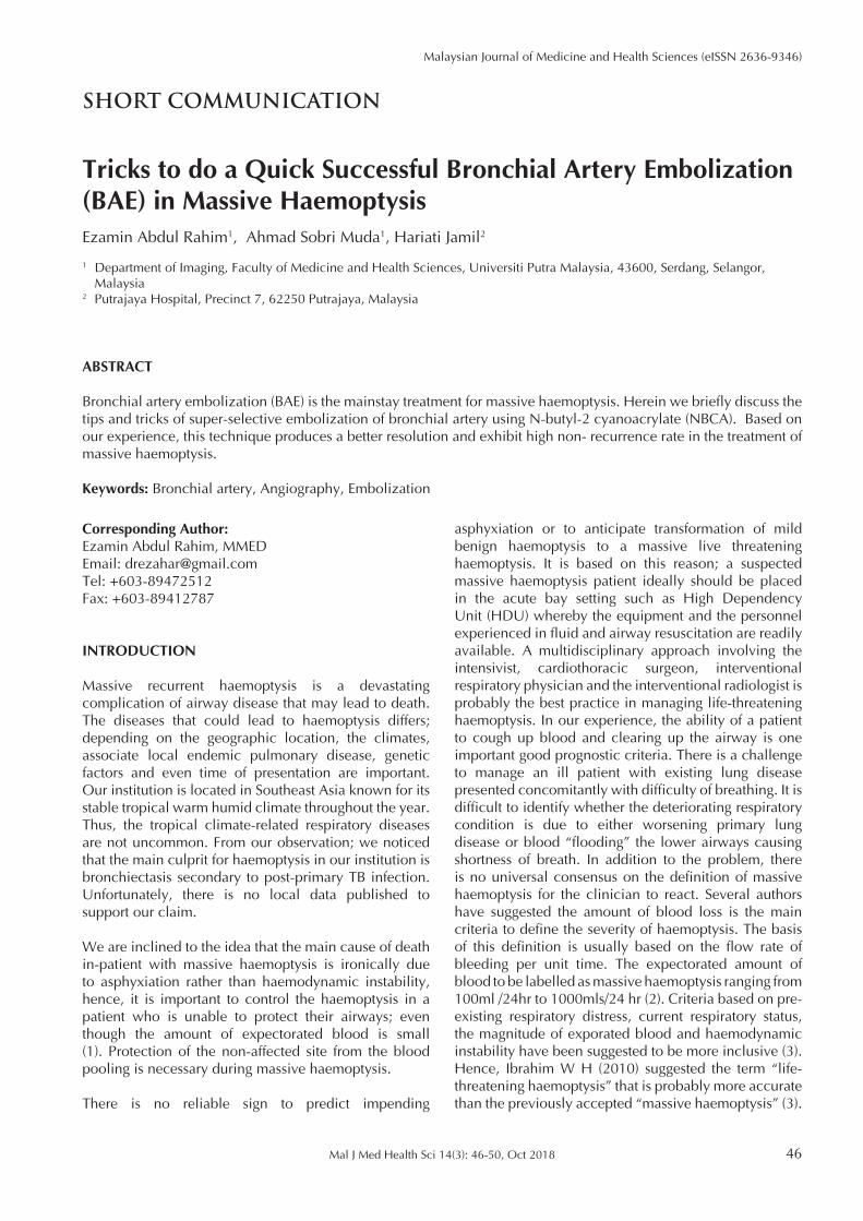
Figure 1: DSA image showing faint contrast opacification within the previously embolized left internal thoracic artery using PVA and coil. This is suggestive of recanalization of the artery (arrowhead). An endovascular coil within the left internal thoracic artery (arrow).
RESULTS
To determine the nature of the vasculature, oblique or cranio-caudal angulations of tube projection, multiple arterial DSA run with magnification view are suggested. The clues for diagnosing abnormal vessels are; the abnormal bronchial artery is usually dilated, especially at the proximal part, the abnormal artery is usually tortuous, the presence of corkscrew appearance at the mid and distal bronchial arteries, and abnormal blushing or hyperaemia of the lung parenchyma. It is very rare to detect an active extravasation of contrast into the alveolar/bronchus during the procedure.
We use multipurpose microcatheter; (Progreat 2.7 Fr- 0.90mm, Terumo or ASAHI Masters PARKWAY HF KIT 2.8 Fr- 0.88mm) as the microcatheter of choice to super-selectively catheterize the bronchial artery and other non-bronchial thoracic arteries. We have experienced few cases of recanalization occurred after PVA and coils deployment (Figure 1). It is for this reason we have adopted a new strategy to use acrylic glue for embolization. We believe that chronic inflammatory process changes and scarred tissue are better embolized using NBCA. The main constraint for NBCA usage is a high cost incurred due to more expensive lipiodol
(ethiodized poppy seed oil) as opposed to PVA. The other reason is the micro-catheter that has been used for the initial embolization will be discarded due to glue material residue within the micro-catheter lumen.DISCUSSION
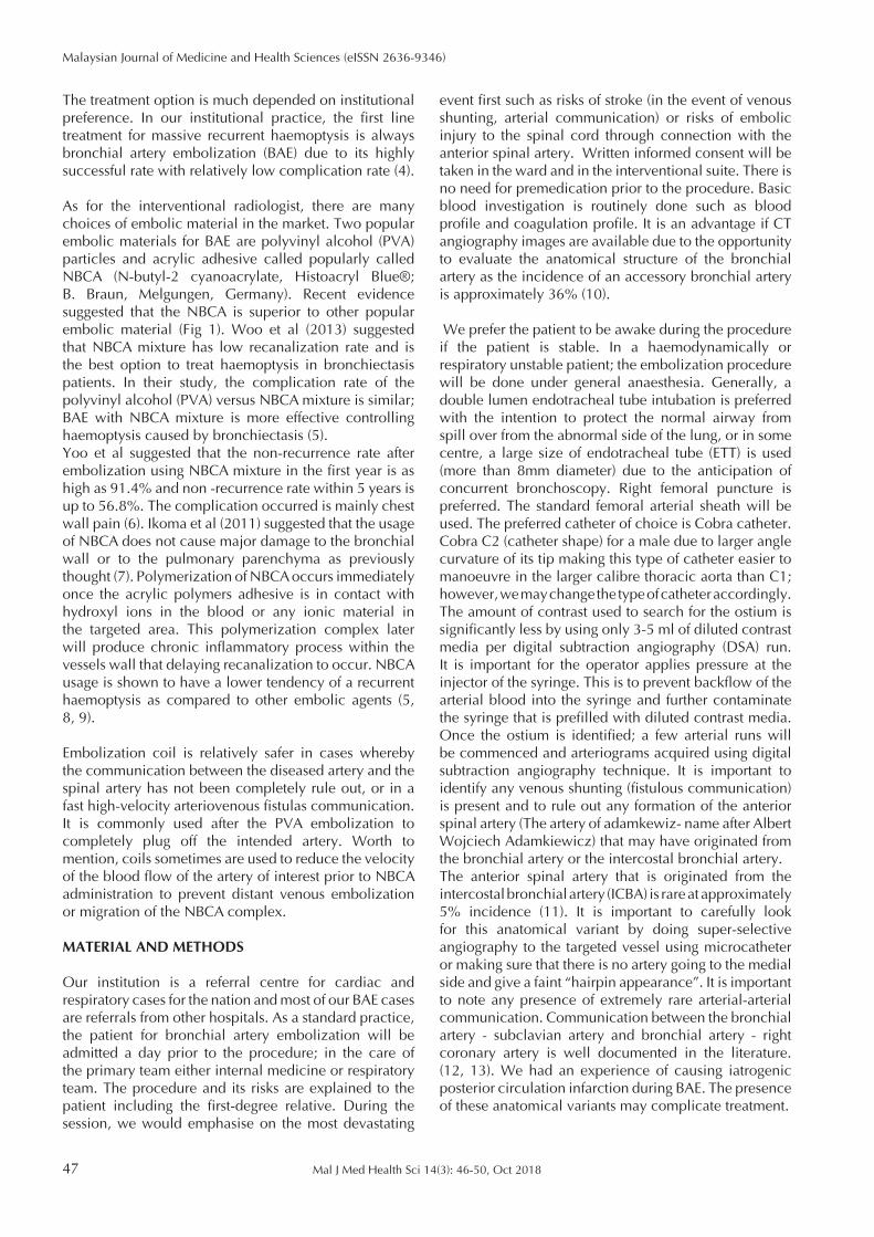
Once the bronchial artery is selectively catheterized, it is best to assess the dynamic flow by injecting a small amount of contrast under fluoroscopy. The decision to use glue (NBCA) or other embolic material is made after acquiring this information. If the patient is intubated, it is an opportunity to ask the anaesthetist to do a “breath-hold” technique to pause the ventilation. The aim is to minimize the motion artefact occurred during the arterial run. If the glue material usage has been chosen, the concentration of the glue is based on the blood flow, wideness area to be blocked and the presence of venous shunting (Figure 2). The standard concentration of NBCA mixture in our institution is about 25-50% dilution using ethiodized poppy seed oil (lipiodol) as the diluent. Adding more diluent can prolong the polymerization time. We had an experienced using 90% NBCA in due to large venous shunting into the pulmonary veins. The most important thing before administration of NBCA mixture is the knowledge about the arterial velocity, prediction of NBCA polymerization time and the
Figure 2: Pre-embolization DSA image of the abnormal bronchial vessels with early venous appearance (white arrows).
Mal J Med Health Sci 14(3): 46-50, Oct 201849
Malaysian Journal of Medicine and Health Sciences (eISSN 2636-9346)
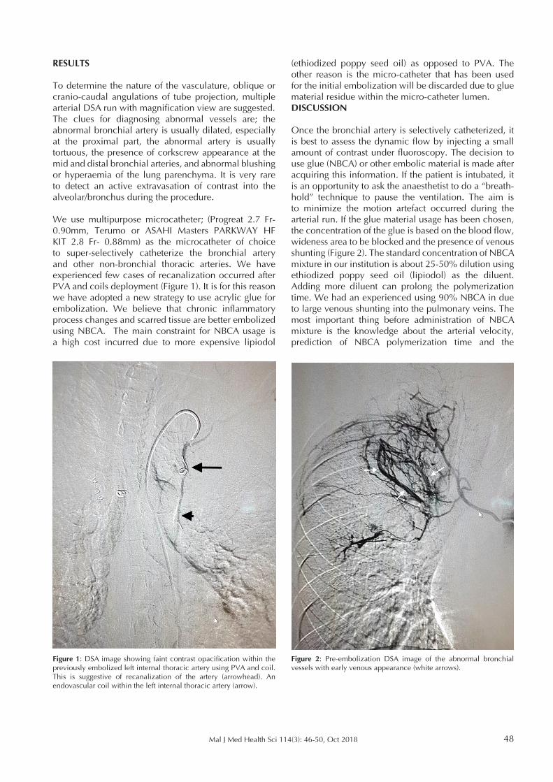
Figure 3: A – pre-embolization DSA image of the left bronchial artery. B - Post embolization image showing glue cast within the embolized left bronchial artery (arrows).
polymerization of NBCA, such the operators need to change a new set latex hand glove and put another layer of clean dry cloth to prevent accidentally mixing ionic fluid into the NBCA mix. We used Dextrose 5% to wash out any blood product in the micro-catheter lumen and hub. We also soaked our glove with dextrose as one of precautionary action.
Under fluoroscopy control; staggered injection by adequately squirting about 0.2 ml in up to every 2-3 seconds is recommended, however, the operators must know and in real time control the injection rate according to the ever-changing dynamicity of the blood flow. The blood flow will be slower once the injection is commenced. It is important to assess for the proximal reflux all the time. Injection is stopped once stagnation of opaque embolic material is seen at the region of interest (Figure 3). Removal of the micro-catheter is done
immediately to prevent micro-catheter adherence to the arterial wall. The radio-opacity of NBCA mixture is less if the Lipiodol concentration is < 50%. In a large area of lung parenchymal involvement; a staged embolization can be organized. A repeat embolization will be done at a later date usually within 1-3 month depending on the severity of the symptoms.
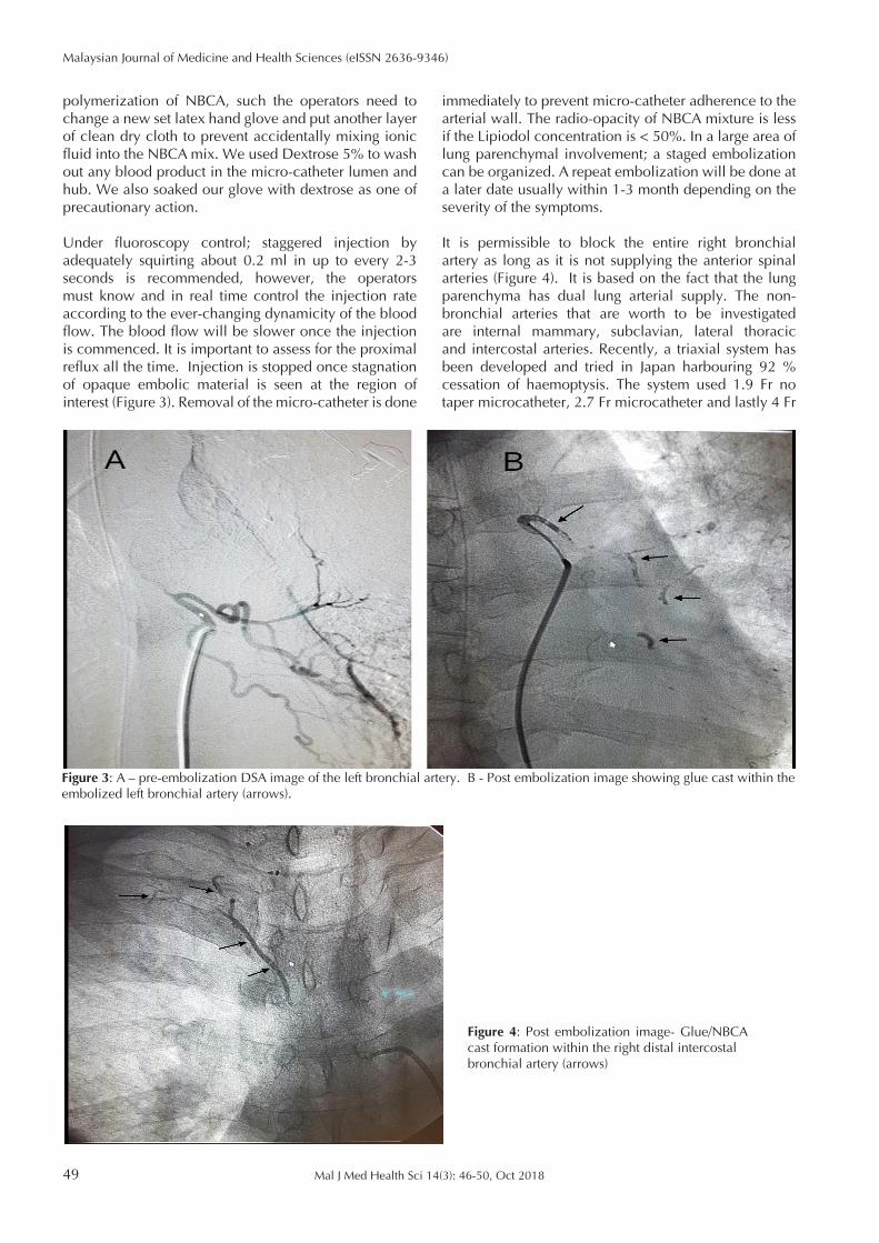
It is permissible to block the entire right bronchial artery as long as it is not supplying the anterior spinal arteries (Figure 4). It is based on the fact that the lung parenchyma has dual lung arterial supply. The non-bronchial arteries that are worth to be investigated are internal mammary, subclavian, lateral thoracic and intercostal arteries. Recently, a triaxial system has been developed and tried in Japan harbouring 92 % cessation of haemoptysis. The system used 1.9 Fr no taper microcatheter, 2.7 Fr microcatheter and lastly 4 Fr
Figure 4: Post embolization image- Glue/NBCA cast formation within the right distal intercostalbronchial artery (arrows)
Mal J Med Health Sci 14(3): 46-50, Oct 2018 50
as guiding catheter into the bronchial artery ostium (14). The NBCA will be injected through 1.9Fr microcatheter, and later after the withdrawal of 1.9Fr microcatheter, the 2.7 fr microcatheter will be used for confirmatory highly selective angiography run to the embolized artery.
The immediate usual complication is chest wall pain, and up to 89% has been reported (15). From our experience, the chest pain is usually mild and involved the anterior wall of the chest depending on the place of the embolization. Most of our patients do not require heavy painkillers or sedation.
CONCLUSION
Super-selective embolization of bronchial artery using NBCA is a safe, quick and reliable method for a successful treatment of massive haemoptysis.
ACKNOWLEDGEMENT
This manuscript is supported by Geran PUTRA GP-IPM/2014/9438300
REFERENCES
1. Ittrich H, Bockhorn M, Klose H, Simon M. The Diagnosis and Treatment of Hemoptysis. Dtsch Arztebl Int. 2017; 114(21):371-38.
2. Sakr L, Dutau H. Massive haemoptysis: an update on the role of bronchoscopy in diagnosis and management. Respiration. 2010; 80(1): 38-58
3. Ibrahim W H. Massive haemoptysis: the definition should be revised European Respiratory Journal. 2008; 32: 1131-1132
4. Cremaschi P, Nascimbene C, Vitulo P, Catanese C, Rota L, Barazzoni GC, Cornalba GP. Therapeutic embolization of bronchial artery: a successful treatment in 209 cases of relapse hemoptysis. Angiology. 1993; 44(4):295–299
5. Woo S, Yoon CJ, Chung JW, Kang SG, Jae HJ, Kim HC, Seong NJ, Kim YJ, Woo YN. Bronchial artery embolization to control hemoptysis: comparison of N-butyl-2-cyanoacrylate and polyvinyl alcohol particles. Radiology 2013; 269(2):594-602.
6. Yoo DH, Yoon CJ, Kang SG, Burke CT, Lee JH, Lee
CT. Bronchial and nonbronchial systemic artery embolization in patients with major hemoptysis: safety and efficacy of N-butyl cyanoacrylate. AJR AM J Roentgenol 2011;196(2): W199-204
7. Ikoma A, Kawai N, Sato M, Tanaka T, Sonomura T, Sahara S, Nakata K, Takasaka I, Minamiguchi H, Nakai M, Mori I. Pathologic evaluation of damage to bronchial artery, bronchial wall and pulmonary parenchyma after bronchial artery embolization with N- butyl cyanoacrylate for massive hemoptysis. J Vascular Interv Radiol. 2011; 22(8): 1212-1215
8. Razavi MK, Murphy K. Embolization of bronchial arteries with N-butyl cyanoacrylate for management of massive haemoptysis: a technical review. Tech Vasc Interv Radiol. 2007; 10(4) 276-282
9. Baltacioğlu F, Cimşit NC, Bostanci K , Yüksel M, Kodalli N. Transarterial microcatheter glue embolization of the bronchial artery for life-threatening hemoptysis: technical and clinical results. Eur J Radiol. 2010; 73(2):380-4.
10. Hartmann IJ, Remy-Jardin M, Menchini L, Teisseir A, Khalil C, Remy J. Ectopic origin of bronchial arteries: assessment with multidetector helical CT angiography. Eur Radiol. 2007; 17(8):1943-53
11. Phillip MB, David AL. CT of the Airways. New Jersey; Humana Press; 2008
12. Cheila MC, Fang YC. An Unexpected Complication of Bronchial Artery Embolization in a Young Patient with Haemoptysis: A Case Report. Chest. 2016;150(4):1031
13. Pathak V, Stavas JM, Ford HJ, Austin CA, Aris RM. Long-term outcomes of the bronchial artery embolization are diagnosis dependent. Lung India. 2016;33(1):3-8
14. Mashashi S, Takeshi H, Saori A, Takuya H, Motoo N, Yoshiyuki O, Keita S, Yuta S. Triaxial system in bronchial arterial embolization for haemoptysis using N-butyl-2-cyanoacrylate. Br J Radiol. 2015; 88(1056): 20150265
15. Cohen AM, Doershuk CF, Stern RC. Bronchial artery embolization to control hemoptysis in cystic fibrosis. Radiology. 1990; 175(2):401-5