Embed Size (px)
Citation preview
Contents iiiContents iii
Trichoscopy A Text and Atlas
Editor-in Chief
Subrata Malakar MBBS MD (Dermatology) DCH
Director and Chief Dermatologist
Rita Skin Foundation
Kolkata, West Bengal, India
Associate Editors
The Health Sciences Publisher New Delhi | London | Panama
BS Chandrashekar MD DNB
Chief Dermatologist
Cutis Academy of Cutaneous Sciences
Bengaluru, Karnataka, India
Samipa S Mukherjee DDV DDVL
FRGUHS (Ped Dermatology)
Dermato-trichologist and Pediatric
Dermatologist
Cutis Academy of Cutaneous Sciences
Bengaluru, Karnataka, India
Purva Mehta DNB
Consultant Dermatologist
Rita Skin Foundation
Kolkata, West Bengal, India
Pratibha Pradhan MD
Consultant Dermatologist
Rita Skin Foundation
Kolkata, West Bengal, India
Jayp
ee B
rothe
rs

Jaypee Brothers Medical Publishers (P) Ltd
Website: www.jaypeebrothers.com Website: www.jaypeedigital.com
© 2017, Jaypee Brothers Medical Publishers
The views and opinions expressed in this book are solely those of the original contributor(s)/author(s) and do not necessarily represent those of editor(s) of the book.All rights reserved. No part of this publication may be reproduced, stored or transmitted in any form or by any means, electronic, mechanical, photocopying, recording or otherwise, without the prior permission in writing of the publishers. All brand names and product names used in this book are trade names, service marks, trademarks or registered trademarks of their respective owners. The publisher is not associated with any product or vendor mentioned in this book.Medical knowledge and practice change constantly. This book is designed to provide accurate, authoritative information about the subject matter in question. However, readers are advised to check the most current information available on procedures included and check information from the manufacturer of each product to be administered, to verify the recommended dose, formula, method and duration of administration, adverse effects and contraindications. It is the responsibility of the practitioner to take all appropriate safety precautions. Neither the publisher nor the author(s)/editor(s) assume any liability for any injury and/or damage to persons or property arising from or related to use of material in this book.This book is sold on the understanding that the publisher is not engaged in providing professional medical services. If such advice or services are required, the services of a competent medical professional should be sought.Every effort has been made where necessary to contact holders of copyright to obtain permission to reproduce copyright material. If any have ��������������� ����������������������������������������������������������� �������������������������������� �
Inquiries for bulk sales may be solicited at: [email protected]
Trichoscopy: A Text and Atlas
First Edition: 2017
ISBN: 978-93-86150-74-5
Printed at
HeadquartersJaypee Brothers Medical Publishers (P) Ltd.4838/24, Ansari Road, DaryaganjNew Delhi 110 002, IndiaPhone: +91-11-43574357Fax: +91-11-43574314E-mail: [email protected]
Jaypee-Highlights Medical Publishers Inc.City of Knowledge, Bld. 235, 2nd Floor, ClaytonPanama City, PanamaPhone: +1 507-301-0496Fax: +1 507-301-0499E-mail: [email protected]
�������������J.P. Medical Ltd.83, Victoria Street, LondonSW1H 0HW (UK)Phone: +44-20 3170 8910Fax: +44(0)20 3008 6180E-mail: [email protected]
Jaypee Brothers Medical Publishers (P) Ltd.17/1-B, Babar Road, Block-B, ShaymaliMohammadpur, Dhaka-1207BangladeshMobile: +08801912003485E-mail: [email protected]
Jaypee Brothers Medical Publishers (P) Ltd.Bhotahity, Kathmandu, NepalPhone: +977-9741283608E-mail: [email protected]
Jayp
ee B
rothe
rs

Dedicated to
My late wife Dr Rita Shah Malakar
Jayp
ee B
rothe
rs

Contents vii
BS Chandrashekar MD DNB
Chief DermatologistCutis Academy of Cutaneous SciencesBengaluru, Karnataka, India
Barnali Chowdhury MD
Consultant DermatologistRita Skin FoundationKolkata, West Bengal, India
Madura C MD FRGUHS
(Dermatosurgery)
Consultant Dermatologist and DermatosurgeonCutis Academy of Cutaneous SciencesBengaluru, Karnataka, India
Pratibha Pradhan MD
Consultant DermatologistRita Skin Foundation
Kolkata, West Bengal, India
Priya Diwaker DDVL
Consultant DermatologistRita Skin FoundationKolkata, West Bengal, India
Purva Mehta DNB
Consultant Dermatologist Rita Skin FoundationKolkata, West Bengal, India
Samipa S Mukherjee DDV DDVL
FRGUHS (Ped Dermatology)
Dermato-trichologist and Pediatric DermatologistCutis Academy of Cutaneous SciencesBengaluru, Karnataka, India
Souvik Sardar MD
Consultant DermatologistRita Skin Foundation
Kolkata, West Bengal, India
Subrata Malakar MBBS MD
(Dermatology) DCH
Director and Chief Dermatologist
Rita Skin Foundation
Kolkata, West Bengal, India
Surit Malakar MBBS, PGT (Dermatology)
Consultant Dermatologist
Rita Skin Foundation
Kolkata, West Bengal, India
Contributors
Jayp
ee B
rothe
rs

Contents ix
Preface
Trichoscopy is fast gaining hold in daily clinical practice of dermatologists across India and the world. This book is writ-
ten by the trichoscopy enthusiasts with a vision to help and enhance structured and step-by-step practice of trichoscopy
by the students and practitioners. The idea of this book was conceived when the authors saw that there was a dearth of
work and data related to trichoscopy in the skin of color and the absence of proper teaching modules related to the same.
This book was envisioned as a ready-reckoner and a basic step-by-step module that helped to arrive at a trichoscopic
diagnosis of a particular condition.
The authors are trichoscopy enthusiasts, who use trichoscopy in the same way that a physician would use his stetho-
scope. They concur with the words of Antonella Tosti and Lidia Rudnicka that the practice of trichology without trichos-
copy is a professional practice gap.
Having said this, the author would like to extend his heartfelt thanks to the two doyens in the world of trichology
at large and trichoscopy as well, Antonella Tosti and Lidia Rudnicka. Their relentless efforts in this field culminated in
producing two of the best books for trichoscopy that are being relied upon from across the world. Since its inception way
back in 1993, when Steven Kossard and Sam Zagarella described dermoscopy with dots in cicatricial alopecia to 2006,
where the term ‘trichoscopy’ was first introduced and used, the knowledge in this field is growing by leaps and bounds
worldwide.
As we compile the trichoscopic data in this book and guide our readers towards making a trichoscopic diagnosis, we
are certain that every reader will make their new observations as they go along and we solemnly support and request
them to share their work and knowledge to add to the existing knowledge in trichoscopy.
We hope this book delivers what you are looking forth to learn in the field of trichoscopy and, thereby, helps in your
clinical practice to achieve maximum output for yourself and your patients.
Subrata Malakar
Jayp
ee B
rothe
rs

Contents xi
I would like to acknowledge my patients who have inspired me a lot to work on this project.
I would like to thank Shri Jitendar P Vij (Group Chairman), Mr Ankit Vij (Group President), Ms Chetna Malhotra Vohra
(Associate Director–Content Strategy), Ms Nedup Denka (Development Editor), Mr Sabyasachi Hazra (Commissioning
Editor, Kolkata Branch) and the entire team of Jaypee Brothers Medical Publishers, New Delhi, India for their tireless
efforts in producing the book in its final form.
Acknowledgments
Jayp
ee B
rothe
rs

Section 1: Introduction
1. Introduction 3 Samipa S Mukherjee
Section 2: Devices and Tools
2. Devices and Tools 9 Pratibha Pradhan
Section 3: Trichoscopic Terminologies
3. Trichoscopic Terminologies 13 Subrata Malakar
Section 4: Trichoscopic Language and Pattern Analysis
4. Follicular and Perifollicular Patterns 55 Subrata Malakar, Purva Mehta
5. Interfollicular Pattern 66 Subrata Malakar, Priya Diwaker
6. Pattern of Hair Shafts 69 Subrata Malakar
7. Pattern of Scaly Scalp 86 Subrata Malakar
Section 5: Normal Scalp
8. Normal Scalp 99 Subrata Malakar
Section 6: Nonscarring Alopecias
9. Androgenetic Alopecia 121 Subrata Malakar, Purva Mehta, Surit Malakar
10. Female Pattern Hair Loss 136 Subrata Malakar, Surit Malakar, Purva Mehta
Contents
Jayp
ee B
rothe
rs

Trichoscopy: A Text and Atlasxiv
11A. Alopecia Areata Incognito 145 Subrata Malakar
11B. Alopecia Areata 148 Subrata Malakar
12. Trichotillomania 166 Subrata Malakar
13. Traction Alopecia 184 Subrata Malakar
14. Telogen Effluvium 188 Subrata Malakar, Purva Mehta, Surit Malakar
15. Anagen Effluvium 193 Subrata Malakar, Barnali Chowdhury
Section 7: Scarring Alopecias
16. Lichen Planopilaris 211 Subrata Malakar
17. Frontal Fibrosing Alopecia 219 Subrata Malakar
18. Discoid Lupus Erythematosus 222 Subrata Malakar, Samipa S Mukherjee, Surit Malakar
19. Acne Keloidalis Nuchae 227 Subrata Malakar, Purva Mehta, Priya Diwaker
20. Central Centrifugal Cicatricial Alopecia 231 Subrata Malakar
21. Folliculitis Decalvans 236 Subrata Malakar
22. Dissecting Cellulitis 241 Subrata Malakar
23. Pseudopelade of Brocq 242 Subrata Malakar
Section 8: Hair Weathering
24. Trichoscopy of Damaged Hair 251 Samipa S Mukherjee, Subrata Malakar
Section 9: Infection and Infestation
25. Tinea Capitis 261 Subrata Malakar
26. Pediculosis Capitis 267 Subrata Malakar
Jayp
ee B
rothe
rs

Contents xv
Section 10: Inflammatory Scalp Diseases
27. Scalp Psoriasis, Seborrheic Dermatitis and Sebopsoriasis 275 Subrata Malakar
Section 11: Systemic Diseases
28. Trichoscopy in Autoimmune Bullous Disorders 293 Souvik Sardar, Subrata Malakar
29. Trichoscopy in Autoimmune Connective Tissue Diseases 300 Souvik Sardar, Subrata Malakar
Section 12: Pediatric Trichology
30. Pediatric Hair Disorders 311 Samipa S Mukherjee, Subrata Malakar
Section 13: Body Hair Disorders
31. Body Hair 325 Subrata Malakar
Section 14: Effect of Dermatology Practice on Hair
32. Trichoscopy in Hair Transplantation 355 Madura C, BS Chandrashekar
Section 15: Algorithms
33. Algorithm 365 Samipa S Mukherjee
Section 16: Monitoring Therapeutic Efficacy
34. Trichoscopic Monitoring and Follow-up 371 Subrata Malakar
Index 383
Jayp
ee B
rothe
rs
FOLLICULAR AND PERIFOLLICULAR PATTERNS
■ Yellow dots
■ White dots
■ Black dots
■ Red dots
■ Blue-gray dots
■ Gray-white halo
■ Peripilar sign
■ Keratotic plugs
■ Empty follicles
■ Loss of follicular openings
■ Perifollicular scales
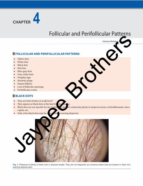
BLACK DOTS
■ They are hairs broken at scalp level1
■ They appear as black dots at the hair follicle opening
■ Black dots are not specific for any conditions and are commonly plenty in alopecia areara, trichotillomania, tinea
capitis, etc.
■ Only a few black dots may be seen in some scarring alopecias.
Fig. 1: Presence of plenty of black dots in alopecia areata. They are not diagnostic as numerous black dots are present in other non-scarring alopecia also.
Subrata Malakar, Purva Mehta
4CHAPTER
Follicular and Perifollicular Patterns
Jayp
ee B
rothe
rs
Section 4: Trichoscopic Language and Pattern Analysis56
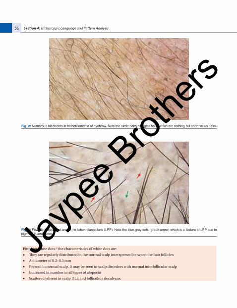
Fig. 2: Numerous black dots in trichotillomania of eyebrow. Note the circle hairs or pigtail hairs which are nothing but short vellus hairs.
Fig. 3: Few black dots (red arrows) in lichen planopilaris (LPP). Note the blue-gray dots (green arrow) which is a feature of LPP due to pigment incontinence.
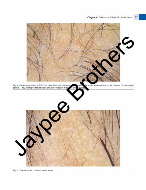
Pinpoint white dots:2 the characteristics of white dots are:
■ They are regularly distributed in the normal scalp interspersed between the hair follicles
■ A diameter of 0.2–0.3 mm
■ Present in normal scalp. It may be seen in scalp disorders with normal interfollicular scalp
■ Increased in number in all types of alopecia
■ Scattered/absent in scalp DLE and folliculitis decalvans.
Jayp
ee B
rothe
rs
Chapter 4: Follicular and Perifollicular Patterns 57
Fig. 4: Pinpoint white dots: 0.2–0.3 mm dots distributed regularly in the interfollicular scalp, dispersed among the mosaics of honeycomb pattern. They correspond to follicular and acrosyringium of eccrine sweat glands.
Fig. 5: Pinpoint white dots in alopecia areata.
Jayp
ee B
rothe
rs
Section 4: Trichoscopic Language and Pattern Analysis58
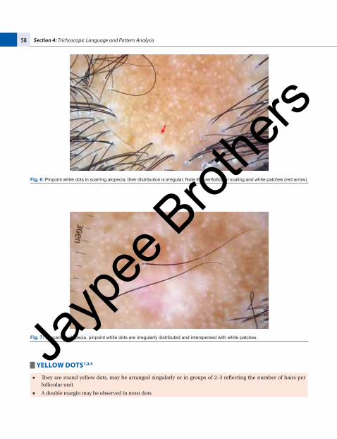
Fig. 6: Pinpoint white dots in scarring alopecia; their distribution is irregular. Note the perifollicular scaling and white patches (red arrow).
Fig. 7: In scarring alopecia, pinpoint white dots are irregularly distributed and interspersed with white patches.
YELLOW DOTS1,3,4
■ They are round yellow dots, may be arranged singularly or in groups of 2–3 reflecting the number of hairs per
follicular unit
■ A double margin may be observed in most dots
Jayp
ee B
rothe
rs
Chapter 4: Follicular and Perifollicular Patterns 59
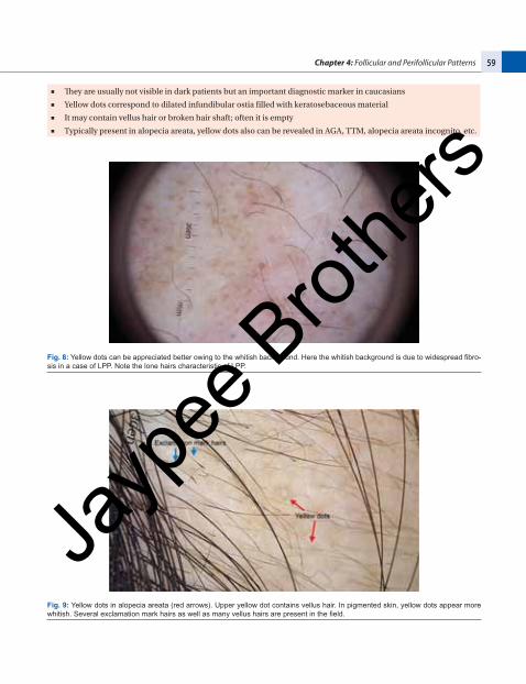
■ They are usually not visible in dark patients but an important diagnostic marker in caucasians
■ Yellow dots correspond to dilated infundibular ostia filled with keratosebaceous material
■ It may contain vellus hair or broken hair shaft; often it is empty
■ Typically present in alopecia areata, yellow dots also can be revealed in AGA, TTM, alopecia areata incognito, etc.
Fig. 8: Yellow dots can be appreciated better owing to the whitish background. Here the whitish background is due to widespread fibro-sis in a case of LPP. Note the lone hairs characteristic of LPP.
Fig. 9: Yellow dots in alopecia areata (red arrows). Upper yellow dot contains vellus hair. In pigmented skin, yellow dots appear more whitish. Several exclamation mark hairs as well as many vellus hairs are present in the field.
Jayp
ee B
rothe
rs
Section 4: Trichoscopic Language and Pattern Analysis60
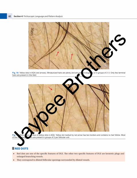
Fig. 10: Yellow dots in AGA (red arrows). Miniaturized hairs are plenty and are present singularly or in groups of 2–3. Only few terminal hairs are present in this field.
Fig. 11: More close-up view of yellow dots in AGA. Yellow dot marked by red arrow has two borders and contains no hair follicle. Most of the miniaturized hairs are present in groups of 2 per follicular unit.
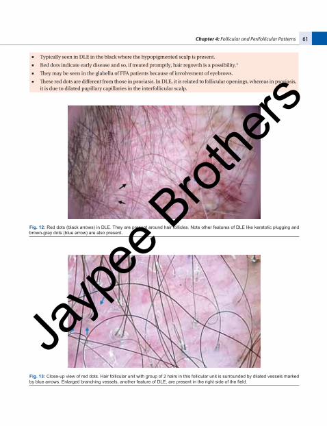
RED DOTS
■ Red dots are one of the specific features of DLE. The other two specific features of DLE are keratotic plugs and
enlarged branching vessels.
■ They correspond to dilated follicular openings surrounded by dilated vessels.
Jayp
ee B
rothe
rs
Chapter 4: Follicular and Perifollicular Patterns 61
■ Typically seen in DLE in the black where the hypopigmented scalp is present.
■ Red dots indicate early disease and so, if treated promptly, hair regowth is a possibility.5
■ They may be seen in the glabella of FFA patients because of involvement of eyebrows.
■ These red dots are different from those in psoriasis. In DLE, it is related to follicular openings, whereas in psoriasis,
it is due to dilated papillary capillaries in the interfollicular scalp.
Fig. 12: Red dots (black arrows) in DLE. They are present around hair follicles. Note other features of DLE like keratotic plugging and brown-gray dots (blue arrow) are also present.
Fig. 13: Close-up view of red dots. Hair follicular unit with group of 2 hairs in this follicular unit is surrounded by dilated vessels marked by blue arrows. Enlarged branching vessels, another feature of DLE, are present in the right side of the field.
Jayp
ee B
rothe
rs
Section 4: Trichoscopic Language and Pattern Analysis62
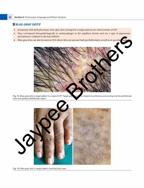
BLUE-GRAY DOTS6
■ In patients with dark phototype, blue-gray dots arranged in a target pattern are characteristic of LPP
■ They correspond histopathologically to melanophages in the papillary dermis and are a sign of pigmentary
incontinence confined to the hair follicles
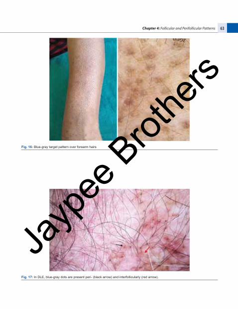
■ Blue-gray dots can also be seen in DLE where they are present both perifollicularly as well as in speckled pattern.
Fig. 14: Blue-gray dots in target pattern in a case of LPP. Target pattern is due to melanin incontinence surrounding only the perifollicular area and sparing interfollicular region.
Fig. 15: Blue-gray dots in target pattern involving body hairs.
Jayp
ee B
rothe
rs
Chapter 4: Follicular and Perifollicular Patterns 63
Fig. 16: Blue-gray target pattern over forearm hairs
Fig. 17: In DLE, blue-gray dots are present peri- (black arrow) and interfollicularly (red arrow).
Jayp
ee B
rothe
rs
Section 4: Trichoscopic Language and Pattern Analysis64
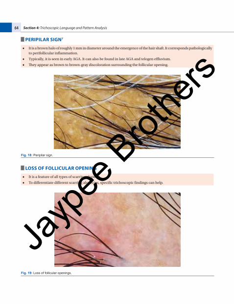
PERIPILAR SIGN7
■ It is a brown halo of roughly 1 mm in diameter around the emergence of the hair shaft. It corresponds pathologically
to perifollicular inflammation.
■ Typically, it is seen in early AGA. It can also be found in late AGA and telogen effluvium.
■ They appear as brown to brown-gray discoloration surrounding the follicular opening.
Fig. 18: Peripilar sign.
LOSS OF FOLLICULAR OPENING
■ It is a feature of all types of scarring alopecia.
■ To differentiate different scarring alopecias, specific trichoscopic findings can help.
Fig. 19: Loss of follicular openings.
Jayp
ee B
rothe
rs
Chapter 4: Follicular and Perifollicular Patterns 65
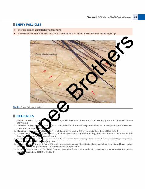
EMPTY FOLLICLES
■ They are seen as hair follicles without hairs.
■ These blank follicles are found in AGA and telogen effluvium and also sometimes in healthy scalp.
Fig. 20: Empty follicular openings.
REFERENCES 1. Ross EK, Vincenzi C, Tosti A. Videodermoscopy in the evaluation of hair and scalp disorders. J Am Acad Dermatol. 2006;55
(5):799-806.
2. Abraham LS, Maceira JP, Estrada BD, et al. Pinpoint white dots in the scalp: dermoscopic and histopathological correlation.
J Am Acad Dermat. 2009;63(4):721.
3. Rudnicka L, Olszewska M, Rakowska A, et al. Trichoscopy update 2011. J Dermatol Case Rep. 2011;5(4):82-8.
4. Lacarrubba F, Dall’ Oglio F, Rita Nasca M, et al. Videodermatoscopy enhances diagnostic capability in some forms of hair
loss. Am J Clin Dermatol. 2004;5(3):205-8.
5. Tosti A, Torres F, Misciali C, et al. Follicular red dots: a novel dermoscopic pattern observed in scalp discoid lupus erythema-
tosus. Arch Dermatol. 2009;145(12):1406-9.
6. Duque-Estrada B, Tamler C, Sodre CT, et al. Dermoscopic pattern of cicatricial alopecia resulting from discoid lupus erythe-
matosus and lichen planopilaris. An Bras Dermatol. 2010;85:179-83.
7. Deloche C, de Lacharriere O, Miscali C, et al. Histological features of peripilar signs associated with androgenetic alopecia.
Arch Dermatol. Res. 2004;295(10):422-8.Jayp
ee B
rothe
rs