Embed Size (px)
Citation preview

Trends in Food Science & Technology 18 (2007) 117e131
Chitin/chitosan:
modifications and
their unlimited
application
potentialdan
overview
K.V. Harish Prashanth andR.N. Tharanathan*
Department of Biochemistry & Nutrition, Central Food
Technological Research Institute, Mysoree570020,
India (Tel.: D91 821 2514876/2512685; fax: D91 821
2517233; e-mail: [email protected])
Use of natural biopolymers for diversified applications in life
sciences has several advantages, such as availability from re-
plenishable agricultural or marine food resources, biocompat-
ibility, biodegradability, therefore leading to ecological safety
and the possibility of preparing a variety of chemically or en-
zymatically modified derivatives for specific end uses. Poly-
saccharides, as a class of natural macromolecules, have the
tendency to be extremely bioactive, and are generally derived
from agricultural feedstock or crustacean shell wastes. Cellu-
lose, starch, pectin, etc. are the biopolymers derived from
the former while chitin and chitosan are obtained from the lat-
ter. In terms of availability, chitin is next to cellulose, available
to the extent of over 10 gigatons annually. The application po-
tential of chitosan, a deacetylated derivative of chitin, is mul-
tidimensional, such as in food and nutrition, biotechnology,
material science, drugs and pharmaceuticals, agriculture and
environmental protection, and recently in gene therapy too.
The net cationicity as well as the presence of multiple reactive
functional groups in the molecule make chitosan a sought-
after biomolecule. The latter offers scope for manipulation
* Corresponding author.
0924-2244/$ - see front matter � 2006 Published by Elsevier Ltd.doi:10.1016/j.tifs.2006.10.022
Review
for preparing a broad spectrum of derivatives for specific end
use applications in diversified areas. The biomedical and ther-
apeutic significance of chitin/chitosan derivatives is a subject
of significant concern to many all over the world. An attempt
is made in this overview to consolidate some of the recent
findings on the biorelated application potential of chitosan
and its derivatives.
IntroductionOf late, a tremendous awareness of the suitability of us-
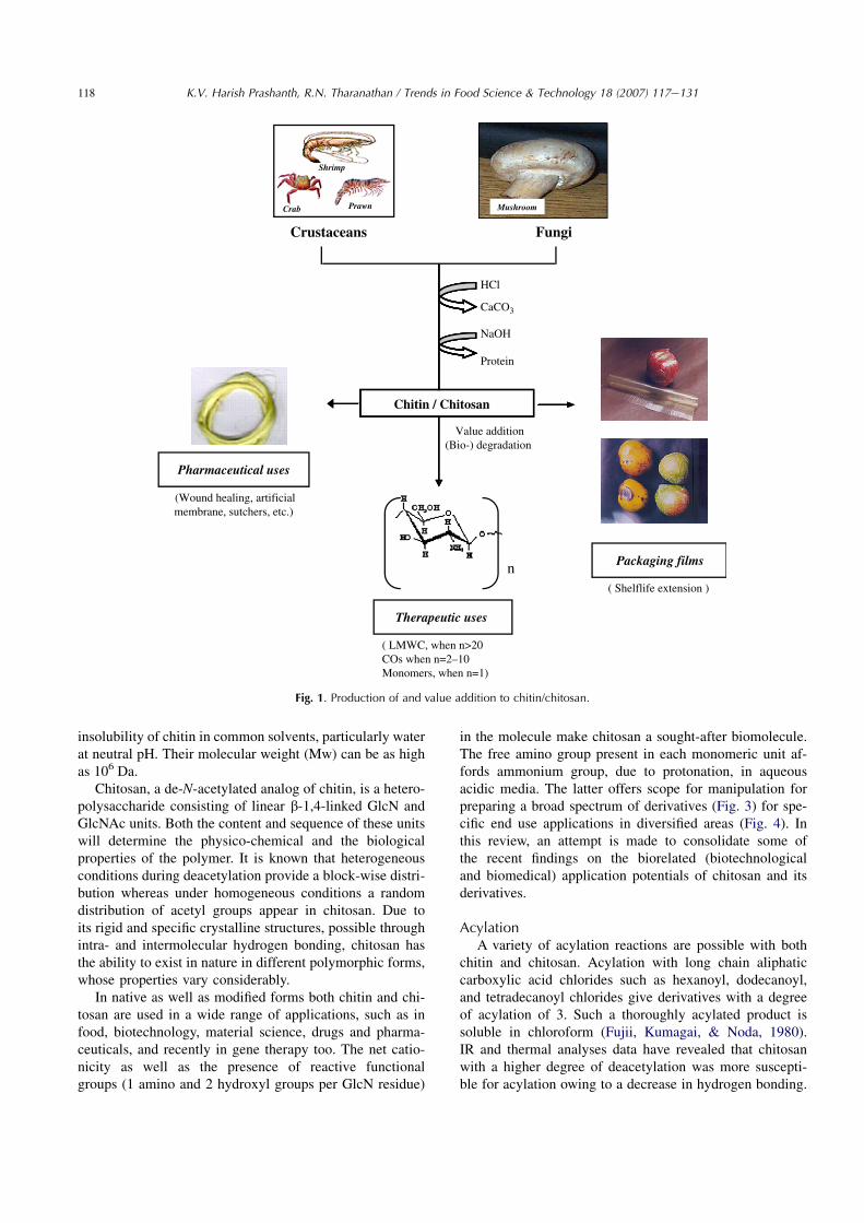
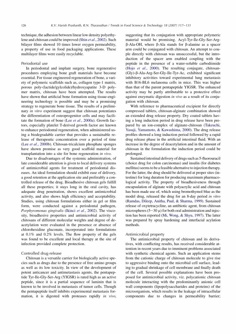
ing natural biopolymers for diversified applications in lifescience is increasing (Tharanathan, 2003). The present dayfolks, being better educated and well informed, prefer togo for organic/natural products, be it a food or a drug,and are prepared to pay a premium price for anything nat-ural and safe. In this regard natural biopolymers have sev-eral advantages, such as availability from replenishableagricultural or marine food resources, biocompatibility,biodegradability, therefore leading to ecological safetyand the possibility of preparing a variety of chemicallyor enzymatically modified derivatives for specific enduses. Polysaccharides, as a class of natural macromole-cules, have the tendency to be extremely bioactive andare generally derived from agricultural feedstock or crusta-cean shell wastes (Ramesh & Tharanathan, 2003). Cellu-lose, starch, pectin, etc. are the biopolymers derivedfrom the former, while chitin and chitosan are derivedfrom the latter. In terms of availability, chitin is next tocellulose, available to the extent of over 10 gigatons(1� 1013 kg). To a considerable extent chitin is also ob-tained from fungi and bacteria (see Fig. 1; Tharanathan& Kittur, 2003).
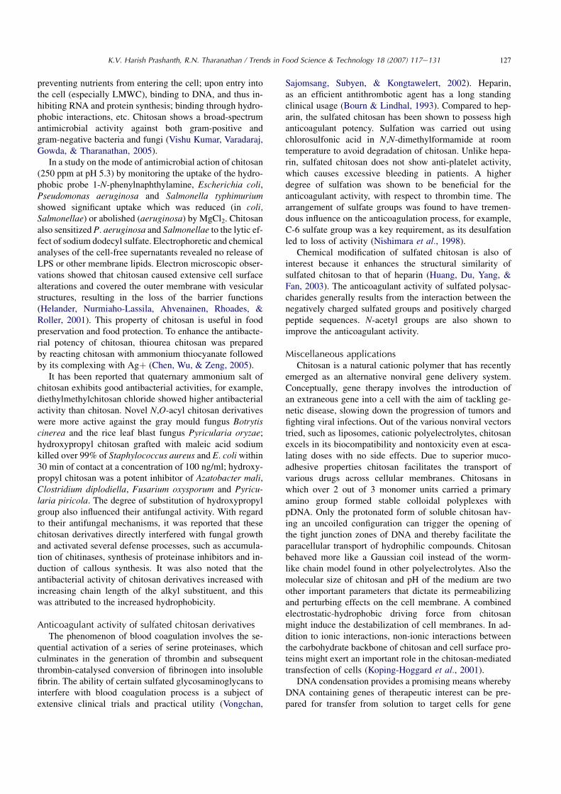
It is believed that both chitin and chitosan are linear co-polymers of D-GlcN and D-GlcNAc residues distributed ran-domly and not blocked together, the residues are linkedentirely in the b-1,4-configuration (Fig. 2); the various depo-lymerization products and structural and spectroscopic stud-ies provide ample proof for this. The b-1,4-configurationresults in a rigid and unbranched structure. The abundanceof hydroxyl groups (1 primary hydroxyl at C-6 and 1 second-ary hydroxyl at C-3) and highly reactive amino group (atC-2) or its N-acetyl counterpart (wholly in chitin) with con-comitant tendency for intra- and intermolecular hydrogenbonds results in the formation of linear aggregates with ex-tensive crystallinity. The latter contributes to the strengthshown by chitinous structures, and also to the virtual
118 K.V. Harish Prashanth, R.N. Tharanathan / Trends in Food Science & Technology 18 (2007) 117e131
Mushroom
Pharmaceutical uses
( LMWC, when n>20COs when n=2–10Monomers, when n=1)
n Packaging films
(Wound healing, artificialmembrane, sutchers, etc.)
Therapeutic uses
FungiCrustaceans
( Shelflife extension )
Value addition(Bio-) degradation
HCl
CaCO3
NaOH
Protein
Chitin / Chitosan
Shrimp
Crab Prawn
Fig. 1. Production of and value addition to chitin/chitosan.
insolubility of chitin in common solvents, particularly waterat neutral pH. Their molecular weight (Mw) can be as highas 106 Da.
Chitosan, a de-N-acetylated analog of chitin, is a hetero-polysaccharide consisting of linear b-1,4-linked GlcN andGlcNAc units. Both the content and sequence of these unitswill determine the physico-chemical and the biologicalproperties of the polymer. It is known that heterogeneousconditions during deacetylation provide a block-wise distri-bution whereas under homogeneous conditions a randomdistribution of acetyl groups appear in chitosan. Due toits rigid and specific crystalline structures, possible throughintra- and intermolecular hydrogen bonding, chitosan hasthe ability to exist in nature in different polymorphic forms,whose properties vary considerably.
In native as well as modified forms both chitin and chi-tosan are used in a wide range of applications, such as infood, biotechnology, material science, drugs and pharma-ceuticals, and recently in gene therapy too. The net catio-nicity as well as the presence of reactive functionalgroups (1 amino and 2 hydroxyl groups per GlcN residue)
in the molecule make chitosan a sought-after biomolecule.The free amino group present in each monomeric unit af-fords ammonium group, due to protonation, in aqueousacidic media. The latter offers scope for manipulation forpreparing a broad spectrum of derivatives (Fig. 3) for spe-cific end use applications in diversified areas (Fig. 4). Inthis review, an attempt is made to consolidate some ofthe recent findings on the biorelated (biotechnologicaland biomedical) application potentials of chitosan and itsderivatives.
AcylationA variety of acylation reactions are possible with both
chitin and chitosan. Acylation with long chain aliphaticcarboxylic acid chlorides such as hexanoyl, dodecanoyl,and tetradecanoyl chlorides give derivatives with a degreeof acylation of 3. Such a thoroughly acylated product issoluble in chloroform (Fujii, Kumagai, & Noda, 1980).IR and thermal analyses data have revealed that chitosanwith a higher degree of deacetylation was more suscepti-ble for acylation owing to a decrease in hydrogen bonding.

119K.V. Harish Prashanth, R.N. Tharanathan / Trends in Food Science & Technology 18 (2007) 117e131
O
H
H
NH
COH
H
O
OO
HH
H H
HO
HO
NH
CO
O
H
O
H
H
NH
COH
H
OO
HH
H H
HO
HO
NH
CO
O
H
O
H
H
NH
COH
H
OHO
H H H
4)-βD-GlcNAc-(1 4)-βD-GlcNAc-(1 4)-βD-GlcNAc-(1
4)-βD-GlcN-(1 4)-βD-GlcNAc-(1 4)-βD-GlcN-(1
O
H
H
CH2OH
H
H
O
OO
HH
H H
HO
HO
NH
CO
O
H
O
H
H
H
H
OO
HH
H H
HO
HO O
H
O
H
H
H
H
OHO
H H H
4)-βD-GlcN- (1
b
a
CH2OH
CH2OH
CH2OH
CH2OH
CH2OH
CH2OH
CH2OH
CH2OH
CH2OH
CH3
CH3
CH3
CH3
CH3
CH3
NH2
NH2
NH2NH2
Fig. 2. Primary structure of (a) chitin and (b) chitosan.
Acylation of chitosan with a cyclic ester (lactone) such asb-propiolactone or g-butyrolactone in an appropriate sol-vent gives derivatives having N-hydroxyalkanoyl groups(Loubaki, Sicsic, & Le Goffic, 1989).
Graft copolymerizationThe possibility of grafting synthetic polymers to chitin
and chitosan has attracted worldwide attention as a newand exciting way to modify and extend their use against
Chitin/Chitosan(Crustacean waste, Fungi)
Derivatization
Depolymerization
O-/N- Carboxyalkylation AcylationSulfation Schiff’s baseEnzymatic substitution Metal chelation Cyanoethylation Nitration, Phosphorylation
Crosslinking Graft copolymerization Polymer networks
Enzymatic
Chitinase/ChitosanaseNon-specific enzymes (Lipase,Protease, Lysozyme,Carbohydrases)
Substitution
Chain elongation
Physical
Chemical
Radiations (UV, γ)UltrasoundMicrowave Thermal treatments
Acids(HCl, HNO2, etc.) Free radicals(H2O2, K2S2O8)
Fig. 3. Multifaceted derivatization potential of chitin/chitosan.

120 K.V. Harish Prashanth, R.N. Tharanathan / Trends in Food Science & Technology 18 (2007) 117e131
Chitin/ChitosanBiorenewable Biocompatible BiodegradableBiofunctional
Food /Nutrition
MaterialScience
Medical Science(Drugs &
Pharmaceuticals)
Microbiological
Immunological
Miscellaneous
Food preservation, Water purification Biotechnology (immobilizationmatrix Dietary supplements, FunctionalfoodsHypocholesterolemic AntioxidantPrebiotics
HydrocolloidElectrochemistry (biosensors) Cosmetics (moisturizer, skin careproducts) Packaging films/Composite coatingformulations Textile finishing (dye binding)Polymeric membranes
HemostasisControlled drug release Oral hygeine, Periodontal use Antitumour, Antiulser Anticoagulant Wound healing, Wound dressing Suture threads, Contact lens
Antibacterial Antifungal
Biological response modifierGene therapeutics Polymeric scaffolds (for cell culture) Immuno potentiator
Agriculture (soil enrichment,increased crop production) Degradation products (low MWchitosan, chitooligomers, monomers)– value additionAnimal feedFlocculating agent Reverse osmosis membranes Polymeric nanoparticles Synthetic polymer blends
Fig. 4. Application potential of chitin/chitosan.
the rapidly growing competition from synthetic polymersthemselves. Research in this field has blossomed quicklyand is still an extremely active subject of study. In spite ofenormous efforts, there is still no large-scale commercial ap-plication of chitin/chitosan graft copolymers. Graft copoly-merization onto chitin and chitosan has not yet beenexplored extensively; it is a rapidly advancing field in poly-mer modification (Table 1; Kurita, 1997). Graft copolymer-ization reaction introduces side chains and makes variousmolecular designs possible, thus affording novel types of tai-lored hybrid materials composed of natural polysaccharidesand synthetic polymers. The properties of the graft co-polymers may be controlled by molecular structure, lengthand number of side chains attached. It is thus one of themost attractive approaches toward constructing versatilemolecular environments. Grafting behavior is generally dis-cussed in terms of grafting percentage, which is a ratio of the
Table 1. Types of chitosan-graft-copolymers
Type Initiator Monomers grafted
Radical-induced
Ce4þ, K2S2O8, Fenton’sreagent (Fe2þ þH2O2),tributyl borane
Acrylonitrile, N-isopropyl acrylamide,methyl methacrylate,vinyl monomers
Radiation-induced
g-rays, 60Co Styrene, 2-hydroxyethylmethacrylate
Microwaveirradiation
Polyacrylamide
Grafting ontomethod
Various catalysts areused [4,40-Azobis(4-cyanovaleric acid)]
Telechelic polymers,polyethylene glycol,poly-dimethyl siloxane
Dendronization (ReductiveN-alkylation)
Polyamidoamine,styrene

121K.V. Harish Prashanth, R.N. Tharanathan / Trends in Food Science & Technology 18 (2007) 117e131
weight of introduced side chains to the weight of the mainchain. Several types of chitosan-graft-copolymers withacrylic, vinyl, nonvinyl, etc. have been prepared for use asflocculants, paper binder-strengthener, slow-release drugcarrier, etc. Conventionally, such graft copolymerizationsare being carried out using a variety of redox systems (seeTable 1), although simultaneous homopolymerization lead-ing to low grafting yield is the major constraint.
Grafting onto chitinCerium(IV), sometimes used for grafting onto cellulose,
is also effective for grafting onto chitin. In its tetravalentstate, Ce(IV) is an excellent oxidizing agent; it forms aredox pair with the NH2 group of chitosan to give macro-radicals. Typical vinyl monomers such as acrylamide andacrylic acid were graft-copolymerized onto powdery chitinin aqueous suspensions. The amount of Ce(IV) affected thepolymerization markedly, and the grafting percentagesreached 240% and 200% with acrylamide and acrylicacid, respectively. The resulting graft copolymers showedimproved solubilities and swelling behaviors. Those havingpoly(sodium acrylate) side chains were soluble in dichloro-acetic acid, whereas those having polyacrylamide chainsswelled highly. Both the copolymers were much morehygroscopic compared to chitin (Kurita, Kawata, Koyama,& Nishimura, 1991).
Methylacrylate and methylmethacrylate (Ren, Miura,Nishi, & Tokura, 1993) were similarly graft-copolymerizedwith chitin using Ce(IV). The extent of swelling of chitin-graft-poly(methylmethacrylate) was dependent on thegrafting percentage. At a grafting percentage of above400%, the swelling became pronounced to give transparentgels from which films could be cast. The glass transitiontemperature of the copolymer was 130 �C as determinedby differential scanning calorimetry (Ren et al., 1993).Grafting of methylmethacrylate onto chitin was also possi-ble with tributylborane in water, although the grafting effi-ciency was not that high. Redox initiators could also beused for grafting (Yazdani-Pedram & Retuert, 1992).
g-ray irradiation on powdery chitin initiates the poly-merization of styrene as in the case of cellulose but thegrafting percentage is low. Water is essential for initiation(Kurita & Inoue, 1989). On addition of a Lewis acid suchas SnCl4 or TiCl4 to iodo-chitin in nitrobenzene, reactivecarbenium species were formed, with which styrene was ef-ficiently graft-copolymerized by a cationic mechanism inthe swollen state (Kurita, 2001). The grafting percentagewas up to 800%. The grafted side chains could be isolatedfrom the copolymers by hydrolytic cleavage of the chitinmain chain with hydrochloric acid to elucidate the graftingbehavior. The polystyrene liberated from the graft copoly-mer with a grafting percentage of 650% was characterizedfor Mn, Mw and Mw/Mn values of 58,000 Da, 87,000 Daand 1.50, respectively. The values of grafting percentageand Mn indicated that one polystyrene chain was attachedto one out of every 44 GlcNAc units on an average. The
resulting chitin-graft-polystyrene was nearly soluble inaprotic polar solvents such as DMSO and DMAc contain-ing 5% LiCl when the grafting percentage was above100%. They showed considerable swelling even in commonlow boiling organic solvents.
Another candidate for the polymeric radical initiator toprepare well-defined graft copolymers is 6-mercapto-chitin(Kurita, Yoshino, Nishimura, & Ishii, 1993). Although mer-capto-chitin is not soluble, it is expected to efficiently initi-ate graft copolymerization owing to the presence of readilydissociating mercapto groups and its swelling in organic sol-vents. Styrene was copolymerized onto mercapto-chitin effi-ciently in DMSO at 80 �C, and the grafting percentage wasalmost 100%. Similarly, methylmethacrylate was graft-copolymerized onto mercapto-chitin efficiently in DMSOat 80 �C to give chitin-graft-poly(methylmethacrylate).The grafting percentage increased with the amount of mono-mer and was above 120% under appropriate conditions. Theresulting graft-copolymers exhibited remarkable affinity forvarious common organic solvents (Kurita, 2001).
Grafting onto chitosanGraft copolymerization onto chitosan has also been at-
tempted by various methods (Jalal Zohuriaan-Mehr, 2005;see Table 1), but it is performed typically with 2,20-azobisisobutyronitrile, Ce(IV), or a redox system. Vinyl mono-mers such as acrylonitrile, methylmethacrylate, methyla-crylate and vinylacetate were graft copolymerized ontochitosan with 2,20-azobisisobutyronitrile in aqueous aceticacid solution or in aqueous suspension. The graftingpercentages were generally low. The chitosan-graft-poly(vinylacetate) was converted into chitosan-graft-poly(viny-lalcohol) by hydrolysis (Blair, Guthrie, Law, & Turkington,1987). Ce(IV) is also a suitable initiator, for graft copoly-merization of polyacrylamide, poly(acrylic acid), andpoly(4-vinylpyridine) with chitosan (Yilmaz, Hasopoglu,Caner, & Yilmaz, 1998). Fenton’s reagent (Fe2þ/H2O2),as a redox initiator, graft-copolymerized methylmethacry-late with chitosan. Here, the reaction between the Fe2þ
and H2O2 leads to free OH�
radicals, which in turn createsmacroradicals on the polymer backbone by means of hy-drogen abstraction. This facilitates grafting. Use of Fe2þ
is advantageous, as it favours binding to chitosan, whichin turn facilitates better yield of radicals, and minimizesany homopolymerization. In addition to higher yield ofgrafting, the reaction takes place at much lowertemperatures.
Potassium persulphate (KPS) is another potential freeradical initiator, wherein the grafting reaction takesplace by two step single electron transfer mechanism fol-lowed by propagation. At 60 �C, KPS actually undergoesthermal degradation generating anionic persulphate freeradicals, which then attack the cationic amino groups(NH3
þ) at C-2 of the GlcN moiety, ultimately resulting inglycosidic bond cleavage, leading to the formation (HarishPrashanth & Tharanathan, 2004; Hsu, Don, & Chiu, 2002)

122 K.V. Harish Prashanth, R.N. Tharanathan / Trends in Food Science & Technology 18 (2007) 117e131
of water-soluble low molecular weight chitosan (LMWC)and chitooligosaccharides (COs). The molecular weightof LMWC was found to be w37 kDa and HPLC analysisof the chitooligomeric fraction showed pentamer, hexamerand higher oligomers. The effect of these degradation prod-ucts on the growth of Ehrlich ascites tumor cells and tumor-induced neovascularization revealed COs (50 mg) to bemore effective compared to LMWC (100 mg) and provedthem to be potent angioinhibitory and antitumor com-pounds as shown by inhibition of angiogenesis and induc-ing apoptosis as a function of DNA fragmentation (HarishPrashanth & Tharanathan, 2004). These degradation prod-ucts also showed scavenging of OH
�and O
�2 radicals and of-
fered protection against calf thymus DNA damage (HarishPrashanth et al., unpublished data). Fluorescence studyshowed binding of LMWC in the minor groove, formingH-bonds to the backbone phosphates without distortingthe DNA double helix structure. It was also observed thatthe fragmented chitosan chains upon cooling at 4 �C over-night led to extensive crosslinking (radical-induced selfassociation), which was reported for the first time, andwhich involved several fragmented chains of depolymer-ized chitosan (Harish Prashanth & Tharanathan, 2006).In fact, secondary interactions such as hydrogen bridgesand hydrophobic interactions between acetylated and non-acetylated units of chitosan fragments lead to a higherdegree of crystallinity and as a result induce conformationalchanges in the molecule. Scanning electron micrograph ofcrosslinked chitosan revealed modification of fibrous nativechitosan to crystalline granule bundles. The latter washighly refractory for swelling and dissolution. The possibil-ity of exhibiting specific biofunctionalites by such cross-linked chitosan needs further investigations.
Recently, KPS-initiated graft-copolymerization of acry-lonitrile and methylmethacrylate (MMA) onto chitosanwas reported (Harish Prashanth & Tharanathan, 2003). Max-imum graft yield of C-g-PAN (249%) was obtained with0.12 M of acrylonitrile and 0.074 mM of KPS at 65 �C for2 h. C-g-PMMA, 0.14 M of MMA at 65 �C gave maximumgrafting (276%). No residual monomers were found byHPLC (Saroja, Gowda, & Tharanathan, 2000). The graft-copolymers could be thermopressed to thin membranousfilms, which were very fragile and brittle. In vitro biodegrad-ability tests on the graft-copolymer revealed preferential(bio-)degradation of the chitosan moiety, leaving the syn-thetic polymeric chains, undegraded (Harish Prashanth,Lakshman, Shamala, & Tharanathan, 2005).
Microwave irradiation, is yet another efficient source ofthermal energy that is used in a variety of modern syntheticreactions. Advantages of using microwave irradiation arethat the reaction could be achieved in a very short durationof time, the workup procedures are simple and the productyields are fairly good. Its utility in the context of green chem-istry has been widely accepted and appreciated for improvedselectivity and eco-friendly reaction conditions. The en-hancement of reaction rate under microwave irradiation is
explained on the basis of dielectric heating of the molecule,which involves rapid energy transfer from the functionalgroups (-OH and -NH2 groups) to reacting molecules. Possi-bly, such a high energy store may be responsible for thebreakage of covalent and glycosidic linkages between theGlcN residues. Microwave irradiation is also known to lowerGibbs energy of activation of the reaction, thus ultimately ef-fecting a free radical-type reaction mechanism. The graftedchitosan derivative was shown to be an efficient adsorbentfor Ca2þ and Zn2þ ions from effluent water.
Grafting polyethylene glycol (PEG) onto chitosan isa convenient approach to prepare water-soluble chitosanderivatives, to be used as a carrier of anticancer drugs. Inspite of their easy solubility, the PEG-g-chitosans, obtainedby the use of carbodiimide, were found to aggregate inaqueous medium, due to hydrogen bonding. Whereaswhen such derivatives were prepared via reductive alkyl-ation by PEG monoaldehydes, followed by further cross-linking, their water solubility was found restricted, andthey gradually reaggregated, finally becoming insoluble(Dal Pozzo et al., 2000). Such a property is highly usefulin wound dressings to prevent tissue adhesion in internalsurgery. Ideally, such materials are insoluble when posi-tioned in the surgical wound, but undergo progressive bio-erosion leading to complete resorption when they are nolonger needed. Based on choice of PEG substitution andcrosslinking, derivatives having high swelling capacitywith enhanced hydrophilicity can be obtained.
Aldehydes are generally used for protecting (via Schiff’sbase formation) the amino groups of chitosan and to allowhydroxyl groups to be modified. Glutaraldehyde, a dialde-hyde, is frequently used for the crosslinking of chitosan(Fig. 4). Crosslinking affects the permeability characteris-tics. Crosslinking through covalent or ionic interactions re-sults in the formation of chitosan hydrogels, which areuseful as drug delivery systems allowing the release of bio-active materials by diffusion, and also as permanent net-works, for example, as polymeric scaffolds in cell culture.
Depolymerization of chitin and chitosanThe very high molecular weight and therefore a very
high viscosity of chitosan precluded its use in several bio-logical applications. For some specific applications, morethan the chitosan, its degradation products, viz. LMWC,COs and monomers, were found to be much more useful.A variety of degradation methods, viz. chemical, physicaland enzymatic, are being worked out to generate these deg-radation products (Fig. 5). Both chitin and chitosan oligo-mers possess additional functional properties such asantitumor activity (Suzuki, 1996; Suzuki et al., 1986;Tsukada et al., 1990), immuno-enhancing effects, andenhancing protective effects against infection with somepathogens in mice (Tokora et al., 1989), antifungal (Hirano& Nagao, 1989; Kendra, Christian, & Hadwiger, 1989) andantimicrobial (Uchida, Izume, & Ohtakara, 1989) activities.Additionally, they have lower viscosity, low molecular

123K.V. Harish Prashanth, R.N. Tharanathan / Trends in Food Science & Technology 18 (2007) 117e131
O
O
O
HNH2 H CH2
OH
HO H
H
H
H
HNH
CH3-COH
HO
n
H
OO
OH
n
O
O
OO
HNH2 H
CH2
CH2
OH
HO
H
HH
H
HNH
CH3-C O
H
HO
n or n*
HO
OCH2
OH
H
H
OH
O
OHHO
NH
CH2
HO
H
HO
HO
HNH2
HO
H
H
H
H
HOH2C
H
H
H
OO
H
C
O
HOHH
Acid hydrolysis
Deamination
Heating
H+
HNO2
+ AcOH
GlcN
GlcN GlcNAc
OH CH2
OH
H
H
HNH
CH3-CO
H
HOHO
OH
GlcNGlcNAc
+ +
Enzymatic Chitos
anas
e
Chitooligomers (n = 2-3)Protease
PectinaseLipase
Chitooligomers (n = 2-8) and LMWC (n*,Mw = 5-20 kDa)
GlcN + GlcNAc +
+ N2 +
O
OHO
HNH2
OH
H
H
HO
OH
HO H
HCH2
2,5-Anhydromannitol oligomers
Chitosan
Microwave oven
O
O
O
O
HNH2 H CH2
OH
OH
HO
H
HH
H
HNH
CH3-CO
H
HO
n
HO
O
CH2
n
H
H
H
OH
O
OHHO
H
HO
H
H
H
H
H
Chitooligomers (n = 2-8) and LMWC(n*, Mw = 5-20 kDa)
O
O
OO
HNH2 H
CH2
CH2OH
OH
HO
H
HH
H
HNH
CH3-CO
H
HO
n/n*
H
HO
n
N = C – C – C – C – C = N
HH
H
H
H
H
HH
Crosslinked chitosan
O
H
H
H
H
OO
O
HH
H H
HOHO O
H
O
H
H
NH
H
OO
O
HH
HH
HO O
H
H
H
HO
Crosslinking
OHC(CH2)3CHO
GlcN + GlcNAc +
NH2CH2OH
NH2
CH2
CH2OH
CH2OH
CH2OH
Fig. 5. Possible depolymerization products and crosslinking of chitosan.
weights and short chain lengths and are soluble in neutralaqueous medium. Subsequently, they seem to be readilyabsorbed in vivo.
Depolymerization processChemical methods
Acid degradative methods are not specific, the hydroly-sis goes randomly generating a large amount of monomers,and later the removal of acid poses problems and also it is
not economical. Chemical treatment using strong acids(viz. nitrous acid and HCl) is a very common and fastmethod to produce a series of chitooligomers, but themethod has some disadvantages such as high cost, lowyield and residual acidity. Recently, LMWC have been pre-pared by salt-assisted acid hydrolysis under microwave ir-radiation (Xing et al., 2005). The mechanism is explainedas due to direct absorption of thermal energy by salt mole-cules, which causes localized superheating of solution.

124 K.V. Harish Prashanth, R.N. Tharanathan / Trends in Food Science & Technology 18 (2007) 117e131
Increase in the conductivity of the solution as well as di-electric loss and microwave coupling of the solvents havea dramatic influence on the rate of heating. The Mw of chi-tosan changed drastically in the presence of salt (from10� 104 to w3� 104 Da), and the composition of the elec-trolyte followed the order Kþ> Ca2þ>Naþ, which wasrelated to the ionic radius of the metals.
Depolymerization of chitosan by the use of nitrous acid(HNO2) is a homogeneous reaction where the number of gly-cosidic bonds broken is roughly stoichiometric to the amountof nitrous acid used (Allan & Peyron, 1995). The mechanisminvolves deamination of deacetylated glucosamine residues(D-units) forming 2,5-anhydro-D-mannose (M-units) at thenew reducing end. Since the latter is unstable, the standardprocedure has been to reduce to 2,5-anhydro-D-mannitolby the use of NaBH4. Nitrous acid induced depolymerizationhas been used previously to study the distribution of N-acetylated units in partially N-acetylated chitosan. It hasbeen found to be specific in the sense that HNO2 attacksthe amino group of D-units, with subsequent cleavage ofthe adjacent glycosidic linkage.
There have been very few reports on the degradation ofchitin or chitosan by free radicals. Nordtveit, Varum, andSmidsrod (1994) demonstrated that the viscosity of chito-san solution decreased rapidly in the presence of hydrogenperoxide (H2O2) and FeCl3, probably due to random depo-lymerization of chitosan. Tanioka et al. (1996) showed thatCu(II), ascorbate, and UVeH2O2 systems also gradually re-duced the molecular weight of chitosan. They postulatedthat the hydroxyl radicals generated in the experimentalsystem caused polymer degradation and that this phenome-non may help to explain the disappearance of chitosanin vivo during biomedical applications. There are severalother methods to degrade chitosan molecules includingthermal degradation and ultrasonic treatment (Allan &Peyron, 1995; Chen, Chang, & Shyru, 1997).
Physical methodsRadiation can provide a useful tool for degradation of
biological polymers and it is often viewed as being thelast process after packaging to control pathogenic andspoilage organisms. Recently, radiation effect on carbohy-drates such as chitosan, sodium alginate, carrageenan, cel-lulose, and pectin has been shown to enhance the use forrecycling these bioresources and reducing the environmen-tal pollution (Hien et al., 2000; Nagasawa, Mitomo, Yoshii,& Kume, 2000). The irradiation effect on chitosan in aceticacid solution with various dose rates and the yield of chito-san oligomers have been investigated (Choi, Ahn, Lee,Byun, & Park, 2002). In another method using 85% phos-phoric acid, low molecular weight chitosans were preparedby irradiation at different reaction temperatures and reac-tion time intervals, wherein the viscosity average molecularweights of chitosan decreased to 7.1� 104 from 21.4� 104
after 35 days of treatment (Jia & Shen, 2002).
Enzymatic methodsAlthough various degradation products of chitosan could
be produced by a variety of methods, enzymatic methodsare gaining importance because they allow regioselectivedepolymerization under mild conditions. Nevertheless, theundesirable level of pyrogenicity caused by the presenceof protein admixtures in such preparations cannot be dis-counted. In the case of enzymatic degradation of chitosan,LMWC with high water solubility were produced by chiti-nase, chitosanase, glucanase, lipase and some proteases(Pantaleone, Yalpani, & Scollar, 1992; Vishu Kumar,Gowda, & Tharanathan, 2004; Vishu Kumar, Varadaraj,Lalitha, & Tharanathan, 2004). Non-specific enzymes(Muzzarelli, 1997), including lysozyme, cellulase, lipase,amylase, papain and pectinase (Grigolon, Azevedo, Santos,& Franco, 2001; Nordtveit, Varum, & Smidsrod, 1996) thatare capable of depolymerizing chitosan are known, andamong these papain, a cysteine protease, is particularly at-tractive because of its plant origin, wide industrial use inmeat tenderization, use in medication for wound debride-ment, and its inhibition by human salivary cystatin.
Biological activity of LMWC and COsPartially depolymerized chitosans, with an average mo-
lecular weight of 10 kDa, seem to have enhanced biochem-ical significance compared to native chitosan. Theirsuperior antibacterial activity has been explained in termsof inhibition of the transcription from DNA (Liu, Guan,Yang, Li, & De Yao, 2001). LMWC modulated plant resis-tance to disease (Vasyukova et al., 2001), stimulated mu-rine peritoneal macrophages, showed antitumor activity(Seo et al., 2000) and were useful in functional food formu-lations (Jeon, Shahidi, & Kim, 2000). LMWC of 20 kDaprevents progression of diabetes mellitus and exhibitshigher affinity for lipopolysaccharide than chitosan ofMw 140 kDa (Hadwiger, Chiang, Victory, & Horovitz,1989; Kondo, Nakatani, Hayashi, & Ito, 2000).
D-Glucosamine oligosaccharides have long attractedmuch attention, as they have physiological functions ina great variety of living organisms, including induction ofphytoalexins (Hadwiger et al., 1989), hemostatic effects(Malette & Quigley, 1984) and antitumor activities (Toda,Shimoji, & Sasaki, 1987). It is thought that the greatestphysiological activities are shown by oligosaccharideswith a chain length greater than the pentasaccharide.Hexa-N-acetylchitohexaose [(GlcNAc)6] has immunopo-tentiating and antitumor functions (Suzuki et al., 1986).They inhibit the growth of fungi and phytopathogens(Kendra & Hadwiger, 1984) and elicit defense mechanismsin plants (Roby, Gadelle, & Toppan, 1987). They also affectthe mitogenic response and chemotactic activities of animalcells (Usami, Minami, Okamoto, Matsubashi, & Shige-masa, 1997). Their lipid binding (Ikeda et al., 1993) prop-erties make them useful ingredients for dietary and foodpreservation applications. LMWC was also shown to re-duce blood glucose and serum triglyceride levels in obese

125K.V. Harish Prashanth, R.N. Tharanathan / Trends in Food Science & Technology 18 (2007) 117e131
diabetic KK-Ay mice (Hayashi & Ito, 2002). It was re-ported that oligochitosans (Mw 1e3 and 3e5 kDa) preventoxidative stress in mice (Shon, Park, Moon, Chang, & Nam,2002).
Hypocholesterolemic activityDue to its beneficial plasma cholesterol level lowering
effect, which plays an important role in the alleviation andtreatment of cardiovascular diseases, chitosan has becomea useful dietary ingredient. The hypocholesterolemic actionof chitosan has been explained to be due to decreasedcholesterol absorption and interference with bile acid ab-sorption, a mechanism similar to those of dietary fiber con-stituents. The cholesterol-lowering action of oral chitosanhas been reported by many (Sugano, Watanabe, Kishi,Izumi, & Ohtakara, 1988), whereas chitin, although exhibit-ing higher excretion of triglycerides in feces, does notdisplay cholesterol-lowering action. Information regardingdigestion and absorption of chitin and chitosan in theGI tract is limited. In an in vivo study in canine GI tract, itwasshown that chitin did not under go any changes in weight andshape, whereas chitosan showed w10% decrease in weightand formed a film.
Enzyme immobilizationThe fact that an enzyme can coexist in various oligo-
meric forms is of major importance for its catalytic expres-sion. Enzyme immobilization is a technique of significantpractical utility, especially to enhance the catalytic poten-tial, resistance to pH and temperature, and continued reus-ability. It is known that chitosan is an excellent basematerial for immobilization of several carbohydrate degrad-ing enzymes, as it exhibits increased thermostability com-pared to the free enzyme. Urease has been immobilizedcovalently onto glutaraldehyde crosslinked chitosan mem-brane, especially to provide resistance to the influence ofinhibitors, such as boric acid, thioglycolic acid, sodiumfluoride and acetohydroxamic acid (Zaborska, 1995). Sim-ilarly, resistance to mechanical stirring of D-amino acidoxidase (a flavoprotein using FAD as cofactor) has beenprovided by enzyme immobilization on crosslinked chito-san matrix (Lemainque, Braun, & LeGoffie, 1988).
Antioxidant propertyThe trend to go for potent, naturally derived antioxidant
molecules over those of synthetic origin is ever increasing.To this class belong chitosan and several of its derivatives,which being safe and non-toxic offer protection from freeradicals, thus retarding the progress of numerous chronicdiseases (Tiwari, 2004). It is known that the antioxidant ef-fect of chitosan varies with its molecular weight and viscos-ity, as shown in cooked comminuted fish flesh modelsystems (Kamil, Jeon, & Shahidi, 2002; Xie, Xu, & Liu,2001). It is attributed to differences in the availability ofnet cationic amino groups in the molecule, which impartintermolecular electrostatic repulsive forces leading to
increase in the hydrodynamic volume of the extended chainconformation. The highly unsaturated fatty acids com-monly found in seafood are particularly sensitive to oxida-tive change during storage. Treatment of herring fishsamples with chitosan, however, showed lower peroxidevalues and total volatile aldehydes than the untreated sam-ples. The low viscosity chitosan showed the strongest anti-oxidative effect (Lin & Chou, 2004).
Wound healing propertyWound healing is a process for promoting rapid dermal
regeneration and accelerated wound healing. A novel asym-metric chitosan membrane consisting of skin surface on toplayer supported by a macroporous sponge-like sublayer hasbeen designed. The chitosan membrane showed controlledevaporative water loss, excellent oxygen permeability andpromoted fluid drainage ability, at the same time effectivelyinhibiting invasion of exogenous microorganisms (Mi et al.,2001). Wound covered with such membrane was hemo-static and healed quickly. Histopathological examinationconfirmed increased epithelialization rate as well as wellorganized deposition of collagen in the dermis. Chitosan-based wound dressing reduced scar tissue (fibroplasias)by inhibiting the formation of fibrin in wounds and it washemostatic and formed a protective film coating (Lloyd,Kennedy, Methacanon, Paterson, & Knill, 1998). Beinga substrate for lysozyme, chitosan degradation productswere internally absorbed, which in turn affected macro-phage activity.
Chitosan-alginate polyelectrolyte complexes, preparedin situ in beads and microspheres, caste as films showedgood wound dressing capability (Yan, Khor, & Lim,2000). Two chitosan films, viz. Chit-LA and Chit-AA,treated for wound healing efficiency, showed completewound closure, good epithelialization and no scar forma-tion (Khan & Peh, 2003). As an effective wound dressingagent with antibacterial properties, chitosan-cellulose blendmembranes have been prepared. They may protect woundsfrom excessive dehydration and infection (Wu et al., 2004).
MembranesChitosan and several of its innumerable derivatives have
the ability to form thin membranous films of use in packag-ing (Kittur, Kumar, & Tharanathan, 1998; Srinivasa,Baskaran, Ramesh, Harish Prashanth, & Tharanathan,2002; Srinivasa, Ramesh, Kumar, & Tharanathan, 2004),encapsulation and drug delivery systems. Due to drugepolymer interactions, high viscosity chitosan films showedbetter sustainable release; and the mechanism of releasefollowed Fiekian diffusion control with subsequent zeroorder release (Puttipipatkhachorn, Nunthanid, Yamamoto,& Peck, 2001). A novel organic (chitosan) and inorganic(tetraethyl orthosilicate) composite membrane has beenprepared, which is pH sensitive and drug permeable (Park,You, Park, Haam, & Kim, 2001). The latter possibly involvedionic interactions. By plasma source ion implantation
126 K.V. Harish Prashanth, R.N. Tharanathan / Trends in Food Science & Technology 18 (2007) 117e131
technique, the adhesion between linear low density polyethy-lene and chitosan could be improved (Shin et al., 2002). Suchbilayer films showed 10 times lower oxygen permeability,a property of use in food packaging applications. Thesemultilayer films were easily recyclable.
Periodontal useIn periodontal and implant surgery, bone regenerative
procedures employing bone graft materials have becomeessential. For tissue engineered regeneration of bone, a vari-ety of polymeric scaffolds such as, collagen type-1 matrix,porous poly-(lactide/gylcolide)/hydroxyapatite 3-D poly-mer matrix, chitosan have been attempted. The resultshave shown that artificial bone formation using tissue-engi-neering technology is possible and may be a promisingstrategy to regenerate bone tissue. The results of a prelimi-nary in vitro experiment suggest that chitosan potentiatesthe differentiation of osteoprogenitor cells and may facili-tate the formation of bone (Lee et al., 2000a). Growth fac-tors, especially platelet T-derived growth factor are knownto enhance periodontal regeneration, when administered us-ing a biodegradable carrier that provides a sustainable re-lease of therapeutic concentration over a period of time(Lee et al., 2000b). Chitosan-tricalcium phosphate spongeshave shown promise as very good scaffold material fortransplantation into a site for bone regeneration in vivo.
Due to disadvantages of the systemic administration, oflate considerable attention is given to local delivery systemsof antimicrobial agents for treatment of periodontal dis-eases. An ideal formulation should exhibit ease of delivery,a good retention at the application site and preferably a con-trolled release of the drug. Bioadhesive chitosan gels fulfillall these properties; it stays long in the oral cavity, hasadequate drug penetration, shows excellent antimicrobialactivity, and also shows high efficiency and acceptability.Studies, using chitosan formulations either in gel or filmform, were conducted against a periodontal pathogen,Porphyromonas gingivalis (Ikinci et al., 2002). The visco-sity, bioadhesive properties and antimicrobial activity ofchitosans of different molecular weights and degree of de-acetylation were evaluated in the presence or absence ofchlorohexidine gluconate, incorporated into formulationsat 0.1% and 0.2% levels. The flow property of the gelswas found to be excellent and local therapy at the site ofinfection provided complete protection.
Controlled drug-releaseChitosan is a versatile carrier for biologically active spe-
cies such as drugs due to the presence of free amino groupsas well as its low toxicity. In view of the development ofpotent anticancer and antimetastasis agents, the pentapep-tide Tyr-Ile-Gly-Ser-Arg (YIGSR) is rated high as an activepeptide, since it is a partial sequence of laminin that isknown to be involved in metastasis of tumor cells. Thoughthe pentapeptide itself inhibits experimental metastasis for-mation, it is digested with proteases rapidly in vivo,
suggesting that its conjugation with appropriate polymericmaterial would be promising. Acyl-Tyr-Ile-Gly-Ser-Arg-b-Ala-OH, where b-Ala stands for b-alanine as a spacerarm could be conjugated with chitosan. An attempt to cou-ple directly with chitosan was unsuccessful, but the intro-duction of the spacer arm enabled coupling with thepeptide in the presence of a water-soluble carbodiimide(Hojo et al., 2000). The resulting conjugate, chitosan-(Gly)-b-Ala-Arg-Ser-Gly-Ile-Tyr-Ac, exhibited significantinhibitory activities toward experimental lung metastasiswith B16-BL6 melanoma cells in mice. This was higherthan that of the parent pentapeptide YIGSR. The enhancedactivity may be partly attributable to a protective effectagainst enzymatic digestion in vivo as a result of its conju-gation with chitosan.
With reference to pharmaceutical excipient for directlycompressed tablets, chitosan-alginate combination showedan extended drug release property. Dry coated tablets hav-ing a long induction period in drug release have been pre-pared by an ion-complex of alginate-chitosan (Takeuchi,Yasuji, Yamamoto, & Kawashima, 2000). The drug releaseprofiles showed a long induction period followed by a rapiddrug release phase in the artificial intestinal fluid. With anincrease in the degree of deacetylation and in the amount ofchitosan in the formulation the induction period could beprolonged.
Sustained intestinal delivery of drugs such as 5-fluorouracil(choice drug for colon carcinomas) and insulin (for diabetesmellitus) seems to be a feasible alternative to injection therapy.For the latter, the drug should be delivered at proper sites (in-testine) for long duration for producing maximum pharmaco-logical activity. The property of bioadhesiveness throughencapsulation of alginate with polyacrylic acid and chitosanhas been made use of, which using bromothymol blue as themodel drug, released the drug for an 8-day period in vitro(Ramdas, Dileep, Anitha, Paul, & Sharma, 1999). Sustainedrelease of oxytetracycline, an antibiotic agent, from chitosanmicrospheres (5e30 m) for both oral administration and injec-tion has been reported (Mi, Wong, & Shyu, 1997). The latterwas prepared by spray hardening and interfacial acylationmethods.
Antimicrobial propertyThe antimicrobial property of chitosan and its deriva-
tives, with conflicting results, has received considerable at-tention in recent years due to imminent problems associatedwith synthetic chemical agents. Such an application stemsfrom the cationic charge of chitosan molecule to give riseto aggressive binding onto the microbial cell surface, lead-ing to gradual shrinkage of cell membrane and finally deathof the cell. Several possible explanations have been pro-posed for antimicrobial activity, viz. polycationic chitosanmolecule interacting with the predominantly anionic cellwall components (lipopolysaccharides and proteins) of themicroorganism, which results in the leakage of intracellularcomponents due to changes in permeability barrier;
127K.V. Harish Prashanth, R.N. Tharanathan / Trends in Food Science & Technology 18 (2007) 117e131
preventing nutrients from entering the cell; upon entry intothe cell (especially LMWC), binding to DNA, and thus in-hibiting RNA and protein synthesis; binding through hydro-phobic interactions, etc. Chitosan shows a broad-spectrumantimicrobial activity against both gram-positive andgram-negative bacteria and fungi (Vishu Kumar, Varadaraj,Gowda, & Tharanathan, 2005).
In a study on the mode of antimicrobial action of chitosan(250 ppm at pH 5.3) by monitoring the uptake of the hydro-phobic probe 1-N-phenylnaphthylamine, Escherichia coli,Pseudomonas aeruginosa and Salmonella typhimuriumshowed significant uptake which was reduced (in coli,Salmonellae) or abolished (aeruginosa) by MgCl2. Chitosanalso sensitized P. aeruginosa and Salmonellae to the lytic ef-fect of sodium dodecyl sulfate. Electrophoretic and chemicalanalyses of the cell-free supernatants revealed no release ofLPS or other membrane lipids. Electron microscopic obser-vations showed that chitosan caused extensive cell surfacealterations and covered the outer membrane with vesicularstructures, resulting in the loss of the barrier functions(Helander, Nurmiaho-Lassila, Ahvenainen, Rhoades, &Roller, 2001). This property of chitosan is useful in foodpreservation and food protection. To enhance the antibacte-rial potency of chitosan, thiourea chitosan was preparedby reacting chitosan with ammonium thiocyanate followedby its complexing with Agþ (Chen, Wu, & Zeng, 2005).
It has been reported that quaternary ammonium salt ofchitosan exhibits good antibacterial activities, for example,diethylmethylchitosan chloride showed higher antibacterialactivity than chitosan. Novel N,O-acyl chitosan derivativeswere more active against the gray mould fungus Botrytiscinerea and the rice leaf blast fungus Pyricularia oryzae;hydroxypropyl chitosan grafted with maleic acid sodiumkilled over 99% of Staphylococcus aureus and E. coli within30 min of contact at a concentration of 100 ng/ml; hydroxy-propyl chitosan was a potent inhibitor of Azatobacter mali,Clostridium diplodiella, Fusarium oxysporum and Pyricu-laria piricola. The degree of substitution of hydroxypropylgroup also influenced their antifungal activity. With regardto their antifungal mechanisms, it was reported that thesechitosan derivatives directly interfered with fungal growthand activated several defense processes, such as accumula-tion of chitinases, synthesis of proteinase inhibitors and in-duction of callous synthesis. It was also noted that theantibacterial activity of chitosan derivatives increased withincreasing chain length of the alkyl substituent, and thiswas attributed to the increased hydrophobicity.
Anticoagulant activity of sulfated chitosan derivativesThe phenomenon of blood coagulation involves the se-
quential activation of a series of serine proteinases, whichculminates in the generation of thrombin and subsequentthrombin-catalysed conversion of fibrinogen into insolublefibrin. The ability of certain sulfated glycosaminoglycans tointerfere with blood coagulation process is a subject ofextensive clinical trials and practical utility (Vongchan,
Sajomsang, Subyen, & Kongtawelert, 2002). Heparin,as an efficient antithrombotic agent has a long standingclinical usage (Bourn & Lindhal, 1993). Compared to hep-arin, the sulfated chitosan has been shown to possess highanticoagulant potency. Sulfation was carried out usingchlorosulfonic acid in N,N-dimethylformamide at roomtemperature to avoid degradation of chitosan. Unlike hepa-rin, sulfated chitosan does not show anti-platelet activity,which causes excessive bleeding in patients. A higherdegree of sulfation was shown to be beneficial for theanticoagulant activity, with respect to thrombin time. Thearrangement of sulfate groups was found to have tremen-dous influence on the anticoagulation process, for example,C-6 sulfate group was a key requirement, as its desulfationled to loss of activity (Nishimara et al., 1998).
Chemical modification of sulfated chitosan is also ofinterest because it enhances the structural similarity ofsulfated chitosan to that of heparin (Huang, Du, Yang, &Fan, 2003). The anticoagulant activity of sulfated polysac-charides generally results from the interaction between thenegatively charged sulfated groups and positively chargedpeptide sequences. N-acetyl groups are also shown toimprove the anticoagulant activity.
Miscellaneous applicationsChitosan is a natural cationic polymer that has recently
emerged as an alternative nonviral gene delivery system.Conceptually, gene therapy involves the introduction ofan extraneous gene into a cell with the aim of tackling ge-netic disease, slowing down the progression of tumors andfighting viral infections. Out of the various nonviral vectorstried, such as liposomes, cationic polyelectrolytes, chitosanexcels in its biocompatibility and nontoxicity even at esca-lating doses with no side effects. Due to superior muco-adhesive properties chitosan facilitates the transport ofvarious drugs across cellular membranes. Chitosans inwhich over 2 out of 3 monomer units carried a primaryamino group formed stable colloidal polyplexes withpDNA. Only the protonated form of soluble chitosan hav-ing an uncoiled configuration can trigger the opening ofthe tight junction zones of DNA and thereby facilitate theparacellular transport of hydrophilic compounds. Chitosanbehaved more like a Gaussian coil instead of the worm-like chain model found in other polyelectrolytes. Also themolecular size of chitosan and pH of the medium are twoother important parameters that dictate its permeabilizingand perturbing effects on the cell membrane. A combinedelectrostatic-hydrophobic driving force from chitosanmight induce the destabilization of cell membranes. In ad-dition to ionic interactions, non-ionic interactions betweenthe carbohydrate backbone of chitosan and cell surface pro-teins might exert an important role in the chitosan-mediatedtransfection of cells (Koping-Hoggard et al., 2001).
DNA condensation provides a promising means wherebyDNA containing genes of therapeutic interest can be pre-pared for transfer from solution to target cells for gene

128 K.V. Harish Prashanth, R.N. Tharanathan / Trends in Food Science & Technology 18 (2007) 117e131
therapy applications. In addition to synthetic condensationagents (PEG), multivalent cations (chitosan) may also facil-itate binding. Such chitosan-based transfection systems areadvantageous because of non-immunogenicity, lack of bio-hazards and the possibility of introducing larger DNA frag-ments into targets over viral vectors in gene therapy.However, the high molecular weight of chitosan precludedits usage because of toxicity problems. Nevertheless, theLMWCs are neither toxic nor haemolytic and they are shownto form complexes with DNA and protect against nucleasedegradation, thereby validating LMWC as components ofa synthetic gene delivery system (Tiwari, 2004).
Chitosan and its amino acid derivatives (poly D, L-lacticacid) have been explored as an extracellular matrix-like sur-face to promote cell adhesion and growth (Zhu et al., 2002).Four kinds of chitosan-amino acid derivatives were pre-pared to minimize the carbohydrate moieties of cell matrixglycoproteins. From detailed cell cultural studies these chi-tosan derivatives were shown to promote chondrogenesis.
Chitosan nanoparticles are shown to enhance oral bio-availability and intestinal absorption of peptide and proteinformulations. By ionotropic gelation of chitosan with tripoly-phophate anions insulin-loaded nanoparticles have been pre-pared (Pan et al., 2002). Enhanced intestinal absorption aswell as relative increase in the pharmacological bioavailabil-ity of insulin was investigated by monitoring the plasma glu-cose level of alloxan-induced diabetic rats. The nanoparticle,having a size in the range of 250e400 nm and polydispersityindex<0.1, positively charged and remaining stable, showedinsulin association of over 80% and its in vitro releaseshowed a great initial burst with a pH-sensitivity. It showedan increased absorption of insulin in vivo (w15%), and thehyperglycemia was prolonged for over 15 h.
Chitosan-poly(acrylic) acid polyionic complexes havebeen prepared for prolonged gastric antibiotic delivery(Torrado, Prada, de la Torre, & Torrado, 2004). Differentpolyionic complexes of amoxicillin, chitosan and polya-crylic acid were prepared and employing a non-invasivemethod the gastric residence time of the formulations wasevaluated, by means of 13C-octanoic acid breath test. Allthe complexes showed extensive swelling, and diffusion ofthe antibiotic was controlled by the degree of polymer-drug interaction.
In the construction of heart valve substitutes, bovinepericardium fixed in buffered glutaraldehyde is presentlybeing used. Calcification limits the durability of such heartvalve substitutes. As an alternative, crosslinking of bioma-cromolecules with glutaraldehyde was tried, which createsvoid spaces in the fiber matrix leading to exposure of poten-tial binding sites for calcification. By hydrogen peroxidedegradation LMWC (2000 Da) was prepared for couplingonto polymer grafted glutaraldehyde crosslinked pericar-dial tissue to prevent calcification in rat subcutaneousmodel (Shanthi & Panduranga Rao, 2001).
The capacity to preconcentrate anions has allowed use ofchitosan and its derivatives in modified electrodes, for
application in sensor and biosensor electrochemistry(Rodrigues, Laranjeira, Stadler, & Drago, 2000).
To facilitate improving water solubility of biologicallyuseful chitosan derivatives, N-methylene phosphonic chito-san has been prepared using a one step reaction that allowedhomogeneous modifications (Heras, Rodrigues, Ramos, &Agullo, 2001). The resulting NH2-CH2-PO3
2� combines itsstrong donor effect with a monodentate ligand as PO3
2�,thus increasing its metal-binding properties, especially forcalcium. The derivative also shows good filmogenic nature.
Phosphated chitin (P-chitin) has been used as an anti-inflammatory agent in a mice model of chitosan-inducedacute respiratory distress syndrome (Khanal et al., 2001).The interstitial pneumonia was thus successfully blockedby a simultaneous intravenous injection of P-chitin. Intrave-nous infusion of some P-chitin formulations dramaticallyreduced lung injury and diminished the accumulation ofneutrophils in the interstitial and alveolar spaces of thelungs. P-chitin with a Mw of 24000, DS of 58% and DAof 4% was found to be most effective in the prevention ofpneumonia.
Attempts have been made to develop new types of antiHIV-1 reagents having different inhibitory mechanismsfrom those of nucleoside analogs, which are toxic, showside effects and are not effective against drug-resistantstrains (Nishimara et al., 1998). A regioselective sulfationof C-2 and/or C-3 groups of chitin has been developedemploying 6-0-trityl chitosan, as a protected intermediate.Sulfation at both 2 and 3 positions showed the highest in-hibitory effect, whereas sulfation at C-6 seems to decreasethe anti HIV-1 activity, but nevertheless the latter showedhigh anticoagulant activity. These new inhibitors of retro-virus infection showed both low cytotoxicity and low anti-coagulant activity.
Chitooligosaccharides stimulate purportedly beneficialgut species (Bifidobacterium and Lactobacillus sp.) openingup the possibility of them acting as prebiotics. Despite thisproperty, in a mixed culture system no increase in the bifiduscounts was observed (Vernazza, Gibson, & Rastall, 2005).Nevertheless, through pure culture studies chitooligosac-charides were shown to be stimulatory to Bifidobacterium bi-fidum and Lactobacillus sp., in low concentrations they led toincreased cell numbers and showed prebiotic effects (Lee,Park, Jung, & Shin, 2002). In vitro studies have shown thatchitooligosaccharides can bind 4 to 5 times its weight of mi-cellar lipids, leading to claims on some brands of slimmingpills for blocking fat absorption, and therefore of use inobesity control (Nauss, Thompson, & Nagyvary, 1983).
Final remarksIn brief, it is very much evident that chitin/chitosan and
their modified derivatives exhibit an unlimited applicationpotential for use in a wide range of faculties. The recentparadigm shift from synthetic packaging materials to biode-gradable packaging films made out of biobased polymersopens up a lot of opportunities for chitosan-derived

129K.V. Harish Prashanth, R.N. Tharanathan / Trends in Food Science & Technology 18 (2007) 117e131
packaging films. Such films are advantageous because oftheir user-friendly and eco-friendly characteristics, andalso the raw materials are generally derived from replenish-able natural resources. A continuous but sustainable use ofsuch hydrocolloid-based biodegradable packaging films au-gurs minimizing plastic wastes, whose volume (over 30%of total wastes generated) and disposal are causing newchallenges and serious threats for the generations tocome. The annual per capita utilization of plastics in Indiais w2 kg/person/year compared to 60 kg/person/year in de-veloped countries. Although plastic packaging films costless (w$2 per kg), chitosan as a raw material is priced atw$15 per kg, whereas other biopolymers (e.g., corn starchand cellulose at w$ 0.8e1.8 per kg) are much cheaper.Nevertheless, for some specialized applications (pharma-ceutical and therapeutic) such premium prices for biode-gradable, safe packaging are well tolerated. In the area ofnanoscience, the use of chitosan and its derivative is im-mense. The biopolymer chitosan is especially very usefulas it can be made available in a variety of morphologies in-cluding fibers, films, hydrogels, membranes, nanoparticlesand microspheres. Although chitosan has received theGRAS (generally recognized as safe) status by the Foodand Drug Administration, USA, its full-fledged usage infood formulations (functional foods) needs official clear-ance. For the latter, additional studies to address regulatoryissues of concern need to be done to substantiate unequiv-ocally the various health-related claims put forth already.Further basic and application oriented studies are expectedto fully utilize the potential of chitin/chitosan for still addi-tional uses and for the benefit of the society.
AcknowledgementsThe authors wish to thank Mrs. Savitha and Ms. Shobha
for their excellent help in typesetting the manuscript.KVHP thanks the Council of Scientific and IndustrialResearch, New Delhi, for the award of a Senior ResearchFellowship.
References
Allan, G. G., & Peyron, M. (1995). Molecular weight manipulationof chitosan I: kinetics of depolymerization by nitrous acid.Carbohydrate Research, 277, 257e272.
Blair, H. S., Guthrie, J., Law, T. K., & Turkington, P. (1987). Chitosanand modified chitosan membranes I. Preparation and character-ization. Journal of Applied Polymer Science, 33, 641e656.
Bourn, M. C., & Lindhal, V. (1993). Glucosaminoglycans and theregulation of blood coagulation. The Biochemical Journal, 289,313e330.
Chen, R. H., Chang, J. R., & Shyru, J. S. (1997). Effects of ultrasoniccondition and storage in acidic solutions on changes in molecularweight and polydispersity of treated chitosan. CarbohydrateResearch, 299, 287e294.
Chen, S., Wu, G., & Zeng, H. (2005). Preparation of high antimicrobialactivity thiourea chitosaneAgþ complex. Carbohydrate Polymers,60, 33e38.
Choi, W. S., Ahn, K. J., Lee, D. W., Byun, M. W., & Park, H. J. (2002).Preparation of chitosan oligomers by irradiation. PolymerDegradation and Stability, 78, 533e538.
Dal Pozzo, A., Vanini, L., Fagnoni, M., Guerrini, A., De Benedittis, M.,& Muzzarelli, R. A. A. (2000). Preparation and characterization ofpoly(ethylene glycol) crosslinked reacetylated chitosans.Carbohydrate Polymers, 42, 201e206.
Fujii, S., Kumagai, H., & Noda, M. (1980). Preparation of poly(acyl)-chitosans. Carbohydrate Research, 83, 389e393.
Grigolon, L. B., Azevedo, A., Santos, R. R., & Franco, T. T. (2001).Enzymatic modification of chitosan by free and immobilizedpapain. In R. A. A. Muzzarelli (Ed.), Chitin enzymology(pp. 78e87). Italy: Atec.
Hadwiger, L. A., Chiang, C., Victory, S., & Horovitz, D. (1989). Themolecular biology of chitosan in plant/pathogen interaction and itsapplication in agriculture. In G. Skjak-Braek, T. Anthonsen, &P. Sandford (Eds.), Chitin and chitosan (pp. 119e138). New York:Elsevier Applied Science.
Harish Prashanth, K. V., Lakshman, Kshama, Shamala, T. R., &Tharanathan, R. N. (2005). Biodegradation of chitosan-graft-polymethylmethacrylate films. International Biodeteriorationand Biodegradation, 56, 115e120.
Harish Prashanth, K. V., & Tharanathan, R. N. (2003). Studies ongraft copolymerization of chitosan with synthetic monomers.Carbohydrate Polymers, 54, 343e351.
Harish Prashanth, K. V., & Tharanathan, R. N. (2004). Depolymerizedproducts of chitosan as potent inhibitors of tumor induced angio-genesis. Biochimica et Biophysica Acta, 1117, 22e29.
Harish Prashanth, K. V., & Tharanathan, R. N. (2006). Crosslinkedchitosandpreparation and characterization. CarbohydrateResearch, 341, 169e173.
Hayashi, K., & Ito, M. (2002). Antidiabetic action of low molecularweight chitosan in genetically obese diabetic KK-Ay mice.Biological and Pharmaceutical Bulletin, 25, 188e192.
Helander, I. M., Nurmiaho-Lassila, E. L., Ahvenainen, R., Rhoades, J.,& Roller, S. (2001). Chitosan disrupts the barrier properties of theouter membrane of gram-negative bacteria. International Journal ofFood Microbiology, 71, 235e244.
Heras, A., Rodrigues, N. M., Ramos, V. M., & Agullo, E. (2001).N-methylene phosphonic chitosanda novel soluble derivative.Carbohydrate Polymers, 44, 233e238.
Hien, N. Q., Nagasawa, N., Tham, L. X., Yoshi, F., Dang, V. H.,Mitomo, H., et al. (2000). Growth-promotion of plants withdepolymerized alginates by irradiation. Radiation Physics andChemistry, 59, 97e101.
Hirano, S., & Nagao, N. (1989). Effects of chitosan, pecticacid, lysozyme, and chitinase on the growth of severalphytopathogens. Agricultural and Biological Chemistry, 53,3065e3066.
Hojo, K., Maeda, M., Mu, Y., Kamada, H., Tsutsumi, Y., Nishiyama, Y.,et al. (2000). Facile synthesis of a chitosan hybrid of a laminin-related peptide and its antimetastatic effect in mice. The Journalof Pharmacy and Pharmacology, 52, 67e73.
Hsu, S. C., Don, T. M., & Chiu, W. Y. (2002). Free radical degradationof chitosan with potassium persulphate. Polymer Degradation andStability, 75, 73e83.
Huang, R., Du, Y., Yang, J., & Fan, L. (2003). Influence of functionalgroups on the in vitro anticoagulant activity of chitosan sulfate.Carbohydrate Research, 338, 483e489.
Ikeda, I., Sugano, M., Yoshida, K., Sasaki, E., Iwamoto, Y., &Hatano, K. (1993). Effects of chitosan hydrolysates on lipidabsorption and on serum and liver lipid concentration in rats.Journal of Agricultural and Food Chemistry, 41, 432e435.
Ikinci, G., Senai, S., Akincibay, H., Kas, S., Ercis, S., Wilson, C. G.,et al. (2002). Effect of chitosan on a periodontal pathogen Por-phyromonas gingivalis. International Journal of Pharmaceutics,235, 121e127.

130 K.V. Harish Prashanth, R.N. Tharanathan / Trends in Food Science & Technology 18 (2007) 117e131
Jalal Zohuriaan-Mehr, M. (2005). Advances in chitin and chitosanmodification through graft co-polymerization: a comprehensivereview. Iranian Polymer Journal, 14, 235e265.
Jeon, Y. J., Shahidi, R., & Kim, S. K. (2000). Preparation of chitin andchitosan oligomers and their applications in physiologicalfunctional foods. Food Reviews International, 16, 159e176.
Jia, Z., & Shen, D. (2002). Effect of reaction temperature and reactiontime on the preparation of low molecular weight chitosan usingphosphoric acid. Carbohydrate Polymers, 49, 393e396.
Kamil, J., Jeon, Y. J., & Shahidi, F. (2002). Antioxidant activity ofchitosans of different viscosity in cooked comminuted flesh ofherring (Clupea harengus). Food Chemistry, 79, 69e77.
Kendra, D. F., Christian, D., & Hadwiger, L. A. (1989). Chitosanoligomers from Fusarium solani/pea interactions, chitinase/b-glucanase digestion of sporelings and from fungal wall chitinactively inhibit fungal growth and enhance disease resistance.Physiological and Molecular Plant Pathology, 35, 215e230.
Kendra, D. F., & Hadwiger, L. A. (1984). Characterization of thesmallest chitosan oligomer that is maximally that is anti fungal toFusarium solani and elicits pisatin formation in Pisum sativum.Experimental Mycology, 8, 276e281.
Khan, T. A., & Peh, K. K. (2003). A preliminary investigation ofchitosan film as dressing for punch biopsy wounds in rats. Journalof Pharmaceutical Sciences, 6, 20e26.
Khanal, D. P., Okamoto, Y., Miyatake, K., Shinobu, T., Shigemase, Y.,Tokura, S., et al. (2001). Protective effects of phosphated chitin(P-chitin) in a mice model of acute respiratory distress syndrome(ARDS). Carbohydrate Polymers, 44, 99e106.
Kittur, F. S., Kumar, K. R., & Tharanathan, R. N. (1998). Functionalpackaging properties of chitosan films. Zeitschrift fur Lebensmittel-Untersuchung und -Forschung, 206, 44e47.
Kondo, Y., Nakatani, A., Hayashi, K., & Ito, M. (2000). Low molecularweight chitosan prevents the progression of low dose streptozoto-cin induced slowly progressive diabetes mellitus in mice.Biological and Pharmacological Bulletin, 23, 1458e1464.
Koping-Hoggard, M., Tubulekas, I., Guan, H., Edwards, K.,Nilsson, M., Varum, K. M., et al. (2001). Chitosan as a non-viralgene delivery system. Structure-property relationships andcharacteristics compared with polyethylenimine in vitro andafter lung administration in vivo. Gene Therapy, 8, 1108e1121.
Kurita, K. (2001). Controlled functionalization of the polysaccharidechitin. Progress in Polymer Science, 26, 1921e1971.
Kurita, K. (1997). In M. F. A. Goosen (Ed.), Application of chitin andchitosan (pp. 297e315). Lancaster, PA: Technomic Publishing.
Kurita, K., & Inoue, S. (1989). Preparation of iodo-chitins and graftcopolymerization on to the derivatives. In G. Skjak Braek,T. Anthonsen, & P. Sandford (Eds.), Chitin and chitosan(pp. 365e371). New York: Elsevier Applied Science.
Kurita, K., Kawata, M., Koyama, Y., & Nishimura, S. (1991). Graftcopolymerization of vinyl monomers onto chitin with cerium (IV)ion. Journal of Applied Polymer Science, 42, 2885e2891.
Kurita, K., Yoshino, H., Nishimura, S., & Ishii, S. (1993). Preparationand biodegradability of chitin derivatives having mercapto groups.Carbohydrate Polymers, 20, 239e245.
Lee, H. W., Park, Y. S., Jung, J. S., & Shin, W. S. (2002). Chitosan oli-gosaccharides, dp 2-8, have prebiotic effects on the Bifidobacte-rium bifidium and Lactobacillus species. Anaerobe, 8, 319e324.
Lee, Y. M., Park, Y. J., Lee, S. J., Ku, Y., Han, S. B., Choi, S. M.,et al. (2000a). Tissue engineered bone formation using chito-san/tricalcium phosphate sponges. Journal of Periodontology,71, 410e417.
Lee, Y. M., Park, Y. J., Lee, S. J., Ku, Y., Han, S. B., Klokkevold, P. Y.,et al. (2000b). The bone regenerative effect of platelet-derivedgrowth factor-BB delivered with a chitosan/tricalcium phosphatesponge carrier. Journal of Periodontology, 71, 418e424.
Lemainque, A., Braun, J., & LeGoffie, F. (1988). Influence of poly-merization of D- amino acid oxidase on the behaviour of the
enzymedimmobilized on chitosan by covalent fixation. EuropeanJournal of Biochemistry, 174, 171e176.
Lin, H. Y., & Chou, C. C. (2004). Antioxidative activities of watersoluble disaccharides of chitosan derivatives. Food ResearchInternational, 37, 883e889.
Liu, X. F., Guan, Y. L., Yang, D. Z., Li, Z., & De Yao, K. (2001).Antibacterial action of chitosan and carboxymethylated chitosan.Journal of Applied Polymer Science, 79, 1324e1335.
Lloyd, L. L., Kennedy, J. F., Methacanon, P., Paterson, M., & Knill, C. J.(1998). Carbohydrate polymers as wound management aids.Carbohydrate Polymers, 37, 315e322.
Loubaki, E., Sicsic, S., & Le Goffic, F. (1989). Chemical modificationof chitosan by glycidyl trimethylammonium chloride. EuropeanPolymer Journal, 25, 397e400.
Malette, W. G., & Quigley Jr., H. J. (1984). Chitosan as hemostatics.US Pat. 4,452,785. Chemical Abstracts, 101, 78864.
Mi, F. L., Shyu, S. S., Wu, Y. B., Lee, S. T., Shyong, J. Y., & Huang, R. N.(2001). Fabrication and characterization of a sponge-like asym-metric chitosan membrane as a wound dressing. Biomaterials, 22,165e173.
Mi, F. L., Wong, T. B., & Shyu, S. S. (1997). Sustained-release ofoxytetracycline from chitosan microspheres prepared by interfacialacylation and spray hardening methods. Journal of Microencap-sulation, 14, 577e591.
Muzzarelli, R. A. A. (1997). Depolymerization of chitins and chitosanswith hemicellulase, lysozyme, papain and lipase. InR. A. A. Muzzarelli, & M. G. Peter (Eds.), Chitin hand book(pp. 153e165). Italy: Atec.
Nagasawa, N., Mitomo, H., Yoshii, F., & Kume, T. (2000). Radiation-induced degradation of sodium alginate. Polymer Degradation andStability, 69, 279e285.
Nauss, J. L., Thompson, J. L., & Nagyvary, J. (1983). The binding ofmicellar lipids to chitosan. Lipids, 18, 714e719.
Nishimara, S. I., Kai, H., Shinada, K., Yoshida, T., Tokura, S., Kurita, K.,et al. (1998). Regioselective syntheses of sulfated polysaccharides:specific anti-HIV-1 activity of novel sulfates. CarbohydrateResearch, 306, 427e433.
Nordtveit, R. J., Varum, K. M., & Smidsrod, O. (1994). Degradation offully water soluble, partially N-acetylated chitosans with lysozyme.Carbohydrate Polymers, 23, 253e260.
Nordtveit, R. J., Varum, K. M., & Smidsrod, O. (1996). Degradation ofpartially N-acetylated chitosans with hen egg white and humanlysozyme. Carbohydrate Polymers, 29, 163e167.
Pan, Y., Li, Y. J., Zhao, H. Y., Zheng, J. M., Xu, H., Wei, G., et al.(2002). Bioadhesive polysaccharide in protein delivery system:chitosan nanoparticles improve the intestinal absorption of insulinin vivo. International Journal of Pharmaceutics, 249, 139e147.
Pantaleone, D., Yalpani, M., & Scollar, M. (1992). Unusualsusceptibility of chitosan to enzymatic hydrolysis. CarbohydrateResearch, 237, 325e332.
Park, S. B., You, J. O., Park, H. Y., Haam, S. J., & Kim, W. S. (2001). Anovel pH-sensitive membrane from chitosan-TEOS IPN: prepara-tion and its drug permeation characteristics. Biomaterials, 22,323e330.
Puttipipatkhachorn, S., Nunthanid, J., Yamamoto, K., & Peck, G. E.(2001). Drug physical state and drug-polymer interaction on drugrelease from chitosan matrix films. Journal of Controlled Release,7, 143e153.
Ramdas, M., Dileep, K. J., Anitha, Y., Paul, W., & Sharma, C. P. (1999).Alginate encapsulated bioadhesive chitosan microspheres forintestinal drug delivery. Journal of Biomaterials Applications, 13,292e296.
Ramesh, H., & Tharanathan, R. N. (2003). Carbohydratesdtherenewable raw materials of high biotechnological value.Critical Reviews in Biotechnology, 23, 149e173.
Ren, L., Miura, Y., Nishi, N., & Tokura, S. (1993). Modification of chitinby ceric salt-initiated graft polymerization-preparation of

131K.V. Harish Prashanth, R.N. Tharanathan / Trends in Food Science & Technology 18 (2007) 117e131
poly(methylmethacrylate) graft chitin derivatives that swell inorganic solvents. Carbohydrate Polymers, 21, 23e27.
Roby, D., Gadelle, A., & Toppan, A. (1987). Chitin oligosaccharides aselicitors of chitinase activity in melon plants. Biochemical andBiophysical Research Communications, 143, 885e892.
Rodrigues, C. A., Laranjeira, M. C. M., Stadler, E., & Drago, V. (2000).Preparation of the pentacyanoferrate (II) on the surface ofN-(4-pyridilmethylidene) chitosan. Carbohydrate Polymers, 41,311e314.
Saroja, N., Gowda, L. R., & Tharanathan, R. N. (2000). Chromato-graphic determination of residual monomers in starch-g-polyacryIonitrile and starch-g-polyacrylate. Chromatographia, 51,345e348.
Seo, W. G., Pae, H. O., Kim, N. Y., Ot, G. S., Park, I. S., Kim, Y. H., et al.(2000). Synergistic cooperation between water-soluble chitosanoligomers and interferon gamma for induction of nitric oxide syn-thesis and tumoricidal activity in murine peritoneal macrophages.Cancer Letters, 159, 189e195.
Shanthi, C., & Panduranga Rao, K. (2001). Chitosan modified poly(glycidylmethacrylate-butyl acrylate) copolymers in grafted bovinepericardial tissuedanticalcification properties. CarbohydratePolymers, 44, 123e131.
Shin, G. H., Lee, Y. H., Lee, J. S., Kim, Y. S., Choi, W. S., & Park, H. J.(2002). Preparation of plastic and biopolymer multiplayer films byplasma source ion implantation. Journal of Agricultural and FoodChemistry, 50, 4608e4614.
Shon, Y. H., Park, I. K., Moon, I. S., Chang, H. W., & Nam, K. S. (2002).Effect of chitosan oligosaccharides on 2,3,7,8-tetrachlorodibenzo-p-dioxin-induced oxidative stress in mice. Biological andPharmaceutical Bulletin, 25, 1161e1164.
Srinivasa, P. C., Baskaran, R., Ramesh, M. N., Harish Prashanth, K. V.,& Tharanathan, R. N. (2002). Storage studies of mango packedusing biodegradable chitosan films. European Food Research andTechnology, 215, 504e508.
Srinivasa, P. C., Ramesh, M. N., Kumar, K. R., & Tharanathan, R. N.(2004). Properties of chitosan films prepared under different dryingconditions. Journal of Food Engineering, 63, 79e85.
Sugano, M., Watanabe, S., Kishi, A., Izumi, M., & Ohtakara, A. (1988).Hypocholesterolemic action of chitosan with different viscosity inrats. Lipids, 23, 187e191.
Suzuki, K., Mikami, T., Okawa, Y., Tokora, A., Suzuki, S., & Suzuki, M.(1986). Antitumor effect of hexa-N-acetylchitohexaose and chito-hexaose. Carbohydrate Research, 151, 403e408.
Suzuki, S. (1996). Studies on biological effects of water soluble lowerhomologous oligosaccharides of chitin and chitosan. FragranceJournal, 15, 61e68.
Takeuchi, H., Yasuji, T., Yamamoto, H., & Kawashima, Y. (2000). Spray-dried lactose composite particles containing an ion complex ofalginate-chitosan for designing a dry-coated tablet having a time-controlled releasing function. Pharmaceutical Research, 17, 94e99.
Tanioka, S., Matsui, Y., Irie, T., Tanigawa, T., Tanaka, Y., Shibata, H., et al.(1996). Oxidative depolymerization of chitosan by hydroxyl radical.Bioscience, Biotechnology, and Biochemistry, 60, 2001e2004.
Tharanathan, R. N. (2003). Biodegradable films and compositecoatingsdpast, present and future. Trends in Food Science &Technology, 14, 71e78.
Tharanathan, R. N., & Kittur, F. S. (2003). Chitindthe undisputedbiomolecule of great potential. Critical Reviews in Food Scienceand Nutrition, 43, 61e87.
Tiwari, A. K. (2004). Antioxidants: new generation therapeutic base fortreatment of polygenic disorders. Current Science, 86, 1092e1102.
Toda, M., Shimoji, K., & Sasaki, J. (1987). Preparation of glucosaminederivatives as immunostimulants and antitumor agents. Eur Pat226,381(1987). Chemical Abstracts, 107, 237216.
Tokora, A., Kobayashi, M., Tatekawa, N., Suzuki, K., Okawa, Y.,Mikami, T., et al. (1989). Protective effect of N-acetylchitohexaose
on Listeria monocytogenes infection in mice. Microbiology andImmunology, 33, 357e367.
Torrado, S., Prada, P., de la Torre, P. M., & Torrado, M. (2004).Chitosan-poly(acrylic) acid polyionic complex: in vivo studyto demonstrate prolonged gastric retention. Biomaterials, 25,917e923.
Tsukada, K., Matsumoto, T., Aizawa, K., Tokoro, A., Naruse, R.,Suzuki, S., et al. (1990). Antimetastatic and growth inhibitoryeffects of N-acetylchitohexaose in mice bearing Lewis lungcarcinoma. Japanese Journal of Cancer Research, 81, 259e265.
Uchida, Y., Izume, M., & Ohtakara, A. (1989). Preparation of chitosanoligomers with purified chitosanase and its application. InG. Skjak-Braek, T. Anthonsen, & P. Sandford (Eds.), Chitin andchitosan (pp. 373e382). New York: Elsevier Applied Science.
Usami, Y., Minami, S., Okamoto, Y., Matsubashi, A., & Shigemasa, Y.(1997). Influence of chain length of N-acetyl-D-glucosamine andD-glucosamine residues on direct and complement mediatedchemotactic activites for canine polymorphonuclear cells.Carbohydrate Polymers, 32, 115e122.
Vasyukova, N. I., Zinoveva, S. V., Illinskaya, L. I., Perekhod, E. A.,Chalenko, G. I., Gerasimova, N. G., et al. (2001). Modulation ofplant resistance to diseases by water-soluble chitosan. AppliedBiochemistry and Microbiology, 37, 103e109.
Vernazza, C. L., Gibson, G. R., & Rastall, R. A. (2005). In vitro fer-mentation of chitosan derivatives by mixed cultures of humanfaecal bacteria. Carbohydrate Polymers, 60, 539e545.
Vishu Kumar, A. B., Gowda, L. R., & Tharanathan, R. N. (2004).Non-specific depolymerization of chitosan by pronase andcharacterization of the resultant products. European Journal ofBiochemistry, 217, 713e723.
Vishu Kumar, A. B., Varadaraj, M. C., Gowda, L. R., & Tharanathan, R. N.(2005). Characterization of chitooligosaccharides prepared by chi-tosanolysis with the aid of papain and pronase, and their bactericidalaction. The Biochemical Journal, 391, 167e175.
Vishu Kumar, A. B., Varadaraj, M. C., Lalitha, R. G., &Tharanathan, R. N. (2004). Low molecular weight chitosans:preparation with the aid of papain and characterization.Biochimica et Biophysica Acta, 1670, 137e146.
Vongchan, P., Sajomsang, W., Subyen, D., & Kongtawelert, P. (2002).Anticoagulant activity of a sulphated chitosan. CarbohydrateResearch, 337, 1239e1242.
Wu, Y. B., Yu, S. H., Mi, C. W., Shyu, S. S., Pong, C. K., & Chao, A. C.(2004). Preparation and characterization of mechanical and anti-bacterial properties of chitosan/cellulose blends. CarbohydratePolymers, 57, 435e440.
Xie, W., Xu, P., & Liu, Q. (2001). Antioxidant activity of water solublechitosan derivatives. Bioorganic and Medicinal Chemistry Letters,11, 1699e1701.
Xing, R., Lius, S., Yu, H., Gao, Z., Wang, P., Li, C., et al. (2005). Salt-assisted acid hydrolysis of chitosan to oligomers under microwaveirradiation. Carbohydrate Research, 340, 2150e2153.
Yan, X., Khor, E., & Lim, L. Y. (2000). PEC films prepared fromchitosan-alginate coacervates. Chemical and PharmaceuticalBulletin, 48, 941e946.
Yazdani-Pedram, M., & Retuert, J. (1992). Homogeneous graftingreaction of vinyl pyrrolidone onto chitosan. Applied PolymerScience, 63, 1321e1326.
Yilmaz, E., Hasopoglu, H., Caner, H., & Yilmaz, O. (1998).Graft-copolymerization of 4-vinyl pyridine onto chitosan-1 byceric ion initiation. European Polymer Journal, 34, 493e497.
Zaborska, W. (1995). Competitive inhibitors of free and chitosanimmobilized urease. Acta Biochimica Polonica, 42, 115e118.
Zhu, H., Ji, J., Lin, R., Gao, C., Feng, L., & Shen, J. (2002). Surfaceengineering of poly(D, L-lactic acid) by entrapment of chitosanbased derivatives for the promotion of chondrogenesis. Journalof Biomedical Materials Research, 62, 532e539.