Embed Size (px)
Citation preview
Trematodes
Pathology and ParasitologyCourse Code: 401
Parasitology-lab # 2
1
Department of Microbiology & ImmunologyFaculty of Pharmacy
Cairo University
Helminthology
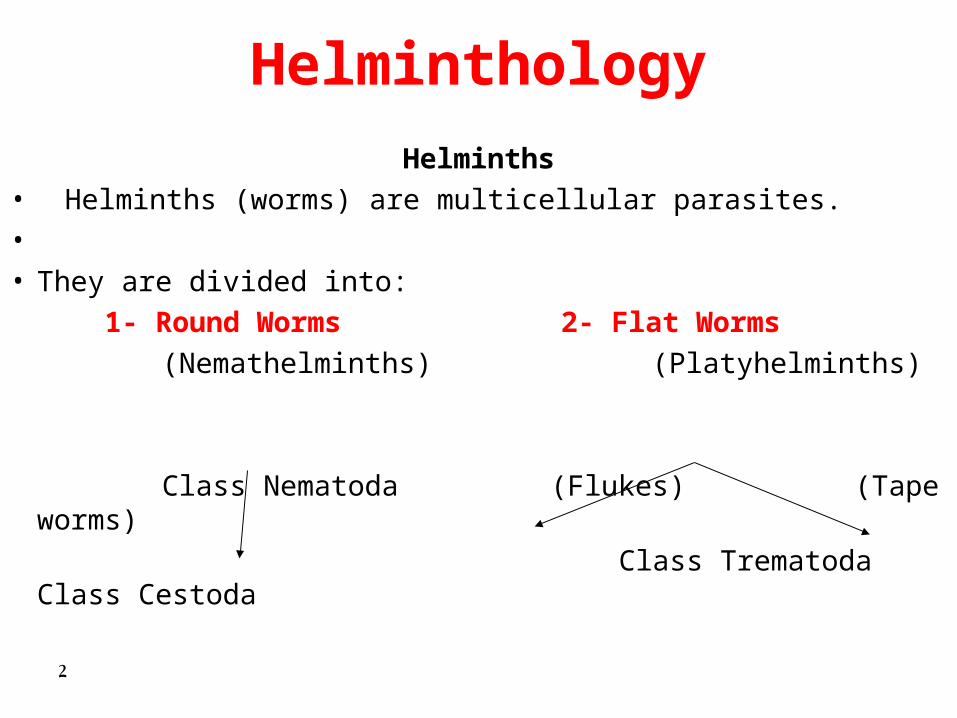
Helminths• Helminths (worms) are multicellular parasites.• • They are divided into:
1- Round Worms 2- Flat Worms
(Nemathelminths) (Platyhelminths)
Class Nematoda (Flukes) (Tape worms)
Class Trematoda Class Cestoda 2
Helminthology
Questions form:• Name of the parasite• Intermediate host• Infective stage• Mode of transmission• Location in the host• Lab diagnosis (Diagnostic stage)• Disease3

Class TrematodaClass Trematoda(Flukes)(Flukes)
4
General Characters
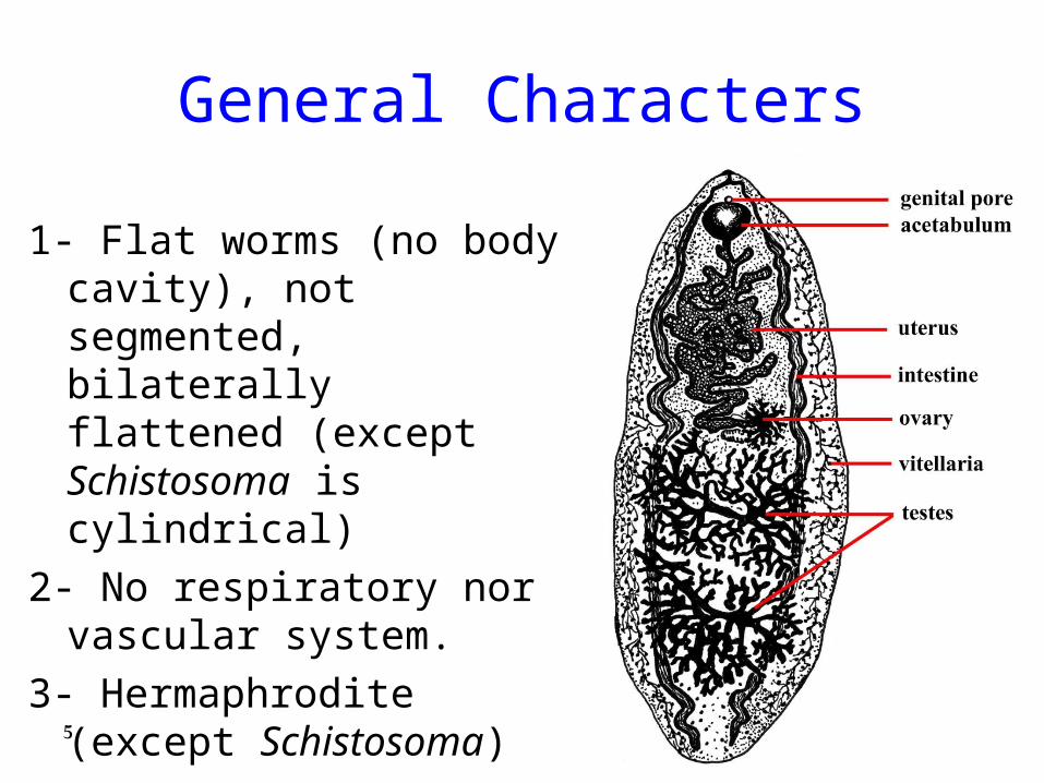
1- Flat worms (no body cavity), not segmented, bilaterally flattened (except Schistosoma is cylindrical)
2- No respiratory nor vascular system.
3- Hermaphrodite (except Schistosoma)
5
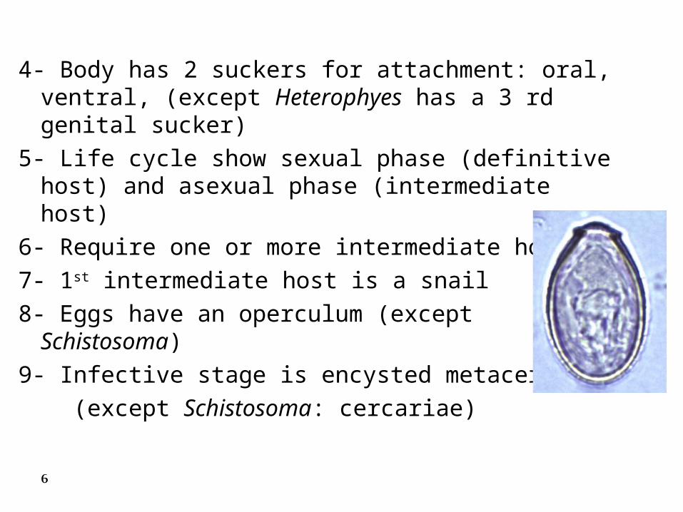
4- Body has 2 suckers for attachment: oral, ventral, (except Heterophyes has a 3 rd genital sucker)
5- Life cycle show sexual phase (definitive host) and asexual phase (intermediate host)
6- Require one or more intermediate host
7- 1st intermediate host is a snail
8- Eggs have an operculum (except Schistosoma)
9- Infective stage is encysted metacercaria
(except Schistosoma: cercariae)
6

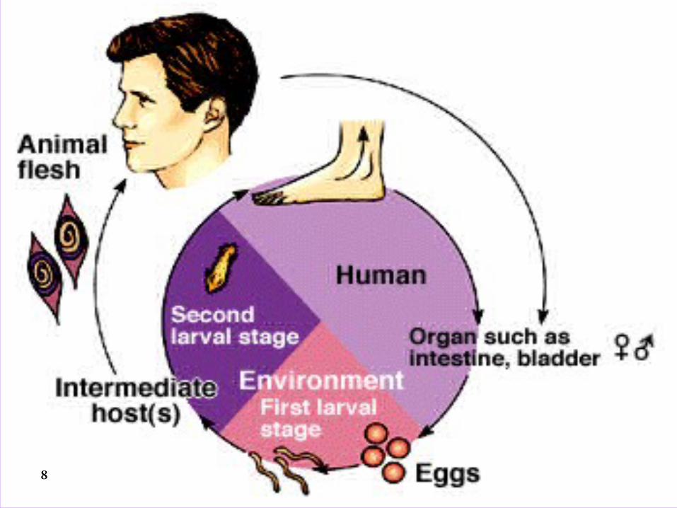
• General life cycle of trematodes1. Eggs are released from human (in feces or urine) in
fresh water.2. Eggs hatch into first larval stage (miracidial larva)
that invade a snail as first intermediate host.3. All trematodes except Schistosoma sp. require a
second intermediate host which is mostly a water plant or animal.
4. The second larval stage (cercarial or metacercarial larva) comes out of the intermediate host(s).
5. Humans are infected through direct penetration of cercaria (Schistosoma) or ingestion of encysted metacercaria in water plant or animal (all other trematodes). 7
8
1- Intestinal Fluke1- Intestinal Fluke Heterophyes heterophyes
• Heterophyes heterophyes adult
• Heterophyes heterophyes eggs
• Heterophyes heterophyes snail
• Heterophyes heterophyes encysted metacercaria
• Heterophyes heterophyes in small intestine
9
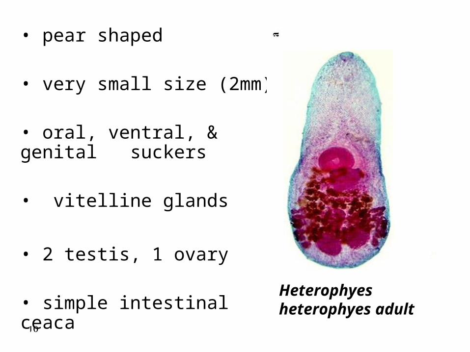
• pear shaped
• very small size (2mm)
• oral, ventral, & genital suckers
• vitelline glands
• 2 testis, 1 ovary
• simple intestinal ceaca Heterophyes heterophyes adult
10
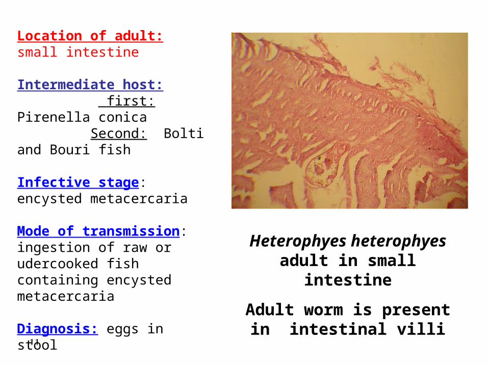
Heterophyes heterophyes adult in small intestine
Adult worm is present in intestinal villi
Location of adult: small intestine
Intermediate host: first: Pirenella conica Second: Bolti and Bouri fish
Infective stage: encysted metacercaria
Mode of transmission: ingestion of raw or udercooked fish containing encysted metacercaria
Diagnosis: eggs in stool
Disease: heterophiasis11
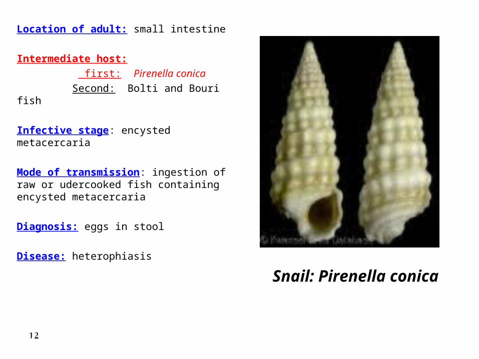
Location of adult: small intestine
Intermediate host:
first: Pirenella conica
Second: Bolti and Bouri fish
Infective stage: encysted metacercaria
Mode of transmission: ingestion of raw or udercooked fish containing encysted metacercaria
Diagnosis: eggs in stool
Disease: heterophiasisSnail: Pirenella conica
12
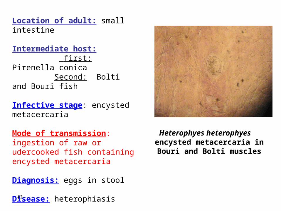
Heterophyes heterophyes encysted metacercaria in Bouri
and Bolti muscles
Location of adult: small intestine
Intermediate host: first: Pirenella conica Second: Bolti and Bouri fish
Infective stage: encysted metacercaria
Mode of transmission: ingestion of raw or udercooked fish containing encysted metacercaria
Diagnosis: eggs in stool
Disease: heterophiasis
13
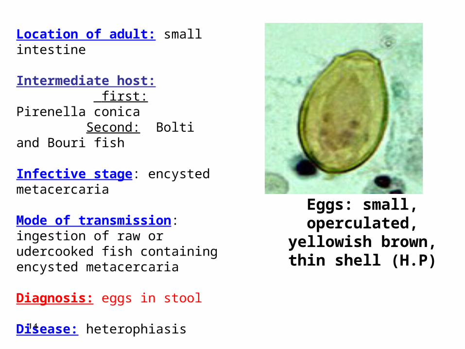
Eggs: small, operculated, yellowish brown, thin shell (H.P)
Location of adult: small intestine
Intermediate host: first: Pirenella conica Second: Bolti and Bouri fish
Infective stage: encysted metacercaria
Mode of transmission: ingestion of raw or udercooked fish containing encysted metacercaria
Diagnosis: eggs in stool
Disease: heterophiasis
14
• Location of adult: small intestine• Intermediate host:
First : Pirenella conica
Second: Bolti and Bouri fish• Infective stage: encysted metacercaria• Mode of transmission: ingestion of raw or
udercooked fish containing encysted metacercaria• Diagnosis: eggs in stool• Disease: heterophiasis
15
2- Liver Fluke2- Liver Fluke Fasciola
• Fasciola hepatica adult
• Fasciola gigantica adult
• Fasciola gigantica snail: (lymnaea cailliaudi )
• Fasciola egg
• Fasciola metacercaria
• Fasciola redia16

17
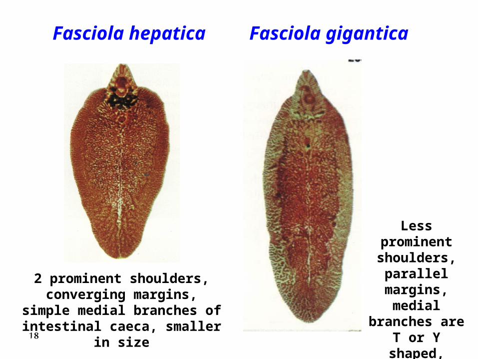
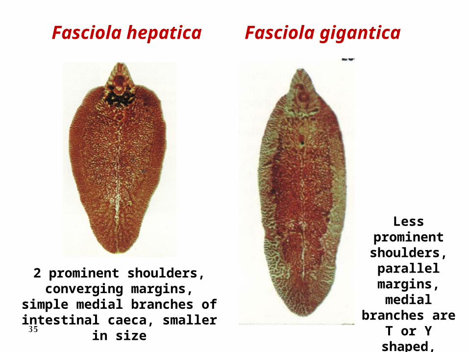
Fasciola hepatica Fasciola gigantica
2 prominent shoulders, converging margins, simple medial branches of intestinal
caeca, smaller in size
Less prominent shoulders,
parallel margins, medial branches
are T or Y shaped, larger
in size18
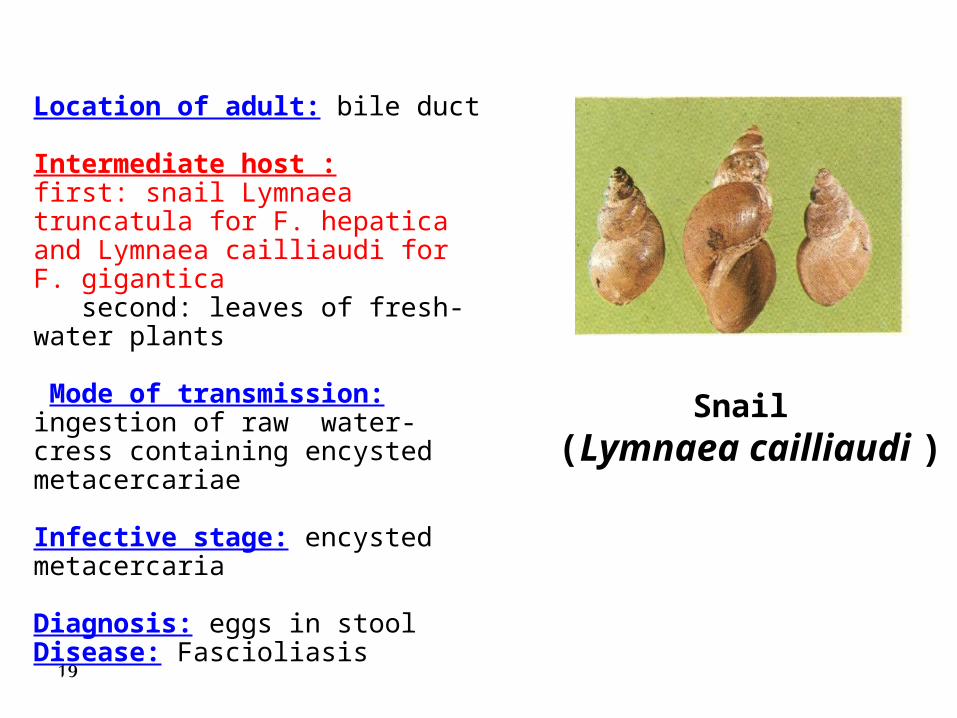
Snail (Lymnaea cailliaudi )
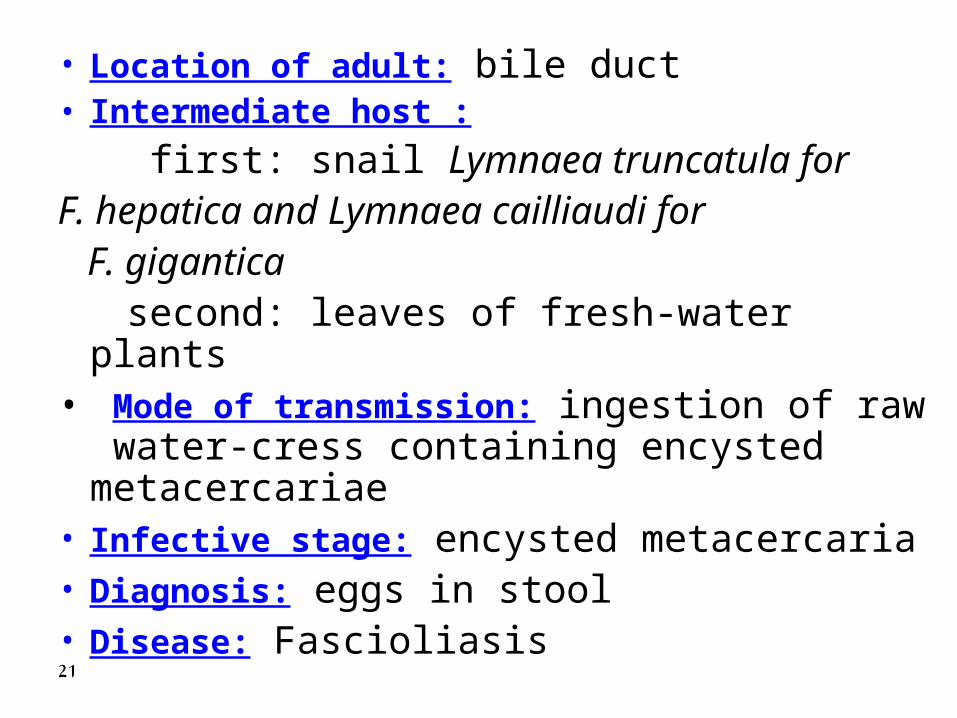
Location of adult: bile duct
Intermediate host :first: snail Lymnaea truncatula for F. hepatica and Lymnaea cailliaudi for F. gigantica second: leaves of fresh-water plants
Mode of transmission: ingestion of raw water-cress containing encysted metacercariae
Infective stage: encysted metacercaria
Diagnosis: eggs in stoolDisease: Fascioliasis
19
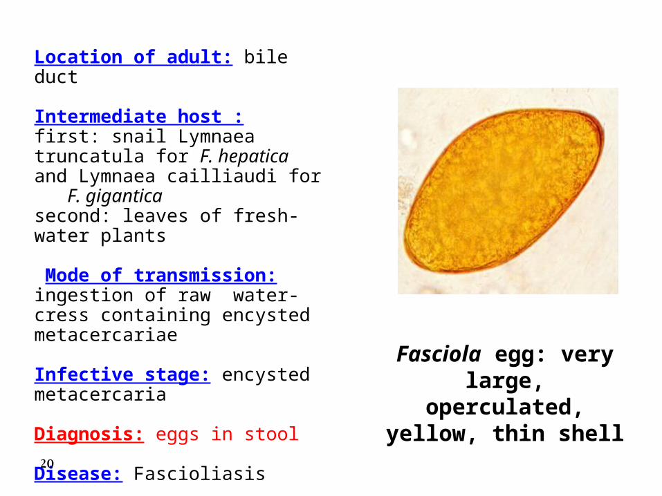
Fasciola egg: very large, operculated, yellow, thin shell
Location of adult: bile duct
Intermediate host :first: snail Lymnaea truncatula for F. hepatica and Lymnaea cailliaudi for F. giganticasecond: leaves of fresh-water plants
Mode of transmission: ingestion of raw water-cress containing encysted metacercariae
Infective stage: encysted metacercaria
Diagnosis: eggs in stool
Disease: Fascioliasis
20
• Location of adult: bile duct• Intermediate host :
first: snail Lymnaea truncatula for F. hepatica and Lymnaea cailliaudi for F. gigantica second: leaves of fresh-water plants• Mode of transmission: ingestion of raw
water-cress containing encysted metacercariae
• Infective stage: encysted metacercaria• Diagnosis: eggs in stool• Disease: Fascioliasis21
33 - -Blood FlukeBlood Fluke Schistosoma spp.
• Schistosoma mansoni male • Schistosoma mansoni female • Schistosoma mansoni male & female• Schistosoma haematobium male • Schistosoma haematobium female• Eggs of Schistosoma mansoni • Eggs of Schistosoma haematobium• Snail of Schistosoma mansoni• Snail of Schistosoma haematobium• S. mansoni cercaria 22
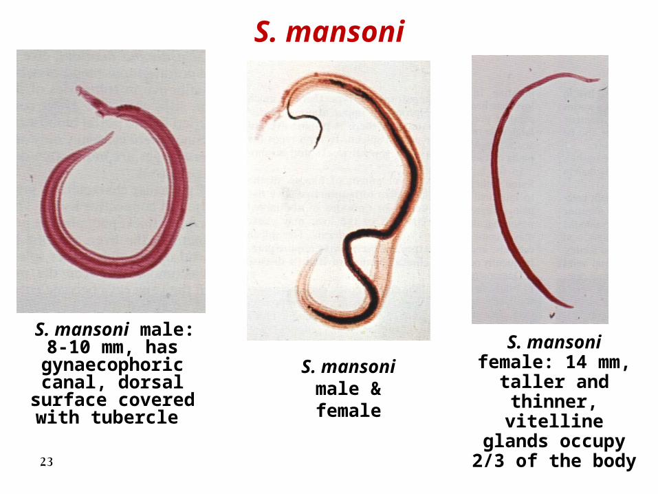
S. mansoni
S. mansoni male: 8-10 mm, has
gynaecophoric canal, dorsal
surface covered with tubercle
S. mansoni female: 14 mm, taller and thinner, vitelline
glands occupy 2/3 of the body
S. mansoni male & female
23
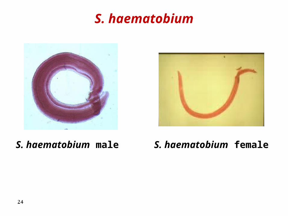
S. haematobium
S. haematobium male S. haematobium female
24
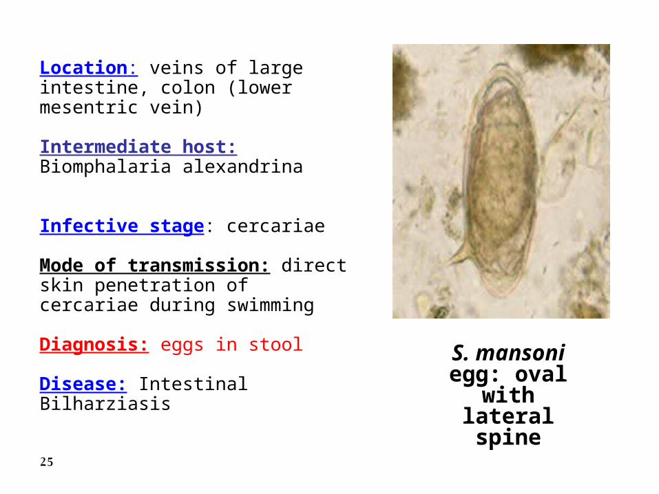
S. mansoni egg: oval with lateral spine
Location: veins of large intestine, colon (lower mesentric vein)
Intermediate host: Biomphalaria alexandrina
Infective stage: cercariae
Mode of transmission: direct skin penetration of cercariae during swimming
Diagnosis: eggs in stool
Disease: Intestinal Bilharziasis
25
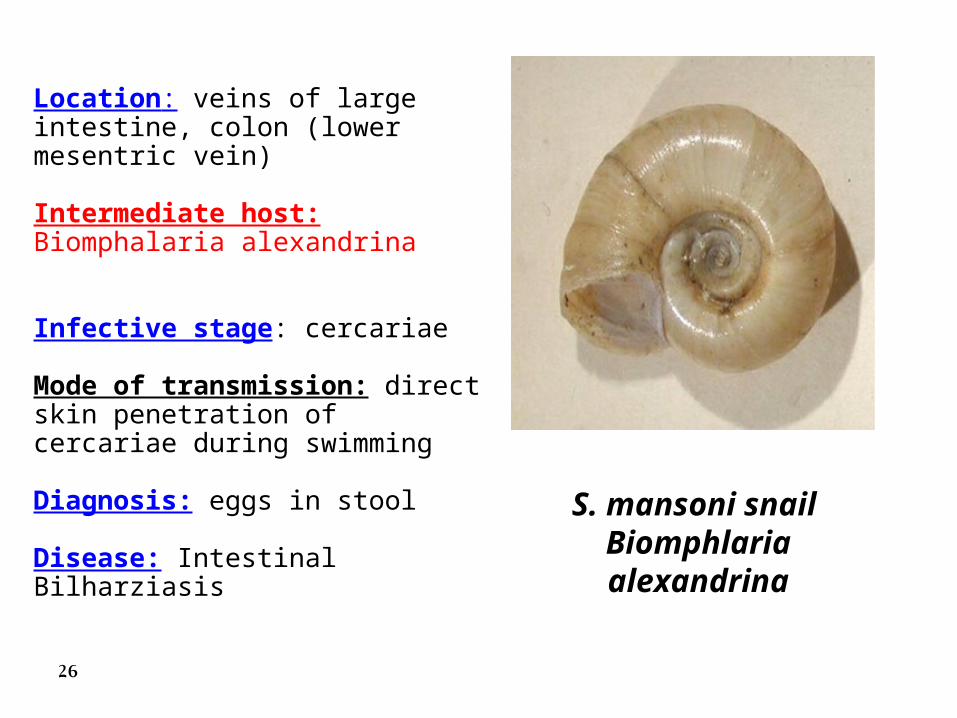
S. mansoni snail Biomphlaria alexandrina
Location: veins of large intestine, colon (lower mesentric vein)
Intermediate host: Biomphalaria alexandrina
Infective stage: cercariae
Mode of transmission: direct skin penetration of cercariae during swimming
Diagnosis: eggs in stool
Disease: Intestinal Bilharziasis
26
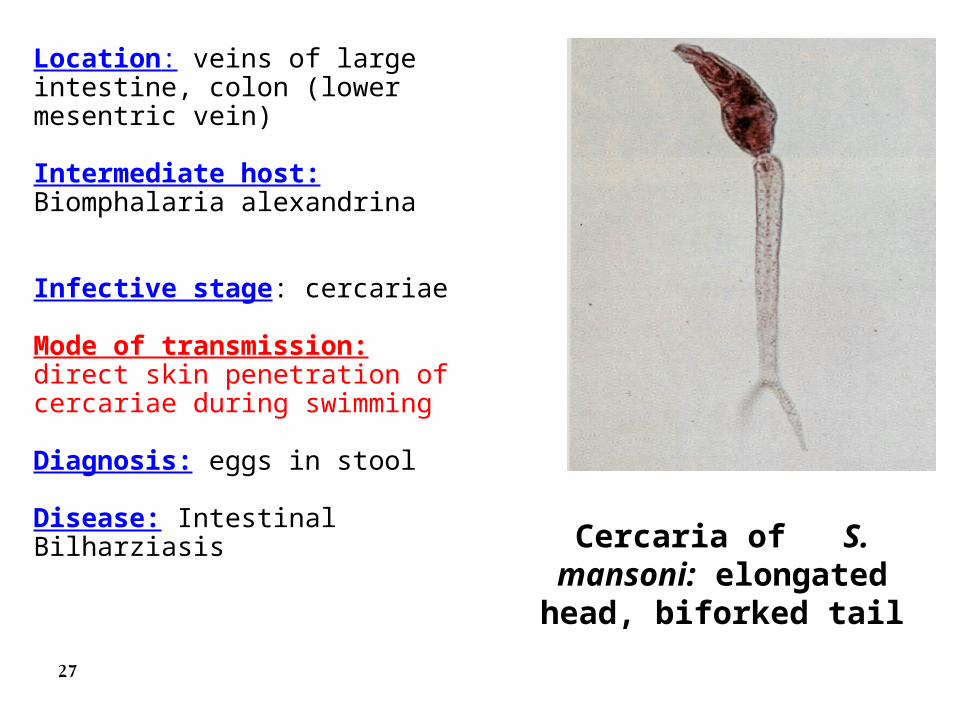
Cercaria of S. mansoni: elongated head, biforked
tail
Location: veins of large intestine, colon (lower mesentric vein)
Intermediate host: Biomphalaria alexandrina
Infective stage: cercariae
Mode of transmission: direct skin penetration of cercariae during swimming
Diagnosis: eggs in stool
Disease: Intestinal Bilharziasis
27
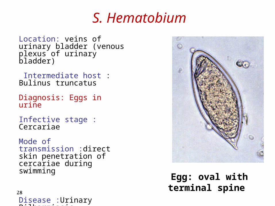
S. Hematobium
Egg: oval with terminal spine
Location: veins of urinary bladder (venous plexus of urinary bladder)
Intermediate host : Bulinus truncatus
Diagnosis: Eggs in urine
Infective stage : Cercariae
Mode of transmission :direct skin penetration of cercariae during swimming
Disease :Urinary Bilharziasis
28
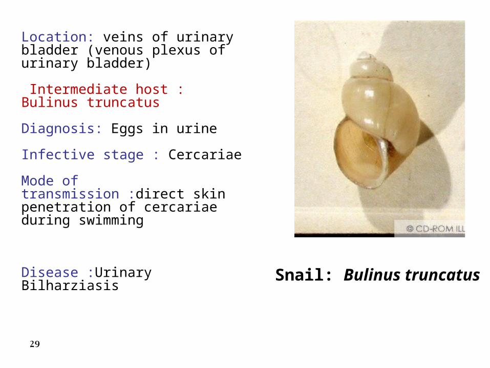
Snail: Bulinus truncatus
Location: veins of urinary bladder (venous plexus of urinary bladder)
Intermediate host : Bulinus truncatus
Diagnosis: Eggs in urine
Infective stage : Cercariae
Mode of transmission :direct skin penetration of cercariae during swimming
Disease :Urinary Bilharziasis
29
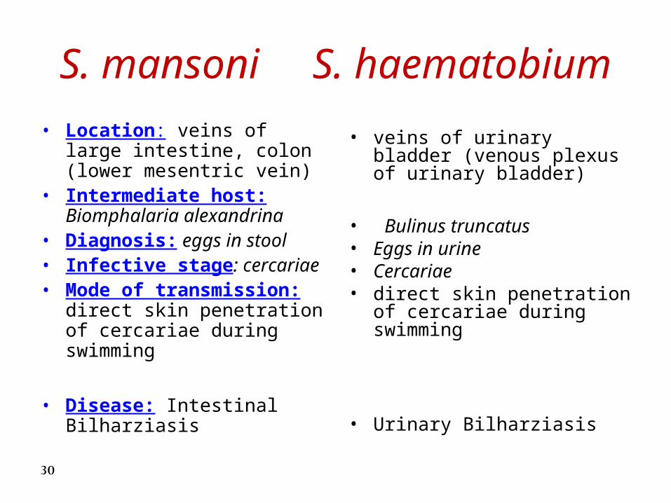
S. mansoni S. haematobium
• Location: veins of large intestine, colon (lower mesentric vein)
• Intermediate host: Biomphalaria alexandrina
• Diagnosis: eggs in stool• Infective stage: cercariae• Mode of transmission: direct
skin penetration of cercariae during swimming
• Disease: Intestinal Bilharziasis
• veins of urinary bladder (venous plexus of urinary bladder)
• Bulinus truncatus• Eggs in urine• Cercariae• direct skin penetration of
cercariae during swimming
• Urinary Bilharziasis
30
Slides of trematodes
31
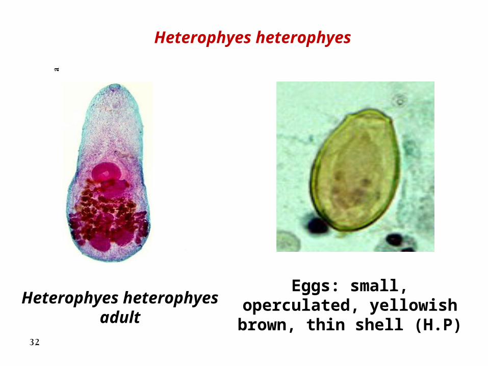
Heterophyes heterophyes adult
Eggs: small, operculated, yellowish brown, thin shell
(H.P)
Heterophyes heterophyes
32
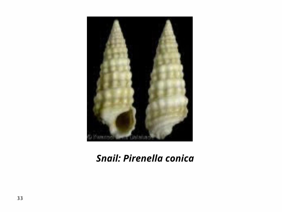
Snail: Pirenella conica
33
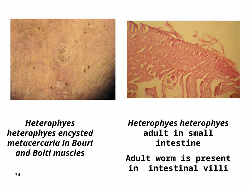
Heterophyes heterophyes adult in small intestine
Adult worm is present in intestinal villi
Heterophyes heterophyes encysted metacercaria in Bouri
and Bolti muscles
34
Fasciola hepatica Fasciola gigantica
2 prominent shoulders, converging margins, simple medial branches of intestinal
caeca, smaller in size
Less prominent shoulders,
parallel margins, medial branches
are T or Y shaped, larger
in size35
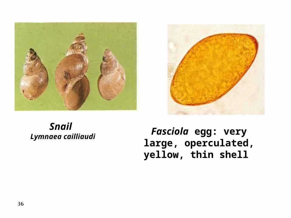
Snail Lymnaea cailliaudi
Fasciola egg: very large, operculated, yellow, thin shell
36
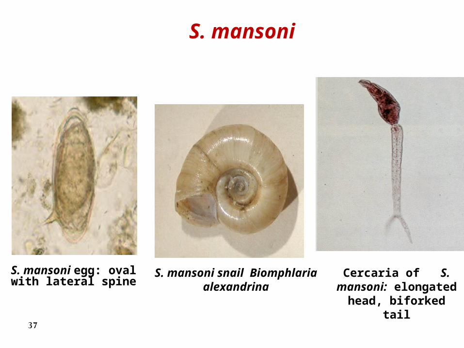
Cercaria of S. mansoni: elongated head, biforked tail
S. mansoni snail Biomphlaria alexandrina
S. mansoni egg: oval with lateral spine
S. mansoni
37
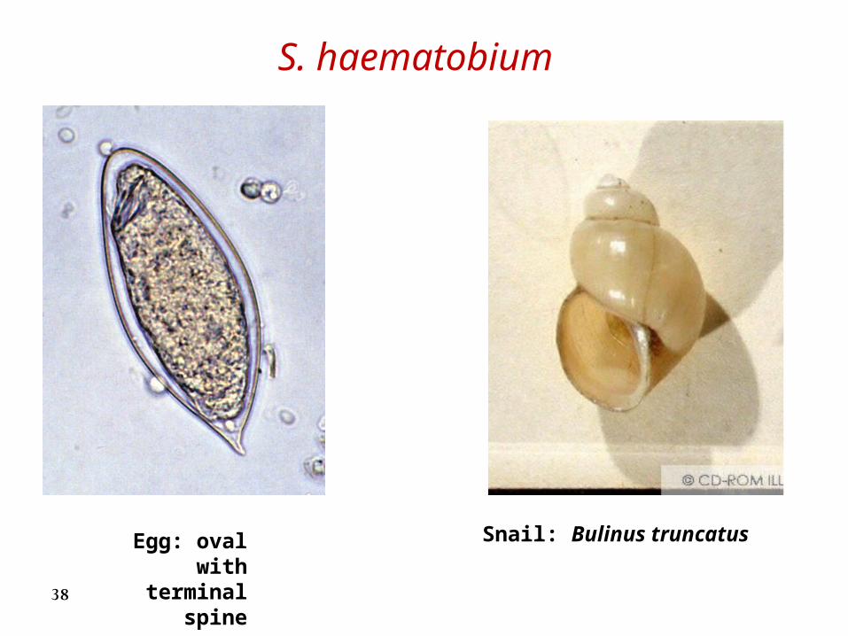
Egg: oval with terminal spine
Snail: Bulinus truncatus
S. haematobium
38