Embed Size (px)
Citation preview
University of North DakotaUND Scholarly Commons
Physical Therapy Scholarly Projects Department of Physical Therapy
2018
Treatment Post Total Knee Arthroplasty withCommon Peroneal Nerve PalsyMegan BerndtUniversity of North Dakota
Follow this and additional works at: https://commons.und.edu/pt-grad
Part of the Physical Therapy Commons
This Scholarly Project is brought to you for free and open access by the Department of Physical Therapy at UND Scholarly Commons. It has beenaccepted for inclusion in Physical Therapy Scholarly Projects by an authorized administrator of UND Scholarly Commons. For more information,please contact [email protected].
Recommended CitationBerndt, Megan, "Treatment Post Total Knee Arthroplasty with Common Peroneal Nerve Palsy" (2018). Physical Therapy ScholarlyProjects. 654.https://commons.und.edu/pt-grad/654
TREATMENT POST TOTAL KNEE ARTHROPLASTY WITH COMMON PERONEAL NERVE PALSY
by
Megan Berndt Bachelor of Arts in Psychology and Associate of Arts and of Science
Minnesota State University Moorhead, 2014 and Williston State College, 2011
A Scholarly Project Submitted to the Graduate Faculty of the
Department of Physical Therapy
School of Medicine
University of North Dakota
in partial fulfillment of the requirements for the degree of
Doctor of Physical Therapy
Grand Forks, North Dakota May, 2018
This Scholarly Project, submitted by Megan Berndt in partial fulfillment of the requirements for the Degree of Doctor of Physical Therapy from the University of North Dakota, has been read by the Advisor and Chairperson of Physical Therapy under whom the work has been done and is hereby approved,
School Advisor)
ii
Title
Department
Degree
PERMISSION
Treatment Post Total Knee Arthroplasty with Common Peroneal Nerve Palsy
Physical Therapy
Doctor of Physical Therapy
In presenting this Scholarly Project in partial fulfillment of the requirements for a graduate degree from the University of North Dakota, I agree that the Department of Physical Therapy shall make it freely available for inspection. I further agree that permission for extensive copying for scholarly purposes may be granted by the professor who supervised my work or, in her absence, by the Chairperson of the department. It is understood that any copying or publication or other use of this Scholarly Project or part thereof for financial gain shall not be allowed without my written permission. It is also understood that due recognition shall be given to me and the University of North Dakota in any scholarly use which may be made of any material in this Scholarly Project.
Signature ~ ~,,-cc:j};
Date { I"';> / \I • •
iii
TABLE OF CONTENTS
LIST OF FIGURES ......................................................................... v
LIST OF TABLES ....... ..................................................................... vi
ACKNOWLEDGEMENTS ... ............................................................ vii
ABSTRACT ................................................................................ viii
CHAPTER I.
II.
BACKGROUND AND PURPOSE ......................... 1
CASE DESCRiPTION .............................. ........... 5
Examination, Evaluation and Diagnosis ....... '" .......... 7
Prognosis and Plan of Care .................................. 11
III. INTERVENTION ............................................. 13
IV. OUTCOMES ............ ....................................... 20
V. DiSCUSSiON .................................................... 22
Reflective Practice ............................... , ............. 24
APPENDiX ...... ........................................................................... 26
REFERENCES ................ ............................................................. 35
iv
LIST OF FIGURES
1. DECISION-MAKING MODEL ............................................................ 10
v
LIST OF TABLES
1. INITIAL KNEE RANGE OF MOTION .................................... .......... 9
2. PLAN OF CARE ........................................................................ 25
3. WEEKLY RIGHT KNEE RANGE OF MOTION PROGRESS ............... . 19
vi
ACKNOWLEDGEMENTS
I would like to thank the patient in this case, the faculty and staff of UND Physical
Therapy, my clinical instructor, and my fellow classmates for their support and
the educational tools necessary to benefit this patient during physical therapy
treatment.
vii
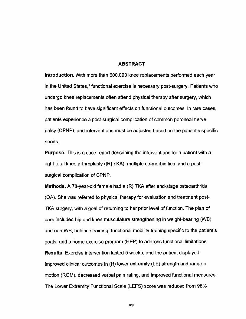
ABSTRACT
Introduction. With more than 600,000 knee replacements performed each year
in the United States,1 functional exercise is necessary post-surgery. Patients who
undergo knee replacements often attend physical therapy after surgery, which
has been found to have significant effects on functional outcomes. In rare cases,
patients experience a post-surgical complication of common peroneal nerve
palsy (CPNP), and interventions must be adjusted based on the patient's specific
needs.
Purpose. This is a case report describing the interventions for a patient with a
right total knee arthroplasty ([R] TKA), multiple co-morbidities, and a post
surgical complication of CPNP.
Methods. A 78-year-old female had a (R) TKA after end-stage osteoarthritis
(OA). She was referred to physical therapy for evaluation and treatment post
TKA surgery, with a goal of returning to her prior level of function. The plan of
care included hip and knee musculature strengthening in weight-bearing (WB)
and non-WB, balance training, functional mobility training specific to the patient's
goals, and a home exercise program (HEP) to address functional limitations.
Results. Exercise intervention lasted 5 weeks, and the patient displayed
improved clinical outcomes in (R) lower extremity (LE) strength and range of
motion (ROM), decreased verbal pain rating, and improved functional measures.
The Lower Extremity Functional Scale (LEFS) score was reduced from 98%
viii
impaired to 46%; however, the patient's further progress in therapy was limited
by co-morbidities, specifically the OA in her left knee and her CPNP.
Conclusion. A combination of strength, balance, and functional training led to
improved functional outcomes for this patient.
ix
CHAPTER I
BACKGROUND AND PURPOSE
According to the Agency for Healthcare Research and Quality (AHRQ),
there are more than 600,000 knee replacements performed each year in the
United States, and joint replacements are going to become the most common
elective surgical procedures by 2030. 1•2 Joint replacement surgery has been
completed since 1968 and has shown to be a reliable procedure, 'improving
patients' quality of life by reducing pain and allowing return to daily functional
activities.2
Arthritis is the most common cause of chronic knee pain which often leads
to the need for a Total Knee Arthroplasty (TKA). There are 3 main subtypes of
arthritis contributing to knee pain: osteoarthritis, rheumatoid arthritis, and post
traumatic arthritis. Osteoarthritis (OA) usually occurs in people over 50-years-old
and is a "wear and tear" type of arthritis. With OA, cartilage that cushions the
bones of the knee wears down and, eventually, the bones rub against their
surfaces leading to knee pain, stiffness, and swelling.2 Treatment of OA with TKA
has shown to be effective with more than 90% of people who undergo TKA
experiencing a dramatic reduction of knee pain.3
George et al4 found that Medicare patients with OA, who received TKA,
improved on 3 different self-reported physical functioning scales, whereas
patients with OA who did not undergo TKA declined over time. A recent
1
systematic review and meta-analysis by Artz et al5 found that physiotherapy and
exercise interventions after TKA provided evidence for short-term effectiveness;
however, long-term effectiveness was well not established. These authors
highlighted the importance of the need for more sufficiently powered studies on
this topic and recommended that future studies address patients' post-surgical
expectations. By understanding patient expectations, interventions may be
adjusted to include patient education to ensure they have realistic expectations
for after their TKA surgery.
Although TKA surgery provides numerous positive benefits for individuals
with severe OA, there are also possible complications from surgery. These
include infection, blood clots, implant problems, continued pain, and
neurovascular injury.2 Other major medical complications such as heart attack or
stroke occur even less frequently.2 Factors that have been associated with an
increased risk of cardiac complications following total joint replacement surgery
include a history of arrhythmia; a history of coronary artery disease, myocardial
infarction, congestive heart failure, or valvular heart disease; revision surgery;
and bilateral surgery.6
Even though surgery generally has favorable outcomes, a rare
complication following TKA is common peroneal nerve palsy (CPNP) resulting in
an acute onset of foot drop. Previous studies?' 8 have found the incidence to
range from 0.5%-1.3%. Factors that have been found to contribute to this injury
are compression of the common peroneal nerve at the fibular neck, positioning
during anesthesia, compression wrapping around the knee, correction of valgus
2
or flexion contracture at the knee, rheumatoid arthritis, previous spinal pathology,
and a high body mass index (8M I). ? The initial recommended treatment for CPNP
is a conservative approach including physical therapy for range of motion
exercises and the use of an ankle-foot orthoses (AFO)J,8 One study? that looked
at time to recovery found that CPNP resolved completely for two-thirds of
patients by one year; however, surgical decompression is recommended if
improvements are not seen with conservative treatments for 3 months.9
In this case report, the patient had several co-morbidities, including the
post-surgical complication of CPNP, bilateral knee OA, and early symptoms of
congestive heart failure (CHF). While skilled physical therapy interventions post
TKA surgery have been found to have positive effects on outcomes, the
complication of CPNP can potentially inhibit progress with rehabilitation. This
made it important to tailor physical therapy interventions to meet the specific
needs of this patient. In a systematic review by Pozzi et ai, 10 the authors
recommended that outpatient physical therapy protocols include strengthening
and intensive functional exercises; and to progress the program as patients
tolerate. Lin et aj11 found that for patients with knee OA, non-weight-bearing
exercises for strength and balance training significantly improved outcomes for
muscle strength and proprioceptive function. Due to the patient's bilateral knee
OA, several interventions were adapted based on this evidence.
Since there are various factors that influence a patient's progress in
physical therapy, to view the individual holistically and account for factors other
than just their disease or pathology, the International Classification of
3
Functioning, Disability, and Health (ICF) Model was utilized. The ICF is beneficial
in organizing information from all aspects of a patient's life and predicting
outcomes. The ICF is a classification system developed by the World Health
Organization (WHO), which provides a common language among health
professionals to describe the different elements of human functioning. 12 This
model takes the emphasis off the diagnosis or disease and focuses on the
abilities of an individual based on the situation, environmental factors, and
personal factors. The ICF Model was used in this case to assist with the
decision-making process for this patient throughout the course of her physical
therapy.
Much of the research for TKA has excluded patients with comorbidities
which is not always generalizable to most patients who undergo joint
replacement surgery.10 The purpose of this case study is to describe the plan of
care and clinical decision making involved with a patient post-TKA surgery who
had multiple co-morbidities and a surgical complication of CPNP.
4
CHAPTER II
CASE DESCRIPTION
The patient, a 78-year-old Caucasian female, was referred to physical
therapy for evaluation and treatment after (R) TKA with cement, which was
performed 5 days prior to her physical therapy examination. The patient showed
signs of CPNP the day of surgery, which was observed during her physical
therapy session in the hospital, as she had minimal ability to dorsiflex her (R)
ankle. She could bear full weight on the leg which she had surgery on, but the
precaution was to not twist her leg while her foot was planted on the ground. Past
medical history included bilateral knee OA for the "past several years,"
hypertension (controlled with medications), early symptoms of congestive heart
failure (checked regularly by her cardiac physician), obesity, asthma, spinal
stenosis, and gastroesophageal reflux disease (GERD). She stated her left (L)
knee was "in worse shape but the doctors decided to do the right one first."
Using the Numeric Pain Rating Scale (NPRS),13 the patient chose a whole
number from 0-10 (0 = no pain, 10 = worst pain) that best reflected her pain. This
scale was used throughout her course of treatment. The NPRS has been found
to have high test-retest reliability.14 Prior to the examination, the patient rated her
pain as 5-6/10. She reported that she had been taking her pain medications
regularly and using her polar ice machine to help relieve pain. The ice machine
5
was included with the cost of surgery, and it circulates ice water through a tube
into a "bladder" that wraps around the knee joint. The patient stated her pain
increased when she would bend and straighten her knee and when she wore the
ankle-foot orthosis (AFO) prescribed to her the day after surgery.
Prior to surgery, the patient could complete most daily activities
independently, with minimal modifications of using her assistive devices and
allowing for more time to complete activities. Occasionally, her husband helped
her with dressing, cooking, and climbing stairs to get to the bedroom and
bathroom. She could walk short distances in the community with a four-wheeled
walker and used two canes in the house. Otherwise, the patient's husband
pushed her in a wheelchair (We) when out in the community. After surgery, the
patient had been receiving assistance from her husband with getting dressed,
transferring, and bathing with a sponge bath.
The patient lived with her husband in a house which had 3 stairs with
handrails to enter the house. She reported there were 15 stairs with handrails to
reach their bedroom and the only bathroom in the house. The patient's husband
stated he made modifications so that there was a bed and commode on the main
level of the house. The patient and her husband were retired and a typical day for
them included "driving into town to go out for breakfast, going to the grocery
store, driving around the country side, and going back to the house to relax and
work on crossword puzzles."
The patient was appropriate to complete an examination to determine her
functional limitations after TKA surgery. Since she was less than a week out of
6
surgery, the plan was to modify the examination procedure to the patient's
tolerance.
Examination, Evaluation and Diagnosis
Examination. The examination was based on components of Magee's
Orthopedic Physical Assessment of the knee 15 and modified due to the patient's
post-surgical status. When the patient arrived at the physical therapy clinic, she
filled out a history form, consent forms, and the Lower Extremity Functional Scale
(LEFS). The LEFS is a patient self-reported tool used to measure lower extremity
musculoskeletal function. There are 20 items which are scored on a 5-point scale
that ranges from 0 to 4. Total score ranges from 0 to 80 points, with higher
scores indicating higher levels of function.16 Sensitivity and specificity have not
been established for knee disorders, but reliability was found to be .85 for TKA.17
The patient completed the LEFS with a score of 2/80, 98% impaired.
She was wheeled in on her we by her husband. The patient was in
apparent distress, and she reported she had 'nausea from pain medication' and
'pain from the ankle orthosis.' The patient was obese but appeared well
nourished. Skin integrity of the surgical incision, which was covered with the
surgical bandage, was assessed, and there were no apparent signs of infection.
Functional mobility was assessed through transfers, gait, bed mobility, and
the LEFS. The patient transferred from sitting in her wheelchair, with her front
wheeled-walker (FWW) in front, to standing with minimal assist (Min. Assist)
(patient can perform 75% or more of task) to moderate assist (Mod. Assist)
(patient can perform 50-74% of task) of 1 person with cueing on hand positioning
7
for safety, leaning forward, and pushing through her upper extremities. The
patient initially grasped her FWW when she was standing and stated it made her
"feel more secure." She was educated on the proper transfer technique by
placing her hands on the stable surface (armrests of the WC) and pushing
through her upper extremities to prevent a fall. The patient ambulated 50 feet
with Min. to Mod. Assist of 1 person, a gait belt, and her FWW, demonstrating a
step-to antalgic gait pattern to the right. She displayed decreased safety as she
would lean onto the walker on her forearms, due to complaints of low back pain,
so she required cueing on placing her hands on the handles and standing up
straight. The patient then transferred from sitting to supine on the plinth with Mod.
Assist of 1 person for help lifting her lower extremities and lowering her upper
trunk onto the table. She completed bed mobility with Min. Assist for moving the
operated lower extremity (LE); however, she was independent with this at home
with the use of a leg lifter strap. Stair negotiation was not assessed at that time
due to the patient's pain level.
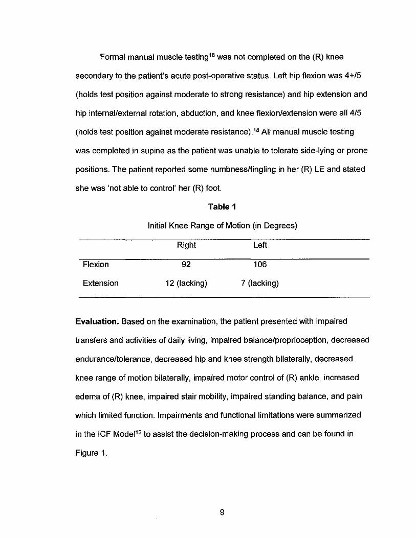
Knee range of motion (ROM) was measured in supine with a standard
goniometer and was based on techniques from the Cram Session in Goniometry
and Manual Muscle Testing book. 18 Active knee flexion was performed by asking
the patient to bend her leg and slide her foot up towards her buttocks as far as
she could go. For knee extension, the patient placed her ankle on a bolster and
was asked to push her knee down towards the table. Measurements can be
found in Table 1. The patient reported knee pain with both knee flexion and
extension.
8
Formal manual muscle testing18 was not completed on the (R) knee
secondary to the patient's acute post-operative status. Left hip flexion was 4+/5
(holds test position against moderate to strong resistance) and hip extension and
hip internal/external rotation, abduction, and knee flexion/extension were all 4/5
(holds test position against moderate resistance).18 All manual muscle testing
was completed in supine as the patient was unable to tolerate side-lying or prone
positions. The patient reported some numbness/tingling in her (R) LE and stated
she was 'not able to control' her (R) foot.
Table 1
Initial Knee Range of Motion (in Degrees)
Right Left
Flexion 92 106
Extension 12 (lacking) 7 (lacking)
Evaluation. Based on the examination, the patient presented with impaired
transfers and activities of daily living, impaired balance/proprioception, decreased
endurance/tolerance, decreased hip and knee strength bilaterally, decreased
knee range of motion bilaterally, impaired motor control of (R) ankle, increased
edema of (R) knee, impaired stair mobility, impaired standing balance, and pain
which limited function. Impairments and functional limitations were summarized
in the ICF ModeP2 to assist the decision-making process and can be found in
Figure 1.
9
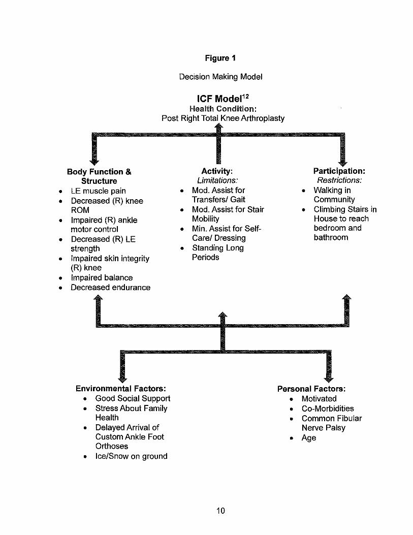
Figure 1
Decision Making Model
ICF Model12
Health Condition: Post Right Total Knee Arthroplasty
Body Function & Structure
• LE muscle pain • Decreased (R) knee
ROM • Impaired (R) ankle
motor control • Decreased (R) LE
strength • Impaired skin integrity
(R) knee • Impaired balance • Decreased endurance
Environmental Factors: • Good Social Support • Stress About Family
Health • Delayed Arrival of
Custom Ankle Foot Orthoses
• Ice/Snow on ground
Activity: Limitations:
• Mod. Assist for Transfers! Gait
• Mod. Assist for Stair Mobility
• Min. Assist for SelfCare! Dressing
• Standing Long Periods
10
Participation: Restrictions:
• Walking in Community
• Climbing Stairs in House to reach bedroom and bathroom
Personal Factors: • Motivated • Co-Morbidities • Common Fibular
Nerve Palsy
• Age
Diagnosis. The patient was status post right total knee arthroplasty using
cement.
Prognosis and Plan of Care
The patient's rehabilitation prognosis was fair due to several co
morbidities and potential barriers which included CPNP limiting (R) ankle
function, bilateral knee pain, low back pain when standing or walking long
periods of time, and an overly helpful husband. Her husband tended to try to do
everything for her which could have limited the patient from maximizing functional
independence. On the other hand, her husband was also beneficial to the patient
as a good support system.
The patient was appropriate for skilled physical therapy interventions due
to her limitations listed above and agreed to complete physical therapy. The
patient was seen 3 days a week for 3 weeks and then 2 days a week for 2 weeks
for 1-hour sessions. The patient's short term goals were to be met within 2 weeks
and included being able to carry out her home exercise program (HEP)
independently for increased tolerance of daily activities and continued
strengthening and to increase strength to 3+/5 for (R) knee flexion and extension
for increased stability during gait and transfers. Long term goals were to bet met
within 6 weeks and included being able to; 1) decrease pain to 1-2/10 to carry out
HEP with minimal pain and for increased tolerance of daily activities, 2) be
independent in HEP to continue progress made in therapy at home, 3) increase
ROM on (R) knee to 115 degrees of flexion and 0 degrees of extension for
increased mobility during gait and transfers, 4) demonstrate strength of at least
11

4/5 for (R) knee flexion and extension for increased stability during daily
activities, gait, and transfers, and 5) score at least 50% or less impaired on the
Lower Extremity Functional Scale (LEFS) for increased tolerance of daily
activities.
12
CHAPTER III
INTERVENTION
The patient attended physical therapy for 13 sessions over the course of 5
weeks. Interventions were based upon a Total Knee Arthroplasty Protocol
provided to the patient by the hospital, prior to surgery, current evidence, and the
patient's specific needs. A detailed outline of the Plan of Care can be found in
Table 2 in the Appendix. The patient's HEP consisted of hip and knee
strengthening exercises, 19,20 ankle ROM, stretching hamstrings and calf
musculature, pain management techniques and modalities for edema, balance
training,21,22 and increased ambulation distance in her house and in the
community as appropriate. A gait belt was always used with this patient during
physical therapy treatment due to decreased balance with ambulation and
transfers. Pictures of and instructions for exercises and transfers were provided
in the binder that was given to the patient when she attended a class at the
hospital prior to surgery.
On the first day of treatment, the examination and evaluation process was
completed, the patient was educated on pertinent anatomy and healing time
frames, signs of infection, modalities for edema and pain control, correct
execution of exercises, and her active role and participation with therapy. The
patient's incision was inspected and showed no signs of infection. She completed
13
strengthening exercises given to her in the hospital to assure correct
performance and was instructed in hamstrings and calf stretches which were
added to the HEP. The patient was instructed to complete passive range of
motion (PROM) and active-assistive range of motion (AAROM) of her (R) ankle
for treatment of her CPNP. For specific interventions, see Table 2 in the
Appendix. Since the patient and her husband reported one of their main
concerns was the AFO, which was "causing her more pain," it was recommended
that she discuss her concerns with the surgeon at the follow-up appointment.
At the second visit, the patient rated her pain as 8/10 (scale of 0 -10), but
she agreed to participate in therapy. She noted that she had seen her primary
care provider that day and had been tested for a possible deep vein thrombosis,
which was negative. The patient began endurance training by warming-up on the
NuStep® bike without any resistance. During ambulation, she continued to
display decreased safety observed in the initial evaluation. She required cueing
on posture and keeping the FWW close to her body, and she was reminded of
the importance of safe hand positioning on the stable surface rather than the
FWW during sit-to-stand transfers. Strengthening exercises were performed in
supine and in sitting and heel slides were completed on a sliding board with a
pillow case. The patient was educated on progressing strengthening exercises by
first increasing number of repetitions and then adding a resistance band.
Functional training of sit-to-stands from a standard height chair were completed
for lower extremity strengthening and to increase independence.23 Patient
14
education was given on carrying out the transfer safely with correct hand
positioning and slowly lowering herself to the chair.
At the third visit, the patient reported she was going to be fitted for a
custom AFO the following week, but she continued to not wear the brace sent
home from the hospital at time of discharge because it caused her pain. Weight
bearing (WB) exercises were added to treatment so the patient could progress
the amount of time spent on her feet. The patient completed WB exercises with
the FWW in front of her for increased balance. She was instructed to hold onto a
counter when she completed these exercises at home with her husband standing
beside her for safety. She required several seated rest breaks throughout the
course of therapy with any WB exercises due to pain in her back and (L) knee
and decreased endurance.
At the fourth visit, the patient reported she had her staples removed the
day before and that she had been "walking more around the house." There were
no signs of infection upon inspection of the incision and it appeared to be healing
well. The patient increased resistance and time completed on the NuStep®. See
Table 2 in the Appendix for resistance levels and additional interventions. A static
knee extension stretch with a towel roll under the ankle was added to the HEP to
aid the patient in progressing towards full extension of her (R) knee.
On the day of the fifth treatment session, the patient still rated pain in her
(R) knee as 8/10. She expressed concern about the incision site stating that it
showed some drainage and that her knee felt "warm" that day. The incision site
was inspected. The patient was educated that slight drainage was normal;
15
however, she and her husband still felt concerned. The patient had an
appointment 4 days prior to this treatment session and reported that everything
looked good after her staples were removed. To ease the patient's concerns, we
asked the surgeon's nurse to inspect the incision site. The nurse reported that
the incision "Iook[ed] even better than the last time [they] saw the patient," and
that it was normal to have some drainage. The patient stated she had not
climbed the staircase to reach the second floor of her house yet, so stair training
was incorporated into the treatment session on a 4-inch step. To progress
strength, she was educated on increasing repetitions and resistance with
exercises, as able.
At the sixth treatment session, the patient rated her (R) knee pain as 5/10,
and she noted her (L) knee was starting to bother her more. She stated she had
an appointment for fitting of an AFO later that morning. Due to fatigue and
complaints of increased pain with activity, the patient completed mostly non-WB
exercises during this treatment session. 11
At the seventh visit, the patient rated her overall pain as 8/10 but stated
that her (R) knee did not "feel that bad." She noted most of the pain was in her
back and her (L) knee and felt that this was limiting her activity the most. The
patient stated that she increased walking distances at home at that she practiced
4 steps on the indoor staircase (8-inch steps with 2 handrails), with Mod. Assist
from her husband for balance and lifting (R) foot onto the step. Additional
repetitions and resistance were added to exercises, per the patient's tolerance.
16
Step-ups were completed in the parallel bars with bilateral upper extremity
support.
On day of the eighth visit, she reported that she had climbed the flight of
stairs in her house, with help from her husband for placement of her (R) foot as
she still had "no control" over it. The patient increased resistance and repetitions
with exercises, and balance exercises were added to treatment. Although her (R)
knee pain was not limiting her and she was progressing with strengthening
exercises, the patient continued to display decreased endurance with ambulation
and stated that her knee "does not want to hold up for longer walking distances."
At the ninth treatment session, the patient noted she "hardly had any pain"
in her (R) knee but that her (L) knee pain ranged from 6-8/10. With ambulation,
the patient showed increased gait speed and decreased antalgic pattern to the
(R) LE, but the antalgic pattern was more notable on the left.
At the 10th visit, treatment was lessened due to complaints of pain in both
knees and a "burning" feeling in her (R) knee. The surgical incision was
inspected and there were no signs of infection. The patient did note that her (R)
knee felt better after completing her exercises and applying a cold pack to her
knee after this therapy session.
At the 11th visit, the patient was reminded to complete her HEP on both
lower extremities, per patient tolerance, for increased strengthening and stability.
She stated that her (L) knee pain increased to 8/10 after completing exercises. It
was recommended that the patient discuss with her surgeon the increased pain
in her (L) knee at her upcoming appointment.
17
At the 12th visit, the patient reported she had "hardly any pain in the (R)
knee" but that her (L) knee pain had "gotten worse with the weather." She noted
that she had been completing MROM ankle exercises every day but that she
still did not have much control over her (R) foot. The patient was evaluated for
stair mobility and she climbed up and down 1 flight of stairs with 1 handrail,
handheld assist, and occasional assist with her (R) foot to clear the step. Initially,
the patient ascended the stairs leading with her (L) LE and complained of
increased pain. She was instructed to try leading with her (R) LE, and she
required less assistance and had decreased pain. The patient was also
instructed to kick out her (L) LE when transferring from sit-to-stand on low
surfaces to decrease compressive forces at her (L) knee. It was recommended
that the patient continue progressing exercises by using ankle weights or
household products, such as a bag of sugar, wrapped around her ankle for
continued strengthening.
The patient's husband stated he was "doing the same exercises with her
at home" and that he thought she was ready to be discharged from therapy. The
patient had a session scheduled at the end of the week, after a follow-up visit
with the surgeon. At the last visit, the patient was instructed in transfer training
to/from the floor. She demonstrated the transfers with Min. Assist to contact
guard assist (eGA) for balance. The patient showed progress with balance
exercises as she tolerated increased time with each exercise; however, (L) knee
pain and minimal function of her (R) foot continued to limit progress towards
increased endurance with ambulation and during weight-bearing activities. See
18
Table 2 in the Appendix for more detailed interventions. The patient decided she
wanted to be discharged from physical therapy and continue with her HEP
independently at home. We spoke with the physician assistant (PA) about the
patient's progress with therapy and the factors that limited her from further
progress. The PA noted they would follow-up with the patient regarding her (L)
knee pain and (R) ankle function to discuss the next course of treatment.
19
CHAPTER IV
OUTCOMES
The patient was discharged after 5-weeks of physical therapy intervention
as she had met all her current goals. Throughout the course of treatment, the
patient was dedicated to completing her HEP, and her husband remained a
strong support system as he motivated and encouraged her to complete her
exercises multiple times per day. She was only able to ambulate household
distances and short distances in the community; however, she had initially
reported that she was not an avid community walker before she had surgery.
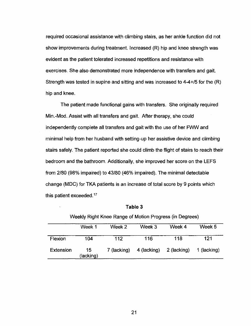
The patient gained adequate range of motion in her (R) knee to carry out
daily tasks. According to Brotzman,24 to descend stairs reciprocally, without hip
or trunk substitution, the patient needs to achieve 115-117 degrees of knee
flexion. This was an important goal that this patient surpassed because she had
15 stairs to reach her bedroom and the only bathroom in her house. The patient
achieved a weekly progression of range of motion, which is shown in Table 3.
Pain in the patient's (R) knee decreased in severity from 8/10 to "no pain" on the
NPRS, but she experienced increased pain in the (L) (non-operated) LE. The
custom fit AFO did not arrive during the 5 weeks of physical therapy, but the
patient faithfully completed AAROM exercises for ankle dorsiflexion and
stretching of the gastrocnemius and soleus musculature in the HEP. She
20
required occasional assistance with climbing stairs, as her ankle function did not
show improvements during treatment. Increased (R) hip and knee strength was
evident as the patient tolerated increased repetitions and resistance with
exercises. She also demonstrated more independence with transfers and gait.
Strength was tested in supine and sitting and was increased to 4-4+/5 for the (R)
hip and knee.
The patient made functional gains with transfers. She originally required
Min.-Mod. Assist with all transfers and gait. After therapy, she could
independently complete all transfers and gait with the use of her FWW and
minimal help from her husband with setting-up her assistive device and climbing
stairs safely. The patient reported she could climb the flight of stairs to reach their
bedroom and the bathroom. Additionally, she improved her score on the LEFS
from 2/80 (98% impaired) to 43/80 (46% impaired). The minimal detectable
change (MOe) for TKA patients is an increase of total score by 9 points which
this patient exceeded. 17
Table 3
Weekly Right Knee Range of Motion Progress (in Degrees)
Week 1 Week 2 Week 3 Week 4 Week 5
Flexion 104 112 116 118 121
Extension 15 7 (lacking) 4 (lacking) 2 (lacking) 1 (lacking) (lacking)
21
CHAPTER V
DISCUSSION
Total knee arthroplasty surgeries are predicted to become one of the most
common elective surgical procedures performed in the future. 1 While benefits of
this procedure have included significantly reducing pain and in turn improving
patient satisfaction and quality of life, it has been found that people who have
undergone TKA show decreased activity levels compared people who were the
same age.25 Disuse of the muscles after a TKA and lack of quadriceps
contraction can lead to muscle atrophy and have lasting effects on an individual's
functional mobility. In the current case, an outpatient physical therapy treatment
plan was implemented within a week after the patient's TKA surgery. At the end
of treatment, which consisted of hip and knee progressive resistive strengthening
in WB and non-WB positions, endurance training, balance training, and functional
mobility training, the patient showed improvements in lower extremity strength
from 3/5 to 4-4+/5, increased knee range of motion to functional levels,
decreased pain in the surgical extremity from 8/10 with activity to "no pain," and
increased independence with functional activities.
Ciolac et aj26 compared older women with knee OA, who had TKA, to
older and young women groups, who did not have musculoskeletal diseases, in a
resistance training program. They found that the program helped to rebuild
22
functional, balance, and lower-limb load deficits that were found in the TKA group
at baseline. Bade and Stevens-Lapsley27 found that a high intensity rehabilitation
program initiated after TKA resulted in improved functional performance
outcomes and strength in the short- and long-term compared to a lower intensity
program. Even though the patient's pain in this case decreased in her (R) knee,
her (L) knee pain increased throughout treatment. Due to this and a co-morbidity
of chronic back pain, interventions were adjusted based on patient tolerance.
Due to these factors, Lin et aP1 found that non-WB interventions for strength
training and balance training both improved outcomes of knee strength and
proprioceptive function.
Another area that limited further progress with therapy for this patient was
a post-surgical complication of CPNP. Zywiel et ai28 found that lower extremity
nerve decompression surgery improved sensation and decreased pain. These
authors proposed that this course of treatment should be considered if
improvements are not seen with conservative management of an AFO and
physical therapy interventions. Park et al7 reported that CPNP completely
resolved in over three-fourths of patients with partial nerve palsy and one-fifth in
those with complete palsy. Poage and Scott9 recommended that patients with
CPNP complete nonsurgical interventions for at least three months.
Treatment areas that were not included in this case, but that could be
utilized in future cases like the current one include Kinesia Taping® for pain and
edema, and neuromuscular electrical stimulation (NMES) to the quadriceps
musculature for increased activation initially.
23
Based on outcomes of the current case, it is recommended that a
combination of therapeutic exercise, balance training, and functional mobility
training be used post-TKA; however, interventions should be customized based
upon each patient's needs. In the rare case of common peroneal nerve injury, it
is recommended that patients complete conservative treatments including
physical therapy and the use of orthotic devices. Patients should consider
seeking surgical treatment if they experience lasting difficulties interfering with
function.
Reflective Practice
Even though this patient showed improved function and outcomes, there
are aspects of this patient's examination and physical therapy that could have
been done differently. In the history portion of the examination, I could have
asked more specific questions about the amount and types of exercises the
patient completed each week. For the examination procedures, I would have
measured girth not only to have an objective measure of whether edema was
decreasing, but also to keep track of if her heart problems were becoming worse
resulting in an increased swelling in the legs. This patient was limited in the
distance and amount of time she could walk/stand; however, it would have been
useful to include more objective measures such as the Stair Climb Test, Timed
"Up & Go," and the 6-Minute Walk Test (or a modified version of the test).
Throughout her physical therapy, the patient was faithful in completing her
exercises and always willing to work hard during the sessions, but her pain often
limited her. Treadmill training could have been a beneficial intervention to try for
24

additional gait training and cardiovascular exercise. I also would have liked to
incorporate NMES into the treatments to try and enhance ankle function, but,
unfortunately the facility did not have all the correct equipment at that time.
Further research on the mirror imaging techniques would also be beneficial.
25

APPENDIX

Table 2
Plan of Care
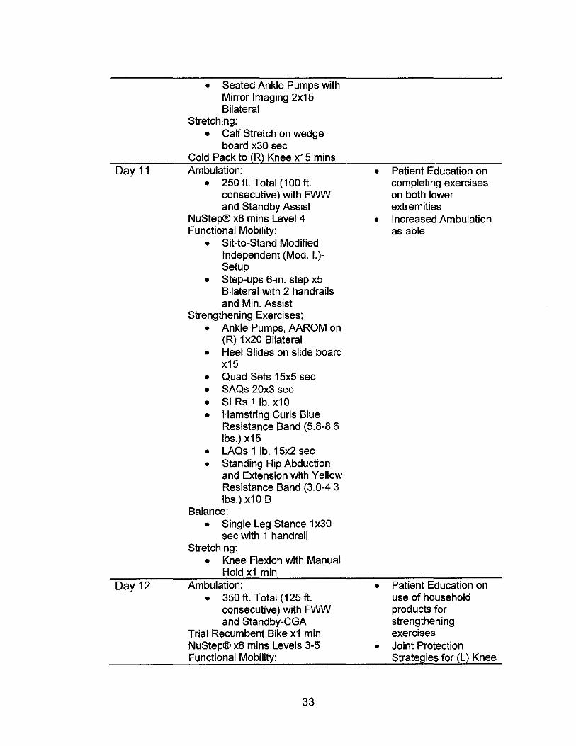
Treatment HEP
Day 1 Initial Evaluation • Static Stretch Patient Education: Hamstrings and Calf
• Anatomy/Healing time with Sheet 3x30 sec frames hold, 3x1day
• Infections signs • Continued
• Modalities for edema/pain Strengthening control Exercises 2 sets x 10
• Active role in PT repetitions, 2x1day
• Correct execution of • Increased exercises Household/Community
Strengthening Exercises: Ambulation as able
• Ankle Pumps 1x20 • Ice machine to (R)
• PROM/AAROM (R) Ankle knee 20 mins, 4-1x20 5x1day
• Short Arc Quads 10x3 sec hold
• Quad Sets 10x5 sec • Supine Hamstring Sets
10x5 sec
• Straight Leg Raises (SLRs) 10x2 sec
• Heel Slides 1 x1 0 Stretching Exercises:
• Static Stretches Hamstrings and Calf 1x20 sec
Day 2 Ambulation: • Progressed repetitions
• 100 ft. Total with Min. and resistance of Assist and FWW (50 ft. exercises as able consecutive) • Increased Ambulation
NuStep® x5 mins Level 1 as able Strengthening Exercises: • Ice machine to (R)
• Ankle Pumps, AAROM on knee 20 mins, 3-(R) 1x20 Bilateral 5x1day
• Heel Slides 10x5 sec
• Quad Sets 10x5 sec
• SLRs 10x3 sec
• Seated Hamstring Curls with Yellow Resistance Band (3.0-4.3Ibs.) 1x10
• Long Arc Quads (LAQs) 10x3 sec
Functional Mobility:
27
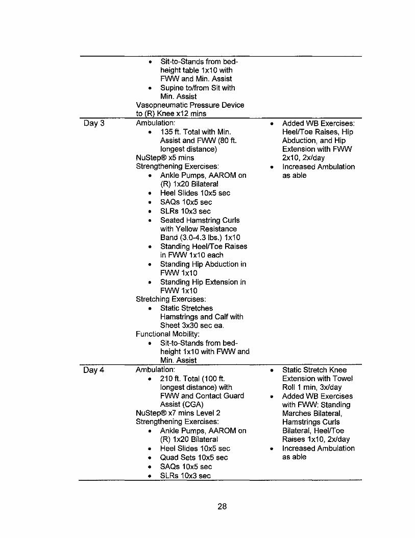
Day 3
Day 4
• Sit-to-Stands from bedheight table 1 x1 0 with FWW and Min. Assist
• Supine to/from Sit with Min. Assist
Vasopneumatic Pressure Device to (R) Knee x12 mins Ambulation:
• 135 ft. Total with Min. Assist and FWW (80 ft. longest distance)
NuStep® x5 mins Strengthening Exercises:
• Ankle Pumps, AAROM on (R) 1 x20 Bilateral
• Heel Slides 10x5 sec • SAQs 10x5 sec • SLRs 10x3 sec • Seated Hamstring Curls
with Yellow Resistance Band (3.0-4.3Ibs.) 1x10
• Standing HeellToe Raises in FWW 1x10 each
• Standing Hip Abduction in FWW 1x10
• Standing Hip Extension in FWW 1x10
Stretching Exercises: • Static Stretches
Hamstrings and Calf with Sheet 3x30 sec ea.
Functional Mobility: • Sit-to-Stands from bed
height 1x10 with FWW and Min. Assist
Ambulation: • 210 ft. Total (100 ft.
longest distance) with FWW and Contact Guard Assist (CGA)
NuStep® x7 mins Level 2 Strengthening Exercises:
• Ankle Pumps, AAROM on (R) 1 x20 Bilateral
• Heel Slides 10x5 sec • Quad Sets 10x5 sec • SAQs 10x5 sec • SLRs 10x3 sec
28
• Added WB Exercises: HeellToe Raises, Hip Abduction, and Hip Extension with FWW 2x10,2x1day
• Increased Ambulation as able
• Static Stretch Knee Extension with Towel Roll 1 min, 3x1day
• Added WB Exercises with FWW: Standing Marches Bilateral, Hamstrings Curls Bilateral, HeellT oe Raises 1x10, 2x1day
• Increased Ambulation as able
• Seated Hamstring Curls with Yellow Resistance Band (3.0-4.3Ibs.) 10x5 sec
• LAQs 10x3 sec
• Standing Marches with FWW 1x7 Bilateral
• Standing Hamstrings Curls with FWW x1 0 Bilateral
• Standing HeelfToe Raises with FWW x1 0 each
Stretching Exercises:
• Knee Extension on bolster x1 min
• Static Stretching Hamstrings and Calf with Sheet 3x20 sec ea.
Functional Mobility:
• Sit-to-Stands from standard chair with armrests 2x5 with FWW
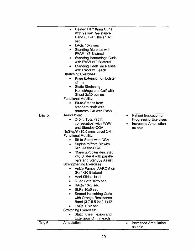
Day 5 Ambulation: • Patient Education on • 245 ft. Total (95 ft. Progressing Exercises
consecutive) with FWW • Increased Ambulation and Standby-CGA as able
NuStep® x10.5 mins Level 2-4 Functional Mobility:
• Sit-to-Stand with CGA
• Supine to/from Sit with Min. Assist-CGA
• Stairs up/down 4-in. step x10 Bilateral with parallel bars and Standby Assist
Strengthening Exercises:
• Ankle Pumps, AAROM on (R) 1 x20 Bilateral
• Heel Slides 1 x11
• Quad Sets 10x5 sec
• SAQs 10x5 sec
• SLRs 10x5 sec
• Seated Hamstring Curls with Orange Resistance Band (3.7-5.5Ibs.) 1x12
• LAQs 10x3 sec Stretching Exercises:
• Static Knee Flexion and Extension x1 min each
Day 6 Ambulation: • Increased Ambulation as able
29
Day?
• 100 ft. consecutive with FWWandCGA
NuStep® x1 0 mins Level 3 Strengthening Exercises:
• Seated AAROM Ankle Pumps with Manual Stretch by Therapist 1 x20 Bilateral
• Seated Knee Flexion with scooter board
• Quad Sets 10x5 sec • LAQs 10x5 sec • Heel Slides 1x10 with slide
board • Hip Abduction 1x10 with
slide board • SAQs 10x5 sec
Stretching Exercises: • Seated Calf Stretch on
wedge board • Static Knee Extension on
bolster x2 mins Cold Pack x1 0 mins (R) Knee Ambulation:
• 145 ft. Total with FWW and Standby-CGA
Functional Mobility: • Sit-to-Stand with Standby
Assist • Supine to/from Sit with
Min. Assist • Step-ups 6 in. step x10
Bilateral NuStep® x8 mins Levels 4-5 Strengthening Exercises:
• Ankle Pumps, AAROM on (R) 1 x20 Bilateral
• Heel Slides x10 • Quad Sets 15x5 sec • SAQs 15x5 sec • SLRs 15x2 sec • Hamstring Curls with
Green Resistance Band (4.6-6.7Ibs.) 1x15
• LAQs 15x3 sec • Standing Hip Abduction
1x15 • Standing Hip Extension
1x15
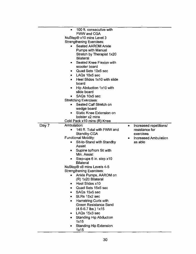
30
• Increased repetitionsl resistance for exercises
• Increased Ambulation as able

Stretching Exercises:
• Static Knee Flexion Stretch with Manual Hold by Therapist x1 min
• Knee Extension with bolster x1 min
Cold Pack x1 0 mins (R) Knee Day 8 Ambulation: • Increased Ambulation
• 200 ft. Total with FWW and as able CGA • Added Resistance
Functional Mobility: Band toWB • Sit-to-Stand with Standby Exercises: Standing
Assist Hip Abduction and
• Supine to/from Sit with Extension, 2x10, CGA 2x1day
• NuStep® x10 mins Level 5 Strengthening Exercises:
• Ankle Pumps, AAROM on (R) 1 x20 Bilateral
• Heel Slides x12
• Quad Sets 15x5 sec
• SLRs 15x2 sec
• Hamstring Curls Green Resistance Band (4.6-6.7 Ibs.) 1x15
• LAQs 20x3 sec • Standing Hip Abduction
and Extension with Yellow Resistance Band (3.0-4.3 Ibs.) 1 x15 Bilateral each
Balance: (on foam mat)
• Standing Feet Shoulder Width x30 sec
• Standing Feet Together x10 sec and x25 sec
• Stride Step x25 sec (L) foot forward, x12 sec (R) foot forward
Stretching Exercises: • Knee Flexion with Manual
Hold x1 min
• Knee Extension on Bolster x1 min
Day 9 Ambulation: • Increased to Blue
• 300 ft. Total (150 ft. Resistance Band (5.8-consecutive) with FWW 8.6 Ibs.) for HS Curls, and Standby Assist 2x10, 2x1day
NuStep® x8 mins Level 4, • Increased Ambulation Bilateral LE only as able
31
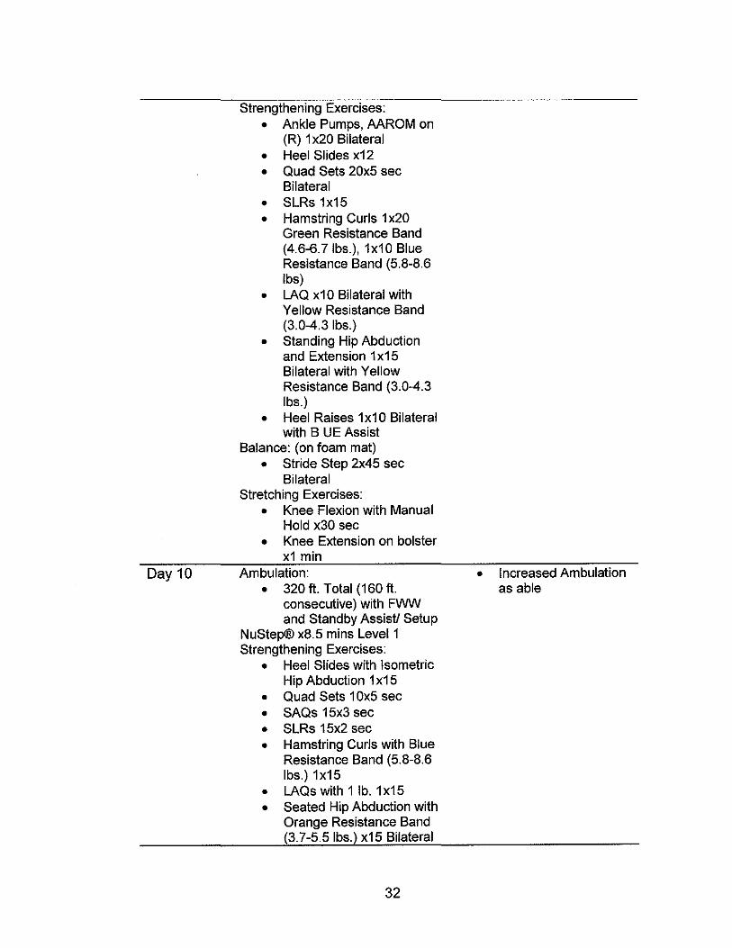
Day 10
Strengthening Exercises: • Ankle Pumps, AAROM on
(R) 1 x20 Bilateral • Heel Slides x12 • Quad Sets 20x5 sec
Bilateral • SLRs 1x15 • Hamstring Curls 1 x20
Green Resistance Band (4.6-6.7Ibs.), 1x10 Blue Resistance Band (5.8-8.6 Ibs)
• LAQ x10 Bilateral with Yellow Resistance Band (3.0-4.3Ibs.)
• Standing Hip Abduction and Extension 1 x15 Bilateral with Yellow Resistance Band (3.0-4.3 Ibs.)
• Heel Raises 1x10 Bilateral with B UE Assist
Balance: (on foam mat) • Stride Step 2x45 sec
Bilateral Stretching Exercises:
• Knee Flexion with Manual Hold x30 sec
• Knee Extension on bolster x1 min
Ambulation: • 320 ft. Total (160 ft.
consecutive) with FWW and Standby Assist! Setup
NuStep® x8.5 mins Level 1 Strengthening Exercises:
• Heel Slides with Isometric Hip Abduction 1 x15
• Quad Sets 10x5 sec • SAQs 15x3 sec • SLRs 15x2 sec • Hamstring Curls with Blue
Resistance Band (5.8-8.6 Ibs.) 1x15
• LAQs with 1 lb. 1x15 • Seated Hip Abduction with
Orange Resistance Band (3.7-5.5 Ibs.) x15 Bilateral
32
• Increased Ambulation as able
• Seated Ankle Pumps with Mirror Imaging 2x15 Bilateral
Stretching:
• Calf Stretch on wedge board x30 sec
Cold Pack to (R) Knee x15 mins Day 11 Ambulation: • Patient Education on
• 250 ft. Total (100 ft. completing exercises consecutive) with FWW on both lower and Standby Assist extremities
NuStep® x8 mins Level 4 • Increased Ambulation Functional Mobility: as able
• Sit-to-Stand Modified Independent (Mod. 1.)-Setup
• Step-ups 6-in. step x5 Bilateral with 2 handrails and Min. Assist
Strengthening Exercises:
• Ankle Pumps, AAROM on (R) 1 x20 Bilateral
• Heel Slides on slide board x15
• Quad Sets 15x5 sec
• SAQs 20x3 sec
• SLRs 1 lb. x10
• Hamstring Curls Blue Resistance Band (5.8-8.6 Ibs.) x15
• LAQs 1 lb. 15x2 sec
• Standing Hip Abduction and Extension with Yellow Resistance Band (3.0-4.3 Ibs.) x10 B
Balance:
• Single Leg Stance 1 x30 sec with 1 handrail
Stretching: • Knee Flexion with Manual
Hold x1 min Day 12 Ambulation: • Patient Education on
• 350 ft. Total (125 ft. use of household consecutive) with FWW products for and Standby-CGA strengthening
Trial Recumbent Bike x1 min exercises NuStep® x8 mins Levels 3-5 • Joint Protection Functional Mobility: Stratel)ies for (L) Knee
33
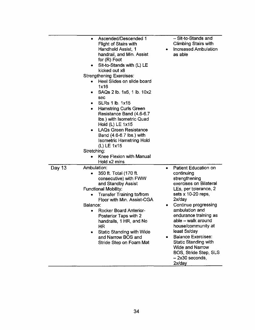
• Ascended/Descended 1 - Sit-to-Stands and Flight of Stairs with Climbing Stairs with Handheld Assist, 1 • Increased Ambulation handrail, and Min. Assist as able for (R) Foot
• Sit-to-Stands with (L) LE kicked out x8
Strengthening Exercises:
• Heel Slides on slide board 1x16
• SAQs 2 lb. 1x6, 1 lb. 10x2 sec
• SLRs 1 lb. 1x15
• Hamstring Curls Green Resistance Band (4.6-6.7 Ibs.) with Isometric Quad Hold (L) LE 1x15
• LAQs Green Resistance Band (4.6-6.7 Ibs.) with Isometric Hamstring Hold (L) LE 1x15
Stretching:
• Knee Flexion with Manual Hold x2 mins
Day 13 Ambulation: • Patient Education on • 350 ft. Total (170 ft. continuing
consecutive) with FWW strengthening and Standby Assist exercises on Bilateral
Functional Mobility: LEs, per tolerance, 2
• Transfer Training to/from sets x 10-20 reps, Floor with Min. Assist-CGA 2x/day
Balance: • Continue progressing
• Rocker Board Anterior- ambulation and Posterior Taps with 2 endurance training as handrails, 1 HR, and No able - walk around HR house/community at
• Static Standing with Wide least 5x1day and Narrow BOS and • Balance Exercises: Stride Step on Foam Mat Static Standing with
Wide and Narrow BOS, Stride Step, SLS - 2x30 seconds, 2x1da~
34
REFERENCES
1. AHRQ Study: Joint Replacement To Become the Most Common Elective
Surgical Procedure in the Next Decades. AHRQ--Agency for Healthcare
Research and Quality: Advancing Excellence in Health Care.
https:llwww.ahrq.gov/news/newsletters/e-newsletter/503.html. Published
February 2,2016. Accessed June 20,2017.
2. Total Knee Replacement-Ortholnfo - AAOS. Total Knee Replacement
Ortholnfo - AAOS. http://orthoinfo.aaos.org/topic.cfm?topic=A00389.
Published August 1,2015. Accessed June 20,2017.
3. Total Knee Replacement Surgery By the Numbers. A Nation in Motion.
http://www.anationinmotion.org/value/total-knee-replacement-surgery
numbers/. Accessed June 20, 2017.
4. George LK, Ruiz D, Sloan FA. The effects of total knee arthroplasty on
physical functioning in the older population. Arthritis & Rheumatism.
2008;58(10):3166-3171. doi:1 0.1 002/art.23888.
5. Artz N, Elvers KT, Lowe CM, Sackley C, Jepson P, Beswick AD.
Effectiveness of physiotherapy exercise following total knee replacement:
systematic review and meta-analysis. BMC Musculoskeletal Disorders.
2015;16(1). doi:10.1186/s12891-015-0469-6.
35
6. Basilico FC, Sweeney G, Losina E, et al. Risk factors for cardiovascular
complications following total joint replacement surgery. Arthritis &
Rheumatism. 2008;58(7):1915-1920. doi:10.1002/art.23607.
7. Park JH, Restrepo C, Norton R, Mandel S, Sharkey PF, Parvizi J.
Common Peroneal Nerve Palsy Following Total Knee Arthroplasty. The
Journal of Arthroplasty. 2013;28(9):1538-1542.
dOi:10.1016/j.arth.2013.02.025.
8. Schinsky MF, Macaulay W, Parks ML, Kiernan H, Nercessian OA Nerve
injury after primary total knee arthroplasty. The Journal of Arthroplasty.
2001 ;16(8):1 048-1 054. doi:1 0.1 054/arth.2001.26591.
9. Poage C, Roth C, Scott B. Peroneal Nerve Palsy. Journal of the American
Academy of Orthopaedic Surgeons. 2016;24(1):1-10. doi:10.5435/jaaos-d-
14-00420.
10. Pozzi F, Snyder-Mackler L, Zeni J. Physical exercise after knee
arthroplasty: A systematic review of controlled trials. Eur J Phys Rehabil
Med.2013;49(6):877-892.
11. Lin D-H, Lin C-HJ, Lin Y-F, Jan M-H. Efficacy of 2 Non-Weight-Bearing
Interventions, Proprioception Training Versus Strength Training, for
Patients With Knee Osteoarthritis: A Randomized Clinical Trial. Journal of
Orthopaedic & Sports Physical Therapy. 2009;39(6):450-457.
doi: 10.2519/jospt.2009.2923.
12. Bellamy J. Background on ICF. APTA
http://www.apta.org/ICF/Background/. Accessed June 20,2017.
36
13. Numeric Pain Rating Scale. Numeric Pain Rating Scale - Physiopedia.
http://www.physio-pedia.com/Numeric_Pain_Rating_Scale#cite_note-pB-
4. Accessed June 20,2017.
14. Ferraz MB, Quaresma MR, Aquino LR, Atra E, Tugwell P, Goldsmith CH.
Reliability of pain scales in the assessment of literate and illiterate patients
with rheumatoid arthritis. J RheumatoI1990;17:1022-4
15. Magee OJ. Orthopedic physical assessment. 6th ed. United States:
Elsevier - Health Sciences Division; January 14, 2014:765-BB7chap 12.
16. The Lower Extremity Functional Scale (LEFS): Scale Development,
Measurement Properties, and Clinical Application. Physical Therapy.
January 1999. doi:10.1093/ptjI79.4.371.
17. Stratford PW, Kennedy OM, Riddle DL. New study design evaluated the
validity of measures to assess change after hip or knee
arthroplasty. Journal of Clinical Epidemiology. 2009;62(3):347-352.
doi: 1 0.1 016/j.jclinepi.200B.06.00B.
1B. Ost LV. Cram Session in Goniometry and Manual Muscle Testing: a
Handbook for Students and Clinicians. Thorofare: SLACK Incorporated;
2013.
19. Alnahdi AH, Zeni JA, Snyder-Mackler L. Hip Abductor Strength Reliability
and Association With Physical Function After Unilateral Total Knee
Arthroplasty: A Cross-Sectional Study. Physical Therapy.
2014;94(B):1154-1162. doi:10.2522/ptj.20130335.
37
20. Piva SR, Teixeira PE, Almeida GJ, et al. Contribution of Hip Abductor
Strength to Physical Function in Patients With Total Knee
Arthroplasty. Physical Therapy. 2011 ;91 (2):225-233.
doi: 1 0.2522/ptj.201 00122.
21. Jogi P, Overend T J, Spaulding SJ, Zecevic A, Kramer JF. Effectiveness
of balance exercises in the acute post-operative phase following total hip
and knee arthroplasty: A randomized clinical trial. SAGE Open Medicine.
2015;3:205031211557076. doi:10.1177/2050312115570769.
22. Liao C-D, Liou T-H, Huang Y-Y, Huang Y-C. Effects of balance training
on functional outcome after total knee replacement in patients with knee
osteoarthritis: a randomized controlled trial. Clinical Rehabilitation.
2013;27(8):697-709. doi:10.1177/0269215513476722.
23. Liu C, Shiroy OM, Jones L Y, Clark DO. Systematic review of functional
training on muscle strength, physical functioning, and activities of daily
living in older adults. European Review of Aging and Physical Activity.
2014;11 (2):95-106. doi:1 0.1 007/511556-014-0144-1.
24. Brotzman BS, Manske RC, Daugherty K, Robert dos Remedios MA
CSCS. Clinical Orthopaedic rehabilitation: An evidence-based approach -
expert consult: Print and online. 3rd ed. Philadelphia, PA: Elsevier Mosby;
May 6, 2011 :chap 6.
25. Liitzner C, Kirschner S, LOtzner J. Patient Activity After TKA Depends on
Patient-specific Parameters. Clinical Orthopaedics and Related
Research®. 2014;472(12):3933-3940. doi:10.1007/s11999-014-3813-5.
38
26. Ciolac E, Silva J, Greve J. Effects of resistance training in older women
with knee osteoarthritis and total knee arthroplasty. Clinics. 2015;70(1):7-
13. doi:10.6061/c1inics/2015(01 )02.
27. Bade MJ, Stevens-Lapsley JE. Early High-Intensity Rehabilitation
Following Total Knee Arthroplasty Improves Outcomes. Journal of
Orthopaedic & Sports Physical Therapy. 2011;41 (12):932-941.
doi: 1 0.2519/jospt.2011.3734.
28. Zywiel MG, Mont MA, Mcgrath MS, Ulrich SO, Bonutti PM, Bhave A.
Peroneal Nerve Dysfunction After Total Knee Arthroplasty. The Journal of
Arthroplasty. 2011 ;26(3):379-385. doi:1 0.1 016/j.arth.2010.03.020.
39