Embed Size (px)
Citation preview

This chapter is an edited version of the manuscript: Den Hartog, L., Huddleston Slater J.J., Vissink,
A., Meijer, H.J., Raghoebar, G.M. Treatment outcome of immediate, early and conventional single-
tooth implants in the aesthetic zone. A systematic review to survival, bone level, soft tissue, aesthetics
and patient satisfaction. Journal of Clinical Periodontology 2008; 35: 1073 - 1086
2.
Treatment outcome of immediate, early and
conventional single-tooth implants in the aesthetic
zone
A systematic review to survival, bone level, soft tissue, aesthetics and patient satisfaction

2
Cha
pter
14
AbstrAct
Aim: To evaluate, through a systematic review of the literature, the out-
come of single-tooth implants in the aesthetic zone with natural adjacent
teeth, thereby addressing immediate, early and conventional implant ap-
proaches.
Material and Methods: MEDLINE (1950-2008), EMBASE (1966-2008), and
CENTRAL (1800-2008) were searched to identify eligible studies. Two re-
viewers independently assessed the methodological quality using specific
study-design related assessment forms.
Results: Out of 86 primarily selected articles, 19 studies fulfilled the inclu-
sion criteria. A meta-analysis showed an overall survival rate of 95.5% (95%
CI: [93.0 – 97.1]) after one year. A stratified meta-analysis revealed no differ-
ences in survival between immediate, early and conventional implant strat-
egies. Minor marginal peri-implant bone resorption was found together
with low incidence of biological and technical complications. No significant
differences in outcome measures were reported in clinical trials comparing
immediate, early or conventional implant strategies.
Conclusion: The included literature suggest that promising short-term re-
sults can be achieved for immediate, early and conventional single-tooth
implants in the aesthetic zone. However, important parameters as aesthetic
outcome, soft tissue aspects and patient satisfaction were clearly underex-
posed. The question whether immediate and early single implant therapies
will result in better treatment outcomes remains inconclusive due to lack
of well-designed controlled clinical studies.

Syst
emat
ic r
evie
w
2
1515
IntroductIon
The application of dental implants for single-tooth replacements has evolved into
a viable prosthodontic alternative to conventional fixed bridgework, resin-bonded
restorations or removable partial dentures. Long-term studies have reported ex-
cellent implant survival rates when applied for single-tooth replacements (Schel-
ler et al. 1998, Romeo et al. 2002). Psychological benefits and tooth structure
conservation adjacent to the tooth to be replaced, are among the advantages of
implant supported restorations.
In the anterior zone, the success of single-tooth implant therapy is not only
determined by high survival rates, but even more by the (long-term) quality of
survival, dictated by a mixture of several factors. Preferably, the appearance of
the peri-implant soft tissue should be in harmony with the mucosa around the
adjacent teeth and the implant crown should be in balance with the neighbouring
dentition (Meijer et al. 2005). Various implant treatment strategies have been pro-
posed for the accomplishment of optimal aesthetics. These include approaches to
rehabilitate the underlying bone structures by augmentation procedures with au-
tologous bone and/or bone substitutes (Weber et al. 1997, Jensen et al. 2006, Pelo
et al. 2007), techniques to manipulate and enhance the architecture of the peri-
implant soft tissue (Zetu & Wang 2005, Esposito et al. 2007) and methods for al-
veolar ridge preservation following tooth extraction (Lekovic et al. 1997, Irinakis &
Tabesh 2007). Furthermore, implants and abutments with specific configurations
have been introduced to sustain the hard and soft tissue (Wohrle 2003, Morton et
al. 2004, Lazzara & Porter 2006, Maeda et al. 2007, Noelken et al. 2007) together
with provisionalization techniques to restore the soft tissue contour (Jemt 1999,
Al-Harbi & Edgin 2007), and the introduction of ceramic customized abutments
and ceramic implant crowns (Canullo 2007, Schneider 2008).
Traditionally, dental implants were placed in healed extraction sites according
to a two-stage surgical procedure and an undisturbed load-free period of three
to six months. In contemporary implantology, however, installation of implants
in fresh extraction sockets and reducing the load-free period by immediate re-
storing implants after insertion have gained attention. Besides shortening of to-
tal treatment time, fewer surgical interventions and eliminating the need for a
temporary prosthesis, these immediate approaches might lead to a reduction of
peri-implant crestal bone loss and a better soft tissue healing thus possibly im-
proving the aesthetics (Esposito et al. 2006, Glauser et al. 2006, Harvey 2007).
On the other hand, there are potential risk factors involved with these techniques
such as enhanced possibility of infection, mismatch between socket wall and im-
plant leading to gap creation and induction of fibrous tissue formation around
the bone-implant interface caused by implant micromovement during eventful
wound healing (Gapski et al. 2003, Esposito et al. 2006). These risk factors may

2
Cha
pter
16
worsen the treatment outcome. This discrepancy needs further study.
The outcome of a single-tooth implant with natural neighbouring teeth may
be dissimilar to cases in which multiple adjacent teeth are replaced by dental im-
plants, because dimensions of the hard and soft tissue between adjacent implants
differ significantly from dimensions found in single implant cases. Single implant
cases take benefit of tissue support of the adjacent dentition (Grunder 2000, Kan
et al. 2003b, Belser et al. 2004). When considering the heights of inter-implant
papillae for instance, studies indicated that these papillae might show inadequacy
for complete enclosure of the inter-implant area with soft tissue, thereby failing to
duplicate the interproximal soft tissue appearance of the adjacent teeth (Tarnow
et al. 1992, Tarnow et al. 2003, Lee et al. 2005). This deficiency may affect the
aesthetic outcome unfavorably. The soft tissue height proximal to single-tooth
implants is on average higher and is suggested to be related to the interproximal
bone level of the adjacent teeth (Grunder 2000, Kan et al. 2003b). Hence, single-
tooth implant therapy may lead to more favorable treatment outcomes compared
to a therapy in which adjacent implants are involved.
To date, several reviews have been published regarding the clinical outcome of
immediate and conventional implant supported single-tooth restorations in par-
tially edentulous patients (Creugers et al. 2000, Berglundh et al. 2002, Belser et
al. 2004, Glauser et al. 2006, Jung et al. 2008). Most of these reviews have mainly
converged on implant survival and addressed to a lesser degree other outcome
measures that determine the quality of survival. Furthermore, none of these re-
views systematically analyzed the literature concerning the efficacy of single-tooth
implants in the aesthetic zone neither did these reviews concentrate explicitly
on the outcome of single implants with natural neighbouring teeth, applied to
replace one missing tooth. However, it is worthwhile for patients and clinicians
to know whether an immediate or conventional single-tooth implant represents
a predictive, reliable and effectual therapy to re-establish function and aesthetics
subsequent to the loss of a single anterior tooth. Therefore, the objective of this
study was to evaluate, through a systematic review of the literature, the outcome
of single-tooth replacements by dental implants in the aesthetic zone in cases in
which the adjacent teeth are natural, thereby focussing on immediate, early and
conventional implant treatment strategies.
MAterIAl And Methods
Types of studies Longitudinal studies (Randomized controlled trials (RCTs), clinical trials, cohort-
studies and case series) were considered for evaluation. Retrospective studies were
excluded. Only case series that investigated at least five patients were contemplated

Syst
emat
ic r
evie
w
2
1717
for inclusion. No time restrictions were implemented. Language was restricted to
papers published in English, German, French, Spanish, Italian and Dutch.
Type of participantsPatients who were treated with an implant-retained single-tooth replacement in the
aesthetic zone neighbored with natural teeth, could be included. The aesthetic zone
was defined as the region in the maxilla or mandible, ranging from second premo-
lar to second premolar (teeth 15-25 and teeth 35-45).
Types of interventionimmediate implant placement: defined as implant placement immediately fol-- lowing extraction of a tooth;
early implant placement: defined as installation of the implant 4 to 8 weeks - after extraction;
conventional implant placement: implant placement ≥ 8 weeks post-extrac-- tion;
immediate loading: application of a load by means of a restoration within 48 - hours of implant placement;
early loading: application of a load by means of a restoration after 48 hours but - less than 3 months after implant placement;
conventional loading: application of a load by means of a restoration ≥ 3 - months after implant placement (Laney 2007).
For studies to be eligible in this review, they had to evaluate endosseous root-form
dental implants with a follow-up of at least 1 year after placement of the implant
crown.
Types of outcome measuresimplant survival, defined as presence of the implant at time of follow-up ex-- aminations;
changes in marginal peri-implant bone level assessed on radiographs;- aesthetics evaluated by dental professionals;- aspects of the peri-implant structures, i.e. level of marginal gingiva, papilla in-- dex (Jemt 1997), probing pocket depth, presence of plaque, bleeding on prob-
ing;
patient satisfaction including aesthetics;- biological and technical complications.-
Search StrategyFor this review, a thorough search of the literature was conducted in databases
of MEDLINE (1950-2008 (via PUBMED) and EMBASE (1966 – 2008). The search

2
Cha
pter
18
was supplemented with a systematic search in the ‘Cochrane Central Register
of Controlled Trials’ (CENTRAL) (1800–2008). The search strategy used, was a
combination of MeSH terms and free text words and is summarized in Table 1.
The search was complemented by checking references of relevant review arti-
cles and eligible studies for additional useful publications. Titles and abstracts of
the searches were scanned independently by two examiners. Full-text documents
were obtained for all possibly relevant articles. Full text analysis was performed
for second selection by two reviewers independently against the stated inclusion
criteria. In case of disagreement, consensus was reached by discussion, if neces-
sary in consultation with a third reviewer.
Table 1. Search strategy.
#1 Search “Dental Implants”[MeSH] OR “Dental Implantation”[MeSH] OR implant*
#2 Search “single implant*” OR “single tooth” OR “single teeth” OR “single crown*” OR
“single restoration*”
#3 Search “aesthetic*” OR “esthetic*” OR “anterior*” OR “front*” OR “incisor*”
#4 Search #1 AND #2 AND #3
Run data search: June 2008.
Quality assessmentMethodological quality was assessed using specific study-design related forms de-
signed by the Dutch Cochrane Collaboration. As there was no checklist available for
the assessment of the quality of case series, a quality-assessment tool was specifi-
cally developed for this review, adapted from the quality form used for clinical trials
(Table 2). Two observers independently generated a score for the included articles,
expressed in the number of plusses given. It was decided that studies scoring 5 or
more plusses were considered to be methodological ‘acceptable’.
Data extraction and synthesisFor each trial the following data were extracted by two review authors independ-
ently and recorded in a data sheet:
number of patients, implants placed, drop-outs and follow-up time. For all - included longitudinal studies of more than one year, follow-up time was cal-
culated as person-years;
details of type of intervention;- details of the outcomes stated, including method of assessment. -
Agreement was reached by a consensus discussion and if necessary a third
reviewer was consulted. If feasible, a meta-analysis was carried out if the outcome
measures could be meaningfully combined.
Syst
emat
ic r
evie
w
2
1919
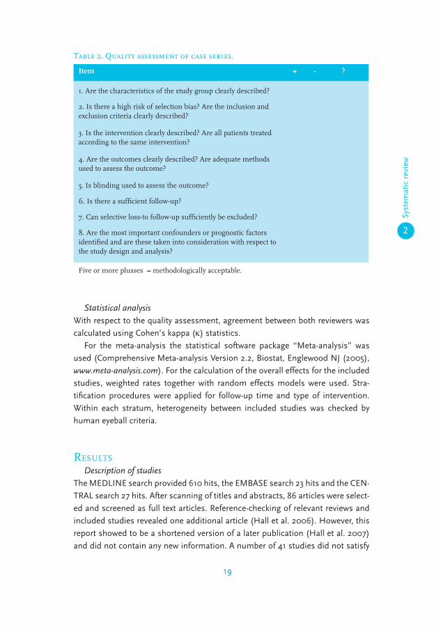
Table 2. Quality assessment of case series.
Item + - ?
1. Are the characteristics of the study group clearly described?
2. Is there a high risk of selection bias? Are the inclusion and exclusion criteria clearly described?
3. Is the intervention clearly described? Are all patients treated according to the same intervention?
4. Are the outcomes clearly described? Are adequate methods used to assess the outcome?
5. Is blinding used to assess the outcome?
6. Is there a sufficient follow-up?
7. Can selective loss-to follow-up sufficiently be excluded?
8. Are the most important confounders or prognostic factors identified and are these taken into consideration with respect to the study design and analysis?
Five or more plusses = methodologically acceptable.
Statistical analysisWith respect to the quality assessment, agreement between both reviewers was
calculated using Cohen’s kappa (κ) statistics.
For the meta-analysis the statistical software package “Meta-analysis” was
used (Comprehensive Meta-analysis Version 2.2, Biostat, Englewood NJ (2005),
www.meta-analysis.com). For the calculation of the overall effects for the included
studies, weighted rates together with random effects models were used. Stra-
tification procedures were applied for follow-up time and type of intervention.
Within each stratum, heterogeneity between included studies was checked by
human eyeball criteria.
results
Description of studiesThe MEDLINE search provided 610 hits, the EMBASE search 23 hits and the CEN-
TRAL search 27 hits. After scanning of titles and abstracts, 86 articles were select-
ed and screened as full text articles. Reference-checking of relevant reviews and
included studies revealed one additional article (Hall et al. 2006). However, this
report showed to be a shortened version of a later publication (Hall et al. 2007)
and did not contain any new information. A number of 41 studies did not satisfy

2
Cha
pter
20
the inclusion criteria because data of single-tooth implants in anterior and poste-
rior zones was not presented separately or adjacent implants were also included,
making it not possible to extract proper data. Furthermore, 14 studies were ex-
cluded due to improper study design (not longitudinal or not prospective) and 5
studies because of a follow-up < 1 year. A total of 26 articles fulfilled the inclusion
and exclusion criteria and were assessed methodologically. Of these 26 studies, 7
studies were excluded. Reasons for exclusion are depicted in Table 3.
Table 3. Studies excluded after quality assessment and reasons for exclusion.
Study Study design Reasons for exclusion
Henriksson 2004
Clinical trial Heterogeneity in clinical procedure (different im-plants, different load-free periods), in/exclusion criteria unclear, no blinding used, prognostic factors/confounders not considered.
Lorenzoni 2003
Case series Patients not treated according to same intervention (immediate and conventional placement included), in/exclusion criteria unclear, no blinding used, prognos-tic factors/confounders not considered.
Ferrara 2006 Case series High risk of selection bias (implants with insufficient primary stability were excluded; method to assess sta-bility not clear), outcomes not clearly described, meth-ods used to assess the outcome unclear, no blinding used, prognostic factors/confounders not considered.
Grunder 2000 Case series Patients characteristics unclear, in/exclusion criteria unclear, no blinding used, prognostic factors/con-founders not considered.
Locante 2004 Case series Patients not treated according to same intervention (immediate and conventional placement included), high risk of selection bias (implants with insufficient primary stability were excluded; method to assess stability not clear), in/exclusion criteria unclear, no adequate methods used to assess the outcome, no blinding used, follow-up routine unclear, prognostic factors/confounders not considered.
Groisman 2003
Case series Patient characteristics unclear, high risk of selection bias (only favorable cases selected), method of assess-ment not clear, no blinding used, prognostic factors/confounders not considered, follow-up routine unclear.
Barone 2006 Case series Patient characteristics not clear, high risk of selection bias (only favorable cases selected), no blinding used, prognostic factors/confounders not considered.

Syst
emat
ic r
evie
w
2
2121
The κ-value for inter-assessor agreement on the methodological quality was 0.89.
Disagreements were generally caused by slight differences in interpretation and
were easily resolved in a consensus meeting. Finally, 19 publications remained for
data extraction. Figure 1 outlines the algorithm of the study selection procedure.
Of the included studies, 5 were RCTs, 2 were clinical trials and 12 were case series.
Six publications presented outcomes of the same patient population, but differed
in follow-up (Palmer et al. 1997, 2000, Cooper et al. 2001, 2007, Jemt & Lekholm
2003, 2005) and results of one study group were reported in two different publi-
cations addressing different topics (Schropp et al. 2005a, 2005b).
Identified articles- MEDLINE search: n = 610- EMASE search: n = 23- CENTRAL search: n = 27
Included for full text analysisn = 86
Included for methodological appraisaln = 26
Included for data analysisn = 19
Excluded articles• Improper study design• Non-topic related• No abstract available• Follow-up < 1 year
Excluded articles• Required data not presented• Improper study design• Follow-up < 1 year
Excluded articlesGrunder 2000; Groisman et al., 2003; Lorenzoni et al., 2003; Locante et al., 2004; Henriksson et al., 2004; Ferrara et al. , 2006; Barone et al., 2006
Most of the studies only evaluated maxillary implants, but three studies did
also include implants placed in the mandible (38 implants in total) (Schropp et
al. 2005a, Schropp et al. 2005b, Romeo et al. 2008). Furthermore, implants were
installed mostly in completely healed extraction sockets or early after extraction
(10 days to 4 weeks) and subsequently were restored according to immediate,
early (1 to 3 weeks after implant placement) or conventional loading protocols.
Restorations that were seated immediate or early after implant placement, were
Figure 1. Algorithm of study selection procedure.

22
Tab
le 4
. St
ud
y ch
arac
teri
stic
s an
d o
utc
om
es o
f in
clu
ded
stu
die
s, a
rran
ged
acc
ord
ing
to
typ
e o
f in
terv
enti
on
an
d s
tud
y d
esig
n.
Stud
yIn
terv
enti
onD
esig
nN
o. o
f pa
tien
ts/
impl
ants
Impl
ant
syst
emR
easo
n(s
) for
too
thlo
ss (n
o.)
Follo
w-
up p
erio
d (y
rs)
No.
of
impl
ant
drop
-ou
ts**
Surv
ival
ra
te (%
)C
han
ge in
mar
-gi
nal
bon
e le
vel
± SD
(mm
)
Kan
20
03
Imm
edia
te p
lace
men
t an
d im
med
iate
load
ing
CS
35/3
5R
epla
ce
Sele
ctF
ract
ure
(15)
, en
dodo
nti
c fa
ilure
(12)
, roo
t res
orp-
tion
(8)
10
100
- 0.2
4 ±
0.3
5* #
De
Rou
ck 2
00
8Im
med
iate
pla
cem
ent
and
imm
edia
te lo
adin
gC
S30
/30
Rep
lace
Se
lect
Fra
ctu
re (1
0),
cari
es/
endo
don
tic
(9),
peri
-od
onta
l (7)
, ro
ot r
esor
ptio
n (4
)
11
97
- 0.8
8 ±
0.5
2* #
Lin
debo
om 2
00
6Im
med
iate
pla
cem
ent v
s co
nve
nti
onal
pla
cem
ent
RC
T
T C25
/25
25/2
5F
rial
it-2
N
R1
2 09
210
0- 0
.51
± 0
.12*
#
- 0.5
2 ±
0.1
5* #
Sch
ropp
20
05
(a)
Ear
ly p
lace
men
t vs
con
ven
tion
al p
lace
men
tR
CT
T C
23/2
322
/22
3iR
oot f
ract
ure
(NR
), en
dodo
nti
c fa
ilure
(NR
), pe
riod
onti
tis
(NR
), ad
van
ced
cari
es le
sion
s (N
R)
2N
RN
RN
R
Sch
ropp
20
05
(b)
Ear
ly p
lace
men
t vs
con
ven
tion
al p
lace
men
tR
CT
T C
23/2
323
/23
3iR
oot f
ract
ure
(NR
), en
dodo
nti
c fa
ilure
(NR
), pe
riod
onti
tis
(NR
), ad
van
ced
cari
es le
sion
s (N
R)
23 2
91
96
- 0.8
± N
R ‡
- 0.7
± N
R ‡
Got
fred
sen
20
04
Ear
ly p
lace
men
t vs
con
ven
tion
al p
lace
men
tC
T
T C
10/1
010
/10
Ast
ra T
ech
Roo
t fra
ctu
re (1
5), a
gen
-es
is (3
), tr
aum
a (2
)5
0 010
010
0- 0
.34
± 0
.57 ¶
- 0.2
6 ±
0.3
8 ¶
Rom
eo 2
00
8Im
med
iate
pla
cem
ent
CS
48/4
8IT
IC
arie
s/en
dodo
nti
c w
ith
ro
ot o
r cr
own
fra
ctu
re
(NR
)
10
100
NR

2323
Tab
le 4
. (C
on
tin
ued
)
Stud
yIn
terv
enti
onD
esig
nN
o. o
f pa
tien
ts/
impl
ants
Impl
ant
syst
emR
easo
n(s
) for
too
thlo
ss (n
o.)
Follo
w-
up p
erio
d (y
rs)
No.
of
impl
ant
drop
-ou
ts**
Surv
ival
ra
te (%
)C
han
ge in
mar
-gi
nal
bon
e le
vel
± SD
(mm
)
Hal
l 20
07
Imm
edia
te lo
adin
g vs
co
nve
nti
onal
load
ing
RC
T
T C14
/14
14/1
4So
uth
ern
Im
plan
tsN
R1
1 29
310
0- 0
.63
± 1
.00
¶
- 0.7
8 ±
1.0
1 ¶
Eri
csso
n 2
00
0Im
med
iate
load
ing
vs
con
ven
tion
al lo
adin
gC
T
T C
14/1
48/
8B
rån
emar
kN
R1.
52 0
85.7
100
- 0.1
4 ±
0.3
6 ¶
- 0.0
7 ±
0.7
9 ¶
Coo
per
200
1E
arly
load
ing
CS
48/5
4A
stra
Tec
hN
R1
39
4.4
- 0.4
± N
R §
Coo
per
200
7E
arly
load
ing
CS
48/5
4A
stra
Tec
hN
R3
119
4.4
-0.4
2 ±
0.5
9§
An
ders
en 2
00
2E
arly
load
ing
CS
8/8
ITI
NR
50
100
+ 0
.53
± N
R§
Mei
jnde
rt 2
00
7C
onve
nti
onal
RC
T9
3/9
3IT
IN
R1
29
7.8
NR
Jem
t 20
03
Con
ven
tion
alC
S10
/10
Brå
nem
ark
Tra
um
a (1
0)
31
100
- 0.3
± 0
.36
¶
Jem
t 20
05
Con
ven
tion
alC
S10
/10
Brå
nem
ark
Tra
um
a (1
0)
62
100
- 0.3
± 0
.24 ¶
Zar
one
200
6C
onve
nti
onal
CS
30/3
4IT
IA
gen
esis
(30
)2
- 3.2
110
0- 1
.2 ±
0.6
1 §
Pal
mer
19
97
Con
ven
tion
al
CS
15/1
5A
stra
Tec
h
NR
21
100
+ 0
.01
± 0
.50
* ¶
Pal
mer
20
00
Con
ven
tion
alC
S15
/15
Ast
ra T
ech
NR
51
100
+ 0
.12
± 0
.49
* ¶
Car
daro
poli
20
06
Con
ven
tion
alC
S16
/16
Brå
nem
ark
NR
15
100
- 1.6
± 0
.57*
#
*Sta
nda
rd d
evia
tion
cal
cula
ted.
**
Defi
ned
as
impl
ants
that
did
not
su
rviv
e an
d im
plan
ts lo
st to
fol
low
-up.
# F
rom
impl
ant p
lace
men
t. ‡
Fro
m h
ealin
g ab
utm
ent p
lace
men
t. §F
rom
tem
-
pora
ry c
row
n p
lace
men
t. ¶
Fro
m d
efin
itiv
e cr
own
pla
cem
ent.
Abb
revi
atio
ns:
NR
= n
ot r
epor
ted,
RC
T=r
ando
miz
ed c
linic
al tr
ial,
CT
= cl
inic
al tr
ial,
CS
= ca
se s
erie
s, T
= te
st g
rou
p,
C =
con
trol
gro
up.
24
Tab
le 5
. O
utc
om
es o
f in
clu
ded
stu
die
s, a
rran
ged
acc
ord
ing
to
typ
e o
f in
terv
enti
on
an
d s
tud
y d
esig
n.
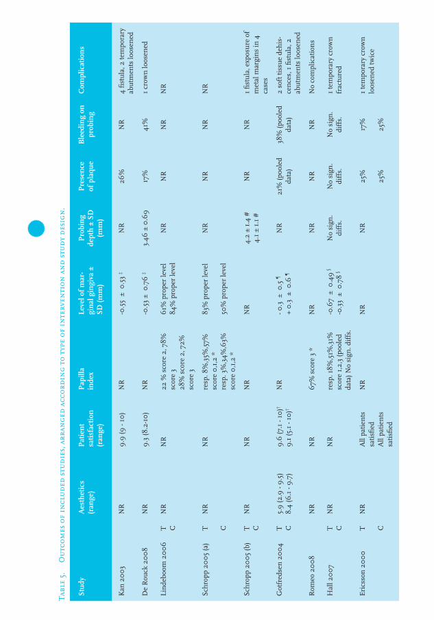
Stud
yA
esth
etic
s
(ran
ge)
Pat
ien
t sa
tisf
acti
on
(ran
ge)
Pap
illa
in
dex
Leve
l of
mar
-gi
nal
gin
giva
±
SD (m
m)
Pro
bin
g de
pth
± SD
(m
m)
Pre
sen
ce
of p
laqu
eB
leed
ing
on
prob
ing
Com
plic
atio
ns
Kan
20
03
NR
9.9
(9 -
10)
NR
-0
.55
± 0
.53
‡N
R
26%
N
R
4 fi
stu
la, 2
tem
pora
ry
abu
tmen
ts lo
osen
ed
De
Rou
ck 2
00
8N
R9
.3 (8
.2-1
0)
NR
-0.5
3 ±
0.7
6 ‡
3.46
± 0
.69
17%
41%
1 cr
own
loos
ened
Lin
debo
om 2
00
6T C
NR
NR
22
% s
core
2, 7
8%
scor
e 3
28%
sco
re 2
, 72%
sc
ore
3
61%
pro
per
leve
l84
% p
rope
r le
vel
NR
N
R
NR
NR
Sch
ropp
20
05
(a)
T C
NR
N
R
resp
. 8%
,35%
,57%
sc
ore
0,1
,2 *
resp
. 3%
,34%
,63%
sc
ore
0,1
,2 *
83%
pro
per
leve
l
50%
pro
per
leve
l
NR
NR
N
RN
R
Sch
ropp
20
05
(b)
T CN
R
NR
N
RN
R4.
2 ±
1.4
#4.
1 ±
1.1
#N
R
NR
1 fi
stu
la, e
xpos
ure
of
met
al m
argi
ns
in 4
ca
ses
Got
fred
sen
20
04
T
C5.
9 (2
.9 -
9.5
)8.
4 (6
.1 -
9.7
)9
.6 (7
.1 -
10)†
9.1
(5.1
- 10
)†
NR
- 0
.3 ±
0.5
¶
+ 0
.3 ±
0.6
¶
NR
21
% (p
oole
d da
ta)
38%
(poo
led
data
)2
soft
tiss
ue
deh
is-
cen
ces,
1 fi
stu
la, 2
ab
utm
ents
loos
ened
Rom
eo 2
00
8N
RN
R6
7% s
core
3 *
NR
NR
NR
NR
No
com
plic
atio
ns
Hal
l 20
07
T CN
RN
R
resp
. 18%
,51%
,31%
sc
ore
1,2,
3 (p
oole
d da
ta) N
o si
gn. d
iffs
.
-0.6
7 ±
0.4
9 §
-0.3
3 ±
0.7
8 §
No
sign
. di
ffs.
No
sign
. di
ffs.
N
o si
gn.
diff
s.1
tem
pora
ry c
row
n
frac
ture
d
Eri
csso
n 2
00
0T C
NR
A
ll pa
tien
ts
sati
sfied
All
pati
ents
sa
tisfi
ed
NR
N
RN
R
25%
25%
17%
25%
1 te
mpo
rary
cro
wn
lo
osen
ed tw
ice

2525
Tab
le 5
. (C
on
tin
ued
)
Coo
per
200
1N
R
NR
N
R
+ 0
.34
± 0
.94
§N
R
0.5
% o
f si
tes
exam
ined
NR
1 ad
jace
nt t
ooth
mi-
grat
ed, 1
per
i-im
plan
t m
uco
siti
s, 1
impl
ant
disc
omfo
rt, 3
cro
wn
s lo
osen
ed; 4
fra
ctu
red
Coo
per
200
7N
RN
RN
R+
0.5
1 ±
1.4
2 §
NR
NR
NR
See
abov
e. N
o n
ew
com
plic
atio
ns
re-
port
ed
An
ders
en 2
00
2N
RN
RN
RN
RN
RN
RN
R1
fist
ula
, 3 c
row
ns
loos
ened
Mei
jnde
rt 2
00
76
6%
acc
epta
ble
8.5
( 6-1
0)
NR
N
RN
R
NR
N
R
NR
Jem
t 20
03
NR
N
R
50%
sco
re 2
, 50
%
scor
e 3
NR
N
R
NR
NR
No
com
plic
atio
ns
Jem
t 20
05
NR
NR
NR
- 0.1
± N
R ¶
NR
NR
NR
No
com
plic
atio
ns
Zar
one
200
63%
not
sat
isfa
ctor
y N
R
resp
. 6%
,12%
,82%
sc
ore
1,2,
3-0
.6 ±
NR
§2.
6 ±
0.2
#18
%
No
blee
din
gN
o im
plan
t-re
late
d co
mpl
icat
ion
s.
Pal
mer
19
97
NR
NR
NR
No
rece
ssio
nN
RN
RN
o bl
eedi
ng
No
soft
tiss
ue
com
-pl
icat
ion
s. 1
cro
wn
lo
osen
ed,
1 po
rcel
ain
fra
ctu
re
Pal
mer
20
00
NR
NR
NR
N
o re
cess
ion
NR
N
RR
are
See
abov
e. N
o n
ew
com
plic
atio
ns
re-
port
ed
Car
daro
poli
20
06
NR
NR
resp
. 14%
,68%
,18%
sc
ore
1,2,
3 -0
.6 ±
0.7
¶2.
4 ±
0.8
NR
9%
N
R
*Mod
ifica
tion
of
Pap
illa
Inde
x. †
Mea
n V
AS-
scor
es f
or a
esth
etic
app
eara
nce
an
d ge
ner
al f
un
ctio
n. #
Stan
dard
dev
iati
on c
alcu
late
d. ‡
Fro
m im
plan
t pla
cem
ent.
§ Fro
m te
mpo
rary
cro
wn
plac
emen
t. ¶
Fro
m d
efin
itiv
e cr
own
pla
cem
ent.
Abb
revi
atio
ns:
T=t
est g
rou
p, C
= c
ontr
ol g
rou
p, N
R =
not
rep
orte
d.

2
Cha
pter
26
all kept out of direct occlusal contact. Two studies reported on immediately re-
stored implants placed directly after tooth extraction. All clinical trials except one
compared the outcome of immediate or early implant placement and immediate
or early implant loading with conventional approaches. This RCT focused on dif-
ferent bone augmentation procedures and all implants were placed and restored
conventionally according to the same protocol (Meijndert et al. 2007). Character-
istics of the included studies are presented in Table 4 and are arranged according
to type of intervention and study design.
Due to methodological diversity of the ‘acceptable’ studies, only data on implant
survival and to a limited degree marginal bone resorption could be meaningfully
combined in a meta- analysis. Therefore, the outcomes are mainly presented as a
descriptive review in the subsequent sections and are depicted in Table 4 and 5.
Implant survival The implant survival rate was defined as the percentage of implants that was still
present at follow-up. All implants that were lost, failed within the first six months
after placement. In some studies implant mobility was detected at second stage
surgery (seven implants) (Schropp et al. 2005b, Lindeboom et al. 2006, Meijndert
et al. 2007) or occurred following placement of the provisional restoration (one
implant) (Cooper et al. 2001), whereas other implants were already in function
when they appeared not to be osseointegrated (six implants) (Ericsson et al.
2000, Cooper et al. 2001, Hall et al. 2006, De Rouck et al. 2008). Altogether, a
total number of 509 single-tooth implants was originally installed in 499 patients
of which 13 patients and 13 implants were lost to follow-up and no information
on survival was available regarding these implants. A total of 14 implants did not
survive.
Since it is generally known that implant loss is most often observed early after
implant installation and/or implant restoration, event rates and survival rates were
calculated in a stratified manner. To that end, results are presented for implants
that were followed up to one year after implant restoration (including implants
that were lost before restoration and consequently were not yet in function) and
implants with an observation period of more than one year after restoration (with
a correction for implants that were lost within the first year after restoration). Re-
sults of the weighted meta-analysis (for study size) of implant loss within one year,
expressed as event rates, are shown in Figure 2. The overall event rate was calcu-
lated as 0.045 (95% conficende interval (CI): 0.029 – 0.070) and can be expressed
as a survival rate of 95.5% (95% CI: 93.0 – 97.1). The weighted meta-analysis (for
person-years and study-size) regarding loss of implants that are more than one
year in function, showed an event rate of 0.007 (95% CI: 0.003 – 0.019).

Syst
emat
ic r
evie
w
2
2727
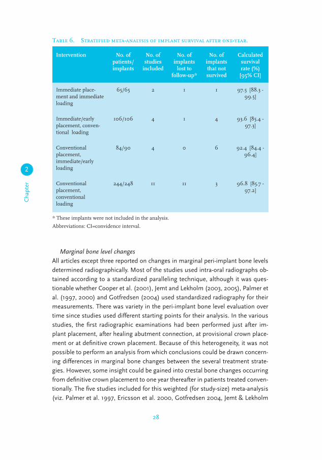
Globally four different treatment strategies could be identified. In this matter,
survival outcomes of immediate and early placed implants that were restored
conventionally were combined as well as implants that were installed convention-
ally but were restored immediately or early. Results of the weighted (for study-
size) stratified meta-analysis are presented in Table 6, revealing no differences
in survival rate after one-year follow-up. Focussing on the studies individually, no
statistically significant differences in implant survival were found in clinical trials
comparing immediate or early implant procedures with conventional ones.
Figure 2. Meta-analysis of implant loss within one year after restoration.
2
Cha
pter
28
Marginal bone level changesAll articles except three reported on changes in marginal peri-implant bone levels
determined radiographically. Most of the studies used intra-oral radiographs ob-
tained according to a standardized paralleling technique, although it was ques-
tionable whether Cooper et al. (2001), Jemt and Lekholm (2003, 2005), Palmer et
al. (1997, 2000) and Gotfredsen (2004) used standardized radiography for their
measurements. There was variety in the peri-implant bone level evaluation over
time since studies used different starting points for their analysis. In the various
studies, the first radiographic examinations had been performed just after im-
plant placement, after healing abutment connection, at provisional crown place-
ment or at definitive crown placement. Because of this heterogeneity, it was not
possible to perform an analysis from which conclusions could be drawn concern-
ing differences in marginal bone changes between the several treatment strate-
gies. However, some insight could be gained into crestal bone changes occurring
from definitive crown placement to one year thereafter in patients treated conven-
tionally. The five studies included for this weighted (for study-size) meta-analysis
(viz. Palmer et al. 1997, Ericsson et al. 2000, Gotfredsen 2004, Jemt & Lekholm
Table 6. Stratified meta-analysis of implant survival after one-year.
Intervention No. of patients/ implants
No. of studies
included
No. of implants
lost to follow-up*
No. of implants that not survived
Calculated survival rate (%) [95% CI]
Immediate place-ment and immediate loading
65/65 2 1 1 97.5 [88.3 - 99.5]
Immediate/early placement, conven-tional loading
106/106 4 1 4 93.6 [85.4 - 97.3]
Conventional placement, immediate/early loading
84/90 4 0 6 92.4 [84.4 - 96.4]
Conventional placement, conventional loading
244/248 11 11 3 96.8 [85.7 - 97.2]
* These implants were not included in the analysis.
Abbreviations: CI=convidence interval.

Syst
emat
ic r
evie
w
2
29
2005, Cardaropoli et al. 2006) (in total 52 implants) revealed a mean marginal
bone loss of 0.20 mm (95% CI: 0.034 – 0.36) during the first year after installa-
tion of the definitive crown (see Figure 3). Data from radiographic examinations
were mostly presented as mean values and consequently no frequency distribu-
tions were given. Cooper et al. (2001) considered the incidence of marginal bone
loss of 48 implants one year after insertion. The latter authors found that after
one year eight implants showed a cortical bone loss of 1.0 to 2.0 mm and three
implants more than 2.0 mm. Finally, the bone level changes detected in the ex-
perimental and conventional study groups of the included clinical trials were not
significantly different.
* * *
Figure 3. Meta-analysis of marginal bone level changes 1 year after instal-lation of the definitive crown.
AestheticsAlbeit all implants reviewed were inserted in the aesthetic zone, only three stud-
ies included the aesthetic outcome in their analysis. Zarone et al. (2006) con-
sidered one implant not being satisfactory because of exposure of the titanium
neck. It was, however, unclear how the aesthetics were measured. At the three-
year control visit Gotfredsen (2004) asked an independent dentist to evaluate
the aesthetic appearance of the implant crowns using a visual analog scale (VAS)
ranging from ‘very unsatisfied’ (score 0) to ‘very satisfied’ (score 10). In the study
by Meijndert et al. (2007), a prosthodontist rated the aesthetics on colour pho-
tographs using an objective rating index. It appeared that 34% of the cases were
judged as poor aesthetics.

2
Cha
pter
30
Peri-implant structuresTo evaluate the quantity of the interproximal gingival papillae, some studies made
use of the papilla index according to Jemt (1997) or a slight modification of this
classification (Schropp et al. 2005a, Romeo et al. 2008). It revealed that in these
studies an increase of tissue volume in the embrasures could be observed during
follow-up. For instance, Jemt and Lekholm (2003) found a mean papilla index of
1.1 at crown placement (score 1 and 2 denote, respectively, less than half of the
height and at least half of the height of the proximal area filled by soft tissue)
while at two-year follow-up a mean score of 2.4 was found (score 3: complete
closure of proximal space with soft tissue). The majority of the papillae analyzed
were associated with papilla index scores of 2 or 3 after follow-up, but no signifi-
cant differences were observed between the different test and control groups.
With respect to the marginal peri-implant mucosal level, Schropp et al. (2005a)
reported that the clinical crown height was acceptable in significantly more cases
in the early placement group than in the conventional group at follow-up; of the
latter almost two-thirds of the crowns were assessed to be too short. The same
difference was found by Gotfredsen (2004), although not reported as significant.
Lindeboom et al. (2006) observed that gingival recession was more prominent in
the immediately-placed implant group, but the sample size was too small to dem-
onstrate a significant difference. Hall et al. (2006) found no statistical significant
differences between immediately or conventionally restored implants. Jemt and
Lekholm (2005) reported that implant crowns were on average 0.7 mm longer
than the contralateral natural crowns after five-year follow-up. The same value
was recorded by Gotfredsen (2004) after five-year and he found that 17 of the 20
implant crowns were too long. The studies by De Rouck et al. (2008) and Kan et
al. (2003) measured the levels of the midfacial gingival level before tooth removal
and after immediate implant placement and restoration. After one-year follow-up,
both studies reported a significant soft tissue loss of respectively 0.53 mm and
0.55 mm at the midfacial aspect.
Only a few studies recorded peri-implant probing pocket depths. Schropp et al.
(2005b) observed a mean reduction in probing depth of 0.5 mm during the two-
year observation period to a mean probing depth of 4.2 mm. The mean probing
depths presented by other studies were clearly lower. Studies that assessed the
presence of plaque on the surfaces of the implant restoration showed high vari-
ance in outcome from 0.5 % to 61% of sites examined. According to bleeding on
probing, the same phenomenon could be observed.
Patient satisfactionFour studies assessed patient satisfaction regarding the final aesthetics and one
study (Gotfredsen, 2004) also evaluated the general functioning of the implant

Syst
emat
ic r
evie
w
22
31
restoration. High satisfaction scores were reported. Three studies (Gotfredsen
2004, Meijndert et al. 2007, De Rouck et al. 2008) made use of a VAS (range
0-10), one study (Kan et al. 2003a) of a scale ranging from very unsatisfied (score
0) to very satisfied (score 10), and in one study (Ericsson et al. 2000) patients
were asked about their satisfaction with the aesthetic outcome.
ComplicationsThe complications described in the various articles were subdivided in biologi-
cal and technical ones. With respect to biological complications, the authors re-
ported on fistula formations, peri-implant mucositis and soft tissue dehiscences.
All fistula subsided after placement of the definitive restoration (Andersen et al.
2002, Kan et al. 2003a) or after non-invasive therapy (Gotfredsen 2004, Schropp
et al. 2005b). In the study by Schropp et al. (2005b) exposure of metal margins
was found in four patients. In three cases, the margin became exposed during
the observation period because of soft tissue recession. In one case, the metal
margin of the crown was present just after crown placement, but became covered
with peri-implant mucosa during function.
Technical complications that were notified were loosening of (temporary) abut-
ments and loosening or fractures of (temporary) crowns. In most of the cases,
abutments could be retightened and crowns could be recemented easily. In the
study by Andersen et al. (2002) three out of eight definitive crowns loosened after
approximately one year. In two of these cases, this was a direct result of a new
trauma.
It could be noticed that not all studies provided data regarding complications
other than implant loss and crestal bone resorption. Concerning the comparative
studies, only Gotfredsen (2004) found more complications in the experimen-
tal ‘early placement’ study group. However, these implants were restored with
standard abutments, while preparable abutments were used for the conventional
implants and the author believed that the technical complications were probably
more related to this difference.
dIscussIon
This systematic review assessed the outcome of single-tooth implants in terms
of implant survival, marginal bone level changes, aesthetics, soft tissue aspects,
patient satisfaction and complications. Aside from the traditional approaches of
implant installation and restoration, more progressive treatment strategies of im-
mediate or early implant placement and immediate or early loading were consid-
ered for evaluation. Unfortunately, we could not draw firm conclusions regarding
the most preferable treatment strategy, owing to the lack of controlled clinical trials.
2
Cha
pter
32
Notwithstanding these limitations, promising results were reported for immediate,
early and conventional single-tooth implant procedures in the aesthetic zone.
The implant survival meta-analysis on implants in the aesthetic zone up to
one year after implant restoration, revealed an overall survival rate of 95.5% (95%
CI: [93.0 – 97.1]) irrespective of the type of intervention. It should be stated that,
with respect to the loss of implants that are more than one year in function, a very
low event rate was calculated (0.007 (95% CI: [0.003 – 0.019]). In general, late
implant losses are attributed to fracture of the implant, overload and peri-implan-
titis in particular (Quirynen et al. 2007). In reference to the last, the strict in- and
exclusion criteria implemented in most of the included trials such as good oral
hygiene, uncontrolled periodontal disease or smoking concomitant with close
follow-up routines, could limit the development of peri-implantitis and thereupon
late implant failure. Of course, in this view, the relative short follow-up periods of
the included studies have to be taken into account.
The high implant survival rate (95.5% after one year) reported in the present
review, are in line with other reviews reporting on survival rates of single-tooth
implants (Creugers et al. 2000, Berglundh et al. 2002, Jung et al. 2008). However,
the last two reviews only included studies with follow-up periods of at least five
year, justifying that a comparison with our calculated survival rate should only be
made with caution. Furthermore, these reviews aggregated implant survival of
diverging indications, including anterior and posterior, and maxillary and man-
dibular single-tooth replacements. Particularly the posterior maxilla constitutes
an area of challenge due to the presence of the maxillary sinus and the low bone
density frequently found here. Long-term implant survival studies have even in-
dicated that the posterior maxilla presents the lowest survival rate (Graziani et
al. 2004). Apparently, this does not count for the survival of maxillary anterior
single-tooth implants.
The more progressive protocols, where implants are immediately installed in
fresh extraction sockets or immediately loaded, scored comparable survival per-
centages as the conventional protocol of installation and restoration. Although
no differences were noted neither in the stratified meta-analysis nor in the in-
cluded clinical trials, these results should only be conceived as a tendency, since
these were based on only a few (randomized) controlled trials and a low number
of patients.
Two studies were included investigating the most escalating approach, viz. im-
mediately loading of immediately placed implants. All implants integrated suc-
cessfully. In these case series only patients were enrolled satisfying strict inclusion
and exclusion criteria like presence of adequate bone volume without the necessity
of bone grafting, an intact labial bony plate after tooth extraction, complementary
soft tissue dimensions and ability to achieve good implant stability. It implies that

Syst
emat
ic r
evie
w
2
33
this modality should be implemented with caution and should be preceded by
careful patient selection and treatment planning. The same hold true for immedi-
ate or early implant loading of implants placed in healed sites. Studies investigat-
ing these approaches, pointed out the importance of good initial implant stability
before loading and all provisional crowns were cleared from occlusion.
It was only possible to combine the outcome measures of implant survival and
to a limited degree crestal bone changes in a stratified meta-analysis. Reasons
were that different outcomes or time points were used or some variables were
not taken into consideration. With reference to the clinical trials, for only one
outcome measure a significant difference was observed. Schropp et al. (2005a)
reported that the level of the marginal peri-implant mucosa was acceptable in
significantly more cases where implants were installed in early healed extraction
sites compared to conventionally healed sites; of the latter almost two-thirds of
the crowns were assessed to be too short. All other clinical trials failed to show
any significant differences.
Remarkably, only three studies assessed the aesthetic outcome of which only
one study made use of an objective aesthetic index. The lack of documentation of
well-defined aesthetic parameters in anterior implant research was demonstrated
earlier by Belser et al. (2004). Nowadays, two instruments are available that aim
to objectify the aesthetic outcome of single-tooth implant crowns, namely the
Implant Crown Aesthetic Index to measure the aesthetics of crown and mucosa
(Meijer et al. 2005) and the Pink Esthetic Score (Furhauser et al. 2005) which
focuses on soft tissue solely. It was concluded that both indexes showed repro-
duciblity, based on calculations of intra- and interobserver agreement. However,
the validity of these indexes was not investigated and although they show good
face validity, the construct validity in particular needs further research. Because
these indexes were developed fairly recently, this could be a prominent reason
that only Meijndert et al. (2007) used the Implant Crown Aesthetic Index, apart
from the fact that the latter authors introduced this index (Meijer et al., 2005).
Meijndert et al. (2007) reported that in 34% of the cases, the aesthetics were not
acceptable, which is a rather high percentage. It must be noted, however, that in
all cases a local bone augmentation procedure was needed prior to implantation
because of severe bone deficiencies. This implies again the significance of the
aesthetic appearance before implant treatment and that the final aesthetics might
be strongly related to that appearance. To illustrate, when the starting point is
favorable, favorable aesthetics could be expected from an implant based single-
tooth replacement, both from the patient’s and professional’s perspectives, while
an unfavorable starting point might lead to satisfactory results from the patient’s
perspective while the professionals objective judgement might be unfavorable.
This incongruity might lead easily to bias in aesthetic implant research.

2
Cha
pter
34
It is widely accepted that randomized controlled trials (RCTs) provide ‘gold
standard’ evidence of the effectiveness of therapies. However, there is scarcity of
existing RCTs in implant research, probably caused by medical-ethical reasons,
costs or workload involved in this type of research. Nevertheless, relevant in-
formation is not exclusively provided by RCTs for matters of longevity. Cohort-
studies, case series and clinical trials could also provide valuable longitudinal
information. Therefore, these types of studies were considered for evaluation too.
It appeared that seven eligible comparative trials could be included, of which four
studies examined immediate or early implant placement, two studies immediate
implant loading and one study focussed on different bone augmentation proce-
dures prior to implantation. Sample sizes were relatively small and presumably
underpowered to demonstrate significant differences between experimental and
conventional single-tooth implant approaches. Furthermore, not all clinical trials
randomly allocated patients to the study groups and for three trials it was unclear
if the outcome assessors were blinded. Probably, some trials were confounded
by the type of prosthetic restoration as Schropp et al. (2005b) and Gotfredsen
(2004) made use of different types of abutments and Ericsson et al. (2000) re-
ported that ceramic or metal-ceramic crowns were utilized. Probably, these vari-
ances could have their influence on parameters like the aesthetic outcome and
patient satisfaction.
The remaining studies included for this review, could be classified as case se-
ries and as a consequence were of a lower level of evidence. Although these stud-
ies were well documented and methodological acceptable within their framework,
results of these studies should be interpreted with caution. Selection and meas-
urement bias will always be present in case series, together with a potential risk
of incorporation bias, favoring the final outcome of the intervention. Moreover,
for most of the case series it was not reported or unclear whether consecutive
recruitment was used. Non-consecutive enrolment may lead to selection of pa-
tients with more favorable pre-operative conditions.
Besides the low number of RCTs and small study groups, one of the major
drawbacks of the reviewed literature was the lack of sufficient follow-up. Eight
of the included studies followed their patients for only one year. It is noteworthy
that, on the other hand, only a small number of patients were lost to follow-up. In
our opinion, the follow-up periods were too short to lead to definitive conclusions
as to whether a single-tooth implant in the aesthetic zone is a reliable therapy
over the long term. However, since there is sufficient evidence in present im-
plantology that implant losses predominately occur within the first months after
placement, the favorable short term survival rates of single implant replacements
in the anterior zone might justify the expectations of a successful long-term sur-
vival. For other parameters including aspects of the peri-implant mucosa, aes-
Syst
emat
ic r
evie
w
2
35
thetic outcome and patient satisfaction, more long-term research is needed, such
as cohort-studies.
In conclusion, evidence from the included literature suggest that single-tooth
implants in the aesthetic zone with natural adjacent teeth will lead to (short-term)
successful treatment outcomes regarding implant survival, marginal bone level
changes and incidence of biological and technical complications. However, with
reference to quality of study design, number of patients included and follow-up
duration, the included studies showed inadequacies. Moreover, other parameters
of utmost importance as the aesthetic outcome, soft tissue aspects, and patient
satisfaction were clearly underexposed. The question whether immediate and ear-
ly implant placement or immediate and early implant loading will result in compa-
rable – or even better – treatment outcomes than conventional implant protocols
of installation and restoration, remains inconclusive. Thus, more well-designed
(randomized) comparative trials are needed investigating objective aesthetic and
satisfaction parameters in particular, to verify these treatment strategies.

2
Cha
pter
36
References
Al-Harbi, S.A. & Edgin, W.A. (2007) Preservation of soft
tissue contours with immediate screw-retained provisional
implant crown. Journal of Prosthetic Dentistry 98, 329-332.
Andersen, E., Haanaes, H.R. & Knutsen, B.M. (2002) Im-
mediate loading of single-tooth ITI implants in the anterior
maxilla: a prospective 5-year pilot study. Clinical Oral
Implants Research 13, 281-287.
Barone, A., Rispoli, L., Vozza, I., Quaranta, A. & Covani, U.
(2006) Immediate Restoration of Single Implants Placed
Immediately After Tooth Extraction. Journal of Periodontology
77, 1914-1920.
Belser, U.C., Schmid, B., Higginbottom, F. & Buser, D.
(2004) Outcome analysis of implant restorations located
in the anterior maxilla: a review of the recent literature.
International Journal of Oral and Maxillofacial Implants 19
Suppl, 30-42.
Berglundh, T., Persson, L. & Klinge, B. (2002) A systematic
review of the incidence of biological and technical complica-
tions in implant dentistry reported in prospective longitudi-
nal studies of at least 5 years. Journal of Clinical Periodontol-
ogy 29 Suppl 3, 197-212.
Canullo, L. (2007) Clinical outcome study of customized
zirconia abutments for single-implant restorations. Interna-
tional Journal of Prosthodontics 20, 489-493.
Cardaropoli, G., Lekholm, U. & Wennstrom, J.L. (2006)
Tissue alterations at implant-supported single-tooth replace-
ments: a 1-year prospective clinical study. Clinical Oral
Implants Research 17, 165-171.
Cooper, L., Felton, D.A., Kugelberg, C.F., Ellner, S., Chaffee,
N., Molina, A.L., Moriarty, J.D., Paquette, D. & Palmqvist,
U. (2001) A multicenter 12-month evaluation of single-tooth
implants restored 3 weeks after 1-stage surgery. International
Journal of Oral and Maxillofacial Implants 16, 182-192.
Cooper, L.F., Ellner, S., Moriarty, J., Felton, D.A., Paquette,
D., Molina, A., Chaffee, N., Asplund, P., Smith, R. & Host-
ner, C. (2007) Three-year evaluation of single-tooth implants
restored 3 weeks after 1-stage surgery. International Journal
of Oral and Maxillofacial Implants 22, 791-800.
Creugers, N.H., Kreulen, C.M., Snoek, P.A. & De Kanter,
R.J. (2000) A systematic review of single-tooth restorations
supported by implants. Journal of Dentistry 28, 209-217.
De Rouck, T., Collys, K., Cosyn, J. (2008) Immediate single-
tooth implants in the anterior maxilla: a 1-year case cohort
study on hard and soft tissue response. Journal of Clinical
Periodontology 35, 649-657.
Ericsson, I., Nilson, H., Lindh, T., Nilner, K. & Randow, K.
(2000) Immediate functional loading of Branemark single
tooth implants. An 18 months’ clinical pilot follow-up study.
Clinical Oral Implants Research 11, 26-33.
Esposito, M., Grusovin, M.G., Maghaireh, H., Coulthard,
P. & Worthington, H.V. (2007) Interventions for replacing
missing teeth: management of soft tissues for dental im-
plants. Cochrane Database of Systematic Reviews CD006697.
Esposito, M.A., Koukoulopoulou, A., Coulthard, P. & Wor-
thington, H.V. (2006) Interventions for replacing missing
teeth: dental implants in fresh extraction sockets (immedi-
ate, immediate-delayed and delayed implants). Cochran
Database of Systematic Reviews CD005968.
Ferrara, A., Galli, C., Mauro, G. & Macaluso, G.M. (2006)
Immediate provisional restoration of postextraction implants
for maxillary single-tooth replacement. International Journal
of Periodontics and Restorative Dentistry 26, 371-377.
Furhauser, R., Florescu, D., Benesch, T., Haas, R., Mailath,
G. & Watzek, G. (2005) Evaluation of soft tissue around
single-tooth implant crowns: the pink esthetic score. Clinical
Oral Implants Research 16, 639-644.
Gapski, R., Wang, H.L., Mascarenhas, P. & Lang, N.P.
(2003) Critical review of immediate implant loading. Clinical
Oral Implants Research 14, 515-527.
Glauser, R., Zembic, A. & Hammerle, C.H. (2006) A sys-
tematic review of marginal soft tissue at implants subjected
to immediate loading or immediate restoration. Clinical Oral
Implants Research 17 Suppl 2, 82-92.
Gotfredsen, K. (2004) A 5-year prospective study of single-
tooth replacements supported by the Astra Tech implant: a
pilot study. Clinical Implant Dentistry and Related Research
6, 1-8.
Graziani, F., Donos, N., Needleman, I., Gabriele, M. &
Tonetti, M. (2004) Comparison of implant survival follow-
ing sinus floor augmentation procedures with implants
placed in pristine posterior maxillary bone: a systematic
review. Clinical Oral Implants Research 15, 677-682.
Groisman, M., Frossard, W.M., Ferreira, H.M., Menezes
Filho, L.M. & Touati, B. (2003) Single-tooth implants in
the maxillary incisor region with immediate provisionaliza-
tion: 2-year prospective study. Practical Procedures Aesthetic
Dentistry Journal 15, 115-22, 124.
Grunder, U. (2000) Stability of the mucosal topography
around single-tooth implants and adjacent teeth: 1-year
results. International Journal of Periodontics and Restorative
Dentistry 20, 11-17.
Hall, J.A., Payne, A.G., Purton, D.G. & Torr, B. (2006) A
randomized controlled clinical trial of conventional and
immediately loaded tapered implants with screw-retained
crowns. International Journal of Prosthodontics 19, 17-19.

Syst
emat
ic r
evie
w
2
37
Hall, J.A., Payne, A.G., Purton, D.G., Torr, B., Duncan, W.J.
& De Silva, R.K. (2007) Immediately restored, single-tapered
implants in the anterior maxilla: prosthodontic and aesthetic
outcomes after 1 year. Clinical Implant Dentistry and Related
Research 9, 34-45.
Harvey, B.V. (2007) Optimizing the esthetic potential of
implant restorations through the use of immediate implants
with immediate provisionals. Journal of Periodontology 78,
770-776.
Henriksson, K. & Jemt, T. (2004) Measurements of soft tis-
sue volume in association with single-implant restorations: a
1-year comparative study after abutment connection surgery.
Clinical Implant Dentistry and Related Research 6, 181-189.
Irinakis, T. & Tabesh, M. (2007) Preserving the socket
dimensions with bone grafting in single sites: an esthetic
surgical approach when planning delayed implant place-
ment. Journal of Oral Implantology 33, 156-163.
Jemt, T. (1997) Regeneration of gingival papillae after
single-implant treatment. International Journal of Periodon-
tics and Restorative Dentistry 17, 327-333
Jemt, T. (1999) Restoring the gingival contour by means
of provisional resin crowns after single-implant treatment.
International Journal of Periodontics and Restorative Dentistry
19, 20-29.
Jemt, T. & Lekholm, U. (2003) Measurements of buccal
tissue volumes at single-implant restorations after local
bone grafting in maxillas: a 3-year clinical prospective study
case series. Clinical Implant Dentistry and Related Research
5, 63-70.
Jemt, T. & Lekholm, U. (2005) Single implants and buccal
bone grafts in the anterior maxilla: measurements of buccal
crestal contours in a 6-year prospective clinical study. Clini-
cal Implant Dentistry and Related Research 7, 127-135.
Jensen, O.T., Kuhlke, L., Bedard, J.F. & White, D. (2006) Al-
veolar segmental sandwich osteotomy for anterior maxillary
vertical augmentation prior to implant placement. Journal of
Oral and Maxillofacial Surgery 64, 290-296.
Jung, R.E., Pjetursson, B.E., Glauser, R., Zembic, A.,
Zwahlen, M. & Lang, N.P. (2008) A systematic review of the
5-year survival and complication rates of implant-supported
single crowns. Clinical Oral Implants Research 19, 119-130.
Kan, J.Y., Rungcharassaeng, K. & Lozada, J. (2003a) Imme-
diate placement and provisionalization of maxillary anterior
single implants: 1-year prospective study. International
Journal of Oral and Maxillofacial Implants 18, 31-39.
Kan, J.Y., Rungcharassaeng, K., Umezu, K. & Kois, J.C.
(2003b) Dimensions of peri-implant mucosa: an evaluation
of maxillary anterior single implants in humans. Journal of
Periodontology 74, 557-562.
Laney, W.R. (2007) Glossary of oral and maxillofacial
implants. Berlin. Quintessence.
Lazzara, R.J. & Porter, S.S. (2006) Platform switching: a
new concept in implant dentistry for controlling postrestora-
tive crestal bone levels. International Journal of Periodontics
and Restorative Dentistry 26, 9-17.
Lee, D.W., Park, K.H. & Moon, I.S. (2005) Dimension of
keratinized mucosa and the interproximal papilla between
adjacent implants. Journal of Periodontology 76, 1856-1860.
Lekovic, V., Kenney, E.B., Weinlaender, M., Han, T.,
Klokkevold, P., Nedic, M. & Orsini, M. (1997) A bone regen-
erative approach to alveolar ridge maintenance following
tooth extraction. Report of 10 cases. Journal of Periodontology
68, 563-570.
Lindeboom, J.A., Tjiook, Y. & Kroon, F.H. (2006) Immedi-
ate placement of implants in periapical infected sites: a
prospective randomized study in 50 patients. Oral Surgery,
Oral Medicine, Oral Pathology, Oral Radiology and Endodon-
tics 101, 705-710.
Locante, W.M. (2004) Single-tooth replacements in the es-
thetic zone with an immediate function implant: a prelimi-
nary report. Journal of Oral Implantology 30, 369-375.
Lorenzoni, M., Pertl, C., Zhang, K., Wimmer, G. & Weg-
scheider, W.A. (2003) Immediate loading of single-tooth
implants in the anterior maxilla. Preliminary results after
one year. Clinical Oral Implants Research 14, 180-187.
Maeda, Y., Miura, J., Taki, I. & Sogo, M. (2007) Biomechani-
cal analysis on platform switching: is there any biomechani-
cal rationale? Clinical Oral Implants Research 18, 581-584.
Meijer, H.J., Stellingsma, K., Meijndert, L. & Raghoebar,
G.M. (2005) A new index for rating aesthetics of implant-
supported single crowns and adjacent soft tissues--the
Implant Crown Aesthetic Index. Clinical Oral Implants
Research 16, 645-649.
Meijndert, L., Meijer, H.J., Stellingsma, K., Stegenga, B. &
Raghoebar, G.M. (2007) Evaluation of aesthetics of implant-
supported single-tooth replacements using different bone
augmentation procedures: a prospective randomized clinical
study. Clinical Oral Implants Research 18, 715-719.
Morton, D., Martin, W.C. & Ruskin, J.D. (2004) Single-stage
Straumann dental implants in the aesthetic zone: considera-
tions and treatment procedures. Journal of Oral and Maxil-
lofacial Surgery 62, 57-66.
Noelken, R., Morbach, T., Kunkel, M. & Wagner, W. (2007)
Immediate function with NobelPerfect implants in the
anterior dental arch. International Journal of Periodontics and
Restorative Dentistry 27, 277-285.
Palmer, R.M., Palmer, P.J. & Smith, B.J. (2000) A 5-year

2
Cha
pter
38
prospective study of Astra single tooth implants. Clinical
Oral Implants Research 11, 179-182.
Palmer, R.M., Smith, B.J., Palmer, P.J. & Floyd, P.D. (1997)
A prospective study of Astra single tooth implants. Clinical
Oral Implants Research 8, 173-179.
Pelo, S., Boniello, R., Gasparini, G., Longobardi, G. & Amo-
roso, P.F. (2007) Horizontal and vertical ridge augmentation
for implant placement in the aesthetic zone. International
Journal of Oral and Maxillofacial Surgery 36, 944-948.
Quirynen, M., Abarca, M., Van,Assche, N., Nevins, M. &
Van Steenberghe, D. (2007) Impact of supportive peri-
odontal therapy and implant surface roughness on implant
outcome in patients with a history of periodontitis. Journal of
Clinical Periodontology 34, 805-815.
Romeo, E., Chiapasco, M., Ghisolfi, M. & Vogel, G. (2002)
Long-term clinical effectiveness of oral implants in the treat-
ment of partial edentulism. Seven-year life table analysis of
a prospective study with ITI dental implants system used
for single-tooth restorations. Clinical Oral Implants Research
13, 133-143.
Romeo, E., Lops, D., Rossi, A., Storelli, S., Rozza, R. & Chia-
pasco, M. (2008) Surgical and Prosthetic management of
interproximal region with single-implant restorations: 1-year
prospective study. Journal of Periodontology 79, 1048-1055.
Scheller, H., Urgell, J.P., Kultje, C., Klineberg, I., Goldberg,
P.V., Stevenson-Moore, P., Alonso, J.M., Schaller, M.,
Corria, R.M., Engquist, B., Toreskog, S., Kastenbaum, F. &
Smith, C.R. (1998) A 5-year multicenter study on implant-
supported single crown restorations. International Journal of
Oral and Maxillofacial Implants 13, 212-218.
Schneider, R. (2008) Implant replacement of the maxil-
lary central incisor utilizing a modified ceramic abutment
(Thommen SPI ART) and ceramic restoration. Journal of
Esthetic and Restorative Dentistry 20, 21-27.
Schropp, L., Isidor, F., Kostopoulos, L. & Wenzel, A. (2005a)
Interproximal papilla levels following early versus delayed
placement of single-tooth implants: a controlled clinical
trial. International Journal of Oral and Maxillofacial Implants
20, 753-761.
Schropp, L., Kostopoulos, L., Wenzel, A. & Isidor, F.
(2005b) Clinical and radiographic performance of delayed-
immediate single-tooth implant placement associated with
peri-implant bone defects. A 2-year prospective, controlled,
randomized follow-up report. Journal of Clinical Periodontol-
ogy 32, 480-487.
Tarnow, D., Elian, N., Fletcher, P., Froum, S., Magner, A.,
Cho, S.C., Salama, M., Salama, H. & Garber, D.A. (2003)
Vertical distance from the crest of bone to the height of the
interproximal papilla between adjacent implants. Journal of
Periodontology 74, 1785-1788.
Tarnow, D.P., Magner, A.W. & Fletcher, P. (1992) The effect
of the distance from the contact point to the crest of bone on
the presence or absence of the interproximal dental papilla.
Journal of Periodontology 63, 995-996.
Weber, H.P., Fiorellini, J.P. & Buser, D.A. (1997) Hard-
tissue augmentation for the placement of anterior dental
implants. Compendium Continuing Education in Dentistry 18,
779-8, 790.
Wohrle, P.S. (2003) Nobel Perfect esthetic scalloped im-
plant: rationale for a new design. Clinical Implant Dentistry
and Related Research 5 Suppl 1, 64-73.
Zetu, L. & Wang, H.L. (2005) Management of inter-dental/
inter-implant papilla. Journal of Clinical Periodontology 32,
831-839.

Syst
emat
ic r
evie
w
2
39