Embed Size (px)
Citation preview

Treatment of TMDs: Bridging the Gap Between Advances in
Research and Clinical Patient Management

Treatment of TMDs: Bridging the Gap Between Advances in Research and Clinical Patient Management
Edited by
Charles S. Greene, DDS
Clinical ProfessorDepartment of OrthodonticsCollege of DentistryUniversity of IllinoisChicago, Illinois
Daniel M. Laskin, DDS, MS
Professor and Chairman EmeritusDepartment of Oral and Maxillofacial SurgerySchools of Dentistry and MedicineVirginia Commonwealth UniversityRichmond, Virginia
Quintessence Publishing Co, IncChicago, Berlin, Tokyo, London, Paris, Milan, Barcelona, Beijing, Istanbul, Moscow, New Delhi, Prague, São Paulo, Seoul, Singapore, and Warsaw

Library of Congress Cataloging-in-Publication Data
Treatment of TMDs : bridging the gap between advances in research and clinical patient management / edited by Charles S. Greene and Daniel M. Laskin. p. ; cm.Treatment of temporomandibular disordersIncludes bibliographical references and index.ISBN 978-0-86715-586-0I. Greene, Charles S., DDS. II. Laskin, Daniel M., 1924- III. Title: Treatment of temporomandibular disorders.[DNLM: 1. Temporomandibular Joint Disorders--therapy. 2. Facial Pain-- physiopathology. 3. Temporomandibular Joint--physiology. 4. Temporomandibular Joint Disorders--physiopathology. WU 140.5]LC Classification not assigned617.5'22--dc23 2012043262
5 4 3 2 1
© 2013 Quintessence Publishing Co Inc Quintessence Publishing Co Inc 4350 Chandler Drive Hanover Park, IL 60133 www.quintpub.com
All rights reserved. This book or any part thereof may not be reproduced, stored in a retrieval system, or transmitted in any form or by any means, electronic, mechanical, photocopying, or otherwise, without prior written permission of the publisher.
Editor: Leah Huffman Design and production: William Jotzat
Printed in China

d e d i c a t i o n
This book is dedicated to the memory of Dr Laszlo Schwartz, who founded the first academic temporomandibular joint (TMJ) center in the United States at Columbia University in 1949. At that time, the generally accepted viewpoint was that abnormalities in dental and jaw relation-ships were the major factors in the development of disorders related to the TMJ. Therefore, procedures such as occlusal adjustment or major restorative dentistry were the preferred therapies. All this eventually changed as the result of his pioneering research and his leader-ship. His textbook, Disorders of the Temporomandibular Joint, published in 1959, represented a major paradigm shift from a mechanical to a biopsychosocial approach to their treatment. Dr Schwartz’s work not only had a profound influence on the future direction of research in the field, but it has also led to improved care of patients with temporomandibular disorders.

c o n t e n t s
Foreword viii Preface ix Contributors x
Section I. Understanding Regional and Widespread Pain Phenomena
1 Sensory Mechanisms of Orofacial Pain 3 Ronald Dubner, Ke Ren, and Barry J. Sessle
2 Pathophysiology of Masticatory Myofascial Pain 17 Rafael Benoliel, Peter Svensson, and Eli Eliav
3 Pathophysiology of Intracapsular Inflammation and Degeneration 33 Rüdiger Emshoff
4 Comorbid Conditions: How They Affect Orofacial Pain 47 Ana Velly, Petra Schweinhardt, and James Fricton
5 How Sleep and Pain Affect Each Other 57 Guido Macaluso, Maria C. Carra, and Gilles J. Lavigne
Section II. Assessing Susceptibility to Pain Development and Chronicity
6 Genetic Determinants of Complex Orofacial Pain Conditions 69 Christian S. Stohler
7 Quantitative Sensory Testing of Pain Responsiveness 79 Peter Svensson, Eli Eliav, and Rafael Benoliel
8 Predicting Treatment Responsiveness: Somatic and Psychologic Factors 91 Richard Ohrbach and Thomas List
Section III. Biomechanics of TMJ Function
9 Biomechanics and Mechanobiology of the TMJ 101 Sandro Palla and Luigi M. Gallo
10 Finite Element Analysis of the TMJ 113 Jan Harm Koolstra
11 Lubrication of the TMJ 123 Yehuda Zadik and Dorrit W. Nitzan

Section IV. Diagnostic Technology
12 Imaging of the TMJ and Associated Structures 133 David C. Hatcher
13 Brain Imaging of Pain Phenomena 141 Geoffrey E. Gerstner, Eric Ichesco, and Tobias Schmidt-Wilcke
14 Synovial Fluid Analysis and Biomarkers of TMJ Disease 155 Regina Landesberg and Sunil Wadhwa
Section V. Therapeutic Advances
15 Developmental and Evolutionary Perspectives on TMJ Tissue Engineering 167 David A. Reed, Robert P. Scapino, Callum F. Ross, Di Chen, and Thomas G. H. Diekwisch
16 Injectable Compounds to Treat TMJ Pain and Degenerative Joint Disease 177 Songsong Zhu and Jing Hu
17 Pharmacologic Management of TMD Pain 185 Stephan A. Schug, Stefan Lauer, and Robert E. Delcanho
Appendix of Abbreviations 195
Index 197

viii
f o r ew o r d
I am very pleased to write a foreword for this textbook. My first reason for this is based on the major shift in the concepts and protocols for managing temporomandibular disorders (TMDs) and orofacial pain that I have seen in my professional career. These changes have occurred as a result of the new knowl-edge we have gained that has enhanced our understanding of these conditions, and the precise goal of this textbook is to bring this type of information to the clinician. Another rea-son that I am pleased to write this foreword is because of my admiration for both Dr Greene and Dr Laskin. Very early in the 1970s, these two individuals boldly questioned univer-sally accepted therapies, and their efforts began a profes-sional movement that demanded more evidence to support our TMD treatments. Acquiring such evidence is essential in offering the best care to our patients. This textbook provides the clinician with an understanding of the basic science and clinical research that supports the use of our current therapies while also pointing the way toward future treatment possibili-ties. These principles are fundamental to good health care.
Many years ago, a link was made by the dental profession between the occlusal relationships of the teeth and orofacial pain. Early on it was observed clinically that in some patients changes in the occlusal condition seemed to be associated with a reduction in pain. Unfortunately, at that time we had very little understanding or appreciation for the scientific method that could be used to better define this association. Instead, we made some assumptions regarding connections between what we knew (occlusion) and what we really did not know well (the pathophysiology of pain). Our early men-tors taught by authority and not necessarily by reason or evi-dence. This seemed to fit nicely with the mechanistic model that we dentists understood and used in managing most of our patients’ common dental problems. However, it eventu-ally became obvious that there were significant inconsisten-cies in achieving success with our orofacial pain patients. We then began to ask more questions that would help us better understand these patients’ problems.
By the late 1980s, the profession began to appreciate and embrace the concept of basing our treatment decisions on scientific studies and not just assuming that our mentors were correct. This stirred up much controversy, not only because it discredited some mentors but also because it forced us to give up concepts that we had accepted that had no scientific merit. We learned, as we have continued to learn, that it is difficult to change belief models.
By the late 1990s, the scientific method became more embraced by the profession and we began to hear the term evidence-based medicine. Significant research funding be-came available, especially for the investigation of pain. How-ever, much of this research was in the basic science domain, leaving the clinician with little connection to the findings. Real-izing the need to link these research findings to the practice of medicine and dentistry, the concept of “translational science”
became a standard goal. Translational science is exactly what this text offers. It presents a state-of-the-art description of the known biology of TMDs and orofacial pain, as well as of developing concepts, in a format that can be translated into the clinical management of patients.
Another important feature that was uncovered by basic science research was that pain is pain. Although there are definitely some unique features of the masticatory structures, we have learned that the mechanism by which nociceptive impulses are initiated, transmitted, and perceived as pain is not unique to the masticatory system but in fact common to all other areas of the body. We have also learned to appre-ciate that dentistry and medicine blend together in the area of orofacial pain. The mechanistic model first embraced by the dental profession can no longer explain the pain our pa-tients experience, especially as it becomes chronic. In fact, most chronic orofacial pain conditions are very similar to other chronic pain conditions managed in the medical field. More-over, many of the chronic pain patients have two or more pain conditions simultaneously. The evidence-based research in orofacial pain has moved us away from teeth to the vast field of understanding human pain and suffering.
Although we have advanced greatly in the field of TMDs and orofacial pain, our knowledge is still incomplete. Yet every day clinicians meet patients who ask for help with their pain and suffering. We must take the best scientific evidence avail-able and determine the most appropriate treatment for each patient. This is not always an easy undertaking, yet it is the most critical task that needs to be accomplished for the pa-tient. This is the concept behind “best practice.” This text will help clinicians make many of these very important decisions for their patients. The most essential factor to consider is to always select the most conservative approach and to do no harm. The human being is a remarkably complex organism with a great ability to adapt and recover. The most conserva-tive approach to therapy is often adequate to enhance this recovery.
I commend Drs Greene and Laskin for their efforts in as-sembling this fine text. I also applaud the contributing au-thors, many of whom have dedicated their life’s work toward gaining a better understanding of why and how our patients suffer and what can be done to help them. The true value of this book will be measured not only by the number of clini-cians who read it but also by how they use this information to reduce the pain and suffering of their patients. This is the ultimate responsibility of the health care provider.
Jeffrey P. Okeson, DMD
Professor and Chair, Department of Oral Health Science Provost's Distinguished Service ProfessorDirector, Orofacial Pain Program College of Dentistry, University of KentuckyLexington, Kentucky

ix
p r e f a c e
The central theme of this book arises from a single question: What is happening in basic and clinical research today that likely will significantly impact the diagnosis and treatment of temporomandibular disorders (TMDs) in the near future? Clearly, the answer to this question must extend far beyond the traditional pain issues that have been the predominant focus of most recent research. The combination of new re-search tools with innovative experimental designs has pro-duced a large body of information about musculoskeletal dis-orders, and much of this can be directly or indirectly applied to the temporomandibular joint (TMJ). However, many dental clinicians are unaware of this type of information because it is presented mainly in medical publications or nonclinical scientific journals. Thus, there is a significant information gap between many of the latest advances in the general field of musculoskeletal disease and their potential applications in the clinical management of patients with various TMDs. This is especially true in regard to the issues of acute versus chronic pain. It is the purpose of this book to help bridge this gap.
The book is divided into five sections, each containing nu-merous chapters that deal with varying aspects of the anat-omy, biochemistry, neurophysiology, and psychology of the common TMDs. Chapters dealing with topics such as the biomechanics of normal and abnormal TMJ function, the complexities of TMJ and masticatory myofascial pain, diag-nostic technology and markers of disease, pharmacologic management of TMDs, and tissue engineering of joint com-ponents provide a strong foundation for discussing other im-
portant issues. Each chapter discusses present knowledge in the particular field and how it may apply to the diagno-sis and treatment of TMD patients. In addition, every chap-ter provides an overview of current new research in the field and its potential for changing future patient care. Included are such clinically relevant topics as the relation of abnormal joint function to joint pathology, the prediction of treatment responsiveness, how sleep disorders affect facial pain, and the role of comorbid conditions in pain response and man-agement. Several chapters also deal with the evolving field of pharmacotherapeutics, including new analgesic drugs, drugs for managing neuropathic pain, and potential drugs for stop-ping or reversing degenerative joint disease. Because of the numerous technical terms used in this book, an appendix of abbreviations has been added.
We are fortunate to have as contributors to this book a group of international authors who are recognized as leading experts in their fields and who have contributed significantly to our current knowledge through their well-known research and publications. We wish to thank them for their time and effort in accepting the challenge of writing chapters with a focus on future clinical applications of their knowledge. Ulti-mately, we hope that the information they have offered in this book will provide the reader with a better understanding of the complexities of the various TMDs, which should help to make their management easier and more successful now as well as in the future.

x
c o n t r i b u t o r s
Rafael Benoliel, BDS (Hons), LDS, RCS (Eng)
ProfessorDepartment of Oral MedicineFaculty of DentistryHebrew University HadassahJerusalem, Israel
Maria C. Carra, DDS, PhD
Postdoctoral ResearcherSchool of DentistryUniversity of ParmaParma, Italy
Di Chen, PhD
Professor and ChairDepartment of BiochemistryRush UniversityChicago, Illinois
Robert E. Delcanho, BDSc, MS
Clinical Associate ProfessorFaculty of Medicine and DentistryUniversity of Western AustraliaPerth, Australia
Thomas G. H. Diekwisch, DDS, PhD(sc), PhD(phil)
Professor and HeadAllan G. Brodie Endowed ChairDepartment of Oral BiologyCollege of DentistryUniversity of IllinoisChicago, Illinois
Ronald Dubner, DDS, PhD
ProfessorDepartment of Neural and Pain SciencesUniversity of Maryland Dental SchoolBaltimore, Maryland
Eli Eliav, DMD, MSc, PhD
Professor and ChairDepartment of Diagnostic SciencesDirector, Center for Temporomandibular Disorders and Orofacial PainSusan and Robert Carmel Endowed Chair in AlgesiologyNew Jersey Dental SchoolUniversity of Medicine and Dentistry of New JerseyNewark, New Jersey
Rüdiger Emshoff, Univ-Doz, Dr med Dr (H)
Associate ProfessorUniversity Clinic of Oral and Maxillofacial SurgeryInnsbruck Medical UniversityInnsbruck, Austria
James Fricton, DDS, MS
ProfessorSchool of DentistryUniversity of MinnesotaMinneapolis, Minnesota
Senior Research InvestigatorHealthPartners Research FoundationBloomington, Minnesota
Luigi M. Gallo, Dr sc techn
ProfessorClinic of Masticatory Disorders, Removable Prosthodontics, Geriatric and Special Care DentistryCenter of Dental MedicineUniversity of ZurichZurich, Switzerland
Geoffrey E. Gerstner, DDS, MS, PhD
Associate ProfessorDepartment of Biologic and Materials SciencesSchool of DentistryDepartment of PsychologyCollege of Literature, Sciences and the ArtsUniversity of MichiganAnn Arbor, Michigan
Charles S. Greene, DDS
Clinical ProfessorDepartment of OrthodonticsCollege of DentistryUniversity of IllinoisChicago, Illinois
David C. Hatcher, DDS, MSc, MRCD(C)
Adjunct ProfessorDepartment of OrthodonticsSchool of DentistryUniversity of the Pacific Clinical ProfessorDepartment of Orofacial Sciences School of DentistryUniversity of California at San FranciscoSan Francisco, California
Private practiceDiagnostic Digital ImagingSacramento, California
Jing Hu, DDS, PhD
Professor and ChairCenter of Orthognathic and TMJ SurgeryDepartment of Oral and Maxillofacial SurgeryWest China School of StomatologySichuan UniversityChengdu, Sichuan, China

xi
Eric Ichesco, BS
Research Laboratory SpecialistDepartment of Biologic and Materials SciencesSchool of DentistryChronic Pain and Fatigue Research CenterSchool of MedicineUniversity of MichiganAnn Arbor, Michigan
Jan Harm Koolstra, PhD
Associate Professor (Dr)Department of Oral Cell Biology and Functional AnatomyAcademic Centre for Dentistry AmsterdamAmsterdam, The Netherlands
Regina Landesberg, DMD, PhD
Associate ProfessorDivision of Oral and Maxillofacial SurgerySchool of Dental MedicineUniversity of ConnecticutFarmington, Connecticut
Daniel M. Laskin, DDS, MS
Professor and Chairman EmeritusDepartment of Oral and Maxillofacial SurgerySchools of Dentistry and MedicineVirginia Commonwealth UniversityRichmond, Virginia
Stefan Lauer, MD
Research FellowDepartment of Anaesthesia and Pain MedicineRoyal Perth HospitalPerth, Australia
Gilles J. Lavigne, DMD, MSc, PhD, FRCD(C)
Professor of Oral MedicineCanada Research Chair in Pain, Sleep and TraumaDean, Faculty of Dental MedicineUniversity of Montreal
Sleep and Biological Rhythm Center Montreal Sacré-Coeur HospitalMontreal, Quebec, Canada
Thomas List, DDS, Odont Dr
Professor and ChairDepartment of Stomatognathic Physiology Faculty of OdontologyMalmö University Malmö, Sweden
Guido M. Macaluso, MD, DDS, MDS
Professor of Clinical DentistrySchool of DentistryUniversity of ParmaParma, Italy
Dorrit W. Nitzan, DMD
ProfessorDepartment of Oral and Maxillofacial SurgerySchool of Dental MedicineHebrew University HadassahJerusalem, Israel
Richard Ohrbach, DDS, PhD
Associate ProfessorDepartment of Oral Diagnostic SciencesBuffalo School of Dental MedicineUniversity at BuffaloBuffalo, New York
Sandro Palla, Dr med dent
Professor EmeritusClinic of Masticatory Disorders, Removable Prosthodontics, Geriatric and Special Care DentistryCenter of Dental MedicineUniversity of ZurichZurich, Switzerland
David A. Reed, PhD
Postdoctoral FellowDepartment of Oral BiologyCollege of DentistryUniversity of IllinoisChicago, Illinois
Ke Ren, MD, PhD
ProfessorDepartment of Neural and Pain SciencesUniversity of Maryland Dental SchoolBaltimore, Maryland
Callum F. Ross, PhD
Associate ProfessorDepartment of Organismal BiologyUniversity of ChicagoChicago, Illinois
Robert P. Scapino, DDS, PhD
Professor EmeritusDepartment of Oral BiologyCollege of DentistryUniversity of IllinoisChicago, Illinois

xii
Tobias Schmidt-Wilcke, MD, MA
Associate ProfessorDepartment of NeurologyUniversity of TübingenTübingen, Germany
Stephan A. Schug, MD
Professor and ChairPharmacology, Pharmacy and Anaesthesiology UnitSchool of Medicine and PharmacologyUniversity of Western Australia
Director of Pain MedicineRoyal Perth HospitalPerth, Australia
Petra Schweinhardt, MD, PhD
Assistant ProfessorFaculty of DentistryMcGill UniversityMontreal, Quebec, Canada
Barry J. Sessle, MDS, PhD, DSc(hc), FRSC, FCAHS
Professor and Canada Research ChairFaculty of DentistryUniversity of TorontoToronto, Ontario, Canada
Christian S. Stohler, DDS, Dr Med Dent
Professor and DeanSchool of DentistryUniversity of MarylandBaltimore, Maryland
Adjunct ProfessorSchool of DentistryUniversity of MichiganAnn Arbor, Michigan
Peter Svensson, DDS, PhD, Dr Odont
ProfessorSection of Clinical Oral PhysiologyDepartment of DentistryAarhus UniversityAarhus, Denmark
Ana Velly, DDS, MS, PhD
Assistant ProfessorFaculty of DentistryCentre for Clinical Epidemiology and Community StudiesJewish General HospitalMcGill UniversityMontreal, Quebec, Canada
Sunil Wadhwa, DDS, PhD
Associate ProfessorDirector, Division of OrthodonticsCollege of Dental MedicineColumbia UniversityNew York, New York
Yehuda Zadik, DMD, MHA
Chief Dental OfficerIsraeli Air Force Surgeon General HeadquartersIsrael Defense Forces, Tel Hashomer
Department of Oral MedicineOral and Maxillofacial Center, Medical CorpsSchool of Dental MedicineHebrew University Hadassah Jerusalem, Israel
Songsong Zhu, DDS, PhD
Associate Professor and Vice-ChairCenter of Orthognathic and TMJ SurgeryDepartment of Oral and Maxillofacial SurgeryWest China School of StomatologySichuan UniversityChengdu, Sichuan, China

IThe five chapters in this section are devoted to topics that ex-pand the understanding of orofacial and temporomandibular disorder (TMD) pain phenomenology. The authors have sum-marized the current research in their respective areas, and they offer projections for future applications of that research to the clinical situation. Advances in these areas are having a profound impact on both researchers and clinicians, and al-ready many of those advances are being applied to the man-agement of TMD patients.
In the Dubner, Ren, and Sessle chapter, the newest con-cepts of pain neurophysiology are well summarized in just one of their sentences: “An emerging concept is that the im-mune cells, glia, and neurons form an integrated network in which activation of an immune response modulates excitabil-ity of pain pathways.” This is one of many fresh insights that their chapter provides regarding pain mechanisms in general and specifically musculoskeletal pain.
Benoliel, Svensson, and Eliav have reviewed the extensive literature on muscle pain, with special emphasis on masti-catory myofascial pain. This review shows that many fac-tors may be involved in the etiology and pathophysiology of such pain, including host susceptibility, genetically influenced physical traits, psychologic issues, and environmental param-eters such as ethnicity, culture, and stress. Thus, this type of pain appears to be more complex than joint pain, which leads them to conclude that in the future “emerging pharma-cotherapeutic targets [will] appear at various levels, including receptors, regulatory proteins, and downstream enzymes.”
Emshoff brings his wide experience in the study of tem-poromandibular joint arthritis to his extensive review of the
literature on that topic. Many of the etiopathologic features of osteoarthritis in general have been elucidated in recent years, and this has shown that detrimental changes in bone, carti-lage, and synovium appear to be interconnected in the patho-genesis of this disease. These findings have led him to con-clude that future therapeutic areas on which to focus should include osteochondral angiogenesis, mitochondrial dysfunc-tion, and chondroprotection through lubrication.
The topic of comorbidity has only recently become well recognized and widely studied in the pain field. The various conditions that are found to coexist in many TMD patients (especially chronic TMD patients) not only complicate the di-agnosis of their facial pain complaints but also clearly affect the management of these problems. As Velly, Schweinhardt, and Fricton point out, clinicians need to identify comorbid conditions in TMD patients early so as to provide proper ther-apy to manage their TMD pain. This may require collaboration with other health care providers as part of a comprehensive rehabilitation treatment program. Their chapter provides the latest information on this important topic, along with sugges-tions for managing such patients clinically.
Macaluso, Carra, and Lavigne have provided an overview of how the topics of sleep and pain have converged in recent years. Sleep studies of pain and non-pain patients have dem-onstrated important differences between them. This has led to the conclusion that sleep deprivation and fragmentation have an essential role in the way pain is perceived and exac-erbated. Sleep problems can exacerbate pain, and intense pain or variable pain intensity can lead to poor sleep. All con-cerned clinicians must be prepared to deal with this reality.
Understanding Regional and Widespread Pain Phenomena
13
Peripheral Mechanisms
The TMJ and masticatory muscles are innervated by the pri-mary afferent (sensory) nerve fibers of the trigeminal nerve. These fibers terminate as sense organs (receptors) that re-spond to peripheral stimulation of the tissues.1–3 The large- diameter, fast-conducting primary afferent nerve fibers (namely, the A-alpha [Aα] and A-beta [Aβ] afferents) end in the tissues, typically with connective tissue or epithelial cell specializations encapsulating their endings. These receptors respond to low-threshold (non-noxious) mechanical stimuli or movements. In primate jaw-closing and lingual muscles, some of these large-diameter afferent endings are associated with muscle spindles and Golgi tendon organs that respond, respectively, to muscle stretch and contractile tension; other orofacial muscles have few, if any, of these specialized endings. Some of the small-diameter, slow-conducting primary afferents (A-delta [Aδ]; C) instead terminate principally as free nerve endings, some of which can respond to non-noxious thermal stimuli (ie, warm or cold thermoreceptors). However, most free nerve endings are activated by noxious stimuli and are therefore termed nociceptors.
Activation of the nociceptive endings in the TMJ and mas-ticatory muscles can ultimately lead to the perceptual, reflex,
and other behavioral responses characterizing musculoskel-etal pain. In contrast, the various low-threshold receptors in these tissues and their afferent inputs to the central nervous system (CNS) play a role in responses evoked by stimuli re-lated to non-noxious joint position, movement, and muscle stretch or tension.4,5 It has been known for several decades that the TMJ is supplied by afferents principally in the auricu-lotemporal branch of the mandibular nerve and that in most mammalian species the richest innervation is in the posterolat-eral aspect of the TMJ capsule. However, there is conflicting data on whether the articular surfaces and disc of the TMJ are innervated. The innervating fibers may not all be sensory (ie, afferents) but may include efferents of the sympathetic nervous system.1–3,6 Free nerve endings are abundant in the TMJ and also in the masticatory muscle tissues, but more specialized receptors are sparse except for those muscles with muscle spindles and Golgi tendon organs.
About 40 years ago, the first electrophysiologic investi-gations were made of the response properties of TMJ and masticatory muscle afferents.1,6–8 They documented that low-threshold non-nociceptive afferents have either slowly adapt-ing or rapidly adapting responses to jaw movement or change in condylar position, and these responses were implicated in the sense of jaw movement and jaw position sense (kinesthe-sia). It became apparent, however, that other primary affer-
Ronald Dubner, DDS, PhD
Ke Ren, MD, PhD
Barry J. Sessle, MDS, PhD, DSc(hc), FRSC, FCAHS
Sensory Mechanisms of Orofacial Pain
This chapter reviews the processes involved in orofacial sensory functions and their clinical correlates. Particular emphasis is given to temporomandibular joint (TMJ) and masticatory muscle pain and their underlying mechanisms.
1

18
Pathophysiology of Masticatory Myofascial Pain2
The early pathophysiologic theories offered “one cause, one disease” hypotheses involving such things as muscle hy-peractivity, altered occlusion, or stress. However, these theo-ries were largely based on cross-sectional studies that are not adequate for establishing causality or possible risk factors. Accumulated data have now indicated a more complex etiol-ogy, and the most current concepts are the multifactorial14,15 and biopsychosocial16 theories. Both of these theories pro-pose a complex interaction between environmental, emotion-al, behavioral, and physical factors and have increased our understanding of the factors involved at a population or group
level. However, specific risk factors may not be active in any given case, and therefore these concepts still do not explain why an individual patient develops MMP. Dworkin et al17,18 ap-proached the question of pathophysiology using prospective studies and showed early on the importance of risk factors such as the psychologic profile and the presence of pain in other sites. These and other studies have established psy-chosocial distress and impaired pain modulation as the two major emerging factors in understanding the etiology of per-sistent MMP.19–22 It has become clear that these factors act within a milieu of further instigating or modulatory factors such
Table 2-1 Diagnostic criteria for masticatory myofascial pain
Myofascial pain*Myofascial pain with or without
limited opening†
Regional, dull, aching pain • Aggravated by mandibular function
Axis I: Physical findings
Complaint of pain of muscle origin• In jaw, temples, face, preauricular, or auricular at rest or during function
Hyperirritable sites or trigger points • Frequently found within a taut band of muscle tissue or fascia• Provocation of these trigger points alters the pain complaint and reveals a pattern of referral• More than 50% reduction of pain is inducible by muscle stretch preceded by trigger point treatment with vapocoolant spray or local anesthetic injection
Signs and symptoms that may accompany pain• Sensation of muscle stiffness • Sensation of acute malocclusion, not clinically verified • Ear symptoms, tinnitus, vertigo, toothache, tension-type headache • Decreased mouth opening; passive stretching increases opening by > 4 mm • Hyperalgesia in the region of referred pain
Pain associated with localized areas of tenderness to palpation in muscle
Pain on palpation in more than three of the following sites and at least one of which is ipsilateral to the pain complaint (right/left [R/L] muscles count for separate sites):• R/L temporalis: posterior, middle, anterior, tendon (8 sites)• R/L masseter: origin, body, insertion (6 sites)• R/L posterior mandibular region (2 sites)• R/L submandibular region (2 sites)• R/L lateral pterygoid region (2 sites)
Myofascial pain as above accompanied by:• Stiffness of muscles• Pain-free unassisted mandibular opening of > 40 mm• With assistance, an increase of ≥ 5 mm in mandibular opening
No psychosocial assessment required Axis II: Psychosocial comorbidity‡
Pain intensity and pain-related disability• Graded chronic pain scale• Jaw disability checklist Depression and somatization• Symptom checklist for depression and somatization (SCL-90)
*American Academy of Orofacial Pain.2 †Research Diagnostic Criteria for Temporomandibular Disorders.3 ‡Other validated measures may be used.4

Nervous System Alterations in MMP Patients
19
as genetics, proinflammatory states, cardiovascular and neu-roendocrine function, trauma, and the social and environmen-tal makeup of the individual23 (Fig 2-1). Baseline data from the Orofacial Pain Prospective Evaluation and Risk Assessment (OPPERA) case-control study support the idea that TMDs are complex multifactorial conditions.29 At this stage, the onset cases in the OPPERA study largely (85%) suffer from both MMP and TMJ disorders,30,31 limiting the conclusions that can be drawn specifically about MMP pathophysiology.
Nervous System Alterations in MMP Patients
Pain modulation and MMP
Complex behavioral influences such as anxiety, depression, belief states, and cognition can separately influence pain per-ception and the pain experience. A key system that is able to
A complex disease model of MMP. Overall, it is reasonable to assume that MMP shares many of the features of other persistent pain conditions.24 The pain in MMP occurs within a framework of nervous system changes initiated by external events and modi-fied by various intrinsic factors (eg, mood, cognitive set, neurodegeneration).24 It can also be viewed as a “gene by pain modulatory circuits by environment” interaction.23 Multiple genes have been identified (eg, COMT, α-adrenoreceptor 2, glucocorticoid receptors, protein kinase, muscarinic receptors, transcription coregulators, and phosphorylators of G proteins25–27) that carry an increased risk for higher pain sensitivity. Environmental factors can increase the risk either through psychosocial mechanisms or physical factors such as trauma. The overall presentation of pain is determined by the interplay of several “brain” factors like context, cognition, mood, learning, memory, sleep, and neurodegeneration that affect inhibitory circuits.23,24 Furthermore, biologic sex and ethnicity may influence the balance between factors.23,24,28
Fig 2-1
a
Environment, function,
comorbidites, lifestyle
Gender, ethnicity
Nervous system
Trauma
Mood catastrophizing
Increased pain sensitivity
Peripheralsensitization
Centralsensitization
Sleep disruption
Disturbed pain modulation
Autonomicnervous system
MMP
Genetics
Psychosocial


Stress-Field Translation and Condyle Metabolism
107
Box-and-whisker plots of the work done to the disc (in mJ) during different jaw movements (median, 25th, and 75th percentiles).
Fig 9-6
Laterotrusioncontralateral
TMJ
Laterotrusionipsilateral
TMJ
Protrusion Open-close0.5 Hz
Open-close1.0 Hz
Wor
k (m
J)
700
600
500
400
300
200
100
0
Mechanical work done to the disc (in mJ) for each TMJ during different jaw movements. Each number on the abscissa represents a TMJ. Notice that all movements lead to significantly larger mechanical work in numbers 8, 9, and 14 than in the others. A—open-close 1.0 Hz; B—open-close 0.5 Hz; C—protrusion; D—laterotrusion ipsilateral TMJ; E—laterotrusion contralateral TMJ.
Fig 9-7
TMJ
Wor
k (m
J)
3,500
3,000
2,500
2,000
1,500
500
0
1,000
1 2 3 4 5 6 7 8 9 10 11 12 13 14 15 EDC
BA
Stress-Field Translation and Condyle Metabolism
Mechanical loading during movement is essential for main-tenance of the articular tissues because, by regulating tissue remodeling, mechanical forces maintain healthy cartilage. However, not all loading conditions have a positive effect on cartilage metabolism. For instance, while cyclic loading or loading within a physiologic range increases proteoglycan synthesis, cartilage overloading, underloading, and static loading cause proteoglycan depletion.62 Mechanical loading leads to compression of the articular cartilage and matrix de-formation, stimulating the chondrocytes’ metabolic activity. In particular, the mechanical loading leads to complex changes within the tissue that include matrix and cell deformation, hy-drostatic pressure gradients, fluid flow, altered matrix water content and changes in osmotic pressure, and ion concentra-tion. Chondrocyte mechanoreceptors such as mechanosen-sitive ion channels and integrins are involved in recognition of these mostly physical changes (mechanotransduction). For instance, activation of the mechanosensitive ion channels by the mechanical stimulation leads to ion influx, in particular calcium ions, and activates intracellular signaling pathways that modulate protein synthesis (see Ragan et al10 for detailed information).
Chondrocytes respond to mechanical stimuli by activating anabolic or catabolic pathways. Changes from anabolic to catabolic signaling can lead to DJD. Consequently, cell-matrix interactions are essential for maintaining the integrity of the articular cartilage, and an intact matrix is essential for chondrocyte survival and transmission of mechanical signals.
The authors’ pilot experiments showed that plowing can compromise cartilage integrity in a force-related manner by causing cell death at the cartilage surface. In addition, plowing alters chondrocyte metabolism by increasing the expression of the catabolic enzyme stromelysin-1 (matrix metalloproteinase 3 [MMP-3]), slightly decreasing that of aggrecan, and augmenting the degree of glycosaminoglycan (GAG) degradation (Figs 9-8 and 9-9). Plowing caused an increase in catabolic activities starting with a compression force of 25N and a decrease of the anabolic activity starting between 50 and 100 N. These results should be interpreted with caution and without inferring that this loading regimen definitely initiates a degenerative process, because the altered metabolism could simply represent remodeling activity.
Cartilage has a poor intrinsic healing capacity.64 Never-theless, after injury, the healthy chondrocytes promote a remodeling process involving the elimination of the damaged matrix and the building of new extracellular matrix (ECM). It is therefore possible that in the plowed cartilage the viable chondrocytes start remodeling the matrix by producing
Current Understanding of Synovial Joint Lubrication
125
has been shown that degradation of HA by hyaluronidase does not detrimentally affect joint lubrication.30 Interestingly, there is no significant difference in the molecular size of HA in the synovial fluid of patients with disc displacement and healthy individuals.31 Thus, it was realized that HA is not a lubricant per se and that adding high–molecular weight HA to the synovial fluid does not affect the friction coefficient. However, a significant increase in the coefficient of friction was observed after the HA in the synovial fluid was changed to low–molecular weight HA,32 thus supporting the possibility that HA has an indirect effect on joint lubrication. Hence, an array of other possible functions of HA in joint movement has been proposed, among which were the roles of a space filler, a wetting agent, a flow barrier within the synovium, and a protector of the cartilage surfaces.33,34 Besides its mechanical role in joint function, HA has been found in vitro to support joint integrity biochemically by acting as a protector against the action of phospholipase, an inhibitor of phagocytosis and chemotaxis, and as an anti-inflammatory agent. It also prevents the formation of scar tissue and angiogenesis.33
According to Swann et al,35 the main synovial lubricant is a large water-soluble proteoglycan, which they termed lubricin and which is also known as superficial zone protein and proteoglycan 4. The multifaceted lubricin, which is encoded by the PRG4 gene, has a molecular weight of 206 kDa and consists of approximately equal proportions of protein and glycosaminoglycans.36 The latter contain negatively charged sugars, which possibly create the strong repulsive hydration forces that enable the molecule to act as a boundary lubricant. It is synthesized and selectively secreted by superficial chondrocytes in the articular cartilage (hence the term superficial zone protein) and by synovial lining fibroblast-like cells. The lubricin in the synovial fluid reduces the coefficient of friction of the articular cartilage surfaces,37 and accordingly it prevents cartilage wear and synovial cell adhesion and proliferation. Several studies also imply that lubricin expression plays a role in condylar cartilage growth.36
It has been proposed that lubricin expression is regulated by mechanical stress; however, its influence regarding the TMJ remains unclear. Exposing synoviocytes, chondrocytes,
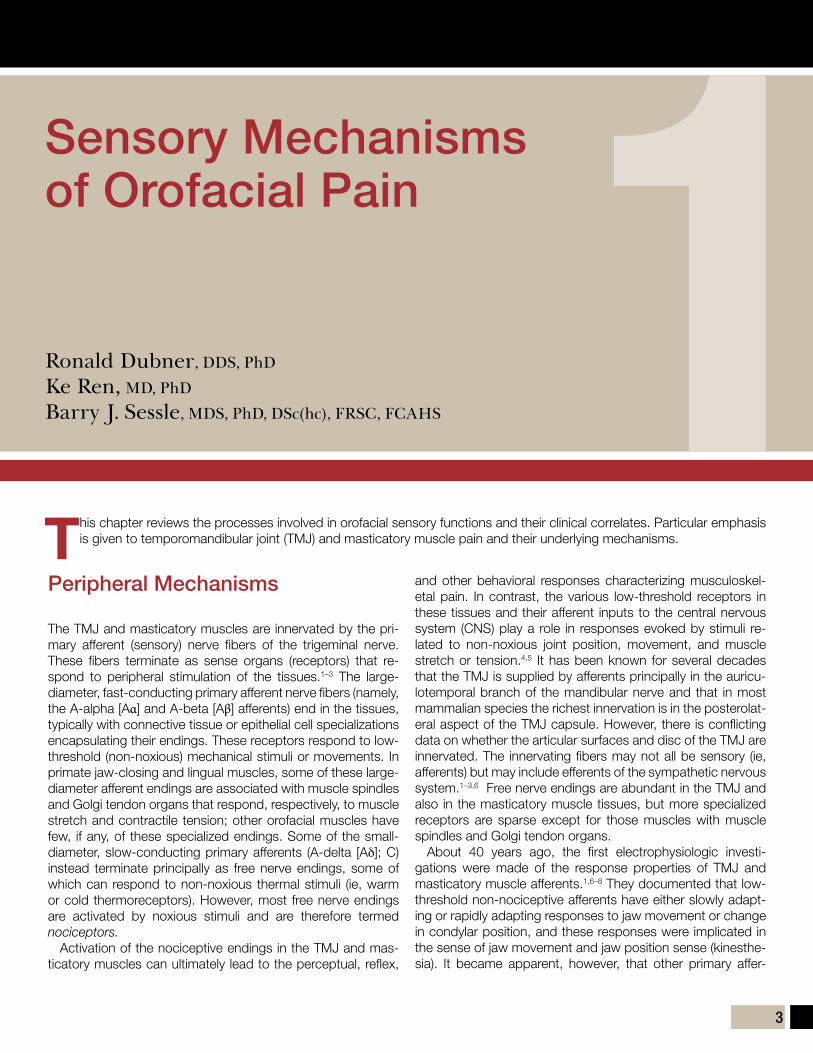
(a) Illustration of a normal TMJ. (b to d) Pressure in the TMJ upon tooth clench-ing (b) and the resulting stuck disc (c and d). When the pressure on the disc ceases, its central area is separated from the bony surface, creating a vac-uum that causes the periphery of the disc to adhere firmly to the surface of the eminence, thus preventing it from sliding (red arrow). At this point, an at-tempt to open the mouth causes pain as the condyle is pulled forward, away from the adhered disc (stars indicate pain locations). (e) The disc is not re-leased on the introduction of a needle. (f) The disc is released following lavage of the upper compartment of the joint. Adhesive forces, rather than only the vacuum effect, are responsible for the immobilization.
Fig 11-1a
b
cd
e
f
197
i n d e x
Page numbers with “f” denote figures; those with “t” denote tables; those with “b” denote boxes.
1,2-dimyristoyl-sn-glycero-3-phosphocholine, 1285-hydroxytryptophan, 4
AA-alpha fibers, 3A-beta fibers, 3Acute orofacial pain, 83–84, 92ADAMTS4, 158–159Adaptation, 120A-delta fibers, 3–4, 6Adenosine 5-triphosphate, 4 Adenosine monophosphate-activated protein kinase, 42Adenoviral vector expressing human insulinlike growth factor-1, 179Advanced glycation end products, 161Aggrecanases, 34, 159, 180Allodynia, 6, 15, 21α2δ modulators, 188–189alpha-amino-3-hydroxy-5-methyl-4-isoxazole-propionate, 9Amygdala, 151Anabolic signaling, 107Anchored disc phenomenon, 127Angiogenesis
factors that affect, 157osteochondral, 42
Animal modelsbiomarkers developed using, 159–161of osteoarthritis, 41
Anterior cingulate cortex, 13, 147f, 148Anterior insula, 149Antidepressants, 53, 186–187, 189Arterial spin labeling, 143Arthrocentesis, 127–129Arthrography, 135Articular bone, 172Articular cartilage
autophagy in, 42avascular properties of, 42, 116collagenous fibers of, 116deforming properties of, 113, 116evolution of, 172metabolism of, 177in osteoarthritis, 34–39, 37fprincipal stress in, 115, 117f
Articular discanteriorly displaced, 118description of, 99, 101energy density vs internal strain energy, 106finite element analysis of, 114friction effects on, 118internal derangement effects on, 119
joint lubrication functions of, 102lubricating system of, 124mechanical behavior of, 118–119plowing force of, 104
Astrocytes, 13Autonomic nervous system, 20Autophagy, 42
BBack pain, 51Benzodiazepines, 186–187Biglycan, 160Bilateral sagittal split osteotomies, 84Biomarkers
aggrecanases, 159animal models used to develop, 159–161collagenases, 158–159C-terminal telopeptide-II, 159interleukin-1β, 156–157monocyte chemoattractant protein, 157prostaglandins, 157of temporomandibular joint-osteoarthritis, 159–161tumor necrosis factor-α, 156–157
Biomechanical modeling, 137–138, 138f, 140Biomedical engineering, 138–140Blood oxygen level–dependent imaging, 142Bone morphogenetic proteins
BMP-2, 167, 179description of, 40
Boundary lubrication, 124Bradykinins, 4Brain-derived neurotrophic factor, 4Brain-imaging studies, 13Brainstem nociceptive processing, 6, 8Bruxism
description of, 24, 62sleep, 61–62
CCalcitonin gene-related peptide, 6Candidate gene studies, 72Cannabinoids, 190–191Cartilage. See Articular cartilage.Catabolic signaling, 107Catastrophizing, 52, 95Catechol-O-methyltransferase, 20, 25–26, 71, 87Central sensitization, 8–12, 14–15, 141, 188, 190Cerebral blood flow, 13C-fibers, 4, 6Chairside screening, 86Chemical condylectomy, 128Chemokines, 157

198
i n d e x
Chondrocyte(s)anabolic and catabolic factors that regulate, 38fdescription of, 34, 36f, 38fhypertrophic-like changes, 36f, 40fmechanical stimuli effects on, 107osteoarthritic, 102sclerostin expression in, 39vascular endothelial growth factor induction in, 38
Chondrocyte receptors, 38Chondroitin sulfate, 159Chronic pain
acute pain progression to, 92description of, 151factors involved in, 50fglutamate and, 191masticatory myofascial pain and, 95prefrontal cortex involvement in, 13risk model for, 96sleep disorders and, 57, 60–62
Cinderella hypothesis, 24Cingulate cortex, 147f, 148–149Cingulotomy, 148Cluster headaches, 26Clustering, 96–97Cognitive-behavioral therapies, 53, 63, 94–95, 97Collagenases, 34Collagenous fibers, 116Community genomics, 74Comorbid pain conditions
description of, 1evidence-based treatment for, 53factors that affect, 52fibromyalgia, 20–21, 26, 49–50headache, 50, 62–63implication of, 51tmasticatory myofascial pain, 26migraine headache, 50neck pain, 51self-management programs for, 53sleep disturbances caused by, 61studies of, 48t–49tsummary of, 53treatment of, 52–53treatment responsiveness affected by, 95
Complete Freund’s adjuvant, 9Complex diseases
characteristics of, 92definition of, 92description of, 72–73immune system’s role in, 73, 73fphenotype, 75–76
Computer-aided design/computer-aided manufacturing technology, 139
Conditioned pain modulation, 83, 83fCondition-specific measures, 93Condylar blastema, 172Condyle-fossa distance, 103f, 104Cone beam computed tomography, 131, 134–135, 136fCoping, 52Corticosteroids, 128, 186COX-1, 157COX-2, 157Craniofacial deformities, 137C-terminal telopeptide-II, 159Cytokines
description of, 126interleukin-1β, 156–157monocyte chemoattractant protein, 157prostaglandins, 157tumor necrosis factor-α, 156–157
DDeep bite, 23Deep sequencing, 77Deep sleep, 58Degenerative joint disease. See Osteoarthritis.Depression, 22, 93, 95Descending modulation, 10–12, 11fDiagnostic imaging, 133–137Diffuse noxious inhibitory controls, 10, 83–84, 86Diffusion tensor imaging, 142Dimethyl sulfoxide, 186Disc displacement, 109, 123, 136fDiscoidin domain receptor 2, 160Disease
complex. See Complex diseases.preclinical symptoms of, 92
Dorsolateral prefrontal cortex, 12Dynamic brain imaging, 131Dynamic loading, 102Dynamic stereometry, 103, 109–110
EEducation-based self care model, 94Effective connectivity magnetic resonance imaging, 142Endochondral ossification, 40–41, 172Endophenotypes, 76Energy density, 106Enkephalin, 10Enzymes, degradative, 158–159Epigenetics, 69, 73–74Epigenome, 74Epigenomics, 74Ethnicity, 21–22Etiology, 91–92Evidence-based treatment, 53Extracellular matrix, 34–35, 41, 107

199
i n d e x
FFibromodulin, 160Fibromyalgia, 20–21, 26, 49–50Fibrous mesenchyme, 169Finite element analysis
applications of, 99, 115–119challenges for, 120description of, 113future of, 119–120history of, 114–115purposes of, 114safety applications of, 119stress and strain values, 115–116surgical planning uses of, 119temporomandibular disorders application of, 119–120temporomandibular joint applications of
adaptation predictions, 120history of, 114–115mechanical behavior, 118–119normal function, 115–116pathologic function, 118
Frictionarticular disc affected by, 118inadequate lubrication as cause of, 127
Functional brain imaging, 77Functional connectivity magnetic resonance imaging, 142Functional magnetic resonance imaging, 142
GGabapentin, 4, 188–189, 191Gene expression, 73–74Gene variants, 71fGene-environment interactions, 69, 72, 75, 77Gene-gene interactions, 69, 72, 75, 77Generic treatments, 96Genes, in complex diseases, 72Genetics
costs of, 70literature regarding, 14masticatory myofascial pain and, 25–26overview of, 69–70single nucleotide polymorphisms, 69
Genome-wide association studies, 70–71, 77Genomic technology
advances in, 70–72genome-wide association studies, 70–71, 77single nucleotide polymorphisms, 70–71
Glia, 13Glial cells, 191Glutamate, 150, 191Glutamate receptors, 21Glutamate transporter, 13Glycosaminoglycan, 107, 108f, 125
Gray matter volume, 146–148Growth factors, 41
HHard tissue imaging, 133–135, 134fHeadaches
in children, 63migraine, 50, 63sleep bruxism associated with, 62sleep disturbances and, 62–63tension-type, 62–63
1H-MRS. See Proton magnetic resonance spectroscopy.
Host susceptibility, 26Human Genome Project, 72Human microbiome, 74–75, 75fHyaline cartilage, 118–119Hyaluronic acid, 34, 35f, 42, 124–125, 128Hyaluronidase, 125Hydrostatic lubrication, 124Hydrostatic pressure, 115Hyperalgesia, 6, 15Hypoxia, 25, 157–158Hypoxia-inducible factor, 38, 158
IImaging. See also Neuroimaging.
advances in, 139–140biomechanical modeling after, 137–138, 138f, 140biomedical engineering uses of, 138–140cone beam computed tomography, 131, 134–135, 136fdiagnostic, 133–137hard tissue, 133–135, 134fmagnetic resonance imaging. See Magnetic resonance imaging.of pain, 13soft tissue, 135–137
IMMPACT. See Initiative on Methods, Measurement, and Pain Assessment in Clinical Trials.
Immune system, 73, 73fIncident-cohort studies, 91Inferior parietal lobule, 147fInflammatory mediators
description of, 4, 155interleukin-1β, 156–157tumor necrosis factor-α, 156–157
Initiative on Methods, Measurement, and Pain Assessment in Clinical Trials, 93
Insomnia, 61, 63Insula, 149–150Insular cortex, 149–150Insulinlike growth factor-1, 179–180Integrated Pain Adaptation Model, 25Interdisciplinary treatment, 52–53Interleukin-1β, 156–157

200
i n d e x
Interleukin-1 receptor antagonist, 180–181Intermediate phenotypes, 76Internal derangements, 93, 119Internal strain energy, 106International Classification of Sleep Disorders, 60–61Intra-articular injections
bone morphogenetic protein 2, 179corticosteroids, 186insulinlike growth factor-1, 179–180interleukin-1 receptor antagonist, 180–181NEL-like molecule 1, 180transforming growth factor beta, 178
Intraoral appliances, 53, 95
JJaw-closing muscles, 113
KKeratan sulfate, 159
LLateral pain system, 146–148Lateral pterygoid muscle, 120Lifestyle, 25, 27Light sleep, 58Liposomes, 128Low-threshold mechanoreceptive neurons, 6, 8Lubrication, of temporomandibular joint, 123–129Lubricin, 34, 42, 125–126
MMacrophage colony-stimulating factor, 41Magnetic resonance imaging
applications of, 136farterial spin labeling, 143description of, 135–137effective connectivity, 142functional, 142functional connectivity, 142history of, 142methods, 142operating principles of, 142–143, 143f
Malocclusions, 23Mandibular advancement appliances, 64Mandibular condylar cartilage, 173Mandibular hypoplasia, 119Masticatory myofascial pain
algorithm of, 19fautonomic nervous system and, 20characteristics of, 17chronic pain and, 95comorbidities, 26definition of, 47
diagnostic criteria for, 18tethnicity and, 21–22genetic factors, 25–26, 151historical perspectives on, 17–19host susceptibility to, 26lifestyle factors, 25, 27nervous system alterations in, 19–21neuropeptides and, 20–21, 26occlusion and, 23pain modulation and, 19–20pressure pain thresholds in, 20psychosocial factors, 22–23sex and, 21skeletal morphologic features, 23sleep disturbances and, 26–27, 61temporomandibular joint disorders and, 23–24trauma as cause of, 22trigger points associated with, 25
Matrix metalloproteinases, 34–35, 108f, 158, 180Maxillofacial surgery, 119Maximal mouth opening, 123–124Mechanical loading, 107Mechanical temporomandibular disorders, 95–96Meckel’s cartilage, 168–169, 172Medial pain system, 148–150Medical care delivery systems, 70Mediolateral stress-field translation, 104–105Medullary dorsal horn, 6Metabolic phenotype, 74Metabotropic glutamate receptors, 9Metagenomic DNA sequencing, 74Methyl salicylate, 186Microbiome, 74–75, 75fMicroglia, 13Migraine headache, 50, 63Mini-anchors, 119Mitochondrial dysfunction, 42Modulus, 81Monocyte chemoattractant protein, 157MRI. See Magnetic resonance imaging.Muscle hypoperfusion, 25Muscle pain
exogenous models of, 24pharmacotherapy for, 53
Muscle relaxants, 187Myalgia, 23–24Myofascial pain. See Masticatory myofascial pain.
NN-acetylaspartate, 148, 150Neck pain, 22, 51NEL-like molecule 1, 180Nerve growth factor, 4, 21, 42, 190

201
i n d e x
Neuroglial cells, 191Neuroimaging. See also Imaging.
antinociceptive areas studied using, 150central pain systems studied using, 146–151description of, 131, 141future applications of, 150–151magnetic resonance imaging. See Magnetic resonance imaging.medial pain systems studied using, 148–150positron emission tomography, 144, 144ftemporomandibular disorder studies, 144, 145t
Neuropathic pain, 84, 86, 190–191Neuropeptides, 20–21, 26Neuroplasticity, 92, 190Neutrophins, 4Next-generation sequencing
bioinformatics platform for, 72description of, 70–72whole genome sequencing, 70, 72
NMDA receptor–ion channel complex, 9fNMDA receptors, 21, 191Nociception
non-neural processes in, 12–13in sleep, 59
Nociceptive afferents, 4, 11Nociceptive-specific neurons, 6, 8, 10Nocturnal migraine headaches, 63Non-neural processes, 12–13Nonsteroidal anti-inflammatory drugs, 53, 185–186Notch1, 172NREM sleep, 58N-type calcium channel, 191Nuclear factor κB, 12, 35Nucleus raphe magnus, 10Numeric rating scales, 81
OObstructive sleep apnea, 61Occlusal interferences, 23Occlusion, 23Octahedral shear stress, 115Onabotulinum toxin, 186, 188Opioid receptors, 10Opioids, 10, 187, 189–190OPPERA. See Orofacial Pain Prospective Evaluation and Risk As-
sessment.Oral appliances, 64Orofacial pain
acute, 83–84bruxism secondary to, 62central mechanisms of, 6–12mandibular advancement appliances for, 64peripheral mechanisms of, 3–6, 5f21st-century trends, 70
Orofacial Pain Prospective Evaluation and Risk Assessment, 19, 85, 96
Orthodontics, 23Osteoarthritis
animal models of, 41articular cartilage destruction in, 34–39, 37f, 42biologic targets in treatment of, 41–43bone changes in, 39–41cartilage abnormalities in, 34–39characteristics of, 155corticosteroids for, 128definition of, 33, 101, 131, 177etiopathogenic mechanisms of, 34–41, 35f–40ffeatures of, 34–41friction and, 127hyaluronic acid effects in, 42inflammatory response in, 41lubricin protective effects in, 42mitochondrial dysfunction in, 42periarticular bone in, 40rat meniscectomy model of, 42research on, 33–34subchondral bone in, 37f, 39f, 39–41synovial inflammatory infiltrates in, 41synoviopathy associated with, 34in temporomandibular joint, 101, 109treatment of, 41–43, 177
Osteochondral angiogenesis, 42Osteophytes, 41Overloading, 127, 127f
PPain
back, 51chronic. See Chronic pain.imaging of, 13masticatory myofascial pain. See Masticatory myofascial pain.neck, 51neuropathic, 84, 86, 190–191orofacial. See Orofacial pain.palpation-induced, 93persistence of, 51, 96progression of, 52fprovocation, 93sleep and, 57, 59–60, 63–64temporomandibular disorder–related. See Temporomandibular
disorder pain.Pain adaptation model, 24–25Pain modulation, 19–20Pain perception
behavioral conditions that affect, 19measurement difficulties for, 72
Pain-related awakenings, 26

202
i n d e x
Pain-related evoked potentials, 149Palpation-induced pain, 93Paradoxical sleep, 58Parafunctional forces, 24Patient-reported outcomes, 93Patient-specific model, 138fPeripheral sensitization, 4–6, 14–15Personalized medicine, 70Pharmacogenomics, 26Pharmacologic treatmentα2δ, 188–189 antidepressants, 53, 186–187, 189benzodiazepines, 186–187cannabinoids, 190–191future applications of, 190–191historical review of, 185–186ketamine, 189, 191for muscle pain, 53muscle relaxants, 187nonsteroidal anti-inflammatory drugs, 53, 185–186onabotulinum toxin, 186, 188opioids, 187, 189–190selective serotonin reuptake inhibitors, 187, 189for temporomandibular disorder pain, 185–191
Phenotypesfunctional brain imaging investigations of, 77intermediate, 76
Phospholipase, 125Phospholipase A2, 126Phospholipids, 124, 126, 128–129Placebo effect, 12, 151Plowing force, 102, 102fPolysomnographic recordings, 58, 58bPositron emission tomography, 144, 144fPositron emission tomography/computed tomography, 144Posterior cingulate cortex, 147f, 149Postherpetic neuralgia, 188Preclinical symptoms, 92Prefrontal cortex, 13Pregabalin, 188–189, 191Pressure pain thresholds, 20–21Primary afferent neurons, 5fPrimary afferents, 3Primary somatosensory cortex, 147fPrincipal stress, in articular cartilage, 115, 117fProinflammatory cytokines, 126PROs. See Patient-reported outcomes.Prostaglandin E2, 20, 157Protein kinase C, 21Proteoglycans, 125, 178Proton magnetic resonance spectroscopy, 142–143Proton pump inhibitors, 188Provocation pain, 93
Psychogenic pain, 85Psychologic factors, 52Psychophysics, 79–80Psychosocial factors, 22–23P-type calcium channel, 191Putative etiology, 91–92
QQuantitative sensory testing (QST)
acute orofacial pain, 83–84afferent nerve fiber functions assessed with, 86applications of, 85–87background of, 79–80conditioned pain modulation, 83, 83fdescription of, 20diffuse noxious inhibitory controls, 83–84, 86future applications of, 85–87history of, 79–83neuropathic pain, 84, 86response-dependent techniques, 82somatosensory sensitivity, 86–87stimuli used in, 79–80stimulus-detection, 80summary of, 87suprathreshold estimation, 80–83, 81ftemporomandibular disorders, 84–85testing algorithms, 80thermal detection thresholds, 84traumatic neuropathic pain, 84, 86triangulation procedure, 81, 82f
RReceptor activator of nuclear factor κB, 41Receptor for advanced glycation end products, 35, 161Rehabilitation treatment model, 92, 96REM sleep, 58Research Diagnostic Criteria for TMD, 76Rheumatic diseases, 33Rheumatoid arthritis, 157Rhythmic masticatory muscle activity, 62Risk factors
description of, 14genetic, 26
Rolling/plowing explants test system, 109
SSatellite glial cells, 12Scaffold biomaterials, 167Schizophrenia, 74Sclerostin, 39Secondary cartilage, 169Segmental modulation, 10Selective serotonin reuptake inhibitors, 187, 189

203
i n d e x
Self care, 96Self-management program, 53Self-report condition-specific measures, 93Serotonin, 20Serotonin noradrenaline reuptake inhibitors, 189Serotonin transporter gene, 25Shear loading, 102Shear stress, 115–116Single nucleotide polymorphisms, 70–71Skeletal morphologic features, 23Sleep
average duration of, 57brain activity during, 58deep, 58definition of, 57fragmentation of, 59light, 58medication effects on, 63nociception attenuation during, 59NREM, 58pain and, 57, 59–60, 63–64paradoxical, 58pathophysiology of, 57–58polysomnographic recordings of, 58, 58bREM, 58in sleep bruxism patients, 62
Sleep arousals, 58, 61Sleep bruxism, 24, 61–62Sleep deprivation
description of, 59migraine headaches precipitated by, 63
Sleep disorders/disturbancesassessment of, 63, 64bchronic pain and, 57, 60chronic widespread pain and, 61–62classification of, 60t, 60–61comorbid pain conditions as cause of, 61description of, 26–27headaches and, 62–63insomnia, 61, 63masticatory myofascial pain and, 26–27, 61temporomandibular disorders and, 61treatment of, 63
Sleep hygiene, 63Sleep-disordered breathing, 61Sleep-related breathing disorders, 61Sleep-related movement disorder, 62SNRIs. See Serotonin noradrenaline reuptake inhibitors.Social support, 52Soft tissue imaging, 135–137Somatosensory cortex, 147f, 148Static loading, 102Stimulus-detection thresholds, 80
Strain, 115–116Stress
disorders related to, 22gene effects during, 76
Stress (force)collagenous fiber resistance to, 116finite element analysis of, 115–116octahedral shear, 115shear, 115–116
Stress relaxation, 118Stress-field translation
condyle metabolism and, 107–109description of, 103–105mediolateral, 104–105recording of, 109
Subchondral bone, in osteoarthritis, 37f, 39f, 39–41Subchondral sclerosis, 40Subnucleus caudalis, 6, 8Suction cup effect, 124Superficial zone protein, 125Suprathreshold estimation, 80–83, 81fSurface-active phospholipids, 124, 126–127Sympathetic nervous system, 25
Synovial cells, 34Synovial chondromatosis, 136fSynovial joints
lubrication, 124–126osteoarthritis and, 155
Synovitis, 159
TTemporomandibular disorder pain
comorbid conditions effect on, 49–51, 51tetiology of, 141factors that affect, 52fibromyalgia and, 49–50migraine headache and, 50persistence of, 51, 96pharmacologic treatment of, 185–191prognosis for, 51progression of, 52fsigns and symptoms of, 47sleep fragmentation as cause of, 61
Temporomandibular disorderscondition-specific measures for, 93definition of, 13, 47finite element analysis applications, 119–120masticatory myofascial pain and, 23–24mechanical, 95–96overview of, 13–15prevalence of, 47, 167primary pain-related, 95

204
i n d e x
Temporomandibular jointanatomy of, 168–169arthralgia of
causes of, 123description of, 47intraoral appliances for, 53
clicking of, 93development of, 168–169disc. See Articular disc.dynamic loading of, 102evolution of, 169–173finite element analysis of. See Finite element analysis.imaging of, 133–140immobilization of, 127load distribution in, 113locking of, 93lubrication of, 123–129osteoarthritis onset in, 101, 109shear loading in, 102static loading of, 102stress-field translation in, 103–105tractional forces in, 102–103
Tension-type headaches, 62–63Testing algorithms, 80Thalamocortical nociceptive processing, 8Thalamus, 147f, 147–148Tissue engineering, 167–174Tissue inhibitors of metalloproteinase 1, 38, 109, 158Toll-like receptors, 12Tractional forces, 102–103Transcription factors, 36fTransforming growth factor beta, 178Transforming growth factor beta-1, 167Transient receptor potential receptors, 4Trauma
masticatory myofascial pain secondary to, 22neuropathic pain associated with, 84, 86
Treatmentadvances in, 165anticipated clinical applications of, 96–97classification of, 93–94comorbid pain conditions, 52–53etiology effects on, 91–92generic, 96goals of, 52interdisciplinary, 52–53multimodal plan of, 53pharmacologic. See Pharmacologic treatment.presenting condition and, matching between, 94rehabilitation model of, 92, 96
Treatment responsivenessbehavioral factors that affect, 95catastrophizing effects on, 95clustering effects on, 96–97comorbid pain conditions effect on, 95factors that affect, 94–96level of analysis effects on, 94measurement of, 92–96objective measures of, 93patient-reported outcomes, 93physical factors that affect, 95predictions about, 92self-report condition-specific measures of, 93
Triangulation, 81, 82fTricyclic antidepressants, 186Trigeminal brain complex, 6, 8Trigeminal brainstem nuclei, 146Trigeminal nociceptive pathways, 14Trigeminal somatosensory pathways, 7fTrigeminal tractotomy, 6Trigger points, 25TRPV1, 190Tumor necrosis factor-α, 4, 156–157, 161
UUnilateral mandibular hypoplasia, 119
VVanilloid receptor 1. See TRPV1.Vascular endothelial growth factor, 38, 42, 157–158Ventroposterior nucleus, 8Vicious cycle theory, 23Viscoelasticity, 118Visual analog scales, 81Vi/Vc neurons, 13Voltage-gated calcium channels, 191von Mises stress, 115Voxel-based morphometry, 142, 149
WWakefulness, 58Whiplash, 22White matter volume, 148–149Whole genome sequencing, 70, 72Wide dynamic range neurons, 6, 8, 10Widespread Pain Index, 49
XX-rays, 133