Embed Size (px)
Citation preview
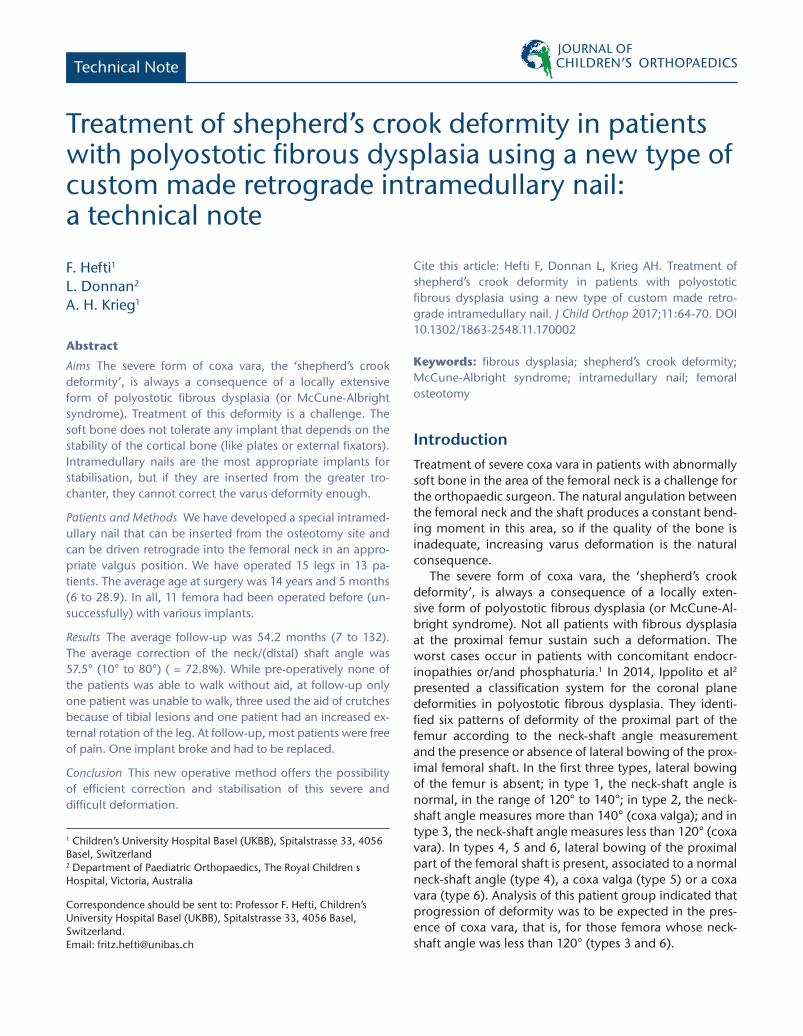
Technical Note
Treatment of shepherd’s crook deformity in patients with polyostotic fibrous dysplasia using a new type of custom made retrograde intramedullary nail: a technical note
F. Hefti1
L. Donnan2
A. H. Krieg1
Abstract
Aims The severe form of coxa vara, the ‘shepherd’s crook deformity’, is always a consequence of a locally extensive form of polyostotic fibrous dysplasia (or McCune-Albright syndrome). Treatment of this deformity is a challenge. The soft bone does not tolerate any implant that depends on the stability of the cortical bone (like plates or external fixators). Intramedullary nails are the most appropriate implants for stabilisation, but if they are inserted from the greater tro-chanter, they cannot correct the varus deformity enough.
Patients and Methods We have developed a special intramed-ullary nail that can be inserted from the osteotomy site and can be driven retrograde into the femoral neck in an appro-priate valgus position. We have operated 15 legs in 13 pa-tients. The average age at surgery was 14 years and 5 months (6 to 28.9). In all, 11 femora had been operated before (un-successfully) with various implants.
Results The average follow-up was 54.2 months (7 to 132). The average correction of the neck/(distal) shaft angle was 57.5° (10° to 80°) ( = 72.8%). While pre-operatively none of the patients was able to walk without aid, at follow-up only one patient was unable to walk, three used the aid of crutches because of tibial lesions and one patient had an increased ex-ternal rotation of the leg. At follow-up, most patients were free of pain. One implant broke and had to be replaced.
Conclusion This new operative method offers the possibility of efficient correction and stabilisation of this severe and difficult deformation.
1 Children’s University Hospital Basel (UKBB), Spitalstrasse 33, 4056 Basel, Switzerland2 Department of Paediatric Orthopaedics, The Royal Children s Hospital, Victoria, Australia
Correspondence should be sent to: Professor F. Hefti, Children’s University Hospital Basel (UKBB), Spitalstrasse 33, 4056 Basel, Switzerland. Email: [email protected]
Cite this article: Hefti F, Donnan L, Krieg AH. Treatment of shepherd’s crook deformity in patients with polyostotic fibrous dysplasia using a new type of custom made retro-grade intramedullary nail. J Child Orthop 2017;11:64-70. DOI 10.1302/1863-2548.11.170002
Keywords: fibrous dysplasia; shepherd’s crook deformity; McCune-Albright syndrome; intramedullary nail; femoral osteotomy
IntroductionTreatment of severe coxa vara in patients with abnormally soft bone in the area of the femoral neck is a challenge for the orthopaedic surgeon. The natural angulation between the femoral neck and the shaft produces a constant bend-ing moment in this area, so if the quality of the bone is inadequate, increasing varus deformation is the natural consequence.
The severe form of coxa vara, the ‘shepherd’s crook deformity’, is always a consequence of a locally exten-sive form of polyostotic fibrous dysplasia (or McCune-Al-bright syndrome). Not all patients with fibrous dysplasia at the proximal femur sustain such a deformation. The worst cases occur in patients with concomitant endocr-inopathies or/and phosphaturia.1 In 2014, Ippolito et al2 presented a classification system for the coronal plane deformities in polyostotic fibrous dysplasia. They identi-fied six patterns of deformity of the proximal part of the femur according to the neck-shaft angle measurement and the presence or absence of lateral bowing of the prox-imal femoral shaft. In the first three types, lateral bowing of the femur is absent; in type 1, the neck-shaft angle is normal, in the range of 120° to 140°; in type 2, the neck-shaft angle measures more than 140° (coxa valga); and in type 3, the neck-shaft angle measures less than 120° (coxa vara). In types 4, 5 and 6, lateral bowing of the proximal part of the femoral shaft is present, associated to a normal neck-shaft angle (type 4), a coxa valga (type 5) or a coxa vara (type 6). Analysis of this patient group indicated that progression of deformity was to be expected in the pres-ence of coxa vara, that is, for those femora whose neck-shaft angle was less than 120° (types 3 and 6).

TREATMENT OF SHEPHERD’S CROOK DEFORMITY IN PATIENTS WITH POLYOSTOTIC FIBROUS DYSPLASIA
J Child Orthop 2017;11:64–70 65
Numerous methods of treatment for femoral defor-mity in fibrous dysplasia have been proposed.3-6 Attempts of valgus osteotomies with angulated blade plates usu-ally have not been very successful due to cut-out of the blade, diaphyseal plate screw failure and fracture or defor-mity below the plate.2,7 A few case reports on the use of a dynamic hip screw describe successful treatment, but the follow-up is short and the fundamental problems are the same as with the angulated blade plate if the disease is extensive.6 The largest series of hip screw fixation is of 23 patients with localised monostotic disease (predominantly intertrochanteric) and minimal deformity. They came to internal fixation because of pain or pathological fracture with good success in the majority of cases.8 Cantilever or monolateral external fixation has a high failure rate in deformity correction in fibrous dysplasia but in selected cases the beam loading ring fixators may have a role.9
It is therefore generally accepted that the most appro-priate implant that can be used in severe cases of femoral neck deformity in polyostotic fibrous dysplasia is an intra-medullary nail. These nails are inserted from the greater trochanter, which means it is technically impossible to cor-rect a severe varus deformity because the pelvis impedes the appropriate insertion of the nail, forcing the entry point to be too far lateral and the correction to be inad-equate (Fig. 1). The corrective potential is therefore lim-ited,4,5 except in mainly diaphyseal deformities.4,10 When using a conventional intramedullary nail, the valgisaton
has to be obtained with other means. This can be achieved in a two-stage procedure with a valgus osteotomy using a blade plate or a dynamic hip screw as the first stage, and after consolidation, stabilisation with an intramedul-lary nail with a blade in the femoral neck as the second stage.3 In other reports, the Fassier-Duval nail had been used and the valgus osteotomy had been stabilised with two angulated pins and cerclage wires.11 A few cases have been described with the use of an intramedullary Expert- Synthes humeral nail,12 and one case report describes a patient with a fracture below a long blade plate, where the correction was done with the retrograde insertion of a conventional intramedullary nail from the fracture site.13 A new device called ‘gap nail’ has been recently presented. It is an intramedullary nail that can be inserted in an anterograde way from the greater trochanter or in a retro-grade way from the knee. It can be combined with a plate that improves local stability at the greater trochanter (as long as the bone is not too soft in this area, which is usu-ally the case in shepherd’s deformity). This nail certainly has a potential for correction of coxa vara, but it is not meant to be inserted from the osteotomy and therefore does not allow the same amount of valgisation and prob-ably does not solve the problem in shepherd’s deformity. To our knowledge, no publication exists describing the use of this nail in such a deformity.
Due to the fact that the trochanteric insertion does not allow an appropriate positioning and direction of the nail, we have developed a special intramedullary nail that can be inserted from the osteotomy site and can be driven retrograde into the femoral neck in an appropriate valgus position. This application also solves the prob-lem that such a medial position of the nail, as required, would otherwise jeopardise the femoral neck vessels if inserted from outside the bone. The paper describes the method and reports on the first 15 cases treated with this method.
Patients and MethodsThe principle of the retrograde intramedullary nail is illus-trated in Figure 2.
Pre-operative planning was performed on orthogo-nal plain radiographs which produced a template that showed the degree of bony resection, correction required and likely position of the intramedullary nail. From this information, a custom-made nail was made for each case by Synthes Switzerland (Fig. 3). In surgery, the patient is placed in a supine position with slight elevation of the affected side. A direct lateral approach is made to the femur which is exposed carefully subperiosteally. A 2.5-mm Kirschner wire is introduced from the lateral cortex down the femoral neck in a central location to guide the
Fig. 1 Basic problem of the trochanteric insertion of an intramedullary nail. Left) In the normal position, the iliac bone forces the nail into a medial direction. Middle) Even in maximal adduction of the leg, the direction of the nail is not enough laterally directed. Right) The theoretical optimal direction of the nail for obtaining a true valgus of the neck.

TREATMENT OF SHEPHERD’S CROOK DEFORMITY IN PATIENTS WITH POLYOSTOTIC FIBROUS DYSPLASIA
66 J Child Orthop 2017;11:64–70
alignment of the osteotomy. The osteotomy is made and enough bone resected to allow the deformity to be fully corrected. The distal canal is then opened with a straight reamer and if any lateral bowing is present this must be fully corrected with one or more additional osteotomies. In the growing child, a smaller male component is first placed into the medullary canal with a special T-handle. This is placed across the distal femoral physis and locked with a transverse small fragment screw (Figs 4 to 6). The female nail is then passed over the male and made flush
with the cut end of the femur. For adolescents or adults, the central telescoping part is not needed. A window is made in the lateral cortex of the femur to give access to the mid part of the nail that has a series of ridges. With the deformity completely corrected, the nail can then be advanced in a retrograde fashion using a small punch inclined against the ridges. With this retrograde method, the proximal end of the nail can be inserted as medial as necessary; if it is appropriate it can even be brought to the metaphysis (Fig. 5) or even into the femoral head. Small holes in the nail at regular distances in the axis of the femoral neck screws help to maintain the orientation of the nail while hammering proximally. For the insertion of the femoral neck screws and the bolting screw in the distal epiphysis, a 3Dimage intensifier is very helpful, as the bone is very thin and axial views cannot be made before the insertion of the proximal screws, because with the leverage the nail would enlarge the hole within the soft bone. At the end of the operation, the correct rotation
Fig. 2 Principle of the retrograde intramedullary nail. Step 1) Pre-operative situation. 2) As the first step, an osteotomy in the trochanteric area with resection of a lateral wedge in the scheduled angle is carried out. Step 3) With adduction of the leg, the reamer is inserted from the osteotomy site. Step 4) The custom-made nail is pushed down until the epiphyseal plate. Step 5) The central pin is pushed down through the nail and is locked through the epiphysis with a screw inserted from the lateral condyle. Step 6) From the osteotomy site a hole is reamed into the femoral neck in the scheduled direction. Penetration of the cranial cortex should be avoided. Step 7) In the middle of the femoral shaft a window is created. The leg is then abducted and the nail that has circular bulging rings can be pushed into the bore hole with the help of a pestle and a hammer. Step 8) Situation with the nail in situ, two femoral neck screws are inserted through the predrilled holes in the nail.
Fig. 3 Photograph of the implant. The proximal part is thicker and has two predrilled holes for the femoral neck screws. The shaft of the implant has bulging rings that allow the pushing of the nail upward.

TREATMENT OF SHEPHERD’S CROOK DEFORMITY IN PATIENTS WITH POLYOSTOTIC FIBROUS DYSPLASIA
J Child Orthop 2017;11:64–70 67
is secured with K-wires through the cortices of both frag-ments of the osteotomy.
The first operation using this nail was undertaken in July 2005. Since then, we have operated 15 legs in 13 patients (seven female, six male). The average age at surgery was 14 years and five months (six years to 28 years and 11 months). All operations have been carried out either by the first or the second author or one of them was assisting. The operations took place in six different cities (Basel, Mel-bourne, Lucerne, Dusseldorf, Vienna and Dresden). The patients and their data are tabulated in Tables 1 and 2. Of the 15 femora, 11 had been operated before (unsuccess-fully) with various implants. In nine cases, the telescopic variant of the retrograde intramedullary nails was used; in six adult patients, the implant was introduced without the telescopic part. All but one case had two femoral neck screws, in one small child two pins were used (case 15, Fig. 5).
The average follow-up was 54.2 months (7 to 132). As this paper’s aim is to introduce a new method and does not primarily present a follow-up study, we also included two patients with less than 24 months of follow-up. If we excluded them, we could not report on an important
complication (case six) and on an important variant of the method (case 15). Pre-operatively, only one patient could walk without aids, four used crutches, and seven were unable to walk. In 13 cases the pain was severe and in two cases it was moderate. The angle between the distal femo-ral shaft and the femoral neck was measured in the frontal plane on radiographs. The average angle was 78.9° (40° to 90°). According to the radiographical classification by Ippolito, four cases were type 3 and 11 cases were type 6.
ResultsThe results are tabulated in Tables 1 and 2. The 15 legs were operated on 13 patients (two patients had simulta-neous surgery on both legs and one patient had surgery on the second side 3.5 years after the first side). The aver-age post-operative neck/(distal) shaft angle was 136.4° (85° to 160°). The average correction of this angle was 57.5° (10° to 80°; 72.8%). Figures 4 and 5 show two illus-trative cases.
Fig. 4 (a) The 14-year-old boy (case 3) with polyostotic fibrous dysplasia und deformation of the femur and a plate in the femoral shaft. (b) Anteroposterior radiograph one year after correction with multiple osteotomies and stabilisation with telescopic retrograde intramedullary nail and two femoral neck screws.
Fig. 5 (a) Six-year-old girl (case 15) with polyostotic fibrous dysplasia und deformation of the femur and two elastic stable intramedullary nails (ESIN). (b) Radiograph six months after surgery. The tip of the retrograde nail was placed into the metaphyseal bone and a very pronounced valgisation was carried out. The leg was primarily place in a hip spica in 40° of abduction and gradually was brought into neutral position in order to avoid a tear to the greater trochanter. Six months post-operatively the child could walk without crutches but with marked external rotation.

TREATMENT OF SHEPHERD’S CROOK DEFORMITY IN PATIENTS WITH POLYOSTOTIC FIBROUS DYSPLASIA
68 J Child Orthop 2017;11:64–70
At follow-up, only one patient was unable to walk, one used the aid of crutches and one patient had an increased external rotation of the leg when walking. One patient still had severe pain, one moderate, two mild and all others were free of pain. One patient, with whole head involve-ment of the disease, did not profit from the surgery as cor-rection was incomplete and there was a screw penetration into the hip joint. This patient continued to have severe pain with great difficulty in walking but has elected not to have any further surgery. A fatigue failure of the nail was seen in one case some eight years after nail insertion. A revision was recently carried out with the insertion of a thicker nail.
There were no other serious complications, namely no infections, nerve lesions or vascular incidents.
DiscussionThe treatment of a shepherd’s crook deformity is one of the most difficult problems in orthopaedic surgery. The soft bone does not tolerate any implant that depends on the stability of the cortical bone. Plates or external fixa-tors are therefore not suitable for this kind of treatment.
All reports on the use of such implants either describe mild deformities (radiological types 1 or 2), have a rather short follow-up time or are anecdotal case reports.6,7,9 The intramedullary nail, however, addresses these issues and is the implant of choice in such severe bone soften-ing conditions.3,4,5,7,10-14 With such nails, deformity of the femoral shaft can be efficiently corrected, if necessary with the help of multiple osteotomies. The varus devia-tion of the femoral neck, however, cannot be corrected easily with a classical intramedullary nail inserted from the greater trochanter (Fig. 2). The use of a retrograde nail inserted through the osteotomy allows overcorrection of the varus deformity at the femoral neck and simultane-ously any diaphyseal malalignment. It is important to use the largest diameter of nail possible as repetitive strain can fatigue smaller nails even after many years of successful bone healing.
Surgical planning can be complex if the true nature of the deformity and the surgical challenges posed are not appreciated. CT scanning with 3D reconstructions greatly improves the visualisation of the proximal femur but should be used guardedly, as if there is very extensive dis-ease; much of the bone may be un-ossified fibrous tissue that will not be visualised in the model produced. Newer
Fig. 6 (a) Three-dimensional reconstructions of CT scans (frontal view) of a 10.7-year-old boy (cases 12 and 13) with polyostotic fibrous dysplasia und shepherd’s crook deformation of the femur. (b) AP radiographs three months post-operatively after correction with multiple osteotomies and stabilisation with telescopic retrograde intramedullary nail and two femoral neck screws on both sides.

TREATMENT OF SHEPHERD’S CROOK DEFORMITY IN PATIENTS WITH POLYOSTOTIC FIBROUS DYSPLASIA
J Child Orthop 2017;11:64–70 69
Table 2. Follow-up data for all patients.
Case # Name Gender Neck-shaft angle at follow up
Angle of Correction Follow-up time (mths)
Complications Walking ability at follow-up
Pain at follow-up
1 J. f 125 35 132 breakage of the nail after 8 years
yes, without aid -
2 R. m 135 60 116 nil yes, without aid nil3 T. m 125 50 38 nil yes, without aid nil4 S. f 115 30 31 nil yes, without aid nil5 A. m 100 25 69 nil yes, without aid +6 P. f 85 10 7 penetration of
screw into the acetabulum
Unable to walk +++
7 D. f 130 55 36 nil yes, without aid nil8 K. f 120 45 51 nil yes, without aid nil9 E. f 135 75 60 proximal screw
migration – removed - gradual recurrence of varus
yes ,use of crutches due to ankle lesion
nil
10 H. m 135 65 35 nil yes nil11 H. m 140 55 37 nil yes nil12 G. m 135 65 54 nil yes ,use of
crutches due to ankle lesion
nil
13 G. m 135 95 54 nil yes with crutches due to tibial deformity
nil
14 K. f 135 60 24 nil yes, without aid nil15 N. f 160 80 15 nil yes, with external
rotationnil
techniques in CT (dual energy scans) and MRI should help with this problem. In association with coxa vara there is usually also severe retroversion, which can also be seen with such a technique. Recent techniques of 3D printing allow the surgeon to perform the procedure virtually but do not reproduce the lack of substance in the bone, lim-ited radiographic visibility of the proximal femur, tension developed in correction and bleeding.
With the introduction of any new intramedullary device, unique issues for removal arise. This nail can only
be removed with a femoral osteotomy; but as the main reason for removal is increasing deformation, this is not an issue. The ridges in the nail have not proved to be prob-lematic for removal as the bone surrounding them remain soft and fibrous and rigid bone ingrowth does not occur.
For the most severe cases, total hip arthroplasty is not an alternative. A prosthesis can only be inserted if there is a long enough healthy area of the femoral shaft so that a solid fixation of the prosthesis is possible. In a recent arti-cle with nine total hip replacements in fibrous dysplasia
Table 1. Pre-operative data for all patients treated with the retrograde nail.
Case # Name Gender Diagnosis Radiographic type pre-op1
Side Pain preop Walking ability pre-op
Previous surgery
Age at surgery with retrograde nail (yrs)
Type of nail
Radiographic type pre-op
Neck-shaft angle pre-op**
1 J. f MAS 6 Lt. severe wheelchair standard IN 7.5 TRIN 3 902 R. m MAS 6 Lt. severe limping, no aid standard IN 5.9 TRIN 5 753 T. m PFD 6 Lt. severe crutches plate 14.5 RIN 5 754 S. f MAS 6 Lt. moderate crutches none 18.0 TRIN 6 855 A. m PFD 3 Lt. severe unable to walk ESIN 13.4 RIN 3 756 P. f PFD 3 Lt. severe unable to walk plate 27.1 RIN 4 757 D. f PFD 3 Rt. severe wheelchair none 9.6 TRIN 5 758 K. f PFD 3 Rt. moderate no aid standard IN 25.4 RIN 4 759 E. f MAS 6 Lt. moderate crutches angular plate 12.9 RIN 6 6010 H. m PFD 6 Rt. moderate crutches standard IN +
plate12.8 TRIN 6 70
11 H. m PFD 6 Lt. none crutches standard IN 12.8 TRIN 6 8512 G. m MAS 6 Rt. severe unable to walk none 10.7 TRIN 6 7013 G. m MAS 6 Lt. severe unable to walk none 10.7 TRIN 6 4014 K. f PFD 6 Lt. severe crutches plate 28.9 RIN 4 7515 N. f PFD 6 Rt. severe unable to walk ESIN 6.0 TRIN 5 80
PFD, polyostotic fibrous dysplasia; MAS, McCune-Albright syndrome; TRIN, telescopic retrograde intramedullary nail (with femoral neck screws or pins); RIN, retrograde intramedullary nail (with femoral neck screws); ESIN, elastic stable intramedullary nail
** angle between the distal part of the shaft and the femoral neck

TREATMENT OF SHEPHERD’S CROOK DEFORMITY IN PATIENTS WITH POLYOSTOTIC FIBROUS DYSPLASIA
70 J Child Orthop 2017;11:64–70
patients, most of them had monostotic disease with less extensive involvement of the femur.15 Correction of the femoral deformity is therefore the only option that offers the possibility to maintain or restore the walking ability of these patients. Eleven of our 12 patients had a significant improvement of their walking ability after the operation, most of them were able to walk without crutches. This has not only a major impact on the quality of life, but it also avoids the additional problem of osteoporosis. In conclu-sion, the retrograde intramedullary nail offers a possibility to correct severe deformations of the femur due to polyos-totic fibrous dysplasia with good medium-term, and maybe even long-term, stability. The nail is custom-made by Syn-thes Switzerland and it takes approximately six weeks to produce it. The procedure has to be planned carefully. It therefore cannot be used in an emergency situation. The operation is technically demanding, requiring very care-ful handling of the extremely soft bone with the ability to manage significant blood loss that can occur in such cases.
Received 4 January 2017; accepted after revision 17 January 2017.
COMPLIANCE WITH ETHICAL STANDARDS
FUNDING STATEMENTNo benefits in any form have been received or will be received from a commercial party related directly or indirectly to the subject of this article.
OA LICENCE TEXTThis article is distributed under the terms of the Creative Commons Attribution-Non Commercial 4.0 International (CC BY-NC 4.0) licence (https://creativecommons.org/licenses/by-nc/4.0/) which permits non-commercial use, reproduction and distribution of the work without further permission provided the original work is attributed.
ACKNOWLEDGEMENTSWe thank the following people for their contributions to this study: Prof. Franz Grill and Ass. Prof. Rudi Ganger (Vienna, Austria), Dr. Chris Harris (Melbourne, Australia), Prof. Rüdiger Krauspe (Dusseldorf Germany), Mrs. Daniela Peier (Basel Switzerland), Mr. Thomas Rickenbacher (Synthes GmbH Oberdorf, Switzerland), Dr. Roberto Sossai (Lucerne, Switzerland) and Dr. Falk Thielemann (Dresden, Germany).
ETHICAL STATEMENTAll procedures performed in studies involving human participants were in accordance with the ethical standards of the institutional and national research committee and with the 1964 Helsinki declaration and its later amendments or comparable ethical standards. A formal approval of the local ethical committees in the cities where the authors are practising was attained. For this type of retrospective study a formal con-sent of the patients/parents is not required. This article does not contain any studies with animals performed by any of the authors.
REFERENCES
1. Leet AI, Chebli C, Kushner H, et al. Fracture incidence in polyostotic fibrous dysplasia and the McCune-Albright syndrome. J Bone Miner Res 2004;19:571-577.
2. Ippolito E, Farsetti P, Boyce AM, et al. Radiographic classification of coronal plane femoral deformities in polyostotic fibrous dysplasia. Clin Orthop Relat Res 2014;472:1558-1567.
3. Ippolito E, Farsetti P, Valentini MB, Potenza V. Two-stage surgical treatment of complex femoral deformities with severe coxa vara in polyostotic fibrous dysplasia. J Bone Joint Surg [Am] 2015;97:119-125.
4. Yang L, Jing Y, Hong D, Chong-Qi T. Valgus osteotomy combined with intramedullary nail for Shepherd’s crook deformity in fibrous dysplasia: 14 femurs with a minimum of 4 years follow-up. Arch Orthop Trauma Surg 2010;130:497-502.
5. Kushare IV, Colo D, Bakhshi H, Dormans JP. Fibrous dysplasia of the proximal femur: surgical management options and outcomes. J Child Orthop 2014;8: 505-511.
6. Guille JT, Kumar SJ, MacEwen GD. Fibrous dysplasia of the proximal part of the femur. Long-term results of curettage and bone-grafting and mechanical realignment. J Bone Joint Surg [Am] 1998;80-A:648-658.
7. Ippolito E, Bray EW, Corsi A, et al; European Pediatric Orthopaedic Society. Natural history and treatment of fibrous dysplasia of bone: a multicenter clinicopathologic study promoted by the European Pediatric Orthopaedic Society. J Pediatr Orthop B 2003;12:155-177.
8. Han I, Choi E, Kim H. Monostotic fibrous dysplasia of the proximal femur: natural history and predisposing factors for disease progression. Bone Joint J 2014;96-B: 673-676.
9. Watanabe K, Tsuchiya H, Sakurakichi K, Matsubara H, Tomita K. Double-level correction with the Taylor Spatial Frame for shepherd’s crook deformity in fibrous dysplasia. J Orthop Sci 2007;12:390-394.
10. Jung ST, Chung JY, Seo HY, Bae BH, Lim KY. Multiple osteotomies and intramedullary nailing with neck cross-pinning for shepherd’s crook deformity in polyostotic fibrous dysplasia: 7 femurs with a minimum of 2 years follow-up. Acta Orthop 2006;77:469-473.
11. Fassier F, Sardar Z, Aarabi M, et al. Results and complications of a surgical technique for correction of coxa vara in children with osteopenic bones. J Pediatr Orthop 2008;28:799-805.
12. Benedetti Valentini M, Ippolito E, Catellani F, Farsetti P. Internal fixation after fracture or osteotomy of the femur in young children with polyostotic fibrous dysplasia. J Pediatr Orthop B 2015;24:291-295.
13. Garvan JD, Trantalis JN. Use of a retrograde femoral nail in a patient with McCune-Albright syndrome. ANZ J Surg 2003;73:1065-1067.
14. Leet AI, Collins MT. Current approach to fibrous dysplasia of bone and McCune-Albright syndrome. J Child Orthop 2007;1:3-17.
15. Sierra RJ, Cabanela ME. Total hip arthroplasty in patients with underlying fibrous dysplasia. Orthopedics 2009;32:320.


![Tachdjian's Pediatric Orthopaedics [Chapter 18] · Congenital Coxa Vara Incidence, 765 Heredity, 765 Clinical Features, 765 Radiographic Findings, 766 Congenital coxa vara is a developmental](https://img.pdfslide.us/doc/110x75/5ba3689909d3f21e368b5a0e/tachdjians-pediatric-orthopaedics-chapter-18-congenital-coxa-vara-incidence.jpg)