Embed Size (px)
Citation preview
86 13 January 1968 Renal Disease-Favre and Wing MEDICAL JOURNAL
nephritis without proteinuria (1.86 and 1.32). In three patientswith low glomerular filtration rate (inulin clearance less than10 ml./min.) we found ratios of from 1.44 up to 2.13, inagreement with the findings of Miller et al. (1952). It ispertinent to note that in all these cases the ratio 5'Cr edeticacid clearance to inulin clearance remains close to 1.00.Therefore 51Cr edetic acid clearance gives a reliable estimateof inulin clearance even in the presence of proteinuria andadvanced renal failure and in other renal diseases wherecreatinine clearance is unreliable.Cr edetic acid is chemically inactive and has no chelating
action. Downes and McDonald (1964), using large doses ofCr edetic acid in the rat, have shown that it does not causerenal damage. The total dose of "Cr edetic acid given for thepurpose of measuring clearance in man is much lower thanthe dose administered when edetic acid is used as a chelatingagent (5 mg. against 2-3,000 mg.). Ninety-eight per cent. of theCr edetic acid administered is excreted through the kidney.Radiation dosage to the patient's kidney is comparatively trivial(Garnett et al., 1967). 51Cr emits monochromatic gammaradiation of 320 keV and is easily counted with any standardwell-type scintillation counter. The ease of manufacture of51Cr edetic acid is an advantage over other labelled substancessuch as 5"Cr inulin (Johnson et al., 1967). The use of thissubstance is tlnerefore safe and convenient.
Summary
Experiments in dogs have shown that the clearance of 51Credetic acid remains identical with the simultaneous clearanceof inulin, and that it is independent of the concentration ofCr edetic acid in the plasma. Simultaneous 51Cr edetic acid,inulin, and endogenous creatinine clearances were measured in
20 patients with renal disease of various origin and a widerange of severity. The correlation between 5'Cr edetic acidand inulin clearances (r=0.992) was found to be better thanthe correlation between creatinine and inulin clearances(r=0.908). 51Cr edetic acid is safe and convenient to use.
We thank Dr. R. F. Jewkes and Miss Barbara Jones, whocarried out the isotope measurements ; Dr. Pierre Verroust for helpwith the dog experiments ; Miss E. M. Clarkson in whose laboratorythe creatinine estimations were performed ; and Mr. B. McAulifffor preparing the figures. We are especially grateful to Mr. N.VealI, of Guy's Hospital, who made available the original suppliesof 5Cr edetic acid.
Supplies of 51Cr edetic acid were obtained from Dr. D. J. Jenkins,Radiochemical Centre, Amersham, Buckinghamrshire, England. Apreservative in the form of 1 %h benzyl alcohol is now, added, andin our hands results with this preparation have been identical withthose obtained with solutions prepared without this preservative.
Requests for reprints should be addressed to A. J. Wing, Depart-ment of Medicine, Charing Cross Hospital Medical School, FulhamHospital, London W.6.
REFERENCES
Berlyne, G. M., Varley, H., Nilwarangkur, S., and Hoerni, M. (1964)Lancet, 2, 874.
Breckenridge, A., and Metcalfe-Gibson, A. (1965). Ibid., 2, 265.Brit. med. 7., 1967, 2, 458.Downes, A. M., and McDonald, I. W. (1964). Brit. 7. Nutr., 18, 153.Foley, T. H., Jones, N. F., and Clapham, W. F. (1966). Lancet, 2, 86.Fiuhfr, J., Kaczmarczyk, J., and Kruttgen, C.-D. (1955). Klin. Wschr., 33,
729.Garnett, E. S., Parsons, V., and Veall, N. (1967). Lancet, 1, 818.Johnson, A. E., Hartley, B., and Gollan, F. (1967). 7. nucl. Med., 8, 97.Miller, B. F., Leaf, A., Mamby, A. R., and Miller, Z. (1952). 7. clin.
Invest., 31, 309.Nelp, W. B., Wagner, H. N., jun., and Reba, R. C. (1964). 7. Lab. clin
Med., 63, 480.Stacy, B. D., and Thorburn, G. D. (1966). Science, 152, 1076.
Treatment of Oral Lichen Planus with Betamethasone
R. A. CAWSON,* M.B., B.D.S., F.D.S. R.C.S., M.C.PATH.
Brit. med. 7., 1968, 1, 86-89
Oral lichen planus is a relatively common form of stomatitisit may be symptom-free, but soreness, sometimes severe, istypical of the erosive form. Soreness of the mouth for 10years or more, with little in the way of remissions, is a parti-cular characteristic of lichen planus, and demands effectivetreatment. There is also a suspicion that persistent oral lichenplanus may be followed by malignant change, as described byWarin (1960) and others. Until recently the usual treatmentwas hydrocortisone (Corlan) pellets, which seem only occa-sionally to have any effect, and tetracycline mouth washes,which often seem to expedite healing of erosions.
Betamethasone 17-valerate (Betnovate) is a synthetic cortico-steroid with much greater anti-inflammatory effect than corti-sone and has been noted by Williams et al. (1964) to causedermal lesions of lichen planus to regress. Betamethasone wasprepared in 0.1-mg. pellets with the original purpose of com-paring it with hydrocortisone pellets in a double-blind trial inthe treatment of recurrent aphthous ulceration. This trialwas extended to include lichen planus. Betamethasone pelletswere also used in the treatment of oral lesions of mucousmembrane pemphigoid as described below.
Present Investigation
Diagnosis.-This was made on clinical grounds in patientswith a pattern of silvery-white striae or papules, with or withouterosions, symmetrically distributed on the oral mucosa,especially on the posterior buccal mucosa. Experience hasshown that clinical diagnosis of lichen planus is reliable-McCarthy and Shklar (1964)-and biopsy examination wascarried out only in cases where lesions were less than typicalin character, especially when only one side of the mouth wasaffected.
Clinical Material and Assessment of Results.-Symptomswere severe enough to require treatment in three cases. Thesepatients were in the first instance given either hydrocortisone(2.5 mg.) or betamethasone (0.1 mg.) pellets at random as partof the double-blind trial. The pellets were allowed to dissolvein the mouth, and were given four times a day. It quicklybecame apparent that one preparation was outstandingly effec-tive while the other seemed ineffective except on rare occasions.The effectiveness of treatment of oral lichen planus can bereadily and objectively assessed by the diminution of size ordisappearance of the lesions. Completely objective comparisoncan be made by means of serial photographs, but it is difficultto obtain an adequate and uniform angle of view. In assessing* Department of Oral Medicine and Pathology, Guy's Hospital Medical
School, London S.E.l.
on 7 March 2019 by guest. P
rotected by copyright.http://w
ww
.bmj.com
/B
r Med J: first published as 10.1136/bm
j.1.5584.86 on 13 January 1968. Dow
nloaded from
13 January 1968 Oral Lichen Planus-Cawson BRMSA R 8MEDICAL JOURNAL °8/
results, slight variations in extent of the lesion were ignored,and only complete clearance or obvious and substantial improve-ment was taken into account.
Results
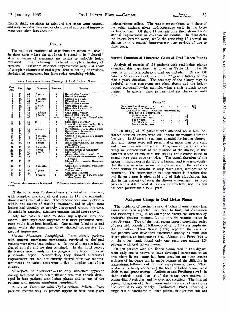
The results of treatment of 30 patients are shown in Table I.In those cases where the condition is stated to be " cleared "after a course of treatment no visible or palpable lesionremained. This " clearing " included complete healing oferosions. "Healed " describes improvement only just shortof complete clearance of oral signs-that is, healing of erosions,abolition of symptoms, but faint striae remaining visible.
TABLE I.-Betamethasone Therapy of Oral Lichen Planus
No. Sex Age Duration Erosions Results
1 F 69 16 years + Healed after 3 months2 F 53 ? + Healed in l month3 M 67 3 months _ Cleared in 2 weeks4 F 41 2 years - Cleared in 1 month5 M 56 - - Healed after l month6 M 67 12 years + Clear after I month7 F 57 12 , Clear after I month. T8 F 57 23 ,, - Did not return9 F 43 2 months - Clear after 2 weeks*10 F 66 5 years + Healed after 3 months11 M 45 5 ,, ± + + Irregular progress. Healed after
1 year12 F 67 24 ,, ± Clear after 2 months*13 F 39 ? - I month treatment only; little
progress.: did not return14 F 58 12 years + Buccal lesion cleared after 3 weeks15 F 59 ? ± Healed on hydrocortisone16 M 20 ? - Cleared in 1 month17 F 62 6 months + Cleared on one side after 3 months18 F 42 6 years + Improved*. T19 F 25 3 months + Healing after 1 month20 M 51 3 years + Initial improvement. No further
progress. T21 F 38 2 months _ Cleared in 4 months22 M 19 1 year + Entirely healed in 4 months*23 F 66 7 years _ Little benefit24 F 60 7 ., + Almost clear after 1 year*. T25 M 48 ? + Considerable improvement after
6 weeks26 F 53 ? - Cleared after 1 month. Remained
clear27 F 50 6 months - Cleared after 2 months. Remained
clear 1 year28 M 43 5 years - Did not return29 F 22 ? - Clear after 1 month30 F 78 6 weeks + + Started to heal in I month; clear
after 5* months
0 Recurs when treatment is stopped. T Denotes those patients who developedthrush.
Of the 30 patients 20 showed very substantial improvement,with complete clearance of oral signs in 13; the remaindershowed weak residual striae. The response was usually obviouswithin one month of starting treatment, and in eight caseslesions had virtually or entirely disappeared within this time.As might be expected, extensive erosions healed more slowly.Only two patients failed to show any response after one
month; later experience suggested that more prolonged treat-ment might have been beneficial. One patient failed to attendagain, while the remainder (five) showed progressive butgradual improvement.Mucous Membrane Pemphigoid.-Three elderly patients
with mucous membrane pemphigoid restricted to the oralmucosa were given betamethasone. In two of them the lesionscleared entirely and no sign remained. In the third patientthe lesions were mainly on the gingivae in relation to severeperiodontal sepsis. Nevertheless, they showed substantialimprovement but had not entirely cleared after two months'treatment; the patient then went to live in another part of thecountry.
Side-effects of Treatment.-The only side-effect apparentduring treatment with betamethasone was that thrush devel-oped in four patients with lichen planus and in one of thepatients with mucous membrane pemphigoid.
Results of Treatment with Hydrocortisone Pellets.-Fromprevious records only 13 patients appear to have been given
hydrocortisone pellets. The results are combined with those offive other patients given hydrocortisone early in the beta-methasone trial. Of these 18 patients only three showed sub-stantial improvement in less than six months. In three casesthe lesions became worse, while the remaining 12 showed nochange or only gradual improvement over periods of one tothree years.
Natural Duration of Untreated Cases of Oral Lichen Planus
Analysis of records of 138 patients with oral lichen planusattending this department is given in Table II. The 30patients in the betamethasone trial are excluded. Of the 138patients 63 attended only once, and 79 gave a history of lessthan a year's duration. The accuracy of the history may bedoubtful in that symptoms are often absent and the lesionsnoticed accidentally-for example, when a visit is made to thedentist. In general, these patients had the disease in mildform.
TABLE IITotal number of casesNo. attending more than once
Duration of lesions when last seen-that is,total period of observation:Up to 6 months6 months to 1 year1 to 5 years.5 to 10 yearsMore than 10 years
.. 13875
15253041
In 60 (80%) of 75 patients who attended on at least onefurther occasion lesions were still present six months after thefirst visit. In 35 cases the patients attended for further observa-tion, and lesions were still present after more than one year.and in one case after 20 years. This, however, is almost cer-tainly an underestimate of the duration of the disease, in thatpatients whose lesions were not unduly troublesome failed toattend more than once or twice. The actual duration of thelesions in most cases is therefore unknown, and it is noteworthythat there is an actual record of improvement in the oral con-dition within six months in only three cases, irrespective oftreatment. The experience in this department is therefore thatoral lichen planus is often mild and of little significance, butthat in the majority of cases the disease is persistent ; in mostpatients it is still present at least six months later, and in a fewhas been present for 5 to 20 years.
Malignant Change in Oral Lichen Planus
The incidence of carcinoma in oral lichen planus is not clear.Cases have been reported from time to time, but Andreasenand Pindborg (1963), in an attempt to clarify the situation byanalysing previous reports, found only 46 recorded cases inover 50 years. Two of the most recent papers presenting seriesof cases with periods of follow-up of up to 10 years emphasizethe difficulties. Thus Warin (1960) reported the cases offive patients who developed carcinoma among 53 with orallichen planus, an incidence of 9%. Altman and Perry (1961),on the other hand, found only one such case among 128patients with oral lichen planus.Of 138 patients with oral lichen planus seen in this depart-
ment only one is known to have developed carcinoma in anarea where lichen planus had been seen, but no more preciseestimate of incidence can be made because of the difficulty inmaintaining follow-up of the mild asymptomatic cases. Thereis also no certainty concerning the form of lichen planus mostliable to malignant change. Andreasen and Pindborg (1963) intheir analysis found that 16 of the lesions were erosive, 11plaque-like, 5 reticular, and 14 were not specified. The intervalbetween diagnosis of lichen planus and appearance of carcinomaalso seemed to vary widely. Desbrosses (1965), reporting asingle case of carcinoma in lichen planus, thought that this was
on 7 March 2019 by guest. P
rotected by copyright.http://w
ww
.bmj.com
/B
r Med J: first published as 10.1136/bm
j.1.5584.86 on 13 January 1968. Dow
nloaded from
most likely to happen with either the hypertrophic form of orallichen planus exposed to chronc trauma or the erosive form,but provided no supporting evidence.A factor which may be overlooked in trying to assess the
incidence of carcinoma in relation to oral lichen planus is thatmany mild asymptomatic cases appear to be noticed only bydental surgeons, as a consequence of the close observation ofthe mouth essential to their work, and are referred to depart-ments of oral medicine such as this. Dermatologists, on theother hand, are likely to see more severe or widespread cases.
causing symptoms, and it is from among these that most reports
of malignant change have originated.If any conclusions can be drawn it would appear that there
is some association between carcinoma and oral lichen planus;but clearly there are difficulties in assessing the incidence, andestimates vary between 10% and less than 1%.
While the possibility of malignant change is an importantreason for treating lichen planus, the severe oral symptoms thatcandle caused by uncomplicated-lichen planus'aret enough justi-fication for seeking an effective form of treatment.
Discussion
Williams et al. (1964) noted the " gratifying results " obtainedin a small number of patients with lichen planus of the skinwho used betamethasone ointment and that betamethasone was
statistically superior to five other topical corticosteroids in a
double-blind trial in the treatment of eczema and psoriasis.The results of local betamethasone therapy in oral lichen
planus are also most striking on both the rapidity and thecompleteness of the response. For quite unknown reasons only
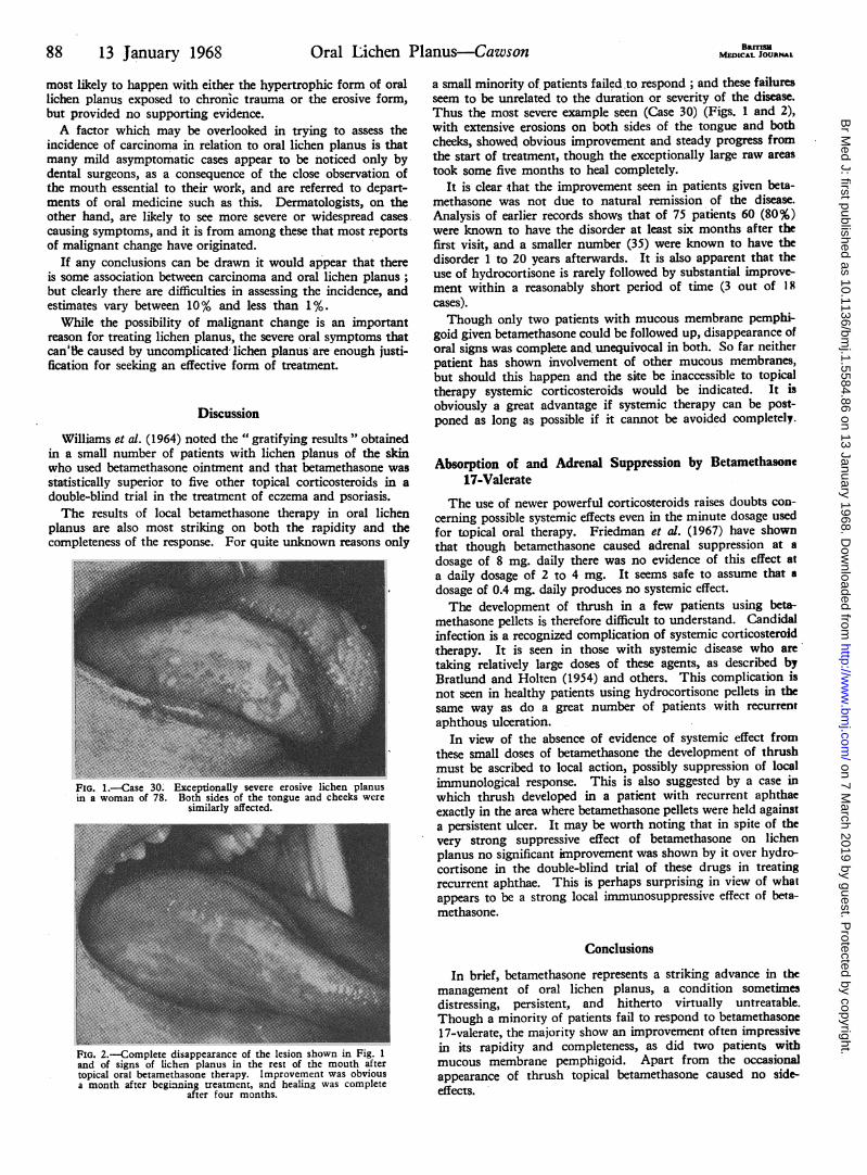
FIG. 1.--Case 30. Exceptionally severe erosive lichen planusin a woman of 78. Both sides of the tongue and cheeks were
similarly affected.
FIG. 2.-Complete disappearance of the lesion shown in Fig. 1
and of signs of lichen planus in the rest of the mouth aftertopical oral betamethasone therapy. Improvement was obviousa month after beginning treatment, and healing was complete
after four months.
BarrMEDCAL JOURNAL
a small minority of patients failed to respond ; and these failuresseem to be unrelated to the duration or severity of the disease.Thus the most severe example seen (Case 30) (Figs. 1 and 2),with extensive erosions on both sides of the tongue and bothcheeks, showed obvious improvement and steady progress fromthe start of treatment, though the exceptionally large raw areastook some five months to heal completely.
It is clear that the improvement seen in patients given beta-methasone was not due to natural remission of the disease.Analysis of earlier records shows that of 75 patients 60 (80%)were known to have the disorder at least six months -after thefirst visit, and a smaller number (35) were known to have thedisorder 1 to 20 years afterwards. It is also apparent that theuse of hydrocortisone is rarely followed by substantial improve-ment within a reasonably short period of time (3 out of 18cases).
Though only two patients with mucous membrane pemphi-goid given betamethasone could be followed up, disappearance oforal signs was comple and unequivocal in both. So far neitherpatient has shown involvement of other mucous membranes,but should this happen and the site be inaccessible to topicaltherapy systemic corticosteroids would be indicated. It isobviously a great advantage if systemic therapy can be post-poned as long as possible if it cannot be avoided completely.
Absorption of and Adrenal Suppression by Betamethasone17-Valerate
The use of newer powerful corticosteroids raises doubts con-
ceminig possible systemic effects even in the minute dosage usedfor topical oral therapy. Friedman et al. (1967) have shownthat though betamethasone caused adrenal suppression at a
dosage of 8 mg. daily there was no evidence of this effect ata daily dosage of 2 to 4 mg. It seems safe to assume that a
dosage of 0.4 mg. daily produces no systemic effect.The development of thrush in a few patients using beta-
methasone pellets is therefore difficult to understand. Candidalinfection is a recognized complication of systemic corticosteroidtherapy. It is seen in those with systemic disease who are
taking relatively large doses of these agents, as described byBratlund and Holten (1954) and others. This complication isnot seen in healthy patients using hydrocortisone pellets in thesame way as do a great number of patients with recurrentaphthous ulceration.
In view of the absence of evidence of systemic effect fromthese small doses of betamethasone the development of thrushmust be ascribed to local action, possibly suppression of localimmunological response. This is also suggested by a case inwhich thrush developed in a patient with recurrent aphthaeexactly in the area where betamethasone pellets were held againsta persistent ulcer. It may be worth noting that in spite of thevery strong suppressive effect of betamethasone on lichenplanus no significant improvement was shown by it over hydro-cortisone in the double-blind trial of these drugs in treatingrecurrent aphthae. This is perhaps surprising in view of whatappears to be a strong local immunosuppressive effect of bea-methasone.
Conclusions
In brief, betamethasone represents a striking advance in the
management of oral lichen planus, a condition sometimesdistressing, persistent, and hitherto virtually untreatable.Though a minority of patients fail to respond to betamethasone17-valerate, the majority show an improvement often impressivein its rapidity and completeness, as did two patients withmucous membrane pemphigoid. Apart from the occasionalappearance of thrush topical betamethasone caused no side-effects.
88 13 January 1968 Oral Lichen Planus Cawson
on 7 March 2019 by guest. P
rotected by copyright.http://w
ww
.bmj.com
/B
r Med J: first published as 10.1136/bm
j.1.5584.86 on 13 January 1968. Dow
nloaded from
13 January 1968 Oral Lichen Planus-awson MWDi'ICSoui 89
In 30 patients with oral lichen planus regarded as severeenough to justify treatment, use of betamethasone (Betnovate)pellets 0.1 mg. was followed by substantial improvement orcomplete clearance of oral lesions in 20. Seven patients showedgradual or incomplete improvement.Only 2 out of 30 patients failed to show any response to
betamethasone, but treatment was not continued for more thanone month in these cases.Of two patients with mucous membrane pemphigoid the use
of betamethasone pellets was followed by complete clearance oforal lesions. A third patient showed substantial improvementuntil he ceased to attend.
The only side-effect of treatment was development of thrusbin four patients.
The assistance of Messrs. Glaxo in their generous supply oftablets and for other help for this trial is gratefully acknowledged.
REFERENCESAltman, J., and Perry, H 0. (1961). Arch. Derm., 84, 179.Andreasen, J. O., and Pindborg, J. J. (1963). Nord. Med., 70, 861.Bratlund, H., and Holten, C. (1954). Dan. med. Bull., 1, 79.Desbrosses, J. L. (1965). Ann. Oto-laryng. (Paris), 82, 841.Friedman, M., Fletcher, J., Hinton, J. M., Lennard-Jones, J. B,
Misiewicz, J. J., and Parrish, J. A. (1967). Brit. med. Y., 1, 335.McCarthy, P. L., and Shkba:-, G. (1964). Diseases of the Oral Mucosa.
New York.Warin, R. P. (1960). Brit. 7. Derm., 72, 288.Williams, D. I., et al. (1964). Lancet, 1, 1177.
Use of Lignocaine in Treatment of Cardiac Arrhythmias
F. H. N. SPRACKLEN,* M.B., CH.B., M.R.C.P.; J. J. KIMERLING,* M.B., B.S., M.R.C.P.
E. M. M. BESTERMAN,* M.A., M.D., F.R.C.P.; J. W. LITCHFIELD,* B.M., F.R.C.P.
Brit. mod. J., 1968, 1, 89-91
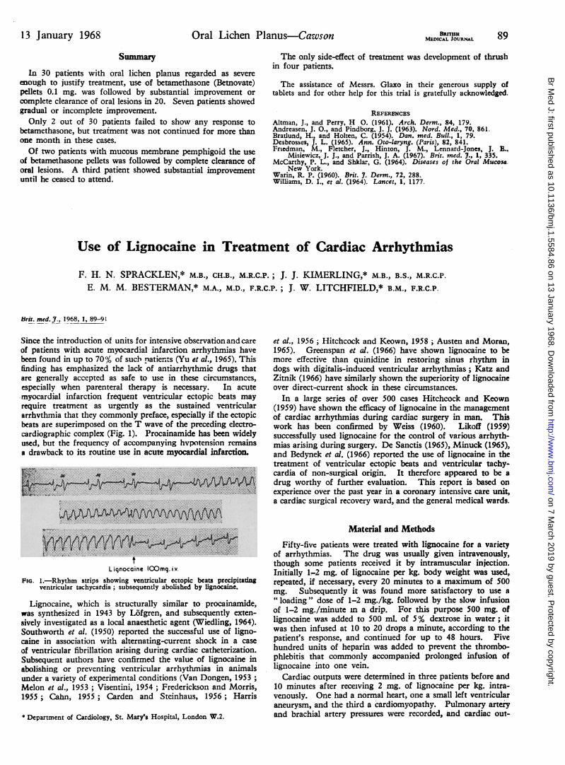
-Since the introduction of. units- for intensive observation and careof patients with acute myocardial infarction arrhythmias havebeen found in up to 70% of such natients (Yu et al., 1965). Thisfinding has emphasized the lack of antiarrhythmic drugs thatare generally accepted as safe to use in these circumstances,especially when, parenteral- therapy is necessary. In acute.myocardial infarction frequent.ventripular ectopic beats mayrequire treatment as urgently as the sustained ventriculararrhythmia that they commonly preface, especially if the ectopicbeats are superimposed on the T wave of the preceding electro-cardiographic complex (Fig. 1). Procainamide has been widelyused, but the frequency of accompanying hvpotension remainsa drawback to its routine use in acute myocardial infarction.
:4
. ME~~~~~~SLiqnocoine OOmqi.v.
FIG. 1.-Rhythm strips showing ventricular ectopic beats precipitatingventricular tachycardia; subsequently abolished by lignocaine.
Lignocaine, which is structurally similar to procainamide,was synthesized in 1943 by Lbfgren, and subsequently exten-sively investigated as a local anaesthetic agent (Wiedling, 1964).Southworth et al. (1950) reported the successful use of ligno-caine in association with alternating-current shock in a caseof ventricular fibrillation arising during cardiac catheterization.Subsequent authors have confirmed the value of lignocaine inabolishing or preventing ventricular arrhythmias in animalsunder a variety of experimental conditions (Van Dongen, 1953;Melon et al., 1953 ; Visentini, 1954; Frederickson and Morris,1955; Cahl, 1955; 'Carden and. Steinhaus, 1956; Harris
et al., 1956; Hitchcock and Keown, 1958; Austen and Moran,1965). Greenspan et al. (1966) have shown lignocaine to bemore effective than quinidine in restoring sinus rhythm indogs with digitalis-induced ventricular arrhythmias; Katz andZitnik (1966) have similarly shown the superiority of lignocaineover direct-current shock in these circumstances.
In a large series of over 500 cases Hitcheock and Keown(1959) have shown the efficacy of lignocaine in the managementof cardiac arrhythmias during cardiac surgery in man. Thiswork has been confirmed by Weiss (1960). Likoff (1959)successfully used lignocaine for the control of various arrhyth-mias arising during surgery. De Sanctis (1965), Minuck (1965),and Bedynek et al. (1966) reported the use of lignocaine in thetreatment of ventricular ectopic beats and ventricular tachy-cardia of non-surgical origin. It therefore appeared to be adrug worthy of further evaluation. This report is based onexperience over the past year in a coronary intensive care unit,a cardiac surgical recovery ward, and the general medical wards.
Material and MethodsFifty-five patients were treated with lignocaine for a variety
of arrhythmias. The drug -was usually given intravenously,though some patients received it by intramuscular injection.Initially 1-.2 mg, of lignocaine per kg. body weight was used,repeated, if necessary, every 20 minutes to a maximum of 500mg. Subsequently it was found more satisfactory to use a" loading " dose of 1-2 mg./kg. followed by the slow infusionof 1-2 mg./minute in a drip. For this purpose 500 mg. oflignocaine was added to 500 ml. of 5% dextrose in water; itwas then infused at 10 to 20 drops a minute, according to thepatient's response, and continued for up to 48 hours. Fivehundred units of heparin was added to prevent the thrombo-phlebitis that commonly accompanied prolonged infusion oflignocaine into one vein.
Cardiac outputs were determined in three patients before and10 minutes after receiving 2 mg. of lignocaine per kg. intra-venously. One' had a normal heart, one a small left ventricularaneurysm, and the third a cardiomyopathy. Pulmonary arteryand brachial artery pressures were rectrded, and cardiac out-* Department of Cardiology, St. Mary's Hospital, London W.2.
on 7 March 2019 by guest. P
rotected by copyright.http://w
ww
.bmj.com
/B
r Med J: first published as 10.1136/bm
j.1.5584.86 on 13 January 1968. Dow
nloaded from