Embed Size (px)
Citation preview

0
Treatment of lipid-rich wastewater using free and immobilized bioemulsifier
and hydrolytic enzymes from indigenous bacterial isolates
Submitted in fulfilment of the academic requirements for the degree of Doctor of Philosophy
(PhD) in the Discipline of Microbiology; School of Life Sciences; College of Agriculture,
Engineering and Science at the University of KwaZulu-Natal (Westville Campus), Durban.
ADEGOKE ISIAKA ADETUNJI
DECEMBER
2017
As the candidate’s supervisor, I have approved this thesis for submission.
Supervisor: Prof. A. O. Olaniran
i
PREFACE
The experimental work described in this thesis was carried out in the Discipline of Microbiology, School
of Life Sciences, University of KwaZulu-Natal (Westville campus), Durban, South Africa from August
2015 to December 2017, under the supervision of Professor A. O. Olaniran.
These studies show original work by the author and have not otherwise been submitted in any form for any
degree or diploma to any tertiary institution. Where use has been made of the work of others, it is duly
acknowledged in the text.
ii
COLLEGE OF AGRICULTURE, ENGINEERING AND SCIENCE
DECLARATION 1 - PLAGIARISM
I, ……………………………………….………………………., declare that,
1. The research reported in this thesis, except where otherwise indicated, is my original research.
2. This thesis has not been submitted for any degree or examination at any other university.
3. This thesis does not contain other persons’ data, pictures, graphs or other information, unless
specifically acknowledged as being sourced from other persons.
4. This thesis does not contain other persons' writing, unless specifically acknowledged as being
sourced from other researchers. Where other written sources have been quoted, then:
a. Their words have been re-written but the general information attributed to them has been referenced
b. Where their exact words have been used, then their writing has been placed in italics and inside
quotation marks, and referenced.
5. This thesis does not contain text, graphics or tables copied and pasted from the Internet, unless
specifically acknowledged, and the source being detailed in the thesis and in the References sections.
Signed
………………………………………………………………………………
CMC Feb 2012
Form EX1-5
iii
COLLEGE OF AGRICULTURE, ENGINEERING AND SCIENCE
DECLARATION 2 - MANUSCRIPTS UNDER REVIEW
Details of contributions to publications that form part and/or include research presented in this thesis
(include publications in preparation, submitted, in press and published and give details of the contributions
of each author to the experimental work and writing of each publication).
Manuscript 1
Title: Indigenous Acinetobacter sp. isolated from lipid-rich wastewater in Durban produced thermostable
glycoprotein bioemulsifiers
Journal: 3 Biotech
Authors:
Adegoke Isiaka Adetunji - Conceptualization, Experimental design, Data collection, Data analysis, Drafting
and Editing of manuscript
Ademola Olufolahan Olaniran – Conceptualization, Experimental design, Manuscript editing
Manuscript 2
Title: Optimization of bioprocess parameters for enhanced protease production by Bacillus aryabhattai
Ab15-ES using response surface methodology
Journal: Bioprocess and Biosystems Engineering
Authors:
Adegoke Isiaka Adetunji - Conceptualization, Experimental design, Data collection, Data analysis, Drafting
and Editing of manuscript
Ademola Olufolahan Olaniran – Conceptualization, Experimental design, Manuscript editing
Manuscript 3
Title: Optimization of culture conditions for enhanced lipase production by an indigenous Bacillus
aryabhattai SE3-PB using response surface methodology
Journal: Biotechnology and Biotechnological Equipment
Authors:
Adegoke Isiaka Adetunji - Conceptualization, Experimental design, Data collection, Data analysis, Drafting
and Editing of manuscript
Form EX1-6
iv
Ademola Olufolahan Olaniran – Conceptualization, Experimental design, Manuscript editing
Manuscript 4
Title: Treatment of lipid-rich wastewater using a mixture of free or immobilized bioemulsifier and
hydrolytic enzymes from indigenous bacterial isolates
Journal: Desalination and Water Treatment
Authors:
Adegoke Isiaka Adetunji - Conceptualization, Experimental design, Data collection, Data analysis, Drafting
and Editing of manuscript
Ademola Olufolahan Olaniran – Conceptualization, Experimental design, Manuscript editing
Signed:
CMC Feb 2012
v
ABSTRACT
The production and discharge of raw and poorly treated lipid-rich wastewater increase yearly due to rapid
urbanization and industrial growth. This results in severe environmental and health hazards by affecting the
normal operations of ecosystems. Biological approach involving synergistic application of low cost
bioemulsifier and hydrolytic enzymes is an efficient, cost-effective, sustainable and eco-friendly
technology for the treatment of high strength lipid-rich wastewater. Therefore, the main objective of this
study was to investigate the potential of a mixture of free or immobilized bioemulsifier and hydrolytic
enzymes (protease and lipase) in the reduction of pollutants present in dairy and poultry processing
wastewater. Glycoprotein bioemulsifiers and hydrolytic enzymes were produced extracellularly by
submerged fermentation from indigenous Acinetobacter sp. and Bacillus aryabhattai, respectively.
Optimization of bioprocess parameters, using response surface methodology, revealed a 4.4- and 7.2-fold
increase in protease and lipase production, respectively. The bioemulsifier from strain AB9-ES (XB9) and
strain AB33-ES (YB33) formed stable emulsions only with edible oils with highest emulsification indices
of 79.6 and 67.9%, respectively obtained against sunflower oil. The emulsifying activity of the
bioemulsifiers was stable over broad range of temperature (4-121 ºC), moderate salinity (1-6%) and pH
(5.0-10.0). Comparative study of biochemical profiling of both free and immobilized hydrolytic enzymes
showed no change in the optimum temperature and pH of both enzyme preparations for maximum activity.
However, in comparison to free enzymes, the immobilized enzymes recorded enhanced stability over the
investigated pH and temperature ranges. Kinetics properties revealed enhanced enzyme-substrate affinity
and increased catalytic efficiency of the immobilized enzymes when compared to soluble enzymes. In
addition, the immobilized enzymes were more stable when stored at 4 and 25 °C and reusable for more than
five consecutive cycles. These hyper-active and highly stable bioproducts were utilized in cocktail in both
soluble and entrapped form for the batch biodegradation of pollutants present in lipid-rich wastewater.
Biodegradability of the wastewater was assessed by measuring the reduction of COD and lipid content at
time intervals under varying incubation conditions. In dairy wastewater treated at 37 °C without pH
vi
adjustment, maximum COD (60.51 and 65.19%) and lipid (47.98 and 63.53%) reduction efficiencies were
recorded at 120 h using free and immobilized bioproducts, respectively. However, under these conditions,
maximum COD (86.44 and 93.65%) and lipid (51.62 and 69.06%) removal efficiencies of poultry
processing wastewater were observed at 120 h when treated with free and immobilized bioproducts,
respectively. At temperature of 50 °C and pH 8.0, there was enhanced reduction of organic pollutants, with
maximum COD (65.96 and 77.52%) and lipid (55.22 and 71.12%) removal efficiencies obtained in dairy
wastewater at 72 h when using free and immobilized bioproducts, respectively. In the case of poultry
processing wastewater, optimum COD (90.29 and 94.72%) and lipid (63 and 76.66%) removal was
recorded at 72 h when treated with free and immobilized bioproducts, respectively. Reusability studies
suggest that the immobilized bioproducts could be reused for up to six and seven times for the treatment of
dairy and poultry processing wastewater, respectively. Findings from this study suggest the efficient, cost-
effectiveness, sustainability and synergistic application of the developed immobilized bioemulsifier and
hydrolytic enzymes in the removal of pollutants present in dairy and poultry processing wastewater.
vii
This thesis is dedicated to:
My beloved parents, late (Pa) Salawudeeen & Alh (Mrs) K. A. Adetunji
for their love, support, prayers and inspiration during the course of my academic career
viii
ACKNOWLEDGEMENTS
The author records deepest gratitude to the following:
• Almighty Allah (SWT) for His continous support towards the successful completion of this
project. He has been faithful and make my dreams come to reality. Alhamdullilah!!
• Prof. A. O. Olaniran for his supervison, advice and support. Many thanks for accepting me to
your laboratory.
• My beloved parents, late (Pa) Salawudeen and Alh (Mrs) K. A. Adetunji, whom I regard as
jewel of inestimable value and pillar of this milestone achievement. Many thanks to Dad for
his valuable legacy of putting education first. Mum has never let me down in all ramification.
Your motivation and supports (morally, spiritually and financially) have really aided the
accomplishment of this project
• My sisters (Alh. Sherifat Adeyemi and Alh. Muinat Akinsola) for your financial support,
motivation and prayers at the time I was helpless. Your regular telephone calls and messages
inspire me
• Laboratory 4 postgraduate students for your love, support and cooperation
• Staff (academic and technical) and students of discipline of Microbiology. You have all
contributed immensely towards completion of this project. Dr. Mokoena is specially thanked
for driving me home when I finish late in the laboratory and when going to campus. Many
thanks to S’fiso Gumbi for hectic sampling from beginning to the end of this project
• Pravash, Gitesh and Neil for providing me dairy and poultry processing wastewater
• University of KwaZulu-Natal, Westville campus and National Research Foundation (NRF)
ix
CONTENTS
PREFACE ………………………………………………………………………………………………….i
ABSTRACT ………………………………………………………………………………….……………v
DEDICATION …………………………………………….………………………………….…………vii
ACKNOWLEDGEMENTS …………………………………………………………………..………..viii
LIST OF TABLES ………………………………………………………………………………….....xxii
LIST OF FIGURES ……………………………………………………………………………….…..xxiv
CHAPTER ONE INTRODUCTION ………………………………………………………........…..….1
1.1 BACKGROUND …………………………………………………………………………….…..1
1.1.1 GENERAL CHARACTERISTICS OF DAIRY WASTEWATER ………………...3
1.1.2 GENERAL CHARACTERISTICS OF POULTRY PROCESSING
WASTEWATER ………………………………………………………………………4
1.1.3 IMPACTS OF LIPID-RICH WASTEWATER ……………………………………..5
1.2 SCOPE OF THIS STUDY …………………………………………………………...…………5
1.3 HYPOTHESIS …………………………………………………………………………………..6
1.4 AIM………………………………………………………….…………..………………………..7
1.5 SPECIFIC OBJECTIVES ………………………….…………………..………………………7
1.6 KEY QUESTIONS TO BE ANSWERED ………………………………………………….….7
1.7 REFERENCES ………………………………………………………………………...…...…...9
CHAPTER TWO PRODUCTION, PROPERTIES AND POTENTIAL APPLICATIONS OF
MICROBIAL SURFACTANTS -- A REVIEW .......................................................................................16
Abstract ......................................................................................................................................................17
1. Introduction ...........................................................................................................................................17
1.1 Biosurfactant/bioemulsifier-producing microorganisms ................................................................19
1.2 Detection methods for biosurfactant and bioemulsifier-producing microorganisms ...................21
1.2.1 Oil spreading assay ..................................................................................................................21
x
1.2.2 Surface and interfacial tension measurement .......................................................................21
1.2.3 Emulsification activity .............................................................................................................22
1.2.4 Drop collapse method ..............................................................................................................22
1.2.5 Cethyltrimethylammonium bromide agar plate method .....................................................23
1.2.6 Blood haemolysis test ...............................................................................................................23
1.3 Classification and chemical nature of biosurfactants ......................................................................23
1.3.1 Glycolipids ................................................................................................................................24
1.3.2 Lipopeptides and lipoproteins .................................................................................................25
1.3.3 Fatty acids, phospholipids and neutral lipids ........................................................................25
1.3.4 Polymeric biosurfactants .........................................................................................................26
1.3.5 Particulate biosurfactants ........................................................................................................26
1.4 Biosurfactant production and influencing factors ...........................................................................26
1.5 Kinetics of growth and biosurfactant production ............................................................................27
1.6 Recovery and purification of biosurfactants ....................................................................................28
1.6.1 Acetone precipitation ...............................................................................................................29
1.6.2 Ethanol precipitation ...............................................................................................................29
1.6.3 Ammonium sulphate precipitation .........................................................................................29
1.6.4 Acid precipitation .....................................................................................................................30
1.6.5 Dialysis and lyophilization .......................................................................................................30
1.6.6 Foam fractionation ...................................................................................................................30
1.6.7 Adsorption and desorption ......................................................................................................31
1.6.8 Ultrafiltration ...........................................................................................................................31
xi
1.6.9 Preparative thin layer chromatography ................................................................................31
1.6.10 Ion exchange chromatography ..............................................................................................31
1.7 Potential applications of biosurfactants/bioemulsifiers ...................................................................32
1.7.1 Bioremediation .........................................................................................................................32
1.7.2 Food industry ............................................................................................................................35
1.7.3 Medicine ....................................................................................................................................35
1.7.4 Cosmetic industry .....................................................................................................................37
1.7.5 Nanotechnology ........................................................................................................................37
1.8 Concluding remarks ...........................................................................................................................37
1.9 References ............................................................................................................................................38
CHAPTER THREE PRODUCTION, PROPERTIES AND POTENTIAL
BIOTECHNOLOGICAL APPLICATIONS OF MICROBIAL PROTEASES -- A REVIEW ........59
Abstract ......................................................................................................................................................60
1. Introduction ...........................................................................................................................................60
1.1 Sources of proteases ............................................................................................................................61
1.1.1 Plant proteases ..........................................................................................................................61
1.1.2 Animal proteases ......................................................................................................................62
1.1.3 Microbial proteases ..................................................................................................................62
1.2 Classification of microbial proteases .................................................................................................63
1.2.1 Serine proteases ........................................................................................................................64
1.2.2 Cysteine proteases ....................................................................................................................64
1.2.3 Metalloproteases .......................................................................................................................64
1.2.4 Aspartic proteases ....................................................................................................................64
xii
1.3 Protease production and influencing factors ...................................................................................65
1.4 Statistical approaches for optimization of protease production .....................................................67
1.4.1 Plackett-Burman design ..........................................................................................................67
1.4.2 Response surface methodology ...............................................................................................69
1.4.2.1 Central composite design ..............................................................................................69
1.4.2.2 Box-Behnken design ......................................................................................................70
1.5 Purification of proteases .....................................................................................................................70
1.5.1 Ultrafiltration ...........................................................................................................................71
1.5.2 Precipitation ..............................................................................................................................71
1.5.3 Ion exchange chromatography ................................................................................................71
1.5.4 Affinity chromatography .........................................................................................................71
1.5.5 Hydrophobic interaction chromatography ............................................................................71
1.6 Immobilization of proteases ...............................................................................................................72
1.7 Characterization of alkaline proteases ..............................................................................................73
1.7.1 Effect of pH on activity and stability ......................................................................................73
1.7.2 Effect of temperature on activity and stability ......................................................................75
1.7.3 Kinetics properties of alkaline proteases ...............................................................................76
1.8 Potential applications of alkaline proteases ......................................................................................76
1.8.1 Detergent industry ...................................................................................................................76
1.8.2 Leather industry .......................................................................................................................77
1.8.3 Food industry ............................................................................................................................77
1.8.4 Medicine ....................................................................................................................................78
xiii
1.8.5 Waste treatment .......................................................................................................................78
1.9 Conclusion and future perspectives ...................................................................................................79
1.10 References ..........................................................................................................................................80
CHAPTER FOUR PRODUCTION, PROPERTIES AND BIOTECHNOLOGICAL
APPLICATIONS OF MICROBIAL LIPASES -- A REVIEW ..............................................................99
Abstract ....................................................................................................................................................100
1. Introduction .........................................................................................................................................100
1.1 Sources of lipases ................................................................................................................................101
1.1.1 Plant lipases .............................................................................................................................101
1.1.2 Animal lipases ..........................................................................................................................101
1.1.3 Microbial lipases .....................................................................................................................101
1.2 Classification of bacterial lipases ......................................................................................................104
1.3 Methods for detection of lipase production ......................................................................................104
1.3.1 Screening methods on solid media .........................................................................................104
1.3.2 Titrimetric methods ................................................................................................................105
1.4 Lipase production and influencing parameters ...............................................................................105
1.5 Optimization of lipase production using statistical experimental designs .....................................107
1.6 Purification of lipases .........................................................................................................................109
1.7 Immobilization of lipases ...................................................................................................................110
1.8 Characterization of alkaline lipases ..................................................................................................110
1.8.1 Effect of temperature on activity and stability ......................................................................110
1.8.2 Effect of pH on activity and stability ......................................................................................112
1.8.3 Kinetics properties of alkaline lipases ....................................................................................113
xiv
1.9 Potential biotechnological applications of alkaline lipases .............................................................113
1.9.1 Lipases in detergent industry .................................................................................................114
1.9.2 Lipases in food industry ..........................................................................................................115
1.9.3 Lipases in cosmetics ................................................................................................................115
1.9.4 Lipases in pulp and paper industry ........................................................................................115
1.9.5 Lipases in bioremediation of lipid-rich wastewater ..............................................................116
1.10 Concluding remarks ........................................................................................................................117
1.11 References .........................................................................................................................................118
CHAPTER FIVE INDIGENOUS ACINETOBACTER SP. ISOLATED FROM LIPID-RICH
WASTEWATER IN DURBAN PRODUCED THERMOSTABLE GLYCOPROTEIN
BIOEMULSIFIERS ................................................................................................................................141
Abstract ....................................................................................................................................................142
1. INTRODUCTION ...............................................................................................................................142
2. MATERIALS AND METHODS ........................................................................................................144
2.1 Sample collection and bacterial isolation procedures ..............................................................144
2.2 Screening of bacteria for bioemulsifier/biosurfactant production .........................................144
2.3 Amplification and sequencing of 16S rRNA gene and phylogenetic analysis .......................145
2.4 Kinetics of growth and bioemulsifier production ....................................................................145
2.5 Extraction and partial purification of bioemulsifier ...............................................................146
2.6 Chemical composition of bioemulsifier ....................................................................................146
2.7 Fourier transforms infrared spectroscopy (FTIR) analysis ...................................................146
2.8 Effect of different hydrophobic substrates on bioemulsifier activity ....................................146
2.9 Bioemulsifier stability studies ...................................................................................................147
xv
2.9.1 Effect of temperature, pH and salinity on bioemulsifier activity ................................147
3. RESULTS AND DISCUSSION ..........................................................................................................147
3.1 Isolation, screening and identification of bioemulsifier-producing bacteria ........................147
3.2 Kinetics of growth and bioemulsifier production ....................................................................150
3.3 Composition of bioemulsifier ....................................................................................................152
3.4 Effect of hydrophobic substrates on emulsifying activity .......................................................154
3.5 Effect of temperature, pH and salinity on emulsifying activity ...............................................155
4. CONCLUSIONS ..................................................................................................................................158
5. REFERENCES ....................................................................................................................................158
CHAPTER SIX OPTIMIZATION OF BIOPROCESS PARAMETERS FOR ENHANCED
PROTEASE PRODUCTION BY BACILLUS ARYABHATTAI AB15-ES USING RESPONSE
SURFACE METHODOLOGY ..............................................................................................................163
Abstract ....................................................................................................................................................164
1. INTRODUCTION ...............................................................................................................................165
2. MATERIALS AND METHODS ........................................................................................................166
2.1 Chemicals ....................................................................................................................................166
2.2 Sample collection and bacterial isolation procedures .............................................................166
2.3 Screening of the bacterial isolate for proteolytic activity ........................................................166
2.4 Identification and phylogenetic analysis of the bacterial isolate ............................................167
2.4.1 Genomic DNA extraction ................................................................................................167
2.4.2 Amplification, sequencing and analysis of 16S rRNA gene .........................................167
2.5 Time course profile of growth and extracellular protease production ...................................167
2.6 Optimization of nutritional parameters for protease production ..........................................168
xvi
2.7 Statistical optimization of protease production .......................................................................168
2.7.1 Response surface methodology .......................................................................................168
3. RESULTS AND DISCUSSION ..........................................................................................................169
3.1 Isolation, screening and identification of protease-producing bacteria ................................169
3.2 Growth kinetics and extracellular protease production .........................................................170
3.3 Selection of best carbon and nitrogen sources for protease production ...............................171
3.4 Optimization of bioprocess parameters for maximum protease production using RSM....173
3.4.1 Experimental validation of the optimized process conditions ………………………..178
4. CONCLUSIONS ..................................................................................................................................179
5. REFERENCES ....................................................................................................................................179
CHAPTER SEVEN OPTIMIZATION OF CULTURE CONDITIONS FOR ENHANCED
LIPASE PRODUCTION BY AN INDIGENOUS BACILLUS ARYABHATTAI SE3-PB USING
RESPONSE SURFACE METHODOLOGY ........................................................................................185
Abstract ....................................................................................................................................................186
1. INTRODUCTION ...............................................................................................................................187
2. MATERIALS AND METHODS ........................................................................................................188
2.1 Chemicals ....................................................................................................................................188
2.2 Sample collection and bacterial isolation procedures .............................................................188
2.3 Screening of bacteria for lipolytic activity ...............................................................................189
2.3.1 Screening of bacteria for lipolytic activity using Tween-20 agar..................................189
2.3.2 Screening of bacteria for lipolytic activity using phenol red agar ................................189
2.4 Molecular identification of lipase-producing bacteria and phylogenetic analysis ................189
2.4.1 Genomic DNA extraction ................................................................................................189
xvii
2.4.2 Amplification, sequencing and analysis of 16S rRNA gene .........................................189
2.5 Kinetics of growth and extracellular lipase production ..........................................................190
2.6 Optimization of culture conditions for maximum lipase production .....................................190
2.6.1 Effect of inducer oils on lipase production .....................................................................190
2.6.2 Response surface methodology .......................................................................................191
3. RESULTS AND DISCUSSION ..........................................................................................................192
3.1 Isolation, screening and identification of lipase-producing bacteria ......................................192
3.2 Kinetics of growth and lipase production .................................................................................193
3.3 Selection of inducer oil for maximum lipase production .........................................................194
3.4 Optimization of culture conditions for improved lipase production using response surface
methodology .....................................................................................................................................195
3.4.1 Experimental validation of the optimized process conditions ……………………..…201
4. CONCLUSIONS ..................................................................................................................................202
5. REFERENCES ....................................................................................................................................202
CHAPTER EIGHT PARTIAL PURIFICATION, IMMOBILIZATION AND
CHARACTERIZATION OF ALKALINE PROTEASE FROM AN INDIGENOUS BACILLUS
ARYABHATTAI AB15-ES ISOLATED FROM LIPID-RICH WASTEWATER ..............................208
Abstract ....................................................................................................................................................209
1. INTRODUCTION ...............................................................................................................................209
2. MATERIALS AND METHODS ........................................................................................................211
2.1 Partial purification of protease .................................................................................................211
2.2 Immobilization of partially purified protease in calcium alginate beads ..............................212
2.3 Protease assay .............................................................................................................................212
xviii
2.4 Determination of immobilization efficiency .............................................................................212
2.5 Characterization of free and immobilized protease ................................................................213
2.5.1 Effect of temperature and pH on the activity of free and immobilized protease .......213
2.5.2 Effect of temperature and pH on the stability of free and immobilized protease........213
2.5.3 Kinetics properties of free and immobilized protease ..................................................213
2.5.4 Storage stability of free and immobilized protease .......................................................214
2.5.5 Reusability potential of the immobilized protease ........................................................214
2.5.6 Scanning electron microscopy of beads with immobilized protease ............................214
3. RESULTS AND DISCUSSION ..........................................................................................................214
3.1 Partial purification of protease .................................................................................................214
3.2 Protease immobilization and scanning electron microscopic analysis ...................................215
3.3 Effect of temperature and pH on the activity of free and immobilized protease from Bacillus
aryabhattai Ab15-ES ........................................................................................................................217
3.4 Thermal and pH stability of the free and immobilized protease from Bacillus aryabhattai
Ab15-ES ............................................................................................................................................219
3.5 Kinetics properties of free and immobilized protease .............................................................221
3.6 Storage stability of free and immobilized protease from Bacillus aryabhattai Ab15-ES .......222
3.7 Reusability potential of immobilized protease from Bacillus aryabhattai Ab15-ES ..............223
4. CONCLUSIONS ..................................................................................................................................224
5. REFERENCES ....................................................................................................................................225
CHAPTER NINE LIPASE FROM AN INDIGENOUS BACILLUS ARYABHATTAI SE3-PB:
PARTIAL PURIFICATION, IMMOBILIZATION AND BIOCHEMICAL PROPERTIES ..........232
Abstract ....................................................................................................................................................234
xix
1. INTRODUCTION ...............................................................................................................................234
2. MATERIALS AND METHODS ........................................................................................................236
2.1 Partial purification of lipase ......................................................................................................236
2.2 Immobilization of partially purified lipase in alginate gel beads ...........................................237
2.3 Lipase assay ................................................................................................................................237
2.4 Determination of immobilization efficiency .............................................................................237
2.5 Biochemical properties of free and immobilized lipase ..........................................................238
2.5.1 Effect of temperature and pH on the activity of free and immobilized lipase from
Bacillus aryabhattai SE3-PB ....................................................................................................238
2.5.2 Effect of temperature and pH on the stability of free and immobilized lipase from
Bacillus aryabhattai SE3-PB ....................................................................................................238
2.5.3 Kinetics properties of free and immobilized lipase .......................................................238
2.5.4 Storage stability of free and immobilized lipase from Bacillus aryabhattai SE3-PB ...238
2.5.5 Reusability potential of immobilized lipase from Bacillus aryabhattai SE3-PB ..........239
2.5.6 Scanning electron microscopy of beads with immobilized lipase .................................239
3. RESULTS AND DISCUSSION ..........................................................................................................239
3.1 Partial purification of lipase ......................................................................................................239
3.2 Lipase immobilization and scanning electron microscopic analysis ......................................240
3.3 Biochemical properties of free and immobilized lipase ...........................................................242
3.3.1 Effect of temperature and pH on the activity of free and immobilized lipase from
Bacillus aryabhattai SE3-PB ....................................................................................................242
3.3.2 Effect of temperature and pH on the stability of free and immobilized lipase from
Bacillus aryabhattai SE3-PB ....................................................................................................244
3.3.3 Kinetics parameters of free and immobilized lipase .....................................................246
xx
3.3.4 Storage stability of free and immobilized lipase from Bacillus aryabhattai SE3-PB ...247
3.3.5 Reusability potential of immobilized lipase from Bacillus aryabhattai SE3-PB ..........248
4. CONCLUSIONS ..................................................................................................................................248
5. REFERENCES ....................................................................................................................................249
CHAPTER TEN TREATMENT OF LIPID-RICH WASTEWATER USING A MIXTURE OF
FREE OR IMMOBILIZED BIOEMULSIFIER AND HYDROLYTIC ENZYMES FROM
INDIGENOUS BACTERIAL ISOLATES ............................................................................................255
Abstract ....................................................................................................................................................256
1. INTRODUCTION ...............................................................................................................................257
2. MATERIALS AND METHODS ........................................................................................................258
2.1 Materials .....................................................................................................................................258
2.2 Immobilization protocol ............................................................................................................258
2.3 Collection of raw lipid-rich wastewater ....................................................................................258
2.4 Physicochemical characterization of lipid-rich wastewater ...................................................258
2.5 Batch biodegradation of lipid-rich wastewater ........................................................................259
2.6 Reusability potential of immobilized protease, lipase and bioemulsifier ...............................259
3. RESULTS AND DISCUSSION ..........................................................................................................259
3.1 Characterization of lipid-rich wastewater ...............................................................................259
3.2 Simultaneous treatment of lipid-rich wastewater using a mixture of free or immobilized
bioemulsifier and enzyme pool ........................................................................................................261
3.3 Reusability potential of immobilized bioproducts ...................................................................267
4. CONCLUSIONS ..................................................................................................................................269
5. REFERENCES ....................................................................................................................................269
xxi
CHAPTER ELEVEN GENERAL DISCUSSION AND CONCLUSION .........................................275
11.1 THE RESEARCH IN PERSPECTIVE ..........................................................................................275
11.2 POTENTIAL FOR FUTURE DEVELOPMENT OF THE STUDY ...........................................280
11.3 REFERENCES ................................................................................................................................282
APPENDIX I – Standard curve for estimation of total protein ............................................................288
APPENDIX II – Standard curve for determination of carbohydrate ..................................................289
APPENDIX III – Replicate values for the physicochemical characterization of dairy and poultry
processing wastewaters ...........................................................................................................................290
xxii
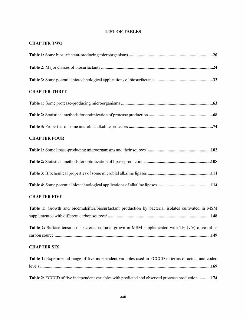
LIST OF TABLES
CHAPTER TWO
Table 1: Some biosurfactant-producing microorganisms ............................................................................20
Table 2: Major classes of biosurfactants .....................................................................................................24
Table 3: Some potential biotechnological applications of biosurfactants ....................................................33
CHAPTER THREE
Table 1: Some protease-producing microorganisms ...................................................................................63
Table 2: Statistical methods for optimization of protease production ..........................................................68
Table 3: Properties of some microbial alkaline proteases ............................................................................74
CHAPTER FOUR
Table 1: Some lipase-producing microorganisms and their sources ..........................................................102
Table 2: Statistical methods for optimization of lipase production ............................................................108
Table 3: Biochemical properties of some microbial alkaline lipases .........................................................111
Table 4: Some potential biotechnological applications of alkaline lipases ................................................114
CHAPTER FIVE
Table 1: Growth and bioemulsifier/biosurfactant production by bacterial isolates cultivated in MSM
supplemented with different carbon sourcesa .............................................................................................148
Table 2: Surface tension of bacterial cultures grown in MSM supplemented with 2% (v/v) olive oil as
carbon source .............................................................................................................................................149
CHAPTER SIX
Table 1: Experimental range of five independent variables used in FCCCD in terms of actual and coded
levels ..........................................................................................................................................................169
Table 2: FCCCD of five independent variables with predicted and observed protease production ...........174
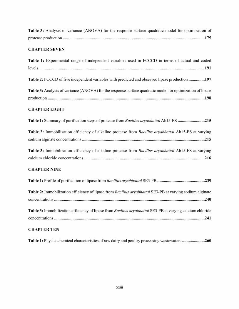
xxiii
Table 3: Analysis of variance (ANOVA) for the response surface quadratic model for optimization of
protease production ....................................................................................................................................175
CHAPTER SEVEN
Table 1: Experimental range of independent variables used in FCCCD in terms of actual and coded
levels.......................................................................................................................................................... 191
Table 2: FCCCD of five independent variables with predicted and observed lipase production ...............197
Table 3: Analysis of variance (ANOVA) for the response surface quadratic model for optimization of lipase
production ..................................................................................................................................................198
CHAPTER EIGHT
Table 1: Summary of purification steps of protease from Bacillus aryabhattai Ab15-ES .........................215
Table 2: Immobilization efficiency of alkaline protease from Bacillus aryabhattai Ab15-ES at varying
sodium alginate concentrations ..................................................................................................................215
Table 3: Immobilization efficiency of alkaline protease from Bacillus aryabhattai Ab15-ES at varying
calcium chloride concentrations ................................................................................................................216
CHAPTER NINE
Table 1: Profile of purification of lipase from Bacillus aryabhattai SE3-PB ............................................239
Table 2: Immobilization efficiency of lipase from Bacillus aryabhattai SE3-PB at varying sodium alginate
concentrations ............................................................................................................................................240
Table 3: Immobilization efficiency of lipase from Bacillus aryabhattai SE3-PB at varying calcium chloride
concentrations ............................................................................................................................................241
CHAPTER TEN
Table 1: Physicochemical characteristics of raw dairy and poultry processing wastewaters .....................260
xxiv
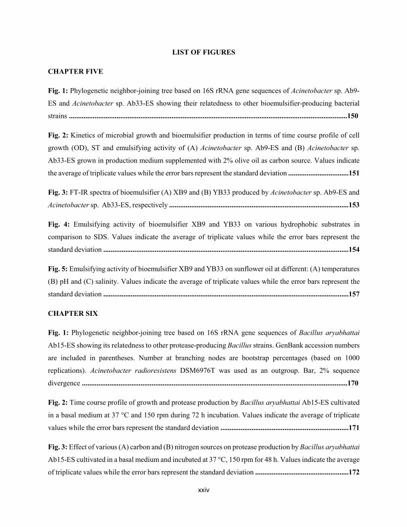
LIST OF FIGURES
CHAPTER FIVE
Fig. 1: Phylogenetic neighbor-joining tree based on 16S rRNA gene sequences of Acinetobacter sp. Ab9-
ES and Acinetobacter sp. Ab33-ES showing their relatedness to other bioemulsifier-producing bacterial
strains ........................................................................................................................................................150
Fig. 2: Kinetics of microbial growth and bioemulsifier production in terms of time course profile of cell
growth (OD), ST and emulsifying activity of (A) Acinetobacter sp. Ab9-ES and (B) Acinetobacter sp.
Ab33-ES grown in production medium supplemented with 2% olive oil as carbon source. Values indicate
the average of triplicate values while the error bars represent the standard deviation .................................151
Fig. 3: FT-IR spectra of bioemulsifier (A) XB9 and (B) YB33 produced by Acinetobacter sp. Ab9-ES and
Acinetobacter sp. Ab33-ES, respectively ..................................................................................................153
Fig. 4: Emulsifying activity of bioemulsifier XB9 and YB33 on various hydrophobic substrates in
comparison to SDS. Values indicate the average of triplicate values while the error bars represent the
standard deviation ......................................................................................................................................154
Fig. 5: Emulsifying activity of bioemulsifier XB9 and YB33 on sunflower oil at different: (A) temperatures
(B) pH and (C) salinity. Values indicate the average of triplicate values while the error bars represent the
standard deviation ......................................................................................................................................157
CHAPTER SIX
Fig. 1: Phylogenetic neighbor-joining tree based on 16S rRNA gene sequences of Bacillus aryabhattai
Ab15-ES showing its relatedness to other protease-producing Bacillus strains. GenBank accession numbers
are included in parentheses. Number at branching nodes are bootstrap percentages (based on 1000
replications). Acinetobacter radioresistens DSM6976T was used as an outgroup. Bar, 2% sequence
divergence .................................................................................................................................................170
Fig. 2: Time course profile of growth and protease production by Bacillus aryabhattai Ab15-ES cultivated
in a basal medium at 37 °C and 150 rpm during 72 h incubation. Values indicate the average of triplicate
values while the error bars represent the standard deviation ......................................................................171
Fig. 3: Effect of various (A) carbon and (B) nitrogen sources on protease production by Bacillus aryabhattai
Ab15-ES cultivated in a basal medium and incubated at 37 °C, 150 rpm for 48 h. Values indicate the average
of triplicate values while the error bars represent the standard deviation ...................................................172
xxv
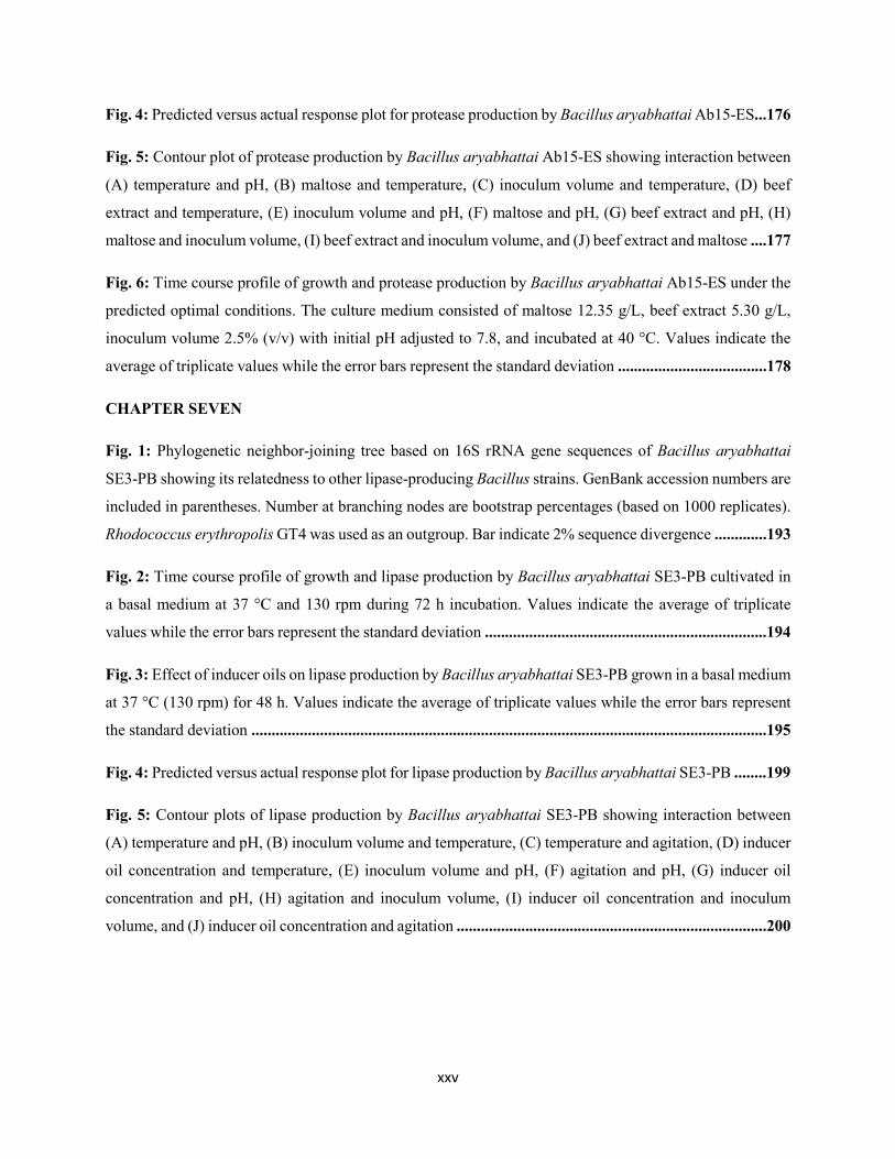
Fig. 4: Predicted versus actual response plot for protease production by Bacillus aryabhattai Ab15-ES...176
Fig. 5: Contour plot of protease production by Bacillus aryabhattai Ab15-ES showing interaction between
(A) temperature and pH, (B) maltose and temperature, (C) inoculum volume and temperature, (D) beef
extract and temperature, (E) inoculum volume and pH, (F) maltose and pH, (G) beef extract and pH, (H)
maltose and inoculum volume, (I) beef extract and inoculum volume, and (J) beef extract and maltose ....177
Fig. 6: Time course profile of growth and protease production by Bacillus aryabhattai Ab15-ES under the
predicted optimal conditions. The culture medium consisted of maltose 12.35 g/L, beef extract 5.30 g/L,
inoculum volume 2.5% (v/v) with initial pH adjusted to 7.8, and incubated at 40 °C. Values indicate the
average of triplicate values while the error bars represent the standard deviation .....................................178
CHAPTER SEVEN
Fig. 1: Phylogenetic neighbor-joining tree based on 16S rRNA gene sequences of Bacillus aryabhattai
SE3-PB showing its relatedness to other lipase-producing Bacillus strains. GenBank accession numbers are
included in parentheses. Number at branching nodes are bootstrap percentages (based on 1000 replicates).
Rhodococcus erythropolis GT4 was used as an outgroup. Bar indicate 2% sequence divergence .............193
Fig. 2: Time course profile of growth and lipase production by Bacillus aryabhattai SE3-PB cultivated in
a basal medium at 37 °C and 130 rpm during 72 h incubation. Values indicate the average of triplicate
values while the error bars represent the standard deviation ......................................................................194
Fig. 3: Effect of inducer oils on lipase production by Bacillus aryabhattai SE3-PB grown in a basal medium
at 37 °C (130 rpm) for 48 h. Values indicate the average of triplicate values while the error bars represent
the standard deviation ................................................................................................................................195
Fig. 4: Predicted versus actual response plot for lipase production by Bacillus aryabhattai SE3-PB ........199
Fig. 5: Contour plots of lipase production by Bacillus aryabhattai SE3-PB showing interaction between
(A) temperature and pH, (B) inoculum volume and temperature, (C) temperature and agitation, (D) inducer
oil concentration and temperature, (E) inoculum volume and pH, (F) agitation and pH, (G) inducer oil
concentration and pH, (H) agitation and inoculum volume, (I) inducer oil concentration and inoculum
volume, and (J) inducer oil concentration and agitation .............................................................................200
xxvi
Fig. 6: Time course profile of growth and lipase production by Bacillus aryabhattai SE3-PB under the
predicted optimal conditions. The culture medium consisted of 2.8% (v/v) inoculum volume and 2% (v/v)
inducer oil concentration with initial pH adjusted to 7.6, and incubated at 40 °C at 193 rpm. Values indicate
the average of triplicate values while the error bars represent the standard deviation ................................201
CHAPTER EIGHT
Fig. 1: Scanning electron microscopy of surface morphology of alginate beads .......................................217
Fig. 2: Effect of temperature on activity of free and immobilized protease from Bacillus aryabhattai Ab15-
ES. Protease activity was measured under standard assay conditions at various temperatures (30-80 °C) at
pH 7.2. Values indicate the average of triplicate values while the error bars represent the standard
deviation.....................................................................................................................................................218
Fig. 3: Effect of pH on activity of free and immobilized protease from Bacillus aryabhattai Ab15-ES.
Enzyme activity was determined at 50 °C. Values indicate the average of triplicate values while the error
bars represent the standard deviation .........................................................................................................219
Fig. 4: Thermal stability of free and immobilized protease from Bacillus aryabhattai Ab15-ES. The enzyme
was pre-incubated at different temperatures (40-50 °C) for 3 h at pH 8.0, and the residual enzyme activities
were estimated at regular intervals under standard assay conditions. The non-heated enzymes were taken
as 100%. Values indicate the average of triplicate values while the error bars represent the standard
deviation ....................................................................................................................................................220
Fig. 5: pH stability of free and immobilized protease from Bacillus aryabhattai Ab15-ES. The enzyme was
pre-incubated at pH values (8.0-9.0) for 3 h at 50 °C, and the residual enzyme activities were estimated at
regular intervals under standard assay conditions. The non-heated enzymes were taken as 100%. Values
indicate the average of triplicate values while the error bars represent the standard deviation ..................221
Fig. 6: Lineweaver-Burk plot of free ( ) and immobilized () protease from Bacillus aryabhattai Ab15-
ES. The enzyme activity was measured at various azocasein concentrations (1.0-10.0 mg/ml) at pH 8.0 and
50 °C. The Km and Vmax values were determined using Lineweaver-Burk plot. S: Substrate concentration;
V: Protease activity ....................................................................................................................................222
Fig. 7: Storage stability of free and immobilized protease at 4 and 25 °C. Values indicate the average of
triplicate values while the error bars represent the standard deviation .......................................................223
xxvii
Fig. 8: Reusability of protease immobilized in alginate gel beads. Values indicate the average of triplicate
values while the error bars represent the standard deviation .......................................................................224
CHAPTER NINE
Fig. 1: SEM image of morphology of alginate gel beads ...........................................................................241
Fig. 2: Effect of temperature on the activity of free and immobilized lipase from Bacillus aryabhattai SE3-
PB. Lipase activity was measured under standard assay conditions at various temperatures (30-80 °C) at
pH 7.2. Values indicate the average of triplicate values while the error bars represent the standard
deviation.....................................................................................................................................................242
Fig. 3: Effect of pH on the activity of free and immobilized lipase from Bacillus aryabhattai SE3-PB.
Lipase activity was determined at 60 °C. Values indicate the average of triplicate values while the error
bars represent the standard deviation .........................................................................................................243
Fig. 4: Thermal stability of free and immobilized lipase from Bacillus aryabhattai SE3-PB. The enzyme
was pre-incubated at different temperatures (55-65 °C) for 3 h at pH 9.5, and the residual enzyme activities
were estimated at regular intervals under standard assay conditions. The non-heated enzymes were taken
as 100%. Values indicate the average of triplicate values while the error bars represent the standard
deviation ....................................................................................................................................................244
Fig. 5: pH stability of free and immobilized lipase from Bacillus aryabhattai SE3-PB. The enzyme was
pre-incubated at pH values (8.5-9.5) for 3 h at 60 °C, and the residual enzyme activities were estimated at
regular intervals under standard assay conditions. The non-heated enzymes were taken as 100%. Values
indicate the average of triplicate values while the error bars represent the standard deviation ..................245
Fig. 6: Lineweaver-Burk plot of free ( ) and immobilized () lipase from Bacillus aryabhattai SE3-PB.
The enzyme activity was measured at various p-NPP concentrations (0.1-50 mM) at pH 9.5 and 60 °C. The
Km and Vmax values were determined using Lineweaver-Burk plot. S: Substrate concentration; V: Protease
activity .......................................................................................................................................................246
Fig. 7: Storage stability of free and immobilized lipase at 4 and 25 °C. Values indicate the average of
triplicate values while the error bars represent the standard deviation .......................................................247
Fig. 8: Reusability of lipase immobilized in alginate gel beads. Values indicate the average of triplicate
values while the error bars represent the standard deviation ....................................................................248
xxviii
CHAPTER TEN
Fig. 1: Reduction in (A) COD and (B) lipid content during treatment of dairy wastewater with free or
immobilized bioemulsifier and hydrolytic enzymes. The wastewater was treated at 37 °C without pH
adjustment. Values indicate the average of triplicate values while the error bars represent the standard
deviation ....................................................................................................................................................262
Fig. 2: Reduction in (A) COD and (B) lipid content during treatment of poultry processing wastewater with
free or immobilized bioemulsifier and hydrolytic enzymes. The wastewater was treated at 37 °C without
pH adjustment. Values indicate the average of triplicate values while the error bars represent the standard
deviation ....................................................................................................................................................263
Fig. 3: Reduction in (A) COD and (B) lipid content during treatment of dairy wastewater with free or
immobilized bioemulsifier and hydrolytic enzymes. The wastewater was treated at 50 °C and pH 8.0.
Values indicate the average of triplicate values while the error bars represent the standard deviation .......265
Fig. 4: Reduction in (A) COD and (B) lipid content during treatment of poultry processing wastewater with
free or immobilized bioemulsifier and hydrolytic enzymes. The wastewater was treated at 50 °C and pH
8.0. Values indicate the average of triplicate values while the error bars represent the standard
deviation.....................................................................................................................................................266
Fig. 5: Reusability potential of immobilized bioemulsifier and hydrolytic enzymes in the treatment of (A)
dairy and (B) poultry processing wastewater .............................................................................................268
1
CHAPTER ONE
INTRODUCTION
1.1 BACKGROUND
Lipid-rich wastewater is defined as a wastewater that consist of lipids along with broad spectrum of
dissolved organic and/or inorganic substances in suspension at high concentrations (Adulkar and Rathod,
2015). Lipid-contaminated wastewater may be of mineral, animal, or vegetable origin from a wide variety
of commercial establishments including slaughterhouses, sausage and meat product factories, restaurants,
fish processing industries, dairy product industries, leather industries, edible oil refineries, wool scouring
factories, petrochemical industries and metal processing industries (Chowdhury et al., 2010; Damasceno et
al., 2014; Bala et al., 2015; Porwal et al., 2015; Vendramel et al., 2015). It is characterized by high chemical
oxygen demand (COD), biochemical oxygen demand (BOD), proteins, total suspended solids (TSS),
nitrogen, phosphorus, sulphates along with many other toxic compounds, depending on the operations and
products from the producing industries (Farizoglu and Uzuner, 2011; Couras et al., 2015). Lipid-rich
wastewater occurs as free- floating oil, unstable oil-water emulsions and highly stable oil-water emulsions
(Awaleh and Soubaneh, 2014). The oil content is usually classified into four categories according to its
physical form, viz. free oil, dispersed oil, emulsified oil and dissolved oil (Coca et al., 2011).
The production and discharge of raw and poorly treated lipid-rich wastewater increase every year due to
rapid urbanization and industrial growth (Affandi et al., 2014). These industries consume large amounts of
water for various processes, equipment and washing facilities as well as, for product production resulting
in the release of huge volume of wastewater, which, if left untreated, could lead to increased disposal and
severe pollution problems, thereby creating environmental hazards and hampering the normal operations
of the ecosystems (Porwal et al., 2015).
Due to stringent regulations for effluent discharge and increasing drive for re-use of treated wastewater,
treatment of lipid-rich wastewater has become an issue of great necessity (Qin et al., 2007). Factors such
as costs, wastewater composition (high, medium, or low strength), treatment efficiency, regulatory
limitations and end use of wastewater influence the choice of treatment methods for lipid-rich wastewater
(Rajasulochana and Preethy, 2016). Various physicochemical techniques have been studied for their
applicability in the treatment of lipid-rich wastewater (Rodrigues et al., 2007; Kushwaha et al., 2011).
These mainly include flotation, sedimentation, ozonation, neutralization, chemical coagulation, gravity
separation, grease trap, oil-water separator, parallel-plate coalesces, cyclone separation, granular media
filtration, ultrafiltration, microfiltration among others (Chipasa, 2001). However, these techniques remain
2
unsatisfactory due to low treatment efficiency in the removal of dissolved and/or emulsified fats,
operational difficulties, high operational costs, generation of secondary pollutants and higher quantities of
solids, and use of chemical agents (Guolin et al., 2011). This makes it imperative for the establishment of
efficient, cost-effective, eco-friendly and sustainable technologies that can serve as alternatives to existing
treatment systems for lipid-rich wastewater.
Biological method is an emerging treatment technology that utilizes metabolic potential of microorganisms
for the elimination of hazardous contaminants present in lipid-rich wastewater under aerobic or anaerobic
conditions, or a combination of both via complete degradation or sequestration (Soleimaninanadegani and
Manshad, 2014; Nzila et al., 2017). It is a method of choice nowadays in comparison to other remediation
technologies due to its cost-effectiveness and wide environmental acceptability (Chanthamalee et al., 2013).
Pollutants removal efficiency is enhanced by the presence of microorganisms with appropriate metabolic
activities, adequate nutrient concentration, pH and temperature (Das and Chandran, 2011). The microbes
utilize the pollutants as source of carbon, thereby converting them into innocuous products through
production of appropriate metabolites such as bioemulsifier or enzymes by direct cell contact (Karigar and
Rao, 2011; Franzetti et al., 2012).
The formation of emulsions through microbial production and release of bioemulsifier is an important
process in the treatment of lipid-rich wastewater (Rahbari-Sisakht et al., 2017). Bioemulsifiers facilitate
enzymatic activity and degradation of fats and oils by increasing their solubility and bioavailability
(Daverey and Pakshirajan, 2011; Damasceno et al., 2012, 2014). This enhances the rate of pollutant
dissolution and utilization by microorganisms (Banat et al., 2010).
The use of biocatalysts is a promising technology for the treatment of high fat-containing wastewater. An
alternative to conventional approaches that is attracting growing interest is the use of enzymes, which
significantly reduce the level of organic pollutants in the wastewater by means of enzymatic catalysis and
enhance better performance of microbial community at the later stage of biological treatment process
(Cammarota and Freire, 2006; Alexandre et al., 2011; Valladão et al., 2011; Demarche et al., 2012).
Enzymes are very versatile, efficient under mild conditions, stable under typical treatment conditions and
selectively act on the target compounds in wastewater (Ferreira-Leitão et al., 2017).
Immobilization of the above-mentioned metabolites improves lipid-rich wastewater treatment efficiency
and further enhances reclamation and re-usability of immobilized bioproducts, thus reducing overall costs
(Jeganathan et al., 2006, 2007; Saranya et al., 2014; Suryanti et al., 2017). In addition, the support materials
protect the bioproducts from harsh environmental conditions, including mechanical stress and extreme
pollutant concentrations (Datta et al., 2013; Mohamad et al., 2015; Lee et al., 2017). This further enhances
3
degradation capacity of the bioproducts in comparison to free form. Immobilization can be achieved by
adsorption, covalent binding, entrapment or cross-linking technique using organic and/or inorganic support
materials (Saranya et al., 2014; Rehm et al., 2016).
1.1.1 GENERAL CHARACTERISTICS OF DAIRY WASTEWATER
In many parts of the world, dairy industry is generally considered as the largest type of food industry and
as a major source of food processing wastewater, contributing to pollution (Britz, 2006). In South Africa,
due to ever-increasing demand for milk and milk products, dairy industry represents one of the largest
sources of industrial effluents generating less than 5 million m3 of waste effluent per annum (WRC, 1989;
Briao and Granhen Tavares, 2007). Since dairy industry produces different products including milk, butter,
yoghurt, ice cream and cheese, the volume and composition of wastewater generated vary depending on
the type of product being produced, the nature and scale of operation and the design of the plant (Liu and
Haynes, 2011).
Most of the wastewater volume generated in dairy industry originates from cleaning processes; equipment
used for production, milk/milk products spills etc. (Kushwaha et al., 2011). Milk loss to the wastewater
stream can amount to 0.5-2.5% of the incoming milk and can be as high as 3-4% (Munavalli and Saler,
2009). Dairy wastewater is usually generated intermittently thus changing the flow rates of this wastewater.
Wastewater generated from milk processing can be separated into two classes, viz. wastewater with high
flow rates and effluent produced in small milk-transformation units (e.g. in cheese production) (Castillo et
al., 2007).
Dairy wastewater is concentrated in nature and contains high amounts of organic molecules such as proteins
(casein), lactose and lipids, contributing largely to high levels of BOD, COD, oil-grease content and
suspended solids (SS) (Kohle et al., 2009; Farizoglu and Uzuner, 2011; Adulkar and Rathod, 2015). In
addition, it is composed of whey, cream, separator and clarifier waters, yoghurt starter culture, detergents,
inorganic salts, sanitizers and stabilizing compounds (Singh et al., 2014). Dairy wastewater appears white
in color with heavy black sludge and strong butyric acid odor due to the decomposition of casein (Shete
and Shinkar, 2013). It is slightly alkaline in nature and becomes acidic quite rapidly upon fermentation of
milk sugar to lactic acid (Pathak et al., 2016). In industrial dairy wastewater, nitrogen originates mainly
from milk proteins in form of either organic or inorganic nitrogen source. Phosphorus is found mainly as
orthophosphate, polyphosphate, or in organic forms (Demirel et al., 2005; Kushwaha et al., 2011).
Suspended solids in dairy wastewater originate from coagulated milk, cheese curd fines or flavoring
ingredients, concentration of which varies in the range of 0.024-4.5 g/L (Kushwaha et al., 2011). The pH
of dairy wastewater varies in the range of 4.7-11; large fluctuations in pH occur due to the use of acidic and
4
caustic cleaning agents (Passeggi et al., 2009; Liu and Haynes, 2011). Significant concentrations of selected
elements including sodium, potassium, calcium, magnesium, iron, cobalt, nickel and manganese are also
present in dairy wastewater (Slavov, 2017). High concentrations of sodium and chloride result from the use
of large amounts of alkaline cleaners in dairy plants (Demirel et al., 2005).
1.1.2 GENERAL CHARACTERISTICS OF POULTRY PROCESSING WASTEWATER
The global meat production in the past few years has recorded a tremendous increase with a projection of
steady doubling growth until 2050 (Mekonnen and Hoekstra, 2012; Bouwman et al., 2013; FAO, 2013).
Poultry industry represents the largest sector of the South African (SA) agricultural sector, contributing
more than 16% of its share of gross domestic product. It has evolved over more than 100 years from a set
of backyard activities into a complex and highly integrated industry with increasing production of chicken.
Approximately 76% of the birds in the SA poultry industry are used for meat production, while the
remaining 24% are used in the egg industry (The South African Poultry Association Industry Profile, 2015).
The annual consumption of poultry products in SA surpasses the overall intake of all other animal protein
sources. In other words, 65.5% of locally produced animal protein consumption is supplied by the poultry
industry (The South African Poultry Industry Profile, 2012).
Poultry processing industries consume considerably large amounts of water for cleaning, rinsing of
carcasses and poultry products as well as for sanitizing and disinfecting facilities and equipment, depending
on the type of process employed, equipment used, productivity of the processing facility and waste
management practices (Bustillo-Lecompte and Mehrvar, 2015). The World Bank Group (2007) describes
a slaughterhouse plant as a meat processing facility that may consume approximately 2.5-40 m3 of water
per metric tons of meat produced. A typical SA poultry processing industry uses 15-20 L of water per bird
processed (CSIR, 2010). Chávez et al. (2005) reported that about 12-24 L of wastewater are generated per
bird slaughtered, and the poultry industry shows substantial environmental impacts related to water
consumption with a consequent generation of greater volume of high-strength wastewater (Hamawand et
al., 2017).
Poultry industry wastewater is characterized by high concentrations of biodegradable organic matter,
mainly lipids and proteins (Valladão et al., 2007). Lipids accounted for more than 67% of the particulate
COD of the slaughterhouse wastewater (Damasceno et al., 2012). Dors et al. (2013) have reported that
poultry industry wastewater contains high concentrations of organic matter inform of COD (39,300 mg/L),
lipids (2,005 mg/L) and SS (8,390 mg/L). In addition, this wastewater is composed of considerable amounts
of phosphorus, nitrogen, organic carbon, heavy metals, nutrients, detergents, disinfectants etc. (Debik and
Coskun, 2009; Bustillo-Lecompte and Mehrvar, 2015). The high contents of organic matter in the poultry
5
processing wastewater can be attributed to residual blood, excreta, fat, feather as well as stomach and
intestinal mucus in the wastewater (Debik and Coskun, 2009).
1.1.3 IMPACTS OF LIPID-RICH WASTEWATER
Lipid-rich wastewater has been considered as an industrial wastewater in the category of agricultural and
food industries, and classified as one of the most hazardous wastewater to the environment by the United
States Environmental Protection Agency (USEPA) (USEPA, 2004). Wastewater from dairy and poultry
processing industries are rich in biodegradable organic matter and nutrients, and normally contain high
levels of fats and proteins contributing to their low biodegradability coefficient (Liu and Haynes, 2011).
Improper disposal of this wastewater, especially when untreated or poorly treated poses serious threats to
the receiving environment by causing air pollution and contamination of surface and underground water
(Shete and Shinkar, 2013). Lipids form a layer on the receiving water surface, thereby reducing the
dissolved oxygen transfer rate into an aerobic process (Abd El-Gawad, 2014). High lipid contents in
biological wastewater treatment plants cause a severe obstacle by interfering with cell-aqueous phase
transfer rates through the formation of lipid coat around the biological floc. This further led to the
development of filamentous microorganism blooms (bulking) and floating sludge with undesirable physical
characteristics (Cammarota and Freire, 2006; Ferreira-Leitão et al., 2017). Furthermore, poor activity
attributed to excessive amounts of lipids in the wastewater inhibit acetogenic-methanogenic
microorganisms, resulting from accumulation of long chain fatty acids (Battimelli et al., 2010; Zhang et
al., 2011). In addition, due to viscous nature of lipids, their presence in wastewater cause clogging of drain
pipes and sewer lines resulting in the release of unpleasant odor, corrosion of sewer lines and unsightly
appearance on the surface of receiving water bodies (Husain et al., 2014; He et al., 2015). Nutrients such
as nitrogen present in lipid-rich wastewater led to eutrophication of receiving water bodies (Kushwaha et
al., 2011). High SS contents in oily wastewater has been an issue of concern. This slows down degradation
rate and results in scum layer production (Hejnfelt and Angelidaki, 2009). In the agricultural sector, release
of oily sludge transforms the physicochemical properties of soil with deleterious effect on the soil
morphology (Robertson et al., 2007). It affects the water absorption capacity of the soil, reduces its
hygroscopic moisture and hydraulic conductivity (Trofimov and Rozanova, 2003; Suleimanov et al., 2005).
Plants grown on such soil encounter inhibition in seed germination coupled with access to fewer nutrients,
which eventually lead to restricted growth (Al-Mutairi et al., 2008).
1.2 SCOPE OF THIS STUDY
It is obvious that the discharge of untreated and inadequately treated lipid-rich wastewater upsurges every
year due to increased productivity from various food processing industries, posing serious environmental
6
and health hazards (Affandi et al., 2014). These industries encounter challenges to accomplish the
requirements imposed by the stringent regulations for wastewater discharge and disposal along with high
costs of treatment of huge volume of wastewater (Qasim and Mane, 2013). Because of raising austere
regulations by environmental authorities for the removal of organic components from the wastewater,
specificity becomes very crucial (Ferreira-Leitão et al., 2017). Conventional effluent treatment technologies
are inept to eliminate all contaminants, especially dissolved and /or emulsified fat in the wastewater.
Combined use of inexpensive bioemulsifier and hydrolase represents a key development that has recently
gained attention for selective removal of target pollutants in the wastewater (Damasceno et al., 2012, 2014;
Silva et al., 2013; Saranya et al., 2014; Suryanti et al., 2017). Immobilization in appropriate support
materials is a cost-effective strategy for improved productivity and operational stability of the bioproducts
(Villegas et al., 2016). However, knowledge on the comparative study of free and immobilized bioproducts
(bioemulsifier and hydrolytic enzymes) for lipid-rich wastewater treatment is currently lacking. It is
therefore imperative to investigate the synergistic application of these bioproducts as a novel approach for
the treatment of high strength lipid-rich wastewater. These bioproducts facilitate degradation of lipid-rich
wastewater by triggering two simultaneous processes namely; emulsification of fat particles and enzymatic
catalysis, leading to increased reduction of organic load in the wastewater and eliminate the problem
associated with the discharge of wastewater.
This research provides a holistic approach for the effective removal of pollutants present in lipid-rich
wastewater. It hopes to establish new knowledge that will aid in the development of methods for the
treatment of lipid-rich wastewater. It will promote the drive for re-use of treated wastewater and conserve
normal operations of the ecosystems. Reports obtained from the physico-chemical characterization of the
wastewater provides information on the state of contamination of the wastewater, and these would be useful
for relevant environmental protection agencies and future scientists. Finally, the use of immobilized
bioemulsifier and hydrolytic enzymes will provide a realistic, efficient, sustainable, and cost-effective
approach for the degradation of pollutants in the wastewater. Therefore, this research focused on
investigating the potential of a mixture of free or immobilized bioemulsifier and hydrolytic enzymes
(protease and lipase) in reduction of pollutants from dairy and poultry processing wastewater collected from
selected sites in KwaZulu-Natal province of South Africa.
1.3 HYPOTHESIS
It is hypothesized that bioemulsifier and hydrolytic enzymes (protease and lipase) produced from
indigenous bacterial isolates will be effective in the reduction of pollutants from dairy and poultry
7
processing wastewater. It is further hypothesized that the immobilized bioproducts will be more efficient
and robust than free form in the degradation of organic load present in the wastewater.
1.4 AIM
1.4.1 To investigate the potential of a mixture of free or immobilized bioemulsifier and hydrolytic enzymes
(protease and lipase) from indigenous bacterial isolates in reduction of pollutants present in dairy and
poultry processing wastewater
1.5 SPECIFIC OBJECTIVES
1.5.1 To collect lipid-rich wastewater samples from selected sites in the KwaZulu-Natal province, South
Africa
1.5.2 To isolate, screen, identify and carry out phylogenetic analysis on the selected bioemulsifier-,
protease- and lipase-producing bacteria
1.5.3 To produce bioemulsifier, protease and lipase from the selected indigenous bacteria isolated from
lipid-rich wastewater by submerged fermentation
1.5.4 To optimize bioprocess parameters for enhanced protease and lipase production from indigenous
bacteria isolated from lipid-rich wastewater using response surface methodology
1.5.5 To immobilize protease and lipase in calcium alginate beads using entrapment method
1.5.6 To determine the effect of physicochemical parameters on emulsifying activity as well as on free and
immobilized protease and lipase activity
1.5.7 To determine the percentage removal efficiency of organic load from lipid-rich wastewater using a
mixture of free or immobilized bioemulsifier and hydrolytic enzymes
1.6 KEY QUESTIONS TO BE ANSWERED
1.6.1 Do indigenous bacteria isolated from lipid-rich wastewater demonstrate bioemulsifying, proteolytic
and lipolytic activity?
1.6.2 How effective is response surface methodology in the optimization of fermentation parameters for
enhanced protease and lipase production?
1.6.3 What are the optimum conditions for maximum bioemulsifying, protease and lipase activity?

8
1.6.4 How stable and reusable is immobilized bioemulsifier and hydrolytic enzymes in the treatment of
lipid-rich wastewater?
1.6.5 How effective is a mixture of free or immobilized bioemulsifier and hydrolytic enzymes in the
treatment of lipid-rich wastewater?
9
1.7 REFERENCES
Abd El-Gawad HS (2014) Oil and grease removal from industrial wastewater using new utility approach.
Adv. Environ. Chem. 2014: 1-6.
Adulkar TV, Rathod VK (2015) Pre-treatment of high fat content dairy wastewater using different
commercial lipases. Desalin. Water Treat. 53: 2450-2455.
Affandi IE, Suratman NH, Abdullah S, Ahmad WA, Zakaria ZA (2014). Degradation of oil and grease
from high-strength industrial effluents using locally isolated aerobic biosurfactant-producing
bacteria. Int. Biodeter. Biodegrad. 30: 1-8.
Alexandre VMF, Valente AM, Cammarota MC, Freire DMG (2011) Performance of anaerobic
bioreactor treating fish-processing plant wastewater pre-hydrolyzed with a solid-enzyme pool.
Renew. Energy, 36: 3439-3444.
Al-Mutairi N, Bufarsan A, Al-Rukaibi F (2008) Ecorisk evaluation and treatability potential of soils
contaminated with petroleum hydrocarbon-based fuels. Chemosphere, 74: 142-148.
Awaleh MO, Soubaneh YD (2014) Wastewater treatment in chemical industries: the concept and current
technologies. Hydrol. Curr. Res. 5: 164-175.
Bala JD, Lalung J, Ismail N (2015) Studies on the reduction of organic load from palm oil mill effluent
(POME) by bacterial strains. Int. J. Recycl. Org. Waste Agric. 4: 1-10.
Banat IM, Franzetti A, Gandolfi I, Bestetti G, Martinotti MG, Fracchia L, Smyth TJ, Marchant R
(2010) Microbial biosurfactants production, applications and future potential. Appl. Microbiol.
Biotechnol. 87: 427-444.
Battimelli A, Torrijos M, Moletta R, Delgenès JP (2010) Slaughterhouse fatty waste saponification to
increase biogas yield. Bioresour. Technol. 101: 3388-3393.
Bouwman L, Goldewijk KK, Van Der Hoek KW, Beusen AHW, Van Vuuren DP, Willems J, Rufino
MC, Stehfest E (2013) Exploring global changes in nitrogen and phosphorus cycles in agriculture
induced by livestock production over the 1900-2050 period. Proc. Natl. Acad. Sci. USA, 110:
20882-20887.
Briao VB, Granhen Tavares CR (2007) Effluent generated by the dairy industry: preventive attitudes and
opportunities. Braz. J. Chem. Eng. 24: 479-487.
10
Britz TJ, van Schalkwyk C, Hung Y (2006) Treatment of dairy processing wastewaters. In Wang LK,
Hung Y, Lo HH, Yapijakis C (Eds), waste treatment in the food processing industry, Taylor and
Francis, Boca Raton, FL, pp. 1-28.
Bustillo-Lecompte CF, Mehrvar M (2015) Slaughterhouse wastewater characteristics, treatment, and
management in the meat processing industry: a review on trends and advances. J. Environ. Manage.
161: 287-302.
Cammarota MC, Freire DMG (2006) A review on hydrolytic enzymes in the treatment of wastewater
with high oil and grease content. Bioresour. Technol. 97: 2195-3010.
Castillo S, Zapico A, Doubrovine N, Lafforgue C, Fonade C (2007) Study of a compact bioreactor for
the in-line treatment of dairy wastewaters: case of effluents produced on breeding farms.
Desalination, 214: 49-61.
Chanthamalee J, Wongchitphimon T, Luepromchai E (2013) Treatment of oily bilge water from small
fishing vessels by PUF-immobilized Gordonia sp. JC11. Water Air Soil Pollut. 224: 1601.
Chávez CP, Castillo LR, Dendooven L, Escamilla-Silva EM (2005) Poultry slaughter wastewater
treatment with an upflow anaerobic sludge blanket (UASB) reactor. Bioresour. Technol. 96: 1730-
1736.
Chipasa KB (2001) Limits of physicochemical treatment of wastewater in the vegetable oil refining
industry. Pol. J. Environ. Stud. 10: 141-147.
Chowdhury P, Viraraghavan T, Srinivasan A (2010) Biological treatment processes for fish processing
wastewater-a review. Bioresour. Technol. 101: 439-449.
Coca J, Gutiérrez G, Benito J (2011) Treatment of oily wastewater. In: Coca-Prados J, Gutiérrez-Cervelló
G (eds) Water Purification and Management. NATO Science for Peace and Security Series:
Environmental Security, Springer, Dordrecht.
Couras CS, Louros VL, Gameiro T, Alves N, Silva A, Capela MI, Arroja LM, Nadais H (2015)
Anaerobic degradation of dairy wastewater in intermittent UASB reactors: influence of effluent
recirculation. Environ. Technol. 36: 2227-2238.
CSIR (2010) A CSIR perspective on water in South Africa-2010.
Damasceno FRC, Cammarota MC, Freire DMG (2012) The combined use of a biosurfactant and an
enzyme preparation to treat an effluent with a high fat content. Coll. Surf. B: Biointerfaces, 95:
241-246.
11
Damasceno FRC, Freire DMG, Cammarota MC (2014) Assessing a mixture of biosurfactant and
enzyme pools in the anaerobic biological treatment of wastewater with a high fat content. Environ.
Technol. 35: 2035-2045.
Das N, Chandran P (2011) Microbial degradation of petroleum hydrocarbon contaminants: An overview.
Biotechnol. Res. Int. Biotechnol. Res. Int. 2011: 1-13.
Datta S, Rene CL, Rajaram YRS (2013) Enzyme immobilization: an overview on techniques and support
materials. 3 Biotech. 3: 1-9.
Daverey A, Pakshirajan K (2011) Pretreatment of synthetic dairy wastewater using the sophorolipid-
producing yeast Candida bombicola. Appl. Biochem. Biotechnol. 163: 720-728.
Debik E, Coskun T (2009) Use of the static granular bed reactor (SGBR) with anaerobic sludge to treat
poultry slaughterhouse wastewater and kinetic modeling. Bioresour. Technol. 100: 2777-2782.
Demarche P, Junghanns C, Nair RR, Agathos SN (2012) Harnessing the power of enzymes for
environmental stewardship. Biotechnol. Adv. 30: 933-953.
Demirel B, Yenigun O, Onay TT (2005) Anaerobic treatment of dairy wastewater: a review. Proc.
Biochem. 40: 2583-2595.
Dors G, Mendes AA, Pereira EB, de Castro HF, Furigo Jr A (2013) Simultaneous enzymatic hydrolysis
and anaerobic biodegradation of lipid-rich wastewater from poultry industry. Appl. Water Sci. 3:
343-349.
FAO (2013) Food Outlook: Biannual Report on Global Food Markets. Meat and Meat Products. Food and
Agriculture Organization (FAO) of the United Nations (UN), Rome, Italy (accessed 26.08.17).
Available online: http://www. fao.org/docrep/019/i3473e/i347e.pdf.
Farizoglu B, Uzuner S (2011) The investigation of dairy industry wastewater treatment in a biological
high performance membrane system. Biochem. Eng. J. 57: 46-54.
Ferreira-Leitão VS, Cammarota MC, Aguieiras ECG, Vasconcelos de Sá LR, Fernandez-Lafuente
R, Freire DMG (2017) The protagonism of biocatalysis in green chemistry and its environmental
benefits. Catalysts, 7: 9.
Franzetti A, Gandolfi I, Raimondi C, Bestetti G, Banat IM, Smyth TJ, Papacchini M, Cavallo M,
Fracchia L (2012) Environmental fate, toxicity, characteristics and potential applications of novel
bioemulsifiers produced by Variovorax paradoxus 7bCT5. Bioresour. Technol. 108: 245-251.
12
Guolin J, Mingming L, Tingting C (2011) Prospects for development of oily sludge treatment. Chem.
Technol. Fuels Oils, 47: 312-326.
Hamawand I, Ghadouani A, Bundschuh J, Hamawand S, Al Juboori RA, Chakrabarty S, Yusaf T
(2017) A critical review on processes and energy profile of the Australian meat processing industry.
Energies, 10: 731.
He X, Zhang Q, Cooney MJ, Yan T (2015) Biodegradation of fat, oil and grease (FOG) deposits under
various redox conditions relevant to sewer environment. Appl. Microbiol. Biotechnol. 99: 6059-
6068.
Hejnfelt A, Angelidaki I (2009) Anaerobic digestion of slaughterhouse by-products. Biomass Bioenergy,
33: 1046-1054.
Husain IA, Alkhatib MF, Jammi MS, Mirghani ME, Bin Zainudin Z, Hoda A (2014) Problems,
control, and treatment of fat, oil, and grease (FOG): a review. J. Oleo Sci. 63: 747-752.
Jeganathan J, Bassi A, Nakhla G (2006) Pre-treatment of high oil and grease pet food industrial
wastewaters using immobilized lipase hydrolyzation. J. Hazard. Mater. B137: 121-128.
Jeganathan J, Nakhla G, Bassi A (2007) Hydrolytic pretreatment of oily wastewater by immobilized
lipase. J. Hazard. Mater. 145: 127-135.
Karigar CS, Rao SS (2011) Role of microbial enzymes in the bioremediation of pollutants: a review.
Enzyme Res. 2011: 1-11.
Kolhe AS, Ingale SR, Bhole RV (2009) Effluent of dairy technology. Shodh, Samiksha aur Mulyankan
Int. Res. J. 2: 459-461.
Kushwaha JP, Srivastava VC, Mall ID (2011) An overview of various technologies for the treatment of
dairy wastewaters. Crit. Rev. Food Sci. Nutri. 51: 442-452.
Lee CW, Jang S-H, Chung H-S (2017) Improving the stability of cold-adapted enzymes by
immobilization. Catalysts, 7: 112.
Liu Y-Y, Haynes RJ (2011) Origin, nature, and treatment of effluents from dairy and meat processing
factories and the effects of their irrigation on the quality of agricultural soils. Critical Rev. Environ.
Sci. Technol. 41: 1531-1599.
Mekonnen MM, Hoekstra AY (2012) A global assessment of the water footprint of farm animal products.
Ecosystems, 15: 401-415.
13
Mohamad NR, Marzuki NHC, Buang NA, Huyop F, Abdul Wahab R (2015) An overview of
technologies for immobilization of enzymes and surface analysis techniques for immobilized
enzymes. Biotechnol. Biotechnol. Equip. 29: 205-220.
Munavalli GR, Saler PS (2009) Treatment of dairy wastewater by water hyacinth. Water Sci. Technol. 59:
713-722.
Nzila A, Thukair A, Saravanan S, Abdur Razzak S (2017) Characterization of aerobic oil and grease-
degrading bacteria in wastewater. Environ. Technol. 38: 661-670.
Passeggi M, Lopez I, Borzacconi L (2009) Integrated anaerobic treatment of dairy industrial wastewater
and sludge. Water Sci. Technol. 59: 501-506.
Pathak U, Das P, Banerjee P, Datta S (2016) Treatment of wastewater from a dairy industry using rice
husk as adsorbent: treatment efficiency, isotherm, thermodynamics, and kinetics modelling. J.
Thermodyn. 2016: 1-7.
Porwal HJ, Mane AV, Velhal SG (2015) Biodegradation of dairy effluent by using microbial isolates
obtained from activated sludge. Water Resour. Ind. 9: 1-15.
Qasim W, Mane AV (2013) Characterization and treatment of selected food industrial effluents by
coagulation and adsorption techniques. Water Res. Ind. 4: 1-12.
Qin JJ, Oo MH, Tao G, Kekre KA (2007) Feasibility study on petrochemical wastewater treatment and
reuse using submerged MBR. J. Membr. Sci. 293: 161-166.
Rahbari-Sisakht M, Pouranfard A, Darvishi P, Ismail AF (2017) Biosurfactant production for
enhancing the treatment of produced water and bioremediation of oily sludge under the conditions
of Gachsaran oil field. J. Chem. Technol. Biotechnol. 92: 1053-1064.
Rajasulochana P, Preethy V (2016) Comparison on efficiency of various techniques in treatment of waste
and sewage water- a comprehensive review. Resource-Efficient Technol. 2: 175-184.
Rehm FBH, Chen S, Rehm BHA (2016) Enzyme engineering for in situ immobilization. Molecules, 21:
1370.
Robertson SJ, McGill WB, Massicotte HB, Rutherford PM (2007) Petroleum hydrocarbon
contamination in boreal forest soils: a mycorrhizal ecosystems perspective. Biol. Rev. 82: 213-240.
Rodrigues A, Xavier S, Bernardes F (2007) Application of photo electrochemical-electro-dialysis
treatment for the recovery and reuse of water from tannery effluents. J. Clean. Prod. 10: 1-7.
14
Saranya P, Swarnalatha S, Sekaran G (2014) Lipoprotein biosurfactant production from an extreme
acidophile using fish oil and its immobilization in nanoporous activated carbon for the removal of
Ca2+ and Cr3+ in aqueous solution. RSC adv. 4: 34144-34155.
Shete BS, Shinkar NP (2013) Dairy industry wastewater sources, characteristics, and its effects on
environment. Int. J. Curr. Eng. Technol. 3: 1611-1615.
Silva JN, Gutarra MLE, Freire DMG, Cammarota MC (2013) Application of home-made enzyme and
biosurfactant in the anaerobic treatment of effluent with high-fat content. J. Bioprocess. Biotechniq.
3: 3
Singh NB, Singh R, Imam MM (2014) Wastewater management in dairy industry: pollution abatement
and preventive attitudes. Int. J. Sci. Environ. Technol. 3: 672-683.
Slavov AK (2017) General characteristics and treatment possibilities of dairy wastewater- a review. Food
Technol. Biotechnol. 55: 14-28.
Soleimaninanadegani M, Manshad S (2014) Enhancement of biodegradation palm oil mill effluents by
local isolated microorganisms. Int. Scholarly Res. Notices, 2014: 1-8.
Suleimanov R, Gabbasova I, Sitdikov R (2005) Changes in the properties of oily gray forest soil during
biological reclamation. Biol. Bull. 32: 93-99.
Suryanti V, Widjonarko DM, Windrawati, Widyaningsih V (2017) Fabrication and characterization of
immobilized biosurfactant produced by Pseudomonas aeruginosa grown on cassava industrial
wastewater into activated allophane as an adsorbent. Mater. Sci. Eng. 176: 012005.
The South Africa Poultry Association Profile (2012) Available: https://www.sapoultry.co.za [2017,
August 25].
The South Africa Poultry Association Profile (2015) Available: https://www.sapoultry.co.za [2017,
August 25].
Trofimov SY, Rozanova M (2003) Transformation of soils properties under the impact of oil pollution.
Eurasian Soil Sci. 36: S82-S87.
USEPA (2004) Technical development document for the final effluent limitations guidelines and standards
for the meat and poultry products point source category; Final Rule; Federal Register, 69: 54476-
54555.
15
Valladão ABG, Cammarota MC, Torres AG, Freire DMG (2011) Profiles of fatty acids and
triacylglycerols and their influence on the anaerobic biodegradability of effluents from poultry
slaughterhouse. Bioresour. Technol. 102: 7043-7050.
Valladão ABG, Freire DMG, Cammarota MC (2007) Enzymatic pre-hydrolysis applied to the anaerobic
treatment of effluents from poultry slaughterhouses. Int. Biodeter. Biodegr. 60: 219-225.
Vendramel S, Bassin JP, Dezotti M, Sant’Anna GL Jr (2015) Treatment of petroleum refinery
wastewater containing heavily polluting substances in an aerobic submerged fixed-bed reactor.
Environ. Technol. 36: 2052-2059.
Villegas LGC, Mashhadi N, Chen M, Mukherjee D, Taylor KE, Biswas N (2016) A short review of
techniques for phenol removal from wastewater. Curr. Pollut. Rep. 2: 157-167.
Water Research Council (WRC) (1989) Water and wastewater management in the dairy industry. WRC
project no 145 TT 38/89. NATSURV series no, 4. Published by the WRC, Pretoria, South Africa.
World Bank Group (2007) Environmental, Health and Safety (EHS) Guidelines for Meat Processing.
General EHS Guidelines: Environmental Wastewater and Ambient Water Quality. (Accessed
26.08.17). Available online: http://www.ifc.org/ehsguidelines.
Zhang L, Lee C-H, Jahng D (2011) Restriction of linoleic acid inhibition of methanization of piggery
wastewater and enhancement of its mineralization by adding calcium ions. J. Chem. Technol.
Biotechnol. 86: 282-289.
16
CHAPTER TWO
A REVIEW ON PRODUCTION, PROPERTIES AND POTENTIAL APPLICATIONS OF
MICROBIAL SURFACTANTS
This chapter has been formatted for submission to the ‘World Journal of Microbiology and
Biotechnology’
17
A review on production, properties and potential applications of microbial surfactants
Adegoke Isiaka Adetunji and Ademola Olufolahan Olaniran*
Discipline of Microbiology, School of Life Sciences, College of Agriculture, Engineering and Science,
University of KwaZulu-Natal (Westville campus), Private Bag X54001, Durban 4000, Republic of South
Africa.
* Corresponding author. Tel: +27 31 260 7400; Fax: +27 31 260 7809
E-mail address: [email protected]
Abstract Microbial surface-active compounds are amphipathic molecules that consist of hydrophilic and
hydrophobic moieties, which permit their partition at the interface between fluid phases of varying degree
of polarity. They are divided into two main groups namely, bioemulsifier and biosurfactant, based on their
molecular weight. These biomolecules are synthesized by diverse groups of microorganisms mainly
bacteria, fungi and yeasts. Their production is significantly influenced by substrate type, fermentation
technology and microbial strains. Microbial surfactants are generally categorized into different groups
based on their chemical nature, ionic charge and producing organisms. Due to their inherent broad
functional properties and diverse synthetic capabilities of the microbes, microbial surfactants are mostly
preferred than their chemical counterparts for various industrial and biomedical applications including
bioremediation, enhanced oil recovery, clean-up of oil-contaminated sites, as additives in cleaning products
and laundry formulations, and as emulsion-stabilizing agents in the medical, food and cosmetic industries.
This review provides an insight on the methods for detection of microbial surfactant production as well as
discusses chemical nature of these surface active compounds. It further elucidates the various factors
influencing biosurfactant production coupled with techniques used for recovery and purification of these
biomolecules for various industrial and environmental applications.
Keywords Microbial surfactants; Bioemulsifier; Biosurfactant; Microorganisms; Production; Application
Introduction
Microbial surface-active compounds (SACs) are amphipathic and structurally diverse group of compounds
consisting of both hydrophilic and hydrophobic domains which preferentially partition between liquid
interfaces with varying degrees of polarity and hydrogen bridges, including oil-water or air-water interfaces
(Santos et al. 2016). Usually, the hydrophobic moiety is a hydrocarbon chain consisting of saturated or
18
unsaturated long-chain fatty acids whereas, the hydrophilic moiety is made up of ionic (cationic or anionic),
non-ionic, amphoteric, amino acids or polysaccharides (Silva et al. 2014a; Mao et al. 2015). They are
categorized into two groups: low-molecular weight and high- molecular weight SACs. The low molecular
weight SACs, also known as biosurfactants are recognized for robust surface activity by reducing surface
and interfacial tension between different phases as well as possess low critical micelle concentration (CMC)
and form stable emulsions (Batista et al. 2006; Uzoigwe et al. 2015). The ability to lower surface and
interfacial tension is caused by the adsorption of the biosurfactant to different phases, resulting in more
interaction and mixing of dissimilar phases, which functions to solubilize hydrophobic substrates (Satpute
et al. 2010a; Walter et al. 2010; Uzoigwe et al. 2015). They composed of biochemical complexes including
a wide range of molecules such as sugars, fatty acids, amino acids and functional groups such as carboxylic
acids. The glycolipids comprise of different sugars linked to β-hydroxyl fatty acids while lipopeptides
consist of cycloheptapeptides with amino acids bonded to fatty acids of different chain length (Uzoigwe et
al. 2015).
On the contrary, bioemulsifiers are high molecular weight polymers that are more effective in forming and
stabilizing oil-water or water-oil emulsions, but do not necessarily reduce surface or interfacial tension
(Gudiña et al. 2015). They consist of polymeric structures including polysaccharides, proteins, lipids, and
complex mixtures such as heteropolysaccharides, lipopolysaccharides, lipoproteins etc. (Perfumo et al.
2009; Smyth et al. 2010a; Sekhon-Randhawa 2014). These combinations confer upon them better
emulsifying potential and ability to stabilize emulsions. Bioemulsifiers are produced by different bacteria
(Rosenberg et al. 1979; Navon-Venezia et al. 1995; Ilori et al. 2005; Martínez-Checa et al. 2007; Dastgheib
et al. 2008; Franzetti et al. 2008, 2012; Zheng et al. 2011, 2012), yeasts (Monteiro et al. 2009, 2010, 2012)
and filamentous fungi (Silva et al. 2014b). Bioemulsifiers are classified into different groups, based on their
inherent chemical structure and the identity of the microorganisms that synthesize them (Ron and
Rosenberg 2001). These include polymeric and particulate surfactants (Kapadia and Yagnik 2013).
The global demands for surfactants has surpassed 2.5 million tons in 2002, was less than $1735.5 million
in 2011 and projected to reach $2210.5 million in 2018, equivalent to the mean annual growth rate of less
than 4% from 2011-2018 (Sekhon et al. 2012). Due to their biological source, bioemulsifiers are considered
to be less toxic, easily biodegradable with greater environmental compatibility; exhibit high selectivity and
specific activity at extreme pH, temperatures and NaCl concentrations. They can be produced in situ from
renewable and readily available economical raw materials, and possess similar or better emulsifying
activities in comparison to chemically-prepared conventional surfactants (Pacwa-Plociniczak et al. 2011;
Franzetti et al. 2012; Vaz et al. 2012; Kapadia and Yagnik 2013; Santos et al. 2013; Banat et al. 2014).
19
Bioemulsifiers possess unique and distinct diverse functional properties including emulsification, wetting,
foaming, solubilization, corrosion-inhibition, dispersion etc., making them a suitable candidate in various
biotechnological applications such as bioremediation; as additives in cleaning and laundry formulations,
and as emulsion-stabilizer in the foods, cosmetics or pharmaceutics (Amaral et al. 2006; Martínez-Checa
et al. 2007; Dastgheib et al. 2008; Banat et al. 2010; Monteiro et al. 2010; Franzetti et al. 2012; Zheng et
al. 2012; Colin et al. 2013).
This review therefore provides an overview of the microbial production of biosurfactants/bioemulsifiers,
detailing detection methods, influencing factors and kinetics of production of these surface active
compounds. It also discusses key features including chemical nature, recovery and purification techniques
as well as potential industrial and biomedical applications of biosurfactants/bioemulsifiers.
Biosurfactants/bioemulsifier-producing microorganisms
In recent decades, there has been a renewed interest in bioprospecting for microorganisms with potential to
produce tensioactive molecules with robust surfactant characteristics (Silva et al. 2014a). Surface-active
compounds are produced by various groups of microorganisms mainly bacteria (Gram-positive and Gram-
negative), fungi, and yeasts. The quantity of biosurfactant production depends on the microorganism type
and source of isolation. Various microbial communities have been reported to produce
biosurfactants/bioemulsifiers (Macdonald et al. 1981; Cooper and Paddock 1983; Cameron et al. 1988;
Singh and Desai 1989; Zinjarde et al. 1997; Abraham et al. 1998; Kakugawa et al. 2002; Lin et al. 2003;
Lukondeh et al. 2003; Rau et al. 2005; Amaral et al. 2006; Maneerat et al. 2006; Perfumo et al. 2006; Morita
et al. 2007; Teichmann et al. 2007; Das et al. 2008a, 2008b; Monteiro et al. 2012; Ozdal et al. 2017). A list
of some biosurfactant-producing microorganisms is shown in Table 1.
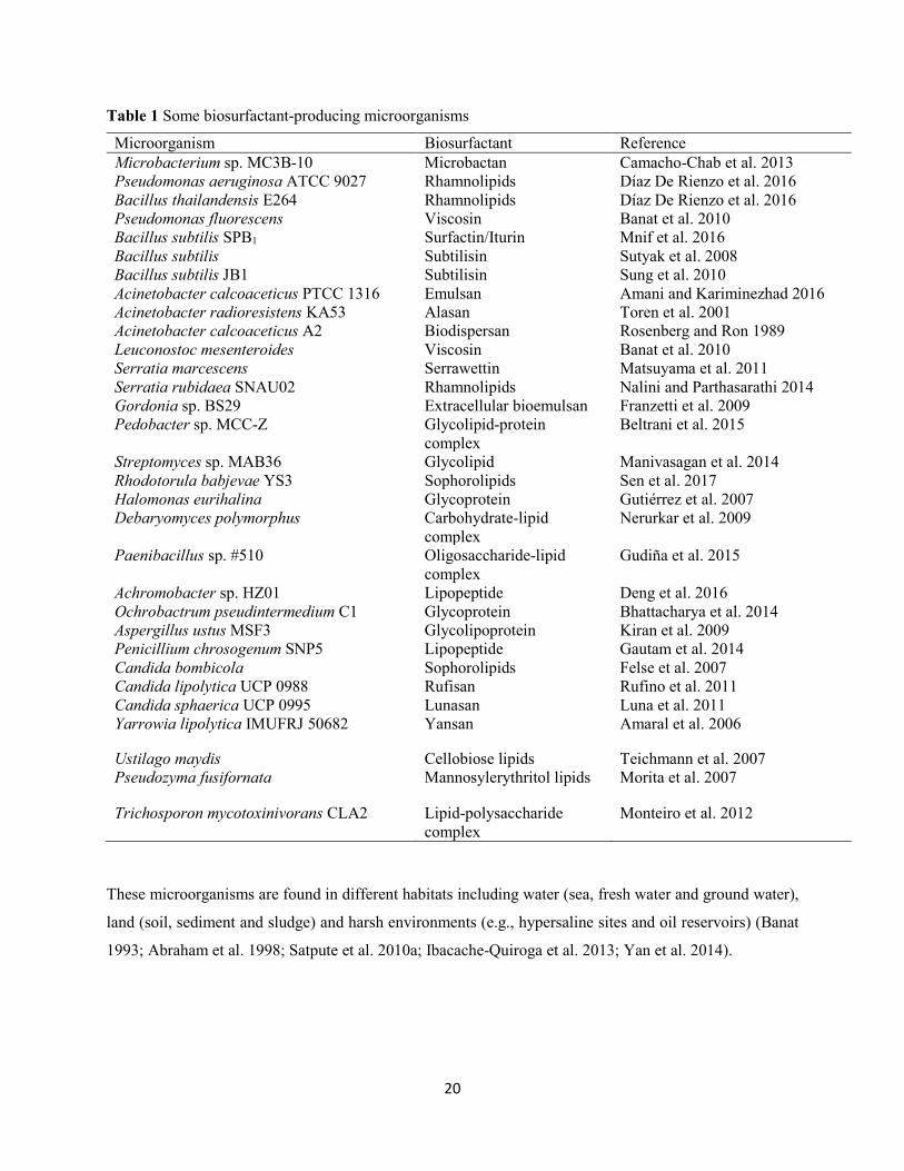
20
Table 1 Some biosurfactant-producing microorganisms
These microorganisms are found in different habitats including water (sea, fresh water and ground water),
land (soil, sediment and sludge) and harsh environments (e.g., hypersaline sites and oil reservoirs) (Banat
1993; Abraham et al. 1998; Satpute et al. 2010a; Ibacache-Quiroga et al. 2013; Yan et al. 2014).
Microorganism Biosurfactant Reference Microbacterium sp. MC3B-10 Microbactan Camacho-Chab et al. 2013 Pseudomonas aeruginosa ATCC 9027 Bacillus thailandensis E264
Rhamnolipids Rhamnolipids
Díaz De Rienzo et al. 2016 Díaz De Rienzo et al. 2016
Pseudomonas fluorescens Viscosin Banat et al. 2010 Bacillus subtilis SPB1 Surfactin/Iturin Mnif et al. 2016 Bacillus subtilis Subtilisin Sutyak et al. 2008 Bacillus subtilis JB1 Subtilisin Sung et al. 2010 Acinetobacter calcoaceticus PTCC 1316 Emulsan Amani and Kariminezhad 2016 Acinetobacter radioresistens KA53 Alasan Toren et al. 2001 Acinetobacter calcoaceticus A2 Biodispersan Rosenberg and Ron 1989 Leuconostoc mesenteroides Viscosin Banat et al. 2010 Serratia marcescens Serrawettin Matsuyama et al. 2011 Serratia rubidaea SNAU02 Rhamnolipids Nalini and Parthasarathi 2014 Gordonia sp. BS29 Extracellular bioemulsan Franzetti et al. 2009 Pedobacter sp. MCC-Z Glycolipid-protein
complex Beltrani et al. 2015
Streptomyces sp. MAB36 Glycolipid Manivasagan et al. 2014 Rhodotorula babjevae YS3 Sophorolipids Sen et al. 2017 Halomonas eurihalina Glycoprotein Gutiérrez et al. 2007 Debaryomyces polymorphus Carbohydrate-lipid
complex Nerurkar et al. 2009
Paenibacillus sp. #510 Oligosaccharide-lipid complex
Gudiña et al. 2015
Achromobacter sp. HZ01 Lipopeptide Deng et al. 2016 Ochrobactrum pseudintermedium C1 Glycoprotein Bhattacharya et al. 2014 Aspergillus ustus MSF3 Glycolipoprotein Kiran et al. 2009 Penicillium chrosogenum SNP5 Lipopeptide Gautam et al. 2014 Candida bombicola Sophorolipids Felse et al. 2007 Candida lipolytica UCP 0988 Rufisan Rufino et al. 2011 Candida sphaerica UCP 0995 Lunasan Luna et al. 2011 Yarrowia lipolytica IMUFRJ 50682 Yansan Amaral et al. 2006
Ustilago maydis Cellobiose lipids Teichmann et al. 2007 Pseudozyma fusifornata Mannosylerythritol lipids Morita et al. 2007
Trichosporon mycotoxinivorans CLA2 Lipid-polysaccharide complex
Monteiro et al. 2012
21
Detection methods for biosurfactant and bioemulsifier-producing microorganisms
High throughput techniques are required for rapid screening of potential biosurfactant-producing
microorganisms in order to discover novel biosurfactant and/or biosurfactant-producing microorganisms
(Maneerat 2005; Satpute et al. 2010a). Due to diverse functional and structural properties, the use of single
screening method for the selection of biosurfactant-producing microorganisms has been very challenging
in providing accurate and reliable results. Hence, the need for adoption of multiple screening tests
concurrently for screening for maximum number of biosurfactant producers among the population of
isolated microorganisms. The different screening methods commonly employed for
biosurfactant/bioemulsifier production are based on surface or interfacial activity, or emulsification or
foaming activities as described below:
Oil spreading assay
It is carried out by adding 10 µl of crude oil to the surface of 40 ml distilled water in a Petri dish to form a
thin oil layer. Thereafter, 10 µl of culture supernatants are added on the oil coated water surface (Morikawa
et al. 2000). Displacement of oil and formation of clear zone indicate the presence of biosurfactant in the
culture supernatant sample. The diameter of clear zone on the oil surface correlates to surfactant activity.
The oil spreading method is rapid, sensitive and reliable technique to detect biosurfactant production by
diverse microorganisms (Youssef et al. 2004; Plaza et al. 2006; Ruggeri et al. 2009; Khopade et al. 2012;
Manivasagan et al. 2014; Panjiar et al. 2015). In addition, it requires less sample volume and no specialized
equipment, and can be employed when the activity and quantity of biosurfactant is low.
Surface and interfacial tension measurement
Surface tension is defined as a force per unit length exerted by a liquid when in close contact with another
liquid or solid. In other words, it is a measure of the free energy per unit area that is linked with an interfacial
or surface tension (Satpute et al. 2010a). Water molecules are bonded together by cohesive forces that form
surface tension. The surface tension of distilled water is calculated to be 72 mN/m, addition of surfactants
caused a reduction in the surface tension (Satpute et al. 2010a). Effectiveness of a biosurfactant depends on
its ability to reduce the surface tension of water to less than 40 mN/m (Abdel-Mawgoud et al. 2010;
Soberón-Chavez 2010). This is typical of a surfactin biosurfactant reported to reduce the surface tension of
water to 27 mN/m (Cooper and Goldenberg 1987; Banat 1993). Similarly, Mulligan (2005) has reported a
surfactin biosurfactant that decreased the surface tension of water to 25 mN/m, and the interfacial tension
of water/hexadecane from 40 mN/m to 1 mN/m. In addition, a rhamnolipid biosurfactant produced by
Pseudomonas aeruginosa has been reported to reduce the surface tension of water to approximately 30
22
mN/m and the interfacial tension of water/oil from 43 mN/m to almost 1 mN/m (Dusane et al. 2010). The
direct measurement of surface tension of culture supernatants is carried out using tensiometer involving du
Nuoy ring method, Wilhelmy plate method, maximum pull force method, or pendant drop methods. It is a
precise and reliable technique for screening of biosurfactant production (Bodour et al. 2003; Youssef et al.
2004; Salihu et al. 2009; Patowary et al. 2017).
Emulsification activity
Among the functional properties of biosurfactant/bioemulsifier is emulsification, which involves dispersion
of one liquid into another, resulting in the mixture of two immiscible liquids (Satpute et al. 2010a; Inès and
Dhouha 2015). It is measured by calculating emulsification index (E24), as described by Cooper and
Goldenberg (1987). In this method, equal volume of hydrocarbon-based compounds and culture
supernatants (2:2 v/v) is mixed together, vortexed for 2 min and allowed to stand for 24 h at room
temperature. The E24 is calculated by dividing the height of the emulsion layer by the total height of liquid
and expressed in percentage (%) (Cooper and Goldenberg 1987). Several authors have employed
emulsification index as a technique for detection of biosurfactant/bioemulsifier production from
microorganisms isolated from diverse environments (Das et al. 2008a; Jagtap et al. 2010; Chen et al. 2012;
Zheng et al. 2012; Belgacem et al. 2015; Ndlovu et al. 2016).
Drop collapse method
Qualitative drop collapse assay for screening of biosurfactant/bioemulsifier-producing microorganisms was
developed by Jain et al. (1991), and predominantly used for the measurement of surface tension of liquids.
It is based on destabilization of liquid droplets by the surface-active compounds (Walter et al. 2010). The
presence of biosurfactant causes the collapse or spread of the liquid droplets over a hydrophobic surface as
a result of interfacial tension that exist between the liquid and the hydrophobic surface (Hsieh et al. 2004;
Walter et al. 2010). A drop of broth culture or supernatant is added onto an oil-coated surface for 1 min.
The spread or collapse of the drop sample suggests the presence of surfactants in the liquid sample (Jain et
al. 1991; Bodour and Miller-Maier 1998; Walter et al. 2010). This is due to reduction in force or interfacial
tension between the liquid and hydrophobic surface. However, if there are no biosurfactants in the culture
sample, the drop remains stable as it is repelled by the hydrophobic surface. A low concentration of
surfactants in the liquid droplets makes the drop collapse assay to be insensitive and inaccurate. This is due
to the fact that, a high concentration of surfactant is needed for a noticeable collapse of the drop on oil
surface (Satpute et al. 2008; Walter et al. 2010). The stability of the liquid droplets is influenced by the
surfactant concentration, an indication that this method is reliable for indirect quantification of pure
biosurfactant by measuring the drop size (Bodour and Miller-Maer 1998; Bodour et al. 2003). This
23
technique is easy to carry out, quick, and requires no sophisticated equipment and large sample volume. It
has been employed for the screening of numerous microbes isolated from different environments for
biosurfactant production (Bodour et al. 2003; Batista et al. 2006; Thavasi et al. 2011; Ibrahim et al. 2013;
Panjiar et al. 2015; Belgacem et al. 2015; Ndlovu et al. 2016).
Cethyltrimethylammonium bromide agar plate method
The cethyltrimethylammonium bromide (CTAB) agar plate method is a semi-quantitative assay developed
by Siegmund and Wagner (1991). It is used for the detection of extracellular glycolipids or other anionic
surfactants. In this assay, the microorganisms are cultivated on a light blue mineral salt agar plate
incorporated with cationic surfactant (CTAB) and the basic dye (methylene blue). The synthesis of anionic
surfactant by the microorganisms is indicated by the formation of a dark blue, insoluble ion pair with CTAB
and methylene blue. Biosurfactant producers are surrounded by dark blue halos. This technique is simple,
specific for anionic biosurfactants and can be applied directly on agar plates or liquid broth using different
substrates or temperatures (Pinzon and Ju 2009). However, CTAB is toxic and suppresses the growth of
some microorganisms.
Blood haemolysis test
This is qualitative screening test for the detection of biosurfactant-producing microorganisms. It was
developed by Mulligan et al. (1984) with the basic principle to determine the ability of biosurfactant to lyse
red blood cells. Bacterial cultures are inoculated on blood agar plate, and then incubated at desired
temperature for 2 d. The lysis of the blood cells and formation of colorless, transparent halo around the
colonies is an indication of the presence of biosurfactant-producing organisms. This may be due to rupturing
of cell membrane by the surface-active compounds (Satpute et al. 2010a). However, this method has been
considered to be non-specific and unreliable (Youssef et al. 2004; Satpute et al. 2008; Fulazzaky et al.
2016).
Classification and chemical nature of biosurfactants
Microbial surface active compounds are commonly categorized based on their chemical components and
microbial source (Pattanath et al. 2008; Banat et al. 2010; Vijayakumar and Saravanan 2015). According
to Rosenberg and Ron (1999), microbial surfactants are grouped into two: low-molecular weight
(glycolipids, lipopeptides, phospholipids) and high-molecular weight surfactants (polymeric and particulate
surfactants) (Table 2).
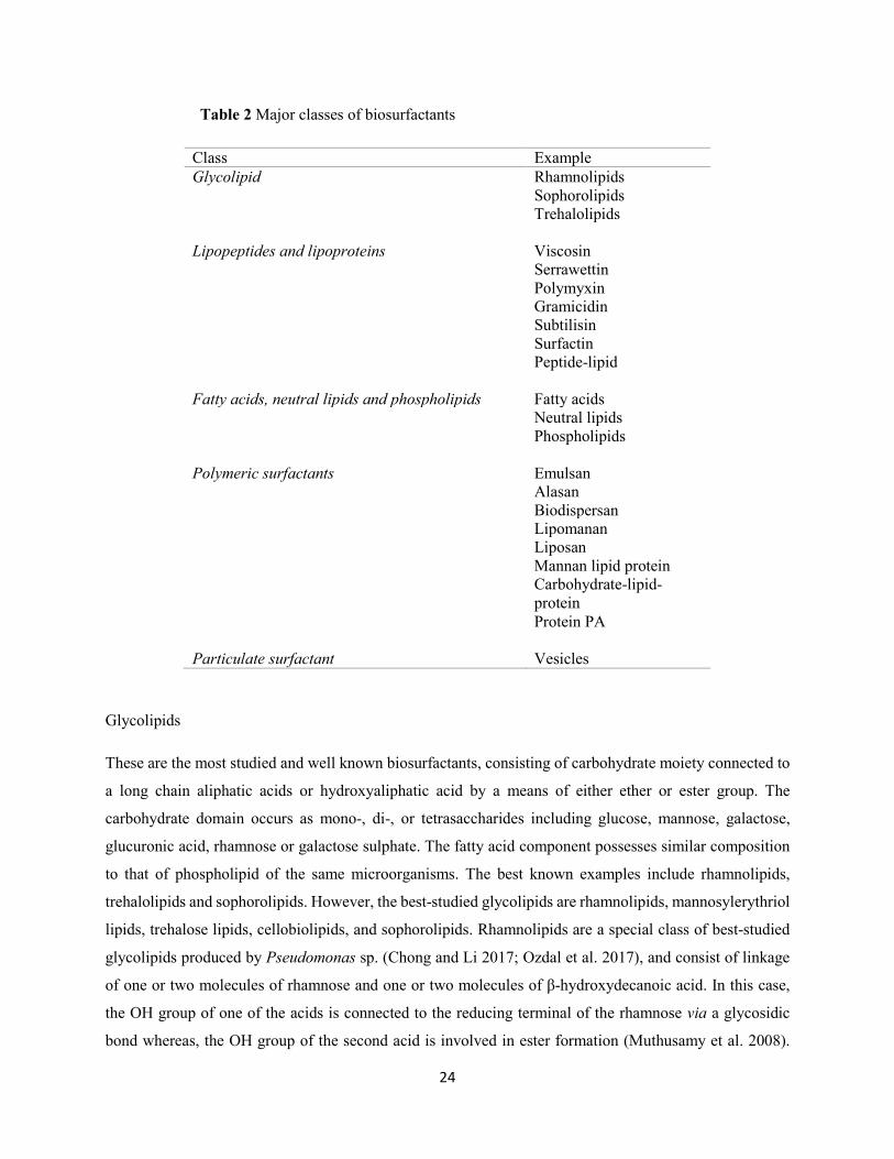
24
Table 2 Major classes of biosurfactants
Class Example Glycolipid Rhamnolipids Sophorolipids Trehalolipids Lipopeptides and lipoproteins Viscosin Serrawettin Polymyxin Gramicidin Subtilisin Surfactin Peptide-lipid Fatty acids, neutral lipids and phospholipids Fatty acids Neutral lipids Phospholipids Polymeric surfactants Emulsan Alasan Biodispersan Lipomanan Liposan Mannan lipid protein Carbohydrate-lipid-
protein Protein PA Particulate surfactant Vesicles
Glycolipids
These are the most studied and well known biosurfactants, consisting of carbohydrate moiety connected to
a long chain aliphatic acids or hydroxyaliphatic acid by a means of either ether or ester group. The
carbohydrate domain occurs as mono-, di-, or tetrasaccharides including glucose, mannose, galactose,
glucuronic acid, rhamnose or galactose sulphate. The fatty acid component possesses similar composition
to that of phospholipid of the same microorganisms. The best known examples include rhamnolipids,
trehalolipids and sophorolipids. However, the best-studied glycolipids are rhamnolipids, mannosylerythriol
lipids, trehalose lipids, cellobiolipids, and sophorolipids. Rhamnolipids are a special class of best-studied
glycolipids produced by Pseudomonas sp. (Chong and Li 2017; Ozdal et al. 2017), and consist of linkage
of one or two molecules of rhamnose and one or two molecules of β-hydroxydecanoic acid. In this case,
the OH group of one of the acids is connected to the reducing terminal of the rhamnose via a glycosidic
bond whereas, the OH group of the second acid is involved in ester formation (Muthusamy et al. 2008).
25
Sophorolipids are a class of glycolipid biosurfactants secreted mainly by yeasts such as Torulopsis
bombicola, Torulopsis petrophilum and Torulopsis apicola (Rahman and Gakpe 2008). They composed of
a dimeric sophorose sugar linked to a long-chain hydroxyl fatty acid through a glycosidic bond. They occur
in two types: acidic (non-lactone) and lactonic sophorolipids. The hydroxyl fatty acids moiety of the acidic
sophorolipids consist of a free carboxyl acid functional group while that of lactonic sophorolipids has a
macrocyclic lactone rings with the 4-hydroxyl group of the sophorose by intramolecular esterification. Both
lactonic and acidic sophorolipids reduced the interfacial tension between n-hexadecane and water from 40
to 5 mN/m with a remarkable stability in pH and temperature (Desai and Banat 1997).
Mannosylerythritol lipids (MELs) are yeast glycolipids and one of the major biosurfactants produced by
Pseudozyma antarctica. It consists of a mixture of four different components: MEL-A, MEL-B, MEL-C
and MEL-D (Kitamoto et al. 1990; Morita et al. 2015). The backbone of these molecules is a mannose-
erythritol disaccharide on which long-chain fatty acid chains are acetylated. Other MEL producers include
Pseudozyma parantarctica (Morita et al. 2012), Kurtzmanomyces sp. I-11 (Kakugawa et al. 2002), and
Ustilago scitaminea NBRC 32730 (Morita et al. 2011).
Lipopeptides and lipoproteins
These are a group of biosurfactants with remarkable surface active properties. A well-studied cyclic
lipopeptide is surfactin produced by Bacillus subtilis ATCC 21332. Surfactin consists of seven amino acids
linked to a carboxyl and hydroxyl groups of a 14-carbon acid (Chen et al. 2015). It is regarded as the most
effective biosurfactant due to its ability to reduce the surface tension of water to 27 mN/m at low
concentration (Kakimuna et al. 1969). Similarly, surfactant BL-86 produced by Bacillus licheniformis has
also been reported to reduce the surface tension of water to 27 mN/m, and the interfacial tension between
water and n-hexadecane to 0.36 mN/m (Desai and Banat 1997). A most important feature of surfactin is its
potential to lyse mammalian red blood cells and form spheroplasts (Satpute et al. 2008).
Fatty acids, phospholipids and neutral lipids
Large amounts of fatty acids and phospholipids are produced by many bacteria and yeast when cultured in
a medium supplemented with n-alkane. The hydrophilic and lipophilic balance is directly proportional to
the length of the hydrocarbon chain in their structures (Satpute et al. 2010a). Phosphatidylethanolamine
synthesized by Acinetobacter sp. form optically clear alkanes-water microemulsions, and that produced by
Rhodococcus erythropolis reduces the interfacial tension between hexadecane and water to less than 1
mN/m and CMC of 30 mg/L (Desai and Banat 1997).
26
Polymeric biosurfactants
The most common polymeric biosurfactants include alasan, liposan, lipomanan, emulsan, and some other
polysaccharide-protein complexes. Emulsan is an efficient bioemulsifier for hydrocarbons in water at
concentrations as low as 0.001% to 0.01% (Lang 2002; Hatha et al. 2007). Liposan is an extracellular water-
soluble emulsifier produced by Candida lipolytica, and consist of 83% carbohydrates and 17% proteins.
Such carbohydrate-protein complex is also produced by Yarrowia lipolytica. Others include mannan lipid
protein (Candida lipolytica), protein PA (Pseudomonas aeruginosa). However, emulsan and biodispersan
produced by Acinetobacter calcoaceticus are the best-studied examples, consisting of a
heteropolysaccharide moiety covalently linked to fatty acids (Rosenberg et al. 1988).
Particulate biosurfactants
The particulate biosurfactants partition extracellular membrane vesicles, forming microemulsion, which
plays a major role in alkane uptake by microbial cells. Vesicles of Acinetobacter sp. has diameter of 20-50
nm, buoyant density of 1.158 cubic g/cm, and consist of protein, phospholipids and lipopolysaccharides
(Chakrabarti 2012; Vijayakumar and Saravanan 2015).
Biosurfactant production and influencing factors
Biosurfactants are secreted extracellularly or intracellularly by microorganisms such as bacteria, yeasts,
and fungi (Banat et al. 2014). The production of biosurfactant compounds occurs on water soluble and
water insoluble substrates by de novo pathway and/or assembly from other substrates, and this may
consequently vary in structure or production domain within the cell depending upon the substrate
composition (Gautam and Tyagi 2006; Satpute et al. 2010b). Factors such as appropriate selection of
microbial strains, substrate type, and fermentation technology play a vital role for a cost-effective and large-
scale production of biosurfactants (Marchant and Banat 2012b; Marchant et al. 2014). Different growth
media are utilized for effective biosurfactant production. In findings by Arima et al. (1968), low yields
(~100 mg/L) of surfactin was reported from complex media such as nutrient broth and Luria Bertani broths.
High yields of surfactin production by Bacillus subtilis strain when cultivated in mineral salt medium
(MSM) has been reported (Yeh et al. 2005). These defined media have proved more effective for the
production of biosurfactants (Wei et al. 2004). It consists of MSM supplemented with water soluble carbon
sources such as glucose, sucrose, glycerol or water-insoluble carbon sources including blended gasoline,
wheat bran, olive oil, hydrocarbons etc. (Prabhu and Phale 2003; Cunha et al. 2004). Govindammal and
Parthasarathi (2013) investigated the influence of glucose, petroleum based substrates, waste fried
vegetable oil, and coconut oil cake on biosurfactant production by Pseudomonas fluorescence MFS03
27
isolated from mangrove forest soil. Vegetable oil and coconut oil were recorded as reliable substrates for
biosurfactant production. Similarly, Sim et al. (1997) studied the effect of vegetable oils (canola and
soybean oils) and glucose on rhamnolipid biosurfactant production by Pseudomonas aeruginosa UW-1.
Results recorded showed a 10-12-fold increase in the yield of rhamnolipid when vegetable oil rather than
glucose was utilized as the sole carbon source. As a result, various authors (Desai and Banat 1997; Banat
et al. 2000; Banat et al. 2014; Satpute et al. 2017) have suggested the use of vegetable oil as inexpensive
and renewable substrates for biosurfactant production. Vegetable oil represents one of the first substrates
reported for high yields of biosurfactants (Raza et al. 2007; George and Jayachandran 2013). Several
authors (Abouseoud et al. 2008; Kiran et al. 2010a; Noudeh et al. 2010) have reported the use of olive oil
as the best carbon source amongst the studied carbon sources for biosurfactant production.
Nitrogen sources form the second most essential supplement for the production of biosurfactants by
microorganisms (Santos et al. 2016). Different organic (urea, yeast extract, peptone, tryptone, corn steep
liquor) and inorganic (sodium nitrate, potassium nitrate, ammonium sulphate) nitrogen sources have been
used in the production of biosurfactants (Abdel-Mawgoud et al. 2010). Santa Anna et al. (2002) investigated
the importance of nitrogen source for the production of biosurfactant by Pseudomonas aeruginosa
cultivated in MSM containing 3% glycerol. Sodium nitrate was found to be more effective than ammonium
sulphate. High yields of sophorose lipids by Torulopsis bombicola and Candida bombicola have been
obtained when using yeast extract and urea as the nitrogen sources (Deshpande and Daniels 1995).
Ammonium nitrate and yeast extract have been reported as best nitrogen source for high yields of
mannosylerythritol lipids by Candida sp. SY16, Candida lipolytica and Candida glabrata (Kim et al. 1999;
Sarubbo et al. 2006, 2007; Rufino et al. 2007, 2008). Nitrogen limitation has been reported for improvement
of biosurfactant production (Abu-Ruwaida et al. 1991). Patel and Desai (1997) observed that biosurfactant
yield was enhanced under nitrogen limiting conditions. In addition, trace elements including Mg2+, K+,
Mn2+ and Fe2+ act as key factors for the multi-enzyme systems associated with biosynthetic pathways of
biosurfactant production (Sen and Swaminathan 2004; Wei et al. 2007; Chen et al. 2015).
Kinetics of growth and biosurfactant production
The kinetics of biosurfactant production exhibit some considerable variations among different systems.
Kinetic parameters are categorized as follows: (a) growth-associated production; (b) production under
growth-limiting conditions; (c) production by resting or immobilized cells, and (d) production with
precursor supplementation (Desai and Banat 1997). The interaction of various fermentation conditions in a
complex way affects the kinetics of biosurfactant production. In growth-linked production, parallel
relationships are noted between cell growth, substrate utilization and biosurfactant production. Zheng et al.
28
(2012) identified a novel bioemulsifier by Aeribacillus pallidus YM-1 cultivated in a MSM supplemented
with 2.0% glucose as carbon source. Maximum cell growth (OD600 = 2.42 ± 0.02) corresponding to a
significant increase in emulsifying activity (60 ± 1%) was achieved during exponential growth phase, an
indication that production kinetics of the bioemulsifier was primarily growth-associated. Gudiña et al.
(2015) studied bioemulsifier production by a novel Paenibacillus sp. #510 grown in MSM added with
paraffin under aerobic conditions. Bioemulsifier production was found to be growth-linked, as parallel
relationship was recorded between biomass production and emulsifying activity. Khopade et al. (2012)
studied the kinetics of biosurfactant production from a marine Streptomyces sp. B3 under batch culture
conditions. Biosurfactant production and surface tension were found to be growth depended. Similar
profiles have been reported by several authors (Franzetti et al. 2008, 2012; Shavandi et al. 2011; Colin et
al. 2013) have reported similar profiles.
Production under growth-limiting conditions is attributed to a drastic increase in the biosurfactant yield
because of the limitation of one or more medium components. Several authors have reported overproduction
of biosurfactants by Pseudomonas sp. when the cultures attain stationary growth phase due to deficient of
nitrogen and iron (El-Sheshtawy et al. 2015; White et al. 2013; Lima et al. 2017).
In the case of production kinetics of biosurfactant by resting or immobilized cells, there is no cell growth;
the cells continue to utilize the carbon source for biosurfactant synthesis (Desai and Banat 1997). This is
noteworthy in the synthesis of rhamnolipid by Pseudomonas sp. (Onwosi and Odibo 2013; Lotfabad et al.
2017), glycolipid by Ochrobactrum intermedium (Ferhat et al. 2017), and lipopeptide surfactin by Bacillus
subtilis ATCC 21332 (Chtioui et al. 2010).
Supplementation of growth medium with biosurfactant precursors induces both qualitative and quantitative
changes in the metabolite. Increased biosurfactant production with yields of about 120-150 g/L following
addition of lipophilic compounds to the fermentation medium has been reported (Lee and Kim 1993).
Similarly, enhanced biosurfactant production from Pseudomonas aeruginosa MTCC 2297 and Bacillus
subtilis ATCC 6633 through supplementation of the culture medium with sugars or sugar byproducts has
also been recorded (Panesar et al. 2011; de Sousa et al. 2014).
Recovery and purification of biosurfactants
The different approaches employed for extraction and purification of biosurfactant compounds affect the
production cost, and these vary according to the microbial growth production process and the
physicochemical characteristics of the biosurfactant(s) produced (Shaligram and Singhal 2010).
Downstream processing costs of biotechnological products account for approximately 70-80% of the total
29
production costs, making commercial production of biosurfactants quite costly (Santos et al. 2016). For
instance, in certain applications such as cosmetics, medicine and food industries, biosurfactant production
is only feasible on a small scale, since advance techniques required for separation of the biomolecules are
not economical on a large scale. Hence, the use of crude biosurfactant preparations for environmental
applications is preferable, since such biosurfactants do not require homogenous purity. The methods
commonly used for effective recovery and/or purification of biosurfactants are influenced by the ionic
charge, solubility and location of the synthesized surfactant compounds relative to the producing organisms
(Helmy et al. 2011; Marchant and Banat 2012a). These methods include the use of organic solvents
(butanol, ethyl acetate) or their combinations (chroloform-methanol, dichloromethane-methanol), and
constitute the most widely used technique for biosurfactant downstream processing. However, this
approach is liable to some limitations including increased production costs, use of large volume of organic
solvents, and high toxicity of organic solvents. Other methods for recovery of biosurfactants involve
precipitation with ammonium sulphate; centrifugation, and adsorption discussed below:
Acetone precipitation
In this process, cell-free culture supernatant is mixed with ice-cold acetone followed by incubation at 4 °C
to recover the precipitate emulsifier. Several authors have employed this approach for the recovery of
biosurfactants and bioemulsifiers (Sharma et al. 2015; Ferhat et al. 2017).
Ethanol precipitation
Similar to acetone, ethanol is regarded as a common solvent for extraction of crude bioemulsifier from the
culture supernatant. The obtained cell free culture supernatant after centrifugation is precipitated using cold
ethanol. This has been proved to be the most effective method for the precipitation of emulsifier from
Acinetobacter calcoaceticus subsp. anitratus SM7 in comparison to other methods (Phetrong et al. 2008).
Ammonium sulphate precipitation
This method was developed by Rosenberg et al. (1979) for extraction of bioemulsifier from Arthrobacter
RAG-1, and is employed for precipitation of high molecular weight bioemulsifiers including emulsan and
protein rich biodispersan. Ammonium sulphate at initial concentration of 30% is added directly to the
culture broth followed by incubation overnight. The obtained precipitate is further re-suspended in
ammonium sulphate until a saturation level is reached. The resultant pellet was centrifuged and extracted
with solvents. The obtained product is further purified and dialyzed. Depending on the type of bioemulsifier
to be precipitated, different concentrations of ammonium sulphate have been used. Bach et al. (2003) added
60% ammonium sulphate to Acinetobacter venetianus RAG-1 cell free culture supernatant whereas, Toren
30
et al. (2001, 2002) precipitated alasan with 65% ammonium sulphate. In another study, Youssef et al. (2005)
obtained a lipopeptide biosurfactant from Bacillus sp. by precipitation of obtained culture supernatant with
40% ammonium sulphate.
Acid precipitation
This method is based on the principle of hydrolysis of culture supernatant using concentrated HCl until a
pH of 2.0 is attained. At such lower pH, protonated biosurfactants become insoluble and precipitate
(Mukherjee et al. 2006; Gudiña et al. 2012). The precipitated biosurfactant is recovered by centrifugation
after incubation overnight at 4 °C. The resultant pellet is dried under vacuum and further extracted using
various solvents (chloroform, methanol ethyl acetate) (Haba et al. 2000; Thaniyavarn et al. 2003; Nitschke
and Pastore 2006; Abbasi et al. 2012; Aparna et al. 2012). This method is simple and require less time for
recovery of crude biosurfactant (Satpute et al. 2010a).
Dialysis and lyophilization
This is carried out by dissolving biosurfactant/bioemulsifier (precipitate) samples in sterile distilled water
and dialyzed against double distilled water using a seamless cellulose tubing dialysis bag of a particular
pore size. The obtained dialysate is then stored at 4 °C in an airtight container for further use. Bioemulsifier
from Acinetobacter calcoaceticus BD4 and BD413 was recovered by ammonium sulphate precipitation.
Further purification was carried by dialysis against cold distilled water followed by lyophilization of
obtained product (Kaplan and Rosenberg 1982). This technique allows simple, rapid and cost-effective
purification of biosurfactants.
Foam fractionation
This is a solvent-free technique used for recovery of biosurfactant compounds that are adsorbed to air
bubbles in the fermentation medium. The production of biosurfactant involves continuous foam formation
resulting from high surface active of the biomolecules. The foam-containing biosurfactant is collected from
the fractionation column and acidified by precipitation using concentrated HCl to lower the pH to 1.0-2.0.
The precipitated sample can further be extracted using organic solvent (Noah et al. 2002). This method
possesses some advantages including prevention of product accumulation that could suppress biomass
growth and product formation; facilitates prolonged biosurfactant production in fed-batch or continuous
mode systems, and prevent biosurfactant denaturation due to their small size and simple structure
(Rangarajan and Sem 2013).
31
Adsorption and desorption
The recovery of some biosurfactant molecules occurs by adsorption and desorption from Amberlite XAD
2 or 16 polystyrene resins. In this method, the biosurfactants are adsorbed on polymer resins followed by
desorption with organic solvents for the collection of purified products (Abalos et al. 2001; Dubey et al.
2005). This technique is a rapid, one-step recovery process with a highly purified biosurfactant obtained.
Adsorption-desorption of biosurfactant on wood-based activated carbon offers a continuous recovery of
biosurfactant from fermentation broth and avoids end product inhibition (Dubey et al. 2005).
Ultrafiltration
This is a low pressure-driven process employed to concentrate and purify biosurfactants using amicon filter
paper or hollow fiber cartridges. Biosurfactants are recovered from culture broth by ultrafiltration with 30
kDa molecular weight cut-offs (MWCO), which allows passage of small molecules (such as salts, amino
acids, other metabolites) while retaining macromolecules of molecular diameter higher than MWCO of the
membrane. The biosurfactant forms micelles at above CMC level, and these are collected in polymeric
membranes. Several authors have reported on the use of ultrafiltration for the recovery of purified
biosurfactants (Lin and Jiang 1997; Zinjarde et al. 1997; Bonilla et al. 2005). This technique is fast, simple,
yields high purity biosurfactants and requires no chemicals.
Preparative thin layer chromatography
In preparative thin layer chromatography, the biosurfactants are purified by applying on a silica (stationary
phase)-coated glass plate of varying thickness, and then allowed to run in a solvent system (mobile phase)
(Satpute et al. 2010a; Biniarz et al. 2017). Bands of the sample are formed based on the affinity of the
sample’s components with the stationary and mobile phases. These bands are visualized under UV light
(non-destructive approach) or using different solvents (destructive method). The obtained bands are further
scraped and extracted with solvents. This method is versatile for detection of quality and purity of
biosurfactants.
Ion exchange chromatography
Biosurfactants with net charge (e.g. negatively charged rhamnolipids) at higher pH are attached to ion-
exchange resins and are eluted with buffer. The biosurfactant is released from the resin following the
addition of a minimum of 0.6 M NaCl to the buffer. This process can be repeated several times using
reusable resin in order to ensure a complete removal of salt from the biosurfactants (Matsufuji et al. 1997;
Satpute et al. 2010a).
32
Potential applications of biosurfactants/bioemulsifiers
In recent years, much attention has been given toward biosurfactants due to their wide range of functional
properties and diverse synthetic capabilities of microorganisms. Of utmost significant is the environmental
acceptability of these surface-active compounds, as they are easily biodegradable with lower toxicity when
compared to synthetic surfactants. These exceptional properties of biosurfactants allow their utilization and
possible substitute of chemically synthesized surfactants in a wide range of biotechnological applications
including petroleum, agriculture, food processing, cosmetics, detergents, leather, textile, paper and
pharmaceutical industries (Rodrigues et al. 2006; Banat et al. 2010; Gudiña et al. 2016). Furthermore,
biosurfactants are used for the recovery of oil residue from storage tank, clean-up of oil spills and
bioremediation of contaminated soil and water (Sobrinho et al. 2013; Silva et al. 2014a). A summary of the
potential applications of biosurfactants in various industries is presented in Table 3, while the major
biotechnological applications are discussed in details below:
Bioremediation
The discharge of contaminants such as hydrocarbons into the environment represents one of the crucial
causes of global pollution and has become a major issue of concern in both industrialized and developing
countries (Lobrero et al. 2012; Luna et al. 2013). This is typical of the largest oil spills in the world, which
occurred in the Gulf of Mexico in 2010, following the explosion of oil off the coastline of the states of
Louisiana and Mississippi (USA), resulting in the release of an estimated 3-4 million barrels of oils spilled
(Rocha et al. 2013).
Different physicochemical approaches including gravity separation, chemical coagulation, cyclone
separation have been developed for the treatment of oil-contaminated sites (Abdelwahab et al. 2009;
Diya’udeen et al. 2011; Padaki et al. 2015). However, these techniques are often expensive and inefficient.
Therefore, biological methods are considered more suitable due to their less hazardous and more selectivity
to specific reactions (Assadi and Tabatabaee 2010). It involves the use of natural degradation capacity of
microorganisms to eliminate or neutralize pollutants from a contaminated site by conversion of the
contaminants into less toxic compounds or complete conversion of such substances into carbon dioxide and
water (Santos et al. 2016). The use of biosurfactant emerges as an alternative approach for enhancing the
dispersal and solubility of contaminants in the aqueous phase, and increase the bioavailability of the
hydrophobic substrates to microorganisms, with subsequent removal of such pollutants through
biodegradation, thus eliminating the need for additional process and results in lesser operational costs
(Kuyukina et al. 2005; Assadi and Tabatabaee 2010; Aparna et al. 2011; Daverey and Pakshirajan 2011;
Damasceno et al. 2012; Olkowska et al. 2012).
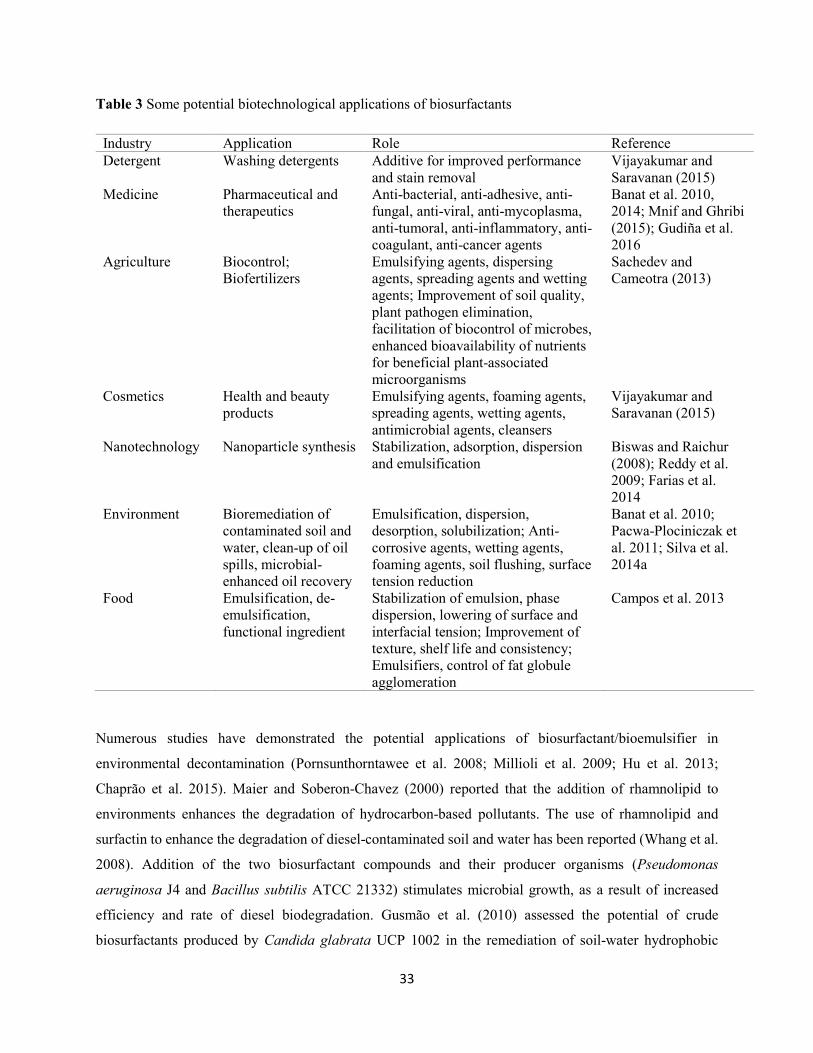
33
Table 3 Some potential biotechnological applications of biosurfactants
Industry Application Role Reference Detergent Washing detergents Additive for improved performance
and stain removal Vijayakumar and Saravanan (2015)
Medicine Pharmaceutical and therapeutics
Anti-bacterial, anti-adhesive, anti-fungal, anti-viral, anti-mycoplasma, anti-tumoral, anti-inflammatory, anti-coagulant, anti-cancer agents
Banat et al. 2010, 2014; Mnif and Ghribi (2015); Gudiña et al. 2016
Agriculture Biocontrol; Biofertilizers
Emulsifying agents, dispersing agents, spreading agents and wetting agents; Improvement of soil quality, plant pathogen elimination, facilitation of biocontrol of microbes, enhanced bioavailability of nutrients for beneficial plant-associated microorganisms
Sachedev and Cameotra (2013)
Cosmetics Health and beauty products
Emulsifying agents, foaming agents, spreading agents, wetting agents, antimicrobial agents, cleansers
Vijayakumar and Saravanan (2015)
Nanotechnology Nanoparticle synthesis Stabilization, adsorption, dispersion and emulsification
Biswas and Raichur (2008); Reddy et al. 2009; Farias et al. 2014
Environment Bioremediation of contaminated soil and water, clean-up of oil spills, microbial-enhanced oil recovery
Emulsification, dispersion, desorption, solubilization; Anti-corrosive agents, wetting agents, foaming agents, soil flushing, surface tension reduction
Banat et al. 2010; Pacwa-Plociniczak et al. 2011; Silva et al. 2014a
Food Emulsification, de-emulsification, functional ingredient
Stabilization of emulsion, phase dispersion, lowering of surface and interfacial tension; Improvement of texture, shelf life and consistency; Emulsifiers, control of fat globule agglomeration
Campos et al. 2013
Numerous studies have demonstrated the potential applications of biosurfactant/bioemulsifier in
environmental decontamination (Pornsunthorntawee et al. 2008; Millioli et al. 2009; Hu et al. 2013;
Chaprão et al. 2015). Maier and Soberon-Chavez (2000) reported that the addition of rhamnolipid to
environments enhances the degradation of hydrocarbon-based pollutants. The use of rhamnolipid and
surfactin to enhance the degradation of diesel-contaminated soil and water has been reported (Whang et al.
2008). Addition of the two biosurfactant compounds and their producer organisms (Pseudomonas
aeruginosa J4 and Bacillus subtilis ATCC 21332) stimulates microbial growth, as a result of increased
efficiency and rate of diesel biodegradation. Gusmão et al. (2010) assessed the potential of crude
biosurfactants produced by Candida glabrata UCP 1002 in the remediation of soil-water hydrophobic
34
pollutants. Removal efficiency of 92.6% was recorded. In another study, a novel biosurfactant, denoted
Lunasan produced by Candida sphaerica UCP 0995 was found to remove 95% of motor oil from
contaminated site (Luna et al. 2011). Gordonia sp. BS29 cultivated in hydrophobic substrate as sole carbon
source has been found to synthesize bioemulsan which effectively degrade crude oil, polycyclic aromatic
hydrocarbons and other recalcitrant branched hydrocarbons from contaminated sites (Franzetti et al. 2008).
The addition of biosurfactants/bioemulsifiers serves as a promising strategy for the biotreatment of oily
wastewater (de Almeida et al. 2016; Rahbari-Sisakht et al. 2017). Zhang et al. (2009) applied rhamnolipids
for the treatment of oily wastewater in an aerated active sludge system. At 20 °C, rhamnolipids (11.2 mg/L)
was found to increase the removal efficiency of oil from 17.7% (without rhamnolipids) to 63%. At 25 °C,
the removal efficiency of oil was above 80% in the presence of rhamnolipids as compared to 22.3% without
rhamnolipids. Similarly, rhamnolipid treatment (22.5 mg/L) for 24 h at 20 °C significantly increased the
removal rate of oil from 24% (in the absence of rhamnolipids) to 92%. The authors therefore observed that
enhanced removal of oils was due to improved solubility, and reduced interfacial tension of the
rhamnolipids. Lima et al. (2011) assessed the removal of oily sludge from oil-contaminated sites using
biosurfactants produced from five bacterial isolates. The biosurfactants led to a reduction in viscosity and
enhanced the formation of oil-water emulsions leading to easier sludge pumping and emulsion breaking for
better oil recovery. In another study, Yan et al. (2012) used a rhamnolipid produced by Pseudomonas
aeruginosa F-2 for the reclamation of oil from oily sludge. Removal efficiency (91.5%) of oil was recorded
from the field pilot-scale studies. Daverey and Pakshirajan (2011) evaluated the pretreatment of synthetic
dairy wastewater using sophorolipid biosurfactant produced by Candida bombicola. The results showed
COD removal efficiency (93%) after 96 h of operation. Furthermore, Cosmann et al. (2017) employed a
surfactin biosurfactant produced by Bacillus subtilis LB5a for the anaerobic treatment of poultry
slaughterhouse effluent. Results obtained revealed no inhibition of microbial consortium of the anaerobic
sludge with consequent soluble COD (SCOD) and O & G removal efficiencies of 80% and 70%,
respectively along with a promising specific methane yield. Daverey and Pakshirajan (2011) evaluated the
pretreatment of synthetic dairy wastewater using sophorolipid biosurfactant produced by Candida
bombicola. The results showed COD removal efficiency (93%) after 96 h of operation. In addition,
Cosmann et al. (2017) employed a surfactin biosurfactant produced by Bacillus subtilis LB5a for the
anaerobic treatment of poultry slaughterhouse effluent. Results obtained revealed no inhibition of microbial
consortium of the anaerobic sludge with consequent soluble COD (SCOD) and O & G removal efficiencies
of 80% and 70%, respectively along with a promising specific methane yield.
Increase in the levels of heavy metals and radionuclides in soil in many industrialized countries poses
serious threats to the ecosystems and human health through either food chain or direct exposure to
35
contaminated soil and water (Chakraborty and Das 2014; Mao et al. 2015). The heavy metals adsorb to the
soil surface in the form of ions or the precipitation of metal compounds. They are removed from soil through
surfactant-associated complexation and ion exchange (Ochoa-Loza et al. 2001; Swarnkar et al. 2012).
Dahrazma and Mulligan (2007) assessed the efficacy of rhamnolipid biosurfactant for the removal of heavy
metals (Cu, Ni and Zn) from sediments obtained from the Lachine Canal in Canada in a continuous flow
configuration system. Rhamnolipid concentration (0.5%, v/v) led to removal efficiencies of Cu (37%), Ni
(27%) and Zn (13%). Addition of 1% NaOH further increased (4-fold) the removal of heavy metals from
the system. Biosurfactant compounds synthesized by Pseudomonas sp. and Alcaligenes sp. have been
employed for the floatation and removal of calcite and scheelite. Recovery rates of 95% (CaWO4) and 30%
(CaCO3) were recorded in comparison to conventional surfactant, found to be incapable of separating these
two minerals (Nitschke and Pastore 2002). Similarly, Juwarkar et al. (2007) investigated the potential of
rhamnolipid for the removal of Cd2+ and Pb from artificially contaminated soil samples. The rhamnolipid
compound was found to remove both free and weakly bound Cd2+ and Pb from the tested soil.
Food industry
In food industries, biosurfactants have several promising applications including formation and stabilization
of emulsions. Emulsification is vital for the formation of consistency and texture in foods; phase dispersion
and solubilization of aromas (Campos et al. 2013; 2014). Generally, emulsifiers in food products stabilize
emulsion by controlling agglomeration of fat globules, stabilization of aerated systems, improvement of
texture and shelf life of starch-containing products, alteration of rheological properties of wheat dough and
enhancement of consistency and texture of fat-based products (Nitschke and Pastore 2002; Campos et al.
2013). Enhancement in the stability of dough, volume, texture and conservation of bakery products
following addition of rhamnolipid biosurfactants has been reported (Van Haesendonck and Vanzeveren
2004). In addition, bioemulsifier produced by Candida utilis is used in processed salad dressing (Campos
et al. 2015). Mannoprotein secreted by Saccharomyces cerevisiae has been reported to stabilize water/oil
emulsions in cookies, mayonnaise and ice cream (Shepherd et al. 1995; Torabizadeh et al. 1996).
Medicine
Several microorganisms have developed multi-drug resistance to commercially available antimicrobial
agents, and this is associated with misuse or abuse of antibiotics. Thus, persistent and chronic infectious
diseases emerged, constituting a serious public health concern (Coates et al. 2011). Furthermore, in the last
decades, the discovery of new antibiotics has reduced significantly, due to challenges in identification of
novel and hyperactive compounds with subsequent high costs needed for their development. Therefore,
36
there is an urgent need for a renewed interest in the development of novel antimicrobial drugs with wide
spectrum activity.
Biosurfactants have been employed in various therapeutic applications due to their bactericidal, fungicidal,
insecticidal and anti-viral activities as well as use as anti-fouling agents. Several biosurfactants displayed
similar or better antimicrobial activity in comparison with conventional antibiotics. For instance, glycolipid
biosurfactant produced by Streptomyces sp. MAB36 was found to exhibit similar inhibitory activity against
Aspergillus niger and Candida albicans when compared with the conventional antifungal nystatin
(Manivasagan et al. 2014). In another study, biosurfactant produced by Nocardiopsis dassonvillei MAD08
was reported to be more effective against Escherichia coli and Staphylococcus epidermidis than
chloramphenicol (Selvin et al. 2009). In addition, sophorolipids from Candida bombicola was found to
inhibit the growth of Gram-negative and Gram-positive bacteria with a minimum inhibitory concentration
of approximately 30 and 1 mg/ml in a contact time of 2 and 4 h, respectively for Escherichia coli
ATCC8739 and Pseudomonas aeruginosa ATCC 9027 as well as 6 and 1 mg/ml in a contact time of 4 h
for Staphylococcus aureus ATCC 6358 and Bacillus subtilis ATCC 6633, respectively (Joshi-Navare and
Prabhune 2013).
Furthermore, some biosurfactant compounds possessed a substantial anti-adhesive and anti-biofilm activity.
Glycolipid biosurfactant synthesized by Brevibacterium casei MSA19 was found to remove pre-formed
biofilms of all the tested pathogenic microorganisms at a concentration of 30 µg/ml (Kiran et al. 2010b).
Similarly, Lunasan-producing Candida sphaerica was reported to inhibit adherence of Pseudomons
aeruginosa, Streptococcus agalactiae and Streptococcus sanguis between 80% and 92% when the
concentration was 10 mg/ml (Luna et al. 2011) whereas, biosurfactant Rufisan showed anti-adherent
activity against most of the tested microorganisms including Streptococcus agalactiae, Streptococcus
mutans, Streptococcus mutans NS, Streptococcus mutans HG, Streptococcus sangui 12, Streptococcus
oralis J22 at a concentration above the CMC (0.3%) (Rufino et al. 2011).
Biosurfactants have been highlighted as potential anti-cancer agents by interfering with cancer progression
processes. These biomolecules are involved in several intracellular molecular recognition steps consisting
of signal transduction, cell differentiation and cell immune response etc. (Rodrigues et al. 2006). For
instance, surfactin biosurfactants are commonly used as potential anti-cancer agent against many cancer
cell lines (Sivapathasekaran et al. 2010; Gudiña et al. 2013; Dey et al. 2015). Iturin A produced by Bacillus
megaterium strain has been found to significantly impair proliferation and inhibit the Akt signaling network
resulting in apoptosis induction in breast cancer cells (MDA-MB-231 and MCF-7) (Dey et al. 2015). In
addition, lipopeptide somocystinamide A secreted from cyanobacteria, Lyngbya majuscula demonstrated
37
noteworthy cytotoxicity against leukaemia, lung, breast and prostate cancer cells with IC50 values between
1.3 µM to 970 nM, based on the cancer model (Wrasidlo et al. 2008).
Cosmetic industry
Biosurfactants possess broad potential application in the cosmetic industry, owing to their multifunctional
properties including emulsification, de-emulsification, foaming, water-binding capacity, and spreading and
wetting, which allow them to be efficiently utilized in cosmetic products such as emulsifiers, solubilizers,
antimicrobial agents, bath products like anti-dandruff shampoo, baby products, toothpaste etc. (Youssef et
al. 2007; Shekhar et al. 2015). Biosurfactants are better skin compatible compared to their synthetic
counterparts (Williams 2009). Lipopeptide biosurfactants with moisturizing and anti-wrinkle properties
demonstrated low cytotoxic effects in human cells. Thus, they are employed as additives in dermatological
products (Mandal et al. 2013). In addition, monoglyceride produced from Pseudomonas fluorescens is a
commonly used surfactant in the cosmetic industry (McNeill and Yamane 1991).
Nanotechnology
The application of biosurfactant in nanotechnology and nanoparticle synthesis is a new development in the
area of green chemistry (Kiran et al. 2010a; Martinez et al. 2014; Santos et al. 2016). Reddy et al. (2009)
observed that silver nanoparticle synthesis could be stabilized for up to two months using surfactin.
Similarly, biosurfactant produced by Pseudomonas aeruginosa has been used to stabilize silver
nanoparticles in the liquid phase (Farias et al. 2014). In another study, glycolipid biosurfactant produced
from Brevibacterium casei MSA 19 has been reported to stabilize silver nanoparticles (Kiran et al. 2010a).
Concluding remarks
The global market for microbial surface-active compounds has increased significantly over decades. This
is due to the outstanding functional properties of these biomolecules, forming a foundation for exploration
in bioremediation, cosmetics, food, medicine and nanotechnology. However, mass production of microbial
surfactants formed a major drawback from economic perspective. Optimization of growth and fermentation
parameters using cheaper renewable substrates coupled with robust downstream processing methods could
pave the way for profitable and cost-effective surfactant production. In addition, the use of recombinant
and mutant hyper-producing microorganisms could provide high yield surfactant production that would be
beneficial for industrial and biotechnological applications.
38
References
Abalos A, Pinazo A, Infante MR, Casals M, García F, Manresa A (2001) Physicochemical and
antimicrobial properties of new rhamnolipids produced by Pseudomonas aeruginosa AT10 from
soybean oil refinery wastes. Langmuir 17:1367-1371
Abbasi H, Hamedi MM, Lotfabad TB, Zahiri HS, Sharafi H, Masoomi F, Moosavi-Movahedi AA, Ortiz A,
Amanlou M, Noghabi KA (2012) Biosurfactant-producing bacterium, Pseudomonas aeruginosa
MA01 isolated from spoiled apples: physicochemical and structural characteristics of isolated
biosurfactant. J Biosci Bioeng 113:211-219
Abdel-Mawgoud AM, Lépine F, Déziel E (2010) Rhamnolipids: diversity of structures, microbial origins
and role. Appl Microbiol Biotechnol 86:1323-1336
Abdelwahab O, Amin NK, El-Ashtoukhy ESZ. 2009. Electrochemical removal of phenol from oil refinery
wastewater. J Hazard Mater 163:711-716
Abouseoud M, Maachi R, Amrane A, Boudergua S, Nabi A (2008) Evaluation of different carbon and
nitrogen sources in production of biosurfactant by Pseudomonas fluorescens. Desalination,
223:143-151
Abraham WR, Meyer H, Yakimov M (1998) Novel glycine containing glucolipids from the alkane using
bacterium Alcanivorax borkumensis. Biochim Biophys Acta, 1393:57-62
Abu-Ruwaida AS, Banat IM, Haditirto S, Khamis A (1991) Nutritional requirements and growth
characteristics of a biosurfactant-producing Rhodococcus bacterium. World J Microbiol Biotechnol
7:53-61
Amani H, Kariminezhad H (2016) Study on emulsification of crude oil in water using emulsan biosurfactant
for pipeline transportation. Petrol Sci Technol 34:216-222
Amaral PFF, da Silva JM, Lehocky M, Barros-Timmons AMV, Coelho MAZ, Marrucho IM, Coutinho JAP
(2006) Production and characterization of a bioemulsifier from Yarrowia lipolytica. Proc Biochem
41:1894-1898
Aparna A, Srinikethan G, Hedge S (2011) Effect of addition of biosurfactant produced by Pseudomonas
sp. on biodegradation of crude oil. In International Proceedings of Chemical, Biological and
Environmental Engineering. Proceedings of the 2nd International Proceedings of Chemical,
Singapore. 26-28 February 2011; vol. 6, pp. 71-75

39
Aparna A, Srinikethan G, Smitha H (2012) Production and characterization of biosurfactant produced by a
novel Pseudomonas sp. 2B. Colloids Surf B: Biointerfaces, 95:23-29
Arima K, Kakinuma A, Tamura G (1968) Surfactin, a crystalline peptide lipid surfactant produced by
Bacillus subtilis: isolation, characterization and its inhibition of fibrin clot formation. Biochem
Biophys Res Commun 31:488-494
Assadi M, Tabatabaee MS (2010) Bisurfactants and their use in upgrading petroleum vacuum distillation
residue: a review. Int J Environ Res 4:549-572
Bach H, Berdichevsky Y, Gutnick D (2003) An exocellular protein from the oil-degrading microbe
Acinetobacter venetianus RAG-1 enhances the emulsifying activity of the polymeric bioemulsifier
emulsan. Appl Environ Microbiol 69:2608-2615
Banat IM (1993) The isolation of a thermophilic biosurfactant producing Bacillus sp. Biotechnol Lett
15:591-594
Banat IM, Franzetti A, Gandolfi I, Bestetti G, Martinotti MG, Fracchia L, Smyth TJ, Marchant R (2010)
Microbial biosurfactants production, applications and future potential. Appl Microbiol Biotechnol
87:427-444
Banat IM, Makkar RS, Cameotra S (2000) Potential commercial applications of microbial surfactants. Appl
Microbiol Biotechnol 53:495-508
Banat IM, Satpute SK, Cameotra SS, Patil R, Nyayanit NV (2014) Cost-effective technologies and
renewable substrates for biosurfactants’ production. Front Microbiol 5:697
Batista SB, Mounteer AH, Amorim FR, Tótola MR (2006) Isolation and characterization of
biosurfactant/bioemulsifier-producing bacteria from petroleum contaminated sites. Bioresour
Technol 97:868-875
Belgacem Z, Bijttebier S, Verreth C, Voorspoels S, Van de Voorde I, Aerts G, Willens KA, Jacquemyn H,
Ruyters S, Lievens B (2015) Biosurfactant production by Pseudomonas strains isolated from flora
nectar. J Appl Microbiol 118:1370-1384
Beltrani T, Chiavarini S, Cicero DO, Grimaldi M, Ruggeri C, Tamburini E, Cremisini C (2015) Chemical
characterization and surface properties of a new bioemulsifier produced by Pedobacter sp. strain
MCC-Z. Int J Biol Macromolec 72:1090-1096
40
Bhattacharya M, Biswas D, Sana S, Datta S (2014) Utilization of waste engine oil by Ochrobactrum
pseudintermedium strain C1 that secretes an exopolysaccharide as a bioemulsifier. Biocatal Agric
Biotechnol 3:167-176
Biniarz P, Łukaszewicz M, Janek T (2017) Screening concepts, characterization and structural analysis of
microbial-derived bioactive lipopeptides: a review. Critical Rev Biotechnol 37:393-410
Biswas M, Raichur AM (2008) Electrokinetic and rheological properties of nano zirconia in the presence
of rhamnolipid biosurfactant. J Am Ceram Soc 91:3197-3201
Bodour AA, Drees KP, Maier RM (2003) Distribution of biosurfactant-producing bacteria in undisturbed
and contaminated arid southwestern soils. Appl Environ Microbiol 69:3280-3287
Bodour AA, Miller-Maier R (1998) Application of a modified drops collapse technique for surfactant
quantitation and screening of biosurfactant producing microorganisms. J Microbiol Methods,
32:273-280
Bonilla M, Olivaro C, Corona M, Vazquez A, Soubes M (2005) Production and characterization of a new
bioemulsifier from Pseudomonas putida ML2. J Appl Microbiol 98:456-463
Camacho-Chab JC, Guézennec J, Chan-Bacab MJ, Ríos-Leal E, Sinquin C, Muñiz-Salazar R, Rosa-García
SCD, Reyes-Estebanez M, Ortega-Morales BO (2013) Emulsifying activity and stability of a non-
toxic bioemulsifier synthesized by Microbacterium sp. MC3B-10. Int J Mol Sci 14:18959-18972
Cameron DR, Cooper DG, Neufeld RJ (1988) The mannoprotein of Saccharomyces cerevisiae is an
effective bioemulsifier. Appl. Environ. Microbiol 54:1420-1425
Campos JM, Stamford TLM, Rufino RD, Luna JM, Stamford TCM, Sarubbo LA (2015) Formulation of
mayonnaise with the addition of a bioemulsifier isolated from Candida utilis. Toxicol Rep 2:1164-
1170
Campos JM, Stamford TLM, Sarubbo LA (2014) Production of a bioemulsifier with potential application
in the food industry. Appl Biochem Biotechnol 172:3234-3252
Campos JM, Stamford TLM, Sarubbo LA, Luna JM, Rufino RD, Banat IM (2013) Microbial biosurfactants
as additives for food industries. Biotechnol Prog 29:1097-1108
Chakrabarti S (2012) Bacterial biosurfactant: characterization, antimicrobial and metal remediation
properties. Ph.D Thesis, National Institute of Technology, Surat, India
41
Chakraborty J, Das S (2014) Biosurfactant-based bioremediation of toxic metals. In Microbial
Biodegradation and Bioremediation, 1st ed, Das S., Ed, Elsevier Rourkella Odisha, India, pp. 167-
201
Chaprão MJ, Ferreira INS, Correa PF, Rufino RD, Luna JM, Silva EJ, Sarubbo LA (2015) Application of
bacterial and yeast biosurfactants for enhanced removal and biodegradation of motor oil from
contaminated sand. Electron J Biotechnol 18:471-479
Chen J, Huang PT, Zhang KY, Ding FR (2012) Isolation of biosurfactant producers, optimization and
properties of biosurfactant produced by Acinetobacter sp. from petroleum-contaminated soil. J
Appl Microbiol 112:660-671
Chen W-C, Juang R-S, Wei Y-H (2015) Applications of a lipopeptide biosurfactant, surfactin, produced by
microorganisms. Biochem Eng J 103:158-169
Chong H, Li Q (2017) Microbial production of rhamnolipids: opportunities, challenges and strategies.
Microb Cell Fact 16:137
Chtioui O, Dimitrov K, Gancel F, Nikov I (2010) Biosurfactant production by immobilized cells of Bacillus
subtilis ATCC 21332 and their recovery by pertraction. Proc Biochem 45:1795-1799
Coates ARM, Halls G, Hu Y (2011) Novel classes of antibiotics or more of the same? Br J Pharmacol
163:184-194
Colin Vl, Pereira CE, Villegas LB, Amoroso MJ, Abate CM (2013) Production and partial characterization
of bioemulsifier from a chromium-resistant actinobacteria. Chemosphere, 90:1372-1378
Cooper DG, Goldenberg BG (1987) Surface-active agents from two Bacillus species. Appl Environ
Microbiol 53:224-229
Cooper DG, Paddock DA (1983) Torulopsis petrophilum and surface activity. Appl Environ Microbiol
46:1426-1429
Cosmann NJ, Gomes BM, Gomes SD, Simiqueli APR, Pastore GM (2017) Use of biosurfactant surfactin
produced from cassava wastewater for anaerobic treatment of effluent from a poultry
slaughterhouse. Afr J Biotechnol 16:224-231
Cunha CD, Rosario M, Rosado AS, Leite GF (2004) Serratia sp. SVGG16: a promising biosurfactant
producer isolated from tropical soil during growth with ethanol-blended gasoline. Proc Biochem
39:2277-2282
42
Dahrazma B, Mulligan CN (2007) Investigation of the removal of heavy metals from sediments using
rhamnolipid in a continuous flow configuration. Chemosphere, 69:705-711
Damasceno FRC, Cammarota MC, Freire DMG (2012) The combined use of a biosurfactant and an enzyme
preparation to treat an effluent with high fat content. Colloids Surf B: Biointerfaces, 95:241-246
Das P, Mukherjee S, Sen R (2008a) The antimicrobial potentials of a lipopeptide biosurfactant derived from
a marine Bacillus circulans. J Appl Microbiol 104:1675-1684
Das P, Mukherjee S, Sen R (2008b) Improved bioavailability and biodegradation of a model polyaromatic
hydrocarbon by a biosurfactant producing bacterium of marine origin. Chemosphere, 72:1229-1234
Dastgheib SMM, Amoozegar MA, Elahi E, Asad S, Banat IM (2008) Bioemulsifier production by a
halothermophilic Bacillus strain with potential applications in microbially enhanced oil recovery.
Biotechnol Lett 30:263-270
Daverey A, Pakshirajan K (2011) Pretreatment of synthetic dairy wastewater using the sophorolipid-
producing yeast Candida bombicola. Appl. Biochem. Biotechnol. 163:720-728
de Almeida DG, Da Silva RCFS, Luna JM, Rufino RD, Santos VA, Banat IM, Sarubbo LA (2016)
Biosurfactants: promising molecules for petroleum biotechnology advances. Front Microbiol
7:1718
de Sousa M, Dantas IT, Felix AKN, de Sant’Ana B, Melo VMM, Goncalves LRB (2014) Crude glycerol
from biodiesel industry as substrate for biosurfactant production by Bacillus subtilis ATCC 6633.
Braz Arch Biol Technol 57:295-301
Deng MC, Li J, Hong YH, Xu XM, Chen WX, Yuan JP, Peng J, Yi M, Wang JH (2016) Characterization
of a novel biosurfactant produced by marine hydrocarbon-degrading bacterium Achromobacter sp.
HZ01. J Appl Microbiol 120:889-899
Desai JD, Banat IM (1997) Microbial production of surfactants and their commercial potential. Microbiol
Mol Biol Rev 61:47-64
Deshpande M, Daniels L (1995) Evaluation of sophorolipid biosurfactant production by Candida
bombicola using animal fat. Bioresour Technol 54:143-150
43
Dey G, Bharti R, Dhanarajan G, Das S, Dey KK, Kumar BNP, Sen R, Mandal M (2015) Marine lipopeptide
Iturin A inhibits Akt mediated GSK3β and FoxO3α signaling and triggers apoptosis in breast
cancer. Sci Rep 5:10316
Diaz De Rienzo MA, Kamalanathan ID, Martin PJ (2016) Comparative study of the production of
rhamnolipid biosurfactants by B. thailandensis E264 and P. aeruginosa ATCC 9027 using foam
fractionation. Proc Biochem 51:820-827
Diya’udeen BH, Daud WMAW, Abdul Aziz AR (2011) Treatment technologies for petroleum refinery
effluents: a review. Proc Saf Environ Prot 89:95-105
Dubey KV, Juwarkar AA, Singh SK (2005) Adsorption-desorption process using wood-based activated
carbon for recovery of biosurfactant from fermented distillery wastewater. Biotechnol Prog 21:860-
867
Dusane DH, Nancharaiah YV, Zinjarde SS, Venugopalan VP (2010) Rhamnolipid mediated distruption of
marine Bacillus pumilus biofilms. Colloids Surf B: Biointerfaces, 81:242-248
El-Sheshtawy HS, Aiad I, Osman ME, Abo-Elnasr AA, Kobisy AS (2015) Production of biosurfactant from
Bacillus licheniformis for microbial enhanced oil recovery and inhibition the growth of sulfate
reducing bacteria. Egypt J Petrol 24:155-162
Farias CBB, Silva AF, Rufino RD, Luna JM, Souza JE, sarubbo LA (2014) Synthesis of silver nanoparticles
using a biosurfactant produced in low-cost medium as stabilizing agent. Eletron J Biotechnol
17:122-125
Felse PA, Shah V, Chan J, Rao KJ, Gross RA (2007) Sophorolipid biosynthesis by Candida bombicola
from industrial fatty acid residues. Enzyme Microb Technol 40:316-323
Ferhat S, Alouaoui R, Badis A, Moulai-Mostefa N (2017) Production and characterization of biosurfactant
by free and immobilized cells from Ochrobactrum intermedium isolated from the soil of southern
Algeria with a view to environmental application. Biotechnol Biotechnol Equip 31:733-742
Franzetti A, Bestetti G, Caredda P, La Colla P, Tamburini E (2008) Surface-active compounds and their
role in the access to hydrocarbons in Gordonia strains. FEMS Microbiol Ecol 63:238-248
Franzetti A, Caredda P, Ruggeri C, La Colla P, Tamburini E, Papacchini M, Bestetti G (2009) Potential
applications of surface active compounds by Gordonia sp. strain BS29 in soil remediation
technologies. Chemosphere, 75:801-807
44
Franzetti A, Gandolfi I, Raimondi C, Bestetti G, Banat, IM, Smyth TJ, Papacchini M, Cavallo M, Fracchia
L (2012) Environmental fate, toxicity, characteristics and potential applications of novel
bioemulsifiers produced by Variovorax paradoxus 7bCTS. Bioresour Technol 108:245-251
Fulazzaky MA, Abdullah S, Salim MR (2016) Supporting data for identification of biosurfactant-producing
bacteria isolated from agro-food industrial effluent. Data Brief 7:834-838
Gautam G, Mishra V, Verma P, Pandey AK, Negi S (2014) A cost effective strategy for production of
biosurfactant from locally isolated Penicillium chrosogenum SNP5 and its application. J
Bioprocess Biotech 4:177
Gautam KK, Tyagi VK (2006) Microbial surfactants: a review. J Oleo Sci 55:155-166
George S, Jayachandran K (2013) Production and characterization of rhamnolipid biosurfactant from waste
frying coconut oil using a novel Pseudomonas aeruginosa D. J Appl Microbiol 114:373-383
Govindammal M, Parthasarathi R (2013) Production and characterization of biosurfactant using renewable
substrates by Pseudomonas fluorescens isolated from mangrove ecosystem. J Appl Chem 2: 55-62
Gudiña EJ, Pereira JFB, Costa R, Evtuguin DV, Coutinho JAP, Teixeira JA, Rodrigues LR (2015) Novel
bioemulsifier produced by a Paenibacillus strain isolated from crude oil. Microb Cell Fact 14:14
Gudiña EJ, Pereira JFB, Rodrigues LR, Coutinho JAP, Teixeira JA (2012) Isolation and study of
microorganisms from oil samples for application in microbial enhanced oil recovery. Int Biodeter
Biodegr 68:56-64
Gudiña EJ, Rangarajan V, Sen R, Rodrigues LR (2013) Potential therapeutic applications of biosurfactants.
Trends Pharmacol 34:667-675
Gudiña EJ, Teixeira JA, Rodrigues LR (2016) Biosurfactants produced by marine microorganisms with
therapeutic applications. Mar Drugs, 14:38
Gusmão CAB, Rufino RD, Sarubbo LA (2010) Laboratory production and characterization of a new
biosurfactant from Candida glabrata UCP1002 cultivated in vegetable fat waste applied to the
removal of hydrophobic contaminant. World J Microbiol Biotechnol 26:1683-1692
Gutiérrez T, Mulloy B, Black K, Green DH (2007) Glycoprotein emulsifiers from two marine Halomonas
species: chemical and physical characterization. J Appl Microbiol 103:1716-1727
Haba E, Espuny MJ, Busquets M, Manresa A (2000) Screening and production of rhamnolipids by
Pseudomonas aeruginosa 47T2 NCIB 40044 from waste frying oils. J Appl Microbiol 88:379-387
45
Hatha AAM, Edward G, Rahman KSMP (2007) Microbial biosurfactants-review. J Mar Atmos Res 3:1-17
Helmy Q, Kardena E, Funamizu N, Wisjnuprapto W (2011) Strategies toward commercial scale of
biosurfactant production as potential substitute for its chemically counterparts. Int J Biotechnol
12:66-86
Hsieh F, Li M, Lin T, Kao S (2004) Rapid detection and characterization of surfactin-producing Bacillus
subtilis and closely related species based on PCR. Curr Microbiol 49:186-191
Hu G, Li J, Zeng G (2013) Recent development in the treatment of oily sludge from petroleum industry: a
review. J Hazard Mater 261:470-490
Ibacache-Quiroga C, Ojeda J, Espinoza-Vergara G, Olivero P, Cuellar M, Dinamarca MA (2013) The
hydrocarbon-degrading marine bacterium Cobetia sp. strain MM1IDA2H-1 produces a
biosurfactant that interferes with quorum sensing of fish pathogens by signal hijacking. Microb
Biotechnol 6:394-405
Ibrahim M, Ijah U, Manga S, Bilbis L, Umar S (2013) Production and partial characterization of
biosurfactant produced by crude oil degrading bacteria. Int Biodeter Biodegr 81:28-34
Ilori MO, Amobi CJ, Odocho AC (2005) Factors affecting biosurfactant production by oil degrading
Aeromonas spp. isolated from a tropical environment. Chemosphere, 61:985-992
Inès M, Dhouha G (2015) Lipopeptide surfactants: production, recovery and pore forming capacity.
Peptides, 71:100-112
Jagtap S, Yavankar S, Pardesi K, Chopade B (2010) Production of bio-emulsifier by Acinetobacter species
isolated from healthy human skin. Indian J Exp Biol 48:70-76
Jain DK, Collins-Thompson DL, Lee H, Trevors JT (1991) A drop-collapsing test for screening surfactant-
producing microorganisms. J Microbiol Methods, 13:271-279
Joshi-Navare K, Prabhune A (2013) A biosurfactant sophorolipid acts in synergy with antibiotics to enhance
their efficiency. BioMed. Res Int 2013:1-8
Juwarkar AA, Nair A, Dubey KV, Singh S, Devotta S (2007) Biosurfactant technology for remediation of
cadmium and lead contaminated soils. Chemosphere, 68:1996-2002
Kakimuna A, Oachida A, Shina T, Sugino H, Isano M, Tanura G, Arima K (1969) Confirmation of the
structure of surfactin by mass spectrometry. Agric Biol Chem 33:1669-1672
46
Kakugawa K, Tamai M, Imamura K, Miyamoto K, Miyoshi S (2002) Isolation of yeast Kurtzmanomyces
sp. I-11, novel producer of mannosylerythritol lipid. Biosci Biotechnol Biochem 66:188-191
Kapadia SG, Yagnik BN (2013) Current trend and potential for microbial biosurfactants. Asian J Exp Biol
Sci 4:1-8
Kaplan N, Rosenberg E (1982) Exopolysaccharide distribution of and bioemulsifier production by
Acinetobacter calcoaceticus BD4 and BD413. Appl Environ Microbiol 44:1335-1341
Khopade A, Ren B, Liu X-Y, Mahadik K, Zhang L, Kokare C (2012) Production and characterization of
biosurfactant from marine Streptomyces species B3. J Colloid Interface Sci 367:311-318
Kim HS, Yoon BD, Choung DH, Oh HM, Katsuragi T (1999) Characterization of a biosurfactant
mannosylerythritol lipid produced from Candida sp. SY 16. Appl Microbiol Biotechnol 52:713-
721
Kiran GS, Hema TA, Gandhimathi R, Selvin J, Thomas TA, Ravji TR, Natarajaseenivasan K (2009)
Optimization and production of a biosurfactant from the sponge-associated marine fungus
Aspergillus ustus MSF3. Colloids Surf B: Biointerfaces, 73:250-256
Kiran GS, Sabarathnam B, Selvin J (2010b) Biofilm distruption potential of a glycolipid biosurfactant from
marine Brevibacterium casei. FEMS Immunol Med Microbiol 59:432-438
Kiran GS, Sabu A, Selvin J (2010a) Synthesis of silver nanoparticles by glycolipid biosurfactant produced
from marine Brevibacterium casei MSA19. J Biotechnol 148:221-225
Kitamoto D, Haneishi K, Nakahara T, Tabuchi T (1990) Production of mannosylerythritol lipids by
Candida Antarctica from vegetable-oils. Agric Biol Chem 54:37-40
Kuyukina MS, Ivshina IB, Makarov SO, Litvinenko LV, Cunningham CJ, Philip JC (2005) Effect of
biosurfactants on crude oil desorption and mobilization in a soil system. Environ Int 31:155-161
Lang S (2002) Biological amphiphiles (microbial biosurfactants). Curr Opin Colloid Interface Sci 7:12-20
Lee LH, Kim JH (1993) Distribution of substrate carbon in sophorose lipid production by Torulopsis
bombicola. Biotechnol Lett 15:263-266.
Lima RA, Andrade RFS, Rodríguez DM, Araújo HWC, Santos VP, Campos-Takaki GM (2017) Production
and characterization of biosurfactant isolated from Candida glabrata using renewable substrates.
Afr J Microbiol Res 11:237-244
47
Lima TMS, Fonseca AF, Leão BA, Mounteer AH, Tótola MR (2011) Oil recovery from fuel oil storage
tank sludge using biosurfactants. J Bioremed Biodegrad 2:12
Lin SC, Jiang HJ (1997) Recovery and purification of the lipopeptide biosurfactant of Bacillus subtilis by
ultrafiltration. Biotechnol Tech 11:413-416
Lin W, Brauers G, Ebel R, Wray V, Sudarsono A, Proksch P (2003) Novel chrome derivatives from the
fungus Aspergillus versicolor isolated from the marine sponge Xestospongia exigua. J Nat Prod
66:57-61
Lobrero R, Estrada JM, Muñoz R, Quijano G (2012) Toluene mass transfer characterization in a biotrickling
filter. Biochem Eng J 60:44-49
Lotfabad TB, Ebadipour N, Roostaazad R, Partovi M, Bahmaei M (2017) Two schemes for production of
biosurfactant from Pseudomonas aeruginosa MR01: applying residues from soybean oil industry
and silica-gel immobilized cells. Coll Surf B: Biointerfaces 152:159-168
Lukondeh T, Ashbolt NJ, Rogers PL (2003) Evaluation of Kluyveromyces marxianus FII 510700 grown on
a lactose-based medium as a source of a natural bioemulsifier. J Ind Microbiol Biotechnol 30:715-
720
Luna JM, Rufino RD, Sarubbo LA, Campos-Takaki GM (2013) Characterization, surface properties and
biological activity of a biosurfactant produced from industrial waste by Candida sphaerica
UCP0995 for application in the petroleum industry. Colloids Surf. B: Biointerfaces, 102:202-209
Luna JM, Rufino RD, Sarubbo LA, Rodrigues LRM, Teixeira JAC, Campos-Takaki GM (2011) Evaluation
antimicrobial and antiadhesive properties of the biosurfactant Lunasan produced by Candida
sphaerica UCP 0995. Curr Microbiol 62:1527-1534
MaCdonald CR, Cooper DG, Zajic JE (1981) Surface active lipids from Nocardia erythropolis grown on
hydrocarbon. Appl Environ Microbiol 41:117-123
Maier R, Soberón-Chávez G (2000) Pseudomonas aeruginosa rhamnolipids: biosynthesis and potential
applications. Appl Microbiol Biotechnol 54:625-633
Mandal SM, Barbosa AE, Franco OL (2013) Lipopeptides in microbial infection control: scope and reality
for industry. Biotechnol Adv 31:338-345
48
Maneerat S (2005) Biosurfactants from marine microorganisms. Songklanakarin J. Sc. Technol. 27:1263-
1272
Maneerat S, Bamba T, Harada K, Kobayashi A, Yamada H, Kawai F (2006) A novel crude oil emulsifier
secreted in the culture supernatant of a marine bacterium, Myroides sp. strain SM1. Appl.
Microbiol. Biotechnol. 70:254-259
Manivasagan P, Sivasankar P, Venkatesan J, Sivakumar K, Kim S-K (2014) Optimization, production and
characterization of glycolipid biosurfactant from the marine actinobacterium, Streptomyces sp.
MAB36. Bioprocess Biosyst Eng 37:783-797
Mao X, Jiang R, Xiao W, Yu J (2015) Use of surfactants for the remediation of contaminated soils: a review.
J Hazard Mater 285:419-435
Marchant R, Banat IM (2012a) Biosurfactants: a sustainable replacement for chemical surfactants?
Biotechnol Lett 34:1597-1605
Marchant R, Banat IM (2012b) Microbial biosurfactants: challenges and opportunities for future
exploitation. Trends Biotechnol 30:558-565
Marchant R, Funston S, Uzoigwe C, Rahman PKSM, Banat IM (2014) Production of biosurfactants from
nonpathogenic bacteria. In Biosurfactant; Taylor & Francis: New York, USA, pp. 73-81
Martinez DST, Faria AF, Berni E, Filho AGS, Almeida G, Caloto-Oliveira A, Grossman MJ, Durrant LR,
Umbuzeiro GA, Alves OL (2014) exploring the use of biosurfactants from Bacillus subtilis in
bionanotechnology: a potential dispersing agent for carbon nanotube ecotoxicological studies.
Proc Biochem 49:1162-1168
Martinezí-Checa F, Toledo FL, El Mabrouki K, Quesada E, Calvo C (2007) Characteristics of bioemulsifier
V2-7 synthesized in culture media added of hydrocarbons: chemical composition, emulsifying
activity and rheological properties. Bioresour Technol 98:3130-3135
Matsufuji M, Nakata K, Yoshimoto A (1997) High production of rhamnolipids by Pseudomonas
aeruginosa growing on ethanol. Biotechnol Lett 19:1213-1215
Matsuyama T, Tonikawa T, Nakagawa Y (2011) Serrawettins and other surfactants produced by Serratia.
In: Soberón-Chávez G (eds) Biosurfactants. Microbiol. Monogr. Vol. 20, Springer, Berlin,
Heidelberg
49
McNeill GP, Yamane TJ (1991) Enzymatic synthesis of surfactants. Am Oil Chem Soc 68:6-10
Millioli VS, Servulo E-LC, Sobral LGS, De Carvalho DD (2009) Bioremediation of crude oil-bearing soil:
evaluating the effect of rhamnolipid addition to soil toxicity and to crude oil biodegradation
efficiency. Global Nest J 11:181-188
Mnif I, Ghribi D (2015) Microbial derived surface active compounds: properties and screening concept.
World J Microbiol Biotechnol 31:1001-1020
Mnif I, Grau-Campistany A, Coronel-León J, Hammami I, Triki MA, Manresa A, Ghribi D (2016)
Purification and identification of Bacillus subtilis SPB1 lipopeptide biosurfactant exhibiting
antifungal activity against Rhizoctonia bataticola and Rhizoctonia solani. Environ Sci Pollut Res
23:6690-6699
Monteiro AS, Bonfim MRQ, Domingues VS, Corrêa A, Siqueira EP, Zani CL, Santos VL (2010)
Identification and characterization of bioemulsifier-producing yeasts isolated from effluents of a
dairy industry. Bioresour Technol 101:5186-5193
Monteiro AS, Coutinho JOPA, Ary C, Rosa CA, Siqueira EP, Santos VL (2009) characterization of new
biosurfactant produced by Trychosporon montevidense CLOA72 isolated from dairy industry
effluents. J Basic Microbiol 49:1-11
Monteiro AS, Domingues VZ, Souza MVD, Lula I, Golcalves DB, de Siqueira EP, dos Santos VL (2012)
Bioconversion of biodiesel refinery waste in the bioemulsifier by Trichosporon mycotoxinivorans
CLA2. Biotechnol Biofuels, 5:29
Morikawa M, Hirata Y, Imanaka T (2000) A study on the structure-function relationship of lipopeptide
biosurfactants. Biochim Biophys Acta, 1488:211-218
Morita T, Fukuoka T, Imura T, Kitamoto D (2012) Formation of the two novel glycolipid biosurfactants,
mannosylribitol lipid and mannosylarabitol lipid, by Pseudozyma parantarctica JCM 11752T.
Appl Microbiol Biotechnol 96:931-938
Morita T, Fukuoka T, Imura T, Kitamoto D (2015) Mannosylerythritol lipids: production and applications.
J Oleo Sci 64:133-141
Morita T, Ishibashi Y, Hirose N, Wada K, Takashi M, Fukuoka T, Imura T, Sakai H, Abe M, Kitamoto D
(2011) Production and characterization of a glycolipid biosurfactant, mannosylerythritol lipid B,
from sugar cane juice by Ustilago scitaminea NBRC 32730. Biosci Biotechnol Biochem 75:1371-
1376
50
Morita T, Konishi M, Fukuoka T, Imura T, Kitamoto D (2007) Physiological differences in the formation
of the glycolipid biosurfactants, mannosylerythritol lipids, between Pseudozyma antarctica and
Pseudozyma aphidis. Appl Microbiol Biotechnol 74:307-315
Mukherjee S, Das P, Sen R (2006) Towards commercial production of microbial surfactants. Trends
Biotechnol 24:509-515
Mulligan CN (2005) Environmental applications for biosurfactants. Environ Pollut 133:183-198
Mulligan CN, Cooper DG, Neufeld RJ (1984) Selection of microbes producing biosurfactants in media
without hydrocarbons. J Ferment Technol 62:311-314
Muthusamy K, Gopalakrishnan S, Ravi TK, Sivachidambaram P (2008) Biosurfactants: properties,
commercial production and application. Curr Sci 94:736-747
Nalini S, Parthasarathi R (2014) Production and characterization of rhamnolipids produced by Serratia
rubidaea SNAU02 under solid-state fermentation and its application as biocontrol agent. Bioresour
Technol 173:231-238
Navon-Venezia S, Zosim Z, Gottlieb A, Legman R, Carmeli S, Ron EZ, Rosenberg E (1995) Alasan, a new
bioemulsifier from Acinetobacter radioresistens. Appl Environ Microbiol 61: 3240-3244
Ndlovu T, Khan S, Khan W (2016) Distribution and diversity of biosurfactant-producing bacteria in a
wastewater treatment plant. Environ Sci Pollut Res 23:9993-10004
Nerurkar AS, Hingurao KS, Suthar HG (2009) Bioemulsifiers from marine microorganisms. J. Sci Ind Res
68:273-277
Nitschke M, Pastore GM (2002) Biosurfactantes: propriedades e aplicacões. Química Nova, 25:772-776
Nitschke M, Pastore GM (2006) Production and properties of a surfactant obtained from Bacillus subtilis
grown on cassava wastewater. Bioresour Technol 97:336-341
Noah KS, Fox SL, Bruhn DF, Thompson DN, Bala GA (2002) Development of continuous surfactin
production from potato process effluent by Bacillus subtilis in an airlift reactor. Appl Biochem
Biotechnol 98-100:803-813
Noudeh GD, Moshafi MH, Kazaeli P, Akef F (2010) Studies on bioemulsifier production by Bacillus
licheniformis PTCC 1595. Afr J Biotechnol 9:362-366
51
Ochoa-Loza FJ, Artiola JF, Maier RM (2001) Stability constants for the complexation of various metals
with a rhamnolipid biosurfactant. J Environ Qual 30:479-485
Olkowska E, Polkowska Z, Namiésnik J (2012) Analytical procedures for the determination of surfactants
in environmental samples. Talanta, 88:1-13
Onwosi CO, Odibo FJC (2013) Rhamnolipid biosurfactant production by Pseudomonas nitroreducens
immobilized on Ca2+ alginate beads and under resting cell condition. Ann Microbiol 63:161-165
Ozdal M, Gurkok S, Ozdal OG (2017) Optimization of rhamnolipid production by Pseudomonas
aeruginosa OG1 using waste frying oil and chicken feather peptone. 3 Biotech 7:117
Pacwa-Plociniczak M, Plaza GA, Piotrowska-Seget Z, Cameotra SS (2011) Environmental applications of
biosurfactants: recent advances. Int J Mol Sci 12:633-654
Padaki M, Murali RS, Abdullah MS, Misdan N, Moslehyani A, Kassim MA, Hilal N, Ismail AF. 2015.
Membrane technology enhancement in oil-water separation: a review. Desalination. 357:197-207
Panesar R, Panesar PS, Bera MB (2011) Development of low cost medium for the production of
biosurfactants. Asian J Biotechnol 3:388-396
Panjiar N, Sachan SG, Sachan A (2015) Screening of bioemulsifier-producing microorganisms isolated
from oil-contaminated sites. Ann Microbiol 65:753-764
Patel RM, Desai AJ (1997) Biosurfactant production by Pseudomonas aeruginosa GS3 from molasses. Lett
Appl Microbiol 25:91-94
Patowary K, Patowary R, Kalita MC, Deka S (2017) Characterization of biosurfactant produced during
degradation of hydrocarbons using crude oil as sole source of carbon. Front Microbiol 8:279
Pattanath KM, Rahman KS, Gakpe E (2008) Production, characterization and applications of
biosurfactants-review. Biotechnology, 7:360-370
Perfumo A, Banat IM, Canganella F, Marchant R (2006) Rhamnolipid production by a novel thermotolerant
hydrocarbon-degrading Pseudomonas aeruginosa AP02-1. J Appl Microbiol 75:132-138
Perfumo A, Smyth TJP, Marchant R, Banat IM (2009) “Production and roles of biosurfactant and
bioemulsifiers in accessing hydrophobic substrates,” In Microbiology of Hydrocarbons, oils, Lipids
and Derived Compounds, ed Kenneth N. Timmis (Berlin; Heidelberg: Springer-Verlag), 1502-
1512
52
Phetrong K, H-Kittikun A, Maneerat S (2008) Production and characterization of bioemulsifier from a
marine bacterium, Acinetobacter calcoaceticus subsp. anitratus SM7. Songklanakarin J Sci
Technol 30:297-305
Pinzon NM, Ju L-K (2009) Improved detection of rhamnolipid production using agar plates containing
methylene blue and cetyl trimethylammonium bromide. Biotechnol Lett 31:1583-1588
Plaza GA, Zjawiony I, Banat IM (2006) Use of different methods for detection of thermophilic
biosurfactant-producing bacteria from hydrocarbon-contaminated and bioremediated soils. J Petrol
Sci Eng 50:71-77
Pornsunthorntawee O, Arttaweeporn N, Paisanjit S, Somboonthanate P, Abe M, Rujiravanit R, Chavadej S
(2008) Isolation and comparison of biosurfactants produced by Bacillus subtilis PT2 and
Pseudomonas aeruginosa SP4 for microbial surfactant-enhanced oil recovery. Biochem Eng J
42:172-179
Prabhu Y, Phale PS (2003) Biodegradation of phenanthrene by Pseudomonas sp. strain PP2: novel
metabolic pathway, role of biosurfactant and cell surface hydrophobicity in hydrocarbon
assimilation. Appl Microbiol Biotechnol 61:342-351
Rahbari-Sisakht M, Pouranfard A, Darvishi P, Ismail AF (2017) Biosurfactant production for enhancing
the treatment of produced water and bioremediation of oily sludge under the conditions of
Gachsaran oil field. J Chem Technol Biotechnol 92:1053-1064
Rahman PKSM, Gakpe E (2008) Production, characterisation and applications of biosurfactants- review.
Biotechnology 7:360-370
Rangarajan V, Sem R (2013) An inexpensive strategy for facilitated recovery of metals and fermentation
products by foam fractionation process. Colloid Surf B: Biointerfaces, 104:99-106
Rau U, Nguyen LA, Roeper H, Koch H, Lang S (2005) Fed-batch bioreactor production of
mannosylerythritol lipids secreted by Pseudozyma aphidis. Appl Microbiol Biotechnol 68:607-613
Raza ZA, Rehman A, Khan MS, Khalid ZM (2007) Improved production of biosurfactant by a
Pseudomonas aeruginosa mutant using vegetable oil refinery wastes. Biodegradation 18:115-121
Reddy AS, Chen C-Y, Baker SC, Chen C-C, Jean J-S, Fan C-W, Chen H-R, Wang J-C (2009) Synthesis of
silver nanoparticles using surfactin: a biosurfactant stabilizing agent. Mater Lett 63:1227-1230
53
Rocha e Silva NMP, Rufino RD, Luna JM, Santos VA, Sarubbo LA (2013) Screening of Pseudomonas
species for biosurfactant production using low-cost substrates. Biocatal Agric Biotechnol 3:132-
139
Rodrigues L, Banat IM, Teixeira J, Oliveira R (2006) Biosurfactants: potential applications in medicine. J
Antimicrob Chemother 57:609-618
Ron EZ, Rosenberg E (2001) Natural roles of biosurfactants. Environ Microbiol 3:229-236
Rosenberg E, Ron EZ (1989) Surface active polymers from the genus Acinetobacter. In: Kalpan DL (Ed),
Biopolymers from renewal resources. New York: Spinger-Verlag, pp. 281-291
Rosenberg E, Ron EZ (1999) High- and low-molecular-mass microbial surfactants. Appl Microbiol
Biotechnol 52:154-162
Rosenberg E, Rubinovitz C, Ron R, Legmann EZ (1988) Purification and chemical properties of
Acinetobacter calcoaceticus A2 biodispersan. Appl Environ Microbiol 54:323-326
Rosenberg E, Zuckerberg A, Rubinovitz C, Gutnick DL (1979) Emulsifier of Anthrobacter RAG-1:
isolation and emulsifying properties. Appl Environ Microbiol 37:402-408
Rufino R, Sarubbo A, Campos G (2007) Enhancement of stability of biosurfactant produced by Candida
lipolytica using industrial residue as a substrate. World J Microbiol Biotechnol 23:729-734
Rufino RD, Luna JM, Sarubbo LA, Rodrigues LRM, Teixeira JAC, Campos-Takaki GM (2011)
Antimicrobial and anti-adhesive potential of a biosurfactant Rufisan produced by Candida
lipolytica UCP 0988. Colloids Surf B: Biointerfaces, 84:1-5
Rufino RD, Sarubbo LA, Benicio BN, Campos-Takaki GM (2008) Experimental design for the production
of tension-active agent by Candida lipolytica. J Ind Microbiol Biotechnol 35:907-914
Ruggeri C, Franzetti A, Bestetti G, Caredda P, La Colla P, Pintus M, Sergi S, Tamburini E (2009) Isolation
and characterization of surface active compound-producing bacteria from hydrocarbon-
contaminated environments. Int Biodeter Biodegr 63:936-942
Sachdev DP, Cameotra SS (2013) Biosurfactants in agriculture. Appl Microbiol Biotechnol 97:1005-1016
Salihu A, Abdulkadir I, Almustapha M (2009) An investigation for potential development on
biosurfactants. Biotechnol Mol Biol Rev 3:111-117
54
Santa Anna IM, Sebastian GV, Menezes EP, Alves TLM, Santos AS, Pereira N. Jr, Freire DMG (2002)
Production of biosurfactant from Pseudomonas aeruginiosa PA1 isolated in oil environments. Braz
J Chem Eng 19:159-166
Santos DKF, Rufino LD, Luna JM, Santos VA, Salgueiro AA, Sarubbo LA (2013) Synthesis and evaluation
of biosurfactant produced by Candida lipolytica using animal fat and corn steep liquor. J Pet Sci
Eng 105:43-50
Santos DKF, Rufino RD, Luna JM, Santos VA, Sarubbo LA (2016) Biosurfactants: Multifunctional
biomolecules of the 21st century. Int J Mol Sci 17:401
Sarubbo L, Moura J, Campos-Takaki G (2006) Production and stability studies of the bioemulsifier
obtained from a new strain of Candida glabrata UCP 1002. Electron J Biotechnol 9:400-406
Sarubbo LA, Farias CBB, Campos-Takaki GM (2007) Co-utilization of canola oil and glucose on the
production of a surfactant by Candida lipolytica. Curr Microbiol 54:68-73
Satpute SK, Banpurkar AG, Dhakephalkar PK, Banat IM, Chopade BA (2010a) Methods for investigating
biosurfactants and bioemulsifiers: a review. Crit Rev Biotechnol 30:127-144
Satpute SK, Bhawsar BD, Dhakephalkar PK, Chopade BA (2008) Assessment of different screening
methods for selecting biosurfactant producing marine bacteria. Indian J Marine Sci 37:243-250
Satpute SK, Bhuyan SS, Pardesi KR, Mujumdar SS, Dhakephalkar PK, Shete AM, Chopade BA (2010b)
“Molecular genetics of biosurfactant synthesis in microorganisms”. In: Biosurfactants ed. Sen R,
editor (New York, NY: Springer Science+ Business meida, LCC Landes Bioscience), pp. 15-33
Satpute SK, Plaza GA, Banpurkar AG (2017) Biosurfactants’ production from renewable natural resources:
example of innovated and smart technology in circular bioeconomy. Manage Syst Prod Eng 1:46-
54
Sekhon KK, Khanna S, Cameotra SS (2012) Biosurfactant production and potential correlation with
esterase activity. J Pet Environ Biotechnol 3:7
Sekhon-Randhawa KK (2014) Biosurfactants produced by genetically manipulated microorganisms:
challenges and opportunities. In: Biosurfactants, eds Kosaric N, Sukan FV, CRC Press, Boca
Raton, FL, pp. 49-67
55
Selvin J, Shanmughapriya S, Gandhimathi R, Kiran GS, Ravji TR (2009) Optimization and production of
novel antimicrobial agents from sponge associated marine actinomycetes Nocardiopsis
dassonvillei MAD08. Appl Microbiol Biotechnol 83:435-445
Sen R, Swaminathan T (2004) Response surface modeling and optimization to elucidate and analyze the
effects of inoculum age and size on surfactin production. Biochem Eng J 21:141-148
Sen S, Borah SN, Bora A, Deka S (2017) Production, characterization, and antifungal activity of a
biosurfactant produced by Rhodotorula babjevae YS3. Microb Cell Fact 16:95
Shaligram NS, Singhal RS (2010) Surfactin- a review on biosynthesis, fermentation, purification and
applications. Food Technol Biotechnol 48:119-134
Sharma D, Ansari MJ, Al-Ghamdi A, Adgaba N, Khan KA, Pruthi V, Al-Waili N (2015) Biosurfactant
production by Pseudomonas aeruginosa DSVP20 isolated from petroleum hydrocarbon-
contaminated soil and its physicochemical characterization. Environ Sci Pollut Res 22:17636-
17643
Shavandi M, Mohebali G, Haddadi A, Shakarami H, Nuhi A (2011) Emulsification potential of newly
isolated biosurfactant-producing bacterium, Rhodococcus sp. strain TA6. Colloids Surf B:
Biointerfaces, 82:477-482
Shekhar S, Sundaramanickam A, Balasubramanian T (2015) Biosurfactant producing microbes and their
potential applications: a review. Crit Rev Environ Sci Technol.45:1522-1554
Shepherd R, Rockey J, Sutherland IW, Roller S (1995) Novel bioemulsifiers from microorganisms for use
in foods. J Biotechnol 40:207-217
Siegmund I, Wagner F (1991) New method for detecting rhamnolipids excreted by Pseudomonas spp.
growth on mineral agar. Biotechnol Tech 5:265-268
Silva NRA, Luna MAC, Santiago ALCMA, Franco LO, Silva GKB, de Souza PM, Okada K, Albuquerque
CDC, da Silva CAA, Campos-Takaki GM (2014b) Biosurfactant-and bioemulsifier produced by a
promising Cunninghamella echinulata isolated from Caatinga soil in the Northeast of Brazil. Int J
Mol Sci 15:15377-15395
Silva RCFS, Almeida DG, Luna JM, Rufino RD, Santos VA, Sarubbo LA (2014a) Applications of
biosurfactants in the petroleum industry and the remediation of oil spills. Int J Mol Sci 15:12523-
12542
56
Sim L, Ward O, Li Z (1997) Production and characterization of a biosurfactant isolated from Pseudomonas
aeruginosa UW-1. J Ind Microbiol Biotechnol 19:232-238
Singh M, Desai JD (1989) Hydrocarbon emulsification by Candida tropicalis and Debaryomyces
polymorphus. Indian J Exp Biol 27:224-226
Sivapathasekaran C, Das P, Mukherjee S, Saravanakumar J, Mandal M, Sen R (2010) Marine bacterium
derived lipopeptides: characterization and cytotoxic activity against cancer cell lines. Int J Pept Res
Ther 16:215-222
Smyth TJP, Perfumo A, Marchant R, Banat IM (2010a) “Isolation and analysis of low molecular weight
microbial glycolipids”, In Handbook of Hydrocarbon and Lipid Microbiology, ed KN. Timmis
(berlin; Heidelberg: Springer-Verlag), 3706-3723
Soberón-Chávez G (2010) Biosurfactants: from genes to applications. Springer-Verlag Berlin Heidelberg.
doi 10.1007/978-3-642-14490-5
Sobrinho HB, Luna JM, Rufino RD, Porto ALF, Sarubbo LA (2013) Biosurfactants: classification,
properties and environmental applications. In Recent Development in Biotechnology, 1st ed,
Studium Press LLC: Houston, TX, USA, 11:1-29
Sung JH, Ahn SJ, Kim NY, Jeong S-K, Kim JK, Chung JK, Lee HH (2010) Purification, molecular cloning,
and biochemical characterization of subtilisin JB1 from a newly isolated Bacillus subtilis JB1. Appl
Biochem Biotechnol 162:900-911
Sutyak KE, Wirawan RE, Aroutcheva AA, Chiindas ML (2008) Isolation of the Bacillus subtilis
antimicrobial peptide subtilosin from the dairy product derived Bacillus amyloliquefaciens. J Appl
Microbiol 104:1067-1074
Swarnkar V, Agrawal N, Tomar R (2012) Sorption of chromate and arsenate by surfactant modified erionite
(E-SMZ). J Dispers Sci Technol 33:919-927
Teichmann B, Linne U, Hewald S, Marahiel MA, Bolker M (2007) A biosynthetic gene cluster for a
secreted cellobiose lipid with antifungal activity from Ustilago maydis. Mol Microbiol 66:525-533
Thaniyavarn J, Roongsawang N, Kameyama T, Haruki M, Imanaka T, Morikawa M, Kanaya S (2003)
Production and characterization of biosurfactants from Bacillus licheniformis F2.2. Biosci
Biotechnol Biochem 67:1239-1244
57
Thavasi R, Sharma S, Jayalakshmi S (2011) Evaluation of screening methods for the isolation of
biosurfactant producing marine bacteria. J Petrol Environ Biotechnol S1: 001
Torabizadeh H, Shojaosadati SA, Tehrani HA (1996) Preparation and characterization of bioemuslfier from
Saccharomyces cerevisiae. Lebensm Wiss Technol 29:734-737
Toren A, Navon-Venezia S, Ron EZ, Rosenberg E (2001) Emulsifying activities of purified alasan proteins
from Acinetobacter radioresistens KA53. Appl Environ Microbiol 67:1102-1106
Toren A, Orr E, Paitan Y, Ron EZ, Rosenberg E (2002) The active component of the bioemulsifier alasan
from Acinetobacter radioresistens KA53 is an OmpA-like protein. J Bacteriol 184:156-170
Uzoigwe C, Burgess JG, Ennis CJ, Rahman PKSM (2015) Bioemulsifiers are not biosurfactants and require
different screening approaches. Front Microbiol 6:245
Van Haesendonck IPH and Vanzeveren ECA (2004) Rhamnolipids in bakery products. W.O. 2004/040984.
International application patent (PCT)
Vaz DA, Gudiña EJ, Alameda EJ, Teixeira JA, Rodrigues LR (2012) Performance of a biosurfactant
produced by a Bacillus subtilis strain isolated from crude oil samples as compared to commercial
chemical surfactants. Colloids Surf B: Biointerfaces, 89:167-174
Vijayakumar S, Saravanan V (2015) Biosurfactants-types, sources and applications. Res J Microbiol
10:181-192
Walter V, Syldatk C, Hausmann R (2010) Screening concepts for the isolation of biosurfactant producing
microorganisms. Adv Exp Med Biol 672:1-13
Wei Y, Lai C, Chang J (2007) Using taguchi experimental design methods to optimize trace element
composition for enhanced surfactin production by Bacillus subtilis ATCC 21332. Proc Biochem
42:40-45
Wei Y, Wang L, Chang J (2004) Optimizing iron supplement strategies for enhanced surfactin production
with Bacillus subtilis. Biotechnol Prog 20:979-983
Whang LM, Liu PWG, Ma CC, Chang SS (2008) Application of biosurfactants, rhamnolipid, and surfactin,
for enhanced biodegradation of diesel-contaminated water and soil. J Hazard Mater 151:155-163
White DA, Hird LC, Ali ST (2013) Production and characterization of a trehalolipid biosurfactant produced
by the novel marine bacterium Rhodococcus sp., strain PML026. J Appl Microbiol 115:744-755
58
Williams K (2009) Biosurfactants for cosmetic applications: overcoming production challenges. MMG 445
Basic Biotechnol 5:78
Wrasidlo W, Mielgo A, Torres VA, Barbero S, Stoletov K, Suyama TL, Klemke RL, Gerwick WH, Carson
DA, Stupack DG (2008) The marine lipopeptide somocystinamide A triggers apoptosis via caspase
8. Proc Natl Acad Sci, USA, 105:2313-2318
Yan P, Lu M, Yang Q, Zhang HL, Zhang ZZ, Chen R (2012) Oil recovery from refinery oily sludge using
a rhamnolipid biosurfactant-producing Pseudomonas. Bioresour Technol 116:24-28
Yan X, Sims J, Wang B, Hamann MT (2014) Marine actinomycete Streptomyces sp. ISP2-49E, a new
source of rhamnolipid. Biochem Syst Ecol 55:292-295
Yeh M, Wei Y, Chang J (2005) Enhanced production of surfactin from Bacillus subtilis by addition of solid
carriers. Biotechnol Prog 21:1329-1334
Youssef N, Simpson DR, Duncan KE, McInerney MG, Folmsbee M, Fincher M, Knapp RM (2007) In situ
biosurfactant production by Bacillus strains injected into a limestone petroleum reservoir. Appl
Environ Microbiol 73:1239-1247
Youssef NH, Duncan KE, McInerney MJ (2005) Importance of 3-hydroxy fatty acid composition of
lipopeptides for biosurfactant activity. Appl Environ Microbiol 71:7690-7695
Youssef NH, Duncan KE, Nagle DP, Savage KN, Knapp RM, McInerney MJ (2004) Comparison of
methods to detect biosurfactant production by diverse microorganisms. J Microbiol Methods,
56:339-347
Zhang H, Long X, Sha R, Zhang G, Meng Q (2009) Biotreatment of oily wastewater by rhamnolipids in
aerated active sludge system. J Zhejiang Univ Sci B, 10:852-859
Zheng C, He J, Wang Y, Wang M, Huang Z (2011) Hydrocarbon degradation and bioemulsifier production
by thermophilic Geobacillus palidus strains. Bioresour Technol 102:9155-9161
Zheng C, Li Z, Su J, Zhang R, Liu C, Zhao M (2012) Characterization and emulsifying property of a novel
bioemulsifier by Aeribacillus pallidus YM-1. J Appl Microbiol 113:44-51
Zinjarde S, Chinnathambi S, Lachke AH, Pant A (1997) Isolation of an emulsifier from Yarrowia lipolytica
NCIM 3589 using a modified mini isoelectric focusing unit. Lett Appl Microbiol 24:117-121
59
CHAPTER THREE
PRODUCTION, PROPERTIES AND POTENTIAL BIOTECHNOLOGICAL APPLICATIONS
OF MICROBIAL PROTEASES -- A REVIEW
This chapter has been formatted for submission to the ‘3 Biotech’
60
Production, properties and potential biotechnological applications of microbial proteases- A Review
Adegoke Isiaka Adetunji and Ademola Olufolahan Olaniran*
Discipline of Microbiology, School of Life Sciences, College of Agriculture, Engineering and Science,
University of KwaZulu-Natal (Westville campus), Private Bag X54001, Durban 4000, Republic of South
Africa.
* Corresponding author. Tel: +27 31 260 7400; Fax: +27 31 260 7809
E-mail address: [email protected]
Abstract Proteases are among the most important classes of hydrolytic enzymes, and occupy a pivotal
position with respect to their applications in both physiological and commercial fields. They catalyze the
cleavage of peptide bonds that exist between amino acid residues in proteins. Proteases are essential
constituents of all forms of life, including plants, animals and microorganisms. However, microorganisms
represent an attractive source due to their high productivity in a relatively short time, rapid growth, broad
biochemical diversity, and susceptibility to genetic manipulation among others. Microbial proteases possess
all the characteristics desired for their biotechnological applications, and constitute approximately 40% of
the total worldwide production of enzymes. Extracellular protease production from microorganisms is
strongly influenced by media components and other physical parameters. Exploitation of statistical
experimental designs for optimization of process parameters for large-scale protease production is among
the most recently developed methods for improving enzyme production from different microorganisms.
The catalytic properties of microbial alkaline proteases help in the discovery of enzymes with high activity
and stability over extreme temperature and pH for various biotechnological applications. The focus of the
present review is to provide an updated overview of the fermentative production of microbial proteases as
well as the various statistical approaches employed for the optimization of these biocatalysts. In addition,
downstream purification and immobilization techniques coupled with biochemical properties of proteases
for potential biotechnological applications were extensively discussed.
Keywords Proteases, Microorganisms, Microbial proteases, Alkaline proteases, Production, Applications
Introduction
Enzymes are biological macromolecules consisting of monomers of amino acids linked together by amide
bonds. They are produced by living organisms, and act as catalyst to accelerate a specific biochemical
reaction required to sustain life (Gurung et al. 2013). Enzymes are highly selective biocatalysts, which
speed up both the rate and specificity of biological reactions by lowering the activation energy without
61
undergoing any permanent change (Aldridge 2013). The catalytic site of these macromolecules is contained
within hydrophobic pockets, which determines their specificity for substrate (Singh et al. 2016a).
The global demands for industrial enzymes is estimated at 4.2 billion dollars in 2014 and anticipated to
raise at a compound annual growth rate of about 7% from 2015 to 2020 to reach about 6.2 billion dollars
(Enzyme Industrial Market 2015). Enzymes play a vital role in numerous biotechnological applications.
Currently, the most commonly used (more than 75%) enzymes for commercial applications are hydrolases,
which catalyze the hydrolysis of various natural molecules (Prakash et al. 2013). However, proteases are
recognized as the leading enzyme, due to their versatility in industrial biotechnology (Gurung et al. 2013).
Proteases are the largest and complex group of enzymes that catalyze the breakdown of proteins by cleavage
of peptide bonds that exist between amino acid residues in a polypeptide chain (Shankar et al. 2011; Ibrahim
et al. 2015b). They constitute one of the most important group of industrial enzymes, accounting for more
than 65% of the total industrial enzyme market (Sundararajan et al. 2011; Annamalai et al. 2014). Proteases
occupy a key position with respect to their applications in both physiological and biotechnological fields
including detergent, pharmaceutical, food and leather industries as well as in environmental pollution
abatement (Sawant and Nagendran 2014; de Souza et al. 2015).
This review therefore focusses on the fermentative production of microbial proteases and provides insight
on various statistical approaches used for optimization of process parameters for enhanced production of
this biocatalyst. Techniques for downstream purification of proteases and their immobilization in/on
appropriate support materials were further discussed. Catalytic properties of alkaline proteases for
exploitation for various biotechnological applications were also discussed.
Sources of proteases
Proteases play a key physiological role in all living organisms. Thus, they are ubiquitous in nature and
found in a wide variety of sources including plants, animals, and microorganisms (Rao et al. 1998).
Plant proteases
Some of the well-known proteases of plant origin include papain, bromelain, and ficin, extracted from
Carica papaya, Ananas comosus and Ficus carica, respectively. They are commonly employed in food and
pharmaceutical industries involving brewing, meat tenderization, milk coagulation, digestion, cancer and
viral treatment as well as in the preparation of soluble protein hydrolysates. Keratinase is used for the
production of lysine by digestion of hair and wool and for prevention of blocking of wastewater systems.
However, despite their enormous applications, protease production from plant sources is influenced by land
62
availability for cultivation and climatic conditions for growth (Rao et al. 1998; González-Rábade et al.
2011).
Animal proteases
The most widely used animal-based proteases include trypsin, chemotrypsin, pepsin and rennin (Rao et al.
1998). Trypsin constitutes a major digestive enzyme found in intestine and responsible for the hydrolysis
of food proteins, and as well serves as target for biocontrol of insect pests. In addition, it is used in the
formulation of microbial growth media and in some specialized medical applications. Chymotrypsin is
found in the pancreatic extract of animals. When in pure form, it is used for various diagnostic and analytical
purposes, especially in the deallergenization of milk protein hydrolysates. Pepsin is an acidic protease found
in the stomach of most vertebrates. Rennet is a pepsin-like protease produced as an inactive precursor in
the stomach of all nursing mammals; its conversion to active renin is by the action of pepsin or autocatalysis.
It is commonly used in the milk industry for the production of stable curd with good flavor (Rao et al.
1998).
Microbial proteases
Microbial proteases are among the most important and extensively studied hydrolytic enzymes since the
beginning of enzymology (Gupta et al. 2002). They constitute 40% of the total worldwide production of
enzymes (Haddar et al. 2009; Raval et al. 2014). The failure of plant and animal proteases to meet global
demands has led to an increased interest in microbial proteases. They are produced by a large number of
microbes including bacteria, fungi, yeasts and actinomycetes (Kumar and Takagi 1999). The various
protease-producing microorganisms are presented in Table 1. The microorganisms represent an excellent
source of proteases due to their rapid growth, broad biochemical diversity, ease of genetic manipulation
and limited space requirement for cultivation. In addition, the microorganisms can be cultivated in large
amounts in a relatively short time by an established fermentation process for the mass production of desired
metabolite. Microbial proteases are commonly extracellular in nature, secreted directly into fermentation
medium by the producing-organisms, thus shortening downstream processing of the enzyme when
compared to plant and animal proteases (Gupta et al. 2002). They have a longer shelf life and can be
preserved for a long period without significant loss of activity. However, of all the microbial sources,
bacterial proteases are of particular interest due to their high catalytic activity and stability at optimal pH
(8-12) and temperature (50-70 °C) and broad substrate specificity (Vijayalakshmi et al. 2011). These
characteristics of bacterial proteases warrant their suitability for use in various biotechnological
applications including detergent, leather processing, textile, food and feed industries as well as in waste
management (Kumar and Takagi 1999; Singhal et al. 2012).
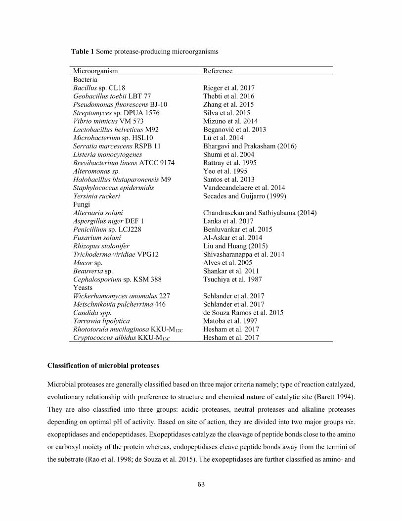
63
Table 1 Some protease-producing microorganisms
Microorganism Reference Bacteria Bacillus sp. CL18 Rieger et al. 2017 Geobacillus toebii LBT 77 Thebti et al. 2016 Pseudomonas fluorescens BJ-10 Zhang et al. 2015 Streptomyces sp. DPUA 1576 Silva et al. 2015 Vibrio mimicus VM 573 Mizuno et al. 2014 Lactobacillus helveticus M92 Beganović et al. 2013 Microbacterium sp. HSL10 Lü et al. 2014 Serratia marcescens RSPB 11 Bhargavi and Prakasham (2016) Listeria monocytogenes Shumi et al. 2004 Brevibacterium linens ATCC 9174 Rattray et al. 1995 Alteromonas sp. Yeo et al. 1995 Halobacillus blutaparonensis M9 Santos et al. 2013 Staphylococcus epidermidis Vandecandelaere et al. 2014 Yersinia ruckeri Secades and Guijarro (1999) Fungi Alternaria solani Chandrasekan and Sathiyabama (2014) Aspergillus niger DEF 1 Lanka et al. 2017 Penicillium sp. LCJ228 Benluvankar et al. 2015 Fusarium solani Al-Askar et al. 2014 Rhizopus stolonifer Liu and Huang (2015) Trichoderma viridiae VPG12 Shivasharanappa et al. 2014 Mucor sp. Alves et al. 2005 Beauveria sp. Shankar et al. 2011 Cephalosporium sp. KSM 388 Tsuchiya et al. 1987 Yeasts Wickerhamomyces anomalus 227 Schlander et al. 2017 Metschnikovia pulcherrima 446 Schlander et al. 2017 Candida spp. de Souza Ramos et al. 2015 Yarrowia lipolytica Matoba et al. 1997 Rhototorula mucilaginosa KKU-M12C Hesham et al. 2017 Cryptococcus albidus KKU-M13C Hesham et al. 2017
Classification of microbial proteases
Microbial proteases are generally classified based on three major criteria namely; type of reaction catalyzed,
evolutionary relationship with preference to structure and chemical nature of catalytic site (Barett 1994).
They are also classified into three groups: acidic proteases, neutral proteases and alkaline proteases
depending on optimal pH of activity. Based on site of action, they are divided into two major groups viz.
exopeptidases and endopeptidases. Exopeptidases catalyze the cleavage of peptide bonds close to the amino
or carboxyl moiety of the protein whereas, endopeptidases cleave peptide bonds away from the termini of
the substrate (Rao et al. 1998; de Souza et al. 2015). The exopeptidases are further classified as amino- and
64
carboxypeptidases, depending on their site of action at the amino or carboxyl terminus of protein. The
endopeptidases are sub-divided into four groups: serine proteases, aspartic proteases, cysteine proteases,
and metalloproteases, based on their catalytic mechanisms (Li et al. 2013).
Serine proteases
Serine proteases are characterized by the presence of a serine residue in the active site, and are generally
recognized by irreversible inhibition by diisopropyl fluorophosphates (DFP), phenylmethylsulfonyl
fluoride (PMSF) and tosyl-L-lysine chloromethyl ketone. In addition, thiol reagents such as p-
chloromercuribenzoate (pCMB) also inhibit some of the serine proteases. These proteases are active at
neutral and alkaline pH, with optimum pH ranging between 7-11. They have broad substrate specificities
and low molecular weight ranging between 18 to 35 kDa (Rao et al. 1998; Ellaiah et al. 2002).
Cysteine proteases
Cysteine proteases, also known as thiol proteases consist of about 20 families, the activity of which is
influenced by a catalytic dyad comprising of cysteine and histidine. They are sensitive to sulphydryl
reagents, including pCMB, iodoacetic acid, iodoacetamide, heavy metals, but are not affected by DFP and
metal-chelating agents. In addition, they are active in the presence of reducing agents such as HCN or
cysteine, dithiothreitol, and ethylene diaminetetraacetic acid (EDTA). Cysteine proteases have neutral pH
optimal or maximally active at acidic pH (Rao et al. 1998).
Metalloproteases
Metalloproteases are the most diverse catalytic form of protease, which depend on divalent metal ions for
their activity (Barett 1995; Vranova et al. 2013). They have optimal pH in the range of 5.0 to 9.0, and are
sensitive to chelating agents, such as EDTA, but are unaffected by serine protease inhibitors or sulphyldryl
agents. Numerous EDTA-inhibited enzymes are activated by ions, such as zinc, calcium, and cobalt. Most
of the bacterial and fungal-derived metalloproteases are zinc-containing enzymes, consisting of one atom
of zinc per molecule of enzyme. The zinc atom is important for enzyme activity. The stability of the protein
structure is enhanced by calcium ions (Ellaiah et al. 2002).
Aspartic proteases
Aspartic proteases, also known as acidic proteases are endopeptidases characterized by the presence of
aspartic acid residues for catalytic activity (Rao et al. 1998). They are commonly found in fungi, but seldom
found in bacteria or protozoa. They are generally divided into two sub-classes viz. pepsin-like enzyme
synthesized by Aspergillus, Penicillium, Rhizopus, and Neurospora, and rennin-like enzymes produced by
65
Endothia and Mucor sp. Most aspartic proteases show maximal activity at low pH (pH 3-4) with isoelectric
points and molecular masses in the range of pH 3.0-4.5 and 30-45 kDa, respectively (Rao et al. 1998;
Srilakshmi et al. 2015). The active-site (aspartic acid residue) is situated within the motif Asp-Xaa-Gly,
where Xaa represents Ser or Thr. Inhibitors such as pepstatin and diazoketone molecules including
diazoacetyl-DL-norleucine methyl ester and 1,2-epoxy-3-(p-nitrophenoxy) propane inhibit aspartic
protease activity in the presence of copper ions (Singh et al. 2016b). They are specific against aromatic or
bulky amino acid residues on both sides of the peptide bond.
Proteases production and influencing factors
Microorganisms generally produce protease extracellularly by submerged fermentation and to a lesser
extent by solid-state fermentation processes during post-exponential or stationary growth phase (Mukherjee
et al. 2008; Reddy et al. 2008). Among the bacterial genera that have been reported for extracellular alkaline
protease production include, Alteromonas, Brevibacterium, Microbacterium, Pseudomonas, Streptomyces,
Vibrio, Bacillus, Xanthomonas (Jisha et al. 2013). However, members of the genus Bacillus have been
reported as main source for large scale production of alkaline proteases (Navaneeth et al. 2009; Queiroga
et al. 2012). Protease production from microorganisms is constitutive or partially inducible in nature, and
the type of substrate utilized in the fermentation medium mostly influences their synthesis. Selection of
appropriate inducible substrate and microorganisms is paramount for the production of desired product
(Beg et al. 2002; Anandan et al. 2007; Bhunia et al. 2012). Under most culture conditions, extracellular
protease production by microorganisms is strongly influenced by media components including variation in
C/N ratio, presence of some readily metabolizable sugars, such as glucose, and metal ions (Beg et al. 2002;
Bhunia et al. 2012). In addition, protease synthesis is also greatly affected by rapidly metabolizable nitrogen
sources, such as amino acids in the fermentation medium (Gupta et al. 2002). The alkaline protease consists
of 15.6% nitrogen; its production is dependent on the presence of both carbon and nitrogen sources in the
culture medium (Kole et al. 1988). Various authors have reported increased yields of protease production
by addition of different carbon and nitrogen sources (Sharma et al. 2017). Sharma et al. (2015) recorded
maximum protease production by a bacterial strain AKS-4 when glucose was used as a carbon source in
the growth media at a concentration of 1% (w/v), resulting in a maximum activity of 59.10 U/ml. In another
study, Sharma et al. (2014) used various carbon sources including glucose, lactose, galactose and starch for
protease production by Bacillus aryabhattai K3. Maximum protease production (622.64 U/ml) was
observed in the presence of lactose (10 g/L).
66
Although complex nitrogen sources are commonly utilized for alkaline protease production, the
requirement for a particular nitrogen supplement differs from one organism to another (Kumar and Takagi
1999; de Souza et al. 2015). In most microorganisms, both organic and inorganic nitrogen sources are
metabolized to produce amino acids, nucleic acids, proteins and other cell wall components (Kumar and
Takagi 1999; Ellaiah et al. 2002). Several authors have employed organic (simple or complex) and
inorganic nitrogen sources for enhancement of protease production. These nitrogen sources have regulatory
effects on protease synthesis. Kumar et al. (2014) studied the effect of organic and inorganic nitrogen
sources on protease production by Marinobacter sp. GA CAS9. Results obtained revealed that organic
nitrogen sources induced higher protease production than inorganic nitrogen sources, with maximum
protease production (249.18 U/ml) recorded in the presence of beef extract. Badhe et al. (2016) studied the
influence of nitrogen sources including ammonium nitrate, ammonium chloride, ammonium sulphate, yeast
extract, potassium nitrate, and sodium nitrate on extracellular protease production by Bacillus subtilis.
Yeast extract was found as the best nitrogen source to stimulate maximum protease production. Urea and
sodium nitrate have been reported as the best organic and inorganic nitrogen sources, respectively for
extracellular protease production by Bacillus licheniformis ATCC 12759 (Akcan 2012).
Increase in the concentration of carbon and nitrogen sources in the fermentation medium suppresses
protease production (Kumar and Takagi 1999; Ellaiah et al. 2002). Inhibition of protease synthesis by
Pseudomonas aeruginosa MCM B-327 in soybean-tryptone media following addition of glucose (95%)
and fructose (60%) has been observed (Zambare et al. 2011). Mehta et al. (2006) recorded a repression in
alkaline protease production due to increase in the concentration of glucose, peptone, yeast extract and
amino acids in the fermentation media.
Several bioprocess parameters including pH, temperature, aeration, inoculum density, agitation speed and
incubation period also influence protease production (Puri et al. 2002; Srividya and Mala 2011; Pathak and
Deshmukh 2012). These parameters are essential to promote the growth of microorganisms for protease
production. Slightly acidic medium pH (6.3-6.5) has been found as optimum for protease production by
Bacillus sp. MIG (Gouda 2006) and Bacillus cereus SIU1 (Singh et al. 2010). Maximum protease
production by Bacillus subtilis NS and Pseudomonas fluorescens was recorded when the initial pH of the
fermentation media was 9.0 (Nisha and Divakaran 2014; Vinoth et al. 2014). Higher initial pH 12.0 for
Bacillus cereus S8 (Lakshmi et al. 2014), 10.5 for Bacillus circulans (Jaswal et al. 2008) and 10.7 for
Bacillus sp. 2-5 (Khosravi-Darani et al. 2008) have also been reported for maximum protease production.
Incubation temperature is a crucial environmental parameter for the production of proteases, since it affects
microbial growth, regulates synthesis and secretion of the enzyme by changing the properties of the cell
wall (Ibrahim et al. 2015b). Optimum temperatures of 30 °C, 37 °C, 40 °C, and 60 °C for protease
67
production by Pseudomonas aeruginosa MCM B-327 (Zambare et al. 2011), Bacillus subtilis AKRS3
(Ravishankar et al. 2012), Bacillus sp. NPST-AK15 (Ibrahim et al. 2015b), and Bacillus polymyxa (Maal
et al. 2009), respectively have been reported.
Statistical approaches for optimization of protease production
Since microbial extracellular protease production is significantly influenced by media components,
optimization of these parameters is imperative in order to maintain a balance between various components
for enhanced protease production (Kumar and Takagi 1999; Chandra et al. 2015). Process optimization has
become a topic of central importance in industrial production processes with special regard to
biotechnology (Reddy et al. 2008). This technique is found suitable for improved productivity of any
product of interest. The conventional one-variable-at-a-time (OVAT) approach involving change of one
independent variable while maintaining all others at a fixed level is the mostly frequently used technique
in biotechnology to obtain maximum cell growth and high yields of desired metabolite. However, this
approach is not only time-consuming and costly for a large number of variables, but also eliminates
combined interactions among the parameters and often leads to misinterpretation of results when
interactions between different components are present (Li et al. 2008). In order to overcome these
challenges, statistical experimental designs such as Plackett-Burman design (PBD), Taguchi methodology,
response surface methodology (RSM) and artificial neural network model are widely used as powerful tools
for optimization processes (Dutta et al. 2004; Oskouie et al. 2007; Chavan et al. 2015) (Table 2).
Plackett-Burman design
Plackett-Burman design is a two-level design used for the selection of significant factors from a huge
number of process parameters, and thus useful in preliminary studies in which the principal objective is to
select variables that can be fixed or eliminated in further optimization processes (Saxena and Singh 2010;
Singh et al. 2017). In this design, there are two types of variables, namely real variables whose concentration
changes during the experiments, and dummy variables whose concentration remains constant during the
experiments. Each variable is represented in two levels, high and low. The use of PBD tremendously
reduces the total number of experiments, since the interaction effects of the variables are not considered
and only those variables that actually affect the production of desired metabolite are screened (Adinarayana
and Ellaiah 2002; Singh et al. 2017). PBD is a reliable approach for evaluation of relative importance of
various variables or medium composition for a specific output (response) (Singh and Tripathi 2008;
Rajeswari et al. 2014). Optimal conditions for maximum extracellular production of alkaline protease by
Bacillus subtilis SHmIIIa has been investigated using PBD (Chandra et al. 2015).
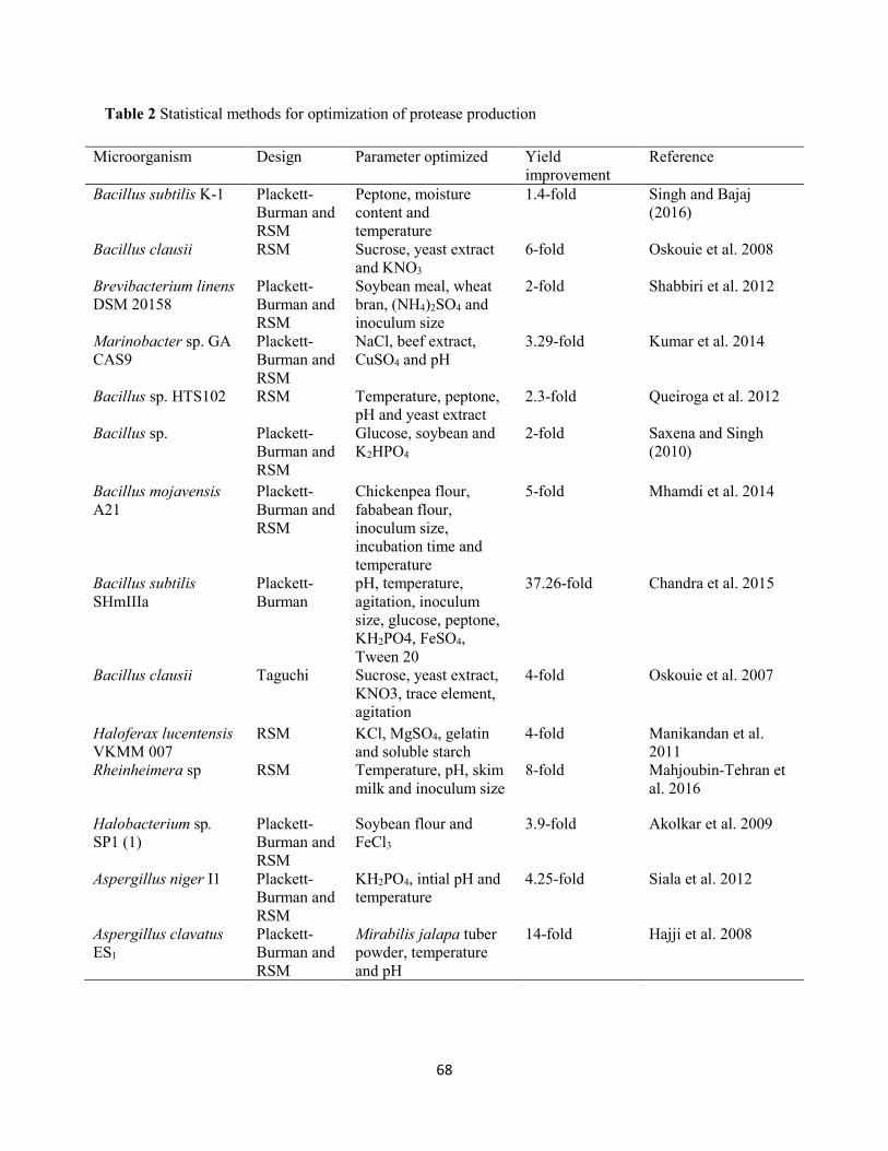
68
Table 2 Statistical methods for optimization of protease production
Microorganism Design Parameter optimized Yield improvement
Reference
Bacillus subtilis K-1 Plackett-Burman and RSM
Peptone, moisture content and temperature
1.4-fold Singh and Bajaj (2016)
Bacillus clausii RSM Sucrose, yeast extract and KNO3
6-fold Oskouie et al. 2008
Brevibacterium linens DSM 20158
Plackett-Burman and RSM
Soybean meal, wheat bran, (NH4)2SO4 and inoculum size
2-fold Shabbiri et al. 2012
Marinobacter sp. GA CAS9
Plackett-Burman and RSM
NaCl, beef extract, CuSO4 and pH
3.29-fold Kumar et al. 2014
Bacillus sp. HTS102 RSM Temperature, peptone, pH and yeast extract
2.3-fold Queiroga et al. 2012
Bacillus sp. Plackett-Burman and RSM
Glucose, soybean and K2HPO4
2-fold Saxena and Singh (2010)
Bacillus mojavensis A21
Plackett-Burman and RSM
Chickenpea flour, fababean flour, inoculum size, incubation time and temperature
5-fold Mhamdi et al. 2014
Bacillus subtilis SHmIIIa
Plackett-Burman
pH, temperature, agitation, inoculum size, glucose, peptone, KH2PO4, FeSO4, Tween 20
37.26-fold Chandra et al. 2015
Bacillus clausii Taguchi Sucrose, yeast extract, KNO3, trace element, agitation
4-fold Oskouie et al. 2007
Haloferax lucentensis VKMM 007
RSM KCl, MgSO4, gelatin and soluble starch
4-fold Manikandan et al. 2011
Rheinheimera sp RSM Temperature, pH, skim milk and inoculum size
8-fold Mahjoubin-Tehran et al. 2016
Halobacterium sp. SP1 (1)
Plackett-Burman and RSM
Soybean flour and FeCl3
3.9-fold Akolkar et al. 2009
Aspergillus niger I1 Plackett-Burman and RSM
KH2PO4, intial pH and temperature
4.25-fold Siala et al. 2012
Aspergillus clavatus ES1
Plackett-Burman and RSM
Mirabilis jalapa tuber powder, temperature and pH
14-fold Hajji et al. 2008
69
In this study, nine process parameters including pH, temperature, agitation speed, inoculum volume,
glucose, peptone, KH2PO4, FeSO4 and Tween 20 at two levels were selected for the design. Out of which,
only six parameters (pH 9.75, agitation speed 225 rpm, inoculum volume 3.5%, glucose 5.5 g/l, peptone
3.5 g/l and KH2PO4) showed significant influence on alkaline protease production. The optimization
process led to 37.26-fold increase in protease production.
Response surface methodology
Response surface methodology is a collection of mathematical and statistical techniques that are useful for
modeling and analysis in applications where response of interest is influenced by several parameters with
the sole purpose of optimizing the response (Oskouie et al. 2008). RSM employs several phases of
optimization, and it can be performed in three basic steps, namely design of experiments for screening of
the parameters followed by the path of steepest ascent/descent, and finally quadratic regression model is
fitted and optimized using canonical regression analysis method (Gupte and Kulkarni 2003; Singh et al.
2017). This method allows building of models for accurate approximation of true response function within
a region around the optimum, using process parameters as independent variables (Puri et al. 2002; Queiroga
et al. 2012). RSM is useful in confirming the effects and interactions of fermentation variables. In addition,
it results in improved productivity, lessens process changeability, gives closer confirmation of the predicted
response to the experimental values and reduces overall costs (Shabbiri et al. 2012). This experimental
design is more useful for optimization of industrial processes and has been applied in various fields
including biomedicine, food and agriculture (Wang et al. 2017). Several fermentation parameters have been
optimized using RSM (Govarthanan et al. 2014; Kandasamy et al. 2016; Singh and Bajaj 2016;
Vijayaraghavan et al. 2017). Experimental designs such as central composite design (CCD), Box-Behnken
design (BBD) and Doehlert design are widely used in RSM to approximate a response function to
experimental data that cannot be described by linear functions (Li et al. 2007).
Central composite design
The CCD is widely employed in RSM for building a second order (quadratic) model for the response
variable without using a complete three factorial experiments (Singh et al. 2017). It consists of a full
factorial or fractional factorial design, a star design in which experimental points are at a distance of α from
its center, and a central point. It has extensively been applied for optimization of protease production (Puri
et al. 2002; Oskouie et al. 2008; Shabbiri et al. 2012; Queiroga et al. 2012; Singh and Bajaj 2016). Statistical
optimization of medium components and culture conditions for alkaline protease production by
Marinobacter sp. GA CAS9 using CCD has been reported (Kumar et al. 2014). Results obtained revealed
that four independent variables including NaCl (60.53 g/l), beef extract (14.73 g/l), CuSO4 (4.73 g/l) and
70
pH (10.7) significantly influenced protease production. The experimental protease production was found to
correspond with the predicted value, an indication of accuracy of the model. Enhancement of protease
production from 298.34 U/ml to 982.68 U/ml was recorded after the optimization process. Saxena and
Singh (2010) employed RSM for the optimization of various bioprocess parameters for protease production
by Bacillus sp. Preliminary screening of bioprocess parameters including glucose, soybean, K2HPO4, NaCl,
MgSO4.7H2O, CaCl2.2H2O, agitation, inoculum size, pH and incubation period for maximum protease
production was carried out using PBD. Variables (glucose, soybean, K2HPO4) that showed significant
effects on protease production were further optimized using CCD. Results obtained showed an
approximately two-fold increase in protease production using basal medium as reference. Singh and Bajaj
(2016) optimized production of thermoalkali-stable protease from Bacillus subtilis K-1 using agro-
industrial waste as a substrate. Results obtained from PBD showed that peptone, moisture content,
temperature, phosphates, and inoculum volume significantly influenced protease production. In addition,
statistical optimization of the selected significant variables, namely peptone, moisture content, and
incubation temperature by CCD resulted in 40% (from initial 728 U/ml to 1020 U/ml) enhancement in
protease production.
Box-Behnken design
Unlike CCD, BBD is independent of quadratic design, and lack an embedded factorial or fractional design
(Ferreira et al. 2007). It requires fewer experiments and a shorter cycle time for a multi-variable
optimization in a three-level (-1, 0 and +1) system (Wang et al. 2017). It is formed by combination of two-
level factorial designs with incomplete block designs. BBD is an attractive, efficient and productive tool
used to enhance the production of microbial metabolites (Govarthanan et al. 2014; Kandasamy et al. 2016;
Wang et al. 2017). Kandasamy et al. (2016) applied RSM-based BBD for the optimization of variables
including pH, incubation time, coffee pulp waste and corncob substrate concentration for enhanced protease
production by Bacillus sp. Maximum yield (920 U/ml) of protease production was achieved after 60 h of
incubation with 3.0 g/l coffee pulp waste, and 2.0 g/l corncob at pH 8.0 and temperature of 37 °C. A 5-fold
increase in protease production by Bacillus mojavensis A21 following optimization of medium components
and culture conditions through BBD has been reported (Mhamdi et al. 2014).
Purification of proteases
Crude proteases are commonly employed for commercial applications, where high enzyme purity is not
required. This makes the application of enzymes cost-effective, since purification into pure form is tedious,
time-consuming and very expensive (Rigo et al. 2008; Mugdha and Usha 2012). The details of downstream
purification techniques are discussed below:
71
Ultrafiltration
Because of the low amounts of enzyme in the cell-free supernatant, excess water is usually removed for the
recovery of the enzyme. This is achieved via membrane separation processes such as ultrafiltration. This
pressure-driven separation process is inexpensive, leads to a slight loss of enzyme activity and used for
purification, concentration and diafiltration or for changing the salt composition (Sullivan et al. 1984;
Kumar and Takagi 1999). However, its major drawbacks include fouling or clogging of membrane resulting
from precipitates formed by the final product.
Precipitation
Precipitation is the most frequently used technique for the separation of enzymes from crude culture
supernatants (Bell et al. 1983). It is carried out by addition of inorganic salt (ammonium sulphate) or organic
solvent (acetone or ethanol) which reduces the solubility of the desired enzymes in an aqueous solution
(Muthulakshmi et al. 2011).
Ion exchange chromatography
Proteases are positively charged biomolecules, and are not bound to anion exchangers (Kumar and Takagi
1999). As a result, cation exchangers form a rational choice for the elution of the bound molecules from the
column by increasing the salt or pH gradient (Gupta et al. 2002). The commonly employed matrices for
ion-exchange chromatography include diethyl amino ethyl and carboxy methyl, which upon binding to the
charged enzyme molecules adsorb the proteins to the matrices. Elution of the adsorbed protein molecule is
achieved by a gradient change in pH or ionic strength of the eluting buffer (Gupta et al. 2002).
Affinity chromatography
The most commonly used adsorbents for protease purification include hydroxyapatite, immobilized N-
benzoyloxycarbonyl phenylalanine agarose, immobilized casein glutamic acid, aprotinin-agarose, and
casein-agarose. However, the ultimate disadvantage of this technique is the high cost of enzyme supports
and the labile nature of some affinity ligands, thus reducing the use at process scale (Kumar and Takagi
1999; Gupta et al. 2002).
Hydrophobic interaction chromatography
Hydrophobic interaction chromatography is based on variation of external hydrophobic amino acid residues
on different proteins, resulting in protein interaction. In aqueous solvent, hydrophobic patches on proteins
preferentially interrelate with other hydrophobic surfaces. These hydrophobic interactions are reinforced
72
by high salt concentrations and higher temperature, and are weakened by the presence of detergents or
miscible organic solvents. The degree of binding of a hydrophobic protein depends on the type and density
of substitution of the matrix, as well as on the nature of buffer conditions (Gupta et al. 2002).
Immobilization of proteases
Enzyme immobilization refers to as physical confinement or localization of enzymes in a certain defined
region of space with retention of their catalytic activities (Mohamad et al. 2015). Immobilization of enzyme
onto appropriate insoluble support is an important tool to fabricate a range of functional properties (Tischer
and Wadekind 1999). Immobilization offers many distinct advantages including possibility of continuous
process, rapid termination of reactions, controlled product formation, and ease of enzyme separation from
reaction mixture (Datta et al. 2013). In addition, insolubilization of enzyme by attachment to a matrix
provides several benefits such as enhanced stability, possible modulation of the catalytic properties,
reduction in the cost of enzymes and enzyme products, and adaptability to various engineering designs
(Asuri et al. 2006; Sheldon 2007; Tian et al. 2009; Hernandez et al. 2011; Saifuddin et al. 2013).
The characteristics of a matrix are crucial in determining the effectiveness of the immobilized enzyme
system (Mohamad et al. 2015). The selection of appropriate support material influences the immobilization
process. Ideal support properties include hydrophilicity, inertness towards enzymes, biocompatibility, non-
toxicity, biodegradability, resistance to microbial attack, resistance to compression and readily affordable
at low cost (Singh 2009). The support materials can be classified as organic and inorganic based on their
chemical components, and can further be sub-divided into natural and synthetic polymers. These include
carboxymethyl cellulose, starch, collagen, modified sepharose, ion exchange resins, active charcoal, silica
(Wu et al. 2012), clay (Kim et al. 2006), aluminium oxide, titanium, diatomaceous earth, hydroxyapatite,
ceramics, celite (Khan et al. 2006; Ansari and Husain 2012; Datta et al. 2013), agarose (Gemenier 1992;
Brena and Viera 2006), treated porous glass (Gorecka and Jastrzebska 2011) and certain polymers (Lee et
al. 2009).
The choice of suitable immobilization technique is paramount for the immobilization process, as it
determines the enzyme activity and characteristics in a particular reaction (Mohamad et al. 2015). Methods
such as adsorption, entrapment, covalent bonding and cross-linking are commonly used for enzyme
immobilization (Huang et al. 2011; Sheldon 2011; Geethanjali and Subash 2013; Park et al. 2013; Sahin et
al. 2015). Immobilization of alkaline protease from Bacillus amyloliquefaciens SP1 by entrapment in
various matrices including alginate, agar and polyacrylamide has been reported (Guleria et al. 2016a). The
immobilized enzyme showed enhanced protease activity and reusability with beads prepared with different
polymers. In addition, Bacillus subtilis M-11 protease immobilized on polysulfone membrane (containing
73
silica gel-3 aminopropyltriethoxysilane) by physical adsorption displayed improved stability and retention
of its activity (77.3%) after ten repeated use (Sahin et al. 2015). Ibrahim et al. (2016) immobilized alkaline
protease from Bacillus sp. NPST-AK15 onto hollow core-mesoporous shell silica nanospheres by covalent
attachment and physical adsorption. The immobilized enzyme recorded significant thermal and organic
solvent stability with a considerable catalytic activity for twelve consecutive batches. Silva et al. (2006)
studied the immobilization of alkaline protease, Esperase by covelent bonding to Eudragit S-100 through
carbodiimide coupling. The immobilized enzyme exhibited a good thermal and storage stability, and
reusability in comparison to the native enzyme.
Characterization of alkaline proteases
Alkaline proteases from different microorganisms are well studied for suitability for various specific
applications based on their properties (Kumar and Takagi 1999). For biotechnological applications,
proteases must possess higher activity and stability under relatively extreme temperature, pH etc. (Sharma
et al. 2017). The essential properties of some microbial alkaline proteases are presented in Table 3 and
discussed in details below:
Effect of pH on activity and stability
A significant level of proteolytic activity over a broad range of pH is required for protease to be employed
for various biotechnological applications (Adinarayana et al. 2003). In general, bacterial alkaline proteases
exhibit high activity at optimum pH range of between 8.0 and 12.0 (Jisha et al. 2013). Optimum pH and
stability of a serine alkaline protease from Aeribacillus pallidus C10 has been reported (Yildirim et al.
2017). The enzyme was found to be active within a broad pH range of 7.0-10.0, with maximum activity
recorded at pH 9.0. The protease retained its activity by more than 70% in the range of pH 6.0-10.5 after 2
h incubation. Alkaline proteases from Bacillus pumilus CBS (Jaouadi et al. 2008), Bacillus strain HUTBS71
(Akel et al. 2009) and Bacillus licheniformis (Sarker et al. 2013) with similar pH stability profiles have
been reported. Ibrahim et al. (2015a) assessed the influence of pH on the activity and stability of protease
produced by Bacillus sp. NPST-AK15. The enzyme was active in a wide pH range (7.0-12.0), with
maximum activity recorded at pH 10.5. The protease was 100% stable at pH 9.0-10.5, retaining 96.6 and
92.3% of its activity at pH 8.0 and 11.0, respectively, and more than 80% of its initial activity retained at
pH 12.0 after 2 h. Protease from Bacillus circulans MTCC 7942 exhibited activity in the range of 8.0-13.0
with optimum activity recorded at pH 10.0. The enzyme maintained its stability in a wide range of pH (7.0-
12.0) for 24 h, retaining 90% activity in the pH range (8.0-12.0) (Patil et al. 2016).
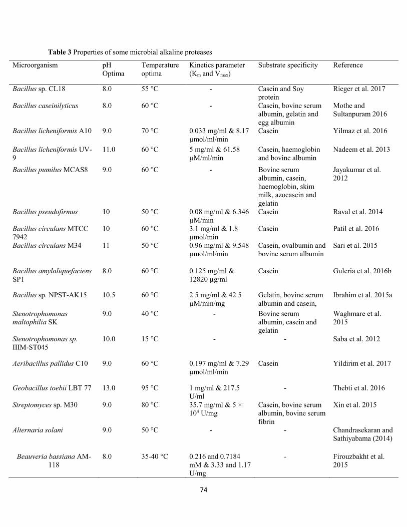
74
Table 3 Properties of some microbial alkaline proteases
Microorganism pH Optima
Temperature optima
Kinetics parameter (Km and Vmax)
Substrate specificity Reference
Bacillus sp. CL18 8.0 55 °C - Casein and Soy protein
Rieger et al. 2017
Bacillus caseinilyticus 8.0 60 °C - Casein, bovine serum albumin, gelatin and egg albumin
Mothe and Sultanpuram 2016
Bacillus licheniformis A10 9.0 70 °C 0.033 mg/ml & 8.17 µmol/ml/min
Casein Yilmaz et al. 2016
Bacillus licheniformis UV-9
11.0 60 °C 5 mg/ml & 61.58 µM/ml/min
Casein, haemoglobin and bovine albumin
Nadeem et al. 2013
Bacillus pumilus MCAS8 9.0 60 °C - Bovine serum albumin, casein, haemoglobin, skim milk, azocasein and gelatin
Jayakumar et al. 2012
Bacillus pseudofirmus 10 50 °C 0.08 mg/ml & 6.346 µM/min
Casein Raval et al. 2014
Bacillus circulans MTCC 7942
10 60 °C 3.1 mg/ml & 1.8 µmol/min
Casein Patil et al. 2016
Bacillus circulans M34 11 50 °C 0.96 mg/ml & 9.548 µmol/ml/min
Casein, ovalbumin and bovine serum albumin
Sari et al. 2015
Bacillus amyloliquefaciens SP1
8.0 60 °C 0.125 mg/ml & 12820 µg/ml
Casein Guleria et al. 2016b
Bacillus sp. NPST-AK15 10.5 60 °C 2.5 mg/ml & 42.5 µM/min/mg
Gelatin, bovine serum albumin and casein,
Ibrahim et al. 2015a
Stenotrophomonas maltophilia SK
9.0 40 °C - Bovine serum albumin, casein and gelatin
Waghmare et al. 2015
Stenotrophomonas sp. IIIM-ST045
10.0 15 °C - - Saba et al. 2012
Aeribacillus pallidus C10 9.0 60 °C 0.197 mg/ml & 7.29 µmol/ml/min
Casein Yildirim et al. 2017
Geobacillus toebii LBT 77 13.0 95 °C 1 mg/ml & 217.5 U/ml
- Thebti et al. 2016
Streptomyces sp. M30 9.0 80 °C 35.7 mg/ml & 5 × 10⁴ U/mg
Casein, bovine serum albumin, bovine serum fibrin
Xin et al. 2015
Alternaria solani 9.0 50 °C - - Chandrasekaran and Sathiyabama (2014)
Beauveria bassiana AM-118
8.0 35-40 °C 0.216 and 0.7184 mM & 3.33 and 1.17 U/mg
- Firouzbakht et al. 2015

75
Similar results have also been reported for proteases from Bacillus tequilensis P15 (Bose et al. 2014),
Bacillus subtilis AP-MSU6 (Maruthiah et al. 2013), Bacillus circulans (Benkiar et al. 2013), Bacillus
lehensis (Joshi and Satyanarayana 2013), and Bacillus alveayuensis CAS 5 (Annamalai et al. 2014) showing
optimal pH in the range of 8.0-12.0. Maximum activity of a serine protease from Bacillus pumilus MCAS8
at pH 9.0 and stability in the range of 7.0-11.0 after 30 min has been observed (Jayakumar et al. 2012).
Remarkably, alkaline serine protease from Bacillus circulans M34 showed maximum activity at optimum
pH of 11.0 and was found to be active over a broad pH ranges (4.0-12.0) (Sari et al. 2014). The enzyme
was stable over a wide pH range, maintaining 97% of its original activity at pH 8.0-11.0 after 1 h.
Effect of temperature on activity and stability
Most of the bacterial alkaline proteases are active and stable at a broad range of temperature, from 50 to 70
°C. The activity of alkaline proteases at broad temperatures and thermostability forms a crucial feature
required for various biotechnological applications. Alkaline proteases from Bacillus sp., Streptomyces sp.
and Thermus sp. are stable at high temperatures; addition of calcium chloride further improves the enzyme
thermostability (Nilegaonkar et al. 2006). In addition, some alkaline proteases possess exceptionally high
thermostability with no decrease in activity at 60-70 °C for up to 3 h (Adinarayana et al. 2003). Ahmetoglu
et al. (2015) investigated the characteristics of protease from Bacillus sp. KG5. The enzyme was found to
be active at 40-45 °C and stable at 50 °C in the presence of 2 mM CaCl2 after 120 min. Thebti et al. (2016)
characterized a haloalkaline protease from Geobacillus toebii LBT 77 newly isolated from Tunisian hot
spring. The enzyme was active between 70 and 100 °C with an optimum activity recorded at 95 °C. The
protease was extremely stable at 90 °C after 180 min. Similar results have also been reported for protease
from Bacillus sp. MLA64 (Lagzian and Asoodeh 2012). This activation and stability at higher temperature
was probably due to the partial thermal inactivation of the protease. Alkaline protease from Bacillus
caseinilyticus was found to be active at 30-60 °C, with maximum activity attained at 60 °C, indicating
thermotolerant nature of the enzyme (Mothe and Sultanpuram 2016). Maximum proteolytic activity of
Bacillus strains HR-08 and KR-8102 isolated from soil of western and northern parts of Iran has been
recorded at 65 and 50 °C, respectively (Moradian et al. 2006). A serine alkaline protease from Bacillus
subtilis DR8806 showed highest activity at 45 °C and was stable up to 70 °C (Farhadian et al. 2015).
Alkaline protease from Bacillus cohnii APT5 has been reported to be active at a broad range of
temperatures, between 30 and 75 °C with maximum activity attained at 50 °C (Tekin et al. 2012). The
enzyme was found to be stable from 40 to 70 °C.
76
Kinetics properties of alkaline proteases
Since enzymes are natural catalysts that accelerate chemical reactions, the speed of any fastidious reaction
being catalyzed by a particular enzyme can only reach a certain maximum value. This is known as
maximum velocity (Vmax) whereas, Michaelis-Menten constant (Km) is the concentration of substrate at
which half of the maximal velocity was attained (Ahmed et al. 2011). The relationship between the rate of
reaction and concentration of substrate depends on the affinity of the enzyme for its substrate. This is usually
expressed as the Km (Ahmed et al. 2011). An enzyme with low Km has a greater affinity for its substrate.
Both Km and Vmax are important for developing an enzyme-based process. Knowledge of such parameters
is essential for assessing the commercial applications of protease under different conditions (Gupta et al.
2002; Sugumaran et al. 2012). Various substrates including casein, azocasein etc. are employed for
determining the kinetic properties of alkaline proteases. Different Km and Vmax values have reported for
various alkaline proteases. The Km and Vmax values of alkaline protease from Bacillus licheniformis A10
were determined to be 0.033 mg/ml and 8.17 µmol/ml/min, respectively in the presence of casein (Yilmaz
et al. 2016). This Km value was found to be lower when compared to that of alkaline proteases from Bacillus
licheniformis UV-9 (Nadeem et al. 2013), Bacillus circulans (Rao et al. 2009) and Bacillus sp. (Jain et al.
2012), suggesting a high affinity of the enzyme for the substrate. In another study, Km and Vmax values of
0.626 mM and 0.0523 mM/min, respectively were recorded for protease from Bacillus licheniformis BBRC
100053 using casein (Nejad et al. 2014). Alkaline protease from Bacillus amyloliquefaciens SP1showed
Km and Vmax values of 0.125 mg/ml and 12820 µg/min, respectively in the presence of casein, indicating
high affinity and efficient catalytic activity of the enzyme (Guleria et al. 2016b).
Potential applications of alkaline proteases
Alkaline proteases are robust enzymes with significant biotechnological applications in detergents, leather
processing, silver recovery, medical, food processing, feeds, and chemical industries, as well as in several
bioremediation processes, contributing to the formation of high value-added products (Jisha et al. 2013;
Aruna et al. 2014). The various applications of alkaline proteases are discussed below:
Detergent industry
The detergent industry forms the largest industrial application of enzymes, accounting for 25-30% of the
total worldwide markets of enzymes and expected to grow faster at a compound annual growth rate of about
11.5% from 2015 to 2020 (Global Market for Enzymes in Industrial Applications 2014). Microbial alkaline
proteases are dominant in commercial applications with a substantial share of market utilized in laundry
detergent (Kumar and Takagi 1999). They are used as additives in detergent formulations for the removal
77
of proteinaceous stains resulting from food, blood, and other body secretions, as well as to improve washing
performance in domestic laundry and cleaning of contact lenses or dentures (Rao et al. 1998; Grbavčić et
al. 2011; Baweja et al. 2016). The use of alkaline proteases in detergent products offers enormous
advantages since these products contain less bleaching agents and phosphates. Thus, rendering beneficial
effects on public and environmental health (Olsen and Falholt 1998; Novozyme 2013). Generally, an ideal
alkaline protease used as detergent additives should have a long shelf life, as well as high activity and
stability over a wide range of pH and temperature. In addition, the enzymes should be efficient at low
amounts and compatible with various detergent components along with chelating and oxidizing agents (Rao
et al. 1998; Kumar and Takagi 1999). This is noteworthy of alkaline proteases from Bacillus cereus,
Bacillus pumilus CBS, Bacillus licheniformis, Bacillus brevis, and Bacillus subtilis AG-1 reported to
exhibit robust detergent compatibility in the presence of calcium chloride and glycine used as stabilizers
(de Nascimento and Martins 2006; Ghafoor and Hasnain 2009; Abou-Elela et al. 2011; Bezawada et al.
2011; Jaouadi et al. 2011).
Leather industry
Leather processing involves series of stages including curing, soaking, liming, dehairing, bating, picking,
degreasing and tanning (Mojsov 2011). Conventional approaches of leather processing involving the use
of hazardous chemicals such as sodium sulfide, lime and amines generate severe health hazards and
environmental pollution (Choudhary et al. 2004). As a result, the use of biodegradable enzymes as
substitutes to chemicals has proved successful in enhancing leather quality and reducing environmental
pollution (Rao et al. 1998; Zambare et al. 2011; Adrio and Demain 2014). Enzymatic dehairing processes
are attractive in preserving the hair and contribute to fall in organic load discharged into effluent. In
addition, it minimizes or eliminates the dependence on toxic chemicals (de Souza and Gutterres 2012). Due
to their elastolytic and keratinolytic activity, alkaline proteases are employed for selective breakdown of
non-collagenous constituents of the skin and for elimination of non-fibrillar proteins during soaking, in
bating, thus producing soft, supple and pliable leather (Singh et al. 2016). Furthermore, microbial alkaline
proteases are employed for quick absorption of water thus, reducing soaking time (Jaouadi et al. 2013).
Alkaline proteases from Bacillus sp. with keratinolytic activity have been reported for dehairing properties
(Arunachalam and Sarita 2009; Vijayaraghavan et al. 2014; Kshetri and Ningthoujam 2016; Rieger et al.
2017).
Food industry
In food industry, alkaline proteases are usually used for various purposes including cheesemaking, baking,
preparation of soya hydrolysates, meat tenderization etc. The catalytic function of proteases is used in the
78
preparation of high nutritional value protein hydrolysate, used as components of dietetic and health
products; in infant formulae and clinical nutritional supplements, and as flavoring agents (Gupta et al. 2002;
Ward 2011). However, the bitter taste of protein hydrolysate formed a crucial barrier for use in food and
health care products. Therefore, proteases (carboxypeptidases A) have a high specificity for debittering
protein hydrolysates. A key application of protease in dairy industry is in the cheese manufacturing, where
the primary role of the enzymes is to hydrolyze specific peptide to generate casein and macropeptides (Rao
et al. 1998; Pai 2003; Qureshi et al. 2015). In addition, proteases play a significant role in meat
tenderization, particularly of beef, since they possess the potential to hydrolyze connective tissue proteins
as well as muscle fiber proteins (Kumar and Takagi 1999). Endo- and exoproteinases are used in baking
industry to modify wheat gluten. The addition of bacterial proteases reduces the mixing time, improves
extensibility and strength of dough and results in enhanced loaf volume (Rao et al. 1998). Alkaline proteases
are employed in processing of soy sauce and soy products. Proteases are also used in the enzymatic
synthesis of aspartame (sweetening agent).
Medicine
A broad diversity and specificity of proteases are employed for the development of various therapeutic
agents. For instance, elastoterase from Bacillus subtilis 316M immobilized on a bandage has been used for
the treatment of burns, purulent wounds, carbuncles, furuncles, and deep abscesses (Kudrya and Simonenko
1994). In addition, alkaline-fibrinolytic protease has been used as a thrombolytic agent (Kim et al. 1996).
Serratiopeptidase, a protease produced by Serratia sp. is the most effective protease for treatment of acute
and chronic inflammation (Vaisar et al. 2007). Collagenases with alkaline protease activity are used for the
preparation of slow-release dosage forms as well as in wound healing, treatment of sciatica in herniated
intervertebral discs, treatment of retained placenta, and as a pretreatment for enhancing adenovirus-
mediated cancer gene therapy (Watanabe 2004). The cytotoxic nature of several proteases allows the
enzymes to be used as efficient antimicrobial agents for clinical purposes (Siritapetawee et al. 2012).
Waste treatment
Alkaline proteases are employed in the treatment of wastes from various food processing industries and
household activities. These enzymes solubilize proteinaceous wastes via a multistep process for the
recovery of liquid concentrates or dry solids of nutritional value for fish or livestock (Karam and Nicell
1997; Karigar and Rao 2011; Pandey et al. 2011). This is achieved by initial adsorption of the enzyme on
the solid substrate followed by cleavage of polypeptide chain that are loosely bound to the surface.
Thereafter, the solubilization of the more compact core occurs at a slower rate depending on the diffusion
of the enzyme surface active sites and core particles (Venugopal et al. 1989). Enzymatic degradation of
79
wastes using alkaline proteases with keratinolytic activity is an attractive method. Among microbial sp.,
some members of the genus Bacillus are reported as keratinase producers for feather degradation (Kojima
et al. 2006; Cortezi et al. 2008; Ni et al. 2011). Enzymatic treatment of waste feather from poultry
slaughterhouses using alkaline protease produced by Bacillus subtilis has been reported (Dalev 1994).
Pretreatment with NaOH, mechanical disintegration, and enzymatic hydrolysis resulted in complete
solubilization of the feathers, releasing a heavy, grayish powder with high protein content which could be
used as additive in feeds, fertilizers etc. In addition, alkaline proteases with keratinolytic activity are used
for degradation of waste material in household refuse, and as a depilatory agent for the removal of hair in
bathtub drains, which caused unpleasant odors (Mukhopadhyay and Chandra 1992; Takami et al. 1992).
Conclusion and future perspectives
Proteases represent one of the largest groups of industrial enzymes, constituting more than 65% of the total
worldwide sale of enzymes. They are ubiquitous in nature, found in a wide array of plants, animals, and
microorganisms. However, microbial proteases are recognized as an excellent source of enzymes due to
their broad biochemical diversity, rapid growth, limited space requirement for cultivation, and susceptibility
to genetic manipulation. Microbial alkaline proteases play a vital role in several industries, mainly food,
detergent, leather and pharmaceutical industries as well as in bioremediation processes. In view of the
growing and diverse potential applications of these proteases, further discovery and engineering of novel
enzymes with robust catalytic activity will be ultimate developments in the near future through newer
methods such as in vitro evolutionary changes of protein primary structures.
Conflict of interest
The authors declare that there is no conflict of interest in the publication of this article
80
References
(2014) Global markets for enzymes in industrial applications. www.bccresearch.com.
http://www.bccresearch.com/pressroom/bio/global-market-industrial-enzymes-reach-nearly-$7.1-
billion-2018. Accessed on 17 October, 2017
(2015) Industrial enzymes market by type (carbohydrases, proteases, non-starch polysaccharides & others),
application (food & beverage, cleaning agents, animal feed & others), brands & by region-global
trends and forecasts to 2020. www.bccresearch.com.http://www.marketsnadmarkets.com/Market-
Reports/industrial-enzymes-market-237327836.html. Accessed on 17 October, 2017
Abou-Elela GM, Ibrahim H, Hassan S, Abd-Elnaby H, El-Toukhy N (2011) Alkaline protease production
by alkaliphilic marine bacteria isolated from Marsa-Matrouh (Egypt) with special emphasis on
Bacillus cereus purified protease. Afr J Biotechnol 10: 4631-4642
Adinarayana K, Ellaiah P (2002) Response surface optimization of the critical medium components for the
production of alkaline protease by a newly isolated Bacillus sp. J Pharm Pharm Sci 5: 272-278
Adinarayana K, Ellaiah P, Prasad DS (2003) Purification and partial characterization of thermostable serine
alkaline protease from a newly isolated Bacillus subtilis PE-11. AAPS Pharm Sci Tech 4: 440-448
Adrio JL, Demain AL (2014) Microbial enzymes: tools for biotechnological processes. Biomolecules 4:
117-139
Ahmed I, Zia MA, Iqbal HMN (2011) Purification and kinetic parameters characterization of an alkaline
protease produced from Bacillus subtilis through submerged fermentation technique. World Appl
Sci J 12: 751-757
Ahmetoglu N, Bekler FM, Acer O, Guven RG, Guven K (2015) Production, purification and
characterization of thermostable metallo-protease from newly isolated Bacillus sp. KG5. EurAsian
J Biosci 9: 1-11
Akcan N (2012) Production of extracellular protease in submerged fermentation by Bacillus licheniformis
ATCC 12759. Afr J Biotechnol 11: 1729-1735
Akel H, Al-Quadan F, Yousef TK (2009) Characterization of a purified thermostable protease from
hyperthermophilic Bacillus strain HUTBS71. Eur J Sci Res 31: 280-288
81
Akolkar A, Bharambe N, Trivedi S, Desai A (2009) Statistical optimization of medium components for
extracellular protease production by an extreme haloarchaeon, Halobacterium sp. SP1 (1). Lett
Appl Microbiol 48: 77-83
Al-Askar AA, AbdulKhair WM, Rashad YM (2014) Production, purification and optimization of protease
by Fusarium solani under solid fermentation and isolation of protease inhibitor protein from Rumex
vesicarius L. J Pure Appl Microbiol 8: 239-250
Aldridge S (2013) Industry backs biocatalysis for greener manufacturing. Nat Biotechnol 31: 95
Alves MH, Campos-Takaki GM, Okada K, Pessoa IHF, Milanez AI (2005) Detection of extracellular
protease in Mucor species. Rev Iberoam Micol 22: 114-117
Anandan D, Marmer WN, Dudley RL (2007) Isolation, characterization and optimization of culture
parameters for production of an alkaline protease isolated from Aspergillus tamari. Ind Microbiol
Biotechnol 34: 339-347
Annamalai N, Rajeswari MV, Balasubramanian T (2014) Extraction, purification and characterization of
thermostable and halostable alkaline protease from Bacillus alveayuensis CAS 5 using marine
wastes. Food Bioprod Process 92: 335-342
Ansari SA, Husain Q (2012) Lactose hydrolysis from milk/whey in batch and continuous processes by
concanavalin A-celite 545 immobilized Aspergillus oryzae β-galactosidase. Food Bioprod Process
90: 351-359
Aruna K, Shah J, Birmole R (2014) Production and partial characterization of alkaline protease from
Bacillus tequilensis strains csgab0139 isolated from spoilt cottage cheese. Int J Appl Biol Pharm
Technol 5: 201-221
Arunachalam C, Sarita K (2009) Protease enzyme: an eco-friendly alternative for leather industry. Indian J
Sci Technol 2: 29-32
Asuri P, Karajanagi SS, Sellitto E, Kim DY, Kane RS, Dordick JS (2006) Water-soluble carbon nanotube-
enzyme conjugates as functional biocatalytic formulations. Biotechnol Bioeng 95: 804-811
Badhe P, Joshi M, Adivarekar R (2016) Optimized production of extracellular proteases by Bacillus subtilis
from degraded abattoir waste. J Biosci Biotechnol 5: 29-36
82
Barett (1995) Proteolytic enzymes: Proteolytic enzymes: aspartic and metallopeptidases. Methods Enzymol
248: 183
Barett AJ (1994) Proteolytic enzymes: serine and cysteine peptidases. Methods Enzymol 244: 1-15
Baweja M, Tiwari R, Singh PK, Nain L, Shukla P (2016) An alkaline protease from Bacillus pumilus MP
27: functional analysis of its binding model toward its applications as detergent additive. Front
Microbiol 7: 1195
Beg QK, Saxena RK, Gupta R (2002) De-repression and subsequent induction of protease synthesis by
Bacillus mojavensis under fed-batch operations. Proc Biochem 37: 1103-1109
Beganović J, Kos B, Pavunc AL, Uroić K, Džidara P, Šuškovic J (2013) Proteolytic activity of probiotic
strain Lactobacillus helveticus M92. Anaerobe 20: 58-64
Bell DJ, Hoare M, Dunnill P (1983) The formation of protein precipitates and their centrifugal recovery.
Adv Biochem Eng Biotechnol 26: 1-72
Benkiar A, Nadia ZJ, Badis A, Rebzani F, Soraya BT, Rekik H, Naili B, Ferradji FZ, Bejar S, Jaouadi B
(2013) Biochemical and molecular characterization of a thermos- and detergent-stable alkaline
serine keratinolytic protease from Bacillus circulans strain DZ100 for detergent formulations and
feather-biodegradation process. Int Biodeter Biodegr 83: 129-138
Benluvankar V, Jebapriya GR, Gnanadoss JJ (2015) Protease production by Penicillium sp. LCJ228 under
solid state fermentation using groundnut oilcake as substrate. Int J Life Sci Pharm Res 5: 2250-
0480
Bezawada J, Yan S, John RP, Tyagi RD, Surampalli RY (2011) Recovery of Bacillus licheniformis alkaline
protease from supernatant of fermented wastewater sludge using ultrafiltration and its
characterization. Biotechnol Res Int 2011: 1-11
Bhargavi PL, Prakasham RS (2016) Enhanced fibrinolytic protease production by Serratia marcescens
RSPB11 through Plackett-Burman and response surface methodological approaches. J Appl Biol
Biotechnol 4: 006-014
Bhunia B, Basak B, Dey A (2012) A review on production of serine alkaline protease by Bacillus spp. J
Biochem Tech 3: 448-457
83
Bose A, Chawdhary V, Keharia H, Subramanian RB (2014) Production and characterization of a solvent-
tolerant protease from a novel marine isolate Bacillus tequilensis P15. Ann Microbiol 64: 343-354
Brena BM, Viera BF (2006) Immobilization of enzymes. In: Guisan JM, editor. Immobilization of enzymes
and cells. 2nd ed. New Jersey (NJ): Humana Press Inc. pp. 123-124
Chandra SJ, Pallavi P, Anuradha BS, Reddy SR (2015) Optimization of bioprocess for enhanced production
of alkaline protease by a Bacillus subtilis SHmIIIa through Blacket-Burman design. Afr J Microbiol
Res 9: 1738-1744
Chandrasekaran M, Sathiyabama M (2014) Production, partial purification and characterization of protease
from a phytopathogenic fungi Alternaria solani (Ell. And Mart. Sorauer). J Basic Microbiol 54:
763-774
Chavan RS, Avhad DN, Rathod VK (2015) Optimization of aqueous two-phase extraction of protease
produced from Bacillus licheniformis NCIM 2042 using response surface methodology. Sep Sci
Technol 50: 45-55
Choudhary RB, Jana AK, Jha MK (2004) Enzyme technology applications in leather processing. Ind J
Chem Technol 11: 659-671
Cortezi M, Contiero J, Lima CJB, Lovaglio RB, Monti R (2008) Characterization of a feather degrading by
Bacillus amyloliquefaciens protease: a new strain. World J Agric Sci 4: 648-656
Dalev PG (1994) Utilisation of waste feathers from poultry slaughter for production of a protein
concentrate. Bioresour Technol 48: 265-267
Datta S, Rene CL, Rajaram YRS (2013) Enzyme immobilization: an overview on techniques and support
materials. 3 Biotech 3: 1-9
de Souza FR, Gutterres M (2012) Application of enzymes in leather processing: a comparison between
chemical and coenzymatic processes. Braz J Chem Eng 29: 471-481
de Souza PM, Bittencourt MLA, Caprara CC, de Freitas M, de Almeida RPC, Silveira D, Fonseca YM,
Filho EXF, Pessoa AJ, Magalhães PO (2015) A biotechnology perspective of fungal proteases.
Braz J Microbiol 46: 337-346
84
de Souza Ramos L, Barbedo LS, Braga-Silva LA, dos Santos ALS, Pinto MR, da Graça Sgarbi DB (2015)
Protease and phospholipase activities of Candida spp. isolated from cutaneous candidiasis. Rev
Iberoam Micol 32: 122-125
Dutta JR, Dutta PK, Banerjee R (2004) Optimization of culture parameters for extracellular protease
production from a newly isolated Pseudomonas sp. using response surface and artificial neural
network models. Proc Biochem 39: 2193-2198
Ellaiah P, Srinivasulu B, Adinarayana K (2002) A review on microbial alkaline proteases. J Sci Ind Res 61:
690-704
Farhadian S, Asoodeh A, Lagzian M (2015) Purification, biochemical characterization and structural
modeling of a potential htrA-like serine protease from Bacillus subtilis DR8806. J Mol Catal B
Enzym 115: 51-58
Ferreira SLC, Bruns RE, Ferreira HS, Matos GD, David JM, Brandão GC, da Silva EGP, Portugal LA, dos
Reis PS, Souza AS, dos Santos WNL (2007) Box-Behnken design: an alternative for the
optimization of analytical methods. Anal Chim Acta 597: 179-186
Firouzbakht H, Zibaee A, Hoda H, Sohani MM (2015) Purification and characterization of the cuticle-
degrading proteases produced by an isolate of Beauveria bassiana using the cuticle of the predatory
bug, Andrallus spinidens Fabricius (Hemiptera: Pentatomidae). J Plant Prot Res 55: 2
Geethanjali S, Subash A (2013) Optimization and immobilization of purified Labeo rohita visceral protease
by entrapment method. Enzyme Res 2013: 1-7
Gemenier P (1992) Materials for enzyme engineering. In: Gemeiner P, editor. Enzyme engineering. 1st ed.
New York (NY): Ellis Horwood, pp. 13-119
Ghafoor A, Hasnain S (2009) Characteristics of an extracellular protease isolated from Bacillus subtilis
AG-1 and its performance in relation to detergent components. Ann Microbiol 59: 559-563
Gonzalez-Rabade N, Badillo-Corona JA, Aranda-Barradas JS, Oliver-Salvador Mdel C (2011) Production
of plant proteases in vivo and in vitro- a review. Biotechnol. Adv. 29: 983-996
Górecka E, Jastrzębska M (2011) Immobilization techniques and biopolymer carriers. Biotechnol Food Sci
75: 65-86
85
Gouda MK (2006) Optimization and purification of alkaline proteases produced by marine Bacillus sp.
MIG newly isolated from eastern harbor of Alexandria. Pol J Microbiol 55: 119-126
Govarthanan M, Park S-H, Kim J-W, Lee K-J, Cho M, Kamala-Kannan S, Oh B-T (2014) Statistical
optimization of alkaline protease production from brackish environment Bacillus sp. SKK11 by
SSF using horse gram husk. Prep Biochem Biotechnol 44: 119-131
Grbavčić S, Bézbradica D, Izrael-Živković L, Avramović N, Milosavić N, Karadzić I, Knežević-Jugović Z
(2011) Production of lipase and protease from an indigenous Pseudomonas aeruginosa strain and
their evaluation as detergent additives: compatibility study with detergent ingredients and washing
performance. Bioresour Technol 102: 11226-11233
Guleria S, Walia A, Chauhan A, Shirkot CK (2016a) Immobilization of Bacillus amyloliquefaciens SP1
and its alkaline protease in various matrices for effective hydrolysis of casein. 3 Biotech 6: 208
Guleria S, Walia A, Chauhan A, Shirkot CK (2016b) Purification and characterization of detergent stable
alkaline protease from Bacillus amyloliquefaciens SP1 isolated from apple rhizosphere. J Basic
Microbiol. 56: 138-152
Gupta R, Beg QK, Lorenz P (2002) Bacterial alkaline proteases: molecular approaches and industrial
applications. Appl Microbiol Biotechnol 59: 15-32
Gupte M, Kulkarni P (2003) A study of antifungal antibiotic production by Thermomonospora sp. MTCC
3340 using full factorial design. J Chem Technol Biotechnol 78: 605-610
Gurung N, Ray S, Bose S, Rai V (2013) A broader view: microbial enzymes and their relevance in
industries, medicine and beyond. Biomed Res Int 2013: 1-18
Haddar A, Bougatef A, Agrebi R, Sellami-Kamoun A, Nasri M (2009) A novel surfactant-stable alkaline
serine-protease from a newly isolated Bacillus mojavensis A21: purification and characterization.
Proc Biochem 44: 29-35
Hajji M, Rebai A, Gharsallah N, Nasri M (2008) Optimization of alkaline protease production by
Aspergillus clavatus ES1 in Mirabilis jalapa tuber powder using statistical experimental design.
Appl Microbiol Biotechnol 79: 915-923
Hernandez K, Fernandez-Lafuente R (2011) Control of protein immobilization: coupling immobilization
and side-directed mutagenesis to improve biocatalyst or biosensor performance. Enzyme Microb
Technol 48: 107-122
86
Hesham AEL, Alrumman SA, Al-Dayel MA, Salah HA (2017) Screening and genetic identification of
acidic and neutral protease-producing yeasts strains by 26S rRNA gene sequencing. Cytol Genet
51: 221-229
Huang XJ, Chen PC, Huang F, Ou Y, Chen MR, Xu ZK (2011) Immobilization of Candida rugosa lipase
on electrospun cellulose nanofiber membrane. J Mol Catal B-Enzym 70: 95-100
Ibrahim ASS, Al-Salamah AA, El-Badawi YB, El-Tayeb MA, Antranikian G (2015a) Detergent-, solvent-
and salt-compatible thermoactive alkaline serine protease from halotolerant alkaliphilic Bacillus
sp. NPST-AK15: purification and characterization. Extremophiles 19: 961-971
Ibrahim ASS, Al-Salamah AA, Elbadawi YB, El-Tayeb MA, Ibrahim SSS (2015b) Production of
extracellular alkaline protease by new halotolerant alkaliphilic Bacillus sp. NPST-AK15 isolated
from hyper saline soda lakes. Electr J Biotechnol 18: 236-243
Ibrahim ASS, Al-Salamah AA, El-Toni AM, Almaary KS, El-Tayeb MA, Elbadawi YB, Antranikian G
(2016) Enhancement of alkaline protease activity and stability via covalent immobilization onto
hollow core-mesoporous shell silica nanospheres. Int J Mol Sci 17: 184
Jain D, Pancha I, Mishra SK, Shrivastav A, Mishra S (2012) Purification and characterization of
haloalkaline thermoactive, solvent stable and SDS-induced protease from Bacillus sp: a potential
additive for laundry detergents. Bioresour Technol 115: 228-236
Jaouadi B, Abdelmalek B, Jaouadib NZ, Bejar S (2011) The bioengineering and industrial applications of
bacterial alkaline proteases: the case of SAPB and KERAB. Prog. Mol. Environ. Bioeng. –from
analysis and modeling to technology applications, Angelo Carpi (ed.) ISBN: 978-953-307-268-5
In Tech
Jaouadi B, Ellouz-Chaabouni S, Rhimi M, Bejar S (2008) Biochemical and molecular characterization of a
detergent-stable serine alkaline protease from Bacillus pumilus CBS with high catalytic efficiency.
Biochimie 90: 1291-1305
Jaouadi NZ, Rekik H, Badis A, Trabelsi S, Belhoul M, Yahiaoui AB, Aicha HB, Toumi A, Bejar S, Jaouadi
B (2013) Biochemical and molecular characterization of a serine keratinase from Brevibacillus
brevis US575 with promising keratin-biodegradation and hide-dehairing activities. PLoS ONE 8:
e76722
87
Jaswal RK, Kocher GS, Virk MS (2008) Production of alkaline protease by Bacillus circulans using
agricultural residues: a statistical approach. Indian J Biotechnol 7: 356-360
Jayakumar R, Jayashree S, Annapurna B, Seshadri S (2012) Characterization of thermostable serine
alkaline protease from an alkaliphilic strain Bacillus pumilus MCAS8 and its application. Appl
Biochem Biotechnol 168: 1849-1866
Jisha VN, Smitha RB, Pradeep S, Sreedevi S, Unni KN, Sajith S, Priji P, Josh MS (2013) Versatility of
microbial proteases. Adv Enzyme Res 1: 39-51
Joshi S, Satyanarayana T (2013) Characteristics and applications of a recombinant alkaline serine protease
from a novel bacterium Bacillus lehensis. Bioresour Technol 131: 76-85
Kandasamy S, Muthusamy G, Balakrishnan S, Duraisamy S, Thangasamy S, Seralathan K-K, Chinnappan
S (2016) Optimization of protease production from surface-modified coffee pulp waste and
corncobs using Bacillus sp. by SSF. 3 Biotech 6: 167
Karam J, Nicell JA (1997) Potential applications of enzymes in waste treatment. J Chem Tech Biotechnol
69: 141-153
Karigar CS, Rao SS (2011) Role of microbial enzymes in the bioremediation of pollutants: a review.
Enzyme Res 2011: 1-11
Khan AA, Akhtar S, Hussain Q (2006) Direct immobilization of polyphenol oxidases on celite 545 from
ammonium sulphate fractionated proteins of potato (Solanum tuberosum). J Mol Catal B, 40: 58-
63
Khosravi-Darani K, Falahatpishe HR, Jalali M (2008) Alkaline protease production on date waste by an
alkalophilic Bacillus sp. 2-5 isolated from soil. Afr J Biotechnol 7: 1536-1542
Kim JB, Grate JW, Wang P (2006) Nanostructures for enzyme stabilization. Chem Eng Sci 61: 1017-1026
Kim W, Choi K, Kim Y, Park H, Choi J, Lee Y, Oh H, Kwon I, Lee S (1996) Purification and
characterization of a fibrinolytic enzyme produced by Bacillus sp. strain CK 11-4 screened from
Chungkook-Jang. Appl Environ Microbiol 62: 2482-2488
Kojima M, kanai M, Tominaga M, Kitazume S, Inoue A, Horikoshi K (2006) Isolation and characterization
of a feather-degrading enzyme from Bacillus pseudofirmus FA30-01. Extremophiles 10: 229-235
88
Kole MM, Draper I, Gerson DF (1988) Production of protease by Bacillus subtilis using simultaneous
control of glucose and ammonium concentrations. J Chem Technol Biotechnol 41: 197-206
Kshetri P, Ningthoujam DS (2016) Keratinolytic activities of alkaliphilic Bacillus sp. MBRL 575 from a
novel habitat, limestone deposit site in Manipur, India. SpringerPlus 5: 595
Kudrya VA, Simonenko IA (1994) Alkaline serine proteinase and lectin isolation from the culture fluid of
Bacillus subtilis. Appl Microbiol Biotechnol 41: 505-509
Kumar CG, Takagi H (1999) Microbial alkaline proteases: from a bioindustrial viewpoint. Biotechnol Adv
17: 561-594
Kumar RS, Ananthan G, Prabhu AS (2014) Optimization of medium composition for alkaline protease
production by Marinobacter sp. GA CAS9 using response surface methodology- a statistical
approach. Biocatal Agric Biotechnol 3: 191-197
Lagzian M, Asoodeh A (2012) An extremely thermotolerant, alkaliphilic subtilisin-like protease from
hyperthermophilic Bacillus sp. MLA64. Int J Biol Macromolec 51: 960-967
Lakshmi BKM, Hemalatha KPJ (2014) Response surface optimization of medium composition for alkaline
protease production by Bacillus cereus strain S8. Int J Pure Appl Biosci 3: 216-223
Lanka S, Anjali CH, Pydipalli M (2017) Enhanced production of alkaline protease by Aspergillus niger
DEF 1 isolated from dairy form effluent and determination of its fibrinolytic ability. Afr J Microbiol
Res 11: 440-449
Lee CH, Lin TS, Mou CY (2009) Mesoporous materials for encapsulating enzymes. Nanotoday 4: 165-179
Li Q, Yi L, Marek P, Iverson BL (2013) Commercial proteases: present and future. FEBS Lett 587: 1155-
1163
Li X, Xu T, Ma X, Guo K, Kai L, Zhao Y, Jia X, Mad Y (2008) Optimization of culture conditions for
production of cis-epoxysuccinic acid hydrolase using response surface methodology. Bioresour
Technol 99: 5391-5396
Li Y, Liu Z, Cui F, Liu Z, Zhao H (2007) Application of Plackett-Burman design and Doehlert design to
evaluate nutritional requirements for xylanase production by Alternaria mali ND-16. Appl
Microbiol Biotechnol 77: 285-291
89
Liu N, Huang L (2015) Partial characterization of an acidic protease from Rhizopus stolonifer RN-11. Open
Biotechnol J 9: 199-203
Lü J, Wu X, Jiang Y, Cai X, Huang L, Yang Y, Wang H, Zeng A, Li A (2014) An extremophile
Microbacterium strain and its protease production under alkaline conditions. J Basic Microbiol 54:
378-385
Ma C, Ni X, Chi Z, Ma L, Gao L (2007) Purification and characterization of an alkaline protease from the
marine yeast Aureobasidium pullulans for bioactive peptide production from different sources. Mar
Biotechnol 9: 343-351
Maal KB, Emtiazi G, Nahvi I (2009) Production of alkaline protease by Bacillus cereus and Bacillus
polymyxa in new industrial culture medium and its immobilization. Afr J Microbiol Res 3: 491-497
Mahjoubin-Tehran M, Shahnavaz B, Ghazi-Birjandi R, Mashreghi M, Fooladi J (2016) Optimization of
protease production by psychrotrophic Rheinheimera sp. with response surface methodology. Appl
Food Biotechnol 3: 236-245
Manikandan M, Kannan V, Velikonja BH, Pašic L (2011) Optimization of growth medium for protease
production by Haloferax lucentensis VKMM 007 by response surface methodology. Braz J
Microbiol 42: 818-824
Maruthiah T, Esakkiraj P, Prabakaran G, Palavesam A, Immanuel G (2013) Purification and
characterization of moderately halophilic alkaline serine protease from marine Bacillus subtilis AP-
MSU 6. Biocatal Agric Biotechnol 2: 116-119
Matoba S, Morano KA, Klionsky DJ, Kim K, Ogrydziak DM (1997) Dipeptidyl aminopeptidase processing
and biosynthesis of alkaline extracellular protease from Yarrowia lipolytica. Microbiology 143:
3263-3272
Mehta VJ, Thumar JT, Singh SP (2006) Production of alkaline protease from an alkaliphilic actinomycete.
Bioresour Technol 97: 1650-1654
Mhamdi S, Haddar A, Mnif IH, Frikha F, Nasri M, Kamoun AS (2014) Optimization of protease production
by Bacillus mojavensis A21 on chickpea and faba bean. Adv Biosci Biotechnol 5: 1049-1060
Mizuno T, Nanko A, Maehara Y, Shinoda S, Miyoshi S-I (2014) A novel extracellular protease of Vibrio
mimicus that mediates maturation of an endogenous hemolysin. Microbiol Immuno 58: 503-512
90
Mohamad NR, Marzuki NHC, Buang NA, Huyop F, Abdul Wahab R (2015) An overview of technologies
for immobilization of enzymes and surface analysis techniques for immobilized enzymes.
Biotechnol Biotechnol Equip 29: 205-220
Mojsov (2011) Applications of enzymes in the textile industry: a review. In: 2nd international congress:
Engineering, Ecology and Materials in the Processing Industry: Jahorina, Bosnia and Herzegovina;
Tehnoloski Fakultet Zvornik, pp. 230-239
Moradian F, Khajeh K, Naderi-Manesh H, Ahmadvand R, Sajedi RH, Sadeghizadeh M (2006) Thiol-
dependent serine alkaline proteases from Bacillus sp. HR-08 and KR-8102: isolation, production,
and characterization. Appl Biochem Biotechnol 134: 77-87
Mothe T, Sultanpuram VR (2016) Production, purification and characterization of a thermotolerant alkaline
serine protease from a novel species Bacillus caseinilyticus. 3 Biotech 6: 53
Mugdha A, Usha M (2012) Enzymatic treatment of wastewater containing dyestuffs using different delivery
systems. Sci Rev Chem Commun 2: 31-40
Mukherjee AK, Adhikari H, Rai SK (2008) Production of alkaline protease by a thermophilic Bacillus
subtilis under solid-state fermentation (SSF) condition using Imperata cylindrical grass and potato
peel as low-cost medium: characterization and application of enzyme in detergent formulation.
Biochem Eng J 39: 353-361
Mukhopadhyay RP, Chandra AL (1992) Application of Streptomycete in the removal of waste keratinous
materials. In: Malik VS, Sridhar P, editors. Industrial Biotechnology. New Delhi: Oxford & IBH
Publishing Co. Pvt. Ltd. Pp. 595-597
Muthulakshmi C, Gomathi D, Kumar DG, Ravikumar G, Kalaiselvi M, Uma C (2011) Production,
purification and characterization of protease by Aspergillus flavus under solid state fermentation.
Jordan J Biol Sci 4: 137-148
Nadeem M, Qazi JI, Syed Q, Gulsher M (2013) Purification and characterization of an alkaline protease
from Bacillus licheniformis UV-9 for detergent formulations. Songklanakarin J Sci Technol 35:
187-195
Nascimento WCA, Martins MLL (2006) Studies on the stability of protease from Bacillus sp. and its
compatibility with commercial detergent. Braz J Microbiol 37: 307-311
91
Navanneth S, Bhuvanesh S, Bhaskar V, Kumar PV, Kandaswamy SKJ, Achary A (2009) Optimization of
medium for the production of subtilisin from Bacillus subtilis MTCC 441. Afr J Biotechnol 8:
6327-6331
Nejad ZG, Yaghmaei S, Moghadam N, Sadeghein B (2014) Some investigations on protease enzyme
production kinetics using Bacillus licheniformis BBRC 100053 and effects of inhibitors on protease
activity. Int J Chem Eng 2014: 1-6
Ni H, Chen QH, Chen F, Fu ML, Dong YC, Cai HN (2011) Improved keratinase production for feather
degradation by Bacillus licheniformis ZJUEL31410 in submerged cultivation. Afr J Biotechnol 10:
7236-7244
Nilegaonkar S, Zambare P, Kanekar P, Dhakephalkar P, Sarnaik S (2006) Production and partial
characterization of dehairing protease from Bacillus cereus MCMB-326. Bioresour Technol 98:
1238-1245
Nisha NS, Divakaran J (2014) Optimization of alkaline protease production from Bacillus subtilis NS
isolated from seawater. Afr J Biotechnol 13: 1707-1713
Novozyme: Enzyme at work (2013) 4th edn. http://novozymes.com/en/about-
us/brochures/Documents/Enzymes_at_work.pdf
Olsen HS, Falholt P (1998) The role of enzymes in modern detergency. J Surfactants Deterg 1: 555-567
Oskouie SFG, Tabandeh F, Yakhchali B, Eftekhar F (2007) Enhancement of alkaline protease production
by Bacillus clausii using Taguchi experimental design. Afr J Biotechnol 6: 2559-2564
Oskouie SFG, Tabandeh F, Yakhchali B, Eftekhar F (2008) Response surface optimization of medium
composition for alkaline protease production by Bacillus clausii. Biochemical Eng J 39: 37-42
Pai JS (2003) Application of microorganisms in food biotechnology. Ind J Biotech 2: 382-386
Pandey D, Singh R, Chand D (2011) An improved bioprocess for synthesis of acetohydroxamic acids using
DTT (dithiothreitol) treated resting cells of Bacillus sp. APB-6. Bioresour Technol 102: 6579-6586
Park JM, Kim M, Park HS, Min J, Kim YH (2013) Immobilization of lysozyme-CLEA onto electrospun
chitosan nanofiber for effective antimicrobial applications. Int J Biol Macromolec 54: 37-43
92
Pathak AP, Deshmukh KB (2012) Alkaline protease production, extraction and characterization from
alkaliphilic Bacillus licheniformis KBDL4: a lonar soda lake isolate. Indian J Exp Biol 50: 569-
576
Patil U, Mokashe N, Chaudhari A (2016) Detergent-compatible, organic solvent-tolerant alkaline protease
from Bacillus circulans MTCC 7942: purification and characterization. Prep Biochem Biotechnol
46: 56-64
Prakash D, Nawani N, Prakash M, Bodas M, Mandal A, Khetmalas M, Kapadnis B (2013) Actinomyces: a
repertory of green catalysts with a potential revenue resource. BioMed Res Int 2013: 1-8
Puri S, Beg QK, Gupta R (2002) Optimization of alkaline protease production from Bacillus sp. by response
surface methodology. Curr Microbiol 44: 286-290
Queiroga AC, Pintado ME, Malcata FX (2012) Use of response surface methodology to optimize protease
synthesis by a novel strain of Bacillus sp. isolated from Portuguese sheep wool. J Appl Microbiol
113: 36-43
Qureshi MA, Khare AK, Pervez A (2015) Enzymes used in dairy industries. Int J Appl Res 1: 523-527
Rajeswari P, Arul JP, Amiya R, Jebakumar SRD (2014) Characterization of saltern based Streptomyces sp.
and statistical media optimization for its improved antibacterial activity. Front Microbiol 5: 753
Rao MB, Tanksale AM, Ghatge MS, Deshpande VV (1998) Microbial and biotechnological aspects of
microbial proteases. Microbiol Mol Biol Rev 62: 597-635
Rao S, Sathish T, Ravichandra P, Prakasham R (2009) Characterization of thermo- and detergent stable
serine protease from isolated Bacillus circulans and evaluation of eco-friendly applications. Proc
Biochem 44: 262-268
Rattray FP, Bockelmann W, Fox PF (1995) Purification and characterization of an extracellular proteinase
from Brevibacterium linens ATCC 9174. Appl Environ Microbiol 61: 3454-3456
Raval VR, Pillai S, Rawal CM, Singh SP (2014) Biochemical and structural characterization of a detergent-
stable serine alkaline protease from seawater haloalkaliphilic bacteria. Proc Biochem 49: 955-962
Ravishankar K, Kumar MA, Saravanan K (2012) Isolation of alkaline protease from Bacillus subtilis
AKRS3. Afr J Biotechnol 11: 13415-13427
93
Reddy LVA, Wee YJ, Yun JS, Ryu HW (2008) Optimization of alkaline protease production by batch
culture of Bacillus sp. RKY3 through Plackett-Burman and response surface methodological
approaches. Bioresour Technol 99: 2242-2249
Rieger TJ, de Oliveira CT, Pereira JQ, Brandelli PA, Daroit DJ (2017) Proteolytic system of Bacillus sp.
CL18 is capable of extensive feather degradation and hydrolysis of diverse protein substrates. Br
Poult Sci 58: 329-335
Rigo E, Rigoni RE, Lodea P, Oliveira D, Freire DMG, Luccio M (2008) Application of different lipases as
pretreatment in anaerobic treatment of wastewater. Environ Eng Sci 25: 1243-1248
Saba I, Qazi PH, Rather SA, Dar RA, Qadri QA, Ahmad N, Johri S, Taneja SC, Shawl S (2012) Purification
and characterization of a cold active alkaline protease from Stenotrophomonas sp., isolated from
Kashmir, India. World J Microbiol Biotechnol 28: 1071-1079
Sahin S, Ozmen I, Kir E (2015) Purification, immobilization, and characterization of protease from local
Bacillus subtilis M-11. Asia-Pac J Chem Eng 10: 241-247
Saifuddin N, Raziah AZ, Junizah AR (2013) carbon nanotubes: a review on structure and their interaction
with proteins. J Chem 2013: 676815
Santos AF, Valle RS, Pacheco CA, Alvarez VM, Seldin L, Santos ALS (2013) Extracellular proteases of
Halobacillus blutaparonensis strain M9, a new moderately halophilic bacterium. Braz J Microbiol
44: 1299-1304
Sari E, Loğoğlu E, Öktemer A (2015) Purification and characterization of organic solvent stable serine
alkaline protease from newly isolated Bacillus circulans M34. Biomed Chromatogr 29: 1356-1363
Sarker PK, Talukdar SA, Deb P, Abu Sayem SM, Mohsina K (2013) Optimization and partial
characterization of culture conditions for the production of alkaline protease from Bacillus
licheniformis P003. Springerplus 2: 506
Sawant R, Nagendran S (2014) Protease: an enzyme with multiple industrial applications. World J Pharm
Pharm Sci 3: 568-579
Saxena R, Singh R (2010) Statistical optimization of conditions for protease production from Bacillus sp.
Acta Biol Szeged 54: 135-141
94
Schlander M, Distler U, Tenzer S, Thines E, Claus H (2017) Purification and properties of yeast proteases
secreted by Wickerhamomyces anomalus 227 and Metschnikovia pulcherrima 446 during growth
in a white grape juice. Fermentation 3: 2
Secades P, Guijarro JA (1999) Purification and characterization of an extracellular protease from the fish
pathogen Yersinia ruckeri and effect of culture conditions on production. Appl Environ Microbiol
65: 3969-3975
Shabbiri K, Adnan A, Jamil S, Ahmad W, Noor B, Rafique HM (2012) Medium optimization of protease
production by Brevibacterium lihens DSM 20158 using statistical approach. Braz J Microbiol 2012:
1051-1061
Shankar S, Rao M, Laxman RS (2011) Purification and characterization of an alkaline protease by a new
strain of Beauveria sp. Proc Biochem 46: 579-585
Sharma AK, Sharma V, Saxena J, Yadav B, Alam A, Prakash A (2015) Optimization of protease production
from bacteria isolated from soil. Appl Res J 1: 388-394
Sharma KM, Kumar R, Panwar S, Kumar A (2017) Microbial alkaline proteases: optimization of production
parameters and their properties. J Genet Eng Biotechnol 15: 115-126
Sharma KM, Kumar R, Vats S, Gupta A (2014) Production, partial purification and characterization of
alkaline protease from Bacillus arybhattai K3. Int J Adv Pharm Biol Chem 3: 290-298
Sheldon RA (2007) Cross-linked enzyme aggregates (CLEAs): stable and recyclable biocatalysts. Biochem
Soc 35: 1583-1587
Sheldon RA (2011) Characteristic features and biotechnological applications of cross-linked enzyme
aggregates (CLEAs). Appl Microbiol Biotechnol 92: 467-477
Shivasharanappa K, Hanchinalmath JV, Sundeep YS, Borah D, Talluri VSSLP (2014) Optimization and
production of alkaline protease from agro byproducts using a novel Trichoderma viridiae strain
VPG 12, isolated from agro soil. Int Lett Nat Sci 9: 78-84
Shumi W, Hossain MDT, Anwar MN (2004) Production of protease from Listeria monocytogenes. Int J
Agric Biol 6: 1097-1100
95
Siala R, Frikha F, Mhamdi S, Nasri M, Kamoun AS (2012) Optimization of acid protease production by
Aspergillus niger I1 on shrimp peptone using statistical experimental design. Sci World J 2012: 1-
11
Silva CJSM, Zhang Q, Shen J, Cavaco-Paulo A (2006) Immobilization of proteases with a water soluble-
insoluble reversible polymer for treatment of wool. Enzyme Microb Technol 39: 634-640
Silva GMM, Bezerra RP, Teixeira JA, Porto TS, Lima-Filho JL, Porto ALF (2015) Fibrinolytic protease
production by new Streptomyces sp. DPUA 1576 from Amazon lichens. Electr J Biotechnol 18:
16-19
Singh BD (2009) Biotechnology expanding horizonsa-amylase immobilization on the silica nanoparticles
for cleaning performance towards starch soils in laundry detergents. J Mol Catal B, 74: 1-5
Singh R, Kumar M, Mittal A, Mehta PK (2016a) Microbial enzymes: industrial progress in 21st century. 3
Biotech 6: 174
Singh R, Mittal A, Kumar M, Mehta PK (2016b) Microbial proteases in commercial applications. J Pharm
Chem Biol Sci 4: 365-374
Singh S, Bajaj BK (2016) Bioprocess optimization for the production of thermoalkali-stable protease from
Bacillus subtilis K-1 under solid-state fermentation. Prep Biochem Biotechnol 46: 717-724
Singh SK, Tripathi VR, Jain RK, Vikram S, Garg SK (2010) An antibiotic, heavy metal resistant and
halotolerant Bacillus cereus SIU1 and its thermoalkaline protease. Microb Cell Fact 9: 59
Singh V, Haque S, Niwas R, Srivastava A, Pasupuleti M, Tripathi CKM (2017) Strategies for fermentation
medium optimization: an in-depth review. Front Microbiol 7: 2087
Singh V, Tripathi C (2008) Production and statistical optimization of a novel olivanic acid by Streptomyces
olivaceus MTCC 6820. Proc Biochem 43: 1313-1317
Singhal P, Nigam VK, Vidyarthi AS (2012) Studies on production, characterization and applications of
microbial alkaline proteases. Int J Adv Biotechnol Res 3: 653-669
Siritapetawee J, Thammasirirak S, Samosornsuk W (2012) Antimicrobial activity of a 48-kDa protease
(AMP48) from Artocarpus heterophyllus latex. Eur Rev Med Pharmacol Sci 16: 132-137
Srilakshmi J, Madhavi J, Lavanya S, Ammani K (2015) Commercial potential of fungal protease: past,
present and future prospects. J Pharm Chem Biol Sci 2: 218-234
96
Srividya S, Mala M (2011) Influence of process parameters on the production of detergent compatible
alkaline protease by a newly isolated Bacillus sp. Y. Turk J Biol 35: 177-182
Sugumaran KR, Ponnusami V, Gowdhaman D, Gunasekar V, Srivastava SN (2012) Thermostable alkaline
protease production from Bacillus thuringiensis MTCC 1953: optimization and kinetic studies. Int
J Chem Tech Res. 4: 198-202
Sullivan TGO, Epstein AC, Korchin SR, Beaton NC (1984) Applications of ultrafiltration in biotechnology.
Chem Eng Prog 80: 68-75
Sundararajan S, Kannan CN, Chittibabu S (2011) Alkaline protease from Bacillus cereus VITSNO4:
potential application as a dehairing agent. J Biosci Bioeng 111: 128-133
Takami H, Nakamura S, Aono R, Horikoshi K (1992) Degradation of human hair by a thermostable alkaline
protease by alkalophilic Bacillus sp. AH-101. Biosci Biotechnol Biochem 56: 1667-1669
Tekin N, Cihan AÇ, Takaç ZS, Tüzün CY, Tunç K, Çökmüş C (2012) Alkaline protease production of
Bacillus cohnii APT5. Turk J Biol 36: 430-440
Thebti W, Riahi Y, Belhadj O (2016) Purification and characterization of a new thermostable, haloalkaline,
solvent stable, and detergent compatible serine protease from Geobacillus toebii strain LBT 77.
Biomed Res Int 2016: 8
Tian X, Anming W, Lifeng H, Haifeng L, Zhenming C, Qiuyan W, Xiaopu Y (2009) Recent advance in the
support and technology used in enzyme immobilization. Afr J Biotechnol 8: 4724-4733
Tischer W, Wedenkind F (1999) Immobilized enzymes: methods and applications. In: Fessner WD, editor.
Top. Curr. Chem. Berlin: Springer Verlag, pp. 100-108
Tsuchiya K, Arai T, Seki K, Kimura T (1987) Purification and some properties of alkaline proteinases from
Cephalosporium sp. KSM388. Agric Biol Chem 51: 2959-2965
Vaisar T, Pennathur S, Green PS, Gharib SA, Hoofnagle AN, Cheung MC, Byun J, Vuletic S, Kassim S,
Singh P, Chea H, Knopp RH, Brunzell J, Geary R, Chait A, Zhao ZQ, Elkon K, Marcovina S,
Ridker P, Oram JF, Heinecke JW (2007) Shotgun proteomics implicates protease inhibition and
complement activation in the anti-inflammatory properties of HDL. J Clin Invest 117: 746-756
Vandecandelaere I, Depuydt P, Nelis HJ, Coenye T (2014) Protease production by Staphylococcus
epidermidis and its effect on Staphylococcus aureus biofilms. Pathog Dis 70: 321-331
97
Venugopal V, Alur MD, Nerkar DP (1989) Solubilization of fish proteins using immobilized microbial
cells. Biotechnol Bioeng 33: 1098-1103
Vijayalakshmi S, Venkat KS, Thankamani V (2011) Optimization and cultural characterization of Bacillus
RV.B2.90 producing alkalophilic thermophilic protease. Re. J Biotechnol 6: 26-32
Vijayaraghavan P, Lazarus S, Vincent SGP (2014) Dehairing protease production by an isolated Bacillus
cereus strain AT under solid-state fermentation using cow dung: biosynthesis and properties. Saudi
J Biol Sci 21: 27-34
Vijayaraghavan P, Rajendran P, Vicent SGP, Arun A, Al-Dhabi NA, Arasu MV, Kwon OY, Kim YO
(2017) Novel sequential screening and enhanced production of fibrinolytic enzyme by Bacillus sp.
IND12 using response surface methodology in solid-state fermentation. Biomed Res Int 2017: 1-
13
Vinoth J, Murugan S, Stalin C (2014) Optimization of alkaline protease production and its fibrinolytic
activity from the bacterium Pseudomonas fluorescens isolated from fish waste discharged soil. Afr
J Biotechnol 13: 3052-3060
Vranova V, Rejsek K, Formanek P (2013) Proteolytic activity in soil: a review. Appl Soil Ecol 70: 23-32
Waghmare SR, Gurav AA, Mali SA, Nadaf NH, Jadhav DB, Sonawane KD (2015) Purification and
characterization of novel organic solvent tolerant 98 kDa alkaline protease from isolated
Stenotrophomonas maltophilia strain SK. Protein Expr Purif 107: 1-6
Wang Y, Wang Q, Wang Y, Han H, Hou Y, Shi Y (2017) Statistical optimization for the production of
recombinant cold-adapted superoxide dismutase in E. coli using response surface methodology.
Bioengineered 2017: 1-7
Ward OP (2011) Proteases. In: Moo-Young M (Ed.), Comprehensive Biotechnology, 2nd edr ed, Waterloo,
Canada, pp. 571-582
Watanabe K (2004) Collagenolytic proteases from bacteria. Appl Microbiol Biotechnol 63: 520-526
Wu C, Zhou G, Jiang X, Ma J, Zhang H, Song H (2012) Active biocatalysts based on Candida rugosa
lipase immobilized in vesicular silica. Proc Biochem 47: 953-959
98
Xin Y, Sun Z, Chen Q, Wang J, Wang Y, Luogong L, Shuhuan L, Dong W, Cui Z, Huang Y (2015)
Purification and characterization of a novel extracellular thermostable alkaline protease from
Streptomyces sp. M30. J Microbiol Biotechnol 25: 1944-1953
Yeo IO, Choi SH, Lee JS, Kim CJ (1995) Characteristics of an alkaline protease from Alteromonas sp.
Agric Chem Biotechnol 38: 106-110
Yildirim V, Baltaci MO, Ozgencli I, Sisecioglu M, Adiguzel A, Adiguzel G (2017) Purification and
biochemical characterization of a novel thermostable serine alkaline protease from Aeribacillus
pallidus C10: a potential additive for detergents. J Enzyme Inhib Med Chem 32: 468-477
Yilmaz B, Baltaci MO, Sisecioglu M, Adiguzel A (2016) Thermotolerant alkaline protease enzyme from
Bacillus licheniformis A10: purification, characterization, effects of surfactants and organic
solvents. J Enzyme Inhib Med Chem 31: 1241-1247
Zambare V, Nilegaonkar S, Kanekar P (2011) A novel extracellular protease from Pseudomonas
aeruginosa MCM B-327: enzyme production and its partial characterization. N Biotechnol 28: 173-
181
Zhang S, Li H, Uluko H, Liu L, Pang X, Lv J (2015) Investigation of protease production by Pseudomonas
fluorescens BJ-10 and degradation on milk proteins. J Food Process Preserv 39: 2466-2472
99
CHAPTER FOUR
PRODUCTION, PROPERTIES AND BIOTECHNOLOGICAL APPLICATIONS OF
MICROBIAL LIPASES -- A REVIEW
This chapter has been formatted for submission to the ‘BioMed Research International’
100
Production, properties and biotechnological applications of microbial lipases -- A Review
Adegoke Isiaka Adetunji and Ademola Olufolahan Olaniran*
Discipline of Microbiology, School of Life Sciences, College of Agriculture, Engineering and Science,
University of KwaZulu-Natal (Westville campus), Private Bag X54001, Durban 4000, Republic of South
Africa.
* Corresponding author. Tel: +27 31 260 7400; Fax: +27 31 260 7809
E-mail address: [email protected]
Lipases are a class of enzymes that catalyze the hydrolysis and synthesis of esters that formed from glycerol
and long-chain fatty acids in oil-water interface, as well as catalyze reverse reactions of esterification, inter-
esterification and trans-esterification in nonaqueous and microaqueous milieu. They are widely distributed
in nature, and microbial lipases are recognized as excellent source due to their stability, substrate specificity,
lower production costs and ease of genetic manipulation. Alkaline lipases from microbial origin have gained
intensified attention for a wide range of applications in food, detergent, and cosmetics industries as well as
in environmental bioremediation. This review provides insight on different sources and types of lipases,
with special emphasis on microbial lipases. Various methods for detection of lipase production and factors
influencing its synthesis were further elucidated. In addition, the review describes the use of statistical
experimental designs as an efficient tool for optimization of lipase production; different techniques for
lipase purification, and immobilization of lipase on a range of support materials. Catalytic characteristics
of the enzyme were further reported with emphasis on the activity, stability and kinetics properties of
alkaline lipases for possible exploitation in various biotechnological applications.
Keywords: Lipases, Alkaline lipases, Production, Characterization, Industrial applications
1. Introduction
Lipases (triacylglycerol acylhydrolases, EC 3.1.1.3) are a class of enzymes that catalyze the hydrolysis of
triglycerides into diglycerides, monoglycerides, glycerol and free fatty acids at the organic-aqueous
interface. In addition, they catalyze a plethora of reactions including esterification, inter-esterification,
trans-esterification, alcoholysis, acidolysis, and aminolysis in non-aqueous and micro-aqueous milieu [4].
Lipases represent the third most commercialized enzymes, after proteases and carbohydrases, and account
for more than one-fifth of the global enzyme market [2, 3]. They are naturally produced by plants, animals
and microorganisms. However, microbial lipases have attracted special industrial attention owing to their
101
stability, selectivity and broad substrate specificity [20]. Lipases are serine hydrolases and their activity
rely on a catalytic triad consisting of Ser-Asp/Glu-His with a consensus sequence (Gly-x-Ser-x-Gly) [5, 6].
The three-dimensional structures of lipases reveal the characteristic α/β hydrolase fold [7]. The active site
of the α/β hydrolase fold enzymes consists of three catalytic residues namely, nucleophilic residue, catalytic
acid residue, and histidine residue [8]. Lipases exhibit chemo-specificity, regio-selectivity and enantio-
selectivity toward substrates [9, 10, 11, 12]. Lipases have emerged as one of the leading biocatalysts with
proven potential biotechnological applications, contributing to the multi-billion dollar under-exploited lipid
technology-based bio-industry [1]. They are employed in various industries ranging from biodiesels, food,
nutraceuticals, detergents, bioremediation, agriculture, cosmetics, leather and paper industries [1]. The
present review aims at providing insight on microbial lipases, with special focus on their production and
properties for exploitation for various biotechnological applications.
2. Sources of lipases
Lipases are ubiquitous in nature and are produced by various plants, animals and microorganisms.
2.1. Plant lipases
In plants, true lipases are abundant in oil bodies of oleaginous seeds including sunflower seeds, linseed, and
bean seeds as well as in cereals [12, 13, 14]. The lipolytic enzymes in the seeds are required for the
mobilization of stored fatty acids, needed as sole carbon and energy source for seedling growth [15]. A
putative lipase involving in the mobilization of fatty acids in the plastoglobuli of chloroplasts has also been
discovered [16]. Latex, a milk sap of rubber trees has been reported to contain lipase activity [17]. In
addition, Carica papaya lipase has emerged as a versatile biocatalyst for many biotechnological
applications including modification of fats and oils, esterification, inter-esterification etc.
2.2. Animal lipases
These include pancreatic and pregastric lipases, obtained from calf, goat and lamb gullet. They are
employed in various industrial processes such as acceleration of cheese ripening and flavor generation, in
addition to lipolysis of butter, fat and cream [18]. Lipase of pancreatic juice source also catalyzed the
hydrolysis of insoluble oil droplets into soluble products.
2.3. Microbial lipases
Lipases of microbial origin represent the most widely used class of enzymes in biotechnological
applications. This is due to their stability at broad ranges of temperature and pH, substrate specificity, high
yields, lower production costs, and ease of genetic manipulation [19, 2, 20, 21]. Many microorganisms
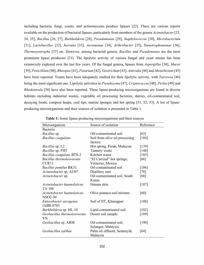
102
including bacteria, fungi, yeasts, and actinomycetes produce lipases [22]. There are various reports
available on the production of bacterial lipases, particularly from members of the genera Acinetobacter [23,
24, 25], Bacillus [26, 27], Burkholderia [28], Pseudomonas [29], Staphylococcus [30], Microbacterium
[31], Lactobacillus [32], Serratia [33], Aeromonas [34], Arthrobacter [35], Stenotrophomonas [36],
Thermosyntropha [37] etc. However, among bacterial genera, Bacillus and Pseudomonas are the most
prominent lipase producers [21]. The lipolytic activity of various fungal and yeast strains has been
extensively explored over the last few years. Of the fungal genera, lipases from Aspergillus [38], Mucor
[39], Penicillium [40], Rhizopus [41], Fusarium [42], Geotrichum [43], Antrodia [44] and Metarhizium [45]
have been reported. Yeasts have been adequately studied for their lipolytic activity, with Yarrowia [46]
being the most significant one. Lipolytic activities in Pseudozyma [47], Cryptococcus [48], Pichia [49] and
Rhodotorula [50] have also been reported. These lipase-producing microorganisms are found in diverse
habitats including industrial wastes, vegetable oil processing factories, dairies, oil-contaminated soil,
decaying foods, compost heaps, coal tips, marine sponges and hot spring [51, 52, 53]. A list of lipase-
producing microorganisms and their sources of isolation is presented in Table 1.
Table 1: Some lipase-producing microorganisms and their sources
Microorganism Source of isolation Reference Bacteria Bacillus sp. Oil-contaminated soil [63] Bacillus coagulans Soil from olive oil processing
factory [184]
Bacillus sp. L2 Hot spring, Perak, Malaysia [139] Bacillus sp. FH5 Tannery waste [148] Bacillus coagulans BTS-3 Kitchen waste [185] Bacillus thermoleovorans CCR11
“El Carrizal” hot springs, Veracruz, Mexico
[66]
Bacillus pumilus RK31 Oil-contaminated soil [186] Acinetobacter sp. AU07 Distillery unit [78] Acinetobacter sp. Oil-contaminated soil, South
Korea [68]
Acinetobacter haemolyticus TA 106
Human skin [187]
Acinetobacter haemolyticus NSO2-30
Olive pomace-soil mixture [60]
Enterobacter aerogenes IABR-0785
Soil of IIT, Kharagpur [188]
Burkholderia sp. HL-10 Lipid-contaminated soil [102] Geobacillus thermoleovorans YN
Desert soil sample [189]
Geobacillus sp. ARM Oil-contaminated soil, Selangor, Malaysia
[190]
Geobacillus zalihae Palm oil effluent, Semenyih, Malaysia
[64]
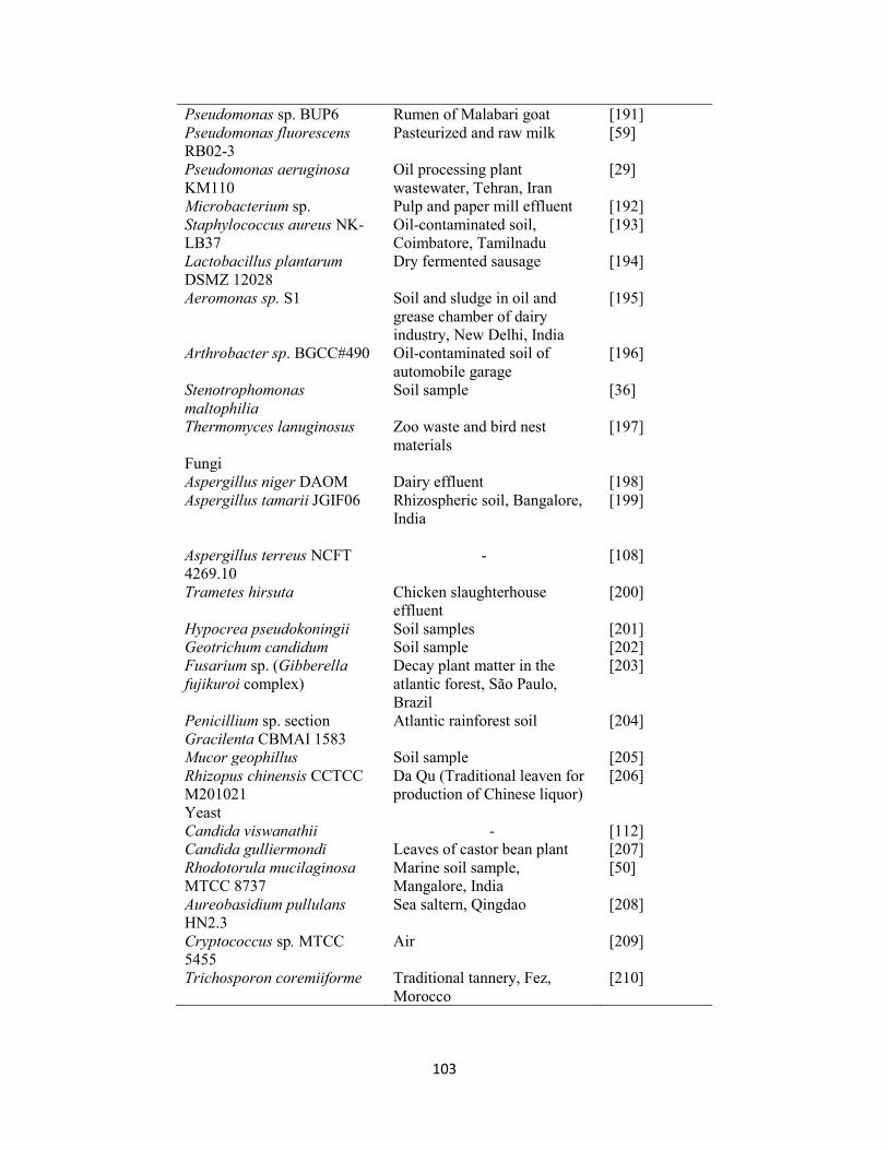
103
Pseudomonas sp. BUP6 Rumen of Malabari goat [191] Pseudomonas fluorescens RB02-3
Pasteurized and raw milk [59]
Pseudomonas aeruginosa KM110
Oil processing plant wastewater, Tehran, Iran
[29]
Microbacterium sp. Pulp and paper mill effluent [192] Staphylococcus aureus NK-LB37
Oil-contaminated soil, Coimbatore, Tamilnadu
[193]
Lactobacillus plantarum DSMZ 12028
Dry fermented sausage [194]
Aeromonas sp. S1 Soil and sludge in oil and grease chamber of dairy industry, New Delhi, India
[195]
Arthrobacter sp. BGCC#490 Oil-contaminated soil of automobile garage
[196]
Stenotrophomonas maltophilia
Soil sample [36]
Thermomyces lanuginosus Zoo waste and bird nest materials
[197]
Fungi Aspergillus niger DAOM Dairy effluent [198] Aspergillus tamarii JGIF06 Rhizospheric soil, Bangalore,
India [199]
Aspergillus terreus NCFT 4269.10
- [108]
Trametes hirsuta Chicken slaughterhouse effluent
[200]
Hypocrea pseudokoningii Soil samples [201] Geotrichum candidum Soil sample [202] Fusarium sp. (Gibberella fujikuroi complex)
Decay plant matter in the atlantic forest, São Paulo, Brazil
[203]
Penicillium sp. section Gracilenta CBMAI 1583
Atlantic rainforest soil [204]
Mucor geophillus Soil sample [205] Rhizopus chinensis CCTCC M201021
Da Qu (Traditional leaven for production of Chinese liquor)
[206]
Yeast Candida viswanathii - [112] Candida gulliermondi Leaves of castor bean plant [207] Rhodotorula mucilaginosa MTCC 8737
Marine soil sample, Mangalore, India
[50]
Aureobasidium pullulans HN2.3
Sea saltern, Qingdao [208]
Cryptococcus sp. MTCC 5455
Air [209]
Trichosporon coremiiforme Traditional tannery, Fez, Morocco
[210]
104
3. Classification of bacterial lipases
Bacterial lipases are classified into eight families; the largest family is sub-divided into six sub-families
[54]. This classification is based on conserved sequence motifs and biocatalytic properties of the enzymes.
True lipases belong to family I, which comprises of Pseudomonas, Bacillus and Staphylococcus lipases.
These lipases possess the conventional catalytic pentapeptide Gly-Xaa-Ser-Xaa-Gly. They are divided into
seven sub-families (I.1-I.7) based on structural features, type of secretion mechanism and requirement for
lipase-specific foldases, and relationship to other enzyme families [54, 11]. Family II lipases are
characterized with Gly-Asp-Ser-Leu motif at the active site, and include esterases of Streptomyces,
Aeromonas and Salmonella. Family III contains lipases of Streptomyces sp., but in contrast to family II
esterase, they are extracellular lipases. Lipases that are similar to mammalian hormone sensitive lipases are
grouped under family IV whereas, lipases of mesophilic bacteria including Pseudomonas oleovorans and
Haemophilus influenza belong to family V. Family VI lipases are the smallest esterases, the active enzymes
are dimeric. Family VIII lipases are large esterases, consisting of amino acid sequence homologous to that
of eukaryotic acetyl choline esterases. Family VIII lipases are similar to β-lactamases. The sequences of
few other lipases that are not grouped into any of the eight super-families are arbitrarily classified as new
family IX and X. This is typical of a cold active lipase reported by De Pascale et al. [6], and grouped to a
novel lipolytic family. However, this classification by Arpigny and Jaeger [54] has been revised several
times. Currently, there are XVI families of lipases available in ESTHER database
(http://bioweb.ensam.inra.fr/esther). In addition, a new classification recently reported in the lipase
engineering database (http://www.led.uni-stuttgart.de) grouped lipases into three classes based on oxyanion
hole: GX, GGGX, and Y.
4. Methods for detection of lipase production
Several techniques have been developed for the screening of microorganisms for lipase production. These
methods either directly use the microorganism under study or measure lipolytic activity in the crude or
purified enzyme preparations [55, 56]. Numerous methods employed for measuring hydrolytic activity and
detection of lipase production are discussed below in details:
4.1. Screening methods on solid media
Lipolysis is monitored directly by changes in the appearance of the substrate (such as tributyrin and triolein)
that are emulsified in the various growth media [57]. The formation of clear halos around the colonies
cultivated on the agar plate is an indication of lipase production [58]. Pseudomonas fluorescens RB02-3
and Acinetobacter haemolyticus NS02-30 were screened for lipolytic activity on tributyrin agar [59, 60].
105
Lipolytic Bacillus sp. LBN 4 has been isolated on tributyrin agar medium using glycerol tributyrate as
substrate [61]. In addition, solid media supplemented with dyes such as phenol red, Victoria Blue B, Spirit
blue, or Nile blue sulfate as pH indicator are also used for determination of lipolytic activity [62]. The drop
in pH due to the release of fatty acid is indicated by a change in the color of the indicators. This chromogenic
method is simple and rapid. However, acidification of the medium resulting from the production of free
fatty acids from microbial lipases gives false results [57]. Phenol red agar, consisting of phenol red dye
(0.01%, w/v), olive oil (0.1%, v/v), CaCl2 (0.1%, w/v) and Agar (2%, w/v) has been used for screening of
Bacillus strain [63]. Geobacillus zalihae sp. nov. was screened for lipolytic activity using triolein agar plate,
comprising of triolein (0.25%, v/v), agar (1%, w/v), nutrient broth (0.8%, w/v) and Victoria Blue (0.01%,
w/v) [64]. Furthermore, fluorescent dye Rhodomine B is also employed for detection of lipolytic organisms
in plate assay containing emulsified olive oil as substrate. The formation of orange fluorescent halos around
colonies under UV irradiation and quantitation of lipase activities range (1 to 30 nkat) suggest production
of lipase [65]. Castro-Ochoa et al. [66] screened Bacillus thermoleovorans CCR11 for lipolytic activity on
Rhodomine B agar consisting of Rhodomine B (0.001%, w/v), nutrient broth (0.8%, w/v), NaCl (0.4%,
w/v), olive oil (3%, v/v), and agar (1%, w/v). Spirit blue agar medium has also been used for the detection
of lipolytic activity of Serratia rubidaea [67] and Acinetobacter sp. [68].
4.2. Titrimetric methods
In this technique, lipase activity is measured on a mechanically stirred emulsion of natural or synthetic
triacylglycerides by neutralization of free fatty acids released with time following addition of titrated NaOH
in order to maintain the pH at a constant end point value [69, 57]. Several authors have reported the use of
olive oil as a substrate for the titrimetric analysis [70, 71]. Rasmey et al. [72] measured the lipolytic activity
of Pseudomonas monteilli 2403-KY120354 in a reaction mixture containing olive oil emulsion incubated
at 37 °C for 1 h. Enzyme activity was terminated following addition of 20 ml acetone: ethanol mixture
(1:1). The liberated free fatty acids was titrated against 0.1 M NaOH using phenolphthalein. 1 unit of lipase
was defined as the amount of enzyme that liberated 1 µmol/min of fatty acids under standard assay
conditions. The other least commonly employed techniques for detection of lipase production include
chromatographic, turbidimetric, fluorimetric, and immunological methods as well as radioactive assays
[57].
5. Lipase production and influencing parameters
Microbial lipases are produced extracellularly by submerged or solid state fermentation during late
exponential or stationary phase [51, 73, 74]. However, submerged fermentation involving the growth of
microorganisms as a suspension in nutrient enriched liquid medium is mostly preferred due to easily
106
engineered process control and significant amounts of extracellular enzyme released in the production
medium [5, 26]. Lipase production is greatly influenced by nutritional and physicochemical factors, such
as temperature, pH, carbon and nitrogen sources, inoculum volume, presence of lipids, inorganic salts,
dissolved oxygen concentration, incubation period, and agitation [5, 53]. However, lipidic carbon sources
represent the ultimate factor that stimulate high lipase production, since the enzymes are inducible in nature
and are therefore generally produced in the presence of a lipid source including oil or other inducers, such
as triacylglycerols, Tweens, hydrolysable esters, fatty acids, bile salts, glycerol etc. [51, 5, 75, 76]. For
instance, lipase production by Bacillus flexus XJU-1 was stimulated by the presence of a surfactant, Tween-
80, which favored the uptake of medium components and lipase release [77]. In addition, various oils such
as olive oil [27], coconut oil [23], cotton seed oil [77] castor oil [78], soybean oil [30] and neem oil [79]
are used as inducers for lipase production. Lipases are produced with a low oil concentration (1-5%) [220,
30]. When a significant amount of oil is used, lipase synthesis reduces due to limitation of oxygen transfer
that result in poor microbial growth [77]. Other carbon sources including sugars, sugar alcohol,
polysaccharides, whey, casamino acids and other complexes sources influence lipase production [80, 81].
Mannitol was found as the best carbon source for lipase production by Streptomyces griseochromogenes
[82]. In some cases, combination of carbohydrate and oil is used for maximum lipase secretion [83, 84, 85,
26]. Various non-conventional carbon sources, including beef tallow, wool scour effluent, whey and n-
hexadecane, as well as cheap agro-industrial residues are also used as lipidic carbon source for lipase
production [85, 86, 87].
The type of nitrogen source in the fermentation medium influences the levels of lipase yield by
microorganisms [88]. Organic nitrogen sources including peptone, yeast extract or a combination of these
resulted in a significant lipase production by most bacterial strains [220, 29, 27]. Optimum lipase production
was also recorded with soybean meal [26] and corn steep liquor [28, 24, 25]. The preference of organic
nitrogen sources by some lipase-producing microorganisms can be attributed to the presence of some
minerals, vitamins or other growth factors that they contain [77]. In some cases, individual amino acids
have been reported to play a significant role in lipase production [88]. This is typical of phenylalanine found
as a preferred nitrogen source for lipase production by Streptomyces griseochromogenes [82]. Inorganic
nitrogen sources such as ammonium chloride, ammonium molybdate and diammonium hydrogen phosphate
are effective for maximum lipase production [89, 90, 91]. However, a higher nitrogen source concentration
inhibits lipase production due to nitrogen metabolite repression [92].
107
6. Optimization of lipase production using statistical experimental designs
The improvement of industrial fermentation processes is centered on designing of fermentation medium,
since its composition considerably influences the yield, concentration and productivity of desired
metabolites [5]. The classical optimization by one variable at a time (OVAT) approach is not only time-
consuming, laborious and expensive, but also fails to depict the interaction effects of the different variables
tested, leading to misinterpretation of results [93]. In order to overcome these difficulties, statistical
experimental designs have been recognized as a preferred method for optimization processes [94, 95, 78]
(Table 2). The significant variables influencing lipase production are usually screened through the Plackett-
Burman design (PBD), and the optimal conditions and interaction effects of these variables are inferred
from the response surface methodology (RSM) using Box-Behnken design (BBD) or central composite
design (CCD) [21, 96, 97].
Plackett-Burman design has been applied for evaluation of medium components for lipase production
through submerged fermentation of microorganisms [98, 99]. PBD, in combination with RSM, has resulted
in enhanced feasibility of process scale-up and commercialization of lipase production from a multitude of
bacteria, fungi, and yeasts [21]. Ruchi et al. [100] screened eleven media components (peptone, tryptone,
NH4Cl, NaNO3, yeast extract, glucose, glycerol, xylose, gum arabic, MgSO4, and NaCl) for lipase
production by Pseudomonas aeruginosa using PBD. The most significant parameters (gum arabic, MgSO4,
tryptone, and yeast extract) were further optimized by RSM. Maximum lipase yield (5.58-fold) was
recorded when tryptone, gum arabic, MgSO4 and yeast extract were at concentrations of 1.01%, 0.02%,
0.10% and 0.02%, respectively. Similarly, the influence of ten medium components (peptone, glucose,
NaCl, MgSO4.7H2O, FeSO4.7H2O, CaCl2, olive oil, KH2PO4, NH4Cl and Na2HPO4) on lipase production
by Alkalibacillus salilacus SR-079 Halo was studied using PBD [101]. Lipase production was maximally
affected by olive oil, KH2PO4, NaCl, and glucose. Further optimization of the selected variables by RSM
resulted in 4.9-fold enhancement in lipase production at optimal levels of glucose (1g/L), NaCl (4.18
mol/L), olive oil (2%) and KH2PO4 (5 g/L).
Combination of OVAT and RSM has been employed for optimization of lipase production. Papagora et al.
[94] optimized lipase production from Debaryomyces hansenii YLL29 using RSM. The simple one-factor-
at-a-time strategy showed that glucose, olive oil and pH were the significant variables influencing lipase
production. Further optimization of the selected variables by RSM led to a 2.28-fold increase in lipase
production at respective optimal levels of glucose (13.1 g/L), olive oil (19 g/L) and pH (6.4). Similarly, Lo
et al. [102] employed RSM and OVAT for the optimization of extracellular lipase production by
Burkholderia sp. HL-10.
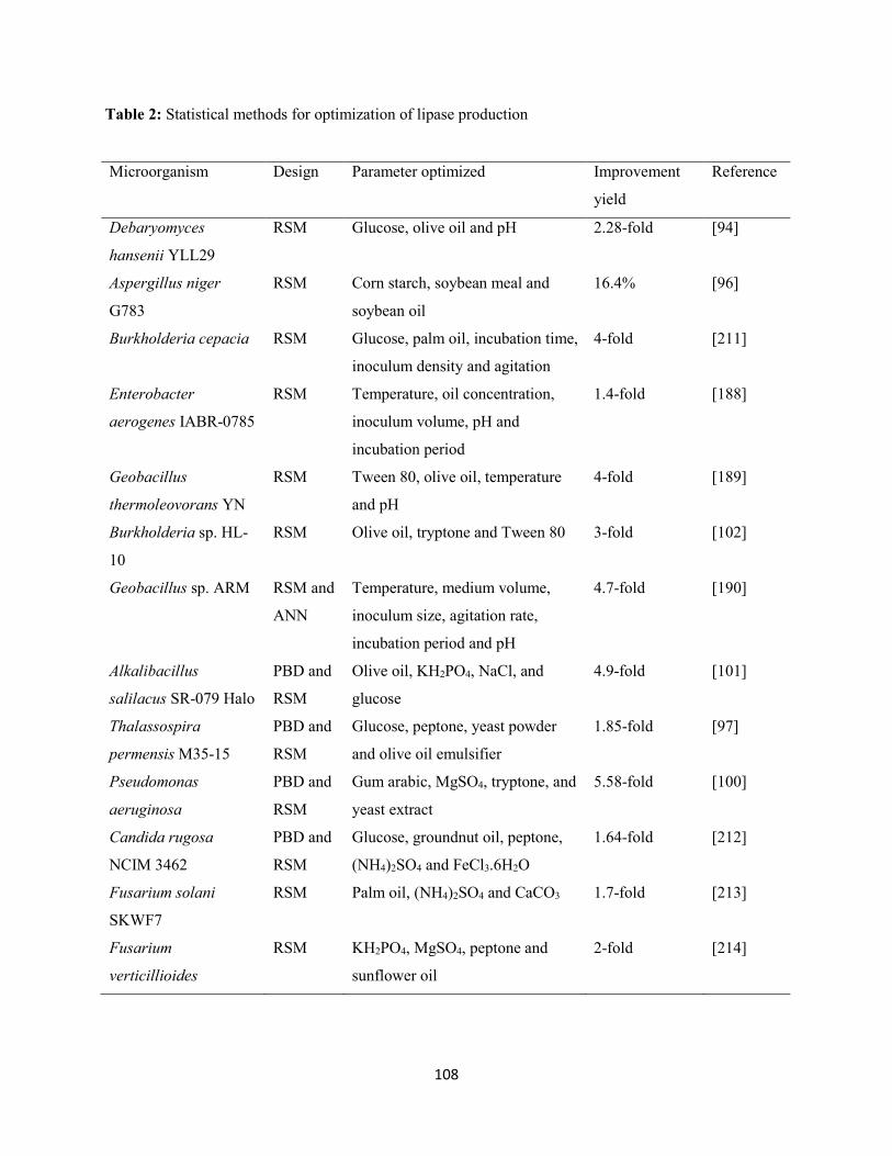
108
Table 2: Statistical methods for optimization of lipase production
Microorganism Design Parameter optimized Improvement
yield
Reference
Debaryomyces
hansenii YLL29
RSM Glucose, olive oil and pH 2.28-fold [94]
Aspergillus niger
G783
RSM Corn starch, soybean meal and
soybean oil
16.4% [96]
Burkholderia cepacia RSM Glucose, palm oil, incubation time,
inoculum density and agitation
4-fold [211]
Enterobacter
aerogenes IABR-0785
RSM Temperature, oil concentration,
inoculum volume, pH and
incubation period
1.4-fold [188]
Geobacillus
thermoleovorans YN
RSM Tween 80, olive oil, temperature
and pH
4-fold [189]
Burkholderia sp. HL-
10
RSM Olive oil, tryptone and Tween 80 3-fold [102]
Geobacillus sp. ARM RSM and
ANN
Temperature, medium volume,
inoculum size, agitation rate,
incubation period and pH
4.7-fold [190]
Alkalibacillus
salilacus SR-079 Halo
PBD and
RSM
Olive oil, KH2PO4, NaCl, and
glucose
4.9-fold [101]
Thalassospira
permensis M35-15
PBD and
RSM
Glucose, peptone, yeast powder
and olive oil emulsifier
1.85-fold [97]
Pseudomonas
aeruginosa
PBD and
RSM
Gum arabic, MgSO4, tryptone, and
yeast extract
5.58-fold [100]
Candida rugosa
NCIM 3462
PBD and
RSM
Glucose, groundnut oil, peptone,
(NH4)2SO4 and FeCl3.6H2O
1.64-fold [212]
Fusarium solani
SKWF7
RSM Palm oil, (NH4)2SO4 and CaCO3 1.7-fold [213]
Fusarium
verticillioides
RSM KH2PO4, MgSO4, peptone and
sunflower oil
2-fold [214]
109
Preliminary studies by OVAT revealed that olive oil, tryptone and Tween-80 exhibited significant effects
on lipase production. Optimization by CCD resulted in almost 3-fold increase in maximum lipase
production at respective optimum concentrations of olive oil (0.65%, v/v), tryptone (2.42%, w/v) and
Tween-80 (0.15%, v/v).
7. Purification of lipases
Most of the commercial applications of enzymes do not always require homogenous preparation of the
enzyme. However, a certain degree of purity is needed, depending upon the final application in industries
such as fine chemicals, pharmaceuticals and cosmetics [103, 5]. For industrial purposes, the purification
techniques employed should be cost-effective, rapid, high yielding and amenable to large-scale operations.
In addition, they should possess potential for continuous product recovery, with a relatively high capacity
and selectivity for the desired product [5]. The extent of purification varies with the order of purification
steps.
Most of the microbial lipases are extracellular in nature and the fermentation process is usually followed
by the recovery of the biomass from the culture broth by centrifugation or filtration. The obtained cell-free
culture supernatant containing the crude enzyme is then concentrated by ultrafiltration, ammonium sulphate
precipitation or extraction with organic solvents [221]. Approximately 80% of all the purification methods
employed thus far used a precipitation step, with 60% of these using ammonium sulphate and 35% using
ethanol, acetone or an acid (usually hydrochloric acid). The precipitation step is normally carried out in the
early stages of recovery, followed by chromatographic separation. However, precipitation techniques
normally give high yield (87%) in comparison to chromatography methods, which result in lower yields of
about 60-70% [104, 221]. Such enzyme preparations are suitable for use in detergent formulations and
wastewater treatment [5, 105, 106]. Partial purification of lipases by ultrafiltration [107], ammonium
sulphate [108], ethanol [109] or a combination of the precipitating agents has been reported [90]. However,
ammonium sulphate is mostly preferred because of its cost-effectiveness, high solubility in water and
exhibit no effect on enzyme structure [110]. Increase in lipase activity depends on the concentration of
ammonium sulphate used [111].
In industrial applications, such as synthetic reactions in pharmaceutical industry, high purity of lipase is
required. Hence, chromatographic techniques involving ion exchange chromatography [26], gel filtration
chromatography [60], affinity chromatography [107], hydrophobic interaction chromatography [112] or a
combination [28] are mostly used. However, these routine procedures of lipase purification are sometimes
time-consuming and result in low final yields. As a result, novel technologies including membrane
processes, immunopurification, hydrophobic interaction chromatography using epoxy-activated spacer arm
110
as a ligand and poly-ethylene glycol-sepharose gel, poly (vinyl alcohol) polymers as column
chromatography stationary phases, and aqueous two-phase systems have recently been applied for
purification of lipase [221].
8. Immobilization of lipases
Immobilization of lipase on/in a variety of support materials permits multiple reuse of the enzyme,
enhanced stability, and easier product recovery [21, 113]. In addition, in comparison to free enzymes,
immobilized lipases allow possible continuous process, rapid termination of reactions, and controlled
product formation [114, 115]. Such immobilized enzymes are used in a wide range of applications [116,
117]. Methods such as adsorption [118], covalent bonding [119], entrapment [120], and cross-linking [121]
are employed for lipase immobilization. For instance, Dandavate et al. [122] covalently immobilized
Candida rugosa lipase onto the surface of silica nanoparticles and glutaraldehyde-activated aminopropyl
glass beads, resulting in easy recovery and reuse of the enzyme. Immobilization of Yarrowia lipolytica
lipase on octyl-agarose and octadecyl-sepabeads by physical adsorption led to higher yields and greater
stability [123]. Among fungi, immobilization of Rhizopus oryzae lipase on silica aerogels [124], and
Thermomyces sp. lipase on regenerated cellulose, glass fiber and polyvinylidene fluoride grafted with 1,4-
diaminobutane and activated with glutaraldehyde [125] have been reported. Among bacterial lipases,
Pseudomonas sp. lipases have been covalently immobilized on porous polymethyl-acrylamide cross-linked
with N,N’methylene bisacrylamide [126] and bacterially produced exopolysaccharide [127]. In addition,
zirconia particles [85], alginate-k-carrageenan hybrid matrix [128], celite carriers [129], polyglutaraldehyde
activated styrene-divinylbenzene copolymer [130], synthetic macroporous alkylated glycidyl epoxy
copolymers [131] are notable support materials reported for immobilization of lipases from bacteria
including Burkholderia sp. and Arthrobacter sp.
9. Characterization of alkaline lipases
Lipases are characterized by determining their pH activity and stability, temperature activity and stability,
kinetic parameters, among others in order to discover new commercial enzymes with better properties
suitable for various biotechnological applications. The essential properties of some microbial alkaline
lipases are highlighted in Table 3 and discussed in details below:
9.1. Effect of temperature on activity and stability
Alkaline lipases exhibit optimum activity and stability at broad temperature ranges, a characteristic for
adaptability to various industrial applications. Generally, bacterial lipases are active in the temperature
range of 30-60 °C [132].
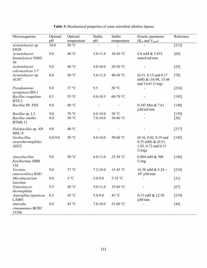
111
Table 3: Biochemical properties of some microbial alkaline lipases
Microorganism Optimal pH
Optimal temperature
Stable pH
Stable temperature
Kinetic parameter (Km and Vmax)
Reference
Acinetobacter sp. EH28
10.0 50 °C - - - [215]
Acinetobacter haemolyticus NS02-30
9.0 40 °C 5.0-11.0 10-30 °C 0.8 mM & 3.833 mmol/ml/min
[60]
Acinetobacter calcoaceticus 1-7
9.0 40 °C 4.0-10.0 20-50 °C - [25]
Acinetobacter sp. AU07
8.0 50 °C 5.0-11.0 40-50 °C (0.51, 0.15 and 0.17 mM) & (16.98, 15.44 and 14.61 U/mg)
[78]
Pseudomonas aeruginosa BN-1
8.0 37 °C 9.5 50 °C - [216]
Bacillus coagulans BTS-3
8.5 55 °C 8.0-10.5 60-70 °C - [185]
Bacillus SP. FH5 9.0 60 °C - - 0.345 Mm & 7.61 µM/ml/min
[148]
Bacillus sp. L2 9.0 70 °C 8.0-10.0 50 °C - [139] Bacillus smithii BTMS 11
8.0 50 °C 7.0-10.0 30-80 °C - [26]
Halobacillus sp. AP-MSU 8
9.0 40 °C - - - [217]
Geobacillus stearothermophilus AH22
8.0-9.0 50 °C 4.0-10.0 50-60 °C (0.16, 0.02, 0.19 and 0.55 mM) & (0.51, 1.03, 0.72 and 0.15 U/mg)
[145]
Anoxybacillus flavithermus HBB 134
9.0 50 °C 6.0-11.0 25-50 °C 0.084 mM & 500 U/mg
[146]
Yersinia enterocolitica KM1
9.0 37 °C 7.2-10.0 15-45 °C 16.58 mM & 5.24 × 10⁵ µM/min
[218]
Microbacterium luteolum
8.0 5 °C 5.0-9.0 5-35 °C - [31]
Talaromyces thermophilus
9.5 50 °C 9.0-11.0 35-60 °C - [87]
Aspergillus japonicus LAB01
8.5 45 °C 5.0-9.0 45 °C 0.13 mM & 12.58 µM/min
[219]
Antrodia cinnamomea BCRC 35396
8.0 45 °C 7.0-10.0 25-60 °C - [44]
112
However, there are reports on lipases that are optimal at both lower and higher temperature ranges [3].
Optimum temperatures of 30, 40, 50, and 60 °C have been reported for lipases produced by Staphylococcus
arlettae JPBW-1 [30], Acinetobacter calcoaceticus 1-7 [25], Bacillus smithii BTMS11 [26], and Bacillus
licheniformis B42 [90], respectively. An extracellular lipase from Pseudomonas sp. has been reported to
exhibit optimal activity at 90 °C with half-life of more than 13 h at this temperature [133]. Among
thermophilic bacteria, lipases from Bacillus and Geobacillus sp. have been reported to be active at
temperature between 50 and 70 °C [134, 66, 107]. Of particular interest are lipases from Geobacillus kaue
and Geobacillus kaustophilus that show optimal activities at 80 °C, which was almost completely
maintained at 90 °C [135]. Similarly, thermotolerant lipases, Lip A and Lip B from Thermosyntropha
lipolytica exhibit maximum activity at around 96 °C, a temperature found to be highest for maximum
activity among lipases [136].
Lipases from many microbial species have been reported to be stable at a broad temperature range of
between 4 and 90 °C [92]. This is notable of thermostable lipase from a Bacillus strain, found to retain
100% of its original activity at 75 °C after 30 min. The enzyme retained 90% of the original activity after
being incubated at 60 °C for 15 h [137]. Similarly, a retention of 90% activity at 20-50 °C for 60 h was
reported for lipase from Acinetobacter calcoaceticus 1-7 [25]. At 30-80 °C, more than 50% activity of
lipase from Bacillus smithii BTMS 11 was retained after 12 h [26]. Lipase from Geobacillus
thermodenitrificans IBRL-nra exhibited no loss of activity at 65 and 70 °C after 60 min. However, 90% of
the original activity was retained at 65 and 70 °C for 3 h and 2 h, respectively [107]. This was found to be
highly stable in comparison to lipase from Geobacillus stearothermophilus with 87.5% original activity
retained after 15 min of exposure at 70 °C [138]. Shariff et al. [139] reported stability of L2 lipase at 60 °C
with a half-life of 2 h. At the same temperature, the enzyme retained more than 40% of its original activity
for 3 h and 45 min. The thermostability of L2 lipase was found to be higher than that of Bacillus
stearothermophilus L1, which was stable at 60 °C for 30 min [40].
9.2. Effect of pH on activity and stability
Bacterial lipases that are active and stable in the wide alkaline pH range (8.0-12.0) represent attractive
biocatalysts in many industrial applications. For instance, alkaline lipases that show optimal activities in
the pH range from 9.0 to 11.0 have been reported from Acinetobacter radioresistens, Bacillus sp.,
Chromobacterium viscosum, Micrococcus sp., and different species of the genus Pseudomonas [141]. In
addition, strains of Bacillus cepacia ATCC 25609 [142], Acinetobacter johnsonii LP28 [24],
Staphylococcus sp. ESW [218), Bacillus sp. LBN 2 [53], and Bacillus flexus XJU-1 [143] exhibit alkaline
113
lipolytic activity. Remarkably, lipases from Bacillus stearothermophilus SB-1, Bacillus atrophaeus SB-2
and Bacillus licheniformis SB-3 are active over a broad pH range (3.0-12.0) [144].
The alkaline lipases exhibit various good stability ranges. This is notable of alkaline lipase from
Acinetobacter calcoaceticus 1-7 found to be stable in the pH range of 4.0-10.0 and retained 100% of its
activity at pH 9.0 for 24 h at 40 °C [25]. In addition, lipase from Pseudomonas aeruginosa KM110 was
found to be stable in the pH range of 7.0-10.0, retaining 65% activity at pH 8.0 for 1 h at 30 °C [29]. Ekinci
et al. [145] reported lipase from Geobacillus stearothermophilus AH22 that retained more than 70% of its
original activity at pH 7.0-10.0 for 30 d at +4 °C. More than 94% activity of lipase from Anoxybacillus
flavithermus HBB 134 was maintained between pH 6.0 to 11.0 at 25 °C for 24 h [146].
9.3. Kinetics properties of alkaline lipases
In many cases, lipases appear to conform to Michaelis-Menten kinetics, characterized by two parameters,
Michaelis-Menten constant (Km) and maximum velocity (Vmax). The Km values of the enzyme vary widely,
however for most industrially relevant enzymes, Km ranges between 10-1 and 10-5 M in the presence of
biotechnologically important substrates [147]. Km and Vmax values of 0.16 mM and 0.52 U/mg for p-
nitrophenyl acetate, 0.02 mM and 1.03 U/mg for p-nitrophenyl butyrate, 0.19 mM and 0.72 U/mg for p-
nitrophenyl octanoate, and 0.55 mM and 0.15 U/mg for p-nitrophenyl laurate have been reported for lipase
from Geobacillus stearothermophilus AH22 [145]. Similarly, Gururaj et al. [78] determined the kinetic
properties of Acinetobacter sp. AU07 lipase in the presence of different fatty acid esters as substrates. The
Km and Vmax values were 0.51 mM and 16.98 U/mg for p-nitrophenyl butyrate, 0.15 mM and 15.44 U/mg
for p- nitrophenyl laurate, 0.17 mM and 14.61 U/mg for p-nitrophenyl myristate. In addition, Km and Vmax
values of 0.8 mM and 3.833 mmol/ml/min, respectively were recorded from Acinetobacter haemolyticus
NS02-30 lipase when using p-nitrophenyl palmitate as substrate [60]. Ghori et al. [148] recorded Km and
Vmax values of 0.345 mM and 7.61 µM/ml/min, respectively in the presence of p-nitrophenyl butyrate as
substrate from lipase secreted by Bacillus sp. FH5.
10. Potential biotechnological applications of alkaline lipases
Microbial lipases constitute an important class of biotechnologically valuable enzymes, mainly due to the
versatility in their enzymatic properties and substrate specificity. This versatility makes lipases the enzyme
of choice for various potential applications in food, detergent, leather, pharmaceutical, textile, cosmetic,
paper industries etc. [149, 2, 20]. Some of the biotechnological applications of alkaline lipases are
summarized in Table 4 and discussed in details below:
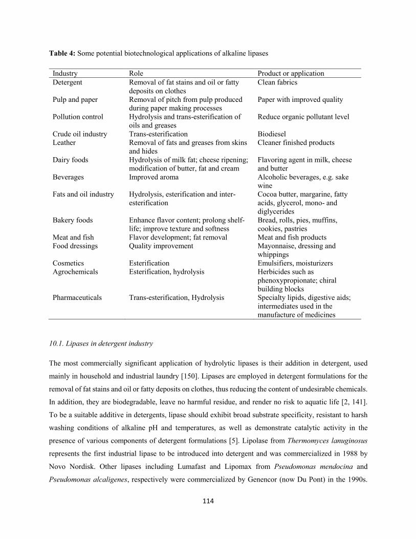
114
Table 4: Some potential biotechnological applications of alkaline lipases
Industry Role Product or application Detergent Removal of fat stains and oil or fatty
deposits on clothes Clean fabrics
Pulp and paper Removal of pitch from pulp produced during paper making processes
Paper with improved quality
Pollution control Hydrolysis and trans-esterification of oils and greases
Reduce organic pollutant level
Crude oil industry Trans-esterification Biodiesel Leather Removal of fats and greases from skins
and hides Cleaner finished products
Dairy foods Hydrolysis of milk fat; cheese ripening; modification of butter, fat and cream
Flavoring agent in milk, cheese and butter
Beverages Improved aroma Alcoholic beverages, e.g. sake wine
Fats and oil industry Hydrolysis, esterification and inter-esterification
Cocoa butter, margarine, fatty acids, glycerol, mono- and diglycerides
Bakery foods Enhance flavor content; prolong shelf-life; improve texture and softness
Bread, rolls, pies, muffins, cookies, pastries
Meat and fish Flavor development; fat removal Meat and fish products Food dressings Quality improvement Mayonnaise, dressing and
whippings Cosmetics Esterification Emulsifiers, moisturizers Agrochemicals Esterification, hydrolysis Herbicides such as
phenoxypropionate; chiral building blocks
Pharmaceuticals Trans-esterification, Hydrolysis Specialty lipids, digestive aids; intermediates used in the manufacture of medicines
10.1. Lipases in detergent industry
The most commercially significant application of hydrolytic lipases is their addition in detergent, used
mainly in household and industrial laundry [150]. Lipases are employed in detergent formulations for the
removal of fat stains and oil or fatty deposits on clothes, thus reducing the content of undesirable chemicals.
In addition, they are biodegradable, leave no harmful residue, and render no risk to aquatic life [2, 141].
To be a suitable additive in detergents, lipase should exhibit broad substrate specificity, resistant to harsh
washing conditions of alkaline pH and temperatures, as well as demonstrate catalytic activity in the
presence of various components of detergent formulations [5]. Lipolase from Thermomyces lanuginosus
represents the first industrial lipase to be introduced into detergent and was commercialized in 1988 by
Novo Nordisk. Other lipases including Lumafast and Lipomax from Pseudomonas mendocina and
Pseudomonas alcaligenes, respectively were commercialized by Genencor (now Du Pont) in the 1990s.
115
Recently, lipases from several microorganisms have been characterized as potent detergent additives [151,
152, 25, 153].
10.2. Lipases in food industry
Fats and oils are indispensable constituents of foods. The nutritional and sensory values as well as physical
properties of a triglyceride are greatly influenced by position of fatty acid in the glycerol backbone, the
chain length of the fatty acid and its degree of unsaturation etc. [154]. The modification of structure and
composition of fat and oil is of great significance in food processing industries that require new economics
and green technologies. Microbial lipases that are regiospecific and fatty acid specific are of enormous
importance to produce several products in food industries. For instance, lipase-catalyzed reactions can be
used to modify and upgrade cheap oil into nutritionally important structured triacylglycerols like cocoa
butter substitutes, low calories triacylglycerols and oleic acid enriched oils [155]. Lipases have also been
used in foods to modify flavor by synthesis of esters of short chain fatty acids and alcohol, which are known
flavor and fragrance compounds [156]. In addition, lipases are used in the removal of fat from meat and
fish products to produce lean meat. The fat is removed during the processing of the fish meat by adding
lipases, a phenomenon known as bio-lipolysis [157]. Lipases also play a substantial role in the fermentative
process of sausage production and to determine changes in long-chain fatty acid released during ripening
[2]. Over decades, microbial lipases have been used for refining rice flavor, modifying soybean milk,
improving aroma, and enhancing fermentation in apple wine [158].
10.3. Lipases in cosmetics
Immobilized lipase from Rhizomucor miehei has been applied as a biocatalyst for the production of
cosmetic products including isopropyl palmitate and 2-ethylhexyl palmitate, used as emollient in personal
care products such as skin and sun-tan creams, bath oils etc. The use of the enzyme instead of commonly
used chemicals gives products of much higher quality with minimum downstream refining. In addition,
wax esters (esters of fatty acids and fatty alcohols) produced from catalytic reaction of lipase from Candida
cylindracea have similar applications in personal care products. Enzymatic production of water-soluble
retinol derivatives from immobilized lipase has been reported [159]. Lipases are also used in hair waving
preparation and as components of topical anti-obese creams or as oral administration [160, 161, 162].
10.4. Lipases in pulp and paper industry
The pulp and paper industry generates large amounts of lignocellulosic biomass every year. The technology
for pulp manufacturing is highly diverse, thus providing numerous opportunities for the application of
microbial enzymes. Pitch (hydrophobic components in wood) creates severe problems in paper mill, by
116
causing sticky deposits in the paper machines as well as holes and spots in the final paper [163]. The
enzymatic pitch control technique using lipase has been a routine practice for large-scale paper making
process [164]. Lipases are used for the removal of pitch from the pulp produced during paper making
processes [165]. Lipases hydrolyze approximately 90% of triglycerides in the pitch into monoglycerides,
glycerol and fatty acids, which are less sticky, and highly hydrophilic [163]. In General, lipases in paper
industry increase the pulping rate of pulp, increase whiteness and intensity, decrease chemical usage,
prolong equipment life, reduce pollution level of wastewater, save energy and time, and reduce composite
site [2]. The addition of lipase from Pseudomonas sp. KWI-56 to a de-inking composition for ethylene
oxide-propylene oxide adduct stearate enhanced whiteness of paper and reduced residual ink spots [166].
10.5. Lipases in bioremediation of lipid-rich wastewater
Lipids are noxious components of industrial and municipal wastewaters, as they contribute greatly to the
organic matter of the wastewater and promote the growth of filamentous microorganisms. Therefore, their
transformation to innocuous products is imperative. The use of biocatalysts serves as a promising
technology for the treatment of high fat-containing wastewater. An alternative to conventional approaches
that is attracting growing interest is the use of enzymes, which significantly reduce the level of organic
pollutants in the wastewater by means of enzymatic catalysis and enhance better performance of microbial
community at the later stage of biological treatment process [167, 168, 169, 170]. Application of lipases
from different sources in the treatment of wastewater from lipid-processing factories, dairies, restaurants
etc. offers a novel approach in enzyme biotechnology. Thus, making the wastewater amenable to
conventional biological treatment [21]. The utilization of a solid enzymatic preparation from Penicillium
restrictum for the treatment of dairy wastewater with high levels of oil and grease (O & G) has been reported
[171]. Results obtained showed 13% higher chemical oxygen demand (COD) removal efficiency with 40%
lower accumulation of O & G in flocs; 1.7 times higher biomass concentration, and 1.3 times higher specific
oxygen uptake rate.
Immobilization of lipase in insoluble supports is an important tool for the treatment of lipid-rich wastewater.
Immobilized lipase has been used for the hydrolysis of O & G contents in pet food industry wastewater,
resulting in COD and O & G reduction of 49 and 45%, respectively without pretreatment, and 65 and 64%,
respectively, with immobilized lipase treatment [172]. Dumore and Mukhopadhyay [173] used
triacylglycerin lipase immobilized in chitosan for the treatment of synthetic oily wastewater. Results
recorded revealed approximately 48% and 47% removal efficiencies of O & G and COD, respectively. In
another study, Kanmani et al. [174] applied partially purified lipase (Staphylococcus pasteuri COM-4A)
immobilized on celite carrier for the treatment of coconut oil mill effluent. Result showed 46% and 24%
117
increase in volatile fatty acids and long-chain fatty acids, respectively with a concomitant reduction in O &
G (52%) and COD (32%).
Several lipase-producing microorganisms have been employed individually or as a consortium in the
remediation of lipid-containing wastewater. These include Bacillus stearothermophilus [175], Bacillus sp.
[176], Burkholderia sp. [177], Raoultella planticola [178], Geotrichum candidum [179], Penicillium
chrysogenum [180], Yarrowia lipolytica [181]. Consortium of microorganisms including Pseudomonas
aeruginosa, Bacillus sp., and Acinetobacter calcoaceticus [182], and Bacillus subtilis, Bacillus
licheniformis, Bacillus amyloliquefaciens, Serratia marcescens, Pseudomonas aeruginosa, and
Staphylococcus aureus [183] have been formulated as potential inoculum for the treatment of high strength
lipid-rich wastewater.
11. Concluding remarks
Lipases are ubiquitous in nature, often found in plants, animals, and microorganisms. However, microbial
lipases are versatile and widely employed for diverse biotechnological applications in food, cosmetics, pulp
and paper, detergent industries as well as in environmental bioremediation process. In lieu of the emergent
worldwide market and potential applications of microbial lipases, the complex downstream processing steps
result in low enzyme yield. Optimization of fermentation parameters through statistical experimental
designs is crucial in order to maintain a balance between various components for enhanced lipase
production. Immobilization of lipases in appropriate support materials allow continuous use of the enzyme
and reduction in the overall costs. The catalytic properties of alkaline lipases provide information on
possible identification of enzyme with high activity and stability for exploitation in various industrial and
environmental applications. Alternative approaches through metagenomics, site-directed mutagenesis, and
cloning and expression of lipase encoding gene in heterologous host should be developed for a robust
catalytic activity. In addition, the use of agro-industrial residues and industrial effluents as substrates for
lipase production will help in alleviating the costs of the enzyme and serve as pollution control strategies.
Conflicts of interest
The authors declare that there are no conflicts of interest concerning the publication of this paper
118
References
[1] N. Verma, S. Thakur, and A. K. Bhatt, “Microbial lipases: industrial applications and properties: a
review,” International Research Journal of Biological Sciences, vol. 1, no. 8, pp. 88-92, 2012.
[2] F. Hasan, A. A. Sha, and A. Hameed, “Industrial applications of microbial lipases,” Enzyme and
Microbial Technology, vol. 39, no. 2, pp. 235-251, 2006.
[3] G. M. Borrelli, and D. Trono, “Recombinant lipases and phospholipases and their uses as biocatalysts
for industrial applications,” International Journal of Molecular Sciences, Vol. 16, no. 9, pp. 20774-
20840, 2015.
[4] B. Joseph, P. W. Ramteke, and G. Thomas, “Cold active microbial lipases: some hot issues and recent
developments,” Biotechnolology Advances, vol. 26, no. 5, pp. 457-470, 2008.
[5] R. Gupta, N. Gupta, and P. Rathi, “Bacterial lipases: an overview of production, purification and
biochemical properties,” Applied Microbiology and Biotechnology, vol. 64, no. 6, pp. 763-781,
2004.
[6] D. de Pascale, A. M. Cusano, F. Autore, E. Parrilli, G. Di Prisco, G. Marino, and M. L. Tutino, “The
cold-active Lip 1 lipase from the antarctic bacterium Pseudoalteromonas haloplanktis TAC125 is
a member of a new bacterial lipolytic enzyme family,” Extremophiles, vol. 12, no. 3, pp. 311-323,
2008.
[7] M. Nardini, B. W. Dijkstra, “α/β hydrolase fold enzyme: the family keeps growing,” Current Opinion
in Structural Biology, vol. 9, no. 6, pp. 732-737, 1999.
[8] K- E. Jaeger, B. W. Dijkstra, and M. T. Reetz, “Bacterial biocatalysts: molecular biology, three-
dimensional structures, and biotechnological applications of lipases,” Annual Review of
Microbiology, vol. 53, pp. 315-351, 1999.
[9] F. Beisson, A. Tiss, C. Riviére, and R. Verger, “Methods for lipase detection and assay: a critical
review,” European Journal of Lipid Science and Technology, vol. 2, pp.133-153, 2000.
[10] J. K. Gupta, and S. K. Soni, “Industrial uses of enzymes,” Journal of Punjab Academy of Science, vol.
2, pp. 75-80, 2000.
[11] K. Jaeger, and T. Eggert, “Lipases for biotechnology,” Current Opinion in Biotechnology, vol. 13, no.
4, pp. 390-397, 2002.
119
[12] P. Villeneuve, “Plant lipases and their applications in oils and fats modification” European Journal of
Lipid Science and Technology, vol. 105, no. 6, pp. 308-317, 2003.
[13] M. Barros, L. F. Fleuri, G. A. Macedo, “Seed lipases: sources, applications and properties-a review,”
Brazilian Journal of Chemical Engineering, vol. 27, no. 1, pp. 15-29, 2010.
[14] M. R. W. Moussavou, C. Brunschwig, B. Baréa, P. Villeneuve, and J. Blin, “Are plant lipases a
promising alternative to catalyze transesterification for biodiesel production?,” Progress in Energy
and Combustion Science, vol. 39, no. 5, pp. 441-456, 2013.
[15] A. L. Quettier, and P. J. Eastmond, “Storage oil hydrolysis during early seedling growth,” Plant
Physiology and Biochemistry, vol. 47, no. 6, pp. 485-490, 2009.
[16] A. K. Padham, M. T. Hopkins, T- W Wang, L. M. McNamara, M. Lo, L. G. L. Richardson, M. D.
Smith, C. A. Taylor, J. E. Thompson, “Characterization of a plastid triacylglycerol lipase from
Arabidopsis,” Plant Physiology, vol. 143, pp. 1372-1384, 2007.
[17] F. W. Paques, and G. A. Macedo, “Plant lipases from latex: properties and industrial applications,”
Química Nova, vol. 29, no. 1, pp. 93-99, 2006.
[18] M. El-Hofi, E- S El-Tanboly, and N. S. Abd-Rabou, “Industrial application of lipases in cheese
making: a review,” International Journal of Food Safety, vol. 13, pp. 293-302, 2011.
[19] L. Brady, A. M. Brzozowski, Z. S. Derewenda, E. Dodson, G. Dodson, S. Tolley, J. P. Turkenburg, L.
Christiansen, B. Huge-Jensen, L. Norskov, L. Thim, and U. Menge, “A serine protease triad forms
the catalytic centre of a triacylglycerol lipase,” Nature, vol. 343, pp. 767-770, 1990.
[20] L. Bora, D. Gohain, and R. Das, “Recent advances in production and biotechnological applications of
thermostable and alkaline bacterial lipases,” Journal of Chemical Technology and Biotechnology,
vol. 88, pp. 1959-1970, 2013.
[21] P. Kanmani, J. Aravind, and K. Kumaresan, “An insight into microbial lipases and their environmental
facet,” International Journal of Environmental Science and Technology, vol. 12, no. 3, pp. 1147-
1162, 2015.
[22] I. S. Babu, and G. H. Rao, “Optimization of process parameters for the production of lipase in
submerged fermentation by Yarrowia lipolytica NCIM 3589,” Research Journal of Microbiology,
vol. 2, no. 1, pp. 88-93, 2007.
120
[23] A. Khoramnia, A. Ebrahimpour, B. K. Beh, O. M. Lai, “Production of a solvent, detergent, and
thermotolerant lipase by a newly isolated Acinetobacter sp. in submerged and solid-state
fermentations,” Journal of Biomedicine and Biotechnology, vol. 2011, pp. 1-12, 2011.
[24] H. Wang, J. Shao, Y. J. Wei, J. Zhang, and W. Qi, “A novel low temperature alkaline lipase from
Acinetobacter johnsonii LP28 suitable for detergent formulation,” Food Technology and
Biotechnology, vol. 49, no. 1, pp. 96-102, 2011.
[25] H. Wang, S. Zhong, H. Ma, J. Zhang, and W. Qi, “Screening and characterization of a novel alkaline
lipase from Acinetobacter calcoaceticus 1-7 isolated from Bohai Bay in China for detergent
formulation,” Brazilian Journal of Microbiology, vol. 43, no. 1, pp. 148-156, 2012.
[26] V. P. Lailaja, and M. Chandrasekaran, “Detergent compatible alkaline lipase produced by marine
Bacillus smithii BTMS11,” World Journal of Microbiology and Biotechnology, vol. 29, no. 8, pp.
1349-1360, 2013.
[27] M. Nerurkar, J. Manasi, P. Sujata, and A. Ravindra, “Application of lipase from marine bacteria
Bacillus sonorensis as an additive in detergent formulation,” Journal of Surfactants and
Detergents, vol. 16, no. 3, pp. 435-443, 2013.
[28] X. Wang, X. Yu, and Y. Xu, “Homologous expression, purification and characterization of a novel
high-alkaline and thermal stable lipase from Burkholderia cepacia ATCC 25416,” Enzyme and
Microbial Technology, vol. 45, no. 2, pp. 94-102, 2009.
[29] E. Mobarak-Qamsari, R. Kasra-Kermanshahi, and Z. Moosavi-nejad, “Isolation and identification of
a novel, lipase-producing bacterium, Pseudomonas aeruginosa KM110,” Iranian Journal of
Microbiology, vol. 3, no. 2, pp. 92-98, 2011.
[30] M. Chauhan, R. S. Chauhan, V. K. Garlapati, “Evaluation of a new lipase from Staphylococcus sp. for
detergent additive capability,” BioMed. Research International, vol. 2013, pp. 1-6, 2013.
[31] B. Joseph, N. Shrivastava, and P. W. Ramteke, “Extracellular cold-active lipase of Microbacterium
luteolum isolated from Gangotri glacier, Western Himalaya: Isolation, partial purification and
characterization,” Journal of Genetic Engineering and Biotechnology, vol. 10, no. 1, pp. 137-144,
2012.
121
[32] M. Esteban-Torres, J. M. Mancheño, B. L. Rivas, and R. Muñoz, “Characterization of a halotolerant
lipase from the lactic acid bacteria Lactobacillus plantarum useful in food fermentations,” LWT-
Food Science and Technology, vol. 60, no. 1, pp. 246-252, 2015.
[33] A. M. Abdou, “Purification and partial characterization of psychrotrophic Serratia marcescens lipase,”
Journal of Dairy Science, vol. 86, no. 1, pp. 127-132, 2003.
[34] N. Velu, K. Divakar, G. Nandhinidevi, and P. Gautam, “Lipase from Aeromonas caviae AU04:
isolation, purification and protein aggregation,” Biocatalysis and Agricultural Biotechnology, vol.
1, no. 1, pp. 45-50, 2012.
[35] A. Pandey, S. Benjamin, C. R. Soccol, P. Nigam, N. Krieger, and V. T. Soccol, “The realm of microbial
lipases in biotechnology,” Biotechnology and Applied Biochemistry, vol. 29, pp. 119-131, 1999.
[36] M. Hasan-Beikdashti, H. Forootanfar, M. S. Safiarian, A. Ameri, M. H. Ghahremani, M. R. Khoshay,
M. A. Faramarzi, “Optimization of culture conditions for production of lipase by a newly isolated
bacterium Stenotrophomonas maltophilia,” Journal of Taiwan Institute of Chemical Engineers,
vol. 43, no. 5, pp. 670-677, 2012.
[37] V. M. Gumerov, A. V. Mardanov, P. M. Kolosov, and N. V. Ravin, “Isolation and functional
characterization of lipase from the thermophilic alkali-tolerant bacterium Thermosyntropha
lipolytica,” Applied Biochemistry and Microbiology, vol 48, no. 4, pp. 338-343, 2012.
[38] N. C. Mhetras, K. B. Bastawde, and D. V. Gokhale, “Purification and characterization of acidic lipase
from Aspergillus niger NCIM 1207,” Bioresource Technology, vol 100, no. 3, pp. 1486-1490, 2009.
[39] S. Ülker, and S. A. Karaoğlu, “Purification and characterization of an extracellular lipase from Mucor
hiemalis f. corticola isolated from soil,” Journal of Bioscience and Bioengineering, vol. 114, no.
4. pp. 385-390, 2012.
[40] T. Tan, M. Zhang, J. Xua, and J. Zhang, “Optimization of culture conditions and properties of lipase
from Penicillium camembertii Thom PG-3,” Process Biochemistry, vol. 39, no. 11, pp. 1495-1502,
2004.
[41] S. Y. Sun, and Y. Xu, “Membrane-bound ‘synthetic lipase’ specifically cultured under solid-state
fermentation and submerged fermentation by Rhizopus chinensis: a comparative investigation,”
Bioresource Technology, vol. 100, no. 3, pp. 1336-1342, 2009.
122
[42] R. Liu, X. Jiang, H. Moua, H. Guan, H. Hwang, and X. Li, “A novel low-temperature resistant alkaline
lipase from a soda lake fungus strain Fusarium solani N4-2 for detergent formulation,”
Biochemical Engineering Journal, vol. 46, no. 3, pp. 265-270, 2009b.
[43] Y. Cai, L. Wang, X. Liao, Y. Ding, and J. Sun, “Purification and partial characterization of two new
cold-adapted lipases from mesophilic Geotrichum sp. SYBC WU-3,” Process Biochemistry, vol.
44, no. 7, pp. 786-790, 2009.
[44] C. -H. Shu, C. -J. Xu, and G. -C. Lin, “Purification and partial characterization of a lipase from
Antrodia cinnamomea,” Process Biochemistry, vol. 41, no. 3, pp. 734-738, 2006.
[45] W. O. B. Silva, L. Santi, M. Berger, A. F. M. Pinto, J. A. Guimaraes, A. Schrank, and M. H. Vainstein,
“Characterization of a spore surface lipase from the biocontrol agent Metarhizium anisopliae,”
Process Biochemistry, vol 44, no. 8, pp. 829-834, 2009.
[46] K. N. S. Yadav, M. G. Adsul, K. B. Bastawde, D. D. Jadhav, H. V. Thulasiram, and D. V. Gokhale,
“Differential induction, purification and characterization of cold active lipase from Yarrowia
lipolytica NCIM 3639,” Bioresource Technology, vol 102, no. 22, pp. 10663-10670, 2011.
[47] N. R. Kamini, T. Fujii, T. Kurosu, and H. Iefuji, “Production, purification and characterization of an
extracellular lipase from the yeast, Cryptococcus sp. S-2,” Process Biochemistry, vol. 36, no. 4, pp.
317-324, 2000.
[48] R. Bussamara, A. M. Fuentefria, E. S. Oliveira, L. Broetto, M. Simcikova, P. Valente, A. Schrank, M.
H. Vainstein, “Isolation of a lipase-secreting yeast for enzyme production in a pilot-plant scale
batch fermentation,” Bioresource Technology, vol. 101, no. 1, pp. 268-275, 2010.
[49] A. Sugihara, T. Senoo, A. Enoki, Y. Shimada, T. Nagao, and Y. Tominaga, “Purification and
characterization of a lipase from Pichia bertonii,” Applied Microbiology and Biotechnology, vol.
43, no. 2, pp. 277-281, 1995.
[50] R. Potumarthi, C. Subhakar, J. Vanajakshi, and A. Jetty, “Effect of aeration and agitation regimes on
lipase production by newly isolated Rhodotorula mucilaginosa MTCC 8737 in stirred tank reactor
using molasses as sole production medium,” Applied Biochemistry and Biotechnology, vol. 151,
no. 2-3, pp. 700-710, 2008.
[51] R. Sharma, Y. Chisti, and U. C. Banerjee, “Production, purification, characterization, and application
of lipases,” Biotechnology Advances, vol. 19, no. 8, pp. 627-662, 2001.
123
[52] G. S. Kiran, S. Shanmughapriya, J. Jayalakshmi, J. Selvin, R. Gandhimathi, and K. Sivaramakrishnan,
“Optimization of extracellular psychrotrophic alkaline lipase produced by marine Pseudomonas sp.
(MS1057),” Bioprocess and Biosystems Engineering, vol. 31, no. 5, pp. 483-492, 2008.
[53] L. Bora, and M. Bora, “Optimization of extracellular thermophilic highly alkaline lipase from
thermophilic Bacillus sp. isolated from hot springs of Arunachal Pradesh India,” Brazilian Journal
of Microbiology, vol. 43, no. 1, pp. 30-42, 2012.
[54] J. L. Arpigny and K –E. Jaeger, “Bacterial lipolytic enzymes: classification and properties,”
Biochemistry Journal, vol. 343, pp. 177-183, 1999.
[55] S. Nair and P. Kumar, “Molecular characterization of a lipase-producing Bacillus pumilus strain
(NMSN-1d) utilizing colloidal water-dispersible polyurethane,” World Journal of Microbiology
and Biotechnology, vol. 23, no. 10, pp. 1441-1449, 2007.
[56] M. Singh, R. S. Singh, and U. C. Banerjee, “Enantioselective transesterification of racemic phenyl
ethanol and its derivatives in organic solvent and ionic liquid using Pseudomonas aeruginosa
lipase,” Process Biochemistry, vol. 45, no. 1, pp. 25-29, 2010.
[57] F. Hasan, A. A. Shah, and A. Hameed, “Methods for detection and characterization of lipases: a
comprehensive review,” Biotechnology Advances, vol. 27, no. 6, pp. 782-798, 2009.
[58] S. Ertuğrul, G. Dönmez, and S. Takaç, “Isolation of lipase producing Bacillus sp. from olive mill
wastewater and improving its enzyme activity,” Journal of Hazardous Material, vol. 149, no. 3,
pp. 720-724, 2007.
[59] R. Boran and A. Ugur, “Partial purification and characterization of the organic solvent-tolerant lipase
produced by Pseudomonas fluorescens RB02-3 isolated from milk,” Preparative Biochemistry and
Biotechnology, vol. 40, no. 4, pp. 229-241, 2010.
[60] N. Sarac and A. Ugur, “A green alternative for oily wastewater treatment: lipase from Acinetobacter
haemolyticus NS02-30,” Desalination and Water Treatment, vol. 1, no. 42, 19750-19759, 2015.
[61] L. Bora and M. C. Kalita, “Production and optimization of thermostable lipase from a thermophilic
Bacillus sp. LBN 4,” Internet Journal of Microbiology, vol. 4, no.1, pp. 1-6, 2007.
[62] A. W. Shelley, H. C. Deeth, and I. C. MacRae, “A numerical taxonomic study of psychrotrophic
bacteria associated with lipolytic spoilage of raw milk,” Journal of Applied Microbiology, vol 62,
no. 3, pp. 197-207, 1987a.
124
[63] L. P. Lee, H. M. Karbul, M. Citartan, S. C. B. Gopinath, T. Lakshmipriya, and T.-H. Tang, “Lipase-
secreting Bacillus species in an oil-contaminated habitat: promising strains to alleviate oil
pollution,” BioMed. Research International, vol. 2015, pp. 1-9, 2015.
[64] R. N. Z. R. Abd Rahman, T. C. Leow, A. Salleh, and M. Basri, “Geobacillus zalihae sp. nov., a
thermophilic lipolytic bacterium isolated from palm oil mill effluent in Malaysia,” BMC
Microbiology, vol. 7, pp. 77, 2007.
[65] E. K. Kim, W. H. Jang, J. H. Ko, J. S. Kang, M. J. Noh, and O. J. Yoo, “Lipase and its modulator from
Pseudomonas sp. strain KFCC 10818: proline-to-glutamine substitution at position 112 induces
formation of enzymatically active lipase in the absence of the modulator,” Journal of Bacteriology,
vol 183, no. 20, pp. 5937-5941, 2001.
[66] L. D. Castro-Ochoa, C. Rodríguez-Gómez, G. Valerio-Alfaro, and R. O. Ros, “Screening, purification
and characterization of the thermoalkalophilic lipase produced by Bacillus thermoleovorans
CCR11,” Enzyme and Microbial Technology, vol. 37, no. 6, pp. 648-654, 2005.
[67] G. Emmanuel, P. Esakkiraj, A. Jebadhas, P. Iyapparaj, A. Palavesam, “Investigation of lipase
production by milk isolate Serratia rubidaea,” Food Technology and Biotechnology, vol. 46, no.
1, pp. 60-65, 2008.
[68] P. Anbu, M. -J. Noh, D. -H. Kim, J. -S. Seo, B. -K. Hur, and K. H. Min, “Screening and optimization
of extracellular lipases by Acinetobacter species isolated from oil-contaminated soil in South
Korea,” Africa Journal of Biotechnology, vol. 10, no. 20, pp. 4147-4156, 2011.
[69] F. Beisson, V. Arondel, and R. Verger, “Assaying Arabidopsis lipase activity,” Biochemical Society
Transactions, vol 28, no. 6, pp. 773-775, 2000.
[70] R. Fujii, Y. Utsunomiya, J. Hiratake, A. Sogabe, and K. Sakata, “Highly sensitive active-site titration
of lipase in microscale culture media using fluorescent organophosphorus ester,” Biochimica et
Biophysica Acta, vol.1631, no. 2, pp. 197-205, 2003.
[71] M. A. Kashmiri, A. Ahmad, and B. W. Butt, “Production, purification and partial characterization of
lipase from Trichoderma viride,” Africa Journal of Biotechnology, vol. 5, no. 10, pp. 878-882,
2006.
125
[72] A. M. Rasmey, A. A. Aboseidah, S. Gaber, and F. Mahran, “Characterization and optimization of
lipase activity produced by Pseudomonas monteilli 2403-KY120354 isolated from ground beef,”
Africa Journal of Biotechnology, vol. 16, no. 2, pp. 96-105, 2017.
[73] H. Alkan, Z. Baysal, F. Uyar, and M. Dogru, “Production of lipase by a newly Bacillus coagulans
under solid-state fermentation using melon wastes,” Applied Biochemistry and Biotechnology, vol.
136, no. 2, pp. 183-192, 2007.
[74] K. Chakraborty and R. Paulraj, “An extra-cellular alkaline metallolipase from Bacillus licheniformis
MTCC 6824: purification and biochemical characterization,” Food Chemistry, vol. 109, no. 4, pp.
727-736, 2008.
[75] A. Zhang, R. Gao, N. Diao, G. Xie, G. Gao, and S. Cao, “Cloning, expression and characterization of
an organic solvent tolerant lipase from Pseudomonas fluorescens JCM5963,” Journal of Molecular
Catalysis B: Enzymatic, vol. 56, no. 2-3, pp. 78-84, 2009a.
[76] H. Zhang, F. Zhang, and Z. Li, “Gene analysis, optimized production and property of marine lipase
from Bacillus pumilus B106 associated with South China Sea sponge Halichondria rugosa,” World
Journal of Microbiology and Biotechnology, vol. 25, no. 7, pp. 1267-1274, 2009b.
[77] F. N. Niyonzima, S. S. More, and U. Muddapur, “Optimization of fermentation culture conditions for
alkaline lipase production by Bacillus flexus XJU-1,” Current Trends in Biotechnology and
Pharmacy, vol 7, no. 3, pp. 793-803, 2013.
[78] P. Gururaj, S. Ramalingam, G. N. Devi, and P. Gautam, “Process optimization for production and
purification of a thermostable, organic solvent tolerant lipase from Acinetobacter sp. AU07,”
Brazilian Journal of Microbiology, vol. 47, no. 3, pp. 647-657, 2016.
[79] R. Gulati, J. Isar, V. Kumar, A. K. Prasad, V. S. Parmar, and R. K. Saxena, “Production of a novel
alkaline lipase by Fusarium globulosum using neem oil, and its applications,” Pure and Applied
Chemistry, vol. 77, no. 1, pp. 251-262, 2005.
[80] E. H. Ghanem, H. A. Al-Sayed, and K. M. Saleh, “An alkalophilic thermostable lipase produced by a
new isolated of Bacillus alcalophilus,” World Journal of Microbiology and Biotechnology, vol 16,
no. 5, pp. 459-464, 2000.
126
[81] N. Rashid, Y. Shimada, S. Ezaki, H. Atomi, and T. Imanaka, “Low temperature lipase from
psychrotrophic Pseudomonas sp. strain KB700A,” Applied and Environmental Microbiology, vol.
67, no. 9, pp. 4064-4069, 2001.
[82] B. Gunalakshmi, M. Sahu, K. Sivakumar, T. Thangaradjou, S. Sudha, and L. Kanan, “Investigation on
lipase producing actinomycete stain LE-1, isolated from shrimp pond,” Research Journal of
Microbiology, vol. 3, pp. 73-81, 2008.
[83] K. Thirunavukarasu, N. G. Edwinoliver, S. Anbarasan, M. K. Gowthaman, H. Iefuji, and N. R. Kamini,
“Removal of triglyceride soil from fabrics by a novel lipase from Cryptococcus sp. S-2,” Process
Biochemistry, vol. 43, no. 7, pp. 701-706, 2008.
[84] S. S. Kumar, L. Kumar, V. Sahai, and R. Gupta, “A thiol-activated lipase from Trichosporon asahii
MSR 54: detergent compatibility and presoak formulation for oil removal from soiled cloth at
ambient temperature,” Journal of Industrial Microbiology and Biotechnology, vol. 26, no. 3, pp.
427-432, 2009.
[85] J. -Y. Wang, C. -L. Ma, Y. -M. Bao, P. -S. Xu, “Lipase entrapment in protamine-induced bio-zirconia
particles: characterization and application to the resolution of (R, S)-1-phenylethanol,” Enzyme and
Microbial Technology, vol 51, no. 1, pp. 40-46, 2012.
[86] R. Aravindan, P. Anbumathi, and T. Viruthagiri, “Lipase applications in food industry,” Indian Journal
of Biotechnology, vol. 6, pp. 141-158, 2007.
[87] I. B. Romdhane, A. Fendri, Y. Gargouri, A. Gargouri, and H. Belghith, “A novel thermoactive and
alkaline lipase from Talaromyces thermophilus fungus for use in laundry detergents,” Biochemical
Engineering Journal, vol. 53, no. 1, pp. 112-120, 2010.
[88] P. K. Ghosh, R. K. Saxena, R. Gupta, R. P. Yadav, S. Davidson, “Microbial lipases: production and
applications,” Science Progress, vol. 79, pp. 119-157, 1996.
[89] P. Rathi, R. K. Saxena, and R. Gupta, “A novel alkaline lipase from Burkholderia cepacia for detergent
formulation,” Process Biochemistry, vol 37, no. 2, pp. 187-192, 2001.
[90] R. A. Bayoumi, S. S. El-louboudey, N. M. Sidkey, and M. A. Abd-El-Rahman, “Production,
purification and characterization of thermos-alkalophilic lipase for application in bio-detergent
industry,” Journal of Applied Sciences Research, vol 3, no. 12, pp. 1752-1765, 2007.
127
[91] V. Brozzoli, S. Crognale, I. Sampedro, F. Federici, A. D’Annibale, and M. Petruccioli, “Assessment
of olive-mill wastewater as a growth medium for lipase production by Candida cylindracea in
bench-top reactor,” Bioresource Technology, vol. 100, no. 13, pp. 3395-3402, 2009.
[92] F. N. Niyonzima and S. S. More, “Microbial detergent compatible lipases,” Journal of Scientific and
Industrial Research, vol 74, pp.105-113, 2015.
[93] D. Bas and I. H. Boyaci, “Modeling and optimization I: usability of response surface methodology,”
Journal of Food Engineering, vol 78, no. 3, pp. 836-845, 2007.
[94] C. Papagora, T. Roukas, and P. Kotzekidou, “Optimization of extracellular lipase production by
Debaryomyces hansenii isolates from dry-salted olives using response surface methodology,” Food
and Bioproducts Processing, vol 91, no. 4, pp. 413-420, 2013.
[95] F. Yang, L. Long, X. Sun, H. Wu, T. Li, and W. Xiang, “Optimization of medium using response
surface methodology for lipid production by Scenedesmus sp.,” Marine Drugs, vol. 12, no. 3, 1245-
1257, 2014.
[96] J. Jia, X. Yang, Z. Wu, Q. Zhang, Z. Lin, H. Guo, C. S. K. Lin, J. Wang, and Y. Wang, “Optimization
of fermentation medium for extracellular lipase production from Aspergillus niger using response
surface methodology,” BioMed Research International, vol. 2015, pp. 1-8, 2015.
[97] W. Kai and Y. Peisheng, “Optimization of lipase production from a novel strain Thalassospira
permensis M35-15 using response surface methodology,” Bioengineered, vol. 7, no. 5, pp. 298-
303, 2016.
[98] A. Rajendran, A. Palanisamy, and V. Thangavelu, “Evaluation of medium components by Plackett-
Burman statistical design for lipase production by Candida rugosa and kinetic modeling,” Chinese
Journal of Biotechnology, vol. 24, no. 3, pp. 436-444, 2008.
[99] A. Rajendran and V. Thangavelu, “Statistical experimental design for evaluation of medium
components for lipase production by Rhizopus arrhizus MTCC 2233 LWT,” Food Science and
Technology, vol 42, no. 5, pp. 985-992, 2009.
[100] G. Ruchi, G. Anshu, and S. K. Khare, “Lipase from solvent tolerant Pseudomonas aeruginosa strain:
production optimization by response surface methodology and application,” Bioresource
Technology, vol 99, no. 11, pp. 4796-4802, 2008.
128
[101] A. Samaei-Nouroozi, S. Rezaei, N. Khoshnevis, M. Doosti, R. Hajihoseini, M. R. Khoshayand, and
M. A. Faramarzi, “Medium-based optimization of an organic solvent-tolerant extracellular lipase
from the isolated halophilic Alkalibacillus salilacus,” Extremophiles, vol. 19, no. 5, pp. 933-947,
2015.
[102] C. -F Lo, C. -Y Yu, I. -C Kuan, and S. -L Lee, “Optimization of lipase production by Burkholderia
sp. using response surface methodology,” International Journal of Molecular Sciences, vol. 13, no.
11, pp. 14889-14897, 2012.
[103] R. K. Saxena, A. Sheoran, B. Giri, W. S. Davidson, “Purification strategies for microbial lipases,”
Journal of Microbiological Methods, vol. 52, no. 1, pp. 1-18, 2003.
[104] M. R. Aires-Barros, M. A. Taipa, J. M. S. Cabral, “Isolation and purification of lipases, In: Wooley
P, Petersen SB (eds) Lipases-their structure, biochemistry and purification, Cambridge University
Press, Cambridge, pp 243-270, 1994.
[105] A. Mugdha and M. Usha, “Enzymatic treatment of wastewater containing dyestuffs using different
delivery systems,” Scientific Reviews and Chemical Communications, vol. 2, no. 1, pp. 31-40,
2012.
[106] C. Vasquez, D. Anderson, M. Oyarzún, A. Carvajal, and C. Palma, “Method for stabilization and
immobilization of enzymatic extracts and its application to the decolorization of textile dyes,”
Biotechnology Letters, vol. 36, no. 10, pp. 1999-2010, 2014.
[107] A. Balan, D. Ibrahim, R. Abdul Rahim, and F. A. A. Rashid, “Purification and characterization of a
thermostable lipase from Geobacillus thermodenitrificans IBRL-nra,” Enzyme Research, vol.
2012, pp. 1-7, 2012.
[108] B. K. Sethi, P. K. Nanda, and S. Sahoo, “Characterization of biotechnologically relevant extracellular
lipase produced by Aspergillus terreus NCFT 4269.10,” Brazilian Journal of Microbiology, vol.
47, no. 1, pp. 143-149, 2016.
[109] A. Ameri, M. Shakibaie, S. Amirpour-Rostami, A. Ameri, M. Adeli-Sardou, P. Khazaeli, H.
Rahmani, and H. Forootanfar, “Partial purification and characterization of a thermoalkalophilic
lipase originated from Bacillus atrophaeus FSHM2 and its application for ester synthesis,”
Biotechnology, vol. 14, no. 4, pp. 154-164, 2015.
129
[110] M. G. M. Purwanto, “The role and efficiency of ammonium sulphate precipitation in purification
process of papain crude extract,” Procedia Chemistry, vol. 18, pp.127-131, 2016.
[111] F. Pabai, S. Kermasha, and A. Morin, “Lipase from Pseudomonas fragi CRDA 323: partial
purification, characterization and interesterification of butter fat,” Applied Microbiology and
Biotechnology, vol 43, no. 1, pp. 42-51, 1995.
[112] A. F. de Almeida, K. B. Dias, A. C. C. da Silva, C. R. F. Terrasan, S. M. Tauk-Tornisielo, and E. C.
Carmona, “Agroindustrial wastes as alternative for lipase production by Candida viswanathii under
solid-state cultivation: purification, biochemical properties, and its potential for poultry fat
hydrolysis,” Enzyme Research, vol. 2016, pp. 1-15, 2016.
[113] C. W. Lee, S. -H Jang, and H. -S Chung, “Improving the stability of cold-adapted enzymes by
immobilization,” Catalysts, vol. 7, no. 4, pp. 112, 2017.
[114] A. A. Homaei, R. Sariri, F. Vianello, and R. Stevanato, “Enzyme immobilization: an update,” Journal
of Chemical Biology, vol. 6, no. 4, pp. 185-205, 2013.
[115] H. Öztürk, E. Pollet, V. Phalip, Y. Güvenilir, and L. Avérous, “Nanoclays for lipase immobilization:
biocatalyst characterization and activity in polyester synthesis,” Polymers, vol. 8, pp. 416, 2016.
[116] S. Datta, L. R. Christena, and Y. R. S. Rajaram, “Enzyme immobilization: an overview on techniques
and support materials,” 3 Biotech. vol. 3, no. 1, pp. 1-9, 2013.
[117] F. B. H. Rehm, S. Chen, and B. H. A. Rehm, “Enzyme engineering for in situ immobilization,”
Molecules, vol. 21, no. 10, pp. 1370, 2016.
[118] P. -C. Chen, Y. -C. Qian, F. Fang, X. -Y. Zhu, and X. -J. Huang, “Adsorption and activity of lipase
on polyphosphazene-modified polypropylene membrane surface,” Catalysts, vol. 6, no. 11, pp.
174, 2016.
[119] E. Yilmaz, K. Can, M. Sezgin, and M. Yilmaz, “Immobilization of Candida rugosa on glass beads
for enantioselective hydrolysis of racemic naproxen methyl ester,” Bioresource Technology, vol.
102, no. 2, pp. 499-506, 2011.
[120] S. Kumar, R. K. Yadav, and S. Negi, “A comparative study of immobilized lipase produced from
Penicillium chrosogenum SNP5 on two different anionic carriers for its pH and thermostability,”
Indian Journal of Biotechnology, vol. 13, pp. 301-305, 2014.
130
[121] M. Kim, J. M. Park, H. J. Um, D. H. Lee, K. H. Lee, F. Kobayashi, Y. Iwasaka, C. S. Hong, J. Min,
and Y. H. Kim, “Immobilization of cross-linked lipase aggregates onto magnetic beads for
enzymatic degradation of polycaprolactone,” Basic Journal of Microbiology, vol. 50, no. 3, pp.
218-226, 2010.
[122] V. Dandavate, J. Jinjala, H. Keharia, and D. Madamwar, “Production, partial purification and
characterization of organic solvent tolerant lipase from Burkholderia multivorans V2 and its
application for ester synthesis,” Bioresource Technology, vol. 100, no. 13, pp. 3374-3381, 2009.
[123] A. G. Cunha, G. Fernández-Lorente, J. V. Bevilaqua, J. Destain, L. M. Paiva, D. M. Freire, R.
Fernández-Lafuente, and J. M. Guisán, “Immobilization of Yarrowia lipolytica lipase-a comparison
of stability of physical adsorption and covalent attachment technique,” Applied Biochemistry and
Biotechnology, vol. 146, no. 1-3, pp. 49-56, 2008.
[124] N. Kharrat, Y. B. Ali, S. Marzouk, Y. -T. Gargouri, and M. Karra-Chaabouni, “Immobilization of
Rhizopus oryzae lipase on silica aerogels by adsorption: comparison with the free enzyme,” Process
Biochemistry, vol. 46, no. 5, pp. 1083-1089, 2011.
[125] G. -J. Chen, C. -H. Kuo, C. -I. Chen, C. -C. Yu, C. -J. Shieh and Y. -C. Liu, “Effects of membranes
with various hydrophobic/hydrophilic properties on lipase immobilized activity and stability,”
Journal of Bioscience and Bioengineering, vol. 113, no. 2, pp. 166-172, 2012.
[126] H. -S. Wu and M. -J. Tsai, “Kinetics of tributyrin hydrolysis by lipase,” Enzyme and Microbial
Technology, vol. 35, no. 6-7, pp. 488-493, 2004.
[127] A. Dimitrijevic, D. Velickovic, M. Rikalovic, N. Avramovic, N. Milosavic, R. Jankov, and I.
Karadzic, “Simultaneous production of exopolysaccharide and lipase from extremophilic
Pseudomonas aeruginosa san-ai strain: a novel approach for lipase immobilization and
purification,” Carbohydrate Polymers, vol. 83, no. 3, pp. 1397-1401, 2011.
[128] R. Abdulla and P. Ravindra, “Characterization of cross linked Burkholderia cepacia lipase in alginate
and k-carrageenan hybrid matrix,” Journal of the Taiwan Instistute of Chemical Engineers, vol. 44,
no. 4, pp. 545-551, 2013.
[129] C. -H. Liu, Y. -H. Lin, C. -Y. Chen, and J. -S. Chang, “Characterization of Burkholderia lipase
immobilized on celite carriers,” Journal of the Taiwan Institute of Chemical Engineers, vol. 40, no.
4, pp. 359-363, 2009a.
131
[130] N. Dizge, B. Keskinler, and A. Tanriseven, “Biodiesel production from canola oil by using lipase
immobilized onto hydrophobic microporous styrene-divinylbenzene copolymer,” Biochemical
Engineering Journal, vol. 44, no. 2-3, pp. 220-225, 2009.
[131] I. Bhushan, R. Parshad, G. N. Qazi, G. Ingavle, C. R. Rajan, S. Ponrathnam, and V. K. Gupta, “Lipase
enzyme immobilization on synthetic beaded macroporous copolymers for kinetic resolution of
chiral drugs intermediates,” Process Biochemistry, vol. 43, no. 4, pp. 321-330, 2008.
[132] D. Litthauer, A. Ginster, and E. V. E. Skein, “Pseudomonas luteola lipase: a new member of the 320-
residue Pseudomonas lipase family,” Enzyme and Microbial Technology, vol. 30, no. 2, pp. 209-
215, 2002.
[133] P. Rathi, S. Bradoo, R. K. Saxena, and R. Gupta, “A hyperthermostable alkaline lipase from
Pseudomonas sp. with the property of thermal activation,” Biotechnology Letters, vol 22, no. 6, pp.
495-498, 2000.
[134] G. D. Haki and S. K. Rakshit, “Developments in industrially important thermostable enzymes: a
review,” Bioresource Technology, vol. 89, no. 1, pp. 17-34, 2003.
[135] P. M. Stathopoulou, A. L. Savvides, A. D. Karagouni, and D. G. Hatzinikolaou, “Unraveling the
lipolytic activity of thermophilic bacteria isolated from a volcanic environment,” BioMed Research
International, vol. 2013, pp. 1-13, 2013.
[136] M. A. Salameh and J. Wiegel, “Purification and characterization of two highly thermophilic alkaline
lipases from Thermosyntropha lipolytica,” Applied and Environmental Microbiology, vol. 73, no.
23, pp. 7725-7731, 2007.
[137] Y. Wang, K. C. Srivastava, G. -J. Shen, and H. Y. Wang, “Thermostable alkaline lipase from a newly
isolated thermophilic Bacillus, strain A30-1 (ATCC 53841),” Journal of Fermentation and
Bioengineering, vol. 79, no. 5, pp. 433-438, 1995.
[138] M. Sifour, H. M. Saeed, T. I. Zaghloul, M. M. Berekaa, and Y. R. Abdel-Fattah, “Purification and
properties of a lipase from thermophilic Geobacillus stearothermophilus strain-5,” International
Journal of Biological Chemistry, vol. 4, no. 4, pp. 203-212, 2010.
[139] F. M. Shariff, R. N. Z. R. Abd Rahman, M. Basri, and A. B. Salleh, “A newly isolated thermostable
lipase from Bacillus sp,” International Journal of Molecular Sciences, vol. 12, no. 5, 2917-2934,
2011.
132
[140] H. -K. Kim, S. -Y. Park, J. -K. Lee, and T. -K. Oh, “Gene cloning and characterization of thermostable
lipase from Bacillus stearothermophilus L1,” Bioscience, Biotechnology and Biochemistry, vol. 62,
no. 1, pp. 66-71, 1998.
[141] F. Hasan, A. A. Shah, S. Javed, and A. Hameed, “Enzymes used in detergents: lipases,” African
Journal of Biotechnology, vol. 9, no. 31, pp. 4836-4844, 2010.
[142] S. Dalal, P. K. Singh, S. Raghava, S. Rawat, and M. N. Gupta, “Purification and properties of the
alkaline lipase from Burkholderia cepacia ATCC 25609,” Biotechnology and Applied
Biochemistry, vol. 51, pp. 23-31, 2008.
[143] F. N. Niyonzima and S. More, “Biochemical properties of the alkaline lipase of Bacillus flexus XJU-
1 and its detergent compatibility,” Biologia, vol. 69, no. 9, pp. 1108-1117, 2014.
[144] S. Bradoo, R. K. Saxena, and R. Gupta, “Two acidothermotolerant lipases from new variants of
Bacillus sp.,” World Journal of Microbiology and Biotechnology, vol 15, no. 1, pp. 87-91, 1999.
[145] A. P. Ekinci, B. Dinçer, N. Baltaş, and A. Adigüzel, “Partial purification and characterization of
lipase from Geobacillus stearothermophilus AH22,” Journal of Enzyme Inhibition and Medicinal
Chemistry, vol. 31, no. 2, pp. 325-331, 2016.
[146] Z. B. Bakir and K. Metin, “Purification and characterization of an alkali-thermostable lipase from
thermophilic Anoxybacillus flavithermus HBB 134,” Journal of Microbiology and Biotechnology,
vol. 26, no. 6, pp. 1087-1097, 2016.
[147] P. D. Fullbrook, “Practical applied kinetics. In: Godfrey T, West S, eds. Industrial enzymology,”
Macmillan, London. Pp 483-501, 1996.
[148] M. I. Ghori, M. J. Iqbal, and A. Hameed, “Characterization of a novel lipase from Bacillus sp. isolated
from tannery wastes,” Brazilian Journal of Microbiology, vol. 42, no. 1, pp. 22-29, 2011.
[149] A. Houde, A. Kademi, and D. Leblanc, “Lipases and their industrial applications: an overview,”
Applied Biochemistry and Biotechnology, vol. 118, no. 1-3, pp. 155-170, 2004.
[150] K. E. Jaeger, S. Ransac, B. Dijkstra, C. Colson, M. Heuvel, and O. Misset, “Bacterial lipases,” FEMS
Microbiology Reviews, vol 15, no. 1, pp. 29-63, 1994.
133
[151] L. Bora and M. C. Kalita, “Production of thermostable alkaline lipase on vegetable oils from a
thermophilic Bacillus sp. DH4, characterization and its potential applications as detergent
additive,” Journal of Chemical Technology and Biotechnology, vol 83, no. 5, pp. 688-693, 2008.
[152] M. Royter, M. Schmidt, C. Elend, H. Hobenreich, T. Schafer, U. T. Bornscheuer, and G. Antranikian,
“Thermostable lipases from the extreme thermophilic anaerobic bacteria Thermoanaerobacter
thermohydrosulfuricus SOL1 and Candanaerobacter subterraneus subsp. tengcongensis,”
Extremophiles, vol. 13, no. 5, pp. 769-783, 2009.
[153] M. Masomian, R. N. Z. R. Abd Rahman, A. B. Salleh, and M. Basri, “A thermostable and organic
solvent-tolerant lipase from Aneurinibacillus thermoaerophilus strain HZ,” Process Biochemistry,
vol. 48, no. 1, pp. 169-175, 2013.
[154] B. Andualema and A. Gessesse, “Microbial lipases and their industrial applications: review,”
Biotechnology, vol. 11, pp. 100-118, 2012.
[155] R. Gupta, P. Rathi, and S. Bradoo, “Lipase mediated upgradation of dietary fats and oils,” Critical
Reviews in Food Science and Nutrition, vol. 43, no. 6, pp. 635-644, 2003.
[156] G. A. Macedo, M. M. S. Lozano, and G. M. Pastore, “Enzymatic synthesis of short chain citronellyl
esters by a new lipase from Rhizopus sp.,” Electronic Journal of Biotechnology, vol. 6, no. 1, pp.
72-75, 2003.
[157] R. J. Kazlauskas and U. T. Bornscheur, “Biotransformations with lipases. In: Biotechnology,” Rehm,
HJ, Pihler G, Stadler A, Kelly PJW (Eds). VCH, New York, pp 37-192, 1998.
[158] E. W. Seitz, “Industrial applications of microbial lipases: a review,” Journal of the American Oil
Chemists’ Society, vol. 51, no. 2, pp. 12-16, 1974.
[159] T. Maugard, B. Rejasse, and M. D. Legoy, “Synthesis of water-soluble retinol derivatives by
enzymatic method,” Biotechnology Progress, vol 18, no. 3, pp. 424-428, 2002.
[160] C. V. Smythe, “Microbiological production of enzymes and their industrial application,” Economic
Botany, vol 5, no. 2, pp. 126-144, 1951.
[161] J. Saphir, “Permanent hair waving,” West Germany Patent 1,242,794, 1967.
[162] P. August, “Lipase containing defatting creams,” West Germany Patent, 2,064,940, 1972.
134
[163] K. E. Jaeger and M. T. Reetz, “Microbial lipases from versatile tools for biotechnology,” Trends in
Biotechnology, vol 16, no. 9, pp. 396-403, 1998.
[164] P. Bajpai, “Application of enzymes in paper and pulp industry,” Biotechnology Progress, vol 15, no.
2, pp. 147-157, 1999.
[165] S. Fakuda, S. Hayashi, H. Ochiai, T. Iiizumi, and K. Nakamura, “Improvers for deinking of
wastepaper,” Japanese Patent, 2: 229-290, 1990.
[166] L. Haalck, H. C. Hedrich, J. Hassink, and F. Spener, “Modification of waste fats by lipase-catalyzed
reaction in solvent-free substrate blends,” Progress in Biotechnology, vol. 8, pp. 505-512, 1992.
[167] M. C. Cammarota and D. M. G. Freire, “A review on hydrolytic enzymes in the treatment of
wastewater with high oil and grease content,” Bioresource Technology, vol. 97, no. 17, pp. 2195-
3010, 2006.
[168] V. M. F. Alexandre, A. M. Valente, M. C. Cammarota, and D. M. G. Freire, “Performance of
anaerobic bioreactor treating fish-processing plant wastewater pre-hydrolyzed with a solid-enzyme
pool,” Renewable Energy, vol. 36, no. 12, pp. 3439-3444, 2011.
[169] A. B. G. Valladão, M. C. Cammarota, A. G. Torres, and D. M. G. Freire, “Profiles of fatty acids and
triacylglycerols and their influence on the anaerobic biodegradability of effluents from poultry
slaughterhouse,” Bioresource Technology, vol. 102, no. 14, pp. 7043-7050, 2011.
[170] V. S. Ferreira-Leitão, M. S. Cammarota, E. C. G. Aguieiras, L. R. Vasconcelos de Sá, R. Fernandez-
Lafuente, and D. M. G. Freire, “The protagonism of biocatalysis in green chemistry and its
environmental benefits,” Catalysts, vol. 7, no. 1, pp. 9, 2017.
[171] D. R. Rosa, M. C. Cammarota, and D. M. G. Freire, “Production and utilization of a novel solid
enzymatic preparation produced by Penicillium restrictum in activated sludge systems treating
wastewater with high levels of oil and grease,” Environmental Engineering Science, vol. 23, no. 5,
pp. 814-823, 2006.
[172] J. Jeganathan, G. Nakhla, and A. Bassi, “Hydrolytic pretreatment of oily wastewater by immobilized
lipase,” Journal of Hazardous Materials, vol. 145, no. 1-2, pp. 127-135, 2007.
[173] N. Dumore and M. Mukhopadhyay, “Removal of oil and grease using immobilized triacylglycerin
lipase,” International Biodeterioration and Biodegradation, vol. 68, pp. 65-70, 2012.
135
[174] P. Kanmani, K. Kumaresan, and J. Aravind, “Pretreatment of coconut mill effluent using celite-
immobilized hydrolytic enzyme preparation from Staphylococcus pasteuri and its impact on
anaerobic digestion,” Biotechnology Progress, vol. 31, no. 5, pp. 1249-1258, 2015.
[175] R. A. Bayoumi, H. M. Atta, and M. H. El-Sehrawy, “Bioremediation of khormah slaughterhouse
wastes by production of thermoalkalistable lipase for application in leather industries,” Life Science
Journal, vol. 9, no. 4, pp. 1324-1335, 2012.
[176] G. Granzotto, P. R. Marcelino, A. Barbosa, E. P. Rodrigues, M. I. Rezende, and A. L. M. De Oliveira,
“Culturable bacterial pool from aged petroleum-contaminated soil: identification of oil-eating
Bacillus strains,” Annals of Microbiology, vol. 62, no. 4, pp. 1681-1690, 2012.
[177] Y. Matsumiya, D. Wakita, A. Kimura, S. Sanpa, and M. Kubo, “Isolation and characterization of a
lipid-degrading bacterium and its application to lipid-containing wastewater treatment,” Journal of
Bioscience and Bioengineering, vol. 103, no. 4, pp. 325-330, 2007.
[178] D. Sugimori, M. Watanabe, and T. Utsue, “Isolation and rapid degradation profile of Raoultella
planticola strain 232-2 capable of efficiently catabolizing edible oils under acidic conditions,”
Applied Microbiology and Biotechnology vol. 97, no. 2, pp. 871-880, 2013.
[179] N. Asses, L. Ayed, H. Bouallagu, I. Rejeb, M. Gargouri, and M. Hamdi, “Use of Geotrichum
candidum for olive mill wastewater treatment in submerged and static culture,” Bioresource
Technology, vol. 100, no. 7, pp. 2182-2188, 2009.
[180] S. Kumar, A. Mathur, V. Singh, S. Nandy, S. K. Khare, and S. Negi, “Bioremediation of waste
cooking oil using a novel lipase produced by Penicillium chrosogenum SNP5 grown in solid
medium containing waste grease,” Bioresource Technology, vol. 120, pp. 300-304, 2012.
[181] C. Gonçalves, M. Lopes, J. P. Ferreira, and I. Belo, “Biological treatment of olive mill wastewater
by non-conventional yeasts,” Bioresource Technology, vol. 100, no. 15, pp. 3759-3763, 2009.
[182] W. Mongkolthanaruk and S. Dharmstthiti, “Biodegradation of lipid-rich wastewater by a mixed
bacterial consortium,” International Biodeterioration and Biodegradation, vol. 50, no. 2, pp. 101-
105, 2002.
[183] M. P. Prasad and K. Manjunath, “Comparative study on biodegradation of lipid-rich wastewater using
lipase producing bacterial species,” Indian Journal of Biotechnology, vol. 10, no. 1, pp. 121-124,
2011.
136
[184] K. L. Daouadji, F. Z. I. Reffas, M. L. Benine, and B. Abbouni, “Optimization of various physical and
chemical parameters for lipase production by Bacillus coagulans,” American- Eurasian Journal of
Agricultural and Environmental Sciences, vol. 15, no. 5, pp. 962-968, 2015.
[185] S. Kumar, K. Kikon, A. Upadhyay, S. S. Kanwar, and R. Gupta, “Production, purification, and
characterization of lipase from thermophilic and alkaliphilic Bacillus coagulans BTS-3,” Protein
Expression and Purification, vol. 41, no. 1, pp. 38-44, 2005.
[186] R. Kumar, A. Sharma, A. Kumar, and D. Singh, “Lipase from Bacillus pumilus RK31: production
and some properties,” World Applied Sciences Journal, vol. 16, no. 7, pp. 940-948, 2012.
[187] S. C. Jagtap and B. A. Chopade, “Purification and characterization of lipase from Acinetobacter
haemolyticus TA 106 isolated from human skin,” Songklanakarin Journal of Science and
Technology, vol. 37, no. 1, pp. 7-13, 2015.
[188] A. Kumari, P. Mahapatra, and R. Banerjee, “Statistical optimization of culture conditions by response
surface methodology for synthesis of lipase with Enterobacter aerogenes,” Brazilian Archives of
Biology and Technology, vol. 52, no. 6, pp. 1349-1356, 2009.
[189] Y. F. Abdel-Fattah, “Optimization of thermostable lipase production from a thermophilic Geobacillus
sp. using Box-Behnken experimental design,” Biotechnology Letters, vol. 24, no. 14, pp. 1217-
1222, 2002.
[190] A. Ebrahimpour, R. N. Z. R. Abd Rahman, D. H. E. Ch’ng, M. Basri, and A. Salleh, “A modeling
study by response surface methodology and artificial neural network on culture parameters
optimization for thermostable lipase production from a newly isolated thermophilic Geobacillus
sp. strain ARM,” BMC Biotechnology, vol 8, pp. 96, 2008.
[191] P. A. Faisal, E. S. Hareesh, P. Priji, K. N. Unni, S. Sajith, S. Sreedevi, M. S. Josh, and S. Benjamin,
“Optimization of parameters for the production of lipase from Pseudomonas sp. BUP6 by solid
state fermentation,” Advances in Enzyme Research, vol. 2, pp. 125-133, 2014.
[192] R. Tripathi, J. Singh, R. K. Bharti, and I. S. Thakur, “Isolation, purification and characterization of
lipase from Microbacterium sp. and its application in biodiesel production,” Energy Procedia, vol.
54, pp. 518-529, 2014.
137
[193] N. Kalyani and N. Saraswathy, “Production of extracellular lipase by a new strain Staphylococcus
aureus NK-LB37 isolated from oil-contaminated soil,” African Journal of Biotechnology, vol. 13,
no. 28, pp. 2858-2866, 2014.
[194] M. F. Lopes, A. L. Leitão, M. Regalla, J. J. Marques, M. J. Carrondo, and M. T. Crespo,
“Characterization of a highly thermostable extracellular lipase from Lactobacillus plantarum,”
International Journal of Food Microbiology, vol 76, no. 1-2, pp. 107-115, 2002.
[195] B. A. Mahdi, A. Bhattacharya, and A. Gupta “Enhanced lipase production from Aeromonas sp. S1
using Sal deoiled seed cake as novel natural substrate for potential application in dairy wastewater
treatment,” Journal of Chemical Technology and Biotechnology, vol. 87, no. 3, pp. 418-426, 2012.
[196] A. Sharma, D. Bardhan, and R. Patel, “Optimization of physical parameters for lipase production
from Arthrobacter sp. BGCC#490,” Indian Journal of Biochemistry and Biophysics, vol. 46, no. 2,
pp. 178-183, 2009.
[197] B. Sreelatha, V. K. Rao, R. R. Kumar, S. Girisham, and S. M. Reddy, “Culture conditions for the
production of thermostable lipase by Thermomyces lanuginosus,” Beni-Suef University Journal of
Basic and Applied Sciences, vol. 6, no. 1, pp. 87-95, 2017.
[198] L. M. Colla, A. L. Primaz, S. Benedetti, R. A. Loss, M. de Lima, C. O. Reinehr, T. E. Bertolin, J. A.
V. Costa, “Surface response methodology for the optimization of lipase production under
submerged fermentation by filamentous fungi. Brazilian Journal of Microbiology, vol. 47, no. 2,
pp. 461-467, 2016.
[199] A. Das, S. Shivakumar, S. Bhatttacharya, S. Shakya, and S. S. Swathi, “Purification and
characterization of a surfactant-compatible lipase from Aspergillus tamarii JGIFO6 exhibiting
energy-efficient removal of oil stains from polycotton fabric,” 3 Biotech. vol. 6, no. 2, pp. 131,
2016.
[200] S. M. V. Pacheco, A. Cruz Júnior, A. F. Morgado, A. Furigo Júnior, O. C. Amadi, J. M. Guisán, and
B. Pessela, “Isolation and screening of filamentous fungi producing extracellular lipase with
potential in biodiesel production,” Advances in Enzyme Research, vol 3, pp. 101-114, 2015.
[201] M. G. Pereira, A. C. Vici, F. D. A. Facchini, A. P. Tristão, J. R. Cursino-Santos, P. R. Sanches, J. A.
Jorge, and M. L. T. M. Polizeli, “Screening of filamentous fungi for lipase production: Hypocrea
pseudokoningii a new producer with a high biotechnological potential,” Biocatalysis and
Biotransformation, vol. 32, no. 1, pp. 74-83, 2014.
138
[202] J. L. Loo, A. Khoramnia, O. M. Lai, K. Long, and H. M. Ghazali, “Mycelium-bound lipase from a
locally isolated strain of Geotrichum candidum,” Molecules, vol. 19, no. 6, pp. 8556-8570, 2014.
[203] B. H. Oliveira, G. V. Coradi, D. Attili-Angelis, C. Scauri, A. H. P. G. Luques, A. M. Barbosa, R. F.
H. Dekker, P. O. Neto, and V. M. G. Lima, “Comparison of lipase production on crambe oil and
meal by Fusarium sp. (Gibberella fujikuroi complex),” European Journal of Lipid Science and
Technology, vol 115, no. 12, pp. 1413-1425, 2013.
[204] D. F. M. Turati, W.G. Morais Júnior, C. R. F. Terrasan, S. Moreno-Perez, B. C. Pessela, G.
Fernandez-Lorente, J. M. Guisan, and E. C. Carmona, “Immobilization of lipase from Penicillium
sp. Section Gracilenta (CBMAI 1583) on different hydrophobic supports: modulation of functional
properties,” Molecules, vol. 22, no. 2, pp. 339, 2017.
[205] S. H. Naqvi, M. U. Dahot, A. Ali, M. Y. Khan, and M. Rafiq, “Production and characterization of
extracellular lipase secreted by Mucor geophillus,” African Journal of Biotechnology, vol. 10, no.
84, pp. 19598-19606, 2011.
[206] Y. Teng, Y. Xu, and D. Wang, “Changes in morphology of Rhizopus chinensis in submerged
fermentation and their effect on production of mycelium-bound lipase,” Bioprocess and Biosystems
Engineering, vol. 32, no. 3, pp. 397-405, 2009.
[207] A. C. D. Oliveira, M. L. Fernandes, and A. B. Mariano, “Production and characterization of an
extracellular lipase from Candida guilliermondii,” Brazilian Journal of Microbiology, vol. 45, no.
4, pp. 1503-1511, 2014.
[208] Z. Liu, Z. Chi, L. Wang, and J. Li, “Production, purification and characterization of an extracellular
lipase from Aureobasidium pullulans HN2.3 with potential application for the hydrolysis of edible
oils,” Biochemical Engineering Journal, vol. 40, no. 3, pp. 445-451, 2008.
[209] K. Thirunavukarasu, S. Purushothaman, M. K. Gowthaman, T. Nakajima-Kambe, C. Rose, and N. R.
Kamini, “Utilization of fishmeal and fish oil for production of Cryptococcus sp. MTCC 5455 lipase
and hydrolysis of polyurethane thereof,” Journal of Food Science and Technology, vol. 52, no. 9,
pp. 5772-5780, 2015.
[210] F. Laachari, S. Elabed, A. Sayari, I. Mohammed, E. Harchali, A. Boubendir, and S. K. Ibnsouda,
“Biochemical characterization of a thermoactive and thermostable lipase from a newly isolated
Trichosporon coremiiforme strain,” African Journal of Biotechnology, vol. 12, no. 28, pp. 4503-
4511, 2013.
139
[211] P. Rathi, V. K. Goswami, V. Sahai, and R. Gupta, “Statistical medium optimization and production
of a hyperthermostable lipase from Burkholderia cepacia in a bioreactor,” Journal of Applied
Microbiology, vol. 93, no. 6, pp. 930-936, 2002.
[212] A. Rajendran and V. Thangavelu, “Optimization of medium composition for lipase production by
Candida rugosa NCIM 3462 using response surface methodology,” Canadian Journal of
Microbiology, vol. 53, no. 5, pp. 643-655, 2007.
[213] P. Kanmani, S. Karthik, J. Aravind, and K. Kumaresan, “The use of response surface methodology
as a statistical tool for media optimization in lipase production from the dairy effluent isolate
Fusarium solani,” ISRN Biotechnology, vol. 2013, pp. 1-8, 2013.
[214] F. D. A. Facchini, A. C. Vici, M. G. Pereira, J. A. Jorge, and M. L. T. M. Polizeli, “Enhanced lipase
production of Fusarium verticillioides by using response surface methodology and wastewater
pretreatment application,” Journal of Biochemical Technology, vol. 6, no. 3, pp. 996-1002, 2015.
[215] E. H. Ahmed, T. Raghavendra, and D. Madamwar, “An alkaline lipase from organic solvent tolerant
Acinetobacter sp. EH28: application for ethyl caprylate synthesis,” Bioresource Technology, vol.
101, no. 10, pp. 3628-3634, 2010.
[216] M. N. Syed, S. Iqbal, S. Bano, A. Khan, S. A. Qader, and A. Azhar, “Purification and characterization
of 60 KD lipase linked with chaperonin from Pseudomonas aeruginosa BN-1,” African Journal of
Biotechnology, vol. 9, no. 45, pp. 7724-7732, 2010.
[217] P. Esakkiraj, G. Prabakaran, T. Maruthiah, H. Immanuel, and A. Palavesam, “Purification and
characterization of halophilic alkaline lipase from Halobacillus sp.,” Proceedings of the National
Academy of Science, India, Section B: Biological Sciences, vol. 86, no. 2, 309-314, 2016.
[218] X. Ji, G. Chen, Q. Zhang, L. Lin, and Y. Wei, “Purification and characterization of an extracellular
cold-adapted alkaline lipase produced by psychrotrophic bacterium Yersinia enterocolitica strain
KM1,” Journal of Basic Microbiology, vol. 55, no. 6, pp. 718-728, 2015.
[219] L. T. A. Souza, J. S. Oliveira, V. L. dos Santos, W. C. B. Regis, M. M. Santoro, and R. R. Resende,
“Lipolytic potential of Aspergillus japonicus LAB01: production, partial purification, and
characterization of an extracellular lipase,” BioMed Research International, vol. 2014, pp. 1-11,
2014.
140
[220] S. Cherif, S. Mnif, F. Hadrich, S. Abdelkafi, and S. Sayadi, “A newly high alkaline lipase: an ideal
choice for application in detergent formulations,” Lipid Health and Disease, vol. 10, pp. 221-228,
2011.
[221] C. H. Tan, P. L. Show, C. W Ooi, E. -P. Ng, J. C. -W. Lan, and T. C. Ling, “Novel lipase purification
methods-a review of the latest developments,” Biotechnology Journal, vol. 10, no. 1, pp. 31-44,
2015.
141
CHAPTER FIVE
INDIGENOUS ACINETOBACTER SP. ISOLATED FROM LIPID-RICH WASTEWATER IN
DURBAN PRODUCED THERMOSTABLE GLYCOPROTEIN BIOEMULSIFIERS
This chapter has been submitted in the current format to the ‘3 Biotech’
142
Indigenous Acinetobacter sp. isolated from lipid-rich wastewater in Durban produced thermostable
glycoprotein bioemulsifiers
Adegoke Isiaka Adetunji and Ademola Olufolahan Olaniran*
Discipline of Microbiology, School of Life Sciences, University of KwaZulu-Natal (Westville campus),
Private Bag X54001, Durban 4000, Republic of South Africa.
* Corresponding author. Tel: +27 31 260 7400; Fax: +27 31 260 7809
E-mail address: [email protected]
Abstract The potential of indigenous bacteria isolated from lipid-rich wastewater to produce bioemulsifier
was investigated using 2% (v/v) olive oil as carbon source. Of the eighteen bioemulsifier/biosurfactant-
producing bacterial isolates, two demonstrated no significant reduction in surface tension, and were
classified as bioemulsifier producers. The isolates were identified as Acinetobacter sp. AB9-ES and
Acinetobacter sp. AB33-ES based on PCR amplification and analysis of their 16S rRNA gene sequences.
Bioemulsifier production by these organisms was found to be growth-associated, with maximum
emulsifying activities of 83.8 ± 0.17% (at 168 h) and 80.8 ± 0.29% (at 120 h) observed for strains AB9-ES
and AB33-ES, respectively. Bioemulsifier yields of 4.52 ± 0.03 and 4.31 ± 0.04 g/L was obtained for strains
AB9-ES and AB33-ES, respectively. FT-IR analysis revealed the glycoprotein nature of both
bioemulsifiers. The bioemulsifier from strain AB9-ES (XB9) and strain AB33-ES (YB33) formed stable
emulsions only with edible oils with highest emulsification indices of 79.6 and 67.9%, respectively obtained
against sunflower oil. The emulsifying activity of the bioemulsifiers was stable over broad range of
temperature (4-121 ºC), moderate salinity (1-6%) and pH (5.0-10.0), suggesting the potential
biotechnological applications of these bioemulsifiers, especially in the remediation of oil-polluted sites.
Keywords Acinetobacter sp.; Bioemulsifier; Emulsifying activity; Lipid-rich wastewater; Remediation
Introduction
Lipid-rich wastewater is characterized by the presence of fats, oils and greases (FOGs) as well as vast array
of dissolved organic and/or inorganic substances in suspension at high concentrations (Adulkar and Rathod
2015). The discharge of untreated and inadequately treated lipid-rich wastewater increases every year due
to rapid urbanization and industrial growth. This poses serious threat to both terrestrial and aquatic
ecosystems (Rupani et al. 2010). Among the most efficient treatment approaches is the addition of microbial
143
surface active compounds (SACs) to the wastewater. This disperses the lipids and other pollutants in the
wastewater, and accelerate their mineralization through increased bioavailability and subsequent uptake by
direct cell contact (Makkar et al. 2011; Franzetti et al. 2012).
Surface active compounds are amphiphilic molecules which form micelles and partition at the interface
between fluid phases with varied degrees of polarity (Santos et al. 2016). They are classified into two
groups: low molecular and high molecular weight SACs. The low molecular weight compounds, also
known as biosurfactants form micelles at the interface of immiscible liquids by reducing surface and
interfacial tension, and blocking hydrogen bonding and hydrophilic/hydrophobic interactions (Luna et al.
2012). They consist of glycolipids, fatty acids, phospholipids, neutral lipids amongst others with molecular
mass ranging between 500-1500 Da (Banat et al. 2010). Conversely, bioemulsifiers are high molecular
weight compounds that emulsify two immiscible liquids even at low concentrations, without necessarily
reducing surface or interfacial tension (Uzoigwe et al. 2015). They consist of polysaccharides,
lipopolysaccharides, proteins, glycoprotein or lipoproteins, which confer upon them better emulsifying
potential and ability to stabilize emulsions (Rosenberg and Ron 1999; Uzoigwe et al. 2015).
Bioemulsifiers are produced by a wide variety of microorganisms including bacteria, yeast, and fungi using
different substrates such as carbohydrates, hydrocarbons, vegetable oils and glycerols for cell growth
(Kitamoto et al. 2002; Sarubbo et al. 2007). However, bioemulsifier production has been reported to be a
common phenomenon among members of the genus Acinetobacter (Rosenberg 1986). Examples include
emulsan, an extracellular polyanionic bioemulsifier produced by Acinetobacter calcoaceticus RAG-1
(Rosenberg and Ron 1999). Others include alasan from Acinetobacter radioresistens (Navon-Venezia et al.
1995), biodispersan from Acinetobacter calcoaceticus A2 (Rosenberg et al. 1988). Many strains of
Acinetobacter isolated from contaminated soil, mud, marine water, fresh water and human skin have been
reported to be bioemulsifier producers (Sar and Rosenberg 1983; Foght et al. 1989; Patil and Chopade 2001;
Phetrong et al. 2008). However, few studies have been carried on bioemulsifier-producing Acinetobacter
sp. from lipid-rich wastewater (Saisa-ard et al. 2013).
In contrast to synthetic surfactants, bioemulsifiers are environmentally-friendly, biocompatible with less
toxicity and higher biodegradability, and specifically active at extreme temperature, pH and salinity. In
addition, bioemulsifier can be produced from renewable cheaper substrates through fermentation processes
(Pansiripat et al. 2010). Due to diverse functional properties such as emulsification, wetting, foaming,
cleansing, phase separation, surface activity and reduction in hydrocarbon viscosity, bioemulsifiers are best
employed in bioremediation, enhanced oil recovery, clean-up of oil-contaminated pipes and vessels, among
others (Martinez-Checa et al. 2007; Dastgheib et al. 2008).
144
In spite of their immense applications, large scale production of bioemulsifier has been limited due to low
yield and high production costs (Banat et al. 2014). Alternative approaches for ameliorating the
aforementioned challenges include the selection of efficient strains for optimum bioemulsifier production
(Marchant et al. 2014). Exploration of native strains could be of great significance for bioemulsifier
production, since native strains can be assumed to perform better in their native environment than exotic
strains. To the best of our knowledge, this is the first report on bioemulsifier production from Acinetobacter
sp. isolated from lipid-rich wastewater in South Africa. This study therefore characterized glycoprotein
bioemulsifier produced by Acinetobacter sp. isolated from lipid-rich wastewater, in Durban, South Africa
against different hydrophobic substrates. The stability of the emulsifying activity of the bioemulsifiers at
extreme temperature, pH and salinity was also determined in order to ascertain their possible industrial and
environment applications.
Materials and methods
Sample collection and bacterial isolation procedures
Lipid-rich wastewater samples were collected from four different sites namely; an industrial wastewater
treatment plant (WWTP), slaughterhouse (SH), edible oil mill (EOM) and soap (SP) industries, all in the
KwaZulu-Natal (KZN) province, South Africa into sterile 500 ml Schott bottles, and transported
immediately using an ice-packed box to the laboratory for further analysis. Samples of lipid-rich wastewater
(10 ml) were transferred to 250 ml Erlenmeyer flasks containing 100 ml mineral salt medium (MSM)
consisting (in g/L): 1.0 KH2PO4, 1.0 K2HPO4, 1.0 NH4NO3, 0.2 MgSO4.7H2O, 0.02 CaCl2.2H2O and 0.05
FeCl3.6H2O supplemented with 2% olive oil, and then incubated at 37 ºC for 48 h at 120 rpm. Cultures (5
ml) were subsequently transferred into the second flask and cultivated under the same conditions. This
process was repeated four times. Samples (0.1 ml) of the last enrichment cultures were serially diluted,
plated onto nutrient agar (NA) and incubated at 37 ºC for 24 h. Pure cultures obtained by sub-culturing on
fresh NA plates were preserved at -80 ºC in a storage medium (500 µl of 80% sterile glycerol and 500 µl
culture suspension).
Screening of bacteria for bioemulsifier/biosurfactant production
Mineral salt medium supplemented with 2% (w/v or v/v) glucose, diesel, olive oil, sunflower oil, canola
oil, castor oil or rice bran oil as sole carbon and energy sources was used. The pH of the medium was
adjusted to 7.0 using 1 N NaOH or 1 N HCl. Un-inoculated control flask was prepared for each carbon
source. Homogenous bacterial culture [1% (v/v) of OD600nm = 1] was inoculated into the MSM in 250 ml
Erlenmeyer flasks and cultures grown at 37 ºC and 120 rpm for 7 d. Bacterial growth was evaluated at 600
145
nm using UV-Vis spectrophotometer (UVmini-1240, Schimadzu, Australia). Cultures with higher increase
in OD600nm when compared to that of control flask were scored positive (+) while those exhibiting no
difference in turbidity were taken as no growth (-). Culture broth was centrifuged at 8000 rpm for 20 min
at 25 ºC. The obtained culture supernatants from respective test carbon sources were evaluated for
bioemulsifier/biosurfactant production using oil spreading test (OST) (Morikawa et al. 2000),
emulsification assay (Cooper and Goldenberg 1987), and surface tension (ST) measurement (Gudiña et al.
2012). Effect of the carbon sources was studied by comparing bioemulsifier/biosurfactant production after
growth on the different carbon sources. All experiments were done in triplicate.
Amplification and sequencing of 16S rRNA gene and phylogenetic analysis
Genomic DNA was isolated from bacterial cultures using boiling method described by (Akinbowale et al.
2007) with some modifications. Amplification of the 16S rRNA gene fragment of the bioemulsifier-
producing bacteria was carried out using universal primers F-5’-CAGGCCTAACACATGCAAGTC-3’
and R-5’-GGGCGGTGTGTACAAGGC-3’ (Marchesi et al. 1998). The PCR reaction mixture consisted of
1× buffer, 1mM MgCl2, 0.2 mM of each dNTPs, 0.4 µM of each primer, 2 U of Taq DNA polymerase and
2 µl template DNA to a total volume of 25 µl. The mixture was subjected to amplification using a
thermocycler (Bio-Rad T100, Singapore) with the following conditions: initial denaturation at 95 ºC for 5
min, followed by 30 cycles of denaturation at 95 ºC for 1 min, annealing at 55 ºC for 1 min, extension at 72
ºC for 1min, 30 sec, and final extension at 72 ºC for 5 min. Amplification products were observed by
electrophoresis in a 1.5% (w/v) agarose gel at 100 V for 45 min in a 1% TAE buffer. After staining in 1
mg/ml ethidium bromide for 15 min, products were viewed by UV illumination (Syngene, UK), and then
sequenced (inqaba Biotechnical Industries, Pretoria, South Africa). The resulting sequences were compared
with sequences in the GenBank database of National Centre for Biotechnology Information (NCBI) using
the Nucleotide Basic Local Alignment Search Tool (BLAST N) program
(www.ncbi.n1m.nih.gov/BLAST). The sequences were aligned using the multiple sequences alignment
tool, Clustal W. Phylogenetic tree was constructed through Molecular Evolutionary Genetics analysis
(MEGA) version 6.0 software (Tamura et al. 2013) using a neighbor-joining method.
Kinetics of growth and bioemulsifier production
Acinetobacter sp. Ab9-ES and Acinetobacter sp. Ab33-ES were subjected to growth and bioemulsifier
production kinetic study in a MSM (100 ml) supplemented with 2% (v/v) of filter-sterilized (0.2 µm) olive
oil. The pH of the medium was adjusted to 7.0 using 1 N NaOH or 1 N HCl. Bacterial culture [1% (v/v) of
OD600nm = 1] was inoculated into the medium in 250 ml Erlenmeyer flasks, and then incubated at 37 ºC for
168 h at 120 rpm. Samples were taken at 24-h intervals to determine bacterial growth, emulsifying activity
146
and ST. Un-inoculated production medium was used as a control. Bacterial growth was determined by
optical density measurement at 600 nm using UV-Vis spectrophotometer (UVmini-1240, Schimadzu,
Australia). Thereafter, samples were centrifuged at 8000 rpm, 25 ºC for 20 min. ST and emulsification
index, EI24 (%) of the culture supernatants were determined, as described previously. All evaluations were
carried out in triplicate.
Extraction and partial purification of bioemulsifier
Bioemulsifier was precipitated from the culture supernatant obtained by centrifugation at 8000 rpm for 20
min with three volumes of cold ethanol followed by incubation overnight at -20 ºC. Thereafter, the
precipitated bioemulsifier was re-dissolved in distilled water, dialyzed in cellulose tube membrane (cut-off:
12.4 KDa) (Sigma-Aldrich, USA) for 24 h against distilled water (1%, w/v) and then lyophilized. The
obtained product was then weighed (Joshi et al. 2008).
Chemical composition of bioemulsifier
Standards methods were used for determining the chemical composition of bioemulsifier. Carbohydrate
content was determined by the phenol-sulfuric acid method of Dubois et al. (1956) using D-glucose as a
standard. Protein content was measured by the method of Lowry et al. (1951) using bovine serum albumin
as a standard. Lipid content was estimated by using the Folch extraction method (Folch et al. 1957). All
experiments were carried out in triplicate.
Fourier transforms infrared spectroscopy (FTIR) analysis
Functional groups of the bioemulsifier samples were elucidated by FT-IR. The FT-IR spectra with a
resolution of 4 cm-1were collected in the wavenumber range of 3500-500 cm-1with 32 scans using a Bruker
ALPHA P platinum FT-IR spectrometer. The analysis of IR spectra was carried out using OPUS 6.5 (Bruker
Optics) software. All measurements were recorded at room temperature.
Effect of different hydrophobic substrates on bioemulsifier activity
The emulsifying activity of the bioemulsifier (1%, w/v) was determined in the presence of different
hydrophobic substrates including edible oils (sunflower oil, canola oil, castor oil, rice bran oil), aromatic
hydrocarbons (benzene, xylene, toluene), aliphatic hydrocarbons (heptane, dodecane, hexane) as well as
hydrocarbon mixtures (motor oil, kerosene, diesel). This was done by vortexing 2 ml of bioemulsifier
solution with the same amount of respective test hydrocarbons for 2 min. The mixture was then allowed to
stand for 24 h. EI24 (%) was calculated as the height of the emulsion layer divided by height of the total
147
mixture multiplied by 100. The results were compared with those obtained with SDS. All evaluations were
carried out in triplicate.
Bioemulsifier stability studies
Effect of temperature, pH and salinity on bioemulsifier activity
The influence of temperature, pH and salinity on the emulsifying activity of bioemulsifier was investigated
against sunflower oil (substrate that gave highest emulsification index) by pretreating the obtained
bioemulsifier solution (1%, w/v) at varying temperatures, pH and NaCl concentrations. Bioemulsifier
solution was exposed to different temperatures (4, 30, 40 50, 60, 70, 80, 90, 100, 110 and 121 ºC) for 20
min and then cooled to room temperature. The effect of pH was assessed by adjusting the pH of the
bioemulsifier solution to different pH ranges (2-13) using 1 N HCl or 1N NaOH solution. Influence of
salinity was investigated by supplementing bioemulsifier solution with different NaCl concentrations (1-
10%, w/v). EI24 (%) was measured as described previously against sunflower oil. SDS was used as a positive
control. All experiments were carried out in triplicate.
Results and discussion
Isolation, screening and identification of bioemulsifier-producing bacteria
A wide variety of industries produces effluent rich in lipids. Effluents discharged by these industries present
potential problems in terms of wastewater management due to their high FOG contents (Brooksbank et al.
2007). In order to isolate indigenous bacteria capable of degrading the FOG through the production of
extracellular SACs, lipid-rich wastewater samples were collected from different sites including WWTP,
SH, EOM and SP industries, all in the KZN province of South Africa. A total of 20 morphologically distinct
bacteria including 5 from WWTP samples, 7 from SH samples, 5 from EOM samples, and 3 from SP
samples were isolated from the lipid-rich wastewater. These bacterial isolates were screened for the ability
to produce SACs by cultivating them in MSM supplemented with filter-sterilized glucose, diesel, olive oil,
sunflower oil, canola oil, castor oil or rice bran oil as carbon sources. Bacterial growth was determined with
respect to each carbon source. The obtained culture supernatants were tested for bioemulsifier/biosurfactant
production using OST and emulsification assay. Eighteen bacterial isolates showing positive results for
both tests were designated as bioemulsifier/biosurfactant producers (Table 1). All these bacterial isolates
tested positive for bioemulsifier/biosurfactant production in the presence of olive oil as carbon source.
However, of the 18 bacterial isolates, 5 tested positive in the presence of glucose, 4 in the presence of
sunflower oil, and 2 in the presence of canola oil, castor oil and rice bran oil. Despite the growth of 5
bacterial isolates on diesel, bioemulsifier/biosurfactant production was not observed on this carbon source.
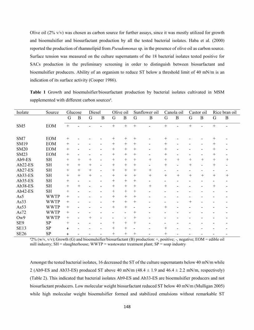
148
Olive oil (2% v/v) was chosen as carbon source for further assays, since it was mostly utilized for growth
and bioemulsifier and biosurfactant production by all the tested bacterial isolates. Haba et al. (2000)
reported the production of rhamnolipid from Pseudomonas sp. in the presence of olive oil as carbon source.
Surface tension was measured on the culture supernatants of the 18 bacterial isolates tested positive for
SACs production in the preliminary screening in order to distinguish between biosurfactant and
bioemulsifier producers. Ability of an organism to reduce ST below a threshold limit of 40 mN/m is an
indication of its surface activity (Cooper 1986).
Table 1 Growth and bioemulsifier/biosurfactant production by bacterial isolates cultivated in MSM
supplemented with different carbon sourcesa.
Isolate Source Glucose Diesel Olive oil Sunflower oil Canola oil Castor oil Rice bran oil G B G B G B G B G B G B G B
SM5 EOM + -
-
- + + + - + - + - + -
SM7 EOM + - - - + + + - + - - - + - SM19 EOM + - - - + + + - + - - - + - SM20 EOM + - - - + + + - + - - - + - SM23 EOM + - - - + + + - + - - - + - Ab9-ES SH + + + - + + + + + + + + + + Ab22-ES SH + + + - + + + - + - + - + - Ab27-ES SH + + + - + + + + - - - - - - Ab33-ES SH + + + - + + + + + + + + + + Ab35-ES SH + - - - + + + - - - - - - - Ab38-ES SH + + - - + + + + + - - - + - Ab42-ES SH + - - - + + + - - - - - - - As5 WWTP + - - - + + + - - - - - - - As33 WWTP + - - - + + + - - - + - - - As53 WWTP + - - - + + - - + - - - - - As72 WWTP + - - - - - + - - - - - - - Ow9 WWTP + - + - - - + - - - - - - - SE9 SP + - - - + + + - - - - - - - SE13 SP + - - - + + - - + - - - - - SE26 SP + - - - + + + - + - - - - -
a2% (w/v, v/v); Growth (G) and bioemulsifier/biosurfactant (B) production: +, positive; -, negative; EOM = edible oil mill industry; SH = slaughterhouse; WWTP = wastewater treatment plant; SP = soap industry
Amongst the tested bacterial isolates, 16 decreased the ST of the culture supernatants below 40 mN/m while
2 (Ab9-ES and Ab33-ES) produced ST above 40 mN/m (48.4 ± 1.9 and 46.4 ± 2.2 mN/m, respectively)
(Table 2). This indicated that bacterial isolates Ab9-ES and Ab33-ES are bioemulsifier producers and not
biosurfactant producers. Low molecular weight biosurfactant reduced ST below 40 mN/m (Mulligan 2005)
while high molecular weight bioemulsifier formed and stabilized emulsions without remarkable ST
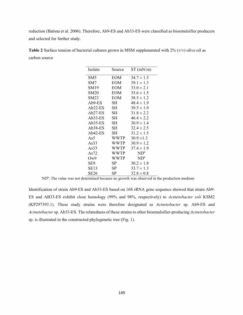
149
reduction (Batista et al. 2006). Therefore, Ab9-ES and Ab33-ES were classified as bioemulsifier producers
and selected for further study.
Table 2 Surface tension of bacterial cultures grown in MSM supplemented with 2% (v/v) olive oil as
carbon source
Isolate Source ST (mN/m)
SM5 EOM 34.7 ± 1.5 SM7 EOM 39.1 ± 1.3 SM19 EOM 33.0 ± 2.1 SM20 EOM 35.6 ± 1.5 SM23 EOM 38.5 ± 1.2 Ab9-ES SH 48.4 ± 1.9 Ab22-ES SH 39.5 ± 1.9 Ab27-ES SH 31.8 ± 2.2 Ab33-ES SH 46.4 ± 2.2 Ab35-ES SH 30.9 ± 1.4 Ab38-ES SH 32.4 ± 2.5 Ab42-ES SH 31.2 ± 1.5 As5 WWTP 30.9 ±1.3 As33 WWTP 30.9 ± 1.2 As53 WWTP 37.4 ± 1.9 As72 WWTP NDb
Ow9 WWTP NDb
SE9 SP 30.2 ± 1.8 SE13 SP 33.7 ± 1.3 SE26 SP 32.8 ± 0.8
NDb: The value was not determined because no growth was observed in the production medium
Identification of strain Ab9-ES and Ab33-ES based on 16S rRNA gene sequence showed that strain Ab9-
ES and AB33-ES exhibit close homology (99% and 98%, respectively) to Acinetobacter soli KSM2
(KP297393.1). These study strains were therefore designated as Acinetobacter sp. Ab9-ES and
Acinetobacter sp. Ab33-ES. The relatedness of these strains to other bioemulsifier-producing Acinetobacter
sp. is illustrated in the constructed phylogenetic tree (Fig. 1).
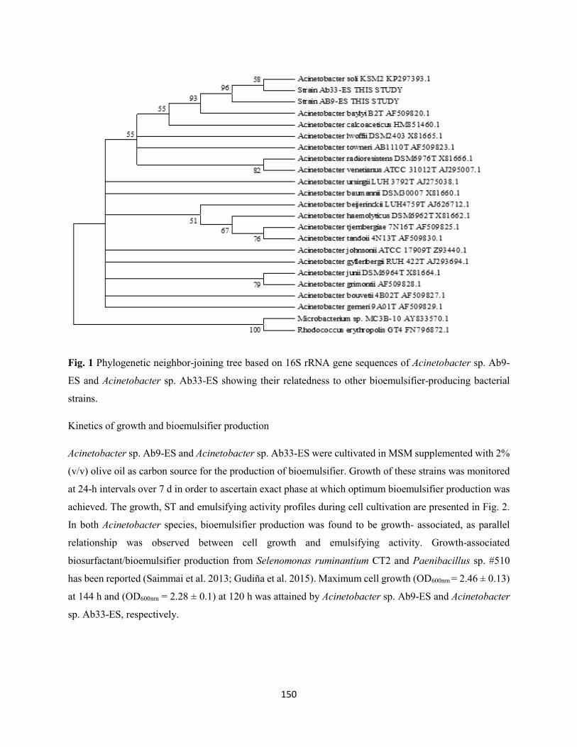
150
Fig. 1 Phylogenetic neighbor-joining tree based on 16S rRNA gene sequences of Acinetobacter sp. Ab9-
ES and Acinetobacter sp. Ab33-ES showing their relatedness to other bioemulsifier-producing bacterial
strains.
Kinetics of growth and bioemulsifier production
Acinetobacter sp. Ab9-ES and Acinetobacter sp. Ab33-ES were cultivated in MSM supplemented with 2%
(v/v) olive oil as carbon source for the production of bioemulsifier. Growth of these strains was monitored
at 24-h intervals over 7 d in order to ascertain exact phase at which optimum bioemulsifier production was
achieved. The growth, ST and emulsifying activity profiles during cell cultivation are presented in Fig. 2.
In both Acinetobacter species, bioemulsifier production was found to be growth- associated, as parallel
relationship was observed between cell growth and emulsifying activity. Growth-associated
biosurfactant/bioemulsifier production from Selenomonas ruminantium CT2 and Paenibacillus sp. #510
has been reported (Saimmai et al. 2013; Gudiña et al. 2015). Maximum cell growth (OD600nm = 2.46 ± 0.13)
at 144 h and (OD600nm = 2.28 ± 0.1) at 120 h was attained by Acinetobacter sp. Ab9-ES and Acinetobacter
sp. Ab33-ES, respectively.
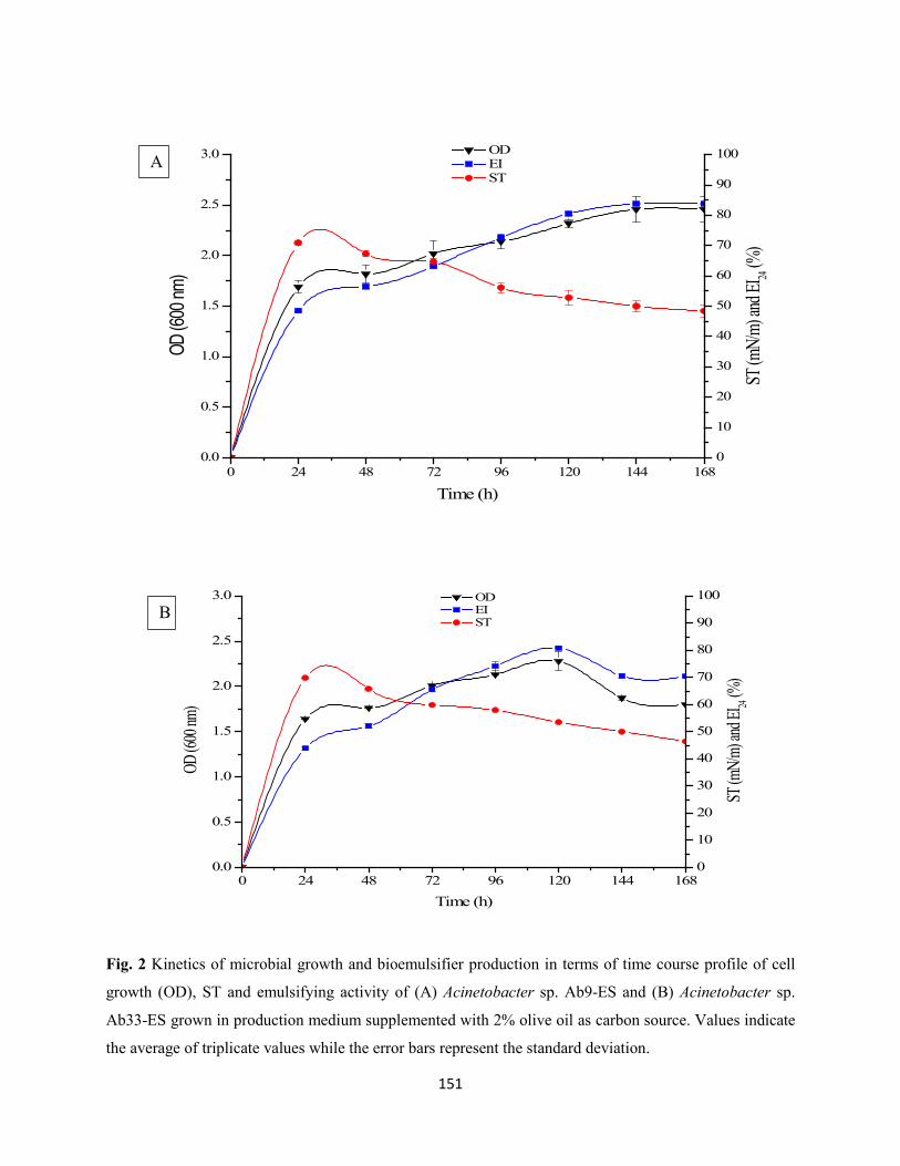
151
0 24 48 72 96 120 144 1680.0
0.5
1.0
1.5
2.0
2.5
3.0 OD EI ST
Time (h)
OD (6
00 nm
)
0
10
20
30
40
50
60
70
80
90
100
ST (m
N/m)
and E
I 24 (%
)
0 24 48 72 96 120 144 1680.0
0.5
1.0
1.5
2.0
2.5
3.0 OD EI ST
Time (h)
OD (6
00 nm
)
0
10
20
30
40
50
60
70
80
90
100
ST (m
N/m)
and E
I 24 (%
)
Fig. 2 Kinetics of microbial growth and bioemulsifier production in terms of time course profile of cell
growth (OD), ST and emulsifying activity of (A) Acinetobacter sp. Ab9-ES and (B) Acinetobacter sp.
Ab33-ES grown in production medium supplemented with 2% olive oil as carbon source. Values indicate
the average of triplicate values while the error bars represent the standard deviation.
A
B
152
Maximum bioemulsifier production was recorded at different phases of growth in both studied
Acinetobacter sp. In Acinetobacter sp. Ab9-ES, the EI24 reached the highest value of 83.8 ± 0.17% at 168
h during stationary phase of growth. This is in agreement with the findings of Pornsunthorntawee et al.
(2008) where maximum biosurfactant production by B. subtilis PT2 was observed during stationary phase.
However, Acinetobacter sp. Ab33-ES attained highest emulsifying activity of 80.8 ± 0.29% at 120 h during
late exponential phase, after which the emulsifying activity decreased probably due to the degradation of
the bioemulsifier. Maximum emulsifying activity of 60 ± 1% of Aeribacillus pallidus YM-1 has been
reported during exponential phase (Zheng et al. 2012). A slight reduction in ST from 70.9 ± 0.85 to 48.4
±1.9 mN/m and 70.0 ± 2.5 to 46.4 ± 2.2 mN/m was observed in the culture supernatants of Acinetobacter
sp. Ab9-ES and Acinetobacter sp. Ab33-ES, respectively as the incubation period increased from 24 to 168
h (Fig. 2). Ethanol precipitation of culture supernatants gave rise to the formation of dried whitish
bioemulsifier powders, denoted as bioemulsifier XB9 and YB33 with a resultant production yields of 4.52
± 0.03 and 4.31 ± 0.04 g/L by Acinetobacter sp. Ab9-ES and Acinetobacter sp. Ab33-ES, respectively.
Composition of bioemulsifier
Preliminary studies on chemical composition of the bioemulsifier XB9 and YB33 were conducted. Results
showed that XB9 consisted predominantly of carbohydrates (65.5%) and proteins (31%) while YB33
composed of carbohydrates (67.8%) and proteins (25%). This was further confirmed by FT-IR analysis
(Fig. 3). The spectra obtained in both bioemulsifiers exhibit some similarities, especially broad absorption
bands at 3275.86 cm-1 (XB9) and 3287.13 cm-1(YB33) which revealed the presence of hydroxyl and amine
groups typical of carbohydrates and peptides, respectively. Absorption peaks in higher frequencies have
been reported to be peculiar features of polysaccharides (Stuart 2004). The presence of weak C-H stretching
bands at 2926.78 and 2978.14 cm-1 in YB33 characterized symmetric stretch (γC-H) of CH2 and CH3 groups
of aliphatic chains, and O-H content of polysaccharides. In addition, the distinct absorption band at 1646.44
cm-1 in YB33 indicates the stretching mode of CO-N bond of acetamido groups of N-acetylated sugars
found mostly in microbial polysaccharides (Christensen 1989), as well as N-H bend of primary amines.
Similarly, a band at 1634 cm-1in XB9 also correspond to N-H bend. Weak absorption bands at 1459.62 and
1405.40 cm-1 in XB9 correspond to C-H stretch of aliphatic amines. Furthermore, absorption bands at
1059.54, 978.69, 861.74 and 724.11 cm-1 indicate C-N, C-H, N-H and C-H groups, respectively of aliphatic
amines. Similarly, in the case of YB33, absorption bands stretching between 877.52-1453.07 cm-1 are
typical of C-H and C-N functional groups of amino sugars within the polysaccharides. The FT-IR spectra
obtained in the present study suggest that the bioemulsifier XB9 and YB33 composed predominantly of
polysaccharides and proteins.
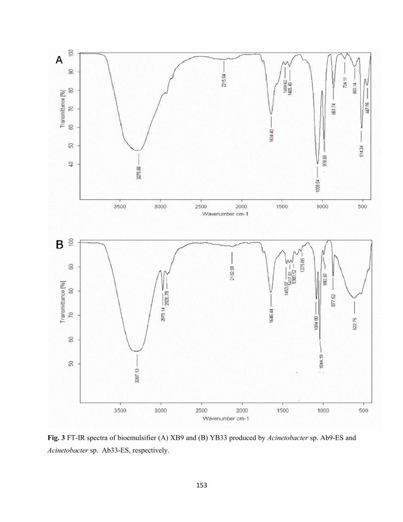
153
Fig. 3 FT-IR spectra of bioemulsifier (A) XB9 and (B) YB33 produced by Acinetobacter sp. Ab9-ES and
Acinetobacter sp. Ab33-ES, respectively.
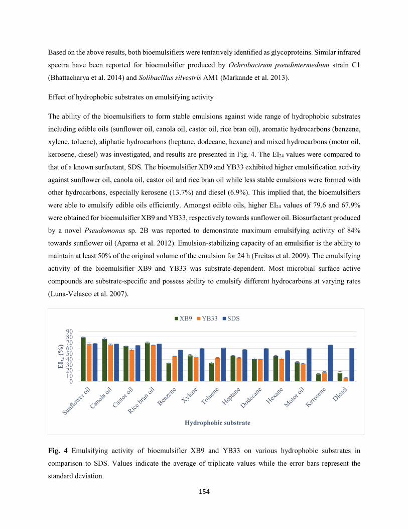
154
Based on the above results, both bioemulsifiers were tentatively identified as glycoproteins. Similar infrared
spectra have been reported for bioemulsifier produced by Ochrobactrum pseudintermedium strain C1
(Bhattacharya et al. 2014) and Solibacillus silvestris AM1 (Markande et al. 2013).
Effect of hydrophobic substrates on emulsifying activity
The ability of the bioemulsifiers to form stable emulsions against wide range of hydrophobic substrates
including edible oils (sunflower oil, canola oil, castor oil, rice bran oil), aromatic hydrocarbons (benzene,
xylene, toluene), aliphatic hydrocarbons (heptane, dodecane, hexane) and mixed hydrocarbons (motor oil,
kerosene, diesel) was investigated, and results are presented in Fig. 4. The EI24 values were compared to
that of a known surfactant, SDS. The bioemulsifier XB9 and YB33 exhibited higher emulsification activity
against sunflower oil, canola oil, castor oil and rice bran oil while less stable emulsions were formed with
other hydrocarbons, especially kerosene (13.7%) and diesel (6.9%). This implied that, the bioemulsifiers
were able to emulsify edible oils efficiently. Amongst edible oils, higher EI24 values of 79.6 and 67.9%
were obtained for bioemulsifier XB9 and YB33, respectively towards sunflower oil. Biosurfactant produced
by a novel Pseudomonas sp. 2B was reported to demonstrate maximum emulsifying activity of 84%
towards sunflower oil (Aparna et al. 2012). Emulsion-stabilizing capacity of an emulsifier is the ability to
maintain at least 50% of the original volume of the emulsion for 24 h (Freitas et al. 2009). The emulsifying
activity of the bioemulsifier XB9 and YB33 was substrate-dependent. Most microbial surface active
compounds are substrate-specific and possess ability to emulsify different hydrocarbons at varying rates
(Luna-Velasco et al. 2007).
Fig. 4 Emulsifying activity of bioemulsifier XB9 and YB33 on various hydrophobic substrates in
comparison to SDS. Values indicate the average of triplicate values while the error bars represent the
standard deviation.
0102030405060708090
EI 2
4(%
)
Hydrophobic substrate
XB9 YB33 SDS
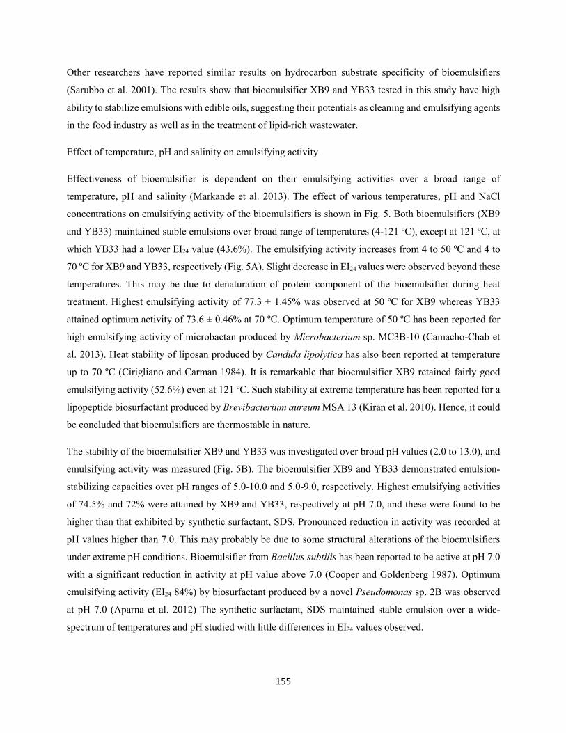
155
Other researchers have reported similar results on hydrocarbon substrate specificity of bioemulsifiers
(Sarubbo et al. 2001). The results show that bioemulsifier XB9 and YB33 tested in this study have high
ability to stabilize emulsions with edible oils, suggesting their potentials as cleaning and emulsifying agents
in the food industry as well as in the treatment of lipid-rich wastewater.
Effect of temperature, pH and salinity on emulsifying activity
Effectiveness of bioemulsifier is dependent on their emulsifying activities over a broad range of
temperature, pH and salinity (Markande et al. 2013). The effect of various temperatures, pH and NaCl
concentrations on emulsifying activity of the bioemulsifiers is shown in Fig. 5. Both bioemulsifiers (XB9
and YB33) maintained stable emulsions over broad range of temperatures (4-121 ºC), except at 121 ºC, at
which YB33 had a lower EI24 value (43.6%). The emulsifying activity increases from 4 to 50 ºC and 4 to
70 ºC for XB9 and YB33, respectively (Fig. 5A). Slight decrease in EI24 values were observed beyond these
temperatures. This may be due to denaturation of protein component of the bioemulsifier during heat
treatment. Highest emulsifying activity of 77.3 ± 1.45% was observed at 50 ºC for XB9 whereas YB33
attained optimum activity of 73.6 ± 0.46% at 70 ºC. Optimum temperature of 50 ºC has been reported for
high emulsifying activity of microbactan produced by Microbacterium sp. MC3B-10 (Camacho-Chab et
al. 2013). Heat stability of liposan produced by Candida lipolytica has also been reported at temperature
up to 70 ºC (Cirigliano and Carman 1984). It is remarkable that bioemulsifier XB9 retained fairly good
emulsifying activity (52.6%) even at 121 ºC. Such stability at extreme temperature has been reported for a
lipopeptide biosurfactant produced by Brevibacterium aureum MSA 13 (Kiran et al. 2010). Hence, it could
be concluded that bioemulsifiers are thermostable in nature.
The stability of the bioemulsifier XB9 and YB33 was investigated over broad pH values (2.0 to 13.0), and
emulsifying activity was measured (Fig. 5B). The bioemulsifier XB9 and YB33 demonstrated emulsion-
stabilizing capacities over pH ranges of 5.0-10.0 and 5.0-9.0, respectively. Highest emulsifying activities
of 74.5% and 72% were attained by XB9 and YB33, respectively at pH 7.0, and these were found to be
higher than that exhibited by synthetic surfactant, SDS. Pronounced reduction in activity was recorded at
pH values higher than 7.0. This may probably be due to some structural alterations of the bioemulsifiers
under extreme pH conditions. Bioemulsifier from Bacillus subtilis has been reported to be active at pH 7.0
with a significant reduction in activity at pH value above 7.0 (Cooper and Goldenberg 1987). Optimum
emulsifying activity (EI24 84%) by biosurfactant produced by a novel Pseudomonas sp. 2B was observed
at pH 7.0 (Aparna et al. 2012) The synthetic surfactant, SDS maintained stable emulsion over a wide-
spectrum of temperatures and pH studied with little differences in EI24 values observed.
156
Investigation of the effect of salinity on emulsifying activity of the bioemulsifiers XB9 and YB33 at NaCl
concentrations ranging from 1-10% (w/v) indicated the stability of both bioemulsifiers up to 6% salinity
(Fig. 5C). Similar results have been observed previously with bioemulsifier produced from Ochrobactrum
pseudintermedium strain C1 (Bhattacharya et al. 2014). Maximum activity (57.4%) was observed at NaCl
concentration of 3% for XB9. On the contrary, YB33 demonstrated highest activity of 61.2% at NaCl
concentration of 5%. Beyond these salinity levels, the emulsifying activity declined drastically. Stability of
these bioemulsifiers at NaCl concentrations up to 6% warrant intensified bioprospection in the remediation
of polluted intertidal zone.
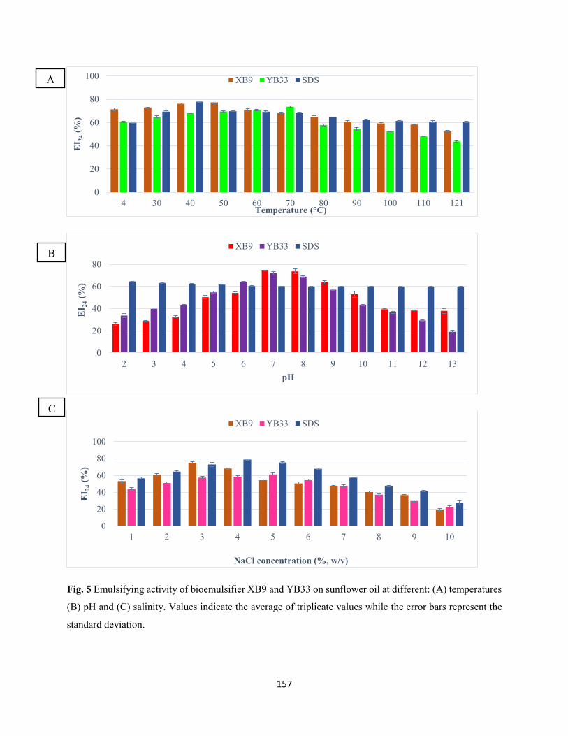
157
Fig. 5 Emulsifying activity of bioemulsifier XB9 and YB33 on sunflower oil at different: (A) temperatures
(B) pH and (C) salinity. Values indicate the average of triplicate values while the error bars represent the
standard deviation.
0
20
40
60
80
100
4 30 40 50 60 70 80 90 100 110 121
EI 2
4(%
)
Temperature (°C)
XB9 YB33 SDS
0
20
40
60
80
2 3 4 5 6 7 8 9 10 11 12 13
EI 2
4(%
)
pH
XB9 YB33 SDS
0
20
40
60
80
100
1 2 3 4 5 6 7 8 9 10
EI 2
4(%
)
NaCl concentration (%, w/v)
XB9 YB33 SDS
A
B
C
158
Conclusions
In the present study, glycoprotein bioemulsifier production from two indigenous Acinetobacter sp. isolated
from lipid-rich wastewater was reported in the presence of olive oil as inducible substrate, thus contributing
to the reduction of process cost. The bioemulsifiers demonstrated high emulsifying activity in the presence
of edible oils including sunflower oil, canola oil, castor oil and rice bran oil, as well as stability at extreme
temperature, moderate salinity and pH. Hence, these bioemulsifiers (XB9 and YB33) represent a good
candidate for various industrial and environmental applications, especially in the remediation of lipid-rich
wastewater and enhanced-oil recovery.
Acknowledgements
The financial support of the National Research Foundation (NRF) of South Africa towards this research is
hereby acknowledged. Opinions expressed and conclusions arrived at, are those of the authors and are not
necessarily to be attributed to the NRF. The first author is thankful to Dr. Ajit Kumar for providing valuable
technical assistance to accomplish the research work.
Conflict of interests
The authors declare that there are no conflicts of interest regarding the publication of this paper
References
Adulkar, TV, Rathod VK (2015) Pre-treatment of high fat content dairy wastewater using different
commercial lipases. Desalin Water Treat 53(9):2450-2455
Akinbowale OL, Peng H, Grant P, Barton MD (2007) Antibiotic and heavy metal resistance in motile
aeromonads and pseudomonads from rainbow trout (Oncorhynchus mykiss) farms in Australia. Int
J Antimicrob Agents 30(2):177-182
Aparna A, Srinikethan G, Smitha H (2012) Production and characterization of biosurfactant produced by a
novel Pseudomonas sp. 2B. Colloids Surf B: Biointerfaces 95:23-29
Banat IM, Franzetti A, Gandolfi I, Bestetti G, Martinotti MG, Fracchia L, Smyth TJ, Marchant R (2010)
Microbial biosurfactants production, applications and future potential. Appl Microbiol Biotechnol
87(2):427-444
Banat IM, Satpute SK, Cameotra SS, Patil R, Nyayanit NV (2014) Cost effective technologies and
renewable substrates for biosurfactants’ production. Front Microbiol 5:697
159
Batista SB, Mounteer AH, Amorim FR, Tótola MR (2006) Isolation and characterization of
biosurfactant/bioemulsifier-producing bacteria from petroleum contaminated sites. Bioresour
Technol 97(6):868-875
Bhattacharya M, Biswas D, Sana S, Datta S (2014) Utilization of waste engine oil by Ochrobactrum
pseudintermedium strain C1 that secretes an exopolysaccharide as a bioemulsifier. Biocatal Agric
Biotechnol 3(4):167-176
Brooksbank AM, Latchford JW, Mudge SM (2007) Degradation and modification of fats, oils and grease
by commercial microbial supplements. World J Microbiol Biotechnol 23(7):977-985
Camacho-Chab JC, Guézennec J, Chan-Bacab MJ, Ríos-Leal E, Sinquin C, Muñiz-Salazar R, De la Rosa-
García SDC, Reyes-Estebanez M, Ortega-Morales BO (2013) Emulsifying activity and stability
of a non-toxic bioemulsifier synthesized by Microbacterium sp. MC3B-10. Int J Mol Sci
14(9):18959-18972
Christensen BE (1989) The role of extracellular polysaccharides in biofilms. J Biotechnol 10(3-4):181-202
Cirigliano MC, Carman GM (1984) Isolation of a bioemulsifier from Candida lipolytica. Appl Environ
Microbiol 48(4):747-750
Cooper DG (1986) Biosurfactants. Microbiol Sci 3(5):145-149
Cooper DG, Goldenberg BG (1987) Surface-active agents from two Bacillus species. Appl Environ
Microbiol 53(2):224-229
Dastgheib SMM, Amoozegar MA, Elahi E, Asad S, Banat IM (2008) Bioemulsifier production by a
halothermophilic Bacillus strain with potential applications in microbially enhanced oil recovery.
Biotechnol Lett 30(2):263-270
Dubois M, Gilles KA, Hamilton JK, Rebers PA, Smith F (1956) Colorimetric method for determination of
sugars and related substances. Anal Chem 28(3):350-356
Foght JM, Gutnick DL, Westlake DWS (1989) Effect of emulsan on biodegradation of crude oil by pure
and mixed bacterial cultures. Appl Environ Microbiol 55(1): 36-42
Folch J, Lees M, Stanley GHS (1957) A simple method for the isolation and purification of total lipids from
animal tissues. J Biol Chem 226(1):497-509
160
Franzetti A, Gandolfi I, Raimondi C, Bestetti G, Banat IM, Smyth TJ, Papacchini M, Cavallo M, Fracchia
L (2012) Environmental fate, toxicity, characteristics and potential applications of novel
bioemulsifiers produced by Variovorax paradoxus 7bCTS. Bioresour Technol 108:245-251
Freitas F, Alves V, Carvalheira M, Costa N, Oliveira R, Reis M (2009) Emulsifying behavior and
rheological properties of the extracellular polysaccharide produced by Pseudomonas oleovorans
grown on glycerol byproduct. Carbohydr Polym 78(3):549-556
Gudiña EJ, Pereira JFB, Costa R, Evtuguin DV, Coutinho JAP, Teixeira JA, Rodrigues LR (2015) Novel
bioemulsifier produced by a Paenibacillus strain isolated from crude oil. Microb Cell Fact 14:14
Gudiña EJ, Pereira JFB, Rodrigues LR, Coutinho JAP, Teixeira JA (2012) Isolation and study of
microorganisms from oil samples for application in microbial enhanced oil recovery. Int Biodeter
Biodegrad 68:56-64
Haba E, Espuny MJ, Busquets M, Manresa A (2000) Screening and production of rhamnolipids by
Pseudomonas aeruginosa 47T2 NCIB40044 from waste frying oils. J Appl Microbiol 88(3):379-
387
Joshi S, Bharucha C, Desai AJ (2008) Production of biosurfactant and antifungal compound by fermented
food isolate Bacillus subtilis 20B. Bioresour Technol 99(11):4603-4608
Kiran GS, Thomas TA, Selvin J, Sabarathnam B, Lipton AP (2010) Optimization and characterization of
a new lipopeptide biosurfactant produced by marine Brevibacterium aureum MSA 13 in solid state
culture. Bioresour Technol 101: 2389-2396
Kitamoto D, Isoda H, Nakahara T (2002) Functions and potential applications of glycolipid biosurfactants:
from energy-saving materials to gene delivery carriers. J Biosci Bioeng 94(3):187–201
Lowry OH, Rosebrough NJ, Farr AI, Randall RJ (1951) Protein measurement with folin phenol reagent. J
Biol Chem 193(1):265-275
Luna JM, Rufino RD, Campos-Takaki GM, Sarubbo LA (2012) Properties of the biosurfactant produced
by Candida sphaerica cultivated in low-cost substrates. Chem Eng Trans 27:67-72
Luna-Velasco MA, Esparza-Garcia F, Canizares-Villanueva RO, Rodriguez-Vazquez R (2007) Production
and properties of a bioemulsifier synthesized by phenanthrene-degrading Penicillium sp. Proc
Biochem 42(3):310-314
Makkar RS, Cameotra SS, Banat IM (2011) Advances in utilization of renewable substrates for
biosurfactant production. AMB Express 1:5
161
Marchant R, Funston S, Uzoigwe C, Rahman PKSM, Banat IM (2014) Production of biosurfactants from
nonpathogenic bacteria. In: Kosaric N, Sukan FV (ed) Biosurfactants: Production and utilization-
processes, technologies, and economics, Boca Raton: CRC Press, pp. 73-82
Marchesi JR, Sato T, Weightman AJ, Martin TA, Fry JC, Hiom SJ, Dymock D, Wade WG (1998) Design
and evaluation of useful bacterium-specific PCR primers that amplify genes coding for bacterial
16S rRNA. Appl Environ Microbiol 64(2):795-799
Markande AR, Acharya SR, Nerurkar AS (2013) Physicochemical characterization of a thermostable
glycoprotein bioemulsifier from Solibacillus silvestris AM1. Proc Biochem 48(11):1800-1808
Martinez-Checa F, Toledo FL, El Mabrouki K, Quesada E, Calvo C (2007) Characteristics of bioemulsifier
V2-7 synthesized in culture media added of hydrocarbons: chemical composition, emulsifying
activity and rheological properties. Bioresour Technol 98(16):3130-3135
Morikawa M, Hirata Y, Imanaka T (2000) A study on the structure-function relationship of the lipopeptide
biosurfactants. Biochem Biophy Acta 1488(3): 211-218
Mulligan CN (2005) Environmental applications for biosurfactants. Environ Pollution 133(2):183-198
Navon-Venezia S, Zosim Z, Gottlieb A, Legmann R, Carmeli S, Ron EZ, Rosenberg E (1995) Alasan, a
new bioemulsifier from Acinetobacter radioresistens. Appl Environ Microbiol 61(9):3240-3244
Pansiripat S, Pornsunthorntawee O, Rujiravanit R, Kitiyanan B, Somboonthanate P, Chavadej S (2010)
Biosurfactant production by Pseudomonas aeruginosa SP4 using sequencing batch reactors: effect
of oil-to-glucose ratio. Biochem Eng J 49(2):185–191
Patil JR, Chopade BA (2001) Studies on bioemulsifier production by Acinetobacter strains isolated from
healthy human skin. J Appl Microbiol 91(2):290-298
Phetrong K, H-Kittikun A, Maneerat S (2008) Production and characterization of bioemulsifier from a
marine bacterium, Acinetobacter calcoaceticus subsp. anitratus SM7. Songklanakarin J Sci
Technol 30(3):297-305
Pornsunthorntawee O, Arttaweeporn N, Paisanjit S, Somboonthanate P, Abe M, Rujiravanit R, Chavadej S
(2008) Isolation and comparison of biosurfactants produced by Bacillus subtilis PT2 and
Pseudomonas aeruginosa SP4 for microbial surfactant-enhanced oil recovery. Biochem Eng J
42(2):172-179
Rosenberg E (1986) Microbial surfactants. Critical Rev Biotechnol 3:109–132
Rosenberg E, Ron EZ (1999) High- and low-molecular mass microbial surfactants. Appl Microbiol
Biotechnol 52(2):154-162
162
Rosenberg E, Rubinovitz C, Gottlieb A, Rosenhak S, Ron EZ (1988) Production of biodispersan by
Acinetobacter calcoaceticus A2. Appl Environ Microbiol 54(2):317-322
Rupani PF, Singh RP, Ibrahim MH, Esa N (2010) Review of current palm oil mill effluent (POME)
treatment methods: vermicomposting as a sustainable practice. World Appl Sci J 11(1):70-81
Saimmai A, Onlamool T, Sobhon V, Maneerat S (2013) An efficient biosurfactant- producing bacterium
Selenomonas ruminantium CT2, isolated from mangrove sediment in south of Thailand. World J
Microbiol Biotechnol 29(1):87-102
Saisa-ard K, Maneerat S, Saimmai A (2013) Isolation and characterization of biosurfactant-producing
bacteria isolated from palm oil industry and evaluation for biosurfactants production using low-
cost substrates. Bio Technologia 94(3):275-284
Santos DKF, Rufino RD, Luna JM, Santos VA, Sarubbo LA (2016) Biosurfactants: Multifunctional
biomolecules of the 21st century. Int J Mol Sci 17(3):401
Sar N, Rosenberg E (1983) Emulsifier production by Acinetobacter calcoaceticus strains. Curr Microbiol
9(6):309-313
Sarubbo LA, Farias CBB, Campos-Takaki GM (2007) Co-utilization of canola oil and glucose on the
production of a surfactant by Candida lipolytica. Curr Microbiol 54(1):68-73
Sarubbo LA, Marcal MC, Neves MLC, Silva MPC, Porto ALF, Campos-Takaki GM (2001) Bioemulsifier
production in batch culture using glucose as carbon source by Candida lipolytica. Appl Biochem
Biotechnol 95(1):59-67
Stuart BH (2004) Organic molecules. In stuart, B.H. (Ed.), Infrared spectroscopy: fundamental and
applications, John Wiley & Sons, New Jersey, USA, pp. 44-70
Tamura K, Stecher G, Peterson D, Filipski A, Kumar S (2013) MEGA 6: Molecular Evolutionary Genetics
Analysis version 6.0. Mol Biol Evo 30(12):2725-2729
Uzoigwe C, Burgess JG, Ennis CJ, Rahman PKSM (2015) Bioemulsifiers are not biosurfactants and require
different screening approaches. Front Microbiol 6: 245
Zheng C, Li Z, Su J, Zhang R, Liu C, Zhao M (2012) Characterization and emulsifying property of a novel
bioemulsifier by Aeribacillus pallidus YM-1. J Appl Microbiol 113(1):44-51
163
CHAPTER SIX
OPTIMIZATION OF BIOPROCESS PARAMETERS FOR ENHANCED PROTEASE
PRODUCTION BY BACILLUS ARYABHATTAI AB15-ES USING RESPONSE SURFACE
METHODOLOGY
This chapter has been submitted in the current format to the ‘Bioprocess and Biosystems
Engineering’
164
Optimization of bioprocess parameters for enhanced protease production by Bacillus aryabhattai
Ab15-ES using response surface methodology
Adegoke Isiaka Adetunji and Ademola Olufolahan Olaniran*
Discipline of Microbiology, School of Life Sciences, College of Agriculture, Engineering and Science,
University of KwaZulu-Natal (Westville campus), Private Bag X54001, Durban 4000, Republic of South
Africa.
* Corresponding author. Tel: +27 31 260 7400; Fax: +27 31 260 7809
E-mail address: [email protected]
Abstract Microbial proteases are the largest and complex group of enzymes that catalyze the hydrolysis of
proteins by cleavage of peptide bonds that exist between amino acid residues in a polypeptide chain. They
constitute more than 40% of the total worldwide production of enzymes, and their production is greatly
influenced by fermentation media composition in addition to culture conditions such as pH, temperature
and inoculum volume. Hence, response surface methodology (RSM) was applied for the optimization of
bioprocess parameters for enhanced extracellular protease production by an indigenous protease-producing
Bacillus aryabhattai Ab15-ES. “One-variable at-a-time” approach was employed for the selection of best
carbon and nitrogen sources for maximum protease production. The optimum conditions for extracellular
protease production obtained from quadratic model of RSM were temperature 40 °C, pH 7.8, inoculum
volume 2.5% (v/v), maltose 12.35 g/L and beef extract 5.30 g/L. The coincidence of observed protease
production (247.84 ± 2.31 U/mL) with predicted protease yield (248.19 U/mL) coupled with high
coefficient of determination (R2 = 0.9880, P < 0.01) confirmed the validity of the model. A 4.4-fold increase
in protease production was obtained in the optimized medium using the basal medium as a reference. The
present study is the first report on protease production and optimization from Bacillus aryabhattai using
response surface methodology. These findings suggest a rational choice of optimized process conditions
for commercial production of protease by Bacillus aryabhattai Ab15-ES for various biotechnological
applications.
Keywords Protease; Bioprocess parameters; Bacillus aryabhattai; Response surface methodology; Lipid-
rich wastewater; Optimization
165
Introduction
Proteases are the largest and most important group of enzymes constituting more than 60% of the total
industrial enzyme market [1, 2]. They play a vital role in industrial biotechnology, especially in detergent,
food, leather, and textile industries; pharmaceutical, organic chemical synthesis and wastewater treatment
[3]. Proteases are ubiquitous in nature, and are produced by plants, animals and microbes. However,
proteases of microbial origin are mostly preferred due to their broad biochemical diversity, ease of genetic
manipulation, feasibility of mass culture, limited space for growth with high productivity, longer shelf life
and less stringent storage requirements [4]. A wide range of microorganisms including bacteria, fungi, and
yeasts produces proteases. However, mass production of proteases for commercial applications has been
recorded among bacteria mainly in the genus Bacillus [5, 6], including Bacillus clausii [7], Bacillus cereus
[8], Bacillus circulans [9], Bacillus lehensis [10], Bacillus licheniformis [6], Bacillus megaterium [11],
Bacillus mojavensis [12], Bacillus sterothermophilus [13], Bacillus pumilus [14] and Bacillus subtilis [15].
In view of the growing trends of proteases which has prompted expansion of their uses, the overall costs of
the enzyme production from downstream purification still formed the ultimate challenge to successful white
biotechnology [16]. Optimization of fermentation conditions is the most important step for improved and
cost-effective bioprocesses [17, 18, 49]. Conventional methods involving change of ‘one-variable-at-a-
time’ is frequently used for the optimization of process parameters to achieve optimum cell density and
high productivity of microbial metabolite. However, these approaches are time-consuming and thus
expensive for simultaneous consideration of many variables [19]. In addition, they are laborious, require
more experimental data and unable to detect interactions and optimal conditions among variables [20, 21].
Due to these drawbacks, statistical experimental designs have been recognized as ideal ways for bioprocess
optimization studies [22, 23]. Statistical optimization by response surface methodology (RSM) is an
efficient experimental strategy to maintain optimal conditions in a multi-variable system [16, 24]. RSM is
an assemblage of mathematical and statistical techniques for modelling and analysis in applications where
a response of interest is influenced by several variables and the objective is to optimize this response [7].
This method helps to identify effective factors, study interactions, select optimum conditions and quantify
the relationships between one or more measured responses and the vital input factors in limited number of
experiments [22, 25, 26]. Several fermentation processes have been optimized using this methodology [27,
28]. Central composite design (CCD) is used to estimate the coefficients of a quadratic model, and it has
three groups of design points: two-level factorial or fractional factorial, axial and central points.
Protease optimization studies by CCD taking cognizance of fermentation medium composition and physical
factors as objective functions have been reported [7, 17, 29, 30, 50]. It has been well established that
166
extracellular protease production by microorganisms is influenced by media composition, especially carbon
and nitrogen sources in addition to culture conditions such as temperature, pH and inoculum volume [22,
31, 51]. Therefore, in the present study, optimization of process parameters was applied for enhanced
production of protease by Bacillus aryabhattai Ab15-ES. “One-variable-at-a-time” approach was used for
selection of best carbon and nitrogen sources. Thereafter, statistical optimization of the selected carbon and
nitrogen sources together with temperature, pH and inoculum size for enhanced protease production was
carried out using RSM.
Materials and methods
Chemicals
Azocasein used as substrate for protease assay was purchased from Sigma-Aldrich (St. Louis, MO, USA).
All other reagents were of analytical grade.
Sample collection and bacterial isolation procedures
Lipid-rich wastewater samples were collected from a poultry processing industry in Estcourt, KwaZulu-
Natal (KZN) province, South Africa into sterile 500 mL Schott bottles. Samples (10 mL) were transferred
to 250 mL Erlenmeyer flasks containing 100 mL mineral salt medium (MSM) consisting (in g/L): 1.0
KH2PO4, 1.0 K2HPO4, 1.0 NH4NO3, 0.2 MgSO4.7H2O, 0.02 CaCl2.2H2O and 0.05 FeCl3.6H2O
supplemented with 2% olive oil, and then incubated at 37 ºC for 48 h at 150 rpm [32]. Enriched cultures (5
mL) were subsequently transferred into the second flask and cultivated under the same conditions. This
process was repeated four times. Samples (0.1 mL) of the last enrichment cultures were serially diluted,
plated onto nutrient agar (NA) and incubated at 37ºC for 24 h. Pure cultures obtained by sub-culturing on
fresh NA plates were preserved at -80 ºC in a storage medium (500 µl of 80% sterile glycerol and 500 µL
culture suspension).
Screening of the bacterial isolate for proteolytic activity
A loopful of bacterial culture was inoculated into 10 mL sterile nutrient broth and then incubated at 150
rpm (37 ºC) for 24 h. Proteolytic activity of the bacterial isolate was detected by addition of 20 µL of
obtained culture supernatant into wells bored on skim milk agar plate [33]. Sterile nutrient broth was used
as a control. Plates were incubated at 37 ºC for 48 h and observed for the formation of clear zones of casein
hydrolysis, indicative of proteolytic activity. All experiments were carried out in triplicate.
167
Identification and phylogenetic analysis of the bacterial isolate
Genomic DNA extraction
Genomic DNA was isolated from bacterial culture grown overnight on NA plate at 37 °C using boiling
method described by [34] with modifications. Freshly grown colonies (5-7) were suspended in 70 µL sterile
distilled water, boiled at 100 ºC for 10 min and then cooled on ice for 5 min. This was centrifuged at 13000
rpm in a micro-centrifuge 5415D (Eppendorf, Hamburg, Germany) for 5 min. Cell-free supernatant was
transferred into fresh sterile Eppendorf tubes and used as template DNA for the PCR assay.
Amplification, sequencing and analysis of 16S rRNA gene
Amplification of the 16S rRNA gene was carried out using universal bacterial primers F-5'
CAGGCCTAACACATGCAAGTC-3' and R-5' GGGCGGTGTGTACAAGGC-3' [35]. The PCR mixture
consisted of 1× buffer, 1mM MgCl2, 0.2 mM of each dNTPs, 0.4 µm of each primer, 2 U of Taq polymerase
and 2 µL template DNA in a total volume of 25 µL. The mixture was subjected to amplification using a
thermocycler (Bio-Rad T100, Singapore) at initial denaturation of 95 ºC for 5 min, followed by 30 cycles
of; denaturation at 95 ºC for 1 min, annealing at 55 ºC for 1 min, extension at 72 ºC for 1min, 30 sec and
final extension at 72 ºC for 5 min. Amplification products were observed by electrophoresis in a 1.5% (w/v)
agarose gel at 100 V for 45 min in a 1% TAE buffer. After staining in 1 mg/mL ethidium bromide for 15
min, products were viewed by UV illumination (Syngene, UK), and then sequenced (inqaba biotech,
Pretoria, South Africa). The resulting sequences were compared with sequences in the GenBank database
of the National Centre for Biotechnology Information (NCBI) using the Nucleotide Basic Local Alignment
Search Tool (BLAST N) program (www.ncbi.n1m.nih.gov/BLAST). The sequences were aligned using the
multiple sequences alignment tool, Clustal W. Phylogenetic tree was constructed with Molecular
Evolutionary Genetics analysis (MEGA) version 6.0 software [36] using a neighbor-joining method.
Time course profile of growth and extracellular protease production
Basal medium used for growth and protease production profiling consisted of (in g/L): glucose, 10; peptone,
5; NaCl, 0.5; CaCl2. 2H2O, 0.1; K2HPO4, 0.3; KH2PO4, 0.4 and MgSO4. 7H2O, 0.1. The pH of the medium
was adjusted to 7.2 with 1N HCl or 1N NaOH. One milliliter of the bacterial culture (OD600nm = 1)) was
inoculated into 99 mL basal medium in 250 mL Erlenmeyer flasks. The flasks were incubated at 150 rpm
(37 °C) for 72 h in a shaking incubator. Samples were taken at 12-h intervals to determine bacterial growth
and protease production. Bacterial growth was determined by measuring optical density at 600 nm using
UV-Vis spectrophotometer (UVmini-1240, Schimadzu, Australia). Thereafter, samples were centrifuged at
168
10000 rpm for 10 min at 4 ºC using high-speed refrigerated centrifuge (Avanti J-26 XP, Beckman Coulter,
USA). The obtained cell-free culture supernatants were then assayed for protease activity. All experiments
were done in triplicate. Protease activity was determined according to the modified method of Secades and
Guijarro [37] using azocasein as a substrate. One unit (U) of enzyme activity was defined as the amount of
enzyme which yielded an increase in absorbance (440 nm) of 0.01 per minute under defined assay
conditions.
Optimization of nutritional parameters for protease production
Preliminary screening of different carbon and nitrogen sources for maximum protease production was
carried out using a “one-variable-at-a-time” approach. Simple and complex carbon sources such as fructose,
sucrose, lactose, maltose, galactose, mannose, soluble starch as well as inorganic and organic nitrogen
sources including NH4Cl, KNO3, yeast extract, skim milk, beef extract, malt extract, and soybean meal
were supplemented separately in the production medium for extracellular protease production. The carbon
and nitrogen sources were added at a concentration of 10 (g/L) and 5 (g/L), respectively. Homogenous
bacterial culture [1% (v/v) of OD600nm = 1] was inoculated into the production medium, and then incubated
on a shaking incubator at 150 rpm for 48 h at 37 ºC. The fermentation medium was centrifuged at 10000
rpm at 4 ºC for 10 min. The obtained cell-free supernatant was assayed for protease activity, as previously
described. All experiments were carried out in triplicate.
Statistical optimization of protease production
Response surface methodology
Response surface methodology (RSM) was used for the optimization of five independent variables
including; maltose, beef extract, temperature, pH and inoculum volume. In this experiment, face centered
CCD (FCCCD) was employed to determine optimum concentration of these variables for extracellular
protease production. A total of 50 experimental runs were generated using Design-Expert 10 (Stat Ease
Inc., Minneapolis, USA). Each numeric factor was varied at three different levels (-1, 0, +1). The central
coded value of the variables was considered as zero. The minimum and maximum ranges of variables
studied, and the full experimental plan with respect to their values in actual and coded levels are listed in
Table 1.
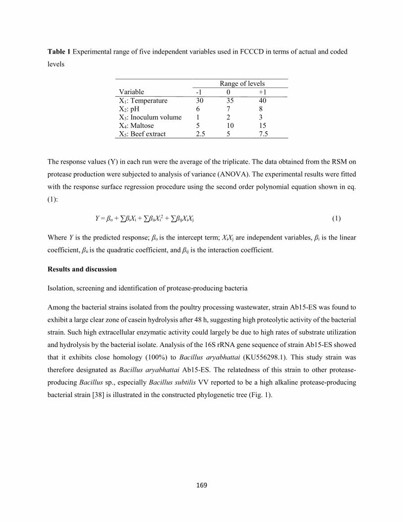
169
Table 1 Experimental range of five independent variables used in FCCCD in terms of actual and coded
levels
Variable
Range of levels -1 0 +1
X1: Temperature 30 35 40 X2: pH 6 7 8 X3: Inoculum volume 1 2 3 X4: Maltose 5 10 15 X5: Beef extract 2.5 5 7.5
The response values (Y) in each run were the average of the triplicate. The data obtained from the RSM on
protease production were subjected to analysis of variance (ANOVA). The experimental results were fitted
with the response surface regression procedure using the second order polynomial equation shown in eq.
(1):
Y = βo + ∑βiXi + ∑βiiXi2 + ∑βijXiXj (1)
Where Y is the predicted response; βo is the intercept term; XiXj are independent variables, βi is the linear
coefficient, βii is the quadratic coefficient, and βij is the interaction coefficient.
Results and discussion
Isolation, screening and identification of protease-producing bacteria
Among the bacterial strains isolated from the poultry processing wastewater, strain Ab15-ES was found to
exhibit a large clear zone of casein hydrolysis after 48 h, suggesting high proteolytic activity of the bacterial
strain. Such high extracellular enzymatic activity could largely be due to high rates of substrate utilization
and hydrolysis by the bacterial isolate. Analysis of the 16S rRNA gene sequence of strain Ab15-ES showed
that it exhibits close homology (100%) to Bacillus aryabhattai (KU556298.1). This study strain was
therefore designated as Bacillus aryabhattai Ab15-ES. The relatedness of this strain to other protease-
producing Bacillus sp., especially Bacillus subtilis VV reported to be a high alkaline protease-producing
bacterial strain [38] is illustrated in the constructed phylogenetic tree (Fig. 1).
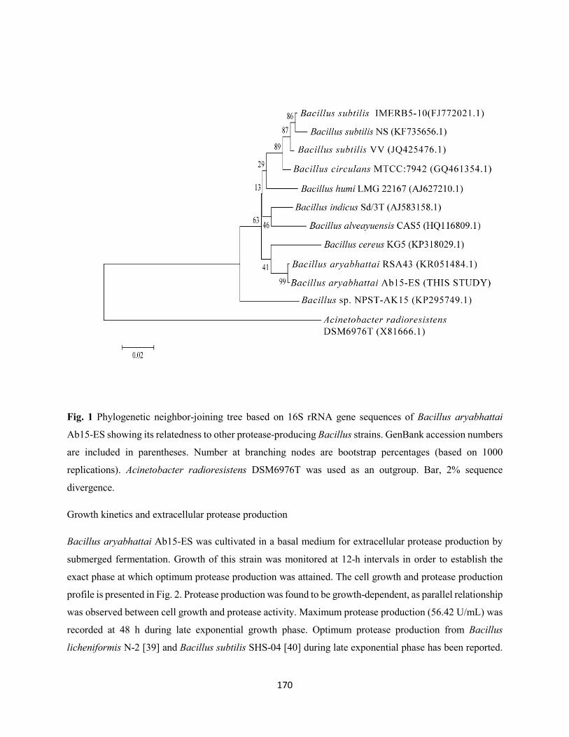
170
Fig. 1 Phylogenetic neighbor-joining tree based on 16S rRNA gene sequences of Bacillus aryabhattai
Ab15-ES showing its relatedness to other protease-producing Bacillus strains. GenBank accession numbers
are included in parentheses. Number at branching nodes are bootstrap percentages (based on 1000
replications). Acinetobacter radioresistens DSM6976T was used as an outgroup. Bar, 2% sequence
divergence.
Growth kinetics and extracellular protease production
Bacillus aryabhattai Ab15-ES was cultivated in a basal medium for extracellular protease production by
submerged fermentation. Growth of this strain was monitored at 12-h intervals in order to establish the
exact phase at which optimum protease production was attained. The cell growth and protease production
profile is presented in Fig. 2. Protease production was found to be growth-dependent, as parallel relationship
was observed between cell growth and protease activity. Maximum protease production (56.42 U/mL) was
recorded at 48 h during late exponential growth phase. Optimum protease production from Bacillus
licheniformis N-2 [39] and Bacillus subtilis SHS-04 [40] during late exponential phase has been reported.
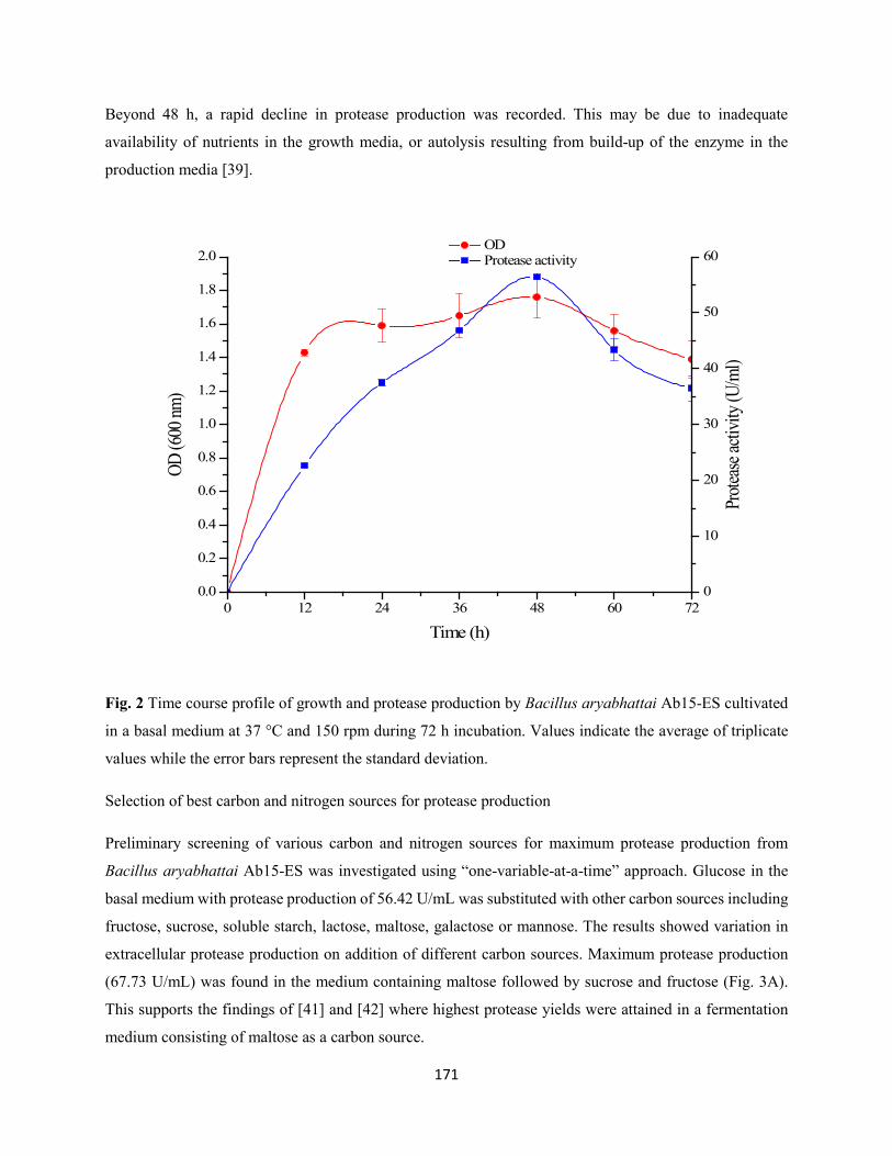
171
Beyond 48 h, a rapid decline in protease production was recorded. This may be due to inadequate
availability of nutrients in the growth media, or autolysis resulting from build-up of the enzyme in the
production media [39].
0 12 24 36 48 60 720.0
0.2
0.4
0.6
0.8
1.0
1.2
1.4
1.6
1.8
2.0 OD Protease activity
Time (h)
OD (6
00 nm
)
0
10
20
30
40
50
60
Prote
ase a
ctivit
y (U/
ml)
Fig. 2 Time course profile of growth and protease production by Bacillus aryabhattai Ab15-ES cultivated
in a basal medium at 37 °C and 150 rpm during 72 h incubation. Values indicate the average of triplicate
values while the error bars represent the standard deviation.
Selection of best carbon and nitrogen sources for protease production
Preliminary screening of various carbon and nitrogen sources for maximum protease production from
Bacillus aryabhattai Ab15-ES was investigated using “one-variable-at-a-time” approach. Glucose in the
basal medium with protease production of 56.42 U/mL was substituted with other carbon sources including
fructose, sucrose, soluble starch, lactose, maltose, galactose or mannose. The results showed variation in
extracellular protease production on addition of different carbon sources. Maximum protease production
(67.73 U/mL) was found in the medium containing maltose followed by sucrose and fructose (Fig. 3A).
This supports the findings of [41] and [42] where highest protease yields were attained in a fermentation
medium consisting of maltose as a carbon source.
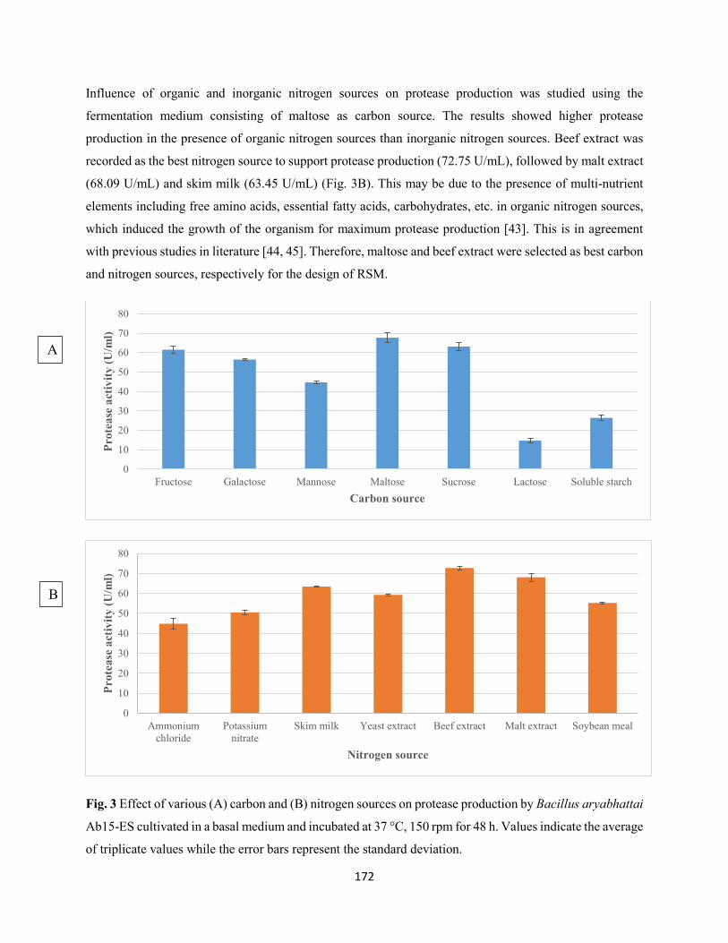
172
Influence of organic and inorganic nitrogen sources on protease production was studied using the
fermentation medium consisting of maltose as carbon source. The results showed higher protease
production in the presence of organic nitrogen sources than inorganic nitrogen sources. Beef extract was
recorded as the best nitrogen source to support protease production (72.75 U/mL), followed by malt extract
(68.09 U/mL) and skim milk (63.45 U/mL) (Fig. 3B). This may be due to the presence of multi-nutrient
elements including free amino acids, essential fatty acids, carbohydrates, etc. in organic nitrogen sources,
which induced the growth of the organism for maximum protease production [43]. This is in agreement
with previous studies in literature [44, 45]. Therefore, maltose and beef extract were selected as best carbon
and nitrogen sources, respectively for the design of RSM.
Fig. 3 Effect of various (A) carbon and (B) nitrogen sources on protease production by Bacillus aryabhattai
Ab15-ES cultivated in a basal medium and incubated at 37 °C, 150 rpm for 48 h. Values indicate the average
of triplicate values while the error bars represent the standard deviation.
0
10
20
30
40
50
60
70
80
Fructose Galactose Mannose Maltose Sucrose Lactose Soluble starch
Prot
ease
act
ivity
(U/m
l)
Carbon source
0
10
20
30
40
50
60
70
80
Ammoniumchloride
Potassiumnitrate
Skim milk Yeast extract Beef extract Malt extract Soybean meal
Prot
ease
act
ivity
(U/m
l)
Nitrogen source
A
B
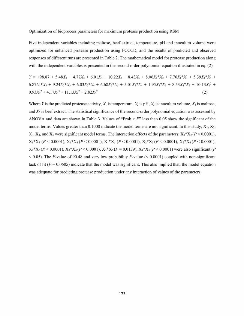
173
Optimization of bioprocess parameters for maximum protease production using RSM
Five independent variables including maltose, beef extract, temperature, pH and inoculum volume were
optimized for enhanced protease production using FCCCD, and the results of predicted and observed
responses of different runs are presented in Table 2. The mathematical model for protease production along
with the independent variables is presented in the second-order polynomial equation illustrated in eq. (2)
Y = +98.87 + 5.48X1 + 4.77X2 + 6.01X3 + 10.22X4 + 8.43X5 + 8.06X1*X2 + 7.76X1*X3 + 5.39X1*X4 +
6.87X1*X5 + 9.24X2*X3 + 6.03X2*X4 + 6.68X2*X5 + 5.01X3*X4 + 1.95X3*X5 + 8.53X4*X5 + 10.13X12 +
0.93X22 + 4.17X3
3 + 11.13X42 + 2.82X5
2 (2)
Where Y is the predicted protease activity, X1 is temperature, X2 is pH, X3 is inoculum volume, X4 is maltose,
and X5 is beef extract. The statistical significance of the second-order polynomial equation was assessed by
ANOVA and data are shown in Table 3. Values of “Prob > F” less than 0.05 show the significant of the
model terms. Values greater than 0.1000 indicate the model terms are not significant. In this study, X1, X2,
X3, X4, and X5 were significant model terms. The interaction effects of the parameters: X1*X2 (P < 0.0001),
X1*X3 (P < 0.0001), X1*X4 (P < 0.0001), X1*X5 (P < 0.0001), X2*X3 (P < 0.0001), X2*X4 (P < 0.0001),
X2*X5 (P < 0.0001), X3*X4 (P < 0.0001), X3*X5 (P = 0.0139), X4*X5 (P < 0.0001) were also significant (P
< 0.05). The F-value of 90.48 and very low probability F-value (< 0.0001) coupled with non-significant
lack of fit (P = 0.0685) indicate that the model was significant. This also implied that, the model equation
was adequate for predicting protease production under any interaction of values of the parameters.
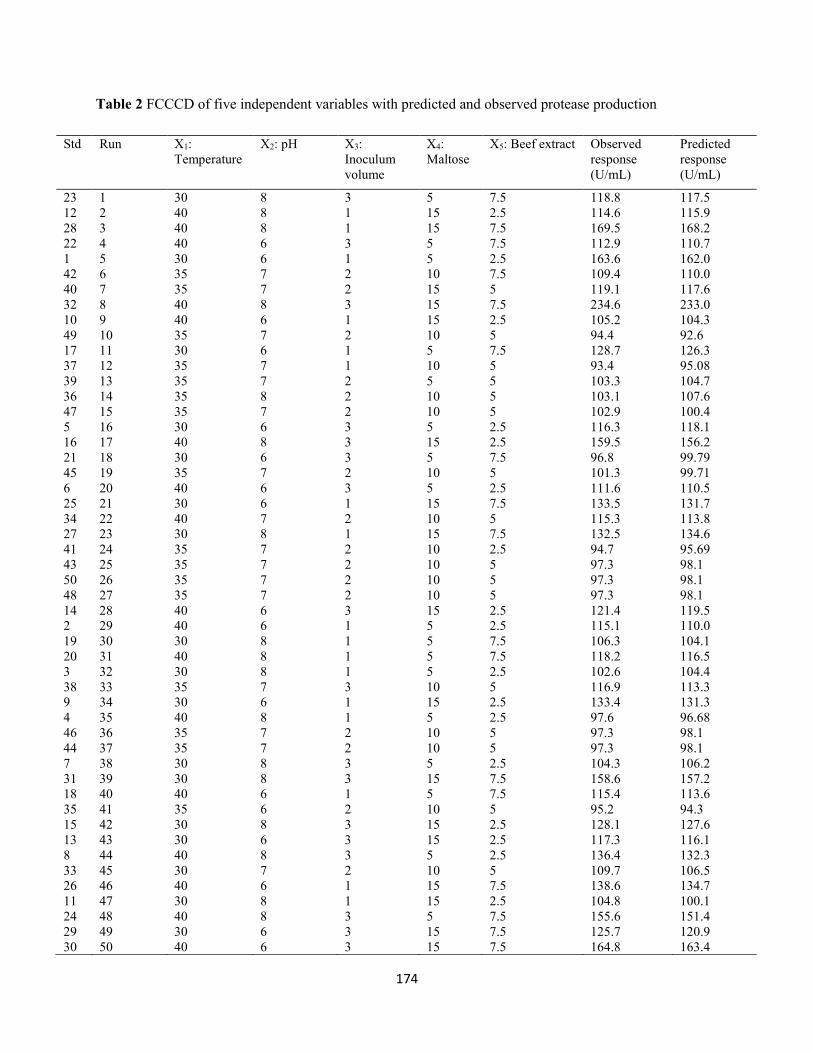
174
Table 2 FCCCD of five independent variables with predicted and observed protease production
Std Run X1: Temperature
X2: pH X3: Inoculum volume
X4: Maltose
X5: Beef extract Observed response (U/mL)
Predicted response (U/mL)
23 1 30 8 3 5 7.5 118.8 117.5 12 2 40 8 1 15 2.5 114.6 115.9 28 3 40 8 1 15 7.5 169.5 168.2 22 4 40 6 3 5 7.5 112.9 110.7 1 5 30 6 1 5 2.5 163.6 162.0 42 6 35 7 2 10 7.5 109.4 110.0 40 7 35 7 2 15 5 119.1 117.6 32 8 40 8 3 15 7.5 234.6 233.0 10 9 40 6 1 15 2.5 105.2 104.3 49 10 35 7 2 10 5 94.4 92.6 17 11 30 6 1 5 7.5 128.7 126.3 37 12 35 7 1 10 5 93.4 95.08 39 13 35 7 2 5 5 103.3 104.7 36 14 35 8 2 10 5 103.1 107.6 47 15 35 7 2 10 5 102.9 100.4 5 16 30 6 3 5 2.5 116.3 118.1 16 17 40 8 3 15 2.5 159.5 156.2 21 18 30 6 3 5 7.5 96.8 99.79 45 19 35 7 2 10 5 101.3 99.71 6 20 40 6 3 5 2.5 111.6 110.5 25 21 30 6 1 15 7.5 133.5 131.7 34 22 40 7 2 10 5 115.3 113.8 27 23 30 8 1 15 7.5 132.5 134.6 41 24 35 7 2 10 2.5 94.7 95.69 43 25 35 7 2 10 5 97.3 98.1 50 26 35 7 2 10 5 97.3 98.1 48 27 35 7 2 10 5 97.3 98.1 14 28 40 6 3 15 2.5 121.4 119.5 2 29 40 6 1 5 2.5 115.1 110.0 19 30 30 8 1 5 7.5 106.3 104.1 20 31 40 8 1 5 7.5 118.2 116.5 3 32 30 8 1 5 2.5 102.6 104.4 38 33 35 7 3 10 5 116.9 113.3 9 34 30 6 1 15 2.5 133.4 131.3 4 35 40 8 1 5 2.5 97.6 96.68 46 36 35 7 2 10 5 97.3 98.1 44 37 35 7 2 10 5 97.3 98.1 7 38 30 8 3 5 2.5 104.3 106.2 31 39 30 8 3 15 7.5 158.6 157.2 18 40 40 6 1 5 7.5 115.4 113.6 35 41 35 6 2 10 5 95.2 94.3 15 42 30 8 3 15 2.5 128.1 127.6 13 43 30 6 3 15 2.5 117.3 116.1 8 44 40 8 3 5 2.5 136.4 132.3 33 45 30 7 2 10 5 109.7 106.5 26 46 40 6 1 15 7.5 138.6 134.7 11 47 30 8 1 15 2.5 104.8 100.1 24 48 40 8 3 5 7.5 155.6 151.4 29 49 30 6 3 15 7.5 125.7 120.9 30 50 40 6 3 15 7.5 164.8 163.4
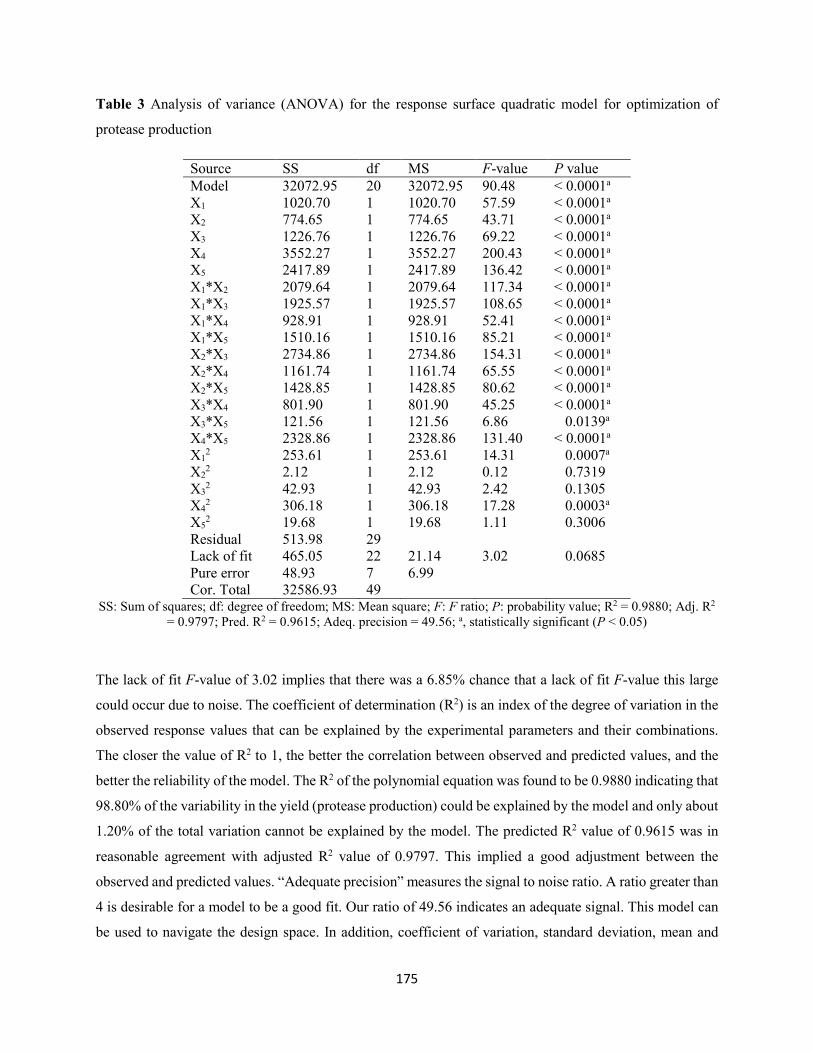
175
Table 3 Analysis of variance (ANOVA) for the response surface quadratic model for optimization of
protease production
Source SS df MS F-value P value Model 32072.95 20 32072.95 90.48 < 0.0001a X1 1020.70 1 1020.70 57.59 < 0.0001a X2 774.65 1 774.65 43.71 < 0.0001a X3 1226.76 1 1226.76 69.22 < 0.0001a X4 3552.27 1 3552.27 200.43 < 0.0001a X5 2417.89 1 2417.89 136.42 < 0.0001a X1*X2 2079.64 1 2079.64 117.34 < 0.0001a X1*X3 1925.57 1 1925.57 108.65 < 0.0001a X1*X4 928.91 1 928.91 52.41 < 0.0001a X1*X5 1510.16 1 1510.16 85.21 < 0.0001a X2*X3 2734.86 1 2734.86 154.31 < 0.0001a X2*X4 1161.74 1 1161.74 65.55 < 0.0001a X2*X5 1428.85 1 1428.85 80.62 < 0.0001a X3*X4 801.90 1 801.90 45.25 < 0.0001a X3*X5 121.56 1 121.56 6.86 0.0139a X4*X5 2328.86 1 2328.86 131.40 < 0.0001a X1
2 253.61 1 253.61 14.31 0.0007a X2
2 2.12 1 2.12 0.12 0.7319 X3
2 42.93 1 42.93 2.42 0.1305 X4
2 306.18 1 306.18 17.28 0.0003a X5
2 19.68 1 19.68 1.11 0.3006 Residual 513.98 29 Lack of fit 465.05 22 21.14 3.02 0.0685 Pure error 48.93 7 6.99 Cor. Total 32586.93 49
SS: Sum of squares; df: degree of freedom; MS: Mean square; F: F ratio; P: probability value; R2 = 0.9880; Adj. R2 = 0.9797; Pred. R2 = 0.9615; Adeq. precision = 49.56; a, statistically significant (P < 0.05)
The lack of fit F-value of 3.02 implies that there was a 6.85% chance that a lack of fit F-value this large
could occur due to noise. The coefficient of determination (R2) is an index of the degree of variation in the
observed response values that can be explained by the experimental parameters and their combinations.
The closer the value of R2 to 1, the better the correlation between observed and predicted values, and the
better the reliability of the model. The R2 of the polynomial equation was found to be 0.9880 indicating that
98.80% of the variability in the yield (protease production) could be explained by the model and only about
1.20% of the total variation cannot be explained by the model. The predicted R2 value of 0.9615 was in
reasonable agreement with adjusted R2 value of 0.9797. This implied a good adjustment between the
observed and predicted values. “Adequate precision” measures the signal to noise ratio. A ratio greater than
4 is desirable for a model to be a good fit. Our ratio of 49.56 indicates an adequate signal. This model can
be used to navigate the design space. In addition, coefficient of variation, standard deviation, mean and
176
predicted residual sum of squares values of 3.55%, 4.21, 118.70 and 1801.44, respectively were obtained
from the model.
The high degree of similarity between the predicted and observed responses is showed by the actual versus
predicted plot (Fig. 4). In this case, even distribution of the points around the diagonal signifies a good fit
of the model.
Fig. 4 Predicted versus actual response plot for protease production by Bacillus aryabhattai Ab15-ES.
For proper understanding of the interaction effects and optimum values of combination of the five
independent variables for maximum protease production, contour plots were generated and are presented
in Fig. 5A-J. These plots were constructed from a pair-wise interaction of the independent parameters, while
keeping other parameters constant at their center point level using a Design Expert 10 software. The
elliptical contour plots indicate a significant interaction among variables while a circular contour plot
implies a negligible interaction [46,47]. From the contour plot, significant interaction was notable among
the parameters, as in case of temperature and inoculum volume.
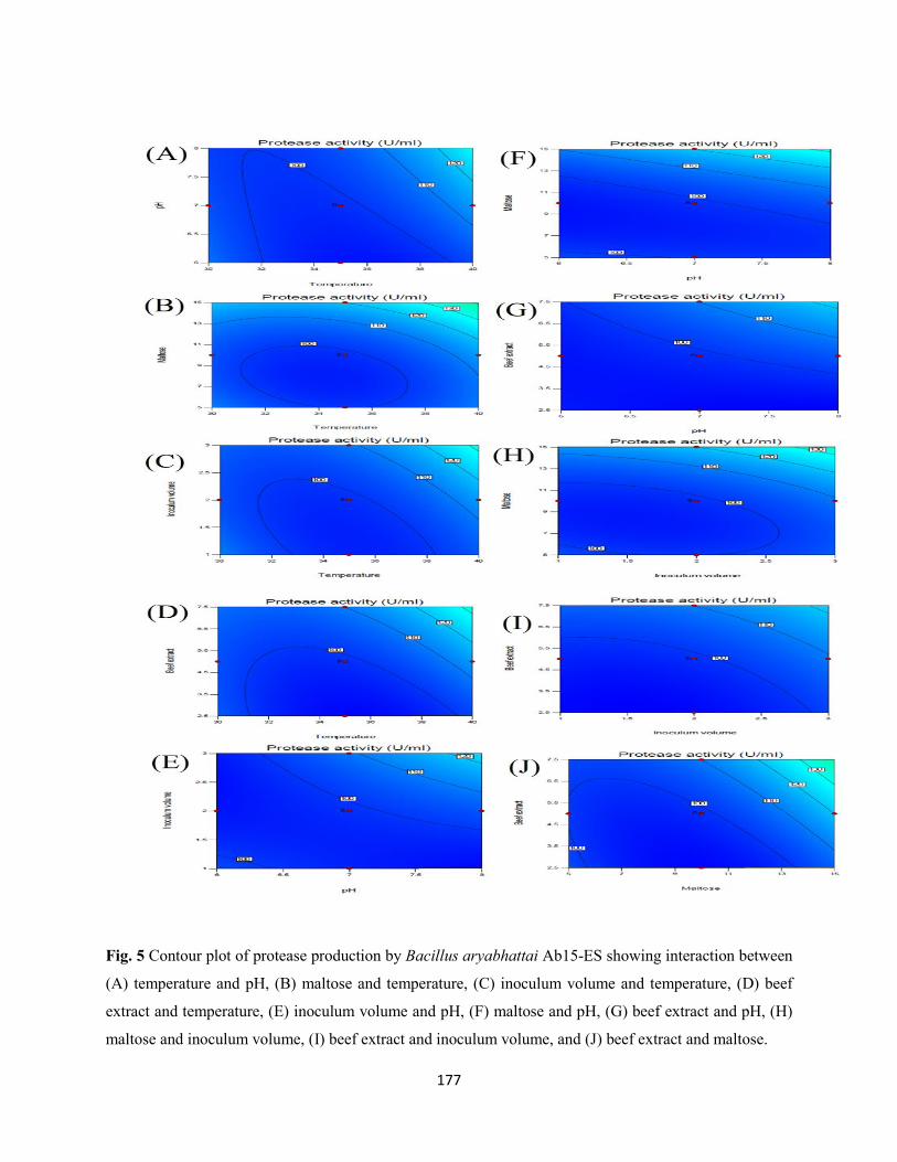
177
Fig. 5 Contour plot of protease production by Bacillus aryabhattai Ab15-ES showing interaction between
(A) temperature and pH, (B) maltose and temperature, (C) inoculum volume and temperature, (D) beef
extract and temperature, (E) inoculum volume and pH, (F) maltose and pH, (G) beef extract and pH, (H)
maltose and inoculum volume, (I) beef extract and inoculum volume, and (J) beef extract and maltose.
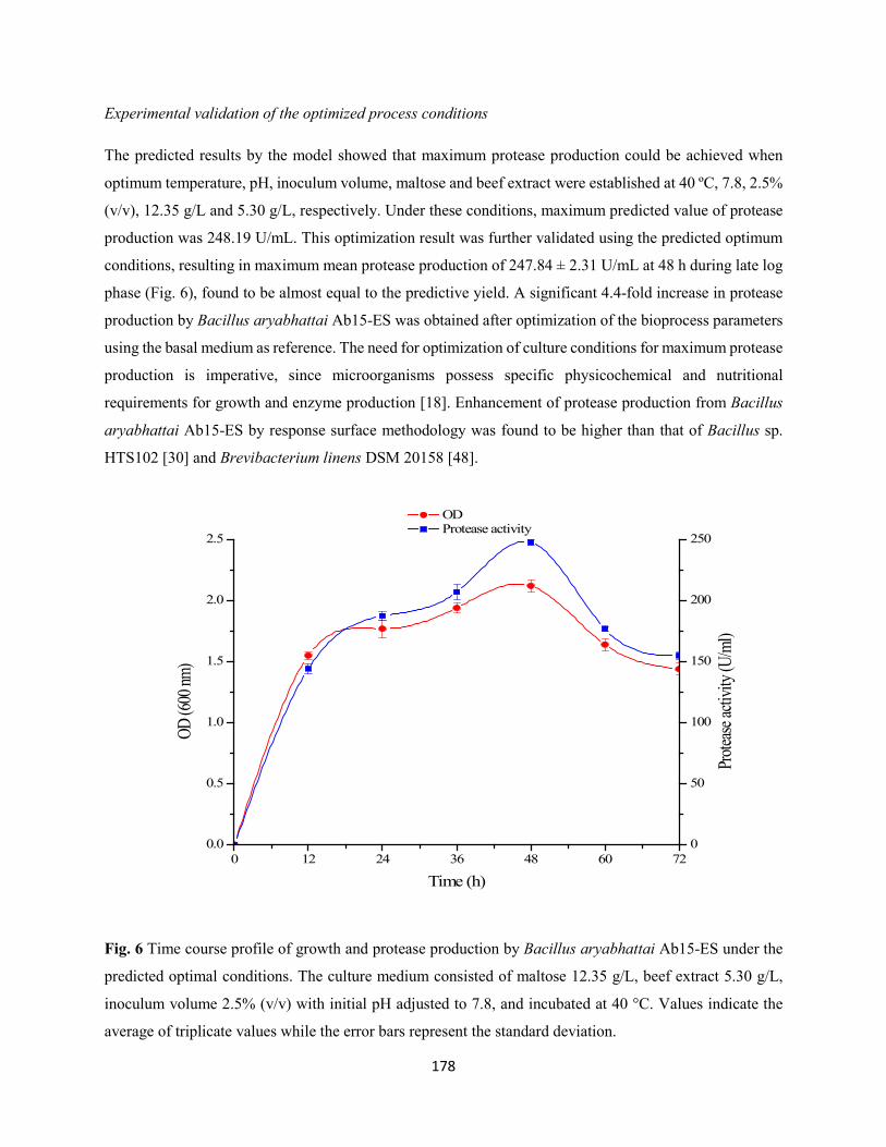
178
Experimental validation of the optimized process conditions
The predicted results by the model showed that maximum protease production could be achieved when
optimum temperature, pH, inoculum volume, maltose and beef extract were established at 40 ºC, 7.8, 2.5%
(v/v), 12.35 g/L and 5.30 g/L, respectively. Under these conditions, maximum predicted value of protease
production was 248.19 U/mL. This optimization result was further validated using the predicted optimum
conditions, resulting in maximum mean protease production of 247.84 ± 2.31 U/mL at 48 h during late log
phase (Fig. 6), found to be almost equal to the predictive yield. A significant 4.4-fold increase in protease
production by Bacillus aryabhattai Ab15-ES was obtained after optimization of the bioprocess parameters
using the basal medium as reference. The need for optimization of culture conditions for maximum protease
production is imperative, since microorganisms possess specific physicochemical and nutritional
requirements for growth and enzyme production [18]. Enhancement of protease production from Bacillus
aryabhattai Ab15-ES by response surface methodology was found to be higher than that of Bacillus sp.
HTS102 [30] and Brevibacterium linens DSM 20158 [48].
0 12 24 36 48 60 720.0
0.5
1.0
1.5
2.0
2.5
OD Protease activity
Time (h)
OD (6
00 nm
)
0
50
100
150
200
250
Prote
ase ac
tivity
(U/m
l)
Fig. 6 Time course profile of growth and protease production by Bacillus aryabhattai Ab15-ES under the
predicted optimal conditions. The culture medium consisted of maltose 12.35 g/L, beef extract 5.30 g/L,
inoculum volume 2.5% (v/v) with initial pH adjusted to 7.8, and incubated at 40 °C. Values indicate the
average of triplicate values while the error bars represent the standard deviation.
179
Conclusions
In the present study, FCCCD was used for the optimization of bioprocess parameters for maximum protease
production from Bacillus aryabhattai Ab15-ES using submerged fermentation process. Protease production
depends mainly on temperature, pH, inoculum volume, maltose and beef extract. The observed protease
production value agreed with that of predicted protease production, thus confirming the validity of the
model. Under these optimum conditions, a 4.4-fold increase in protease production was obtained using the
basal medium as reference. These findings suggest that Bacillus aryabhattai Ab15-ES can be used for mass
production of protease for various biotechnological applications, thus minimizing overall costs of the
enzyme production.
Financial support
The financial support of the National Research Foundation (NRF) of South Africa towards this research is
hereby acknowledged. Opinions expressed and conclusions arrived at, are those of the authors and are not
necessarily to be attributed to the NRF.
References
1. Annamalai N, Rajeswari MV, Balasubramanian T (2014) Extraction, purification and application of
thermostable and halostable alkaline protease from Bacillus alveayuensis CAS 5 using marine wastes. Food
Bioprod Process 92:335-342
2. de Souza PM, de Assis Bittencourt ML, Caprara CC, de Freitas M, de Almeida RPC, Silveira D, Fonseca
YM, Filho EXF, Pessoa Junior A, Magalhães PO (2015) A biotechnology perspective of fungal proteases.
Braz J Microbiol 46:337-346
3. Sawant R, Nagendran S (2014) Protease: an enzyme with multiple industrial applications. World J Pharm
Pharm Sci 3:568-579
4. Li Q, Yi L, Marek P, Iverson BL (2013) Commercial proteases: present and future. FEBS Lett 587:1155-
1163
5. Navanneth S, Bhuvanesh S, Bhaskar V, Kumar PV, Kandaswamy SKJ, Achary A (2009) Optimization
of medium for the production of subtilisin from Bacillus subtilis MTCC 441. Afr J Biotechnol 8:6327-6331
180
6. Lin S, Zhang M, Liu J, Jones GS (2015) Construction and application of recombinant strain for the
production of an alkaline protease from Bacillus licheniformis. J Biosci Bioeng 119:284-288
7. Oskouie SFG, Tabandeh F, Yakhchali B, Eftekhar F (2008) Response surface optimization of medium
composition for alkaline protease production by Bacillus clausii. Biochem Eng J 39:37-42
8. Uyar F, Porsuk I, Kizil G, Yilmaz EI (2011) Optimal conditions for production of extracellular protease
from newly isolated Bacillus cereus strain CA15. EurAsian J Biosci 5:1-9
9. Benkiar A, Nadia ZJ, Badis A, Rebzani F, Soraya BT, Rekik H, Naili B, Ferradji FZ, Bejar S, Jaouadi B
(2013) Biochemical and molecular characterization of a thermos- and detergent-stable alkaline serine
keratinolytic protease from Bacillus circulans strain DZ100 for detergent formulations and feather-
biodegradation process. Int Biodeter Biodegr 83:129-138
10. Joshi S, Satyanarayana T (2013) Characteristics and applications of a recombinant alkaline serine
protease from a novel bacterium Bacillus lehensis. Bioresour Technol 131:76-85
11. Asker MM, Mahmoud MG, El Shebwy K, Abdel Aziz MS (2013) Purification and characterization of
two thermostable protease fractions from Bacillus megaterium. J Genet Eng Biotechnol 11:103-109
12. Haddar A, Agrebi R, Bougatef A, Hmidet N, Sellami-Kamoun A, Nasri M (2009) Two detergent stable
alkaline serine-proteases from Bacillus mojavensis A21: Purification, characterization and potential
application as a laundry detergent additive. Bioresour Technol 100:3366-3373
13. Sookkheo B, Sinchaikul S, Phutrakul S, Chen ST (2000) Purification and characterization of the highly
thermostable proteases from Bacillus stearothermophilus TLS33. Protein Expr Purif 20:142-151
14. Jaouadi B, Ellouz-Chaabouni S, Rhimi M, Bejar S (2008) Biochemical and molecular characterization
of a detergent-stable serine alkaline protease from Bacillus pumilus CBS with high catalytic efficiency.
Biochimie, 90:1291-1305
15. Maruthiah T, Esakkiraj P, Prabakaran G, Palavesam A, Immanuel G (2013) Purification and
characterization of moderately halophilic alkaline serine protease from marine Bacillus subtilis AP-MSU
6. Biocatal Agric Biotechnol 2:116-119
16. Gupta R, Beg Q, Lorenz P (2002) Bacterial alkaline proteases: molecular approaches and industrial
applications. Appl Microbiol Biotechnol 59:15-32
181
17. Reddy LVA, Wee Y-J, Yun J-S, Ryu H-W (2008) Optimization of alkaline protease production by batch
culture of Bacillus sp. RKY3 through Plackett-Burman and response surface methodological approaches.
Bioresour Technol 99:2242-2249
18. Bhunia B, Dey A (2012) Statistical approach for optimization of physicochemical requirements on
alkaline protease production from Bacillus licheniformis NCIM 2042. Enzyme Res 2012:1-13
19. Rao YK, Tsay K-J, Wu W-S, Tzeng Y-M (2007) Medium optimization of carbon and nitrogen sources
for the production of spores from Bacillus amyloliquefaciens B128 using response surface methodology.
Proc Biochem 42:535-541
20. Cazetta ML, Celligoi MA, Buzato JB, Scarmino IS (2007) Fermentation of molasses by Zymomonas
mobilis: effects of temperature and sugar concentration on ethanol production. Bioresour Technol 98:2824-
2828
21. Li X, Xu T, Ma X, Guo K, Kai L, Zhao Y, Jia X, Mad Y (2008) Optimization of culture conditions for
production of cis-epoxysuccinic acid hydrolase using response surface methodology. Bioresour Technol
99:5391-5396
22. Puri S, Beg QK, Gupta R (2002) Optimization of alkaline protease production from Bacillus sp. by
response surface methodology. Curr Microbiol 44:286-290
23. Beg QK, Sahai V, Gupta R (2003) Statistical media optimization and alkaline protease production from
Bacillus mojavensis in a bioreactor. Proc Biochem 39:203-209
24. Montgomeryd DC, Runger GC (2002) Applied statistics and probability for Engineers. 3rd ed., John
Wiley and Sons, NY, USA
25. Ahuja SK, Ferreira GM, Morreira AR (2004) Application of Plackett-Burman design and response
surface methodology to achieve exponential growth of aggregated shipworm bacterium. Biotechnol Bioeng
85:666-675
26. Bandaru VVR, Somalanka SR, Mendu DR, Madicherla NR, Chityala A (2006) Optimization of
fermentation conditions for the production of ethanol from sago starch by co-immobilized
amyloglucosidase and cells of Zymomonas mobilis using response surface methodology. Enzyme Microb
Technol 38:209-214
182
27. Walia NK, Sekhon KK, Chaudhary DP, Cameotra SS, Srivastava P, Kumar A (2014) Optimization of
fermentation parameters for bioconversion of corn to ethanol using response surface methodology. J Pet
Environ Biotechnol 5:178
28. Tajabadi N, Ebrahimpour A, Baradaran A, Rahim RA, Mahyudin NA, Manap MYA, Abubakar F, Saari
N (2015) Optimization of γ-aminobutyric acid production by Lactobacillus plantarum Taj-Apis362 from
honeybees. Molecules, 20:6654-6669
29. Wang Q, Hou Y, Xu Z, Miao J, Li G (2008) Optimization of cold-active protease production by the
psychrophilic bacterium Colwellia sp. NJ341 with response surface methodology. Bioresour Technol
99:1926-1931
30. Queiroga AC, Pintado ME, Malcata FX (2012) Use of response surface methodology to optimize
protease synthesis by a novel strain of Bacillus sp. isolated from Portuguese sheep wool. J Appl Microbiol
113:36-43
31. Akhtar N, Mahmud ASM, Khan MS, Taznin T, Haque ME, Sultana S, Sultana S (2013) Effects of
cultural conditions on the production of extracellular protease by Streptomyces albolongus and
Streptomyces aburaviensis. Enzyme Eng 2:110
32. Abd Rahman RNZR, Leow TC, Salleh AB, Basri M (2007) Geobacillus zalihae sp. nov., a thermophilic
lipolytic bacterium isolated from palm oil mill effluent in Malaysia. BMC Microbiol 7:77
33. Loperana L, Ferrari MD, Díaz AL, Ingold G, Pérez LV, Carvallo F, Travers D, Menes RJ, Lareo C
(2009) Isolation and selection of native microorganisms for the aerobic treatment of simulated dairy
wastewaters. Bioresour Technol 100:1762-1766
34. Akinbowale OL, Peng H, Grant P, Barton MD (2007) Antibiotic and heavy metal resistance in motile
aeromonads and pseudomonads from rainbow trout (Oncorhynchus mykiss) farms in Australia. Int J
Antimicrob Agents, 30:177-182
35. Marchesi JR, Sato T, Weightman AJ, Martin TA, Fry JC, Hiom SJ, Dymock D, Wade WG (1998)
Design and evaluation of useful bacterium-specific PCR primers that amplify genes coding for bacterial
16S rRNA. Appl Environ Microbiol 64:795-799
36. Tamura K, Stecher G, Peterson D, Filipski A, Kumar S (2013) MEGA 6: Molecular Evolutionary
Genetics Analysis version 6.0. Mol Biol Evo 30:2725-2729
183
37. Secades P, Guijarro JA (1999) Purification and characterization of an extracellular protease from the
fish pathogen Yersinia ruckeri and effect of culture conditions on production. Appl Environ Microbiol
65:3969-3975
38. Vijayaraghavan P, Vijayan A, Arun A, Jenisha JK, Vincent SGP (2012) Cow dung: a potential biomass
substrate for the production of detergent-stable dehairing protease by alkaliphilic Bacillus subtilis strain
VV. SpringerPlus, 1:76
39. Nadeem M, Qazi JI, Syed Q, Baig S (2008) Optimization of process parameters for alkaline protease
production by Bacillus licheniformis N-2 and kinetics studies in batch fermentation. Turk J Biol 32:243-
251
40. Olajuyigbe FM (2013) Optimized production and properties of thermostable alkaline protease from
Bacillus subtilis SHS-04 grown on groundnut (Arachis hypogaea) meal. Adv Enzyme Res 1:112-120
41. Raj A, Khess N, Pujari N, Bhattacharya S, Das A, Rajan SS (2012) Enhancement of protease production
by Pseudomonas aeruginosa isolated from dairy effluent sludge and determination of its fibrinolytic
potential. Asian Pac J Trop Biomed S1845-S1851
42. Vijayaraghavan P, Lazarus S, Vincent SGP (2014) De-hairing protease production by an isolated
Bacillus cereus strain AT under solid-state fermentation using cow dung: biosynthesis and properties. Saudi
Biol Sci 21:27-34
43. Esakkiraj P, Sankaralingam S, Usha R, Palavesam A, Immanuel G (2011) Solid-state protease
production using anchovy waste meal by moderate halophile Serratia proteamaculans AP-CMST isolated
from fish intestine. Ann Microbiol 61:749-755
44. Umuyaparvathi S, Meenakshi S, Arumugam M, Balasubramanian T (2013) Purification and
characterization of protease from Bacillus cereus SU12 isolated from oyster Saccostrea cucullata. Afr J
Biotechnol 12:5897-5908
45. Kumar RS, Ananthan G, Prabhu AS (2014) Optimization of medium composition for alkaline
production of Marinobacter sp. GA CAS9 using response surface methodology- A statistical approach.
Biocat Agric Biotechnol 3:191-197
46. Muralidhar RV, Chirumamila RR, Marchant R, Nigam P (2001) A response surface approach for the
comparison of lipase production by Candida cylindracea using two different carbon sources. Biochem Eng
J 9:17-23
184
47. Xu H, Sun LP, Shi YZ, Wu YH, Zhang B, Zhao DQ (2008) Optimization of cultivation conditions for
extracellular polysaccharide and mycelium biomass by Morchella esculenta As51620. Biochem Eng J
39:66-73
48. Shabbiri K, Adnan A, Jamil S, Ahmad W, Noor B, Rafique HM (2012) Medium optimization of
protease production by Brevibacterium linens DSM 20158 using statistical approach. Braz J Microbiol
2012:1051-1061
49. Singh V, Haque S, Niwas R, Srivastava A, Pasupuleti M, Tripathi CKM (2017) Strategies for
fermentation medium optimization: an in-depth review. Front Microbiol 7:2087
50. Singh S, Bajaj BK (2016) Bioprocess optimization for production of thermoalkali-stable protease from
Bacillus subtilis K-1 under solid-state fermentation. Prep Biochem Biotechnol 46:717-724
51. Sharma KM, Kumar R, Panwar S, Kumar A (2017) Microbial alkaline proteases: optimization of
production parameters and their properties. J Genet Eng Biotechnol 15:115-126
185
CHAPTER SEVEN
OPTIMIZATION OF CULTURE CONDITIONS FOR ENHANCED LIPASE PRODUCTION
BY AN INDIGENOUS BACILLUS ARYABHATTAI SE3-PB USING RESPONSE SURFACE
METHODOLOGY
This chapter has been submitted in the current format to the ‘Biotechnology and
Biotechnological Equipment’
186
Optimization of culture conditions for enhanced lipase production by an indigenous Bacillus
aryabhattai SE3-PB using response surface methodology
Adegoke Isiaka Adetunji and Ademola Olufolahan Olaniran*
Discipline of Microbiology, School of Life Sciences, College of Agriculture, Engineering and Science,
University of KwaZulu-Natal (Westville campus), Private Bag X54001, Durban 4000, Republic of South
Africa.
* Corresponding author. Tel: +27 31 260 7400; Fax: +27 31 260 7809
E-mail address: [email protected]
ABSTRACT
Lipases are enzymes that catalyze the hydrolysis of fats into fatty acids and glycerol at the water-lipid
interface as well as involved in a range of bioconversion reactions in non-aqueous and micro-aqueous
environments. In this present study, optimization of culture conditions for extracellular lipase production
by an indigenous lipase-producing bacterial strain isolated from lipid-rich wastewater was carried out using
response surface methodology. The study isolate was identified as Bacillus aryabhattai SE3-PB by PCR
amplification and analysis of 16S rDNA. Sunflower oil was found to induce maximum lipase production.
Face centered central composite design revealed that temperature (40 °C), pH (7.6), inoculum volume
(2.8%, v/v), agitation (193 rpm) and inducer oil concentration (2%, v/v) were optimum culture conditions
for maximum lipase production. The observed lipase production (264.02 ± 1.94 U/ml) found to be in
agreement with predicted lipase yield (265.82 U/ml) coupled with high coefficient of determination (R2 =
0.9919, P < 0.01) confirmed the validity of the model. A 7.2-fold increase in lipase production was obtained
in the optimized medium relative to the basal medium. These findings provide first report on production
and optimization of lipase by Bacillus aryabhattai SE3-PB, and suggest a rational choice of optimum
process conditions for commercial production of lipase by Bacillus aryabhattai SE3-PB for various
biotechnological applications, especially in the remediation of lipid-rich wastewater.
KEYWORDS: Lipase; Bacillus aryabhattai SE3-PB; lipid-rich wastewater; face centered central
composite design; response surface methodology
187
Introduction
Lipases (triacylglycerol acylhydrolases EC 3.1.1.3) are enzymes that catalyze the hydrolysis of lipids into
di-acylglycerides, mono-acylglycerides, glycerol and fatty acids over an oil-water interface [1-3]. In
addition, they are involved in a variety of bioconversion reactions including esterification,
interesterification, transesterification, alcoholysis, acidolysis and aminolysis in non-aqueous and micro-
aqueous milieu [4-6]. Because of their versatility, lipases are employed as enzyme of choice for numerous
biotechnological applications including food, leather, cosmetic, detergent, paper and textile industries as
well as in pharmaceutical and treatment of fat-containing waste effluents [7-9].
Lipases are ubiquitous in nature and are produced by plants, animals, and microorganisms [4,10]. However,
microbial lipases formed the most widely used class of enzymes due to their selectivity, lower production
cost, ease of genetic manipulation, high productivity, regiospecificity, stereospecificity, broad substrate
specificity, and ability to catalyze heterogeneous reactions at the interface of water soluble and water
insoluble systems [8,11-13]. Microorganisms such as bacteria, yeast, fungi and actinomycetes have been
reported as potential producers of lipase [14-17]. They are found in different habitats including industrial
wastes, vegetable oil processing industries, dairy industries, oil-contaminated sites, among others [18].
Lipase production from many species of Bacillus including Bacillus pumilus [19], Bacillus subtilis [20],
Bacillus thermoleovorans [21], Bacillus cereus [22], Bacillus coagulans [23], Bacillus megaterium [24],
and Bacillus licheniformis [25] has been reported. However, this is the first report on production and
optimization of lipase from Bacillus aryabhattai.
The production of extracellular bacterial lipases is influenced by fermentation parameters such as
temperature, pH, inoculum size, agitation, carbon sources, inducer sources and concentrations [26-28]. The
search for optimal conditions of the above-mentioned parameters for lipase production in fermentation
processes is imperative in order to obtain high yields of the enzyme at lower costs. A classical and
conventional practice of achieving these is by ‘one variable-at-a-time approach’. This approach has been
proved to be laborious, time-consuming and costly for simultaneous consideration of many variables in
addition to its inability to depict interactive effects among factors and locate optimum conditions among
variables. In order to overcome these drawbacks, statistical experimental designs by response surface
methodology (RSM) have been recognized as an effective approach in many biotechnological processes
[26,29].
Response surface methodology is a collection of mathematical and statistical techniques for designing
experiments, modelling and searching for optimum conditions of factors influencing the responses. It is
widely used to identify effective factors, study interactions, select optimum conditions and quantify the
188
relationships between one or more measured responses and the vital input factors in a limited number of
experiments [11]. Furthermore, RSM has been successfully applied to evaluate and optimize the effect of
process parameters in the production of lipase [28,30,31]. A central composite design (CCD) is the most
commonly used response surface design when the experimental design is defined by the upper and lower
limits of each parameter [32]. Several bioprocess parameters have been optimized for enhanced lipase
production using CCD [15,16,33,34,].
The continuous demand for industrial lipase production with potential biotechnological applications
necessitated the need to bioprospect for native and robust/hyperactive bacterial strains capable of mass
production of this enzyme. In this study, optimization of culture conditions for enhanced extracellular lipase
production by an indigenous Bacillus aryabhattai SE3-PB was investigated using RSM. Effect of various
inducer oils on lipase production was also studied. Thereafter, optimization of the selected inducer
concentrations along with other process parameters, including temperature, pH, agitation speed, and
inoculum volume for enhanced lipase production was carried out using face centered CCD (FCCCD).
Materials and methods
Chemicals
p-nitrophenyl palmitate (p-NPP) used as substrate for lipase assay was purchased from Sigma-Aldrich (St.
Louis, MO, USA). All other reagents were of analytical grade.
Sample collection and bacterial isolation procedures
Lipid-rich wastewater samples were collected from an oil mill industry in Pietermaritzburg, KwaZulu-Natal
(KZN) province, South Africa in sterile 500 ml Schott bottles. Samples (10 ml) were transferred to 250 ml
Erlenmeyer flasks containing 100 ml mineral salt medium (MSM) consisting (in g/L): 1.0 KH2PO4, 1.0
K2HPO4, 1.0 NH4NO3, 0.2 MgSO4.7H2O, 0.02 CaCl2.2H2O and 0.05 FeCl3.6H2O supplemented with 1%
olive oil, and then incubated at 37 ºC for 48 h at 130 rpm [35]. Enriched cultures (5 ml) were subsequently
transferred into a second flask and cultivated under the same conditions. This process was repeated four
times. Samples (0.1 ml) of the last enrichment cultures were serially diluted, plated onto nutrient agar (NA)
and incubated at 37 ºC for 24 h. Pure cultures obtained by sub-culturing on fresh NA plates were preserved
as glycerol stock at -80 ºC.
189
Screening of bacteria for lipolytic activity
Lipolytic activity of the bacteria was assessed by using solid media containing different lipid sources, such
as olive oil and Tween-20. Enzymatic activity was detected by visual observation and measurement of clear
zone on the agar surface.
Screening of bacteria for lipolytic activity using Tween-20 agar
Tween-20 agar consisting (in %, w/v, v/v): peptone 1, NaCl 0.5, CaCl2.2H2O 0.01, agar 2 and Tween-20 1
was prepared as described by Gopinath et al. [36]. A loopful of bacterial culture was inoculated into 10 ml
sterile nutrient broth and then incubated at 130 rpm (37 ºC) for 24 h. Twenty microliter of obtained culture
supernatant was added into wells bored on Tween-20 agar plate, and then incubated at 37 ºC for 48 h. Sterile
nutrient broth was used as a control. Visible precipitate of calcium salts around the wells resulting from
formation of fatty acid from lipid hydrolysis indicates the presence of lipolytic activity. All experiments
were carried out in triplicate.
Screening of bacteria for lipolytic activity using phenol red agar
Phenol red agar consisting (in %, w/v, v/v): phenol red dye 0.01, olive oil 0.1, CaCl2 0.1 and agar 2 was
prepared as previously described [37]. The pH of the medium was adjusted to 7.3 using 1N NaOH or 1N
HCl. Culture supernatant (20 µl) was added into the agar wells, and then incubated at 37 ºC for 48 h. The
change in the color of phenol red dye to yellow was used as an indication of lipolytic activity. All
experiments were carried out in triplicate.
Molecular identification of lipase-producing bacteria and phylogenetic analysis
Genomic DNA extraction
Genomic DNA was isolated from bacterial culture grown overnight on NA plate at 37°C using boiling
method described by Akinbowale et al. [38] with modifications. Freshly grown colonies (5-7) were
suspended in 70 µl sterile distilled water and then boiled at 100 ºC for 10 min before cooling on ice for 5
min. This was centrifuged at 13000 rpm in a micro-centrifuge 5415D (Eppendorf, Hamburg, Germany) for
5 min. Cell-free supernatant was transferred into fresh sterile Eppendorf tubes and used as template DNA
for the PCR reaction.
Amplification, sequencing and analysis of 16S rRNA gene
Amplification of the 16S rDNA of the lipase-producing bacterial isolate was carried out using the universal
bacterial primers F-5'-CAGGCCTAACACATGCAAGTC-3' and R-5'-GGGCGGTGTGTACAAGGC-3'
190
[39]. The PCR mixture consisted of 1× buffer, 1mM MgCl2, 0.2 mM of each dNTPs, 0.4 µm of each primer,
2 U of Taq polymerase and 2 µl template DNA in a total volume of 25 µl. The mixture was subjected to
amplification using a thermocycler (Bio-Rad T100, Singapore) at initial denaturation of 95 ºC for 5 min,
followed by 30 cycles of; denaturation at 95 ºC for 1 min, annealing at 55 ºC for 1 min, extension at 72 ºC
for 1min, 30 sec and final extension at 72 ºC for 5 min. Amplification products were observed by
electrophoresis in a 1.5% (w/v) agarose gel at 100 V for 45 min in a 1% TAE buffer. After staining in 1
mg/ml ethidium bromide for 15 min, products were viewed by UV illumination (Syngene, UK), and then
sequenced (inqaba biotech, Pretoria, South Africa). The resulting sequences were compared with sequences
in the GenBank database of National Centre for Biotechnology Information (NCBI) using the Nucleotide
Basic Local Alignment Search Tool (BLAST N) program (www.ncbi.n1m.nih.gov/BLAST). The
sequences were aligned using the multiple sequences alignment tool, Clustal W. Phylogenetic tree was
constructed with Molecular Evolutionary Genetics analysis (MEGA) version 6.0 software [40] using a
neighbor-joining method.
Kinetics of growth and extracellular lipase production
The production medium composed (in %, w/v, v/v): peptone 0.5, yeast extract 0.5, NaCl 0.05, CaCl2 0.005,
and olive oil 1.0 emulsified with gum arabic 0.5 [41]. The pH of the medium was adjusted to 7.2 with 1 N
NaOH or 1 N HCl. One milliliter of the bacterial culture (OD600nm = 1) was inoculated into 99 ml basal
medium in 250 ml Erlenmeyer flasks. The flasks were then incubated at 37 ºC (130 rpm) for 72 h in a
shaking incubator. Samples were taken at 12-h intervals to determine bacterial growth and lipase
production. Bacterial growth was determined by measuring optical density at 600 nm using UV-Vis
spectrophotometer (UVmini-1240, Schimadzu, Australia). Subsequently, samples were centrifuged at
10000 rpm for 10 min at 4 ºC using high-speed refrigerated centrifuge (Avanti J-26 XP, Beckman Coulter,
USA). The obtained culture supernatants (crude enzyme) were then assayed for lipase activity. Lipase
activity was determined as described by Winkler and Stuckmann [42] with some modifications using p-
NPP as a substrate. One unit (U) of lipase activity was defined as the amount of enzyme that liberated 1
µmol of p-nitrophenol ml-1 min-1 under standard assay conditions. All experiments were done in triplicate.
Optimization of culture conditions for maximum lipase production
Effect of inducer oils on lipase production
Various vegetable oils including palm oil, castor oil, sunflower oil, canola oil or rice bran oil were screened
for the ability to induce maximum lipase production by Bacillus aryabhattai SE3-PB. Olive oil in the
fermentation medium for lipase production was replaced with (1%, v/v) of the tested oils while keeping the
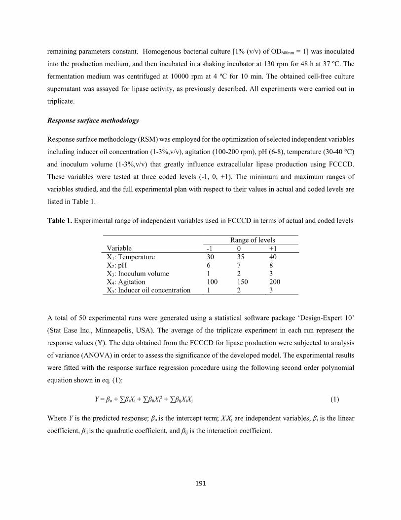
191
remaining parameters constant. Homogenous bacterial culture [1% (v/v) of OD600nm = 1] was inoculated
into the production medium, and then incubated in a shaking incubator at 130 rpm for 48 h at 37 ºC. The
fermentation medium was centrifuged at 10000 rpm at 4 ºC for 10 min. The obtained cell-free culture
supernatant was assayed for lipase activity, as previously described. All experiments were carried out in
triplicate.
Response surface methodology
Response surface methodology (RSM) was employed for the optimization of selected independent variables
including inducer oil concentration (1-3%,v/v), agitation (100-200 rpm), pH (6-8), temperature (30-40 °C)
and inoculum volume (1-3%,v/v) that greatly influence extracellular lipase production using FCCCD.
These variables were tested at three coded levels (-1, 0, +1). The minimum and maximum ranges of
variables studied, and the full experimental plan with respect to their values in actual and coded levels are
listed in Table 1.
Table 1. Experimental range of independent variables used in FCCCD in terms of actual and coded levels
Variable
Range of levels -1 0 +1
X1: Temperature 30 35 40 X2: pH 6 7 8 X3: Inoculum volume 1 2 3 X4: Agitation 100 150 200 X5: Inducer oil concentration 1 2 3
A total of 50 experimental runs were generated using a statistical software package ‘Design-Expert 10’
(Stat Ease Inc., Minneapolis, USA). The average of the triplicate experiment in each run represent the
response values (Y). The data obtained from the FCCCD for lipase production were subjected to analysis
of variance (ANOVA) in order to assess the significance of the developed model. The experimental results
were fitted with the response surface regression procedure using the following second order polynomial
equation shown in eq. (1):
Y = βo + ∑βiXi + ∑βiiXi2 + ∑βijXiXj (1)
Where Y is the predicted response; βo is the intercept term; XiXj are independent variables, βi is the linear
coefficient, βii is the quadratic coefficient, and βij is the interaction coefficient.
192
Results and discussion
Isolation, screening and identification of lipase-producing bacteria
Lipid-rich wastewater samples were collected from an edible oil mill industry in Pietermaritzburg,
KwaZulu-Natal province of South Africa. The rational choice of this site was to provide a good environment
for lipolytic organisms to grow. Samples were enriched in MSM supplemented with 1% (v/v) olive oil as a
sole carbon source in order to stimulate the growth of lipolytic organisms [35]. Lipase-producing bacteria
isolated from lipid-rich wastewater have been reported [43-46]. Among the isolated bacterial strains
screened for extracellular lipase production on solid media consisting of Tween-20 as inducible substrate,
strain SE3-PB was found to exhibit higher lipolytic activity. This was based on the formation of large visible
calcium salt precipitates around the agar well, resulting from the breakdown of Tween-20 into fatty acids
(Result not shown). Tween-20 is a commonly used substrate for detecting bacterial lipolytic activity, as it
promotes optimal contact between cells and/or enzymes and substrate [47]. Further screening of strain SE3-
PB for lipolytic activity was carried out using a chromogenic approach involving the use of phenol red dye,
and olive oil as inducible substrate. Phenol red is a pH indicator with end point at pH 7.3-7.4. The release
of fatty acids from lipolysis reaction of olive oil caused a decrease in the pH of the dye, resulting in yellow
coloration, an indication of lipolytic activity of the tested bacterial strain [37]. Analysis of the 16S rRNA
gene sequence of strain SE3-PB showed that it exhibits close homology (100%) to Bacillus aryabhattai
(KX781238.1). This study strain was therefore designated as Bacillus aryabhattai SE3-PB. The relatedness
of this strain to other lipase-producing Bacillus sp. is illustrated in the constructed phylogenetic tree (Figure
1).
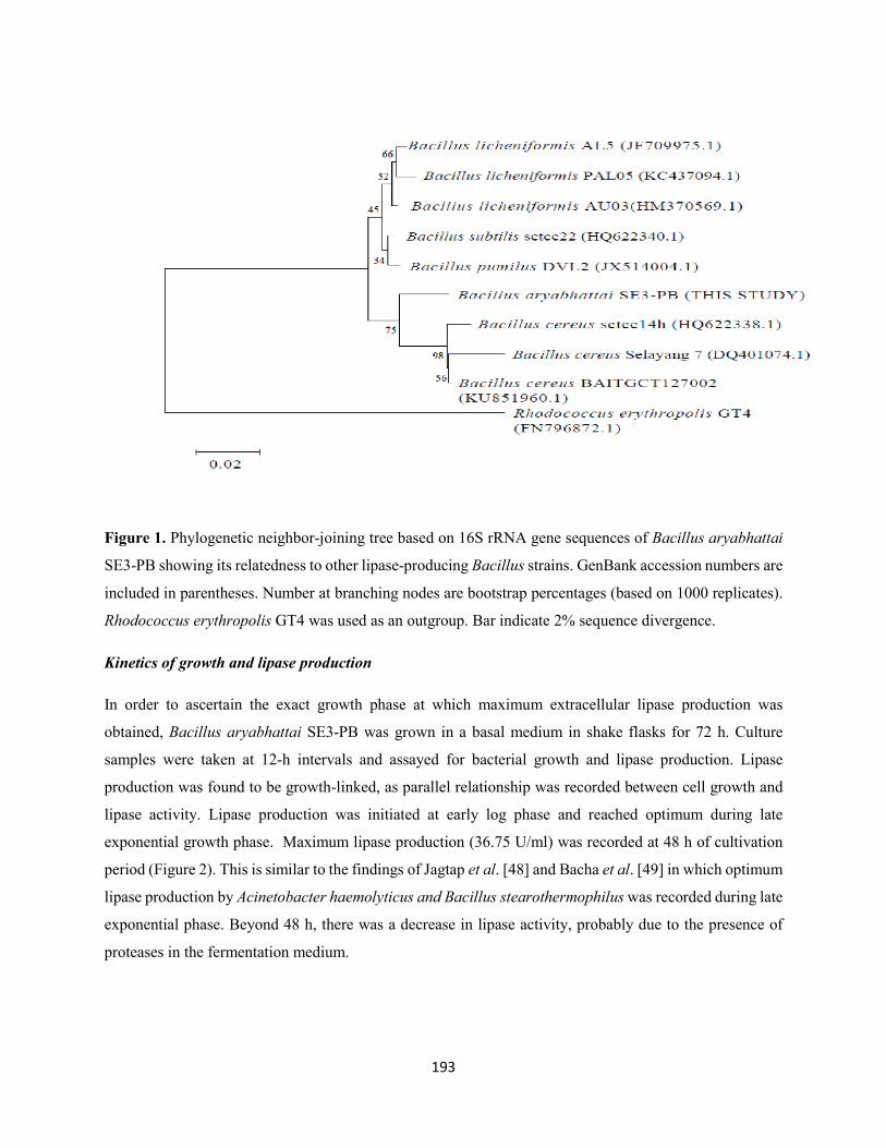
193
Figure 1. Phylogenetic neighbor-joining tree based on 16S rRNA gene sequences of Bacillus aryabhattai
SE3-PB showing its relatedness to other lipase-producing Bacillus strains. GenBank accession numbers are
included in parentheses. Number at branching nodes are bootstrap percentages (based on 1000 replicates).
Rhodococcus erythropolis GT4 was used as an outgroup. Bar indicate 2% sequence divergence.
Kinetics of growth and lipase production
In order to ascertain the exact growth phase at which maximum extracellular lipase production was
obtained, Bacillus aryabhattai SE3-PB was grown in a basal medium in shake flasks for 72 h. Culture
samples were taken at 12-h intervals and assayed for bacterial growth and lipase production. Lipase
production was found to be growth-linked, as parallel relationship was recorded between cell growth and
lipase activity. Lipase production was initiated at early log phase and reached optimum during late
exponential growth phase. Maximum lipase production (36.75 U/ml) was recorded at 48 h of cultivation
period (Figure 2). This is similar to the findings of Jagtap et al. [48] and Bacha et al. [49] in which optimum
lipase production by Acinetobacter haemolyticus and Bacillus stearothermophilus was recorded during late
exponential phase. Beyond 48 h, there was a decrease in lipase activity, probably due to the presence of
proteases in the fermentation medium.
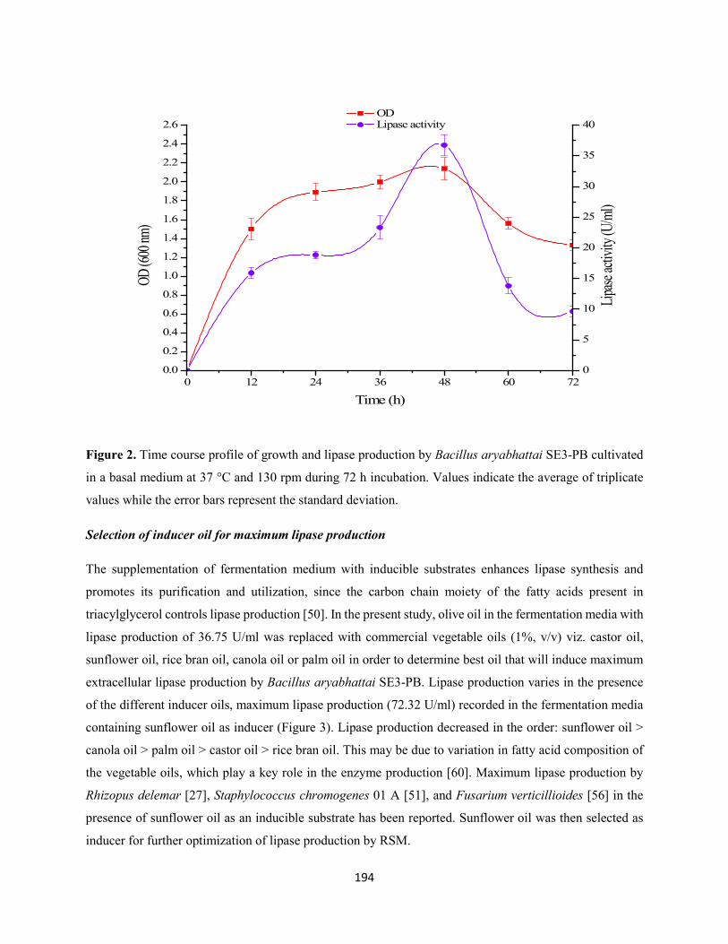
194
0 12 24 36 48 60 720.0
0.2
0.4
0.6
0.8
1.0
1.2
1.4
1.6
1.8
2.0
2.2
2.4
2.6 OD Lipase activity
Time (h)
OD (6
00 nm
)
0
5
10
15
20
25
30
35
40
Lipase
activ
ity (U
/ml)
Figure 2. Time course profile of growth and lipase production by Bacillus aryabhattai SE3-PB cultivated
in a basal medium at 37 °C and 130 rpm during 72 h incubation. Values indicate the average of triplicate
values while the error bars represent the standard deviation.
Selection of inducer oil for maximum lipase production
The supplementation of fermentation medium with inducible substrates enhances lipase synthesis and
promotes its purification and utilization, since the carbon chain moiety of the fatty acids present in
triacylglycerol controls lipase production [50]. In the present study, olive oil in the fermentation media with
lipase production of 36.75 U/ml was replaced with commercial vegetable oils (1%, v/v) viz. castor oil,
sunflower oil, rice bran oil, canola oil or palm oil in order to determine best oil that will induce maximum
extracellular lipase production by Bacillus aryabhattai SE3-PB. Lipase production varies in the presence
of the different inducer oils, maximum lipase production (72.32 U/ml) recorded in the fermentation media
containing sunflower oil as inducer (Figure 3). Lipase production decreased in the order: sunflower oil >
canola oil > palm oil > castor oil > rice bran oil. This may be due to variation in fatty acid composition of
the vegetable oils, which play a key role in the enzyme production [60]. Maximum lipase production by
Rhizopus delemar [27], Staphylococcus chromogenes 01 A [51], and Fusarium verticillioides [56] in the
presence of sunflower oil as an inducible substrate has been reported. Sunflower oil was then selected as
inducer for further optimization of lipase production by RSM.
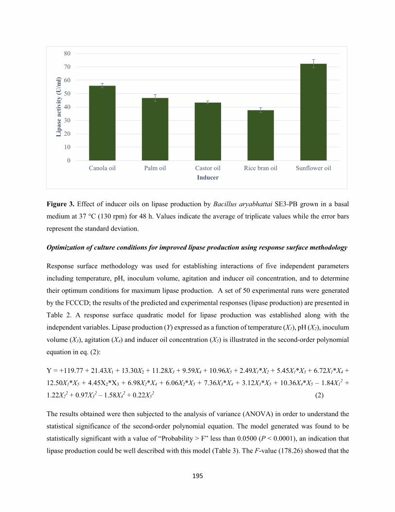
195
Figure 3. Effect of inducer oils on lipase production by Bacillus aryabhattai SE3-PB grown in a basal
medium at 37 °C (130 rpm) for 48 h. Values indicate the average of triplicate values while the error bars
represent the standard deviation.
Optimization of culture conditions for improved lipase production using response surface methodology
Response surface methodology was used for establishing interactions of five independent parameters
including temperature, pH, inoculum volume, agitation and inducer oil concentration, and to determine
their optimum conditions for maximum lipase production. A set of 50 experimental runs were generated
by the FCCCD; the results of the predicted and experimental responses (lipase production) are presented in
Table 2. A response surface quadratic model for lipase production was established along with the
independent variables. Lipase production (Y) expressed as a function of temperature (X1), pH (X2), inoculum
volume (X3), agitation (X4) and inducer oil concentration (X5) is illustrated in the second-order polynomial
equation in eq. (2):
Y = +119.77 + 21.43X1 + 13.30X2 + 11.28X3 + 9.59X4 + 10.96X5 + 2.49X1*X2 + 5.45X1*X3 + 6.72X1*X4 +
12.50X1*X5 + 4.45X2*X3 + 6.98X2*X4 + 6.06X2*X5 + 7.36X3*X4 + 3.12X3*X5 + 10.36X4*X5 – 1.84X12 +
1.22X22 + 0.97X3
2 – 1.58X42 + 0.22X5
2 (2)
The results obtained were then subjected to the analysis of variance (ANOVA) in order to understand the
statistical significance of the second-order polynomial equation. The model generated was found to be
statistically significant with a value of “Probability > F” less than 0.0500 (P < 0.0001), an indication that
lipase production could be well described with this model (Table 3). The F-value (178.26) showed that the
0
10
20
30
40
50
60
70
80
Canola oil Palm oil Castor oil Rice bran oil Sunflower oil
Lip
ase
activ
ity (U
/ml)
Inducer

196
model was significant and suitable for simulation of lipase production with any interaction of the five
variables [52]. There is only a 0.01% chance that a large “model F-value” could occur due to noise. The
high F-value and very low probability depict the model good prediction of lipase production.
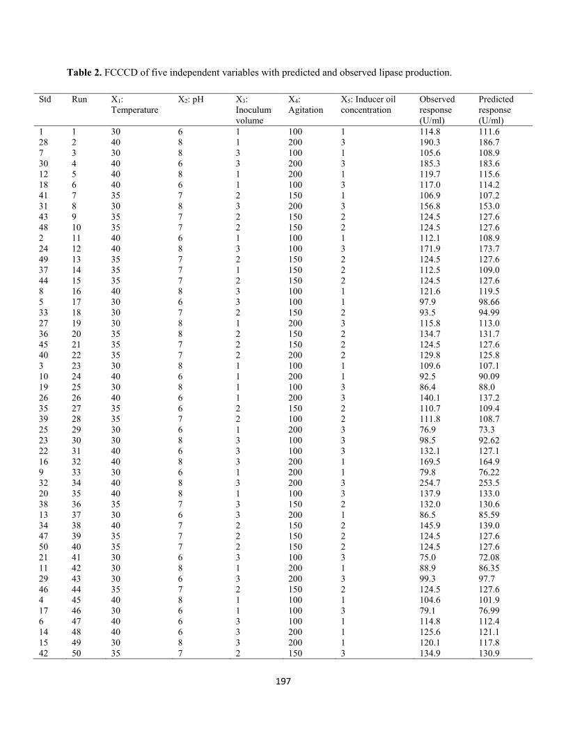
197
Table 2. FCCCD of five independent variables with predicted and observed lipase production.
Std Run X1: Temperature
X2: pH X3: Inoculum volume
X4: Agitation
X5: Inducer oil concentration
Observed response (U/ml)
Predicted response (U/ml)
1 1 30 6 1 100 1 114.8 111.6 28 2 40 8 1 200 3 190.3 186.7 7 3 30 8 3 100 1 105.6 108.9 30 4 40 6 3 200 3 185.3 183.6 12 5 40 8 1 200 1 119.7 115.6 18 6 40 6 1 100 3 117.0 114.2 41 7 35 7 2 150 1 106.9 107.2 31 8 30 8 3 200 3 156.8 153.0 43 9 35 7 2 150 2 124.5 127.6 48 10 35 7 2 150 2 124.5 127.6 2 11 40 6 1 100 1 112.1 108.9 24 12 40 8 3 100 3 171.9 173.7 49 13 35 7 2 150 2 124.5 127.6 37 14 35 7 1 150 2 112.5 109.0 44 15 35 7 2 150 2 124.5 127.6 8 16 40 8 3 100 1 121.6 119.5 5 17 30 6 3 100 1 97.9 98.66 33 18 30 7 2 150 2 93.5 94.99 27 19 30 8 1 200 3 115.8 113.0 36 20 35 8 2 150 2 134.7 131.7 45 21 35 7 2 150 2 124.5 127.6 40 22 35 7 2 200 2 129.8 125.8 3 23 30 8 1 100 1 109.6 107.1 10 24 40 6 1 200 1 92.5 90.09 19 25 30 8 1 100 3 86.4 88.0 26 26 40 6 1 200 3 140.1 137.2 35 27 35 6 2 150 2 110.7 109.4 39 28 35 7 2 100 2 111.8 108.7 25 29 30 6 1 200 3 76.9 73.3 23 30 30 8 3 100 3 98.5 92.62 22 31 40 6 3 100 3 132.1 127.1 16 32 40 8 3 200 1 169.5 164.9 9 33 30 6 1 200 1 79.8 76.22 32 34 40 8 3 200 3 254.7 253.5 20 35 40 8 1 100 3 137.9 133.0 38 36 35 7 3 150 2 132.0 130.6 13 37 30 6 3 200 1 86.5 85.59 34 38 40 7 2 150 2 145.9 139.0 47 39 35 7 2 150 2 124.5 127.6 50 40 35 7 2 150 2 124.5 127.6 21 41 30 6 3 100 3 75.0 72.08 11 42 30 8 1 200 1 88.9 86.35 29 43 30 6 3 200 3 99.3 97.7 46 44 35 7 2 150 2 124.5 127.6 4 45 40 8 1 100 1 104.6 101.9 17 46 30 6 1 100 3 79.1 76.99 6 47 40 6 3 100 1 114.8 112.4 14 48 40 6 3 200 1 125.6 121.1 15 49 30 8 3 200 1 120.1 117.8 42 50 35 7 2 150 3 134.9 130.9
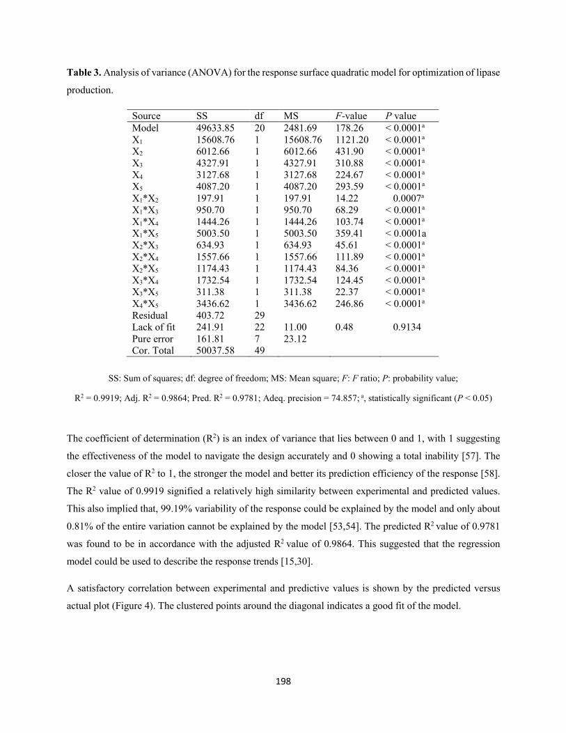
198
Table 3. Analysis of variance (ANOVA) for the response surface quadratic model for optimization of lipase
production.
SS: Sum of squares; df: degree of freedom; MS: Mean square; F: F ratio; P: probability value;
R2 = 0.9919; Adj. R2 = 0.9864; Pred. R2 = 0.9781; Adeq. precision = 74.857; a, statistically significant (P < 0.05)
The coefficient of determination (R2) is an index of variance that lies between 0 and 1, with 1 suggesting
the effectiveness of the model to navigate the design accurately and 0 showing a total inability [57]. The
closer the value of R2 to 1, the stronger the model and better its prediction efficiency of the response [58].
The R2 value of 0.9919 signified a relatively high similarity between experimental and predicted values.
This also implied that, 99.19% variability of the response could be explained by the model and only about
0.81% of the entire variation cannot be explained by the model [53,54]. The predicted R2 value of 0.9781
was found to be in accordance with the adjusted R2 value of 0.9864. This suggested that the regression
model could be used to describe the response trends [15,30].
A satisfactory correlation between experimental and predictive values is shown by the predicted versus
actual plot (Figure 4). The clustered points around the diagonal indicates a good fit of the model.
Source SS df MS F-value P value Model 49633.85 20 2481.69 178.26 < 0.0001a X1 15608.76 1 15608.76 1121.20 < 0.0001a X2 6012.66 1 6012.66 431.90 < 0.0001a X3 4327.91 1 4327.91 310.88 < 0.0001a X4 3127.68 1 3127.68 224.67 < 0.0001a X5 4087.20 1 4087.20 293.59 < 0.0001a X1*X2 197.91 1 197.91 14.22 0.0007a X1*X3 950.70 1 950.70 68.29 < 0.0001a X1*X4 1444.26 1 1444.26 103.74 < 0.0001a X1*X5 5003.50 1 5003.50 359.41 < 0.0001a X2*X3 634.93 1 634.93 45.61 < 0.0001a X2*X4 1557.66 1 1557.66 111.89 < 0.0001a X2*X5 1174.43 1 1174.43 84.36 < 0.0001a X3*X4 1732.54 1 1732.54 124.45 < 0.0001a X3*X5 311.38 1 311.38 22.37 < 0.0001a X4*X5 3436.62 1 3436.62 246.86 < 0.0001a Residual 403.72 29 Lack of fit 241.91 22 11.00 0.48 0.9134 Pure error 161.81 7 23.12 Cor. Total 50037.58 49
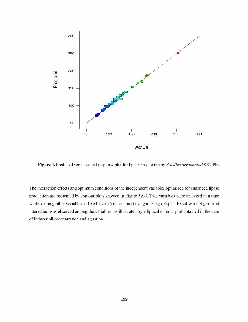
199
Figure 4. Predicted versus actual response plot for lipase production by Bacillus aryabhattai SE3-PB.
The interaction effects and optimum conditions of the independent variables optimized for enhanced lipase
production are presented by contour plots showed in Figure 5A-J. Two variables were analyzed at a time
while keeping other variables at fixed levels (center point) using a Design Expert 10 software. Significant
interaction was observed among the variables, as illustrated by elliptical contour plot obtained in the case
of inducer oil concentration and agitation.
Actual
Pred
icted
50
100
150
200
250
300
50 100 150 200 250 300
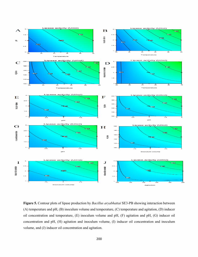
200
Figure 5. Contour plots of lipase production by Bacillus aryabhattai SE3-PB showing interaction between
(A) temperature and pH, (B) inoculum volume and temperature, (C) temperature and agitation, (D) inducer
oil concentration and temperature, (E) inoculum volume and pH, (F) agitation and pH, (G) inducer oil
concentration and pH, (H) agitation and inoculum volume, (I) inducer oil concentration and inoculum
volume, and (J) inducer oil concentration and agitation.
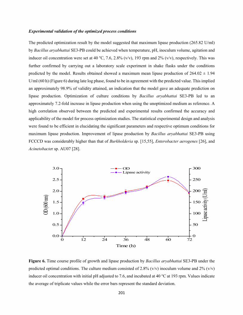
201
Experimental validation of the optimized process conditions
The predicted optimization result by the model suggested that maximum lipase production (265.82 U/ml)
by Bacillus aryabhattai SE3-PB could be achieved when temperature, pH, inoculum volume, agitation and
inducer oil concentration were set at 40 °C, 7.6, 2.8% (v/v), 193 rpm and 2% (v/v), respectively. This was
further confirmed by carrying out a laboratory scale experiment in shake flasks under the conditions
predicted by the model. Results obtained showed a maximum mean lipase production of 264.02 ± 1.94
U/ml (60 h) (Figure 6) during late log phase, found to be in agreement with the predicted value. This implied
an approximately 98.9% of validity attained, an indication that the model gave an adequate prediction on
lipase production. Optimization of culture conditions by Bacillus aryabhattai SE3-PB led to an
approximately 7.2-fold increase in lipase production when using the unoptimized medium as reference. A
high correlation observed between the predicted and experimental results confirmed the accuracy and
applicability of the model for process optimization studies. The statistical experimental design and analysis
were found to be efficient in elucidating the significant parameters and respective optimum conditions for
maximum lipase production. Improvement of lipase production by Bacillus aryabhattai SE3-PB using
FCCCD was considerably higher than that of Burkholderia sp. [15,55], Enterobacter aerogenes [26], and
Acinetobacter sp. AU07 [28].
0 12 24 36 48 60 720.0
0.5
1.0
1.5
2.0
2.5
3.0 OD Lipase activity
Time (h)
OD (60
0 nm)
0
50
100
150
200
250
300
Lipase
activi
ty (U/m
l)
Figure 6. Time course profile of growth and lipase production by Bacillus aryabhattai SE3-PB under the
predicted optimal conditions. The culture medium consisted of 2.8% (v/v) inoculum volume and 2% (v/v)
inducer oil concentration with initial pH adjusted to 7.6, and incubated at 40 °C at 193 rpm. Values indicate
the average of triplicate values while the error bars represent the standard deviation.
202
Conclusions
In the present study, optimization of culture conditions for enhanced extracellular lipase production by
Bacillus aryabhattai SE3-PB was investigated using FCCCD. Lipase production was found to be
significantly influenced by temperature, pH, inoculum volume, agitation and inducer oil concentration. The
high similarity of observed lipase production with the predicted lipase yield confirmed the validity and
accuracy of the generated model. An overall 7.2-fold increase in lipase production was recorded after
optimization of culture conditions by RSM. This study further reiterates the use of FCCCD in determining
significant parameters and optimum conditions for maximum production of desired microbial metabolite.
Optimum conditions of the parameters investigated will provide a baseline for further study involving large
scale and cost-effective production of lipase by Bacillus aryabhattai SE3-PB.
Acknowledgements
The financial support of the National Research Foundation (NRF) of South Africa towards this research is
hereby acknowledged. Opinions expressed and conclusions arrived at, are those of the authors and are not
necessarily to be attributed to the NRF. The first author is thankful to Dr. Ajit Kumar for providing technical
assistance during the conduct of the experiments.
Disclosure statement
The authors declare that there are no conflicts of interest.
References
[1] Gilham D, Lehner R. Techniques to measure lipase and esterase activity in vitro. Methods 2005;36:139-
147.
[2] Angkawidjaja C, Kanaya S. Family I.3 lipase: Bacterial lipases secreted by the type I secretion system.
Cellular Mol Life Sci. 2006;63:2804-2817.
[3] Treichel H, de Oliveira D, Mazutti MA, di Luccio M, Oliveira JV. A review on microbial lipases
production. Food Bioproc Technol. 2010;3:182-196.
203
[4] Joseph B, Ramteke PW, Thomas, G. Cold active microbial lipases: some hot issues and recent
developments. Biotechnol Adv. 2008;26:457-470.
[5] Casas-Godoy L, Duquesne S, Bordes F, Sandoval G, Marty A. Lipases: an overview. Methods Mol
Biol. 2012;861:3-30.
[6] Singh AK, Mukhopadhyay M. Overview of fungal lipases: A review. Appl Biochem Biotechnol.
2012;166:486-520.
[7] Bora L, Gohain D, Das R. Recent advances in production and biotechnological applications of
thermostable and alkaline bacterial lipases. J Chem Technol Biotechnol. 2013;88:1959-1970.
[8] Hasan F, Shah AA, Hameed A. Industrial applications of microbial lipases. Enzyme Microb Technol.
2006;39:235-251.
[9] Kapoor M, Gupta MN. Lipase promiscuity and its biochemical applications. Proc Biochem.
2012;47:555-569.
[10] Dutta S, Ray L. Production and characterization of an alkaline thermostable crude lipase from an
isolated strain of Bacillus cereus C7. Appl Biochem Biotechnol. 2009;159:142-154.
[11] Gupta N, Mehra G, Gupta R. A glycerol-inducible thermostable lipase from Bacillus sp.: medium
optimization by a Plackett-Burman design and by response surface methodology. Can J Microbiol.
2004;50:361-368.
[12] Dutra JCV, Terzi SC, Bevilaqua JV, Damaso MCT, Couri S, Langone MAP. Lipase production in
solid state fermentation monitoring biomass growth of Aspergillus niger using digital image processing.
Appl Biochem Biotechnol. 2008;147:63-75.
[13] Griebeler N, Polloni AE, Remonatto D, Arbter F, Ardanega R, Cechet JL. Isolation and screening of
lipase producing fungi with hydrolytic activity. Food Bioproc Technol. 2011;4:578-586.
[14] Aly MM, Tork S, Alo-Garni SM, Nawar L. Production of lipase from genetically improved
Streptomyces exfoliates LP10 isolated from oil-contaminated soil. Afr J Microbiol Res. 2012;6:1125-1137.
[15] Lo C-F, Yu C-Y, Kuan I-C, Lee S-L. Optimization of lipase production by Burkholderia sp. using
response surface methodology. Int J Mol Sci. 2012;13:14889-14897.
204
[16] Colla LM, Primaz AL, Benedetti S, Loss RA, de Lima M, Reinehr CO, Bertolin TE, Costa JAV.
Surface response methodology for the optimization of lipase production under submerged fermentation by
filamentous fungi. Braz J Microbiol. 2016;47:461-467.
[17] de Almeida AF, Dias KB, da Silva ACC, Terrasan CRF, Tauk-Tornisielo SM, Carmona EC.
Agroindustrial wastes as alternative for lipase production by Candida viswanathii under solid-state
cultivation: purification, biochemical properties, and its potential for poultry fat hydrolysis. Enzyme Res.
2016;2016:15.
[18] Sharma R, Christi Y, Banerjee UC. Production, purification, characterization, and application of
lipases. Biotechnol Adv. 2001;19:627-662.
[19] Kumar R, Sharma A, Kumar A, Singh D. Lipase from Bacillus pumilus RK31: production, purification
and some properties. World Appl Sci J. 2012;16:940-948.
[20] Shah KR, Bhatt SA. Purification and characterization of lipase from Bacillus subtilis Pa2. J Biochem
Technol. 2011;3:292-295.
[21] Castro-Ochoa LD, Rodríguez-Gómez C, Valerio-Alfaro G, Ros RO. Screening, purification and
characterization of the thermoalkalophilic lipase produced by Bacillus thermoleovorans CCR11. Enzyme
Microb Technol. 2005;37:648-654.
[22] Awad GEA, Mostafa H, Danial EN, Abdelwahed NAM, Awad HM. Enhanced production of
thermostable lipase from Bacillus cereus ASSCRC-P1 in waste frying oil based medium using statistical
experimental design. J Appl Pharm Sci. 2015;5:007-015.
[23] Daouadji KL, Reffas FZI, Benine ML, Abbouni B. Optimization of various physical and chemical
parameters for lipase production by Bacillus coagulans. Am Eurasian J Agric Environ Sci. 2015;15:962-
968.
[24] Sekhon A, Dahiya N, Tewari RP, Hoondal GS. Production of extracellular lipase from Bacillus
megaterium AKG-1 in submerged fermentation. Indian J Biotechnol. 2006;5:179-183.
[25] Kaur G, Singh A, Sharma R, Sharma V, Verma S, Sharma PK. Cloning, expression, purification and
characterization of lipase from Bacillus licheniformis isolated from hot spring of Himachal Pradesh, India.
3 Biotech. 2016;6:49.
205
[26] Kumari A, Mahapatra P, Banerjee R. Statistical optimization of culture conditions by response surface
methodology for synthesis of lipase with Enterobacter aerogenes. Braz Arch Biol Technol. 2009;52:1349-
1356.
[27] Açikel Ü, Erşan M, Açikel YS. The effect of the composition of growth medium and fermentation
conditions on the production of lipase by R. delemar. Turk J Biol. 2011;35:35-44.
[28] Gururaj P, Ramalingam S, Devi GN, Gautam P. Process optimization for production and purification
of a thermostable, organic solvent tolerant lipase from Acinetobacter sp. AU07. Braz J Microbiol.
2016;47:647-657.
[29] Yin XL, You QH, Jiang ZH. Optimization of enzyme assisted extraction of polysaccharides from
Tricholoma matsutake by response surface methodology. Carbohydr Polym. 2011;86:1358-1364.
[30] Açikel Ü, Erşan M, Açikel YS. Optimization of critical medium components using response surface
methodology for lipase production by Rhizopus delemar. Food Bioprod Proc. 2010;88:31-39.
[31] Jia J, Yang X, Wu Z, Zhang Q, Lin Z, Guo H, Lin CSK, Wang J, Wang Y. Optimization of fermentation
medium for extracellular lipase production from Aspergillus niger using response surface methodology.
Biomed Res Int. 2015;2015:8.
[32] Neter J, Kutner MH, Nachtsheim CJ, Wasserman W. Applied linear statistical models, fourth ed.
McGraw-Hill Inc., Chicago, 1996.
[33] Rathi P, Goswami VK, Sahai V, Gupta R. Statistical medium optimization and production of a
hyperthermostable lipase from Burkholderia cepacia in a bioreactor. J Appl Microbiol. 2002;93:930-936.
[34] Papagora C, Roukas T, Kotzekidou P. Optimization of extracellular lipase production by
Debaryomyces hansenii isolates from dry-salted olives using response surface methodology. Food Bioprod
Proc. 2013;91:413-420.
[35] Abd Rahman RNZR, Leow TC, Salleh A, Basri M. Geobacillus zalihae sp. nov., a thermophilic
lipolytic bacterium isolated from palm oil mill effluent in Malaysia. BMC Microbiol. 2007;7:77.
[36] Gopinath SCB, Anbu P, Hilda A. Extracellular enzymatic activity profiles in fungi isolated from oil-
rich environments. Mycoscience 2005;46:119-126.
[37] Singh R, Gupta N, Goswami VK, Gupta R. A simple activity staining protocol staining for lipases and
esterases. Appl Microbiol Biotechnol. 2006;70:679-682.
206
[38] Akinbowale OL, Peng H, Grant P, Barton MD. Antibiotic and heavy metal resistance in motile
aeromonads and pseudomonads from rainbow trout (Oncorhynchus mykiss) farms in Australia. Int J
Antimicrob Agents, 2007;30:177-182.
[39] Marchesi JR, Sato T, Weightman AJ, Martin TA, Fry JC, Hiom SJ, Dymock D, Wade WG. Design
and evaluation of useful bacterium-specific PCR primers that amplify genes coding for bacterial 16S rRNA.
Appl Environ Microbiol. 1998;64:795-799.
[40] Tamura K, Stecher G, Peterson D, Filipski A, Kumar S. MEGA 6: Molecular Evolutionary Genetics
Analysis version 6.0. Mol Biol Evol. 2013;30:2725-2729.
[41] Kumar S, Kiko K, Upadhyay A, Kanwar SS, Gupta R. Production, purification, and characterization
of lipase from thermophilic and alkaliphilic Bacillus coagulans BTS-3. Protein Expres Purif. 2005;41:38-
44.
[42] Winkler UK, Stuckmann M. Glocogen, hyaluronate, and some other polysaccharides greatly enhance
the formation of exolipase by Serratia marcescens. J Bacteriol. 1979;138:663-670.
[43] Bhumibhamon O, Koprasertsak A, Funthong S. Biotreatment of high fat and oil wastewater by lipase
producing microorganisms. Kasetsart J Nat Sci. 2002;36:261-267.
[44] Shon HK, Tian D, Kwon D-Y, Jin C-S, Lee T-J, Chung W-J. Degradation of fat, oil and greases (FOGs)
by lipase-producing bacterium Pseudomonas sp. Strain D2D3. J Microbiol Biotechnol. 2002;12:583-591.
[45] Mobarak-Qamsari E, Kasra-Kermanshahi R, Moosavi-nejad Z. Isolation and identification of a novel,
lipase-producing bacterium, Pseudomonas aeruginosa KM110. Iranian J Microbiol. 2011;3:92-98.
[46] Sarac N, Ugur A. A green alternative for oily wastewater treatment: lipase from Acinetobacter
haemolyticus NS02-30. Desalin Water Treat. 2016;57:19750-19759.
[47] Thomson CA, Delaquis PJ, Mazza G. Detection and measurement of microbial lipase activity: a
review. Critical Rev Food Sci Nutr. 1999;39:165-187.
[48] Jagtap S, Gore S, Yavankar S, Paedesi K, Chopade B. Optimization of medium for lipase production
by Acinetobacter haemolyticus from healthy human skin. Indian J Exp Biol. 2010;48:936-941.
[49] Bacha AB, Moubayed NMS, Abid I. Thermostable, alkaline and detergent-tolerant lipase from a newly
isolated thermophilic Bacillus stearothermophilus. Indian J Biochem Biophys. 2015;52:179-188.
207
[50] Sethi BK, Nanda PK, Sahoo S. Characterization of biotechnologically relevant extracellular lipase
produced by Aspergillus terreus NCFT 4269.10. Braz. J Microbiol. 2016;47:143-149.
[51] Golani M, Hajela K, Pandey GP. Screening, identification, characterization and production of bacterial
lipase from oil spilled soil. Int J Curr Microbiol Appl Sci. 2016;5:745-763.
[52] Sifour M, Zaghloul TI, Saeed HM, Berekaa MM, Abdel-Fatah YR. Enhanced production of lipase by
the thermophilic Geobacillus stearothermophilus strain-5 using statistical experimental designs. New
Biotechnol. 2010;27:330-336.
[53] Shabbiri K, Adnan A. Bio-statistically optimized production of lipases by Brevibacterium linens DSM
20158. World Appl Sci J. 2011;13:1059-1066.
[54] Gupta N, Sahai V, Gupta R. Alkaline lipase from a novel strain Burkholderia multivorans: Statistical
medium optimization and production in a bioreactor. Proc Biochem. 2007;42:518-526.
[55] Liu C-H, Lu W-B, Chang J-S. Optimizing lipase production of Burkholderia sp. by response surface
methodology. Proc Biochem. 2006;41:1940-1944.
[56] Facchini FDA, Vici AC, Pereira MG, Jorge JA, Polizeli MDTM. Enhanced lipase production of
Fusarium verticillioides by using response surface methodology and wastewater pretreatment application.
J Biochem Tech. 2015;6:996-1002.
[57] Whiteman J, Kana EBG. Comparative assessment of the artificial neural network and response surface
modelling efficiencies for biohydrogen production on sugar cane molasses. Bioenergy Resour. 2014;7:295-
305.
[58] Doddapaneni KK, Tatineni R, Potumarthi R, Mangamoori LN. Optimization of media constituents
through response surface methodology for improved production of alkaline protease by Serratia rubidea. J
Chem Technol Biotechnol. 2007;82:721-729.
[59] Joglekar AM, May AT. Product excellence through design of experiments. Cereal Food World.
1987;32:857-868.
[60] Wang D, Xu Y, Shan T. Effects of oils and oil-related substrates on the synthetic activity of
membrane-bound lipase from Rhizopus chinensis and optimization of the lipase fermentation
media. Biochem Eng J. 2008;41:30-37.
208
CHAPTER EIGHT
PARTIAL PURIFICATION, IMMOBILIZATION AND CHARACTERIZATION OF
ALKALINE PROTEASE FROM AN INDIGENOUS BACILLUS ARYABHATTAI AB15-
ES ISOLATED FROM LIPID-RICH WASTEWATER
This chapter has been formatted for submission to the ‘Applied Biochemistry and Biotechnology’
209
Partial purification, immobilization and characterization of alkaline protease from an indigenous
Bacillus aryabhattai Ab15-ES isolated from lipid-rich wastewater
Adegoke Isiaka Adetunji and Ademola Olufolahan Olaniran*
Discipline of Microbiology, School of Life Sciences, College of Agriculture, Engineering and Science,
University of KwaZulu-Natal (Westville campus), Private Bag X54001, Durban 4000, Republic of South
Africa.
* Corresponding author. Tel: +27 31 260 7400; Fax: +27 31 260 7809
E-mail address: [email protected]
Abstract In the present study, partially purified alkaline protease from Bacillus aryabhattai Ab15-ES was
immobilized in alginate gel beads by entrapment method. Maximum activities of 68.76 and 71.06% were
recorded at optimum conditions of 2% (w/v) sodium alginate and 0.3 M calcium chloride, respectively for
the entrapped enzyme. Biochemical and kinetic properties of free and immobilized protease were
investigated by determining their activity and stability profiles in terms of pH and temperature. Both
enzyme preparations exhibited maximum activity at optimum pH and temperature of 8.0 and 50 °C.
However, in comparison to free enzyme, the immobilized protease showed improved stability at pH 8.0-
9.0 and at temperature range of 40-50 °C. In addition, the immobilized protease exhibited higher Vmax and
increased (1.65-fold) substrate affinity than soluble enzyme. The immobilized protease was found to be
more stable than free enzyme, retaining 80.88 and 38.37% of its initial activity when stored at 4 and 30 °C,
respectively. After repeated use for seven times, protease entrapped in alginate beads maintained 32.93%
of its original activity. These findings suggest enhanced catalytic efficiency and sustainable use of the
developed immobilized protease for various industrial and environmental applications.
Keywords Bacillus aryabhattai Ab15-ES; Alkaline protease; Immobilization; Alginate beads; Enzyme
kinetics
Introduction
Proteases are enzymes that catalyze the hydrolysis of proteins into simpler subunits by cleavage of peptide
bond that exists between two amino acids of a polypeptide chain [1-3]. They occupy a pivotal position,
constituting more than 65% of total industrial enzyme market [4-6], which is projected to increase further
in coming years [7]. Protease have versatile applications in various industries including detergent,
pharmaceutical, leather, silk, food, silver recovery and agriculture as well as in wastewater treatment and
210
organic chemical synthesis [6,8-10]. Proteases are ubiquitous in nature, and are produced from plants,
animals and microbes [11]. The inability of plant and animal proteases to meet contemporary global demand
has led to an increased interest in microbial proteases. Microbial proteases serve as an excellent source of
commercial enzyme since microorganisms can be cultivated in large quantities at relatively short time
through an established fermentation process for the generation of abundant yield of the desired metabolite.
They also have broad biochemical diversity and are susceptible to genetic manipulation [12]. Microbial
proteases account for approximately 40% of the total worldwide production of the enzyme [13-15].
Microorganisms such as bacteria, molds and yeasts have been reported for extracellular protease production
[16-17].
Free enzymes are generally soluble, unstable and susceptible to harsh reaction conditions including extreme
temperature, very high or low pH, high ionic strength, high concentration of reactants and presence of
inhibitors [18]. Enzymes may not function optimally under such extreme conditions which are often
encountered in bioprocesses [19]. In order to ameliorate the aforementioned challenges, immobilization of
protease in suitable support materials represents a promising alternative [7,20-22]. The use of immobilized
enzymes in bioprocesses has many advantages including increased stability and activity over broad range
of pH and temperatures, localization, ease of product separation and handling, recovery yield and
reusability, rapid termination of reactions, controlled product formation, adaptability to various engineering
designs with a consequent decrease in running cost and autolysis rate [18,23]. However, selection of
suitable support materials and immobilization techniques is paramount to overcome the drawbacks of
immobilization. The general methods employed for enzyme immobilization include physical adsorption,
ionic binding, covalent binding, cross-linking and entrapment methods [24]. Amongst the different
immobilization techniques, entrapment method has been mostly considered due to its simplicity and non-
toxicity [25].
Entrapment method involves physical restriction of the enzyme within a confine space or network in a semi-
permeable capsule, obtained by gelation of polyanionic or polycationic polymers in the presence of
multivalent counterions thus, retaining enzyme catalytic activity and capacity to be used continuously
[7,26,27]. It is carried out in a 3-dimensional gel lattice, made of either natural (agar, cellulose, alginate,
carrageenan) or synthetic (polyacrylamide, polyurethane, polyvinyl, polypropylene) polymers [28,29].
However, calcium alginate beads are mostly preferred owing to their mild gelling properties, easy
formulation, biocompatibility, cost-effectiveness and potential to improve enzyme stability and functional
properties [30-32]. Alginate is a water-soluble anionic linear polysaccharide composed of sequential
arrangements of 1,4-linked β-D-mannuronic acid and α-L-glucuronic acid in different proportion, and can
be precipitated by addition of Ca2+ ions, producing microspheres with good strength and flexibility [33-34].
211
It is employed in many biotechnological applications as support material for releasing and encapsulating
cells and enzymes [35].
Proteases are classified as acidic proteases, neutral proteases and alkaline proteases based on optimal pH
of activity [11,36]. Alkaline proteases are proteases that are active at neutral to alkaline pH conditions. They
occur as serine or metalloproteases, and are commonly exploited for various commercial applications due
to their higher activity and stability at broad pH and temperature ranges [37-39]. Many microbial species
are known as alkaline protease producers. However, among bacteria, members of the genus Bacillus
including Bacillus pumilus [40], Bacillus licheniformis [41], Bacillus caseinilyticus [42], Bacillus subtilis
[43-44], Bacillus megaterium [45], Bacillus coagulans [46] and Bacillus alveayuensis [6] are notable for
commercial scale production of alkaline proteases for various biotechnological and environmental
applications [8,47].
Due to the emergent market and potential application of proteases, there is a continuing interest in the
isolation of indigenous hyperactive proteolytic bacteria with robust catalytic activity suitable for various
industrial and environmental applications. In the present study, extracellular alkaline protease from Bacillus
aryabhattai Ab15-ES isolated from poultry processing wastewater was partially purified and immobilized
by entrapment in alginate beads. Thereafter, comparative study of biochemical and kinetics properties of
the free and immobilized proteases was carried out to ascertain their possible biotechnological applications.
Materials and Methods
Partial purification of protease
Bacillus aryabhattai Ab15-ES was cultivated in optimized medium consisting in g/L: Maltose, 12.35; Beef
extract, 5.30; NaCl, 0.5; CaCl2.2H2O, 0.1; K2HPO4, 0.3; KH2PO4, 0.4; MgSO4.7H2O, 0.1; pH was adjusted
to 7.8. The inoculated culture medium was then incubated at 40 °C for 48 h at 150 rpm. The cell free culture
supernatant (crude protease) obtained by centrifugation at 10000 rpm for 10 min at 4 °C was partially
purified by ammonium sulphate precipitation and dialysis. Solid ammonium sulphate was added to the
crude enzyme by continuous stirring up to 60% saturation. The saturated solution was kept undisturbed at
4 ºC overnight. The precipitate was collected by centrifugation at 10000 rpm for 10 min at 4 ºC, re-
dissolved in a small amount (1:50) of 0.05 M Tris-HCl buffer (pH 7.2) and then dialyzed overnight against
the same buffer at 4 °C.
212
Immobilization of partially purified protease in calcium alginate gel beads
Immobilization of protease was carried out according to the entrapment method of Guleria et al. [7] with
modifications using sodium alginate as support material. Varying amounts of sodium alginate were added
to 0.05 M Tris-HCl buffer (pH 7.2) to attain different concentrations (1-5%). Two milliliter aliquot of
sodium alginate suspension was mixed with 0.5 ml of the partially purified protease. This mixture was
extruded dropwise into a 5 ml CaCl2 solution (0.1- 0.5 M) using a hypodermic syringe from a 5 cm height
with constant stirring. The beads obtained were kept at 4 °C for 1 h for curing. Thereafter, the cured beads
of about 3 mm diameter were recovered from the solution by filtration, washed 3-4 times under mild
agitation with 0.05 M Tris-HCl buffer (pH 7.2) and distilled water to remove unbound enzyme, and then
stored at 4 °C until use [26]. A similar method was used for the preparation of control alginate beads without
the enzyme.
Protease assay
Protease activity was determined according to the modified method of Secades and Guijarro [48] using
azocasein as a substrate. Enzyme solution (120 µl) was added to 480 µl of azocasein solution (1%, w/v of
azocasein in 0.05 M Tris-HCl buffer, pH 7.2), and the mixture was incubated at 37 °C for 30 min. The
reaction was terminated by the addition of 600 µl of 10% chilled trichloroacetic acid (TCA) followed by
further incubation on ice for 30 min to allow undigested protein to precipitate. Thereafter, coagulated
protein was removed from the reaction mixture by centrifugation at 10000 rpm for 10 min at 4 °C using
high- speed refrigerated centrifuge (Avanti J-26 XP, Beckman Coulter, USA). The obtained supernatant
(800 µl) was neutralized by adding 200 µl of 1.8 N NaOH. The absorbance was measured at 440 nm using
a UV-Vis spectrophotometer (UVmini-1240, Schimadzu, Australia). The blank consisted of TCA-substrate
solution. The activity of immobilized protease was determined using the procedure described above, except
that 0.5 g of microspheres was used for the assay. Lowry method was used for the estimation of total protein
using bovine serum albumin as a standard [49]. One unit (U) of enzyme activity was defined as the amount
of enzyme that yielded an increase in absorbance (440 nm) of 0.01 per minute under defined assay
conditions.
Determination of immobilization efficiency
The immobilization efficiency, Y described as the percentage of bound enzyme activity observed in the
immobilizate is calculated using equation (1) [50]:
𝑌𝑌 (%) = 𝑂𝑂𝑂𝑂𝑂𝑂𝑂𝑂𝑂𝑂𝑂𝑂𝑂𝑂𝑂𝑂 𝑎𝑎𝑎𝑎𝑎𝑎𝑎𝑎𝑂𝑂𝑎𝑎𝑎𝑎𝑎𝑎𝐼𝐼𝐼𝐼𝐼𝐼𝐼𝐼𝑂𝑂𝑎𝑎𝐼𝐼𝑎𝑎𝐼𝐼𝑂𝑂𝑂𝑂 𝑎𝑎𝑎𝑎𝑎𝑎𝑎𝑎𝑂𝑂𝑎𝑎𝑎𝑎𝑎𝑎
𝑋𝑋 100 (1)
213
Where immobilized activity was determined by measuring the total residual enzyme activity that remains
in the enzyme solution after immobilization and subtracting this from the total starting activity [50].
Characterization of free and immobilized protease
Effect of temperature and pH on the activity of free and immobilized protease
The temperature at which optimum protease activity was achieved for both free and immobilized proteases
was determined by carrying out enzyme assay at various temperatures (30-80 ºC) for 30 min in 0.05 M
Tris-HCl buffer (pH 7.2). Protease activity was measured, as described previously. The optimum pH for
free and immobilized protease activity was determined at the optimum temperature for 30 min at different
pH values ranging from 3.0-11.0. The buffers used include: 0.05 M citrate buffer (pH 3.0-4.5), 0.05 M
acetate buffer (pH 5.0-5.5), 0.05 M phosphate buffer (pH 6.0-7.0), 0.05 M Tris-HCl buffer (pH 7.5-9.0),
and Glycine-NaOH buffer (9.5-11.0). Azocasein was dissolved in the respective buffers, and then used to
assay for protease activity, as described above. The relative protease activities as percentage (%) of
maximum enzyme activity were calculated and plotted against the respective temperature or pH. All
experiments were carried out in triplicate.
Effect of temperature and pH on the stability of free and immobilized protease
The thermal stability of the free and immobilized protease was determined by pre-incubating the enzyme
in 0.05 M Tris-HCl buffer (pH 8.0) at different temperatures (40, 45, 50 ºC) for up to 3 h. Aliquots were
withdrawn at desired intervals and assayed for residual activity under standard assay conditions. Un-heated
enzyme was considered as a control (100%). The pH stability of the enzyme was assessed by pre-incubating
the enzyme in the respective buffers (0.05 M) for up to 3 h at the optimum temperature. Aliquots were
withdrawn at time intervals, and assayed for residual protease activity under standard experimental
conditions. All experiments were done in triplicate.
Kinetics properties of free and immobilized protease
The influence of substrate (azocasein) concentrations ranging from 1 to 10 mg/ml on free and immobilized
protease activity was studied by measuring enzyme activity, as previously described under standard assay
conditions. Lineweaver-Burk plot was constructed by plotting reciprocals of substrate concentrations
against the reciprocals of enzyme velocity. From the plot, Michaelis-Menten constant (Km) and maximum
velocity (Vmax) values were determined [51].
214
Storage stability of free and immobilized protease
The storage stability of free and immobilized enzyme was determined by preserving the enzymes at 4 and
25 °C. At interval of 5 d for up to 30 d, samples of the stored enzyme were assayed for protease activity.
Residual activity was calculated against the initial enzyme activity taken as 100%.
Reusability potential of the immobilized protease
In order to assess the reusability potential of the protease entrapped in the calcium alginate beads, the
azocasein hydrolysis reaction was repeated several times at 37 °C for 30 min. Thereafter, after every use,
the beads were separated, washed with Trs-HCl buffer (pH 7.2) and added into fresh substrate solution.
Protease activity was measured after every reaction cycle and relative activity was calculated by comparing
the enzyme activity to that of the freshly prepared beads in the first cycle taken as 100%.
Scanning electron microscopy of beads with immobilized protease
The surface morphology of alginate beads containing immobilized protease was studied using ZEISS EVO
LS15 scanning electron microscope.
Results and Discussion
Partial purification of protease
Bacillus aryabhattai Ab15-ES was cultivated in a previously optimized fermentation medium for
extracellular protease production. The results of the purification procedures of obtained crude enzyme are
summarized in Table 1. A 2.19-fold increase in specific activity was obtained for the partially purified
protease compared to the crude enzyme, with a recovery of 60.98%. Sahin et al. [22] and Zhu et al. [52]
reported a 2.1-fold purification and 64% recovery, respectively following ammonium sulphate precipitation
of protease from Bacillus subtilis M-11 and Geobacillus sp. YMTC 1049. Similarly, Mothe et al. [42]
recorded a 1.65-fold increase in protein purification with 48% recovery during ammonium sulphate
precipitation of protease from Bacillus caseinilyticus.
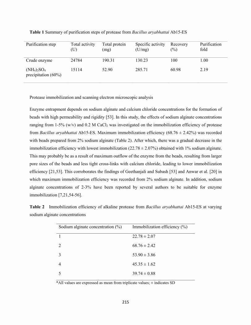
215
Table 1 Summary of purification steps of protease from Bacillus aryabhattai Ab15-ES
Purification step Total activity (U)
Total protein (mg)
Specific activity (U/mg)
Recovery (%)
Purification fold
Crude enzyme 24784 190.31 130.23 100 1.00
(NH4)2SO4 precipitation (60%)
15114 52.90 285.71 60.98 2.19
Protease immobilization and scanning electron microscopic analysis
Enzyme entrapment depends on sodium alginate and calcium chloride concentrations for the formation of
beads with high permeability and rigidity [53]. In this study, the effects of sodium alginate concentrations
ranging from 1-5% (w/v) and 0.2 M CaCl2 was investigated on the immobilization efficiency of protease
from Bacillus aryabhattai Ab15-ES. Maximum immobilization efficiency (68.76 ± 2.42%) was recorded
with beads prepared from 2% sodium alginate (Table 2). After which, there was a gradual decrease in the
immobilization efficiency with lowest immobilization (22.78 ± 2.07%) obtained with 1% sodium alginate.
This may probably be as a result of maximum outflow of the enzyme from the beads, resulting from larger
pore sizes of the beads and less tight cross-links with calcium chloride, leading to lower immobilization
efficiency [21,53]. This corroborates the findings of Geethanjali and Subash [53] and Anwar et al. [20] in
which maximum immobilization efficiency was recorded from 2% sodium alginate. In addition, sodium
alginate concentrations of 2-3% have been reported by several authors to be suitable for enzyme
immobilization [7,21,54-56].
Table 2 Immobilization efficiency of alkaline protease from Bacillus aryabhattai Ab15-ES at varying
sodium alginate concentrations
Sodium alginate concentration (%) Immobilization efficiency (%)
1 22.78 ± 2.07
2 68.76 ± 2.42
3 53.90 ± 3.86
4 45.35 ± 1.62
5 39.74 ± 0.88
*All values are expressed as mean from triplicate values; ± indicates SD
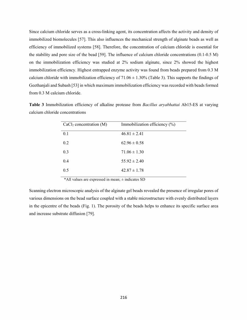
216
Since calcium chloride serves as a cross-linking agent, its concentration affects the activity and density of
immobilized biomolecules [57]. This also influences the mechanical strength of alginate beads as well as
efficiency of immobilized systems [58]. Therefore, the concentration of calcium chloride is essential for
the stability and pore size of the bead [59]. The influence of calcium chloride concentrations (0.1-0.5 M)
on the immobilization efficiency was studied at 2% sodium alginate, since 2% showed the highest
immobilization efficiency. Highest entrapped enzyme activity was found from beads prepared from 0.3 M
calcium chloride with immobilization efficiency of 71.06 ± 1.30% (Table 3). This supports the findings of
Geethanjali and Subash [53] in which maximum immobilization efficiency was recorded with beads formed
from 0.3 M calcium chloride.
Table 3 Immobilization efficiency of alkaline protease from Bacillus aryabhattai Ab15-ES at varying
calcium chloride concentrations
CaCl2 concentration (M) Immobilization efficiency (%)
0.1 46.81 ± 2.41
0.2 62.96 ± 0.58
0.3 71.06 ± 1.30
0.4 55.92 ± 2.40
0.5 42.87 ± 1.78
*All values are expressed in mean; ± indicates SD
Scanning electron microscopic analysis of the alginate gel beads revealed the presence of irregular pores of
various dimensions on the bead surface coupled with a stable microstructure with evenly distributed layers
in the epicentre of the beads (Fig. 1). The porosity of the beads helps to enhance its specific surface area
and increase substrate diffusion [79].
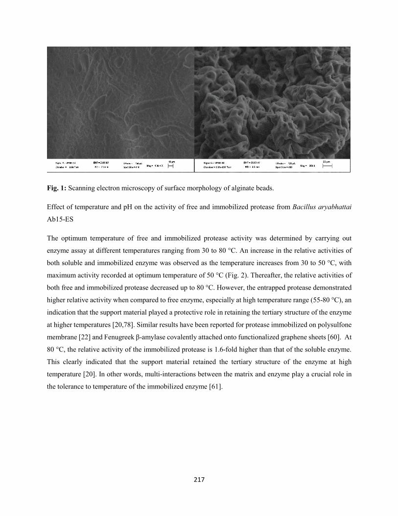
217
Fig. 1: Scanning electron microscopy of surface morphology of alginate beads.
Effect of temperature and pH on the activity of free and immobilized protease from Bacillus aryabhattai
Ab15-ES
The optimum temperature of free and immobilized protease activity was determined by carrying out
enzyme assay at different temperatures ranging from 30 to 80 °C. An increase in the relative activities of
both soluble and immobilized enzyme was observed as the temperature increases from 30 to 50 °C, with
maximum activity recorded at optimum temperature of 50 °C (Fig. 2). Thereafter, the relative activities of
both free and immobilized protease decreased up to 80 °C. However, the entrapped protease demonstrated
higher relative activity when compared to free enzyme, especially at high temperature range (55-80 °C), an
indication that the support material played a protective role in retaining the tertiary structure of the enzyme
at higher temperatures [20,78]. Similar results have been reported for protease immobilized on polysulfone
membrane [22] and Fenugreek β-amylase covalently attached onto functionalized graphene sheets [60]. At
80 °C, the relative activity of the immobilized protease is 1.6-fold higher than that of the soluble enzyme.
This clearly indicated that the support material retained the tertiary structure of the enzyme at high
temperature [20]. In other words, multi-interactions between the matrix and enzyme play a crucial role in
the tolerance to temperature of the immobilized enzyme [61].
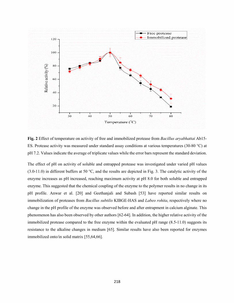
218
Fig. 2 Effect of temperature on activity of free and immobilized protease from Bacillus aryabhattai Ab15-
ES. Protease activity was measured under standard assay conditions at various temperatures (30-80 °C) at
pH 7.2. Values indicate the average of triplicate values while the error bars represent the standard deviation.
The effect of pH on activity of soluble and entrapped protease was investigated under varied pH values
(3.0-11.0) in different buffers at 50 °C, and the results are depicted in Fig. 3. The catalytic activity of the
enzyme increases as pH increased, reaching maximum activity at pH 8.0 for both soluble and entrapped
enzyme. This suggested that the chemical coupling of the enzyme to the polymer results in no change in its
pH profile. Anwar et al. [20] and Geethanjali and Subash [53] have reported similar results on
immobilization of proteases from Bacillus subtilis KIBGE-HAS and Labeo rohita, respectively where no
change in the pH profile of the enzyme was observed before and after entrapment in calcium alginate. This
phenomenon has also been observed by other authors [62-64]. In addition, the higher relative activity of the
immobilized protease compared to the free enzyme within the evaluated pH range (8.5-11.0) suggests its
resistance to the alkaline changes in medium [65]. Similar results have also been reported for enzymes
immobilized onto/in solid matrix [55,64,66].
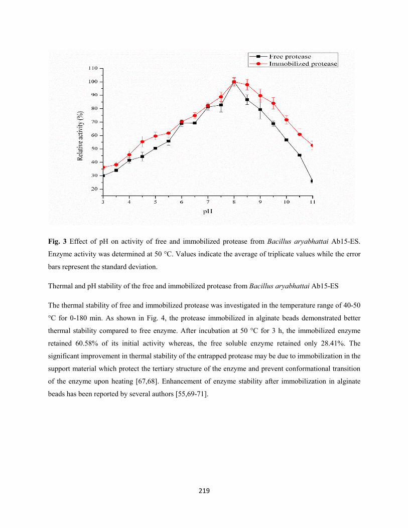
219
Fig. 3 Effect of pH on activity of free and immobilized protease from Bacillus aryabhattai Ab15-ES.
Enzyme activity was determined at 50 °C. Values indicate the average of triplicate values while the error
bars represent the standard deviation.
Thermal and pH stability of the free and immobilized protease from Bacillus aryabhattai Ab15-ES
The thermal stability of free and immobilized protease was investigated in the temperature range of 40-50
°C for 0-180 min. As shown in Fig. 4, the protease immobilized in alginate beads demonstrated better
thermal stability compared to free enzyme. After incubation at 50 °C for 3 h, the immobilized enzyme
retained 60.58% of its initial activity whereas, the free soluble enzyme retained only 28.41%. The
significant improvement in thermal stability of the entrapped protease may be due to immobilization in the
support material which protect the tertiary structure of the enzyme and prevent conformational transition
of the enzyme upon heating [67,68]. Enhancement of enzyme stability after immobilization in alginate
beads has been reported by several authors [55,69-71].
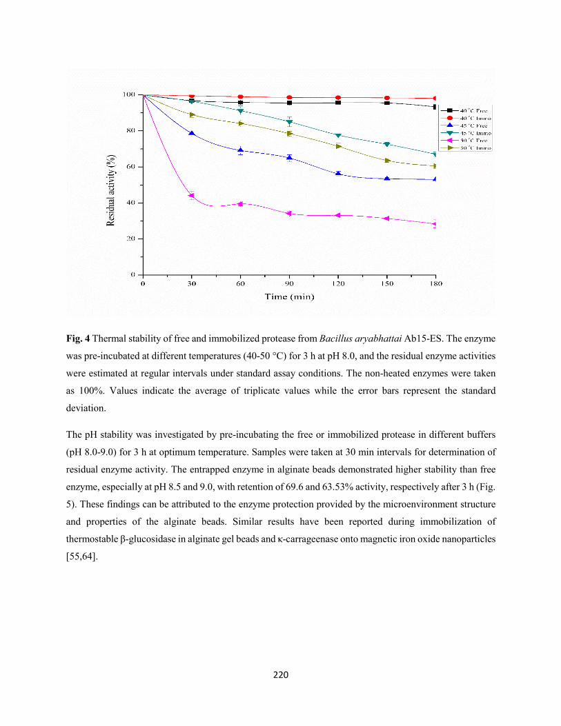
220
Fig. 4 Thermal stability of free and immobilized protease from Bacillus aryabhattai Ab15-ES. The enzyme
was pre-incubated at different temperatures (40-50 °C) for 3 h at pH 8.0, and the residual enzyme activities
were estimated at regular intervals under standard assay conditions. The non-heated enzymes were taken
as 100%. Values indicate the average of triplicate values while the error bars represent the standard
deviation.
The pH stability was investigated by pre-incubating the free or immobilized protease in different buffers
(pH 8.0-9.0) for 3 h at optimum temperature. Samples were taken at 30 min intervals for determination of
residual enzyme activity. The entrapped enzyme in alginate beads demonstrated higher stability than free
enzyme, especially at pH 8.5 and 9.0, with retention of 69.6 and 63.53% activity, respectively after 3 h (Fig.
5). These findings can be attributed to the enzyme protection provided by the microenvironment structure
and properties of the alginate beads. Similar results have been reported during immobilization of
thermostable β-glucosidase in alginate gel beads and κ-carrageenase onto magnetic iron oxide nanoparticles
[55,64].
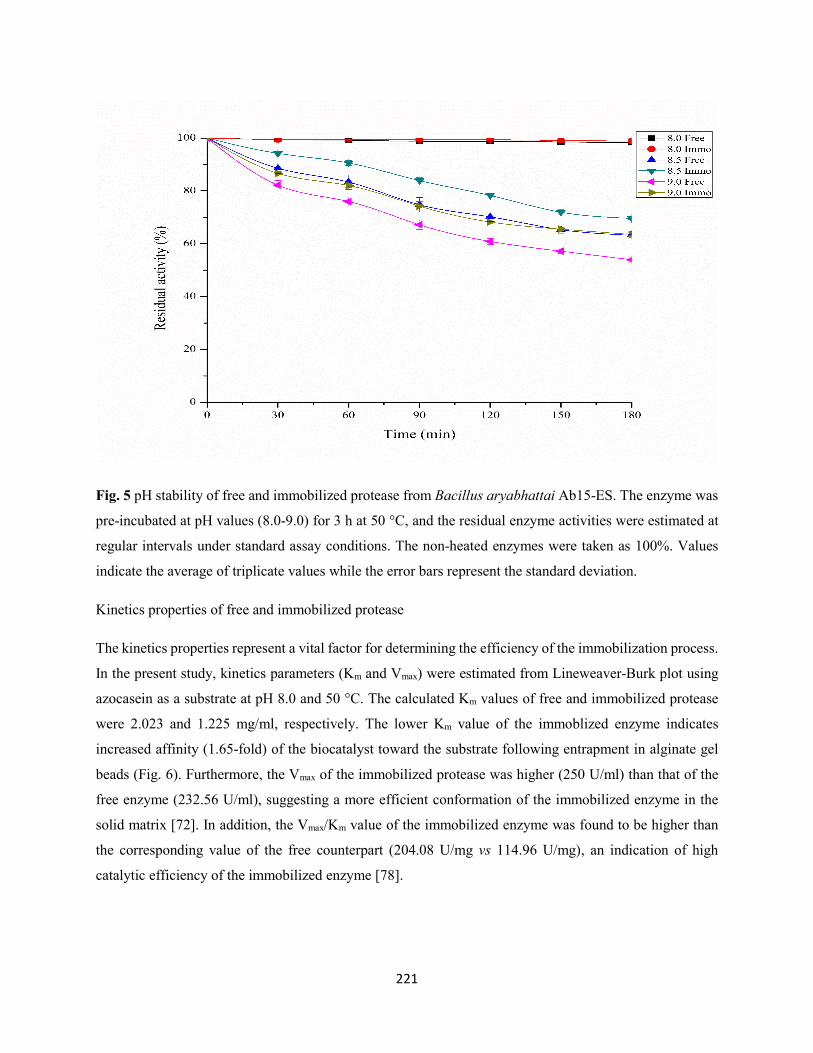
221
Fig. 5 pH stability of free and immobilized protease from Bacillus aryabhattai Ab15-ES. The enzyme was
pre-incubated at pH values (8.0-9.0) for 3 h at 50 °C, and the residual enzyme activities were estimated at
regular intervals under standard assay conditions. The non-heated enzymes were taken as 100%. Values
indicate the average of triplicate values while the error bars represent the standard deviation.
Kinetics properties of free and immobilized protease
The kinetics properties represent a vital factor for determining the efficiency of the immobilization process.
In the present study, kinetics parameters (Km and Vmax) were estimated from Lineweaver-Burk plot using
azocasein as a substrate at pH 8.0 and 50 °C. The calculated Km values of free and immobilized protease
were 2.023 and 1.225 mg/ml, respectively. The lower Km value of the immoblized enzyme indicates
increased affinity (1.65-fold) of the biocatalyst toward the substrate following entrapment in alginate gel
beads (Fig. 6). Furthermore, the Vmax of the immobilized protease was higher (250 U/ml) than that of the
free enzyme (232.56 U/ml), suggesting a more efficient conformation of the immobilized enzyme in the
solid matrix [72]. In addition, the Vmax/Km value of the immobilized enzyme was found to be higher than
the corresponding value of the free counterpart (204.08 U/mg vs 114.96 U/mg), an indication of high
catalytic efficiency of the immobilized enzyme [78].
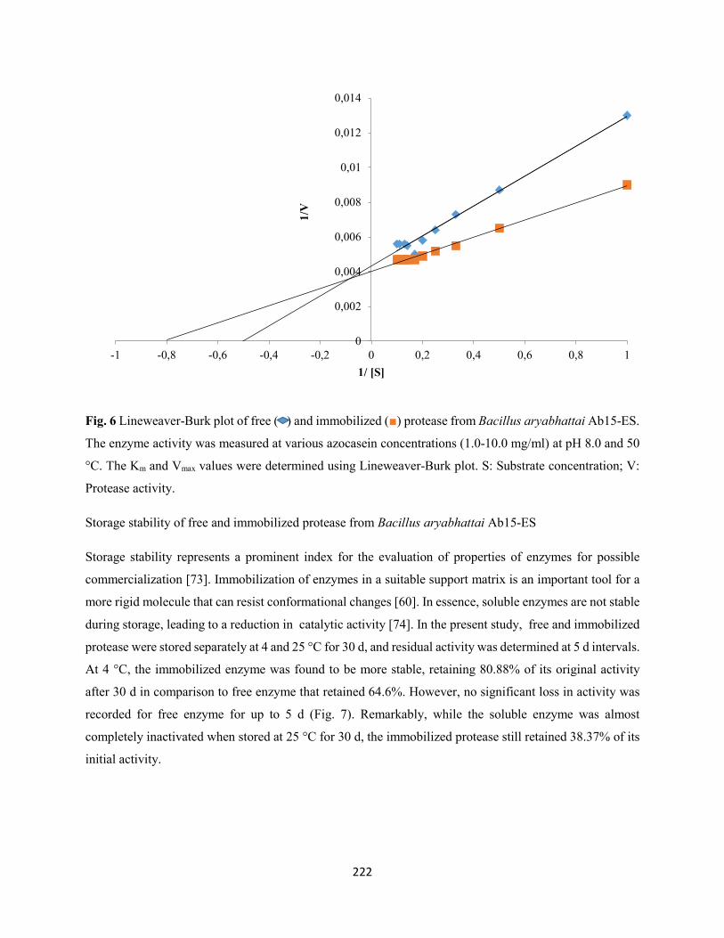
222
Fig. 6 Lineweaver-Burk plot of free ( ) and immobilized () protease from Bacillus aryabhattai Ab15-ES.
The enzyme activity was measured at various azocasein concentrations (1.0-10.0 mg/ml) at pH 8.0 and 50
°C. The Km and Vmax values were determined using Lineweaver-Burk plot. S: Substrate concentration; V:
Protease activity.
Storage stability of free and immobilized protease from Bacillus aryabhattai Ab15-ES
Storage stability represents a prominent index for the evaluation of properties of enzymes for possible
commercialization [73]. Immobilization of enzymes in a suitable support matrix is an important tool for a
more rigid molecule that can resist conformational changes [60]. In essence, soluble enzymes are not stable
during storage, leading to a reduction in catalytic activity [74]. In the present study, free and immobilized
protease were stored separately at 4 and 25 °C for 30 d, and residual activity was determined at 5 d intervals.
At 4 °C, the immobilized enzyme was found to be more stable, retaining 80.88% of its original activity
after 30 d in comparison to free enzyme that retained 64.6%. However, no significant loss in activity was
recorded for free enzyme for up to 5 d (Fig. 7). Remarkably, while the soluble enzyme was almost
completely inactivated when stored at 25 °C for 30 d, the immobilized protease still retained 38.37% of its
initial activity.
0
0,002
0,004
0,006
0,008
0,01
0,012
0,014
-1 -0,8 -0,6 -0,4 -0,2 0 0,2 0,4 0,6 0,8 1
1/V
1/ [S]
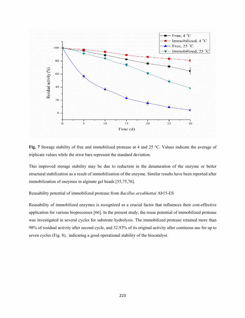
223
Fig. 7 Storage stability of free and immobilized protease at 4 and 25 °C. Values indicate the average of
triplicate values while the error bars represent the standard deviation.
This improved storage stability may be due to reduction in the denaturation of the enzyme or better
structural stabilization as a result of immobilization of the enzyme. Similar results have been reported after
immobilization of enzymes in alginate gel beads [55,75,76].
Reusability potential of immobilized protease from Bacillus aryabhattai Ab15-ES
Reusability of immobilized enzymes is recognized as a crucial factor that influences their cost-effective
application for various bioprocesses [66]. In the present study, the reuse potential of immobilized protease
was investigated in several cycles for substrate hydrolysis. The immobilized protease retained more than
98% of residual activity after second cycle, and 32.93% of its original activity after continous use for up to
seven cycles (Fig. 8), indicating a good operational stability of the biocatalyst.
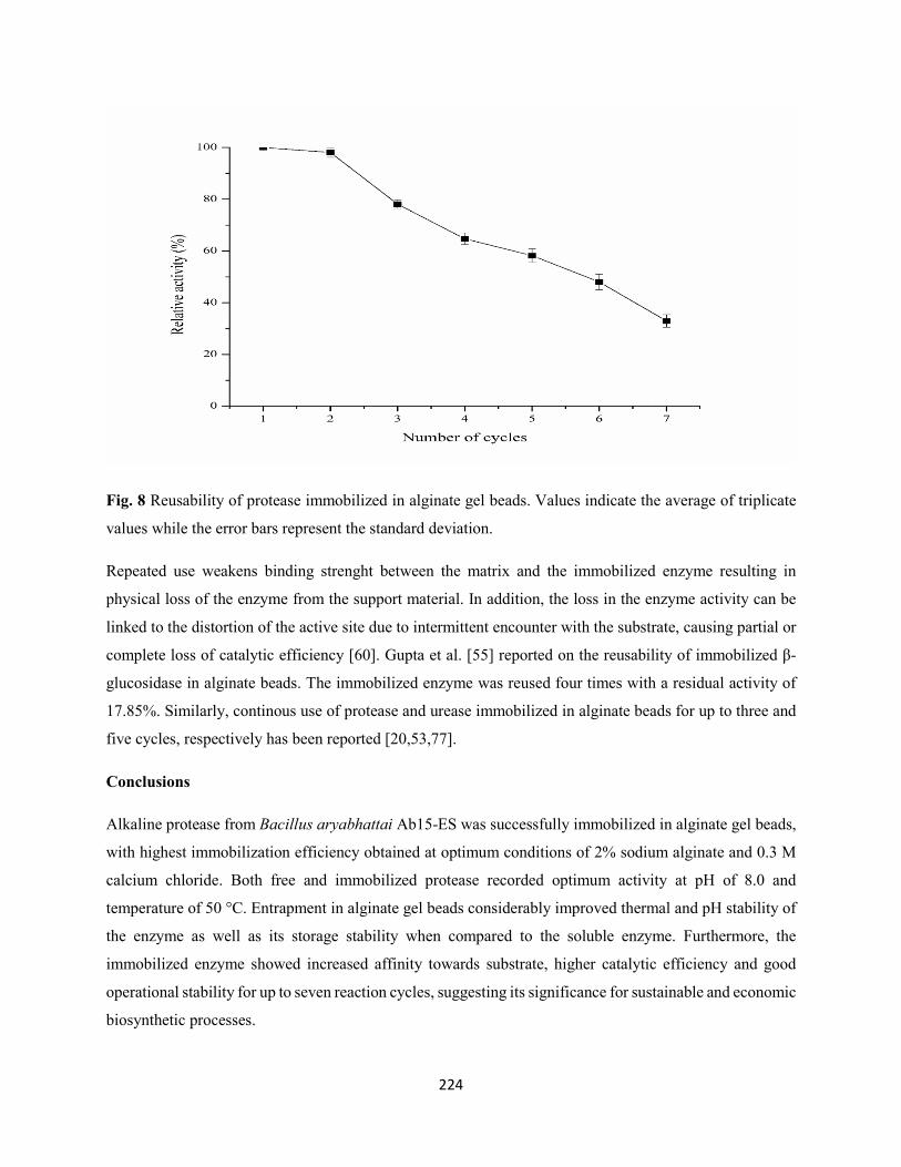
224
Fig. 8 Reusability of protease immobilized in alginate gel beads. Values indicate the average of triplicate
values while the error bars represent the standard deviation.
Repeated use weakens binding strenght between the matrix and the immobilized enzyme resulting in
physical loss of the enzyme from the support material. In addition, the loss in the enzyme activity can be
linked to the distortion of the active site due to intermittent encounter with the substrate, causing partial or
complete loss of catalytic efficiency [60]. Gupta et al. [55] reported on the reusability of immobilized β-
glucosidase in alginate beads. The immobilized enzyme was reused four times with a residual activity of
17.85%. Similarly, continous use of protease and urease immobilized in alginate beads for up to three and
five cycles, respectively has been reported [20,53,77].
Conclusions
Alkaline protease from Bacillus aryabhattai Ab15-ES was successfully immobilized in alginate gel beads,
with highest immobilization efficiency obtained at optimum conditions of 2% sodium alginate and 0.3 M
calcium chloride. Both free and immobilized protease recorded optimum activity at pH of 8.0 and
temperature of 50 °C. Entrapment in alginate gel beads considerably improved thermal and pH stability of
the enzyme as well as its storage stability when compared to the soluble enzyme. Furthermore, the
immobilized enzyme showed increased affinity towards substrate, higher catalytic efficiency and good
operational stability for up to seven reaction cycles, suggesting its significance for sustainable and economic
biosynthetic processes.
225
Acknowledgements: The financial support of the National Research Foundation (NRF) of South Africa
towards this research is hereby acknowledged. Opinions expressed and conclusions arrived at, are those of
the authors and are not necessarily to be attributed to the NRF.
Conflicts of interest: The authors declare no conflict of interest
Author contributions: Adegoke Isiaka Adetunji designed and conducted the experiments; analyzed the
data and drafted the manuscript while Ademola Olufolahan Olaniran conceptualized and designed the
experiment as well as edited the manuscript.
References
1. Ellaiah, P., Srinivasulu, B., & Adinarayana, K. (2002). A review on microbial alkaline proteases. Journal
of Scientific and Industrial Research, 61, 690-704.
2. Gupta, A., & Khare, S. K. (2007). Enhanced production and characterization of a solvent-stable protease
from solvent tolerant Pseudomonas aeruginosa PseA. Enzyme and Microbial Technology, 42, 11-16.
3. Shankar, S., Rao, M., & Laxman, R. S. (2011). Purification and characterization of an alkaline protease
by a new strain of Beauveria sp. Process Biochemistry, 46, 579-585.
4. Sundararajan, S., Kannan, C. N., & Chittibabu, S. (2011). Alkaline protease from Bacillus cereus
VITSN04: Potential application as a dehairing agent. Journal of Bioscience and Bioengineering, 111,
128-133.
5. Zambare, V., Nilegaonkar, S., & Kanekar, P. (2011). A novel extracellular protease from Pseudomonas
aeruginosa MCMB-327: enzyme production and its partial characterization. New Biotechnology, 28,
173-181.
6. Annamalai, N., Rajeswari, M. V., & Balasubramanian, T. (2014). Extraction, purification and
application of thermostable and halostable alkaline protease from Bacillus alveayuensis CAS 5 using
marine wastes. Food and Bioproducts Processing, 92, 335-342.
7. Guleria, S., Walia, A., Chauhan, A., & Shirkot, C. K. (2016). Immobilization of Bacillus
amyloliquefaciens SP1 and its alkaline protease in various matrices for effective hydrolysis of casein. 3
Biotech, 6, 208.
226
8. Deng, A. H., Wu, J., Zhang, Y., Zhang, G. Q., & Wen, T. Y. (2010). Purification and characterization
of a surfactant-stable high-alkaline protease from Bacillus sp. B001. Bioresource Technology, 101:
7100-7106.
9. Jisha, V. N., Smitha, R. B., Pradeep, S., Sreedevi, S., Unni, K. N., Sajith, S., Priji, P., Josh, M. S., &
Benjamin, S. (2013). Versatility of microbial proteases. Advanced Enzyme Research, 1, 39-51.
10. Sawant, R., & Nagendran, S. (2014). Protease: an enzyme with multiple industrial applications. World
Journal of Pharmacy and Pharmaceutical Sciences, 3, 568-579.
11. Rao, M. B., Tanksale, A. M., Ghatge, M. S, & Deshpande, V. V (1998). Molecular and biotechnological
aspects of microbial proteases. Microbiolology and Molecular Biology Reviews, 62, 597-635.
12. Gupta, R., Beg, Q. K., Khan, S., & Chauhan, B. (2002a). An overview of fermentation, downstream
processing and properties of microbial alkaline proteases. Applied Microbiology and Biotechnology,
60, 381-395.
13. Kumar, C. G, & Takagi, H. (1999). Microbial alkaline proteases: from a bioindustrial viewpoint.
Biotechnology Advances, 17, 561-594.
14. Haddar, A., Bougatef, A., Agrebi, R., Sellami-Kamoun, A., & Nasri, M. (2009). A novel surfactant-
stable alkaline serine-protease from a newly isolated Bacillus mojavensis A21: purification and
characterization. Process Biochemistry, 44, 29-35.
15. Raval, V. R., Pillai, S., Rawal, C. M., & Singh, S. P. (2014). Biochemical and structural characterization
of a detergent-stable serine alkaline protease from seawater haloalkaliphilic bacteria. Process
Biochemistry, 49, 955-962.
16. Tang, X. Y., Wu, B., Ying, H. J., & He, B. F. (2010). Biochemical properties and potential applications
of a solvent-stable protease from a highly-yield protease producer Pseudomonas aeruginosa PT 121.
Applied Biochemistry and Biotechnology, 160, 1017-1031.
17. Devi, P. R., VijayaRaghavan, P., Vasudheven, I., Joshua, L., & VijaKumar, M. (2011). Purification and
characterization of protease from Rhizopus oligosporus. International Journal of Biological
Technology, 2, 46-49.
18. Mugdha, A., & Usha, M. (2012). Enzymatic treatment of wastewater containing dyestuffs using
different delivery systems. Scientific Reviews and Chemical Communications, 2, 31-40.
19. Karam, J., & Nicell, J. A. (1997). Potential applications of enzymes in waste treatment. Journal of
Chemical Technology and Biotechnology, 69, 141-153.
20. Anwar, A., Ul Qader, S. A., Raiz, A., Iqbal, S., & Azhar, A. (2009). Calcium alginate: a support material
for immobilization of proteases from newly isolated strain of Bacillus subtilis KIBGE-HAS. World
Applied Sciences Journal, 7: 1281-1286.
227
21. Kumar, D., Kumar, V., Verma, A. K., Singh, P., & Dubey, A. (2014). Characterization and
immobilization of partially purified alkaline protease extracted from rhizospheric soil bacterium,
Bacillus megaterium strain EN-2 and Bacillus subtilis strain EN-3. African Journal of Microbiological
Research, 8, 33-39.
22. Sahin, S., Ozmen, I., & Kir, E. (2015). Purification, immobilization and characterization of protease
from local Bacillus subtilis M-11. Asia-Pacific Journal of Chemical Engineering, 10, 241-247.
23. Massolini, G., & Calleri, E. (2005). Immobilized trypsin systems coupled on-line to separation
methods: recent developments and analytical applications. Journal of Separation Science, 28, 7-21.
24. Hasirci, N., Aksoy, S., & Tumturk, H. (2006). Activation of poly (dimer acid-co-alkyl polyamine)
particles for covelent immobilization of α-amylase. Reactive and Functional Polymers, 66, 1546-1551.
25. Gangadharan, D., Nampoothiri, K. M., Sivaramakrishnan, S., & Pandey, A. (2009). Immobilized
bacterial α-amylase for effective hydrolysis of raw and soluble starch. Food Research International,
42, 436-442.
26. Won, K., Kim, S., Kim, K. J., Park, H. W., & Moon, S. (2005). Optimization of lipase entrapment in
Ca-alginate beads. Process Biochemistry, 40, 2149-2154.
27. Bonine, B. M., Polizelli, P. P, & Bonilla-Rodriguez, G. O. (2014). Immobilization of a plant lipase
from Pachira aquatica in alginate and alginate/PVA beads. Enzyme Research, 2014, 7.
28. Datta, S., Rene, C. L., & Rajaram, Y. R. S. (2013). Enzyme immobilization: an overview on techniques
and support materials. 3 Biotech, 3, 1-9
29. Mohamad, N. R., Marzuki, N. H. C., Buang, N. A., Huyop, F., & Abdul Wahab, R. (2015). An overview
of technologies for immobilization of enzymes and surface analysis techniques for immobilized
enzymes. Biotechnology and Biotechnological Equipment, 29, 205-220.
30. Tumturk, H., Demirel, G., Altinok, H., Aksoy, S., & Hasirci, N. (2008). Immobilization of glucose
isomerase in surface-modified alginate gel beads. Journal of Food Biochemistry, 32, 234-246.
31. Ölcer, Z., & Tanriseven, A. (2010). Co-immobilization of dextransucrase and dextranase in alginate.
Process Biochemistry, 45, 1645-1651.
32. Jamal, F., Qidwai, T., Singh, D., & Pandey, P. K. (2012). Biocatalytic activity of immobilized pointed
gourd (Trichosanthes dioica) peroxidase-concanavalin A complex on calcium alginate pectin gel.
Journal of Molecular Catalysis B: Enzymatic, 74, 125-131.
33. Mahajan, R., Gupta, V. K., & Sharma, J. (2010). Comparison and suitability of gel matrix for entrapping
higher content of enzymes for commercial applications. Indian Journal of Pharmaceutical Sciences,
72, 223-228.
228
34. Buchholz, K., Kasche, V., & Bornscheuer, U. T. (2012). Characterization of immobilized catalyst, in
Biocatalysts and Enzyme Technology (Buchholz, K., Kasche, V., Bornscheuer, U. T., Ed), Boschstraẞe,
Weinheim, Germany. pp. 411-448.
35. Blandino, A., Macias, M., & Cantero, D. (2000). Glucose oxidase release from calcium alginate gel
capsules. Enzyme and Microbial Technology, 27, 319-324.
36. Sabotic, J., & Kos, J. (2012). Microbial and fungal protease inhibitors- current and potential
applications. Applied Microbiology and Biotechnology, 93, 1351-1375.
37. Gupta, R., Beg, Q. K., & Lorenz, P. (2002b). Bacterial alkaline proteases: molecular approaches and
industrial applications. Applied Microbiolology and Biotechnology, 59, 15-32.
38. Devi, M. K., Banu, A. R., Gnanaprabhal, G. R., Pradeep, B. V., & Palaniswamy, M. (2008).
Purification, characterization of alkaline protease enzyme from native isolate Aspergillus niger and its
compatibility with commercial detergents. Indian Journal of Science and Technology, 1, 1-6.
39. Vijayaraghavan, P., & Vincent, S. G. P. (2012). Cow dung as a novel, inexpensive substrate for the
production of a halo-tolerant alkaline protease from Halomonas sp. PVI for eco-friendly applications.
Biochemical Engineering Journal, 69, 57-60.
40. Jaouadi, B., Ellouz-Chaabouni, S., Rhimi, M., & Bejar, S. (2008). Biochemical and molecular
characterization of a detergent-stable serine alkaline protease from Bacillus pumilus CBS with high
catalytic efficiency. Biochimie, 90, 1291-1305.
41. Sellammi-Kamoun, A., Haddar, A., Ali, N. E., Ghorbel-Frikha, B., Kanoun, S., & Nasri, M. (2008).
Stability of thermostable alkaline protease from Bacillus licheniformis RP1 in commercial solid laundry
detergent formulations. Microbiological Research, 163, 299-306.
42. Mothe, T., & Sultanpuran, V. R. (2016) Purification and characterization of a thermotolerant alkaline
serine protease from a novel species of Bacillus caseinilyticus. 3 Biotech, 6, 53.
43. Maruthiah, T., Esakkiraj, P., Prabakaran, G., Palavesam, A., & Immanuel, G. (2013). Purification and
characterization of moderately halophilic alkaline serine protease from marine Bacillus subtilis AP-
MSU 6. Biocatalysis and Agricultural Biotechnology, 2, 116-119.
44. Ghafoor, A., & Hasnain, S. (2010). Purification and characterization of extracellular protease from
Bacillus subtilis EAG-2 strain isolated from ornamental plant nursery. Polish Journal of Microbiology,
59, 107-112.
45. Rajkumar, R., Jayappriyan, K. R., & Rengasamy, R. (2011). Purification and characterization of a
protease produced by Bacillus megaterium RRM2: application in detergent and dehairing industries.
Journal of Basic Microbiology, 51, 614-624.
229
46. Benkiar, A., Nadia, Z. J., Badis, A., Rebzani, F., Soraya, B. T., Rekik, H., Naili, B., Ferradji, F. Z.,
Bejar, S., & Jaouadi, B. (2013). Biochemical and molecular characterization of a thermos- and
detergent-stable alkaline serine keratinolytic protease from Bacillus circulans strain DZ100 for
detergent formulations and feather-biodegradation process. International Biodeterioration and
Biodegradation, 83, 129-138.
47. Joo, H. S., Kumar, C. G., Park, G. C., Paik, S. R., & Chang, C. S. (2004). Bleach-resistant alkaline
protease produced by a Bacillus sp. isolated from the Korean polychaete, Periserrula leucophryna.
Process Biochemistry, 39, 1441-1447.
48. Secades, P., & Guijarro, J. A. (1999). Purification and characterization of an extracellular protease from
the fish pathogen Yersinia ruckeri and effect of culture conditions on production. Applied and
Environmental Microbiology, 65, 3969-3975.
49. Lowry, O. H., Rosebrough, N., Farr, A. L., & Rondall, R. L. (1951). Protein measurement with the
Folin-phenol reagent. Journal of Biological Chemistry, 193, 265-273.
50. Sheldon, R. A., & van Pelt, S. (2013). Enzyme immobilization in biocatalysis: why, what, and how.
Chemical Society Reviews, 42, 6223-6235.
51. Lineweaver, H., & Burk, D. (1934). The determination of enzyme dissociation constants. Journal of
Amercian Chemical Society, 56, 658-666.
52. Zhu, W., Cha, D., Cheng, G., Peng, Q., & Shen, P. (2007). Purification and characterization of a
thermostable protease from a newly isolated Geobacillus sp. YMTC 1049. Enzyme and Microbial
Technology, 40, 1592-1597.
53. Geethanjali, S., & Subash, A. (2013). Optimization and immobilization of purified Labeo rohita
visceral protease by entrapment method. Enzyme Research, 2013, 1-7.
54. Farag, A. M., & Hassan, M. A. (2004). Purification, characterization and immobilization of a keratinase
from Aspergillus oryzae. Enzyme and Microbial Technology, 34, 85-93.
55. Gupta, K. A., Kumar, V., Dubey, A., & Verma, A. K. (2014). Kinetic characterization and effect of
immobilized thermostable β-glucosidase in alginate gel beads on sugarcane juice. ISRN Biochemistry,
2014, 1-8.
56. Thu, T. T. M., & Krasaekoopt, W. (2016). Encapsulation of protease from Aspergillus oryzae and lipase
from Thermomyces lanuginoseus using alginate and different copolymer types. Agriculture and
Natural Resources, 50, 155-161.
57. Sun, J., Liu, J., Liu, Y., & Li, Z. (2011). Optimization of entrapping conditions of nitrifying bacteria
and selection of entrapping agent: In proceedings of the 2nd International Conference on Environmental
Science and Technology (IPCBEE), 6: IACSIT Press.
230
58. Konsoula, Z., & Kyriakides, M. L. (2006). Thermostable α-amylase production by Bacillus subtilis
entrapped in calcium alginate gel capsules. Enzyme and Microbial Technology, 39, 690-696.
59. Elibol, M., & Moreira, A. R. (2003). Production of extracellular alkaline protease by immobilization
of the marine bacterium Teredinobacter turnirae. Process Biochemistry, 38, 1445-1450.
60. Srivastava, G., Singh, K., Talat, M., Srivastava, O. N., & Kayastha, A. M. (2014). Functionalized
graphene sheets as immobilization matrix for Fenugreek β-amylase: enzyme kinetics and stability
studies. PLoS one, 9: e113408.
61. He, S., Song, D., Chen, M., & Cheng, H. (2017). Immobilization of lipases on magnetic collagen fibers
and its applications for short-chain ester synthesis. Catalysts, 7: 178.
62. Tükel, S. S., Hürrem, F., Yildirim, D., & Alptekin, O. (2013). Preparation of crosslinked enzyme
aggregates (CLEA) of catalase and its characterization. Journal of Molecular Catalysis B: Enzymatic,
97: 252-257.
63. Saravanakumar, T., Palvannan, T., Kim, D. H., & Park, S. M. (2014). Optimized immobilization of
peracetic acid producing recombinant acetyl xylan esterase on chitosan coated-Fe3O4 magnetic
nanoparticles. Process Biochemistry, 49, 1920-1928.
64. Xiao, A., Xu, C., Lin, Y., Ni, H., Zhu, Y., & Cai, H. (2016). Preparation and characterization of κ-
carrageenase immobilized onto magnetic iron oxide nanoparticles. Electronic Journal of
Biotechnology, 19, 1-7.
65. Quioroga, E., Illanes, C. O., Ochoa, N. A., & Barberis, S. (2011). Performance improvement of
araujiain, a cysteine phytoprotease by immobilization within calcium alginate beads. Process
Biochemistry, 46, 1029-1034.
66. Ibrahim, A. S. S., Al-Salamah, A. A., El-Toni, A. M., Almaary, K. S., El-Tayeb, M. A., Elbadawi, Y.
B., & Antranikian, G. (2016). Enhancement of alkaline protease activity and stability via covalent
immobilization onto hollow core-mesoporous shell silica nanospheres. International Journal of
Molecular Sciences, 17, 184.
67. Mateo, C., Palomo, J. M., Fernandez-Lorente, G., Guisan, J. M., & Fernandez-Lafuente, R. (2007).
Improvement of enzyme activity, stability and selectivity via immobilization techniques. Enzyme and
Microbial Technology, 40, 1451-1463.
68. Ranjbakhsh, E., Bordbar, A. K., Abbasi, M., Khosropour, A. R., & Shams, E. (2012). Enhancement of
stability and catalytic activity of immobilized lipase on silica-coated modified magnetite nanoparticles.
Chemical Engineering Journal, 179, 272-276.
69. Ahmed, S. A., Al-domany, R. A., El-Shayeb, N. M. A., Radwan, H. H., & Saleh, A. (2008).
Optimization, immobilization of extracellular alkaline protease and characterization of its enzymatic
properties. Research Journal of Agriculture and Biological Sciences, 4, 434-446.
231
70. Zanphorlin, L. M., Facchini, F. D. A., Vasconcelos, F., Bonugli-Santos, R. C., Rodrigues, A., Sette, L.
D., Gomes, E., & Bonilla-Rodrigues, G. O. (2010). Production, partial characterization and
immobilization in alginate beads of an alkaline protease from a new thermophilic fungus
Myceliophthora sp. Journal of Microbiolology, 48, 331-336.
71. Danial, E. N., Hamza, A. H., & Mahmoud, R. H. (2015). Characteristics of immobilized urease on
grafted alginate bead systems. Brazilian Archives of Biology and Technology, 58, 147-153.
72. Su, R., Shi, P., Zhu, M., Hong, F., & Li, D. (2012). Studies on the properties of graphene oxide-alkaline
protease bio-composites. Bioresource Technology, 115, 136-140.
73. De Queiroz, A., Passes, E., Alves, S., Silva, G., Higa, O., & Vítolo, M. (2006). Alginate-poly (vinyl
alcohol) core-shell microspheres for lipase immobilization. Journal of Applied Polymer Science, 102,
1553-1560.
74. Çevik, E., Şenel, M., & Abasiyanik, M. F. (2011). Immobilization of urease on copper chelated EC-
Tribeads and reversible adsorption. African Journal of Biotechnology, 10, 6590-6597.
75. Yahşi, A., Şahin, F., Demirel, G., & Tümtürk, H. (2005). Binary immobilization of tyrosinase by using
alginate gel beads and poly (acrylamide-co-acrylic acid) hydrogels. International Journal of Biological
Macromolecules, 36, 253-258.
76. Bhushan, B., Pal, A., & Jain, V. (2015). Improved enzyme catalytic characteristics upon glutaraldehyde
cross-linking of alginate entrapped xylanase isolated from Aspergillus flavus MTCC 9390. Enzyme
Research, 2015,1-9.
77. Pithawala, K., Mishra, N., & Bahadur, A. (2010). Immobilization of urease in alginate, paraffin and
lac. Journal of Serbian Chemical Society, 75, 175-183.
78. Cao, S-L., Huang, Y-M., Li, X-H., Xu, P., Wu, H., Li, N., Lou, W-Y., & Zong, M-H. (2016).
Preparation and characterization of immobilized lipase from Pseudomonas cepacia onto magnetic
cellulose nanocrystals. Science Reports, 6, 20420.
79. Zhan, J. F., Jiang, S. T., & Pan, L. J. (2013). Immobilization of phospholipase A1 using polyvinyl
alcohol-alginate matrix and evaluation of the effects of immobilization. Brazilian Journal of Chemical
Engineering, 30, 721-728.
232
CHAPTER NINE
LIPASE FROM AN INDIGENOUS BACILLUS ARYABHATTAI SE3-PB: PARTIAL
PURIFICATION, IMMOBILIZATION AND BIOCHEMICAL PROPERTIES
This chapter has been formatted for submission to the ‘Archives of Microbiology’
233
Lipase from an indigenous Bacillus aryabhattai SE3-PB: partial purification, immobilization and
biochemical properties
Adegoke Isiaka Adetunji and Ademola Olufolahan Olaniran*
Discipline of Microbiology, School of Life Sciences, College of Agriculture, Engineering and Science,
University of KwaZulu-Natal (Westville campus), Private Bag X54001, Durban 4000, Republic of South
Africa.
* Corresponding author. Tel: +27 31 260 7400; Fax: +27 31 260 7809
E-mail address: [email protected]
Acknowledgements
The financial support of the National Research Foundation (NRF) of South Africa towards this research is
hereby acknowledged. Opinions expressed and conclusions arrived at, are those of the authors and are not
necessarily to be attributed to the NRF.
Conflict of interest: The authors declare no conflict of interest
234
Abstract
Extracellular lipase from an indigenous Bacillus aryabhattai SE3-PB was partially purified by ammonium
sulphate precipitation, and then immobilized in alginate beads by entrapment method. Biochemical
properties of both free and immobilized lipases were further investigated. After optimization of
immobilization conditions, maximum immobilization efficiencies of 77 ± 1.53% and 75.99 ± 3.49% were
recorded at optimum concentrations of 2% (w/v) sodium alginate and 0.2 M calcium chloride, respectively
for the entrapped enzyme. There was no change in the optimum temperature and pH of both enzyme
preparations, with maximum activity attained at 60 °C and 9.5, respectively. In comparison to free lipase,
the entrapped enzyme demonstrated improved stability over the studied pH range (8.5-9.5) and temperature
(55-65 °C) when incubated for 3 h. Furthermore, the immobilized lipase showed enhanced enzyme-
substrate affinity and higher catalytic efficiency when compared to soluble enzyme. The entrapped enzyme
was also found to be more stable, retaining 61.51 and 49.44% of its original activity after being stored for
30 d at 4 and 25 °C, respectively. In addition, the insolubilized enzyme exhibited good reusability with
18.46% relative activity after being repeatedly used for six times. These findings suggest the efficacy of the
prepared immobilized lipase for various biotechnological applications.
Keywords Bacillus aryabhattai SE3-PB; Lipase; Immobilization; Alginate beads; Biotechnological
applications
Introduction
Lipases (EC 3.1.1.3; triacylglycerol acylhydrolases) are biocatalysts that involved in the hydrolysis of ester
bonds present in fats and oils into di-glycerides, mono-glycerides, glycerol and free fatty acids at the oil-
water interface (Treichel et al. 2010). In addition, lipases also catalyze synthesis reactions including
esterification, trans-esterification, inter-esterification, alcoholysis, acidolysis, and aminolysis in non-
aqueous and micro-aqueous milieu (Casas-Godoy et al. 2012; Kanmani et al. 2015). The potential of lipases
to catalyze different reactions using wide range of substrates in the absence of expensive cofactors coupled
with high activity and stability in organic solvents led to lipases being grouped as third largest class of
commercialized enzymes after proteases and carbohydrases (Hasan et al. 2006; Casas-Godoy et al. 2012).
Lipases are produced by numerous species of animals, plants, bacteria, and fungi (Dutta and Ray 2009;
Nagarajan 2012; Kanmani et al. 2015). However, lipase of microbial origin have gained vast attraction
owing to their good stability, substrate specificity, ease of genetic manipulation, lower production cost and
higher productivity at shorter generation time (Hasan et al. 2006). Lipases are commonly used in food, pulp
235
and paper, detergent, tannery, textile, pharmaceutical, agrochemical and cosmetic industries as well as in
treatment of oil-contaminated wastewater (Hasan et al. 2006; Sangeetha et al. 2011).
Free enzymes are generally soluble in homogenous catalysis system, leading to product contamination and
eliminating their recovery for reuse in the active form from most of the reaction mixtures, thus can only be
used once in solutions (Homaei et al. 2013). Moreover, free enzymes are not only unstable and inactivated
by different environmental conditions (ionic strength, temperature, pH, inhibitors), but too costly for
biotechnological applications (Guisan 2006; Lee et al. 2010; Hernandez and Fernandez-Lafuente 2011;
Sangeetha et al. 2011). In order to abate the aforementioned challenges and make enzyme utilization in
bioprocesses more favorable, immobilization has been recognized as an important strategy in enzyme
technology.
Enzyme immobilization involves retention of catalytic activity of the enzyme in a certain defined region in
order to permit its reusability (Mohamad et al. 2015). It offers several advantages including increased
activity and stability over broader range of temperature and pH, easier product recovery, cost reduction,
recovery yield, possibility of continuous process, rapid termination of reactions, controlled product
formation and adaptability to a variety of configurations and specific processes in reactors (Spahn and
Minteer 2008; Homaei et al. 2013).
The choice of appropriate immobilization technique is key to a successful immobilization process, as it
plays a paramount role in determining the enzyme activity and characteristics in a particular reaction
(Mohamad et al. 2015). Effectiveness of each technique depends on reaction conditions, process of product
formation and cost evaluation (Bhushan et al. 2015). Methods such as physical adsorption, entrapment,
covalent binding and cross-linking are employed for lipase immobilization (Minovska et al. 2005;
Fernandez-Lafuente 2010). However, entrapment in appropriate support material is known as the most
efficient technique due to its ability to enhance mechanical stability, minimize enzyme leaching, and
prevent chemical interaction of the enzyme with the support material (Shen et al. 2011). In addition,
entrapment permits modification of encapsulating material and create optimal microenvironment for the
enzyme (Mohamad et al. 2015). Such microenvironment can be achieved with a range of support materials
(Mendes et al. 2011; Datta et al. 2013; Santos et al. 2015).
Gelation of polyanionic or polycationic polymers (e.g. alginate) in the presence of counter-ions represents
an easy and commonly employed approach for enzyme entrapment. However, alginate is by far the most
widely used polymer for immobilization and microencapsulation technologies due to its mild gelling
properties, non-toxicity, biocompatibility, thermostability, ease of formulation, low cost, effective particle
size and availability (Won et al. 2005; Yadav et al. 2012). Alginate is an anionic linear heteropolysaccharide
236
extracted from marine brown algae and seaweeds, and consist of 1,4-linked β-D-mannuronic acid and α-L-
guluronic acid residues arranged sequentially in different proportions. Polyvalent cations such as calcium
chloride bind to the biopolymer whenever there are two neighboring guluronic acid residues forming an
ionotropic gelation of alginate (Buchholz et al. 2012). Lipase immobilization by entrapment using alginate
beads has been reported (Shafei and Allam 2010; Bonine et al. 2014; Zhang et al. 2013, 2014).
Lipases are serine hydrolases consisting of consensus sequence G-X1- S-X2-G as the catalytic moiety
around the active site serine, where G = glycine, S = serine, X1 = histidine, X2 = glutamic or aspartic acid
(Jaeger and Reetz 1998). The active site of lipase is covered by a lid-like α-helical structure. The lid moves
away upon binding to a lipid interface, causing the active site of lipase fully accessible, thus promoting
hydrophobic interaction of the enzyme and lipid surface (Jaeger and Reetz 1998). The three-dimensional
structure of lipase revealed the presence of a characteristic (α/β) hydrolase fold (Carr and Ollis 2009). When
act on emulsified substrates, lipases showed interfacial activation and possess different kind of selectivity
towards substrates. These include chemo-specificity, regio-specificity, and enantio-selectivity (Villeneuve
2003; Borrelli and Trono 2015). Characterization of lipase determines its appropriateness for various
environmental and industrial applications. Parameters such as pH, temperature, organic solvents, metal
ions, detergents/surfactants, and other inhibitors enhance or suppress lipase activity (Hasan et al. 2009).
Owing to the versatility and specific activity of lipase to catalyze wide range of bioconversion reactions,
the need arise to bioprospect for indigenous bacterial strains with robust catalytic efficiency. This study
therefore focused on partial purification of lipase produced by Bacillus aryabhattai SE3-PB isolated from
lipid-rich wastewater. Subsequently, comparative study of biochemical properties of the free and
immobilized lipases was investigated in order to establish their possible biotechnological applications.
Materials and methods
Partial purification of lipase
Bacillus aryabhattai SE3-PB was grown in optimized medium consisting (in %, w/v, v/v): peptone 0.5,
yeast extract 0.5, NaCl 0.05, CaCl2 0.005, sunflower oil 2 and pH was adjusted to 7.6. The flasks were
incubated at 40 °C for 48 h at 193 rpm. The cell-free culture supernatant regarded as crude enzyme was
precipitated by ammonium sulphate at 80% saturation. Precipitates were harvested by centrifugation at
10000 rpm for 10 min at 4 ºC, resuspended in a minimal amount of 0.05 M Tris-HCl buffer (pH 7.2) to
allow solubilization of proteins and then dialyzed against the same buffer at 4 °C overnight to remove
residual ammonium sulphate. The lipase activity and protein content of the partially purified enzyme were
determined.
237
Immobilization of partially purified lipase in alginate gel beads
Varying concentrations (1-5%) of sodium alginate were prepared by adding different quantities to 0.05 M
Tris-HCl buffer (pH 7.2). Partially purified lipase was immobilized by mixing in equal volume (1:1) with
sodium alginate solution by continuous stirring to obtain homogenous suspension. The enzyme-alginate
mixture was added dropwise to 5 ml of CaCl2 solution (0.1-0.5 M) from a 5 cm height using a hypodermic
syringe. The beads were kept for curing at 4 °C for 1 h (Bonine et al. 2014). Beads of about 3 mm diameter
were collected from the solution by filtration and then washed 3-4 times under mild agitation for 15 min
with Tris-HCl buffer (0.05 M, pH 7.2) and distilled water to remove unbound enzyme (Won et al. 2005).
Similar approach was used for the preparation of control calcium alginate beads without the enzyme.
Lipase assay
Lipase activity was determined as described by Winkler and Stuckmann (1979) with some modifications
using p-NPP as a substrate. A quantity of 30 mg of p-NPP was dissolved in 10 ml isopropanol, mixed with
90 ml of 0.05 M Tris-HCl buffer (pH 7.2) containing 207 mg of sodium deoxycholate and 100 mg of gum
arabic. A 2.4 ml freshly prepared substrate solution was pre-warmed at 37 ºC and then mixed with 100 µl
of the enzyme solution. The reaction mixture was further incubated at 37 ºC for 15 min. For the immobilized
enzyme, 0.6 g of microspheres was used. Absorbance was measured against the enzyme-free blank at 410
nm using UV-Visible spectrophotometer (UVmini-1240, Schimadzu, Australia). Total protein estimation
was determined according to the method of Lowry et al. (1951) using bovine serum albumin as a standard.
One unit of lipase activity was defined as the amount of enzyme that liberated 1 µmol of p-nitrophenol per
ml min-1 under standard assay conditions. The assays were carried out in triplicate.
Determination of immobilization efficiency
The immobilization efficiency, Y is described as the percentage of bound enzyme activity observed in the
immobilizate, and is illustrated in the eq. (1) (Sheldon and van Pelt 2013):
𝑌𝑌 (%) = Observed activityImmobilized activity
𝑋𝑋 100 (1)
Where immobilized activity was determined by measuring the total residual enzyme activity that remains
in the enzyme solution after immobilization and subtracting this from the total starting activity (Sheldon
and van Pelt 2013).
238
Biochemical properties of free and immobilized lipase
Effect of temperature and pH on the activity of free and immobilized lipase from Bacillus aryabhattai SE3-
PB
The influence of temperature on activity of free and immobilized lipase was assessed by carrying out
enzyme assay at temperatures ranging between 30-80 ºC at pH 7.2. Lipase activity was measured according
to a standard protocol using p-NPP as a substrate. The optimum pH of free and immobilized lipase activity
was determined by carrying out enzyme assay at optimum temperature for 15 min using p-NPP as a
substrate in buffer solutions of pH values ranging from 3.0-11.0. The buffers used include 0.05 M citrate
buffer (pH 3.0-4.5), 0.05 M acetate buffer (pH 5.0-5.5), 0.05 M phosphate buffer (pH 6.0-7.0), 0.05 M Tris-
HCl buffer (pH 7.5-9.0) and 0.05 M Glycine-NaOH buffer (pH 9.5-11.0). The relative lipase activities were
calculated as percentage (%) of maxiumum enzyme activity and the values were plotted against the
respective temperature or pH. All experiments were done in triplicate.
Effect of temperature and pH on the stability of free and immobilized lipase from Bacillus aryabhattai SE3-
PB
The thermal stability of free and immobilized lipase was determined by pre-incubating the enzyme samples
in 0.05 M Tris-HCl buffer (pH 9.5) for up to 3 h at 55, 60 and 65 °C. Aliquots were withdrawn at 30 min
intervals and assayed for residual enzyme activity using the standard p-NPP method. The non-heated
enzyme was considered as a control (100%). The pH stability of the free and immobilized lipases was
investigated by pre-incubating the enzyme samples in respective buffers (pH 8.5, 9.0 and 9.5) at optimum
temperature for up to 3 h. Aliquots were withdrawn at 30 min intervals. Residual lipase activity was
measured under standard assay conditions and calculated against the initial enzyme activity (100%). All
experiments were carried out in triplicate.
Kinetics properties of free and immobilized lipase
Lipase assay was carried out in 0.05 M reaction buffer (Tris-HCl, pH 9.5) with varying concentrations (0.1-
50 mM) of p-NPP. The reaction mixture was incubated at 60 ºC for 15 min. Michaelis-Menten constant
(Km) and maximum velocity (Vmax) values were calculated from Lineweaver-Burk plot.
Storage stability of free and immobilized lipase from Bacillus aryabhattai SE3-PB
The storage stability of free and entrapped lipase was determined by preserving the enzyme at 4 and 25 ºC
for 30 d. Enzyme samples were taken at 5-d intervals and assayed for residual activity in the presence of p-
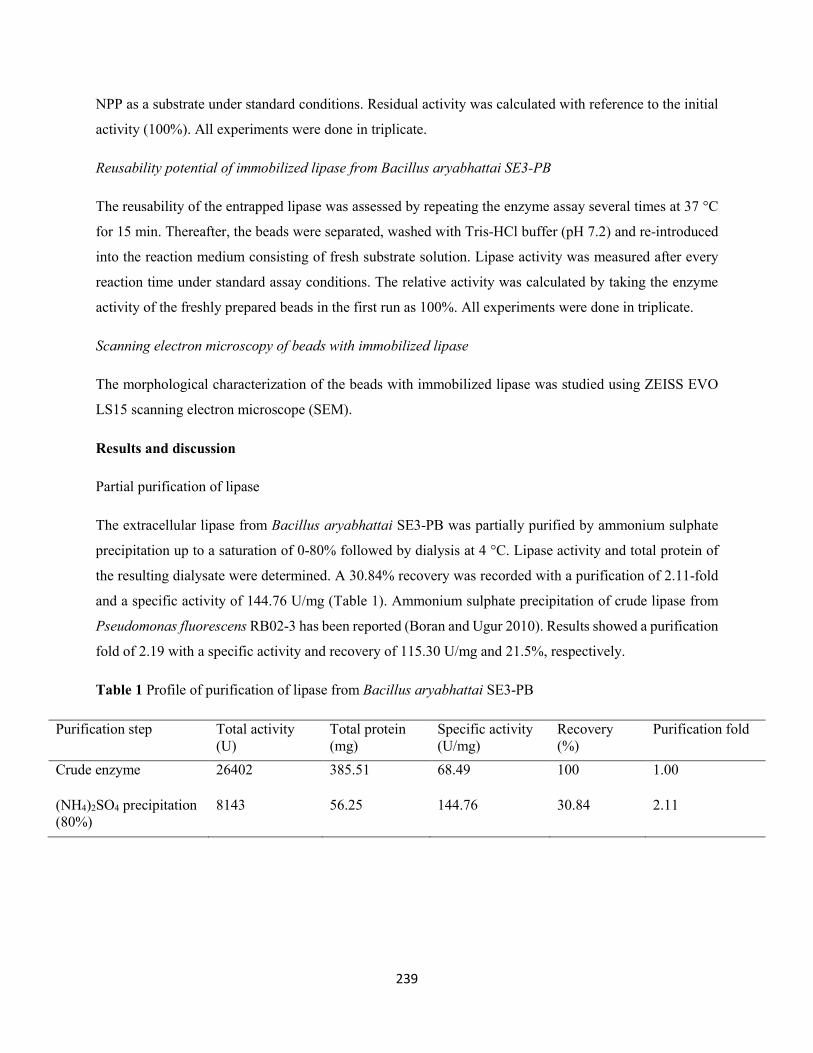
239
NPP as a substrate under standard conditions. Residual activity was calculated with reference to the initial
activity (100%). All experiments were done in triplicate.
Reusability potential of immobilized lipase from Bacillus aryabhattai SE3-PB
The reusability of the entrapped lipase was assessed by repeating the enzyme assay several times at 37 °C
for 15 min. Thereafter, the beads were separated, washed with Tris-HCl buffer (pH 7.2) and re-introduced
into the reaction medium consisting of fresh substrate solution. Lipase activity was measured after every
reaction time under standard assay conditions. The relative activity was calculated by taking the enzyme
activity of the freshly prepared beads in the first run as 100%. All experiments were done in triplicate.
Scanning electron microscopy of beads with immobilized lipase
The morphological characterization of the beads with immobilized lipase was studied using ZEISS EVO
LS15 scanning electron microscope (SEM).
Results and discussion
Partial purification of lipase
The extracellular lipase from Bacillus aryabhattai SE3-PB was partially purified by ammonium sulphate
precipitation up to a saturation of 0-80% followed by dialysis at 4 °C. Lipase activity and total protein of
the resulting dialysate were determined. A 30.84% recovery was recorded with a purification of 2.11-fold
and a specific activity of 144.76 U/mg (Table 1). Ammonium sulphate precipitation of crude lipase from
Pseudomonas fluorescens RB02-3 has been reported (Boran and Ugur 2010). Results showed a purification
fold of 2.19 with a specific activity and recovery of 115.30 U/mg and 21.5%, respectively.
Table 1 Profile of purification of lipase from Bacillus aryabhattai SE3-PB
Purification step Total activity (U)
Total protein (mg)
Specific activity (U/mg)
Recovery (%)
Purification fold
Crude enzyme 26402 385.51 68.49 100 1.00
(NH4)2SO4 precipitation (80%)
8143 56.25 144.76 30.84 2.11
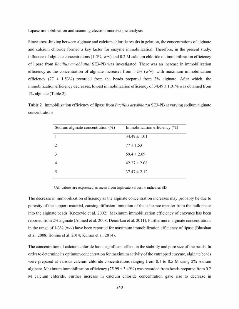
240
Lipase immobilization and scanning electron microscopic analysis
Since cross-linking between alginate and calcium chloride results in gelation, the concentrations of alginate
and calcium chloride formed a key factor for enzyme immobilization. Therefore, in the present study,
influence of alginate concentrations (1-5%, w/v) and 0.2 M calcium chloride on immobilization efficiency
of lipase from Bacillus aryabhattai SE3-PB was investigated. There was an increase in immobilization
efficiency as the concentration of alginate increases from 1-2% (w/v), with maximum immobilization
efficiency (77 ± 1.53%) recorded from the beads prepared from 2% alginate. After which, the
immobilization efficiency decreases, lowest immobilization efficiency of 34.49 ± 1.01% was obtained from
1% alginate (Table 2).
Table 2 Immobilization efficiency of lipase from Bacillus aryabhattai SE3-PB at varying sodium alginate
concentrations
*All values are expressed as mean from triplicate values; ± indicates SD
The decrease in immobilization efficiency as the alginate concentration increases may probably be due to
porosity of the support material, causing diffusion limitation of the substrate transfer from the bulk phase
into the alginate beads (Knezevic et al. 2002). Maximum immobilization efficiency of enzymes has been
reported from 2% alginate (Ahmed et al. 2008; Demirkan et al. 2011). Furthermore, alginate concentrations
in the range of 1-3% (w/v) have been reported for maximum immobilization efficiency of lipase (Bhushan
et al. 2008; Bonine et al. 2014; Kumar et al. 2014).
The concentration of calcium chloride has a significant effect on the stability and pore size of the beads. In
order to determine its optimum concentration for maximum activity of the entrapped enzyme, alginate beads
were prepared at various calcium chloride concentrations ranging from 0.1 to 0.5 M using 2% sodium
alginate. Maximum immobilization efficiency (75.99 ± 3.49%) was recorded from beads prepared from 0.2
M calcium chloride. Further increase in calcium chloride concentration gave rise to decrease in
Sodium alginate concentration (%) Immobilization efficiency (%)
1 34.49 ± 1.01
2 77 ± 1.53
3 59.4 ± 2.69
4 42.27 ± 2.08
5 37.47 ± 2.12
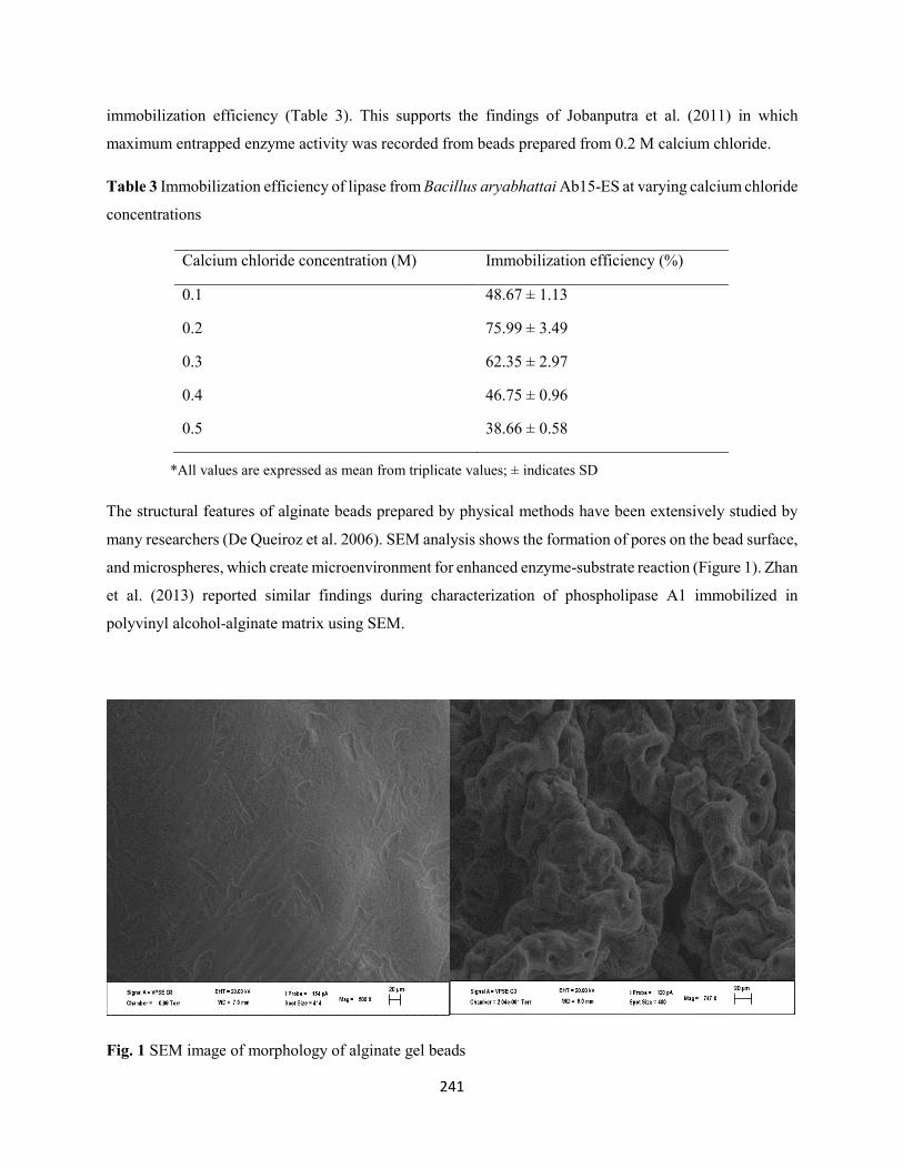
241
immobilization efficiency (Table 3). This supports the findings of Jobanputra et al. (2011) in which
maximum entrapped enzyme activity was recorded from beads prepared from 0.2 M calcium chloride.
Table 3 Immobilization efficiency of lipase from Bacillus aryabhattai Ab15-ES at varying calcium chloride
concentrations
*All values are expressed as mean from triplicate values; ± indicates SD
The structural features of alginate beads prepared by physical methods have been extensively studied by
many researchers (De Queiroz et al. 2006). SEM analysis shows the formation of pores on the bead surface,
and microspheres, which create microenvironment for enhanced enzyme-substrate reaction (Figure 1). Zhan
et al. (2013) reported similar findings during characterization of phospholipase A1 immobilized in
polyvinyl alcohol-alginate matrix using SEM.
Fig. 1 SEM image of morphology of alginate gel beads
Calcium chloride concentration (M) Immobilization efficiency (%)
0.1 48.67 ± 1.13
0.2 75.99 ± 3.49
0.3 62.35 ± 2.97
0.4 46.75 ± 0.96
0.5 38.66 ± 0.58
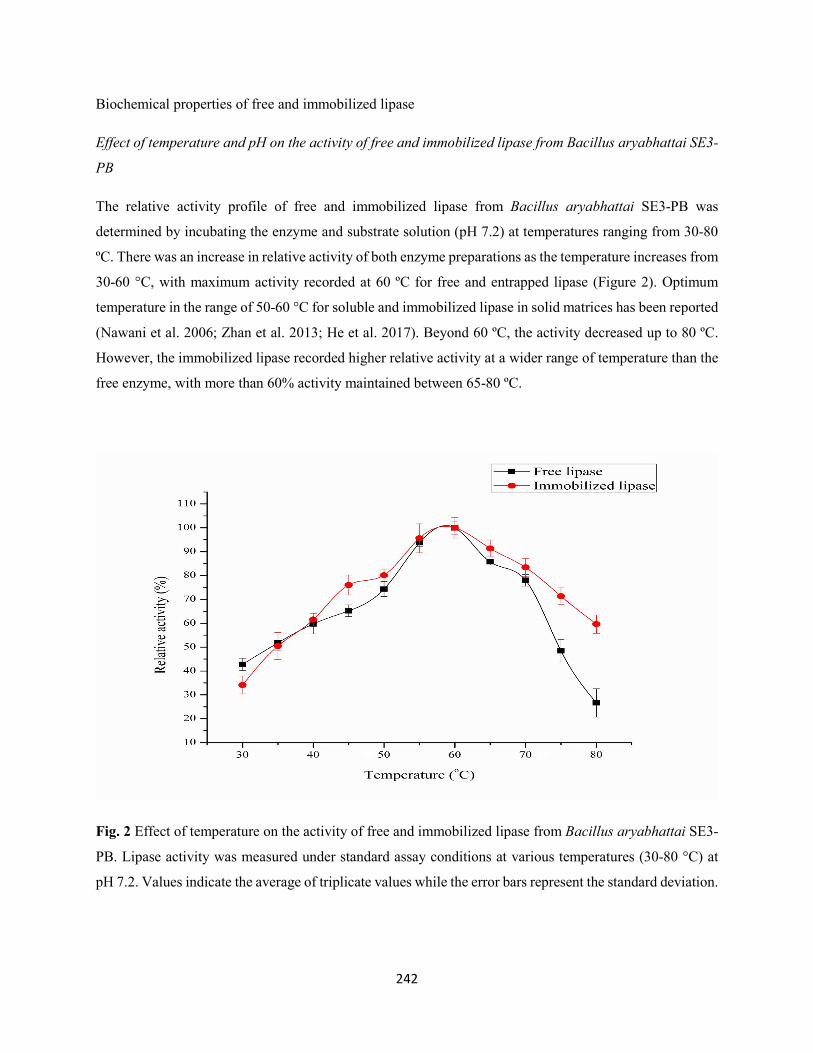
242
Biochemical properties of free and immobilized lipase
Effect of temperature and pH on the activity of free and immobilized lipase from Bacillus aryabhattai SE3-
PB
The relative activity profile of free and immobilized lipase from Bacillus aryabhattai SE3-PB was
determined by incubating the enzyme and substrate solution (pH 7.2) at temperatures ranging from 30-80
ºC. There was an increase in relative activity of both enzyme preparations as the temperature increases from
30-60 °C, with maximum activity recorded at 60 ºC for free and entrapped lipase (Figure 2). Optimum
temperature in the range of 50-60 °C for soluble and immobilized lipase in solid matrices has been reported
(Nawani et al. 2006; Zhan et al. 2013; He et al. 2017). Beyond 60 ºC, the activity decreased up to 80 ºC.
However, the immobilized lipase recorded higher relative activity at a wider range of temperature than the
free enzyme, with more than 60% activity maintained between 65-80 ºC.
Fig. 2 Effect of temperature on the activity of free and immobilized lipase from Bacillus aryabhattai SE3-
PB. Lipase activity was measured under standard assay conditions at various temperatures (30-80 °C) at
pH 7.2. Values indicate the average of triplicate values while the error bars represent the standard deviation.
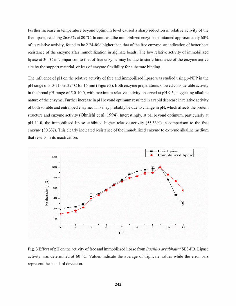
243
Further increase in temperature beyond optimum level caused a sharp reduction in relative activity of the
free lipase, reaching 26.65% at 80 °C. In contrast, the immobilized enzyme maintained approximately 60%
of its relative activity, found to be 2.24-fold higher than that of the free enzyme, an indication of better heat
resistance of the enzyme after immobilization in alginate beads. The low relative activity of immobilized
lipase at 30 ºC in comparison to that of free enzyme may be due to steric hindrance of the enzyme active
site by the support material, or less of enzyme flexibility for substrate binding.
The influence of pH on the relative activity of free and immobilized lipase was studied using p-NPP in the
pH range of 3.0-11.0 at 37 °C for 15 min (Figure 3). Both enzyme preparations showed considerable activity
in the broad pH range of 5.0-10.0, with maximum relative activity observed at pH 9.5, suggesting alkaline
nature of the enzyme. Further increase in pH beyond optimum resulted in a rapid decrease in relative activity
of both soluble and entrapped enzyme. This may probably be due to change in pH, which affects the protein
structure and enzyme activity (Ohnishi et al. 1994). Interestingly, at pH beyond optimum, particularly at
pH 11.0, the immobilized lipase exhibited higher relative activity (55.53%) in comparison to the free
enzyme (30.3%). This clearly indicated resistance of the immobilized enzyme to extreme alkaline medium
that results in its inactivation.
Fig. 3 Effect of pH on the activity of free and immobilized lipase from Bacillus aryabhattai SE3-PB. Lipase
activity was determined at 60 °C. Values indicate the average of triplicate values while the error bars
represent the standard deviation.
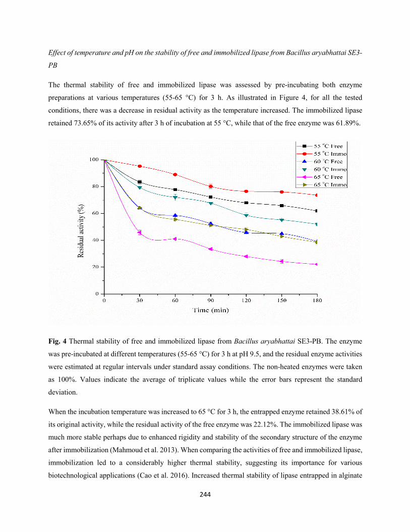
244
Effect of temperature and pH on the stability of free and immobilized lipase from Bacillus aryabhattai SE3-
PB
The thermal stability of free and immobilized lipase was assessed by pre-incubating both enzyme
preparations at various temperatures (55-65 °C) for 3 h. As illustrated in Figure 4, for all the tested
conditions, there was a decrease in residual activity as the temperature increased. The immobilized lipase
retained 73.65% of its activity after 3 h of incubation at 55 °C, while that of the free enzyme was 61.89%.
Fig. 4 Thermal stability of free and immobilized lipase from Bacillus aryabhattai SE3-PB. The enzyme
was pre-incubated at different temperatures (55-65 °C) for 3 h at pH 9.5, and the residual enzyme activities
were estimated at regular intervals under standard assay conditions. The non-heated enzymes were taken
as 100%. Values indicate the average of triplicate values while the error bars represent the standard
deviation.
When the incubation temperature was increased to 65 °C for 3 h, the entrapped enzyme retained 38.61% of
its original activity, while the residual activity of the free enzyme was 22.12%. The immobilized lipase was
much more stable perhaps due to enhanced rigidity and stability of the secondary structure of the enzyme
after immobilization (Mahmoud et al. 2013). When comparing the activities of free and immobilized lipase,
immobilization led to a considerably higher thermal stability, suggesting its importance for various
biotechnological applications (Cao et al. 2016). Increased thermal stability of lipase entrapped in alginate
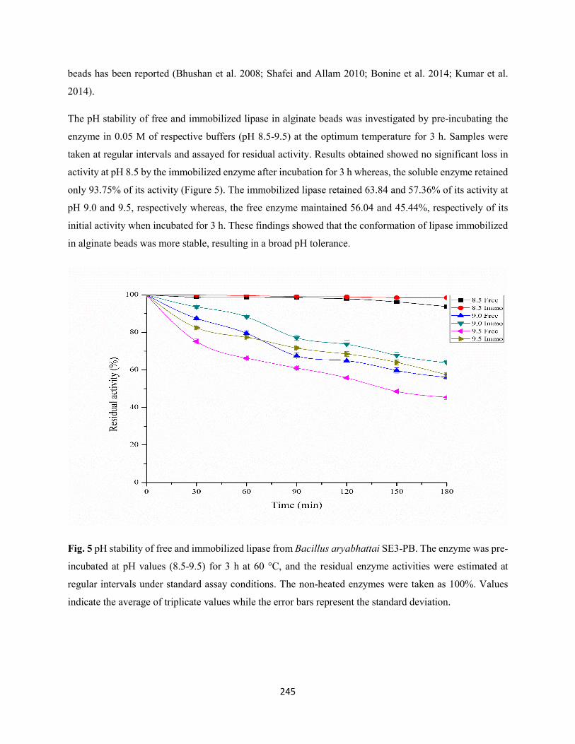
245
beads has been reported (Bhushan et al. 2008; Shafei and Allam 2010; Bonine et al. 2014; Kumar et al.
2014).
The pH stability of free and immobilized lipase in alginate beads was investigated by pre-incubating the
enzyme in 0.05 M of respective buffers (pH 8.5-9.5) at the optimum temperature for 3 h. Samples were
taken at regular intervals and assayed for residual activity. Results obtained showed no significant loss in
activity at pH 8.5 by the immobilized enzyme after incubation for 3 h whereas, the soluble enzyme retained
only 93.75% of its activity (Figure 5). The immobilized lipase retained 63.84 and 57.36% of its activity at
pH 9.0 and 9.5, respectively whereas, the free enzyme maintained 56.04 and 45.44%, respectively of its
initial activity when incubated for 3 h. These findings showed that the conformation of lipase immobilized
in alginate beads was more stable, resulting in a broad pH tolerance.
Fig. 5 pH stability of free and immobilized lipase from Bacillus aryabhattai SE3-PB. The enzyme was pre-
incubated at pH values (8.5-9.5) for 3 h at 60 °C, and the residual enzyme activities were estimated at
regular intervals under standard assay conditions. The non-heated enzymes were taken as 100%. Values
indicate the average of triplicate values while the error bars represent the standard deviation.
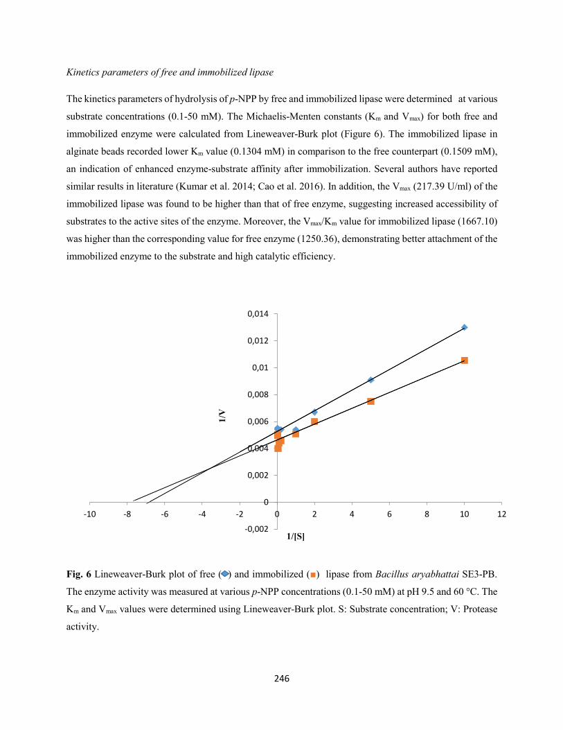
246
Kinetics parameters of free and immobilized lipase
The kinetics parameters of hydrolysis of p-NPP by free and immobilized lipase were determined at various
substrate concentrations (0.1-50 mM). The Michaelis-Menten constants (Km and Vmax) for both free and
immobilized enzyme were calculated from Lineweaver-Burk plot (Figure 6). The immobilized lipase in
alginate beads recorded lower Km value (0.1304 mM) in comparison to the free counterpart (0.1509 mM),
an indication of enhanced enzyme-substrate affinity after immobilization. Several authors have reported
similar results in literature (Kumar et al. 2014; Cao et al. 2016). In addition, the Vmax (217.39 U/ml) of the
immobilized lipase was found to be higher than that of free enzyme, suggesting increased accessibility of
substrates to the active sites of the enzyme. Moreover, the Vmax/Km value for immobilized lipase (1667.10)
was higher than the corresponding value for free enzyme (1250.36), demonstrating better attachment of the
immobilized enzyme to the substrate and high catalytic efficiency.
Fig. 6 Lineweaver-Burk plot of free ( ) and immobilized () lipase from Bacillus aryabhattai SE3-PB.
The enzyme activity was measured at various p-NPP concentrations (0.1-50 mM) at pH 9.5 and 60 °C. The
Km and Vmax values were determined using Lineweaver-Burk plot. S: Substrate concentration; V: Protease
activity.
-0,002
0
0,002
0,004
0,006
0,008
0,01
0,012
0,014
-10 -8 -6 -4 -2 0 2 4 6 8 10 12
1/V
1/[S]
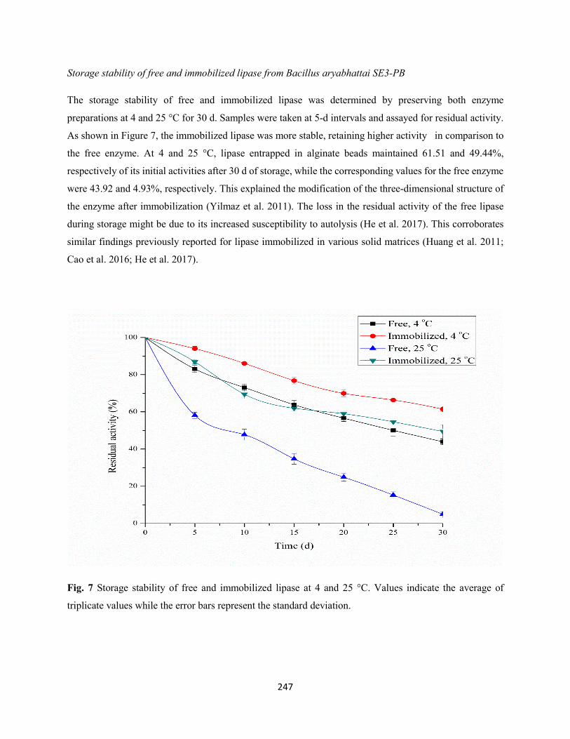
247
Storage stability of free and immobilized lipase from Bacillus aryabhattai SE3-PB
The storage stability of free and immobilized lipase was determined by preserving both enzyme
preparations at 4 and 25 °C for 30 d. Samples were taken at 5-d intervals and assayed for residual activity.
As shown in Figure 7, the immobilized lipase was more stable, retaining higher activity in comparison to
the free enzyme. At 4 and 25 °C, lipase entrapped in alginate beads maintained 61.51 and 49.44%,
respectively of its initial activities after 30 d of storage, while the corresponding values for the free enzyme
were 43.92 and 4.93%, respectively. This explained the modification of the three-dimensional structure of
the enzyme after immobilization (Yilmaz et al. 2011). The loss in the residual activity of the free lipase
during storage might be due to its increased susceptibility to autolysis (He et al. 2017). This corroborates
similar findings previously reported for lipase immobilized in various solid matrices (Huang et al. 2011;
Cao et al. 2016; He et al. 2017).
Fig. 7 Storage stability of free and immobilized lipase at 4 and 25 °C. Values indicate the average of
triplicate values while the error bars represent the standard deviation.
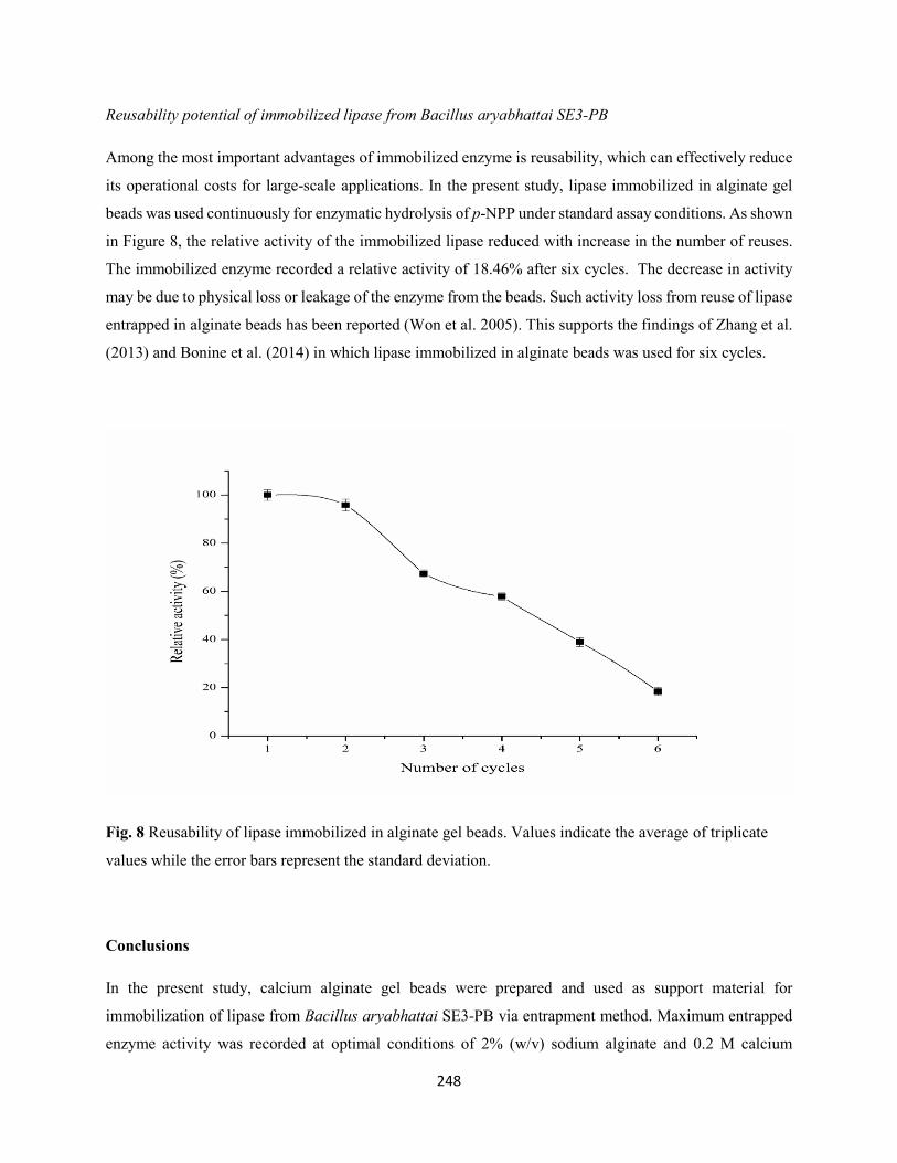
248
Reusability potential of immobilized lipase from Bacillus aryabhattai SE3-PB
Among the most important advantages of immobilized enzyme is reusability, which can effectively reduce
its operational costs for large-scale applications. In the present study, lipase immobilized in alginate gel
beads was used continuously for enzymatic hydrolysis of p-NPP under standard assay conditions. As shown
in Figure 8, the relative activity of the immobilized lipase reduced with increase in the number of reuses.
The immobilized enzyme recorded a relative activity of 18.46% after six cycles. The decrease in activity
may be due to physical loss or leakage of the enzyme from the beads. Such activity loss from reuse of lipase
entrapped in alginate beads has been reported (Won et al. 2005). This supports the findings of Zhang et al.
(2013) and Bonine et al. (2014) in which lipase immobilized in alginate beads was used for six cycles.
Fig. 8 Reusability of lipase immobilized in alginate gel beads. Values indicate the average of triplicate
values while the error bars represent the standard deviation.
Conclusions
In the present study, calcium alginate gel beads were prepared and used as support material for
immobilization of lipase from Bacillus aryabhattai SE3-PB via entrapment method. Maximum entrapped
enzyme activity was recorded at optimal conditions of 2% (w/v) sodium alginate and 0.2 M calcium
249
chloride. Both free and immobilized lipase had maximum activity at optimum temperature and pH of 60
°C and 9.5, respectively. However, the entrapped enzyme exhibited improved pH, temperature and storage
stability as well as enhanced affinity for the substrate and higher catalytic efficiency when compred to the
soluble counterpart. Furthermore, the entrapped lipase showed better reusability for up to six consecutive
cycles during repeated hydrolysis of p-NPP, suggesting the efficient, longevity and cost-effectiveness of
the developed immobilized biocatalyst for various biotechnological applications.
References
Ahmed SA, El-Sayed MEM, Hassan O, Saleh NA, Goda HA (2008) Studies on the activity and stability of
immobilized Bacillus acidocaldarius alpha-amylase. Aust J Basic Appl Sci 2:466-474
Bhushan B, Pal A, Jain V (2015) Improved enzyme catalytic characteristics upon glutaraldehyde cross-
linking of alginate entrapped xylanase isolated from Aspergillus flavus MTCC 9390. Enzyme Res
2015:1-9
Bhushan I, Parshad R, Qazi GN, Gupta VK (2008) Immobilization of lipase by entrapment in Ca-alginate
beads. J Bioact Compat Polym 23:552-562
Bonine BM, Polizelli PP, Bonilla-Rodriguez GO (2014) Immobilization of a plant lipase from Pachira
aquatica in alginate and alginate/PVA beads. Enzyme Res 2014:1-7
Boran R, Ugur A (2010) partial purification and characterization of the organic solvent-tolerant lipase
produced by Pseudomonas fluorescens RB02-3 isolated from milk. Prep Biochem Biotechnol
40:229-241
Borrelli GM, Trono D (2015) Recombinant lipases and phospholipases and their use as biocatalysts for
industrial applications. Int J Mol Sci 16:20774-20840
250
Buchholz K, Kasche V, Bornscheuer UT (2012) Characterization of immobilized catalyst, in Biocatalysts
and Enzyme Technology, Buchholz K, Kasche V, Bornscheuer UT, Eds., Boschstraẞe, Weinheim,
Germany, 411-448
Cao S-L, Huang Y-M, Li X-H, Xu P, Wu H, Li N, Lou W-Y, Zong M-H (2016) Preparation and
characterization of immobilized lipase from Pseudomonas cepacia onto magnetic cellulose
nanocrystals. Sci Rep 6:20420
Carr PD, Ollis DL (2009) α-β fold: An update. Protein Pept Lett 16:137-148
Casas-Godoy L, Duquesne S, Bordes F, Sandoval G, Marty A (2012) Lipases: an overview. Methods Mol
Biol 861:3-30
Datta S, Christena LR, Rajaram YRS (2013) Enzyme immobilization: an overview on techniques and
support materials. 3 Biotech 3:1-9
De Queiroz AAA, Passos ED, Alves SB, Silva GS, Higa OZ, Vítolo M (2006) Alginate-poly (vinyl alcohol)
core-shell microspheres for lipase immobilization. J Appl Polym Sci 102:1553-1560
Demirkan E, Dincbas S, Sevinc N, Ertan F (2011) Immobilization of B. amyloliquefaciens α-amylase and
comparison of some of its enzymatic properties with the free form. Romanian Biotechnol Lett
16:6690-6701
Dutta S, Ray L (2009) Production and characterization of an alkaline thermostable crude lipase from an
isolated strain of Bacillus cereus C7. Appl Biochem Biotechnol 159:142-154
Fernandez-Lafuente R (2010) Lipase from Thermomyces lanuginosus: uses and prospects as an industrial
biocatalyst. J Mol Catal.: Enzym 62:197-212
Guisan JM (2006) Immobilization of enzymes as the 21st century begins. In: Guisan JM, editor.
Immobilization of enzymes and cells. 2nd ed. New Jersey (NJ): Humana Press Inc. 1-13
251
Hasan F, Shah AA, Hameed A (2006) Industrial applications of microbial lipases. Enzyme Microb Technol
39:235-251
Hasan F, Shah AA, Hameed A (2009) Methods for detection and characterization of lipases: A
comprehensive review. Biotechnol Adv 27:782-798
He S, Song D, Chen M, Cheng H (2017) Immobilization of lipases on magnetic collagen fibers and its
applications for short-chain ester synthesis. Catalysts, 7:178
Hernandez K, Fernandez-Lafuente R (2011) Control of protein immobilization: coupling immobilization
and side-directed mutagenesis to improve biocatalyst or biosensor performance. Enzyme Microb
Technol 48:107-122
Homaei AA, Sariri R, Vianello F, Stevanato R (2013) Enzyme immobilization: an update. J Chem Biol
6:185-205
Huang XJ, Chen PC, Huang F, Ou Y, Chen MR, Xu ZK (2011) Immobilization of Candida rugosa lipase
on electrospun cellulose nanofiber membrane. J Mol Catal B Enzym 70:95-100
Jaeger KE, Reetz MT (1998) Microbial lipases form versatile tools for biotechnology. Trends Biotechnol.
16:396-403
Jobanputra AH, Karode BA, Chincholkar SB (2011) Calcium alginate as supporting material for the
immobilization of rifamycin oxidase from Chryseobacterium species. Biotechnol Bioinfor Bioeng
1:529-535
Kanmani P, Aravind J, Kumaresan K (2015) An insight into microbial lipases and their environmental facet.
Int J Environ Sci Technol 12:1147-1162
252
Knezevic Z, Bobic S, Milutinovic A, Obradovic B, Mojovic L, Bugarski B (2002) Alginate-immobilized
lipase by electrostatic extrusion for the purpose of palm oil hydrolysis in lecithin/isooctane system.
Proc Biochem 38:313-318
Kumar S, Yadav RK, Negi S (2014) A comparative study of immobilized lipase produced from Penicillium
chrosogenum SNP5 on two different anionic carriers for its pH and thermostability. Indian J.
Biotechnol 13:301-305
Lee HK, Lee JK, Kim MJ, Lee CJ (2010) Immobilization of lipase on single walled carbon nanotubes in
ionic liquid. Bull Korean Chem Soc 31:650-652
Lowry OH, Rosebrough N, Farr AL, Rondall RL (1951) Protein measurement with the Folin-phenol
reagent. J Biol Chem 193:265-273
Mahmoud KA, Lam E, Hrapovic S, Luong JHT (2013) Preparation of well-dispersed gold/magnetite
nanoparticles embedded on cellulose nanocrystals for efficient immobilization of papain enzyme.
ACS Appl Mater Int 5:4978-4985
Mendes AA, Freitas L, Carvalhoak AK, Oliveira PC, Castro HF (2011) Immobilization of a commercial
lipase from Penicillium camembertii (Lipase G) by different strategies. Enzyme Res 40:611-618
Minovska V, Winkelhausen E, Kuzmanove S (2005) Lipase immobilized by different techniques on various
support materials applied in oil hydrolysis. J Serb Chem Soc 70:609-624
Mohamad NR, Marzuki NHC, Buang NA, Huyop F, Abdul Wahab R (2015) An overview of technologies
for immobilization of enzymes and surface analysis techniques for immobilized enzymes.
Biotechnol Biotechnol Equip 29:205-220
Nagarajan S (2012) New tools for exploring “old friends-microbial lipases”. Appl Biochem Biotechnol
168:1163-1196
253
Nawani N, Singh R, Kaur J (2006) Immobilization and stability studies of a lipase from thermophilic
Bacillus sp: the effect of process parameters on immobilization of enzyme. Electron J Biotechnol
9:560-565
Ohnishi K, Yoshida Y, Toita J, Sekiguchi J (1994) Purification and characterization of a novel lipolytic
enzyme from Aspergillus oryzae. J Ferment Bioeng 78:413-419
Sangeetha R, Arulpandi I, Geetha A (2011) Bacterial lipases as potential industrial biocatalysts: an
overview. Res J Microbiol 6:1-24
Santos JCSD, Barbosa O, Ortiz C, Berenguer-Murcia A, Rodrigues RC, Fernandez-Lafuente R (2015)
Importance of the support properties for immobilization or purification of enzymes. ChemCatChem
7:2413-2432
Shafei MS, Allam RF (2010) Production and immobilization of partially purified lipase from Penicillium
chrosogenum. Malaysian J Microbiol 6:196-202
Sheldon RA, van Pelt S (2013) Enzyme immobilization in biocatalysis: why, what, and how. Chem Soc
Rev 42:6223-6235
Shen Q, Yang R, Hua X, Ye F, Zhang W, Zhao W (2011) Gelatin-templated biomimetic calcification for
β-galactosidase immobilization. Proc Biochem 46:1565-1571
Spahn C, Minteer SD (2008) Enzyme immobilization in biotechnology. Recent Pat Eng 2:195-200
Treichel H, de Oliveira D, Mazutti MA, Di Luccio M, Oliveira JVA (2010) A review on microbial lipases
production. Food Bioproc Technol 3:182-196
Villeneuve P (2003) Plant lipases and their applications in oils and fats modification. Eur J Lipid Sci
Technol 105:308-317
254
Winkler UK, Stuckmann M (1979) Glocogen, hyaluronate, and some other polysaccharides greatly enhance
the formation of exolipase by Serratia marcescens. J Bacteriol 138:663-670
Won K, Kim S, Kim KJ, Park HW, Moon S (2005) Optimization of lipase entrapment in Ca-alginate beads.
Proc Biochem 40:2149-2154
Yadav RR, Mudliar SN, Shekh AY, Fulke AB, Devi SS, Krishnamurthi K, Juwarkar A, Chakrabarti T
(2012) Immobilization of carbonic anhydrase in alginate and its influence on transformation of CO2
to calcite. Proc Biochem 47:585-590
Yilmaz E, Can K, Sezgin M, Yilmaz M (2011) Immobilization of Candida rugosa lipase on glass beads for
enantioselective hydrolysis of racemic naproxen methyl ester. Bioresour Technol 102: 499-506.
Zhan JF, Jiang ST, Pan LJ (2013) Immobilization of phospholipase A1 using a polyvinyl alcohol-alginate
matrix and evaluation of the effects of immobilization. Braz J Chem Eng 30:721-728
Zhang S, Shang W, Yang X, Zhang S, Zhang X, Chen J (2013) Immobilization of lipase using alginate
hydrogel beads and enzymatic evaluation in hydrolysis of p-nitrophenol butyrate. Bull Korean Soc
34:2741-2745
Zhang S, Shang W, Yang X, Zhang X, Huang Y, Zhang S, Chen J (2014) Immobilization of lipase with
alginate hydrogel beads and the lipase-catalyzed kinetic resolution of α-phenyl ethanol. J Appl
Polym Sci 131:40178
255
CHAPTER TEN
TREATMENT OF LIPID-RICH WASTEWATER USING A MIXTURE OF FREE OR
IMMOBILIZED BIOEMULSIFIER AND HYDROLYTIC ENZYMES FROM INDIGENOUS
BACTERIAL ISOLATES
This chapter has been submitted in the current format to the ‘Desalination and Water Treatment’
256
Treatment of lipid-rich wastewater using a mixture of free or immobilized bioemulsifier and
hydrolytic enzymes from indigenous bacterial isolates
Adegoke Isiaka Adetunji & Ademola Olufolahan Olaniran*
Discipline of Microbiology, School of Life Sciences, College of Agriculture, Engineering and Science,
University of KwaZulu-Natal (Westville campus), Private Bag X54001, Durban 4000, Republic of South
Africa.
* Corresponding author. Tel: +27 31 260 7400; Fax: +27 31 260 7809
e-mail address: [email protected]
Abstract
The combined use of free or immobilized bioemulsifier produced by Acinetobacter sp. and enzyme pool
(protease and lipase) from Bacillus aryabhattai in the reduction of organic pollutants present in lipid-rich
wastewater was investigated. Physicochemical characterization of the raw wastewater revealed high
pollutant load in poultry processing wastewater in comparison to dairy wastewater. Biodegradability of the
wastewater was assessed by measuring the reduction of COD and lipid content at time intervals under
varying incubation conditions. In dairy wastewater treated at 37 °C without pH adjustment, maximum COD
(60.51 & 65.19%) and lipid (47.98 & 63.53%) reduction efficiencies were recorded at 120 h using free and
immobilized bioproducts, respectively. However, under these conditions, maximum COD (86.44 &
93.65%) and lipid (51.62 & 69.06%) removal efficiencies of poultry processing wastewater were observed
at 120 h when treated with free and immobilized bioproducts, respectively. At temperature of 50 °C and
pH 8.0, there was improved reduction of organic pollutants, with maximum COD (65.96 & 77.52%) and
lipid (55.22 & 71.12%) removal efficiencies obtained in dairy wastewater at 72 h when using free and
immobilized bioproducts, respectively. In the case of poultry processing wastewater, optimum COD (90.29
& 94.72%) and lipid (63 & 76.66%) removal was recorded at 72 h when treated with free and immobilized
bioproducts, respectively. Reusability studies suggest that the immobilized bioproducts could be reused for
up to six and seven times for the treatment of dairy and poultry processing wastewaters, respectively. This
study suggests the efficient and synergistic application of the developed immobilized bioemulsifier and
hydrolytic enzymes in the treatment of high fat-containing wastewater.
Keywords: Lipid-rich wastewater; Bioproducts; Biodegradation; Organic pollutants; Hydrolytic enzymes
257
1. Introduction
Lipids (fats, oils and greases) and proteins are among the most indispensable components of natural foods
as well as many synthetic compounds and emulsions [1,2]. They constitute a vital constituent of wastewater
discharged from restaurants, oil mill industries, dairy industries and slaughterhouses [3,4]. These industries
utilize large amounts of water for various processes, resulting in significant effluent volume with huge
amounts of organic matter [5]. In addition, this wastewater is characterized by high concentrations of
chemical oxygen demand (COD), biochemical oxygen demand (BOD), suspended solids along with many
toxic compounds, causing severe environmental pollution [6,7]. These include formation of oil films on
water surfaces, which prevent transfer of dissolved oxygen into aerobic process. Aggregates of oil droplets
caused blockage of sewer lines, resulting in unpleasant odor; flotation, mass transfer problems for soluble
substrates, and reduction of sludge acetogenic and methanogenic activities [8-10]. Therefore, effective
techniques to lessen the negative environmental impacts of lipid-rich wastewater is warranted [7].
Various physicochemical approaches including dissolved air flotation and chemical coagulation are
employed for the treatment of lipid-rich wastewater [11-13]. However, these methods are encountered with
drawbacks of high operational costs due to coagulation agents and generation of sludge with sedimentation
problems [14]. Hence, the need for adoption of biological technologies involving application of bioproducts
for an efficient, cost-effective, sustainable and eco-friendly treatment of high fat-containing wastewater
[15]. The use of biosurfactants facilitate enzymatic activity and degradation of lipids by accelerating their
solubility and bioavailability, thus eliminating the need for additional processes [16-18]. In addition,
application of hydrolytic enzymes reduce the lipid and protein levels in the effluent via enzymatic catalysis
and further enhance performance of the microbial population at a later phase of biological treatment
[4,6,8,19-22].
In order to make bioemulsifiers and enzymes more favorable for use in biotechnological processes, different
approaches, including immobilization have been employed for a cost-effective practice [23,24].
Insolubilization of bioproducts in a suitable support matrix offers many distinct advantages including
resistance to environmental changes, easy recovery, multiple reuse, continuous operation, enhanced
stability of the bioproducts under operational conditions and rapid termination of reactions [19,25-27].
Several studies on the combined use of free bioemulsifier and enzyme pool for oily wastewater treatment
have been reported [14,17,28,29]. However, to the best of our knowledge, no studies have been reported
on the comparative study of a mixture of free or immobilized bioemulsifier and hydrolytic enzymes
(protease, lipase) for lipid-rich wastewater treatment. Therefore, this present study investigates the
synergistic effects of free or immobilized extracellular glycoprotein bioemulsifier secreted from
258
Acinetobacter sp. Ab9-ES and hydrolytic enzymes (protease and lipase) produced from Bacillus
aryabhattai in the reduction of pollutants from two different lipid-rich wastewater types.
2. Materials and methods
2.1. Materials
The bioproducts (hydrolytic enzymes and bioemulsifier) used in this experiment were produced by
submerged fermentation from Bacillus aryabhattai and Acinetobacter sp. Ab9-ES, respectively. They
exhibited robust activities at optimum pH and temperature of 8.0 and 50 °C (protease), 9.5 and 60 °C
(lipase) and 7.0 and 50 °C (bioemulsifier). Sodium alginate used as support material for immobilization
was purchased from Sigma-Aldrich (St. Louis, MO, USA). All other reagents were of analytical grade.
2.2. Immobilization protocol
The bioproducts were immobilized separately by entrapment method using the methods of Guleria et al.
[30] and Ferhat et al. [31] with modifications. Bioproduct solution was added to a suitable amount of 2%
(w/v) sodium alginate suspension. The mixture was extruded dropwise through a syringe into a CaCl2
solution. When in contact with CaCl2 solution, the alginate drops solidified and form beads, thus entrapping
the bioproducts. The resultant beads were left to solidify for 1 h before washing with sterile distilled water
for the removal of excess Ca2+ ions.
2.3. Collection of raw lipid-rich wastewater
Raw lipid-rich wastewater samples were collected from two different sources namely, dairy and poultry
processing industries, all in the KwaZulu-Natal province of South Africa, into separate autoclaved 1 L
Schott bottles. The wastewaters were generated mainly from production line, equipment, and floor cleaning
operations.
2.4. Physicochemical characterization of lipid-rich wastewater
Raw lipid-rich wastewater samples were characterized for BOD, COD, TSS, total dissolved solid (TDS),
turbidity, salinity, resistivity, electrical conductivity (EC), total nitrogen (TN), total phosphorus (TP), pH
and temperature in accordance with standard methods [32]. Partition gravimetric method of Kirschman and
Pomeroy [33] was used for the determination of lipid content while protein concentration was estimated
according to the method of Lowry et al. [34] using bovine serum albumin (Sigma-Aldrich, St. Louis, MO,
USA) as a standard. All analyses were performed in triplicate.
259
2.5. Batch biodegradation of lipid-rich wastewater
The synergistic effects of bioemulsifier and enzyme pool in the biodegradation of pollutants present in
lipid-rich wastewater were investigated in 1 L Erlenmeyer flasks containing 500 ml sterile lipid-rich
wastewater. The pH of the wastewater was adjusted to 8.0 using 1 N NaOH or 1 N HCl. Five percent (v/v)
each of the free bioproducts or 2.5, 3 and 3.75 g of immobilized protease, lipase and bioemulsifier,
respectively were added to the wastewater samples. This was followed by incubation under aerobic
condition at 37 and 50 °C (150 rpm) for 120 h in a shaking incubator. A control flask without enzyme or
bioemulsifier preparation was kept under similar conditions. Biodegradability of the lipid-rich wastewater
samples was assessed by determining percentage removal efficiencies of COD and lipid contents at 24, 72
and 120 h using the formula shown in eq. (1) [35]. All analyses were conducted in triplicate.
Removal efficiency (%) = Ccontrol−CfCcontrol
× 100 (1)
where Ccontrol is the value of COD or lipid content of the control lipid-rich wastewater, and Cf is the amount
of these parameters after treatment.
2.6. Reusability potential of immobilized protease, lipase and bioemulsifier
Batch biodegradation of lipid-rich wastewater was repeated several times using a combination of
immobilized protease, lipase and bioemulsifier. After each cycle, the beads were separated, washed with
sterile distilled water and then added into a fresh wastewater sample. Percentage removal efficiencies of
COD and lipid content were determined after every cycle. All experiments were carried out in triplicate.
3. Results and discussion
3.1. Characterization of lipid-rich wastewater
The physicochemical characteristics of dairy and poultry processing wastewaters were determined using
standard procedures, and the mean values of the parameters are shown in Table 1. Both wastewater types
are characterized with unpleasant odor coupled with whitish (dairy wastewater) and brownish (poultry
processing wastewater) appearance. Furthermore, wastewater from poultry processing industry contained
high concentrations of organic matter in form of lipids (88900 ± 10468 mg/L), COD (7518 ± 378 mg/L),
BOD5 (707 ± 13.58 mg/L), TDS (807000 ± 0.00 mg/L) and TSS (4667 ± 2887 mg/L) in comparison to
dairy industry wastewater with lipid content (53367 ± 4306 mg/L), COD (5693 ± 17.56 mg/L), BOD5 (691
± 16.64 mg/L), TDS (5700 ± 0.00 mg/L) and TSS (2333 ± 1528 mg/L).
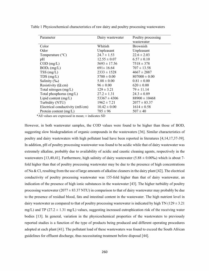
260
Table 1 Physicochemical characteristics of raw dairy and poultry processing wastewaters
*All values are expressed in mean; ± indicates SD
However, in both wastewater samples, the COD values were found to be higher than those of BOD,
suggesting slow biodegradation of organic compounds in the wastewaters [36]. Similar characteristics of
poultry and dairy wastewaters with high pollutant load have been reported in literatures [4,14,17,37-39].
In addition, pH of poultry processing wastewater was found to be acidic while that of dairy wastewater was
extremely alkaline, probably due to availability of acidic and caustic cleaning agents, respectively in the
wastewaters [13,40,41]. Furthermore, high salinity of dairy wastewater (5.88 ± 0.00‰) which is about 7-
fold higher than that of poultry processing wastewater may be due to the presence of high concentrations
of Na & Cl, resulting from the use of large amounts of alkaline cleaners in the dairy plant [42]. The electrical
conductivity of poultry processing wastewater was 155-fold higher than that of dairy wastewater, an
indication of the presence of high ionic substances in the wastewater [43]. The higher turbidity of poultry
processing wastewater (2077 ± 83.37 NTU) in comparison to that of dairy wastewater may probably be due
to the presence of residual blood, fats and intestinal content in the wastewater. The high nutrient level in
dairy wastewater as compared to that of poultry processing wastewater is indicated by high TN (129 ± 3.21
mg/L) and TP (27.2 ± 1.31 mg/L) values, suggesting increased eutrophication risk of the receiving water
bodies [13]. In general, variation in the physicochemical properties of the wastewaters to previously
reported studies is a function of the type of products being produced and different operating procedures
adopted at each plant [41]. The pollutant load of these wastewaters was found to exceed the South African
guidelines for effluent discharge, thus necessitating treatment before disposal [44].
Parameter Dairy wastewater Poultry processing wastewater
Color Whitish Brownish Odor Unpleasant Unpleasant Temperature (°C) 24.7 ± 1.53 22.6 ± 2.03 pH 12.55 ± 0.07 6.57 ± 0.10 COD (mg/L) 5693 ± 17.56 7518 ± 378 BOD5 (mg/L) 691± 16.64 707 ± 13.58 TSS (mg/L) 2333 ± 1528 4667 ± 2887 TDS (mg/L) 5700 ± 0.00 807000 ± 0.00 Salinity (‰) 5.88 ± 0.00 0.81 ± 0.00 Resistivity (Ω.cm) 96 ± 0.00 620 ± 0.00 Total nitrogen (mg/L) 129 ± 3.21 79 ± 11.14 Total phosphorus (mg/L) 27.2 ± 1.31 24.3 ± 0.89 Lipid content (mg/L) 53367 ± 4306 88900 ± 10468 Turbidity (NTU) 1962 ± 7.21 2077 ± 83.37 Electrical conductivity (mS/cm) 10.42 ± 0.00 1614 ± 0.58 Protein content (mg/L) 785 ± 96 507 ± 40
261
3.2. Simultaneous treatment of lipid-rich wastewater using a mixture of free or immobilized bioemulsifier
and enzyme pool
The COD and lipid content represent crucial parameters for assessing the biodegradability of oily
wastewater, as they give an idea about the amount of organic matter that was degraded during the treatment
process [29,45]. In the present study, synergistic application of bioemulsifier and enzyme pool (protease
and lipase) in the treatment of dairy and poultry processing wastewaters was investigated by carrying out a
batch biodegradation experiment under different conditions. The rational for the choice of protease, lipase
and bioemulsifier was due to high protein and lipid contents in the wastewater. High activity of bioproducts
is a key prerequisite for industrial wastewater treatment [8]. Five percent (v/v) each of the bioproducts
containing 151.14 U/ml, 81.43 U/ml and 83.3% of protease, lipase and emulsifying activities, respectively
was used for the treatment of the wastewaters. Similar experiments were carried out concurrently using
required amounts of immobilized protease, lipase and bioemulsifier with similar activities to the free
bioproducts. In the first set of experiment, sterile wastewater samples without pH adjustment was incubated
at 37 °C (150 rpm) for 120 h. Kinetics of degradation of pollutants in the wastewater was assessed by
determining the reduction efficiencies of COD and lipid content at time intervals using standard methods.
There was a significant reduction in the COD and lipid contents of the wastewater in comparison to control
during treatment with free or immobilized bioproducts for 120 h. Maximum COD (60.51 & 65.19%) and
lipid (47.98 & 63.53%) reduction efficiencies were recorded after 120 h in dairy wastewater when treated
with the free and immobilized bioproducts, respectively (Fig. 1A & B). Maximum COD (86.44 & 93.65%)
and lipid (51.62 & 69.06%) removal efficiencies found to be higher than those of dairy wastewater were
obtained after 120 h treatment of poultry processing wastewater using free and entrapped bioproducts,
respectively (Fig. 2A & B). The lower degradation of dairy wastewater compared to poultry processing
wastewater suggests higher affinity of the bioproducts to poultry effluent constituents [3]. This may
probably be due to the presence of high salt content inhibiting the degradation of pollutants in the dairy
wastewater [46].
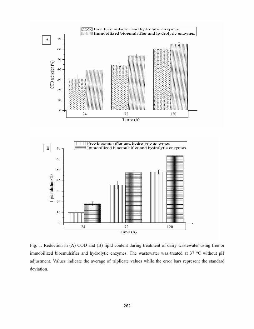
262
Fig. 1. Reduction in (A) COD and (B) lipid content during treatment of dairy wastewater using free or
immobilized bioemulsifier and hydrolytic enzymes. The wastewater was treated at 37 °C without pH
adjustment. Values indicate the average of triplicate values while the error bars represent the standard
deviation.
24 72 120
24 72 120
A
B
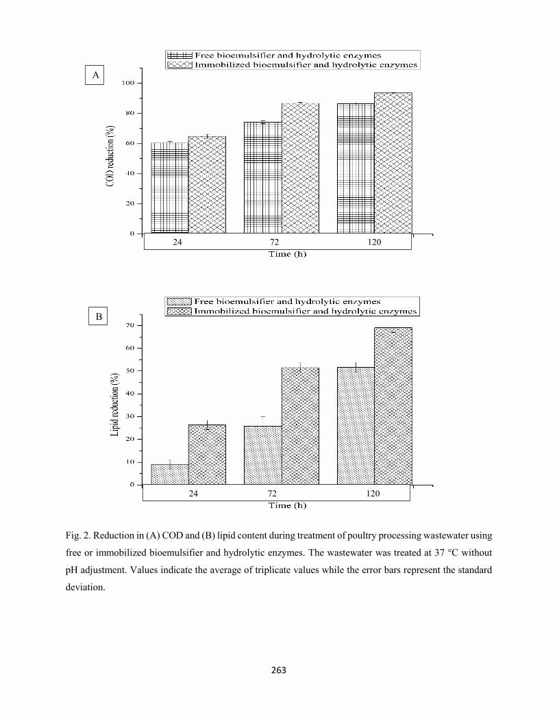
263
Fig. 2. Reduction in (A) COD and (B) lipid content during treatment of poultry processing wastewater using
free or immobilized bioemulsifier and hydrolytic enzymes. The wastewater was treated at 37 °C without
pH adjustment. Values indicate the average of triplicate values while the error bars represent the standard
deviation.
A
B
24 72 120
24 72 120
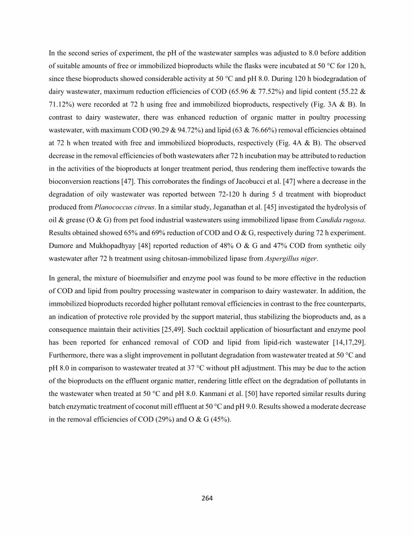
264
In the second series of experiment, the pH of the wastewater samples was adjusted to 8.0 before addition
of suitable amounts of free or immobilized bioproducts while the flasks were incubated at 50 °C for 120 h,
since these bioproducts showed considerable activity at 50 °C and pH 8.0. During 120 h biodegradation of
dairy wastewater, maximum reduction efficiencies of COD (65.96 & 77.52%) and lipid content (55.22 &
71.12%) were recorded at 72 h using free and immobilized bioproducts, respectively (Fig. 3A & B). In
contrast to dairy wastewater, there was enhanced reduction of organic matter in poultry processing
wastewater, with maximum COD (90.29 & 94.72%) and lipid (63 & 76.66%) removal efficiencies obtained
at 72 h when treated with free and immobilized bioproducts, respectively (Fig. 4A & B). The observed
decrease in the removal efficiencies of both wastewaters after 72 h incubation may be attributed to reduction
in the activities of the bioproducts at longer treatment period, thus rendering them ineffective towards the
bioconversion reactions [47]. This corroborates the findings of Jacobucci et al. [47] where a decrease in the
degradation of oily wastewater was reported between 72-120 h during 5 d treatment with bioproduct
produced from Planococcus citreus. In a similar study, Jeganathan et al. [45] investigated the hydrolysis of
oil & grease (O & G) from pet food industrial wastewaters using immobilized lipase from Candida rugosa.
Results obtained showed 65% and 69% reduction of COD and O & G, respectively during 72 h experiment.
Dumore and Mukhopadhyay [48] reported reduction of 48% O & G and 47% COD from synthetic oily
wastewater after 72 h treatment using chitosan-immobilized lipase from Aspergillus niger.
In general, the mixture of bioemulsifier and enzyme pool was found to be more effective in the reduction
of COD and lipid from poultry processing wastewater in comparison to dairy wastewater. In addition, the
immobilized bioproducts recorded higher pollutant removal efficiencies in contrast to the free counterparts,
an indication of protective role provided by the support material, thus stabilizing the bioproducts and, as a
consequence maintain their activities [25,49]. Such cocktail application of biosurfactant and enzyme pool
has been reported for enhanced removal of COD and lipid from lipid-rich wastewater [14,17,29].
Furthermore, there was a slight improvement in pollutant degradation from wastewater treated at 50 °C and
pH 8.0 in comparison to wastewater treated at 37 °C without pH adjustment. This may be due to the action
of the bioproducts on the effluent organic matter, rendering little effect on the degradation of pollutants in
the wastewater when treated at 50 °C and pH 8.0. Kanmani et al. [50] have reported similar results during
batch enzymatic treatment of coconut mill effluent at 50 °C and pH 9.0. Results showed a moderate decrease
in the removal efficiencies of COD (29%) and O & G (45%).
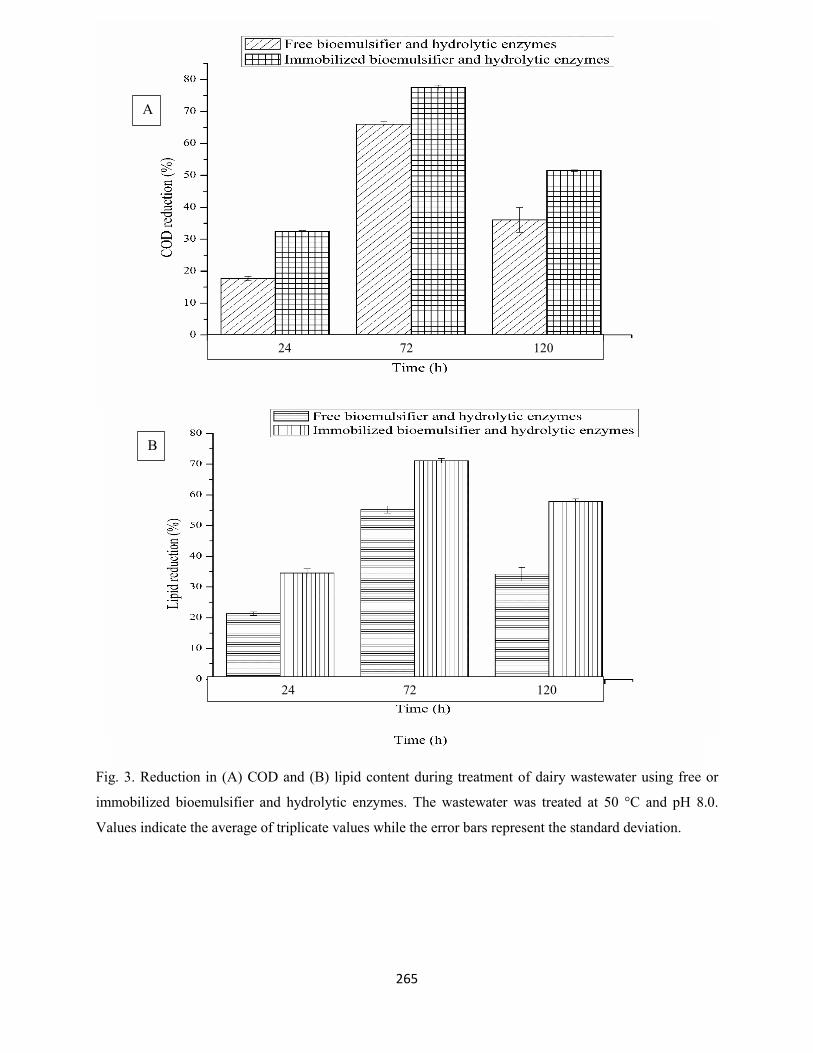
265
Fig. 3. Reduction in (A) COD and (B) lipid content during treatment of dairy wastewater using free or
immobilized bioemulsifier and hydrolytic enzymes. The wastewater was treated at 50 °C and pH 8.0.
Values indicate the average of triplicate values while the error bars represent the standard deviation.
A
24 72 120
24 72 24 72 120
B
24 72 120
24 72 120
A
B
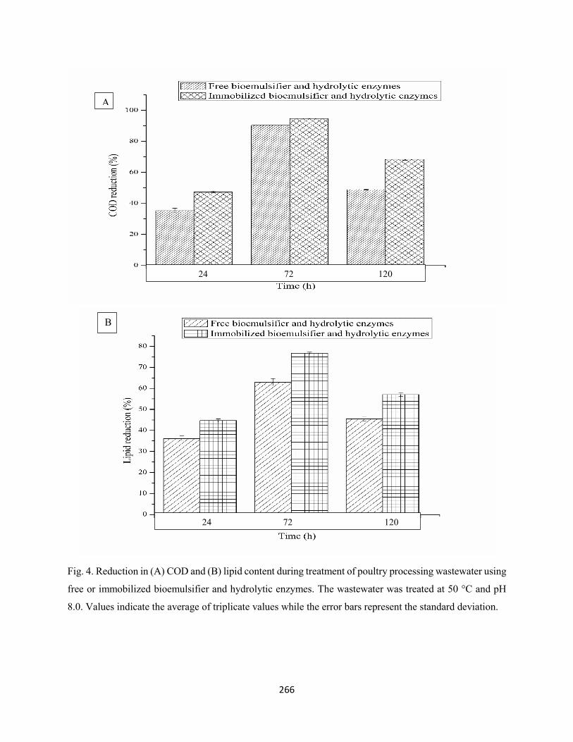
266
Fig. 4. Reduction in (A) COD and (B) lipid content during treatment of poultry processing wastewater using
free or immobilized bioemulsifier and hydrolytic enzymes. The wastewater was treated at 50 °C and pH
8.0. Values indicate the average of triplicate values while the error bars represent the standard deviation.
24 72 120
A
B
24 72 120
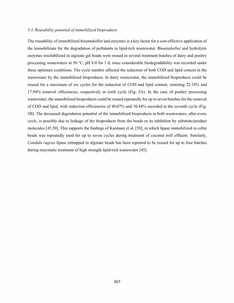
267
3.3. Reusability potential of immobilized bioproducts
The reusability of immobilized bioemulsifier and enzymes is a key factor for a cost-effective application of
the immobilizate for the degradation of pollutants in lipid-rich wastewater. Bioemulsifier and hydrolytic
enzymes insolubilized in alginate gel beads were reused in several treatment batches of dairy and poultry
processing wastewaters at 50 °C, pH 8.0 for 3 d, since considerable biodegradability was recorded under
these optimum conditions. The cycle number affected the reduction of both COD and lipid content in the
wastewater by the immobilized bioproducts. In dairy wastewater, the immobilized bioproducts could be
reused for a maximum of six cycles for the reduction of COD and lipid content, retaining 22.18% and
17.94% removal efficiencies, respectively at sixth cycle (Fig. 5A). In the case of poultry processing
wastewater, the immobilized bioproducts could be reused repeatedly for up to seven batches for the removal
of COD and lipid, with reduction efficiencies of 40.07% and 30.44% recorded at the seventh cycle (Fig.
5B). The decreased degradation potential of the immobilized bioproducts in both wastewaters, after every
cycle, is possibly due to leakage of the bioproducts from the beads or its inhibition by substrate/product
molecules [45,50]. This supports the findings of Kanmani et al. [50], in which lipase immobilized in celite
beads was repeatedly used for up to seven cycles during treatment of coconut mill effluent. Similarly,
Candida rugosa lipase entrapped in alginate beads has been reported to be reused for up to four batches
during enzymatic treatment of high strength lipid-rich wastewater [45].
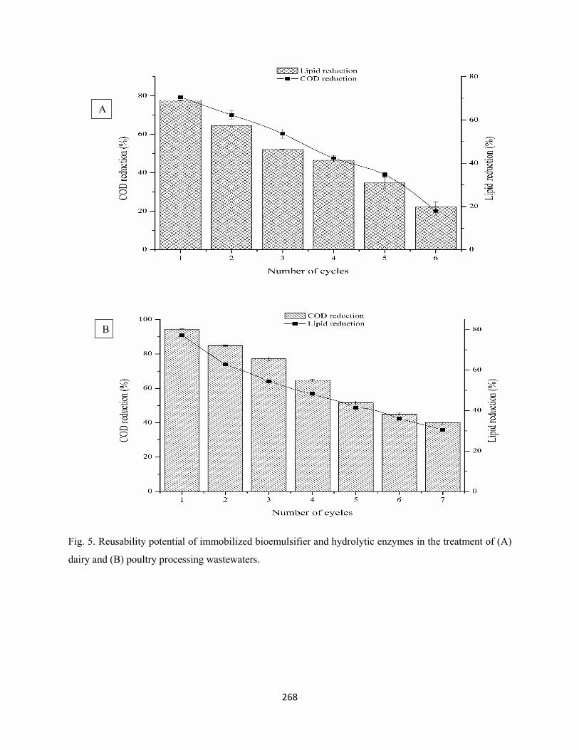
268
Fig. 5. Reusability potential of immobilized bioemulsifier and hydrolytic enzymes in the treatment of (A)
dairy and (B) poultry processing wastewaters.
A
B
269
1. Conclusions
Mixture of free or immobilized bioemulsifier and hydrolytic enzymes was employed for the degradation of
pollutants present in dairy and poultry processing wastewaters. Despite the high pollutant load of poultry
processing wastewater, there was higher removal of COD and lipid from this wastewater at all the treatment
conditions in comparison to dairy wastewater. In addition, the immobilized bioproducts were found to be
more efficient in the degradation of organic load when compared to the free counterparts. Furthermore, the
immobilized bioproducts could be reused for the treatment of dairy and poultry processing wastewaters.
The synergistic application of these bioproducts was found to be extremely advantageous in that, it is rapid,
efficient and cost-effective in the reduction of pollutants present in lipid-rich wastewater in addition to
eliminating the problem of generating solid waste. Thus, representing an attracting remedy to combat severe
pollution problems emanating from the discharge of untreated and inadequately treated lipid-rich
wastewater.
Acknowledgements: The financial support of the National Research Foundation (NRF) of South Africa
towards this research is hereby acknowledged. Opinions expressed and conclusions arrived at, are those of
the authors and are not necessarily to be attributed to the NRF.
Conflict of interest: The authors declare no conflict of interest
References
[1] L. Masse, D.I. Massé, Effect of soluble organic, particulate organic and hydraulic shock loads on
anaerobic sequencing batch reactors treating of slaughterhouse wastewater at 20 °C, Proc. Biochem.
40 (2005) 1225-1232.
[2] K.B. Chipasa, K. Mędrzycka, Behavior of lipids in biological wastewater treatment processes. J. Ind.
Microbiol. Biotechnol. 33 (2006) 635-645.
[3] A.B.G. Valladão, M.C. Cammarota, A.G. Torres, D.M.G. Freire, Profiles of fatty acids and
triacylglycerols and their influence on the anaerobic biodegradability of effluents from poultry
slaughterhouse, Bioresour. Technol. 102 (2011) 7043-7050.
270
[4] B.A. Mahdi, A. Bhattacharya, A. Gupta, Enhanced lipase production from Aeromonas sp. S1 using Sal
deoiled seed cake as novel natural substrate for potential application in dairy wastewater treatment,
J. Chem. Technol. Biotechnol. 87 (2012) 418-426.
[5] H.J. Porwal, A.V. Mane, S.G. Velhal, Biodegradation of dairy effluent by using microbial isolates
obtained from activated sludge, Water Resour. Ind. 9 (2015) 1-15.
[6] T.V. Adulkar, V.K. Rathod, Pre-treatment of high fat content dairy wastewater using different
commercial lipases, Desalin. Water Treat. 53 (2015) 2450-2455.
[7] C.F. Bustillo-Lecompte, M. Mehrvar, Slaughterhouse wastewater characteristics, treatment, and
management in the meat processing industry: a review on trends and advances, J. Environ. Manage.
161 (2015) 287-302.
[8] M.C. Cammarota, D.M.G Freire, A review on hydrolytic enzymes in the treatment of wastewater with
high oil and grease content, Bioresour. Technol. 97 (2006) 2195-3010.
[9] M. Hatamoto, H. Imachi, A. Ohashi, H. Harada, Identification and cultivation of anaerobic, syntrophic
long-chain fatty acid-degrading microbes from mesophilic and thermophilic methanogenic sludges,
Appl. Environ. Microbiol. 73 (2007) 1332-1340.
[10] V.S. Ferreira-Leitão, M.C. Cammarota, E.C.G. Aguieiras, L.R. Vasconcelos de Sá, R. Fernandez-
Lafuente, D.M.G. Freire, The protagonism of biocatalysis in green chemistry and its environmental
benefits, Catalysts 7(2017) 9.
[11] J.P. Kushwaha, V.C. Srivastava, I.D. Mall, An overview of various technologies for the treatment of
dairy wastewaters, Crit. Rev. Food Sci. Nutri. 51 (2011) 442-452.
[12] I. Hamawand, A. Ghadouani, J. Bundschuh, S. Hamawand, R.A. Al Juboori, S. Chakrabarty, T. Yusaf,
A critical review on processes and energy profile of the Australian meat processing industry,
Energies, 10 (2017) 731.
[13] A.K. Slavov, General characteristics and treatment possibilities of dairy wastewater- a review, Food
Technol. Biotechnol. 55 (2017) 14-28.
[14] F.R.C. Damasceno, D.M.G. Freire, M.C. Cammarota, Assessing a mixture of biosurfactant and enzyme
pools in the anaerobic biological treatment of wastewater with high fat content, Environ. Technol.
35 (2014) 2035-2045.
271
[15] D. Alberton, A.D. Mitchell, J. Cordova, P. Peralta-Zamora, N. Krieger, Production of fermented solid
containing lipases of Rhizopus microsporus and its application in the pre-hydrolysis of a high-fat
dairy wastewater, Food Technol. Biotechnol. 48 (2010) 28-35.
[16] A. Daverey, K. Pakshirajan, Pretreatment of synthetic dairy wastewater using the sophorolipid-
producing yeast Candida bombicola, Appl. Biochem. Biotechnol. 163 (2011) 720-728.
[17] F.R.C. Damasceno, M.C. Cammarota, D.M.G. Freire, The combined use of a biosurfactant and an
enzyme preparation to treat an effluent with high fat content, Coll. Surf. B: Biointerfaces. 95 (2012)
241-246.
[18] N.J. Cosmann, B.M. Gomes, S.D. Gomes, A.P.R. Simiqueli, G.M. Pastore, Use of biosurfactant
surfactin produced from cassava wastewater for anaerobic treatment of effluent from a poultry
slaughterhouse, Afr. J. Biotechnol. 16 (2017) 224-231.
[19] J. Jeganathan, G. Nakhla, A. Bassi, Hydrolytic pretreatment of oily wastewater by immobilized lipase,
J. Hazard. Mater. 145 (2007) 127-135.
[20] D.R. Rosa, I.C.S. Duarte, N.K. Saavedra, M.B. Varesche, M. Zaiat, M.C. Cammarota, D.M.G. Freire,
Performance and molecular evaluation of an anaerobic system with suspended biomass for treating
wastewater with high fat content after enzymatic hydrolysis, Bioresour. Technol. 100 (2009) 6170-
6176.
[21] V.M.F Alexandre, A.M. Valente, M.C. Cammarota, D.M.G. Freire, Performance of anaerobic
bioreactor treating fish-processing plant wastewater pre-hydrolyzed with a solid-enzyme pool,
Renew. Energy, 36 (2011) 3439-3444.
[22] F.D.A. Facchini, A.C. Vici, M.G. Pereira, J.A. Jorge, M.L.T.M. Polizeli, Enhanced lipase production
of Fusarium verticillioides by using response surface methology and wastewater pretreatment
application, J. Biochem. Tech. 6 (2015) 996-1002.
[23] A. Mugdha, M. Usha, Enzymatic treatment of wastewater containing dyestuffs using different delivery
systems, Sci. Rev. Chem. Commun. 2 (2012) 31-40.
[24] N.R. Mohamad, N.H.C. Marzuki, N.A. Buang, F. Huyop, R. Abdul Wahab, An overview of
technologies for immobilization of enzymes and surface analysis techniques for immobilized
enzymes, Biotechnol. Biotechnol. Equip. 29 (2015) 205-220.
272
[25] A.A. Homaei, R. Sariri, F. Vianello, R. Stevanato, Enzyme immobilization: an update, J. Chem. Biol.
6 (2013) 185-205.
[26] P. Saranya, S. Swarnalatha, G. Sekaran, Lipoprotein biosurfactant production from an extreme
acidophile using fish oil and its immobilization in nanoporous activated carbon for the removal of
Ca2+ and Cr3+ in aqueous solution, RSC adv. 4 (2014) 34144-34155.
[27] A.S.S. Ibrahim, A.A. Al-Salamah, A.M. El-Toni, K.S. Almaary, M.A. El-Tayeb, Y.B. Elbadawi, G.
Antranikian, Enhancement of alkaline protease activity and stability via covalent immobilization
onto hollow core-mesoporous shell silica nanospheres, Int. J. Mol. Sci. 17 (2016) 184.
[28] R. Tarntip, T. Sirichom, Isolation of proteolytic, lipolytic, and bioemulsifying bacteria for
improvement of the aerobic treatment of poultry processing wastewater, Afr. J. Microbiol. Res. 5
(2011) 5493-5497.
[29] J.do.N Silva, M.L.E. Gutarra, D.M.G. Freire, M.C. Cammarota, Application of home-made enzyme
and biosurfactant in the anaerobic treatment of effluent with high fat content, Bioprocess. Biotech.
3 (2013) 139.
[30] S. Guleria, A. Walia, A. Chauhan, C.K. Shirkot, Immobilization of Bacillus amyloliquefaciens SP1
and its alkaline protease in various matrices for effective hydrolysis of casein, 3 Biotech. 6 (2016)
208.
[31] S. Ferhat, R. Alouaoui, A. Badis, N. Moulai-Mostefa, Production and characterization of biosurfactant
by free and immobilized cells from Ochrobactrum intermedium isolated from the soil of southern
Algeria with a view to environmental application, Biotechnol. Biotechnol. Equip. 31 (2017) 733-
742.
[32] A.E. Greenberg, L.S. Clesceri, A.D. Eaton, Standard methods for the examination of water and
wastewater, American Public Health Association/American Water Works Association/Water
Pollution Control Federation, Washington DC, (2005).
[33] H.D. Kirschman, R. Pomeroy, Determination of oil in oil field wastewater, Anal. Chem. 21 (1949)
793.
[34] O.H. Lowry, N.J. Rosebrough, A.I. Farr, R.J. Randall, Protein measurement with Folin phenol reagent,
J. Biol. Chem. 193 (1951) 265-275.
273
[35] P. Piro, M. Carbone, G. Tomei, Assessing settleability of dry and wet weather flows in an urban area
serviced by combined sewer, Water Air Soil Pollut. 214 (2011) 107-117.
[36] W. Qasim, A.V. Mane, Characterization and treatment of selected food industrial effluents by
coagulation and adsorption techniques, Water Res. Ind. 4 (2013) 1-12.
[37] A.A. Mendes, E.B. Pereira, A. Jr. Furigo, F. de Castro, Anaerobic biodegradability of dairy wastewater
pretreated with porcine pancreas lipase, Braz. Arch. Biol. Technol. 53 (2010) 1279-1284.
[38] G. Dors, A.A. Mendes, E.B. Pereira, H.F. de Castro, A. Jr Furigo, Simultaneous enzymatic hydrolysis
and anaerobic biodegradation of lipid-rich wastewater from poultry industry, Appl. Water Sci. 3
(2013) 343-349.
[39] U. Pathak, P. Das, P. Banerjee, S. Datta, Treatment of wastewater from a dairy industry using rice husk
as adsorbent: treatment efficiency, isotherm, thermodynamics, and kinetics modelling, J.
Thermodyn. 2016 (2016) 1-7.
[40] S. Bayar, Y.S. Yildiz, A.E. Yilmaz, S. Irdemez, The effect of stirring speed and current density on
removal efficiency of poultry slaughterhouse wastewater by electrocoagulation method,
Desalination, 280 (2011) 103-107.
[41] Y-Y Liu, R.J. Haynes, Origin, nature, and treatment of effluents from dairy and meat processing
factories and the effects of their irrigation on the quality of agricultural soils, Critical Rev. Environ.
Sci. Technol. 41 (2011) 1531-1599.
[42] B. Demirel, O. Yenigun, T.T. Onay, Anaerobic treatment of dairy wastewater: a review, Proc.
Biochem. 40 (2005) 2583-2595.
[43] J.N. Edokpayi, J.O. Odiyo, T.A.M. Msagati, E.O. Popoola, Removal efficiency of faecal indicator
organisms, nutrients and heavy metals from a peri-urban wastewater treatment plant in
Thohoyandou, Limpopo province, South Africa. Int. J. Environ. Res. Public Health 12 (2015) 7300-
7320.
[44] Government Gazette. No. 36820 (2013), Wastewater values applicable to the irrigation of any land or
property up to 2000 cubic metres; Minister of Water and Environmental Affairs: South Africa.
[45] J. Jeganathan, A. Bassi, G. Nakhla, Pre-treatment of high oil and grease pet food industrial wastewaters
using immobilized lipase hydroxylation, J. Hazard. Mater. B137 (2006) 121-128.
274
[46] O. Lefebvre, S. Quentin, M. Torrijos, J.J. Godon, J.P. Delgenès, R. Moletta, Impact of increasing NaCl
concentrations on the performance and community composition of two anaerobic reactors, Appl.
Microbiol. Biotechnol. 75 (2007) 61-69.
[47] D.F.C. Jacobucci, M.R.G. Oriani, L.R. Durrant, Reducing COD level on oily effluent by utilizing
biosurfactant-producing bacteria, Braz. Arch. Biol. Technol. 52 (2009) 1037-1042.
[48] N.S. Dumore, M. Mukhopadhyay, Removal of oil and grease using immobilized triacylglycerin, Int.
Biodeter. Biodegr. 68 (2012) 65-70.
[49] V. Minovska, E. Winkelhausen, S. Kuzmanova, Lipase immobilized by different techniques on various
support materials applied in oil hydrolysis, J. Serb. Chem. Soc. 70 (2005) 609-624.
[50] P. Kanmani, K. Kumaresan, J. Aravind, Pretreatment of coconut mill effluent using celite-immobilized
hydrolytic enzyme preparation from Staphylococcus pasteuri and its impact on anaerobic digestion,
Biotechnol. Prog. 31 (2015) 1249-1258.
275
CHAPTER ELEVEN
GENERAL DISCUSSION AND CONCLUSION
11.1 THE RESEARCH IN PERSPECTIVE
Dairy and poultry processing industries contribute tremendously to global industrial pollution. These
industries consume large amounts of water for various processes, resulting in the discharge of huge volume
of effluent containing high concentrations of organic molecules including fats and proteins, contributing to
their low biodegradability coefficient (Liu and Haynes, 2011). Improper disposal of this wastewater,
especially when untreated or inadequately treated poses serious threats to the receiving environment by
causing air pollution and contamination of surface and underground water (Shete and Shinkar, 2013).
Hence, the need for establishment of efficient, cost-effective, eco-friendly and sustainable technologies that
can serve as alternatives to existing treatment systems for lipid-rich wastewater. Biological technology
involving application of bioemulsifier and hydrolytic enzymes offers a promising approach for the effective
treatment of high strength lipid-rich wastewater. These bioproducts facilitate degradation of lipid-rich
wastewater by triggering two simultaneous processes namely; emulsification of fat particles and enzymatic
catalysis, leading to increased reduction of organic load in the wastewater and alleviating the problem
associated with the discharge of untreated wastewater (Damasceno et al. 2012, 2014; Parthipan et al., 2017).
This project thus aimed at investigating the synergistic effects of a mixture of free or immobilized
bioemulsifier and hydrolytic enzymes from indigenous bacterial isolates in the reduction of pollutants from
dairy and poultry processing wastewaters.
Production and characterization of thermostable glycoprotein bioemulsifiers from indigenous
Acinetobacter sp. isolated from lipid-rich wastewater in Durban was described in Chapter Five. High
throughput techniques for screening and selection of bioemulsifier/biosurfactant-producing
microorganisms are required to bioprospect for novel bioemulsifiers or bioemulsifier-producing microbial
strains (Satpute et al., 2010). The use of single technique for the screening of bioemulsifier/biosurfactant-
producing microorganisms is inaccurate and unreliable due to diverse functional and structural properties
of the surface active compounds. As a result, Acinetobacter sp. Ab9-ES and Ab33-ES were screened for
bioemulsifier/biosurfactant production by growing in the presence of different carbon sources including
glucose, diesel or edible oils. The obtained culture supernatants were tested for surface/emulsifying activity
using oil spreading test and emulsification assay. These study strains showed positive results for both tests,
especially when tested with olive oil, since this edible oil was mostly utilized for growth and
bioemulsifier/biosurfactant production by the bacterial strains. Surface tension (ST) was measured on the
culture supernatants of Acinetobacter sp. in order to distinguish between bioemulsifier and biosurfactant
276
producers. The ST of the bacterial culture supernatants was found to be above the threshold of 40 mN/m,
an indication that they are bioemulsifier producers (Monteiro et al., 2009; Franzetti et al., 2012; Lima et
al., 2017; Patowary et al., 2017). Among microbial population, extracellular bioemulsifier production has
been reported to be a common phenomenon among members of the genus Acinetobacter (Patil and
Chopade, 2001; Jagtap et al., 2010). Growth of these organisms on water-insoluble carbon sources results
in emulsification of the substrates in the culture medium, leading to the production of bioemulsifiers.
Bioemulsifier production by these study strains was found to be growth-associated, with maximum
emulsifying activities recorded during stationary (168 h) and exponential (120 h) growth phase for strains
Ab9-ES and Ab33-ES, respectively. It is well established that bioemulsifier production is influenced by
environmental factors, substrate composition and media concentration (Banat et al., 2014; Santos et al.,
2016). Interaction of these parameters with each other in a multifaceted way affects the kinetics of
biosurfactant production. Similar profile has been reported by several other bioemulsifier-producing
microorganisms (Zheng et al., 2012; Gudiña et al., 2015; Heryani and Putra, 2017). The potential of these
bioemulsifiers to emulsify different hydrophobic substrates was investigated in the presence of edible oils;
aliphatic, aromatic and mixed hydrocarbons. The bioemulsifiers exhibited higher emulsification activity
against edible oils, especially sunflower oil while less stable emulsions were formed with other
hydrocarbons. The poor emulsification of the aliphatic, aromatic and mixed hydrocarbons may probably be
due to the inability of the bioemulsifier to stabilize the microscopic droplets (Rufino et al., 2016). The
emulsifying activity of the bioemulsifiers was substrate-dependent. Most microbial surface active
compounds are substrate-specific and possess ability to emulsify different hydrocarbons at varying rates
(Rufino et al., 2014; Bhattacharya et al., 2014). Findings from this study showed that the bioemulsifiers
produced by Acinetobacter sp. possess higher emulsion-stabilizing capacity with different edible oils,
therefore forming a potential biomolecule for the treatment of lipid-rich wastewater.
Effectiveness of bioemulsifier for various applications is dependent on their emulsifying activities over a
broad range of temperature, pH and salinity (Markande et al. 2013; Rufino et al., 2016). These
environmental factors affect the activity of bioemulsifiers in diverse ways. The emulsifier from
Acinetobacter sp. in this study was subjected to varying conditions of pH, temperature, and salinity to
determine the emulsifier’s properties. The bioemulsifiers maintained stable emulsions over broad range of
temperatures. However, slight decrease in emulsifying activity was observed at higher temperatures. This
may be due to denaturation of protein component of the bioemulsifier during heat treatment (Sarrubo et al.,
2006). Different bioemulsifiers with prolonged stability after exposure to higher temperatures have been
reported (Shavandi et al., 2011). However, other surface active compounds lose some emulsifying activity
at those temperatures (Vecino-Bello et al., 2012). The bioemulsifiers demonstrated emulsion-stabilizing
277
capacity over acidic to alkaline pH ranges, with maximum emulsifying activity recorded at neutral pH,
found to be higher than that exhibited by synthetic surfactant. This corroborates the findings of Lima and
Alegre (2009) and Aparna et al. (2012) in which bioemulsifiers produced by Saccharomyces lipolytica
CCT-0913 and Pseudomonas sp. 2B, respectively were active at neutral pH. Similar profile has been
observed at neutral pH in the stability of bioemulsifiers produced by Rhocococcus sp. TA6 and
Lactobacillus pentosus (Shanvandi et al., 2011; Vecino-Bello et al., 2012). Bhattacharya et al. (2014) state
that the decrease in emulsifying activity at extreme acidic pH, as reported in this study may be due to acid
hydrolysis of carbohydrate moieties present in the biopolymer. Pronounced reduction in the emulsifying
activity recorded at higher pH values may probably be due to some structural alterations of the
bioemulsifiers under extreme pH conditions. In addition, emulsifying activity increases as the NaCl
concentrations increase until attaining optimum at moderate concentration. Beyond these salinity levels,
the emulsifying activity declined drastically. The salinity had significant effect on hydrocarbon
emulsification and stability due to ionization caused by the interaction of the salt ions with the hydrophobic
substrates (Amodu et al., 2014). In general, the robust characteristics of these bioemulsifiers are very
beneficial for applications under extreme conditions of temperature, moderate pH and salinity in
bioremediation of oil-contaminated sites.
Chapter Six investigates the statistical optimization of bioprocess factors for enhanced production of
protease from Bacillus aryabhattai Ab15-ES using response surface methodology (RSM). Bacillus
aryabhattai Ab15-ES isolated from lipid-rich wastewater was screened for proteolytic activity using skim
milk agar. The formation of clear zone around the agar wells, resulting from hydrolysis of casein was an
indication of protease production (Olajuyigbe and Ajele, 2005). Such high extracellular enzymatic activity
could largely be due to high rates of substrate utilization and hydrolysis by the bacterial isolate.
Extracellular protease production by microbial strains is constitutive or partially inducible in nature. As a
result, the type of substrate utilized in the fermentation medium mostly influences the enzyme synthesis.
Therefore, selection of appropriate inducible substrate and microorganisms is paramount for the production
of desired metabolite (Bhunia et al., 2012). Preliminary studies of protease production by Bacillus
aryabhattai Ab15-ES yielded maximum enzyme production during late exponential phase of growth, and
this was found to be growth-linked. Optimum protease production from Bacillus licheniformis N-2
(Nadeem et al., 2008) and Bacillus subtilis SHS-04 (Olajuyigbe, 2013) during late exponential phase has
been reported. The rapid decline in protease production beyond optimum may be due to depletion of
nutrients in the growth media, or autolysis resulting from build-up of the enzyme in the production media
(Nadeem et al., 2008). The formation and accumulation of extracellular enzymes in the fermentation media
depend on media constituents such as carbon and nitrogen sources, which are vital parameters influencing
278
enzyme production (Akolkar et al., 2009). As a result, conventional optimization studies involving change
of ‘one-variable-at-a-time’ was used for the optimization of process parameters to achieve optimum cell
density and enhanced production of microbial metabolite. Of the carbon sources tested, maltose was found
to result in maximum protease production by Bacillus aryabhattai Ab15-ES. This supports the findings of
Raj et al. (2012) and Vijayaraghavan et al. (2014) where highest protease yields were attained in a medium
containing maltose as a carbon source. The optimum protease synthesis recorded in the presence of beef
extract may be due to the presence of multi-nutrient elements including free amino acids, essential fatty
acids, carbohydrates, etc. in the organic nitrogen sources, which induced the growth of the organism for
maximum protease production (Esakkiraj et al., 2011). Several authors have recorded similar results
(Umuyaparvathi et al., 2013; Kumar et al., 2014). However, these approaches are time-consuming and thus
expensive for simultaneous consideration of many variables. In addition, they are laborious, require more
experimental data and unable to detect interactions and optimal conditions among variables (Li et al., 2008).
Hence, the need for statistical optimization of bioprocess parameters for maximum protease production
(Singh and Bajaj, 2016). Statistical experimental method is widely used in biotechnology to maintain a
balance between various parameters for enhanced protease production (Chandra et al., 2015). The central
composite design (CCD) of RSM used in the present study was very useful in confirming the effects and
interactions of fermentation variables. In addition, it results in enhanced productivity, reduces process
changeability, and gives closer confirmation of the predicted response to the experimental values.
Enhancement of protease production from Bacillus aryabhattai Ab15-ES by RSM was achieved under the
optimum conditions of temperature, pH, inoculum volume, maltose and beef extract, resulting in a 4.4-fold
enhancement in protease production using the basal medium as reference. This was found to be higher than
that of Bacillus sp. HTS102 (Queiroga et al., 2012) and Brevibacterium linens DSM 20158 (Shabbiri et al.,
2012). These results suggest that the fermentation parameters greatly influence extracellular protease
production by the bacterial strain and their interactions are crucial in the synthesis of the enzyme (Rao et
al., 2008).
Bacillus aryabhattai SE3-PB was isolated from lipid-rich wastewater, and found to exhibit lipolytic activity
following screening on Tween-20 and phenol red agar (Gopinath et al., 2006; Singh et al., 2006). Lipase
production from Bacillus aryabhattai SE3-PB by submerged fermentation started at early log phase and
reached optimum during late exponential growth phase. The decrease in lipase production recorded beyond
exponential phase may probably be due to the presence of inhibitors (e.g. protease) or limitation of
nutrients in the culture medium. The supplementation of fermentation medium with inducible substrates
enhances lipase production, since the carbon chain moiety of the fatty acids present in triacylglycerol
controls lipase production (Sethi et al., 2016). In the present study, olive oil in the fermentation medium
279
for preliminary lipase production was replaced with commercial vegetable oils viz. palm oil, castor oil, rice
bran oil, sunflower oil or canola oil in order to determine best oil that will induce maximum extracellular
lipase production by Bacillus aryabhattai SE3-PB. Lipase production varies in the presence of the different
inducer oils, with maximum lipase production recorded in the fermentation media containing sunflower oil
as inducer (Chapter Seven). In a similar study, Facchini et al. (2015) and Golani et al. (2016) recorded
maximum lipase production by Fusarium verticillioides and Staphylococcus chromogenes 01 A,
respectively in the presence of sunflower oil as an inducible substrate. Face centered CCD of RSM used in
the present study for optimization of fermentation conditions including temperature, pH, inoculum volume,
agitation and sunflower oil concentration for improved lipase production from Bacillus aryabhattai SE3-
PB indicated the importance of these parameters at different levels, and their interactions. Lipase production
was significantly influenced by temperature, pH, inoculum volume, agitation and sunflower oil
concentration. A high similarity recorded between the predicted and observed results suggests that the
model was accurate for process optimization, resulting in 7.2-fold enhancement in lipase production when
using unoptimized medium as reference. Improvement of lipase production by Bacillus aryabhattai SE3-
PB was largely higher than that of Burkholderia sp. (Lo et al., 2012) and Acinetobacter sp. AU07 (Gururaj
et al., 2016). The present study suggests a rational choice of optimum processing conditions for large scale
and cost-effective production of lipase by Bacillus aryabhattai SE3-PB for various biotechnological
applications.
Biochemical and kinetics properties of free and immobilized protease and lipase from Bacillus aryabhattai
were investigated in this study and reported in chapter Eight and Nine. The partially purified enzymes were
immobilized in alginate beads by entrapment method. Enzyme immobilization was greatly influenced by
varying sodium alginate and calcium chloride concentrations. Comparative study of biochemical profiling
of both free and immobilized enzymes revealed no change in the optimum temperature and pH of both
enzyme preparations for maximum activity. This suggested that the chemical coupling of the enzymes to
the polymer had no effects on temperature or pH profile. Ibrahim et al. (2016) and Xiao et al. (2016) have
also observed similar phenomenon in which no change was recorded in the optimum temperature and pH
of biocatalysts before and after immobilization. However, in comparison to free enzymes, the immobilized
enzymes recorded enhanced stability over the investigated pH and temperature ranges. Ranjbakhsh et al.
(2012) and Cao et al. (2016) observed that the significant improvement in the stability of the entrapped
enzyme might be due to immobilization in the support material, which protect the tertiary structure of the
enzyme and prevent conformational transition of the enzyme. Kinetics properties of the enzymes revealed
enhanced enzyme-substrate affinity and increased catalytic efficiency of the immobilized enzymes when
compared to soluble enzymes. In addition, the immobilized enzymes were more stable when stored at 4
280
and 25 °C for 30 d and reusable at several consecutive cycles, indicating a good operational stability of the
biocatalysts (Srivastava et al., 2014). This was not surprising, since soluble enzymes are not stable under
prolonged storage thus, leading to a reduction in catalytic activity (Çevik et al., 2011). Reusability of
insolubilized enzymes is recognized as one of the most vital factors that influence their cost-effective
application for various bioprocesses (Ibrahim et al., 2016). Findings from this study suggest the efficient
and sustainable use of the developed immobilized catalytic system for various environmental and
biotechnological applications.
The present study investigates the synergistic effects of a mixture of free or immobilized extracellular
glycoprotein bioemulsifier and hydrolytic enzymes (protease and lipase) in the reduction of organic load
from dairy and poultry processing wastewaters. Kinetics study of degradation of organic load in the
wastewater revealed maximum chemical oxygen demand (COD) and lipid removal efficiencies in dairy and
poultry processing wastewaters when treated with free or immobilized bioproducts (Chapter Ten).
However, in comparison to dairy wastewater, there was enhanced reduction of organic load from poultry
processing wastewater, suggesting higher affinity of the bioproducts to poultry effluent constituents
(Valladão et al., 2011). The immobilized bioproducts were found to be more effective in the degradation
of pollutants from both wastewater types when compared to the free counterpart, an indication of protective
role provided by the support material in stabilizing the bioproducts thus, maintaining their activities
(Homaei et al., 2013). Such cocktail application of biosurfactant and enzyme pool has been reported for
enhanced removal of COD and lipid from lipid-rich wastewater (Damasceno et al., 2012, 2014). In
addition, the immobilized bioproducts were reusable consecutively for the reduction of pollutants in the
wastewater. The decreased degradation potential of the immobilized bioproducts in both wastewater types,
after every cycle, is possibly due to leakage of the bioproducts from the beads or its inhibition by
substrate/product molecules (Jeganathan et al., 2006). This supports the findings of Kanmani et al. (2015),
in which lipase immobilized in celite beads was repeatedly used for up to seven cycles during treatment of
coconut mill effluent. The synergistic application of these bioproducts was found to be extremely
advantageous in that, it is rapid, efficient and cost-effective in the reduction of pollutants present in lipid-
rich wastewater in addition to eliminating the problem of generating solid waste. Thus, representing an
attractive remedy to combat severe pollution problems emanating from the discharge of untreated and
inadequately treated lipid-rich wastewater.
11.2 POTENTIAL FOR FUTURE DEVELOPMENT OF THE STUDY
Biological method is an emerging treatment technology that utilizes metabolic potential of microorganisms
for the elimination of hazardous pollutants present in lipid-rich wastewater under aerobic or anaerobic
281
conditions, or a combination of both via complete degradation or sequestration (Soleimaninanadegani and
Manshad, 2014; Nzila et al., 2017). In the present study, the potential of a mixture of free or immobilized
bioemulsifier and hydrolytic enzymes (protease and lipase) for the reduction of pollutants present in dairy
and poultry processing wastewaters was investigated under aerobic conditions. Anaerobic degradation of
these wastewater types using these bioproducts could be a promising strategy for large-scale production of
biogas (methane), thus serving as a cheap and alternative source of energy. This energy can be used to
power a biological wastewater treatment plant making them self-sustainable. This will help to lower the
reliance on non-renewable energy sources such as fossil fuels, thus reducing carbon footprint and
expenditure on energy production. In addition, utilization of agro-industrial residues and industrial effluents
can serve as a cheaper substrate for a cost-effective production of bioemulsifier and hydrolytic enzymes.
Cloning and expression of bioproduct (bioemulsifier, protease, lipase) genes from Acinetobacter sp. and
Bacillus aryabhattai in suitable prokaryotic hosts could be a rapid technique for large scale production of
these metabolites. Furthermore, these bioproducts could be applied for the treatment of wastewater with
various organic load in order to ascertain the robustness of the system. Optimization of process parameters
using statistical experimental designs for the treatment of lipid-rich wastewater could help in the selection
of significant factor, study interaction among the parameters and search for optimum conditions for
maximum degradation of pollutants in the dairy and processing wastewaters. Future studies may also focus
on proper understanding of the behavior of pollutants in the lipid-rich wastewater and mechanisms of their
degradation through combined use of bioemulsifier and hydrolytic enzymes.
282
11.3 REFERENCES
Akolkar A, Bharambe N, Trivedi S, Desai A (2009) Statistical optimization of medium components for
extracellular protease production by an extreme haloarchaeon, Halobacterium sp. SP1(1). Lett.
Appl. Microbiol. 48: 77-83.
Amodu OS, Ntwampe SK, Ojumu TV (2014) Emulsification of hydrocarbons by biosurfactants:
exclusive use of agrowaste. Bioresour. 9: 3508-3525.
Aparna A, Srinikethan G, Smitha H (2012) Production and characterization of biosurfactant produced
by a novel Pseudomonas sp. 2B. Colloids Surf B: Biointerfaces, 95: 23-29.
Banat IM, Satpute SK, Cameotra SS, Patil R, Nyayanit NV (2014) Cost effective technologies and
renewable substrates for biosurfactants’ production. Front. Microbiol. 5: 697.
Bhattacharya M, Biswas D, Sana S, Datta S (2014) Utilization of waste engine oil by Ochrobactrum
pseudintermedium strain C1 that secretes an exopolysaccharide as a bioemulsifier. Biocatal. Agric.
Biotechnol. 3: 167-176.
Bhunia B, Basak B, Dey A (2012) A review on production of serine alkaline protease by Bacillus spp. J.
Biochem. Tech. 3: 448-457.
Cao S-L, Huang Y-M, Li X-H, Xu P, Wu H, Li N, Lou W-Y, Zong M-H (2016) Preparation and
characterization of immobilized lipase from Pseudomonas cepacia onto magnetic cellulose
nanocrystals. Sci. Rep. 6: 20420.
Çevik E, Şenel M, Abasiyanik M F (2011) Immobilization of urease on copper chelated EC-Tribeads and
reversible adsorption. Afr. J. Biotechnol. 10: 6590-6597.
Chandra SJ, Pallavi P, Anuradha BS, Reddy SR (2015) Optimization of bioprocess for enhanced
production of alkaline protease by a Bacillus subtilis SHmIIIa through Blacket-Burman design. Afr.
J. Microbiol. Res. 9: 1738-1744.
Damasceno FRC, Cammarota MC, Freire DMG (2012) The combined use of a biosurfactant and an
enzyme preparation to treat an effluent with high fat content. Coll. Surf. B: Biointerfaces, 95: 241-
246.
Damasceno FRC, Freire DMG, Cammarota MC (2014) Assessing a mixture of biosurfactant and
enzyme pools in the anaerobic biological treatment of wastewater with high fat content. Environ.
Technol. 35: 2035-2045.
283
Esakkiraj P, Sankaralingam S, Usha R, Palavesam A, Immanuel G (2011) Solid-state protease
production using anchovy waste meal by moderate halophile Serratia proteamaculans AP-CMST
isolated from fish intestine. Ann. Microbiol. 61: 749-755.
Facchini FDA, Vici AC, Pereira MG, Jorge JA, Polizeli MDTM (2015) Enhanced lipase production of
Fusarium verticillioides by using response surface methodology and wastewater pretreatment
application. J. Biochem. Tech. 6: 996-1002.
Franzetti A, Gandolfi I, Raimondi C, Bestetti G, Banat IM, Smyth TJ, Papacchini M, Cavallo M,
Fracchia L (2012) Environmental fate, toxicity, characteristics and potential applications of novel
bioemulsifiers produced by Variovorax paradoxus 7bCTS. Bioresour. Technol. 108: 245-251.
Golani M, Hajela K, Pandey GP (2016) Screening, identification, characterization and production of
bacterial lipase from oil spilled soil. Int. J. Curr. Microbiol. Appl. Sci. 5: 745-763.
Gopinath SCB, Anbu P, Hilda A (2005) Extracellular enzymatic activity profiles in fungi isolated from
oil-rich environments. Mycoscience, 46: 119-126.
Gudiña EJ, Pereira JFB, Costa R, Evtuguin DV, Coutinho JAP, Teixeira JA, Rodrigues LR (2015)
Novel bioemulsifier produced by a Paenibacillus strain isolated from crude oil. Microb. Cell Fact.
14: 14.
Gururaj P, Ramalingam S, Devi GN, Gautam P (2016) Process optimization for production and
purification of a thermostable, organic solvent tolerant lipase from Acinetobacter sp. AU07. Braz.
J. Microbiol. 47: 647-657.
Heryani H, Putra MD (2017) Kinetic study and modeling of biosurfactant production using Bacillus sp.
Electronic J. Biotechnol. 27: 49-54.
Homaei AA, Sariri R, Vianello F, Stevanato R (2013) Enzyme immobilization: an update. J. Chem. Biol.
6: 185-205.
Ibrahim ASS, Al-Salamah AA, El-Toni AM, Almaary KS, El-Tayeb MA, Elbadawi YB, Antranikian
G (2016) Enhancement of alkaline protease activity and stability via covalent immobilization onto
hollow core-mesoporous shell silica nanospheres. Int. J. Mol. Sci. 17: 184.
Jagtap S, Yavankar S, Pardesi K, Chopade B (2010) Production of bioemulsifier by Acinetobacter
species isolated from healthy human skin. Indian J. Exp. Biol. 48: 70-76.
284
Jeganathan J, Bassi A, Nakhla G (2006) Pre-treatment of high oil and grease pet food industrial
wastewaters using immobilized lipase hydroxylation. J. Hazard. Mater. B137: 121-128.
Kanmani P, Kumaresan K, Aravind J (2015) Pretreatment of coconut mill effluent using celite-
immobilized hydrolytic enzyme preparation from Staphylococcus pasteuri and its impact on
anaerobic digestion. Biotechnol. Prog. 31: 1249-1258.
Kumar RS, Ananthan G, Prabhu AS (2014) Optimization of medium composition for alkaline
production of Marinobacter sp. GA CAS9 using response surface methodology- A statistical
approach. Biocat. Agric. Biotechnol. 3: 191-197.
Li X, Xu T, Ma X, Guo K, Kai L, Zhao Y, Jia X, Mad Y (2008) Optimization of culture conditions for
production of cis-epoxysuccinic acid hydrolase using response surface methodology. Bioresour.
Technol. 99: 5391-5396.
Lima ÁS, Alegre RM (2009) Evaluation of emulsifier stability of biosurfactant produced by
Saccharomyces lipolytica CCT-0913. Braz. Arch. Biol. Technol. 52: 285-290.
Lima RA, Andrade RFS, Rodríguez DM, Araújo HWC, Santos VP, Campos-Takaki GM (2017)
Production and characterization of biosurfactant isolated from Candida glabrata using renewable
substrates. Afr. J Microbiol. Res. 11: 237-244.
Liu Y-Y, Haynes RJ (2011) Origin, nature, and treatment of effluents from dairy and meat processing
factories and the effects of their irrigation on the quality of agricultural soils. Critical Rev. Environ.
Sci. Technol. 41: 1531-1599.
Lo C-F, Yu C-Y, Kuan I-C, Lee S-L (2012) Optimization of lipase production by Burkholderia sp. using
response surface methodology. Int. J. Mol. Sci. 13: 14889-14897.
Markande AR, Acharya SR, Nerurkar AS (2013) Physicochemical characterization of a thermostable
glycoprotein bioemulsifier from Solibacillus silvestris AM1. Proc. Biochem. 48: 1800-1808.
Monteiro AS, Coutinho JOPA, Ary C, Rosa CA, Siqueira EP, Santos VL (2009) Characterization of
new biosurfactant produced by Trychosporon montevidense CLOA72 isolated from dairy industry
effluents. J. Basic Microbiol. 49: 1-11.
Nadeem M, Qazi JI, Syed Q, Baig S (2008) Optimization of process parameters for alkaline protease
production by Bacillus licheniformis N-2 and kinetics studies in batch fermentation. Turk. J. Biol.
32: 243-251.
285
Nzila A, Thukair A, Saravanan S, Abdur Razzak S (2017) Characterization of aerobic oil and grease-
degrading bacteria in wastewater. Environ. Technol. 38: 661-670.
Olajuyigbe FM (2013) Optimized production and properties of thermostable alkaline protease from
Bacillus subtilis SHS-04 grown on groundnut (Arachis hypogaea) meal. Adv. Enzyme Res. 1: 112-
120.
Olajuyigbe FM, Ajele TO (2005) Production dynamics of extracellular protease from Bacillus species.
Afr. J. Biotechnol. 4: 776-779.
Parthipan P, Preetham E, Machuca LL, Rahman PKSM, Murugan K, Rajasekar A (2017)
Biosurfactant and degradative enzymes mediated crude oil degradation by bacterium Bacillus
subtilis A1. Front. Microbiol. 8: 193.
Patil JR, Chopade BA (2001) Studies on bioemulsifier production by Acinetobacter strains isolated from
healthy human skin. J. Appl. Microbiol. 91: 290-298.
Patowary K, Patowary R, Kalita MC, Deka S (2017) Characterization of biosurfactant produced during
degradation of hydrocarbons using crude oil as sole source of carbon. Front. Microbiol. 8: 279.
Queiroga AC, Pintado ME, Malcata FX (2012) Use of response surface methodology to optimize
protease synthesis by a novel strain of Bacillus sp. isolated from Portuguese sheep wool. J. Appl.
Microbiol. 113: 36-43.
Raj A, Khess N, Pujari N, Bhattacharya S, Das A, Rajan SS (2012) Enhancement of protease production
by Pseudomonas aeruginosa isolated from dairy effluent sludge and determination of its
fibrinolytic potential. Asian Pac. J. Trop. Biomed. 2012: S1845-S1851.
Ranjbakhsh E, Bordbar AK, Abbasi M, Khosropour AR, Shams E (2012). Enhancement of stability
and catalytic activity of immobilized lipase on silica-coated modified magnetite nanoparticles.
Chem. Eng. J. 179: 272-276.
Rao CS, Sathish T, Mahalaxmi M, Laxmi GS, Rao RS, Prakasham RS (2008) Modelling and
optimization of fermentation factors for enhancement of alkaline protease production by isolated
Bacillus circulans using feed-forward neural network and genetic algorithm. J. Appl. Microbiol.
104: 889-898.
Rufino RD, Luna JM, de Campos Takaki GM, Sarubbo LA (2014) Characterization and properties of
the biosurfactant produced by Candida lipolytica UCP 0988. Eletron. J. Biotechnol. 17: 34-38.
286
Rufino RD, Motta Silveira GN, Luna JM, Sarubbo LA (2016) Conservation of the biosurfactant
produced by Pseudomonas aeruginosa for environmental applications. Chem. Eng. Trans. 49: 535-
540.
Santos DKF, Rufino RD, Luna JM, Santos VA, Sarubbo LA (2016) Biosurfactants: Multifunctional
biomolecules of the 21st century. Int. J. Mol. Sci. 17: 401.
Sarubbo L, Moura J, Campos-Takaki G (2006) Production and stability studies of the bioemulsifier
obtained from a new strain of Candida glabrata UCP 1002. Electron. J. Biotechnol. 9: 400-406.
Satpute SK, Banpurkar AG, Dhakephalkar PK, Banat IM, Chopade BA (2010) Methods for
investigating biosurfactants and bioemulsifiers: a review. Crit. Rev. Biotechnol. 30: 127-144.
Sethi BK, Nanda PK, Sahoo S (2016) Characterization of biotechnologically relevant extracellular lipase
produced by Aspergillus terreus NCFT 4269.10. Braz. J. Microbiol. 47: 143-149.
Shabbiri K, Adnan A, Jamil S, Ahmad W, Noor B, Rafique HM (2012) Medium optimization of
protease production by Brevibacterium linens DSM 20158 using statistical approach. Braz. J.
Microbiol. 2012: 1051-1061.
Shavandi M, Mohebali G, Haddadi A, Shakarami H, Nuhi A (2011) Emulsification potential of a newly
isolated biosurfactant-producing bacterium, Rhodococcus sp. strain TA6. Colloids Surf. B:
Biointerfaces, 82: 477-482.
Shete BS, Shinkar NP (2013) Dairy industry wastewater sources, characteristics, and its effects on
environment. Int. J. Curr. Eng. Technol. 3: 1611-1615.
Singh R, Gupta N, Goswami VK, Gupta R (2006) A simple activity staining protocol staining for lipases
and esterases. Appl. Microbiol. Biotechnol. 70: 679-682.
Singh S, Bajaj BK (2016) Bioprocess optimization for the production of thermoalkali-stable protease from
Bacillus subtilis K-1 under solid-state fermentation. Prep. Biochem. Biotechnol. 46: 717-724.
Soleimaninanadegani M, Manshad S (2014) Enhancement of biodegradation palm oil mill effluents by
local isolated microorganisms. Int. Scholarly Res. Notices, 2014: 1-8.
287
Srivastava G, Singh K, Talat M, Srivastava ON, Kayastha AM (2014). Functionalized graphene sheets
as immobilization matrix for Fenugreek β-amylase: enzyme kinetics and stability studies. PLoS
one, 9: e113408.
Umuyaparvathi S, Meenakshi S, Arumugam M, Balasubramanian T (2013) Purification and
characterization of protease from Bacillus cereus SU12 isolated from oyster Saccostrea cucullata.
Afr. J. Biotechnol. 12: 5897-5908.
Valladão ABG, Cammarota MC, Torres AG, Freire DMG (2011) Profiles of fatty acids and
triacylglycerols and their influence on the anaerobic biodegradability of effluents from poultry
slaughterhouse. Bioresour. Technol. 102: 7043-7050.
Vecino-Bello X, Devesa-Rey R, Cruz JM, Moldes AB (2012) Study of the synergistic effects of salinity,
pH, and temperature on the surface-active properties of biosurfactants produced by Lactobacillus
pentosus. J. Agric. Food Chem. 60: 1258-1265.
Vijayaraghavan P, Lazarus S, Vincent SGP (2014) De-hairing protease production by an isolated
Bacillus cereus strain AT under solid-state fermentation using cow dung: biosynthesis and
properties. Saudi Biol. Sci. 21: 27-34.
Xiao A, Xu C, Lin Y, Ni H, Zhu Y, Cai H (2016) Preparation and characterization of κ-carrageenase
immobilized onto magnetic iron oxide nanoparticles. Electron. J. Biotechnol. 19: 1-7.
Zheng C, Li Z, Su J, Zhang R, Liu C, Zhao M (2012) Characterization and emulsifying property of a
novel bioemulsifier by Aeribacillus pallidus YM-1. J. Appl. Microbiol. 113: 44-51.
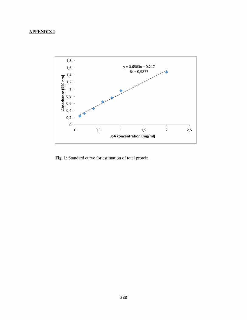
288
APPENDIX I
Fig. 1: Standard curve for estimation of total protein
y = 0,6583x + 0,217R² = 0,9877
0
0,2
0,4
0,6
0,8
1
1,2
1,4
1,6
1,8
0 0,5 1 1,5 2 2,5
Abso
rban
ce (5
50 n
m)
BSA concentration (mg/ml)
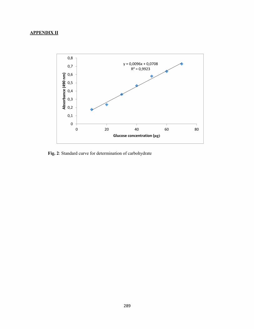
289
APPENDIX II
Fig. 2: Standard curve for determination of carbohydrate
y = 0,0096x + 0,0708R² = 0,9923
0
0,1
0,2
0,3
0,4
0,5
0,6
0,7
0,8
0 20 40 60 80
Abso
rban
ce (4
90 n
m)
Glucose concentration (µg)
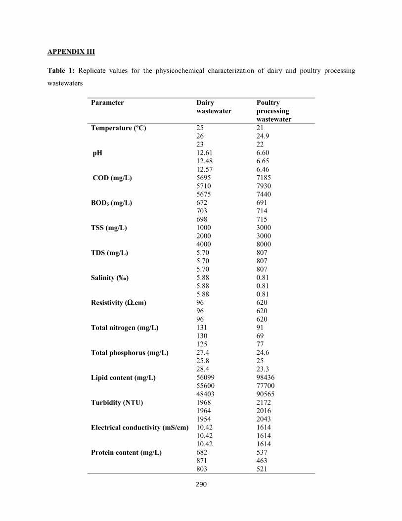
290
APPENDIX III
Table 1: Replicate values for the physicochemical characterization of dairy and poultry processing
wastewaters
Parameter Dairy wastewater
Poultry processing wastewater
Temperature (ºC) 25 21 26 24.9 23 22
pH 12.61 6.60 12.48 6.65 12.57 6.46
COD (mg/L) 5695 7185 5710 7930 5675 7440
BOD5 (mg/L) 672 691 703 714 698 715
TSS (mg/L) 1000 3000 2000 3000 4000 8000
TDS (mg/L) 5.70 807 5.70 807 5.70 807
Salinity (‰) 5.88 0.81 5.88 0.81 5.88 0.81
Resistivity (Ω.cm) 96 620 96 620 96 620
Total nitrogen (mg/L) 131 91 130 69 125 77
Total phosphorus (mg/L) 27.4 24.6 25.8 25 28.4 23.3
Lipid content (mg/L) 56099 98436 55600 77700 48403 90565
Turbidity (NTU) 1968 2172 1964 2016 1954 2043
Electrical conductivity (mS/cm) 10.42 1614 10.42 1614 10.42 1614
Protein content (mg/L) 682 537 871 463 803 521

291