Embed Size (px)
Citation preview
Treatment of DVT
Michael Miller, Jr., MD
Instructor
Duke University Medical Center
Friday, May 22, 2009
Multidisciplinary Vascular Conference

Course Objectives
• DVT Incidence and Etiology
• Imaging.
• Anticoagulation
• Systemic Thrombolysis
• Catheter Directed Thrombolysis
• To Filter or Not to Filter? That is the question.
• Mechanical Thrombectomy Devices.
• Venous Intervention.
Incidence
• Two million people in the U.S affected by
DVT.
• Over 250 thousand new cases per year.
• Iliofemoral DVT represents approximately
20% of new cases.
NIH Consensus Conf JAMA 1986
Anderson et al. Arch Intern Med 1991; 151:933-38
Pathogenesis
• Virchow’s Triad:
– Endothelial Injury.
– Abnormal Blood Flow (Stasis).
– Hypercoagulability.
• Extension of Calf Vein thrombosis
– 10-29%
Risk Factors
• Postoperative patient
• Orthopedic Surgery
• Old Age
• Malignancy
• Pregnancy / Post Partum
• Spinal Injury
• Birth Control Pills
• Obesity
• Intravenous Foreign Body
• Prior DVT
• Cardiac Disease
• Immobilization
• Leg Trauma
• Blood Coagulation Disorder

Course Objectives
• DVT Incidence and Etiology
• Imaging.
• Anticoagulation
• Systemic Thrombolysis
• Catheter Directed Thrombolysis
• To Filter or Not to Filter? That is the question.
• Mechanical Thrombectomy Devices.
• Venous Intervention.
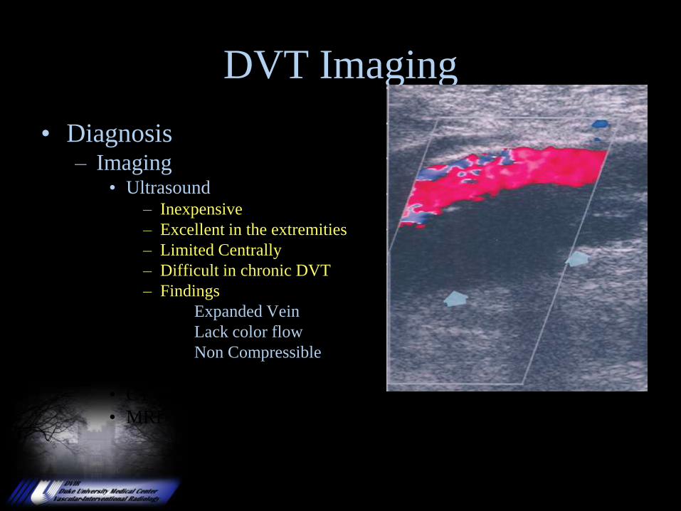
DVT Imaging
• Diagnosis– Imaging
• Ultrasound– Inexpensive
– Excellent in the extremities
– Limited Centrally
– Difficult in chronic DVT
– Findings
Expanded Vein
Lack color flow
Non Compressible
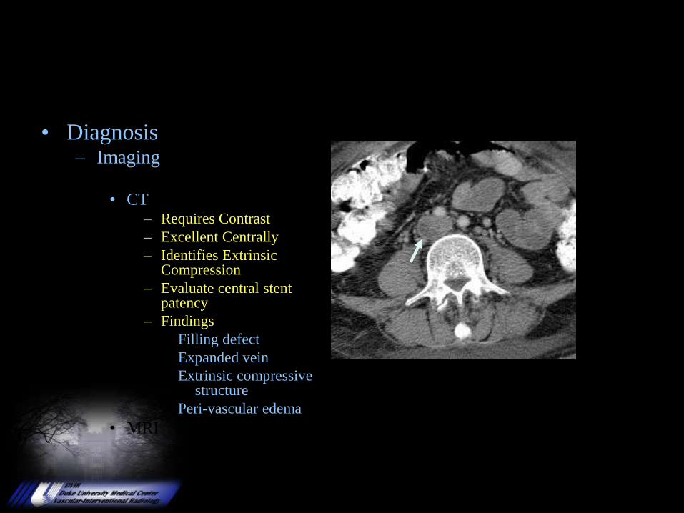
• CT
• MRI
• Diagnosis– Imaging
• Ultrasound
• CT– Requires Contrast
– Excellent Centrally
– Identifies Extrinsic Compression
– Evaluate central stent patency
– Findings
Filling defect
Expanded vein
Extrinsic compressive structure
Peri-vascular edema
• MRI
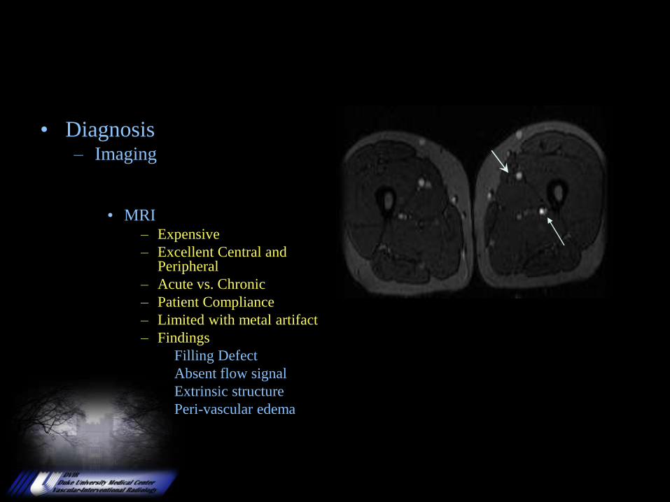
DVT Imaging
• Diagnosis– Imaging
• Ultrasound
• CT
• MRI– Expensive
– Excellent Central and Peripheral
– Acute vs. Chronic
– Patient Compliance
– Limited with metal artifact
– Findings
Filling Defect
Absent flow signal
Extrinsic structure
Peri-vascular edema
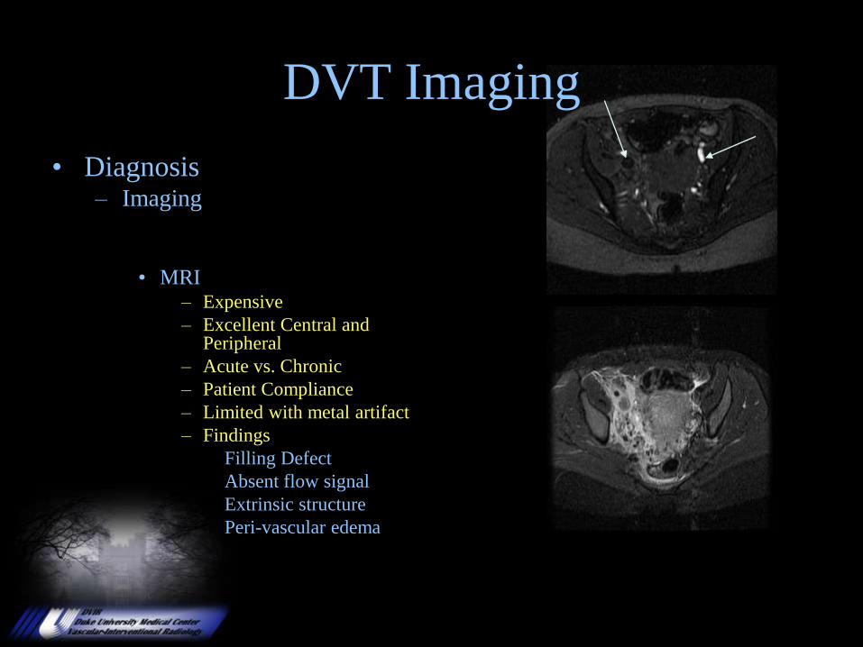
DVT Imaging
• Diagnosis– Imaging
• Ultrasound
• CT
• MRI– Expensive
– Excellent Central and Peripheral
– Acute vs. Chronic
– Patient Compliance
– Limited with metal artifact
– Findings
Filling Defect
Absent flow signal
Extrinsic structure
Peri-vascular edema
DVT Imaging
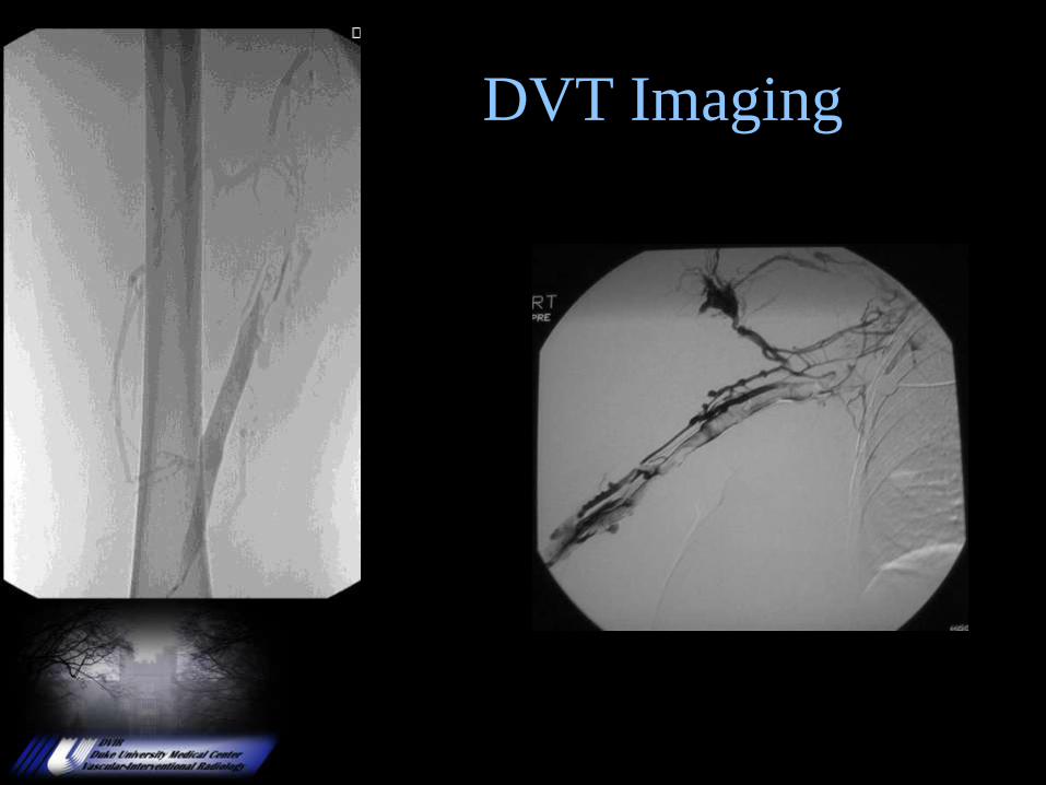
DVT Imaging
Venogram

Course Objectives
• DVT Incidence and Etiology
• Imaging.
• Anticoagulation
• Systemic Thrombolysis
• Catheter Directed Thrombolysis
• To Filter or Not to Filter? That is the question.
• Mechanical Thrombectomy Devices.
• Venous Intervention.
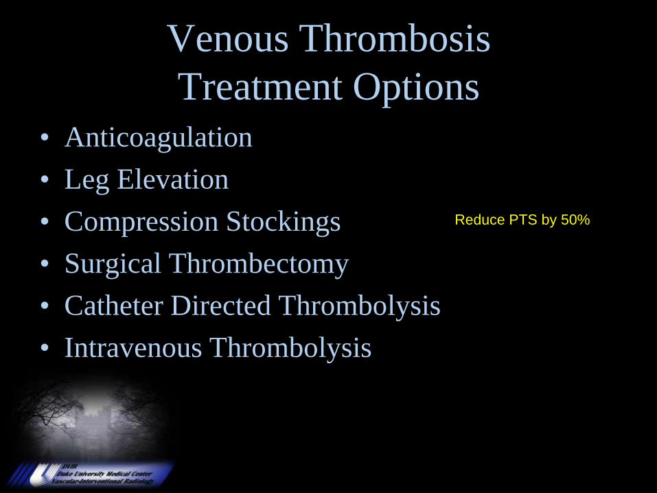
• Anticoagulation
• Leg Elevation
• Compression Stockings
• Surgical Thrombectomy
• Catheter Directed Thrombolysis
• Intravenous Thrombolysis
Venous Thrombosis
Treatment Options
Reduce PTS by 50%
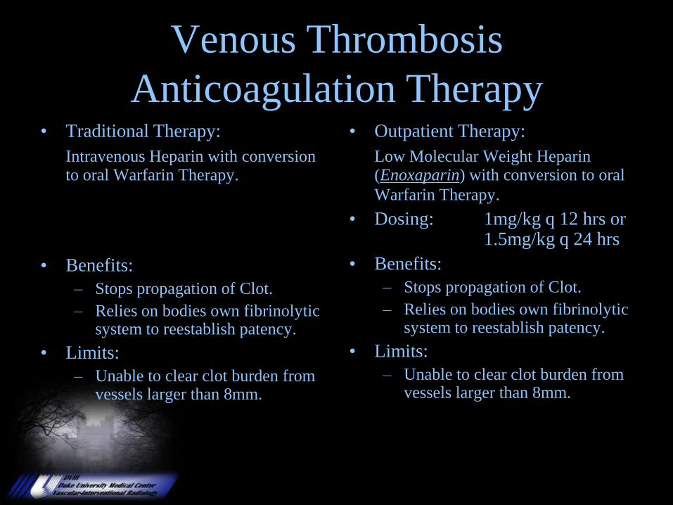
Venous Thrombosis
Anticoagulation Therapy• Traditional Therapy:
Intravenous Heparin with conversion to oral Warfarin Therapy.
• Benefits:
– Stops propagation of Clot.
– Relies on bodies own fibrinolytic system to reestablish patency.
• Limits:
– Unable to clear clot burden from vessels larger than 8mm.
• Outpatient Therapy:
Low Molecular Weight Heparin (Enoxaparin) with conversion to oral
Warfarin Therapy.
• Dosing: 1mg/kg q 12 hrs or 1.5mg/kg q 24 hrs
• Benefits:
– Stops propagation of Clot.
– Relies on bodies own fibrinolytic system to reestablish patency.
• Limits:
– Unable to clear clot burden from vessels larger than 8mm.
Venous Thrombosis
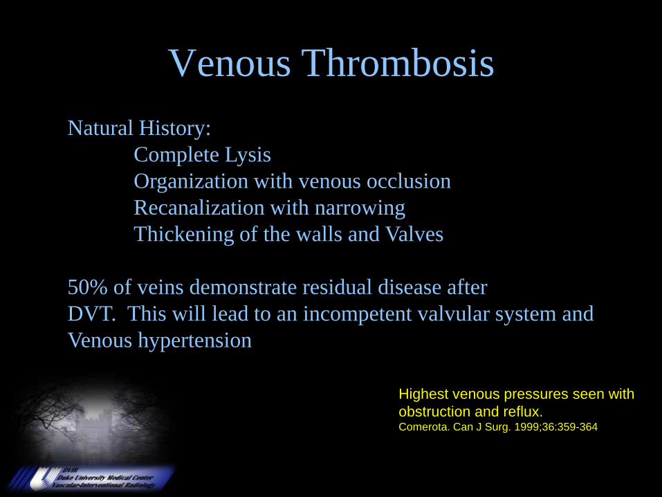
Natural History:
Complete Lysis
Organization with venous occlusion
Recanalization with narrowing
Thickening of the walls and Valves
50% of veins demonstrate residual disease after
DVT. This will lead to an incompetent valvular system and
Venous hypertension
Highest venous pressures seen with
obstruction and reflux.Comerota. Can J Surg. 1999;36:359-364
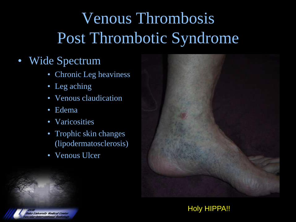
Venous Thrombosis
Post Thrombotic Syndrome
• Wide Spectrum• Chronic Leg heaviness
• Leg aching
• Venous claudication
• Edema
• Varicosities
• Trophic skin changes
(lipodermatosclerosis)
• Venous Ulcer
Holy HIPPA!!
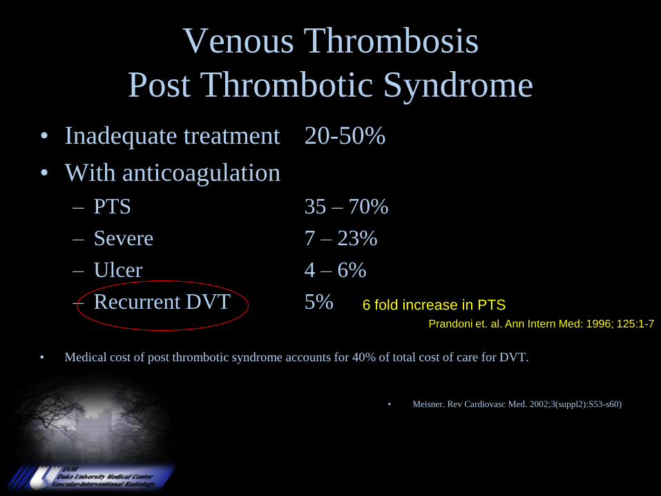
• Inadequate treatment 20-50%
• With anticoagulation
– PTS 35 – 70%
– Severe 7 – 23%
– Ulcer 4 – 6%
– Recurrent DVT 5%
• Medical cost of post thrombotic syndrome accounts for 40% of total cost of care for DVT.
• Meisner. Rev Cardiovasc Med. 2002;3(suppl2):S53-s60)
Venous Thrombosis
Post Thrombotic Syndrome
6 fold increase in PTS
Prandoni et. al. Ann Intern Med: 1996; 125:1-7
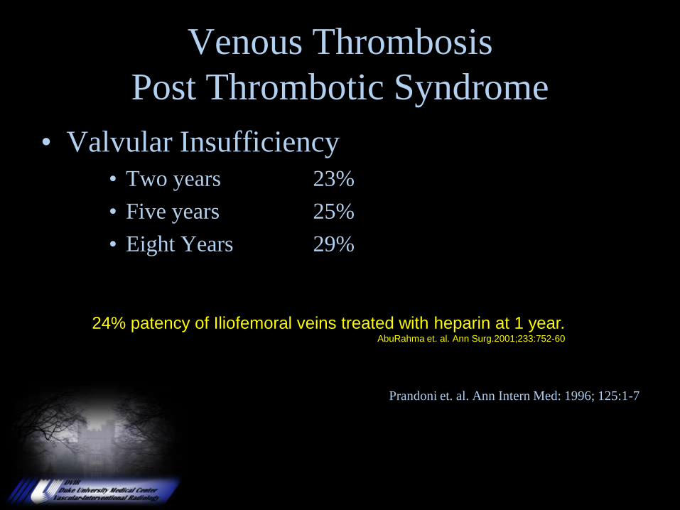
• Valvular Insufficiency• Two years 23%
• Five years 25%
• Eight Years 29%
Prandoni et. al. Ann Intern Med: 1996; 125:1-7
Venous Thrombosis
Post Thrombotic Syndrome
24% patency of Iliofemoral veins treated with heparin at 1 year.AbuRahma et. al. Ann Surg.2001;233:752-60

Course Objectives
• DVT Incidence and Etiology
• Imaging.
• Anticoagulation
• Systemic Thrombolysis
• Catheter Directed Thrombolysis
• To Filter or Not to Filter? That is the question.
• Mechanical Thrombectomy Devices.
• Venous Intervention.
Venous Thrombosis
Thrombolysis
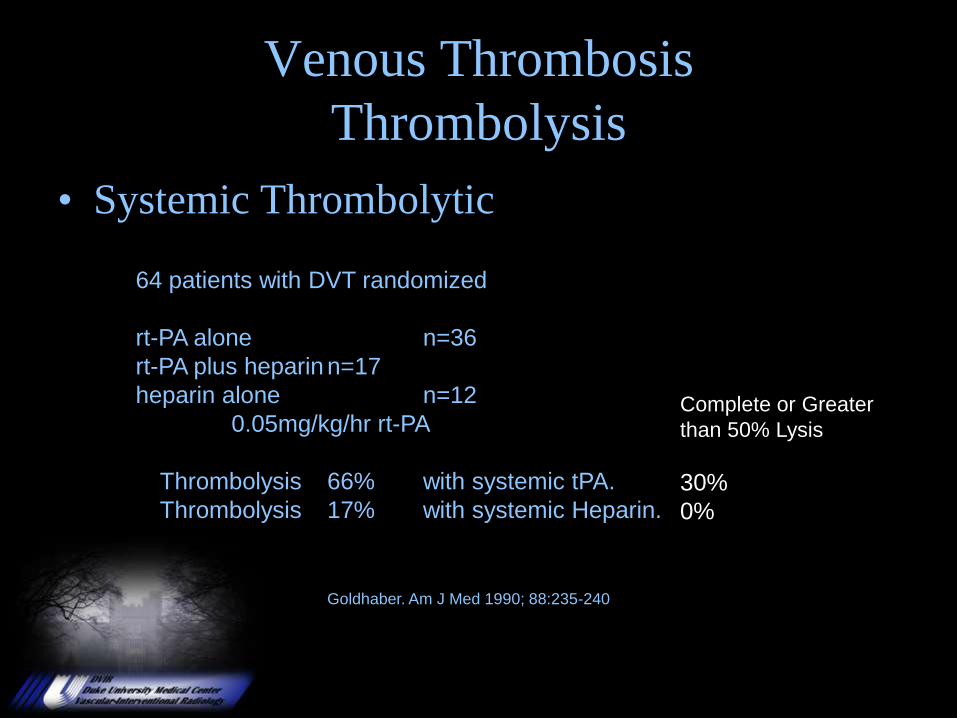
• Systemic Thrombolytic
64 patients with DVT randomized
rt-PA alone n=36
rt-PA plus heparin n=17
heparin alone n=12
0.05mg/kg/hr rt-PA
Thrombolysis 66% with systemic tPA.
Thrombolysis 17% with systemic Heparin.
Goldhaber. Am J Med 1990; 88:235-240
Complete or Greater
than 50% Lysis
30%
0%

Course Objectives
• DVT Incidence and Etiology
• Imaging.
• Anticoagulation
• Systemic Thrombolysis
• Catheter Directed Thrombolysis
• To Filter or Not to Filter? That is the question.
• Mechanical Thrombectomy Devices.
• Venous Intervention.
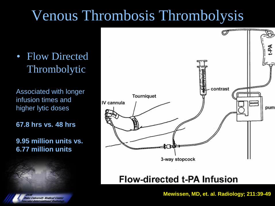
Venous Thrombosis Thrombolysis
• Flow Directed
Thrombolytic
Associated with longer
infusion times and
higher lytic doses
67.8 hrs vs. 48 hrs
9.95 million units vs.
6.77 million units
Mewissen, MD, et. al. Radiology; 211:39-49
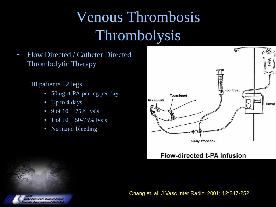
Venous Thrombosis
Thrombolysis• Flow Directed / Catheter Directed
Thrombolytic Therapy
10 patients 12 legs
• 50mg rt-PA per leg per day
• Up to 4 days
• 9 of 10 >75% lysis
• 1 of 10 50-75% lysis
• No major bleeding
Chang et. al. J Vasc Inter Radiol 2001; 12:247-252
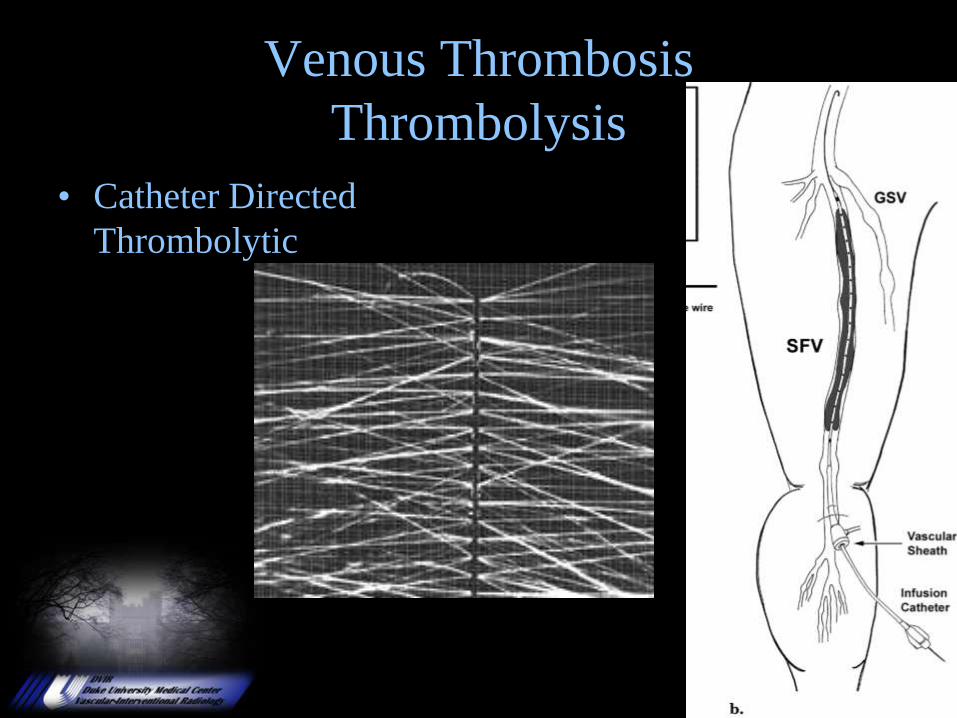
Venous Thrombosis
Thrombolysis
• Catheter Directed
Thrombolytic
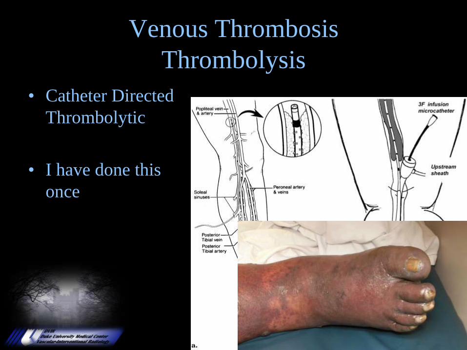
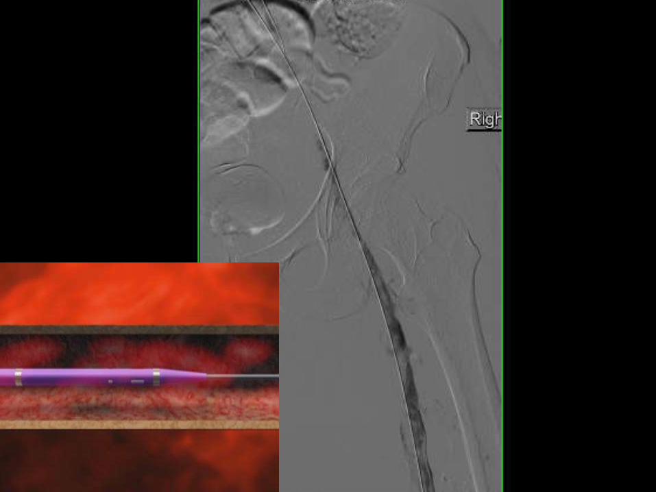
Venous Thrombosis
Thrombolysis
• Catheter Directed
Thrombolytic
• I have done this
once
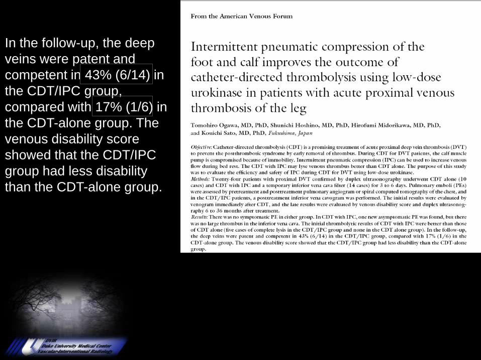
In the follow-up, the deep
veins were patent and
competent in 43% (6/14) in
the CDT/IPC group,
compared with 17% (1/6) in
the CDT-alone group. The
venous disability score
showed that the CDT/IPC
group had less disability
than the CDT-alone group.
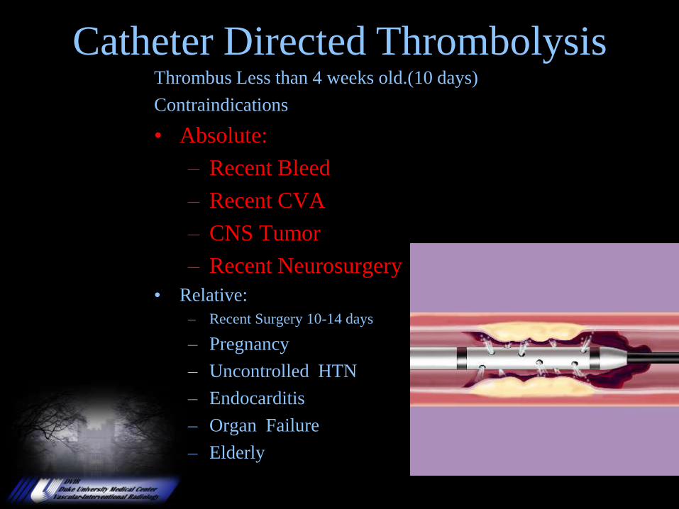
Thrombus Less than 4 weeks old.(10 days)
Contraindications
• Absolute:
– Recent Bleed
– Recent CVA
– CNS Tumor
– Recent Neurosurgery
• Relative:
– Recent Surgery 10-14 days
– Pregnancy
– Uncontrolled HTN
– Endocarditis
– Organ Failure
– Elderly
Catheter Directed Thrombolysis
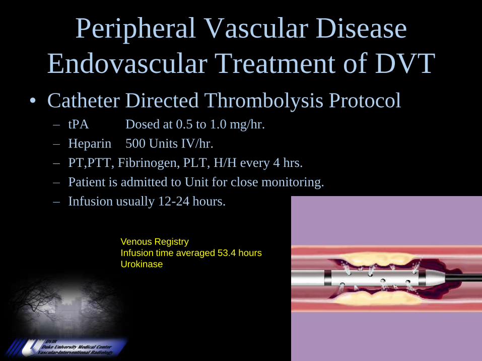
Peripheral Vascular Disease
Endovascular Treatment of DVT
• Catheter Directed Thrombolysis Protocol– tPA Dosed at 0.5 to 1.0 mg/hr.
– Heparin 500 Units IV/hr.
– PT,PTT, Fibrinogen, PLT, H/H every 4 hrs.
– Patient is admitted to Unit for close monitoring.
– Infusion usually 12-24 hours.
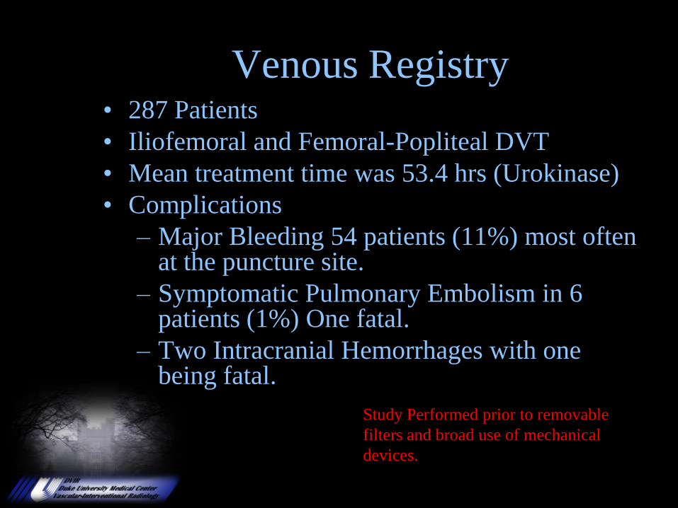
Venous Registry
Infusion time averaged 53.4 hours
Urokinase
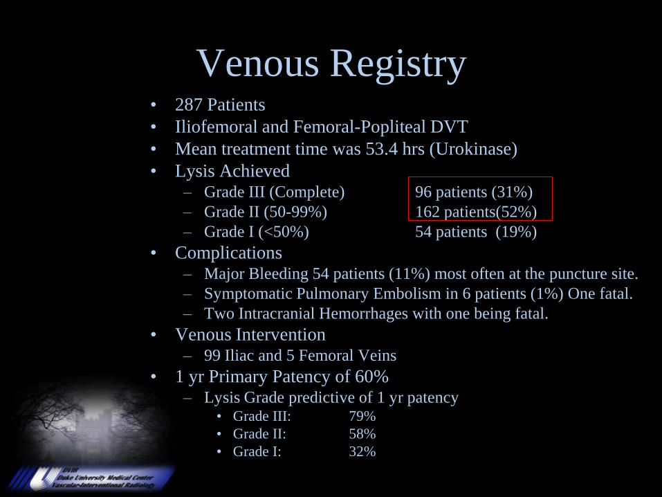
Venous Registry• 287 Patients
• Iliofemoral and Femoral-Popliteal DVT
• Mean treatment time was 53.4 hrs (Urokinase)
• Lysis Achieved– Grade III (Complete) 96 patients (31%)
– Grade II (50-99%) 162 patients(52%)
– Grade I (<50%) 54 patients (19%)
• Complications– Major Bleeding 54 patients (11%) most often at the puncture site.
– Symptomatic Pulmonary Embolism in 6 patients (1%) One fatal.
– Two Intracranial Hemorrhages with one being fatal.
• Venous Intervention – 99 Iliac and 5 Femoral Veins
• 1 yr Primary Patency of 60%– Lysis Grade predictive of 1 yr patency
• Grade III: 79%
• Grade II: 58%
• Grade I: 32%
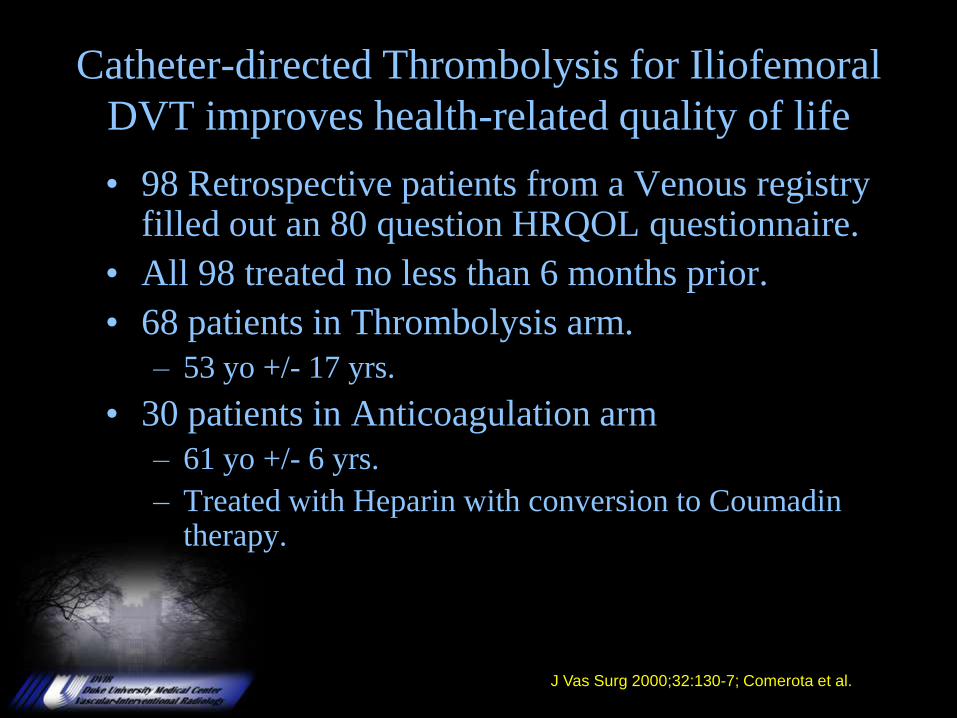
Catheter-directed Thrombolysis for Iliofemoral
DVT improves health-related quality of life
• 98 Retrospective patients from a Venous registry filled out an 80 question HRQOL questionnaire.
• All 98 treated no less than 6 months prior.
• 68 patients in Thrombolysis arm.
– 53 yo +/- 17 yrs.
• 30 patients in Anticoagulation arm
– 61 yo +/- 6 yrs.
– Treated with Heparin with conversion to Coumadin therapy.
J Vas Surg 2000;32:130-7; Comerota et al.
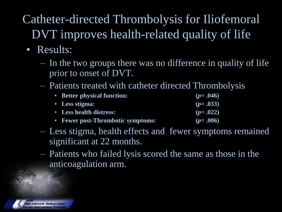
• Results:
– In the two groups there was no difference in quality of life prior to onset of DVT.
– Patients treated with catheter directed Thrombolysis• Better physical function: (p= .046)
• Less stigma: (p= .033)
• Less health distress: (p= .022)
• Fewer post-Thrombotic symptoms: (p= .006)
– Less stigma, health effects and fewer symptoms remained significant at 22 months.
– Patients who failed lysis scored the same as those in the anticoagulation arm.
Catheter-directed Thrombolysis for Iliofemoral
DVT improves health-related quality of life
•J Vas Surg 2000;32:130-7; Comerota et al.
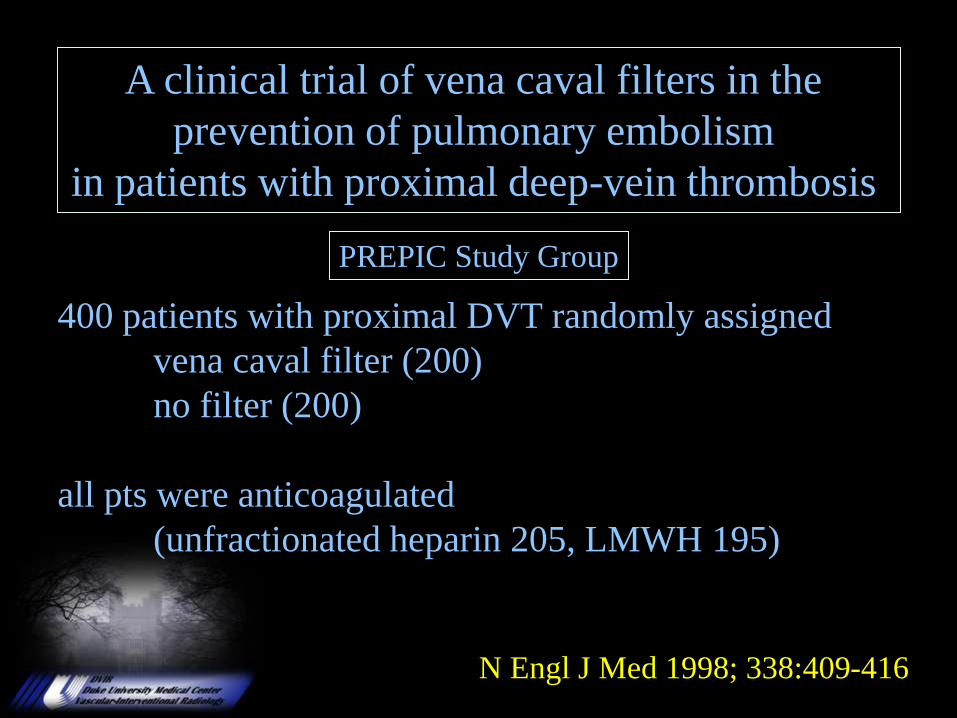
A clinical trial of vena caval filters in the
prevention of pulmonary embolism
in patients with proximal deep-vein thrombosis
PREPIC Study Group
N Engl J Med 1998; 338:409-416
400 patients with proximal DVT randomly assigned
vena caval filter (200)
no filter (200)
all pts were anticoagulated
(unfractionated heparin 205, LMWH 195)
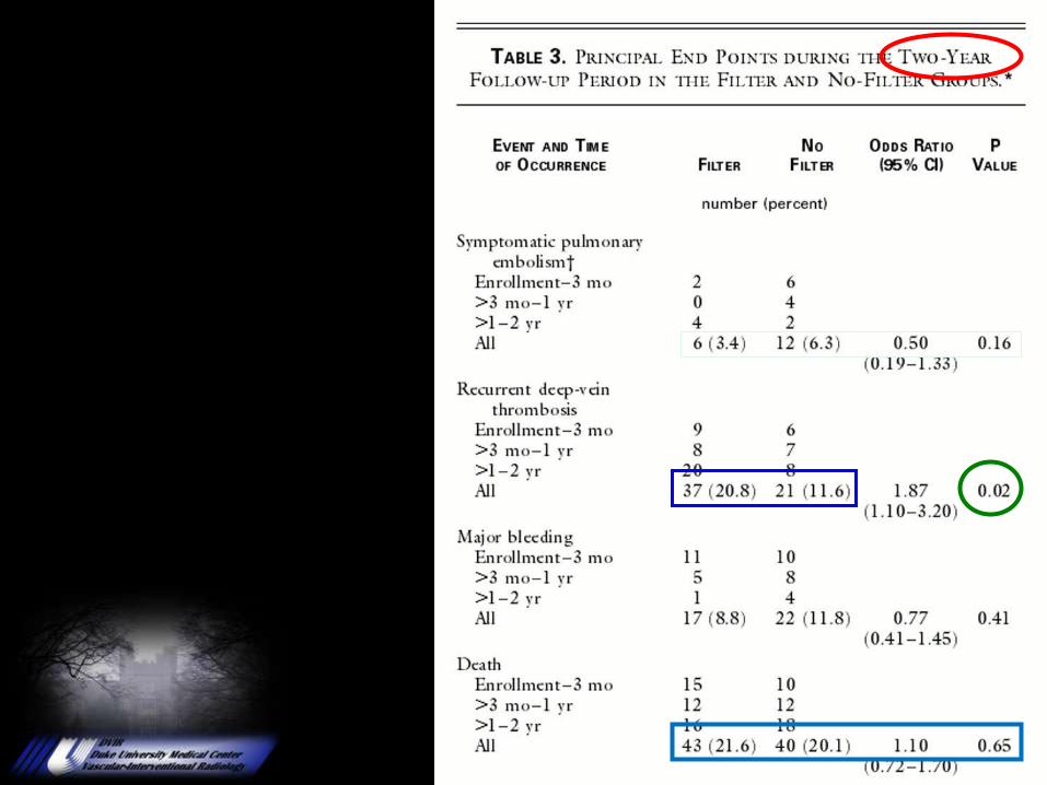
Follow up
Visits @ 3 mo, 1 yr
Telephone 2 yrs

Course Objectives
• DVT Incidence and Etiology
• Imaging.
• Anticoagulation
• Systemic Thrombolysis
• Catheter Directed Thrombolysis
• To Filter or Not to Filter? That is the question.
• Mechanical Thrombectomy Devices.
• Venous Intervention.
Venous Registry• 287 Patients
• Iliofemoral and Femoral-Popliteal DVT
• Mean treatment time was 53.4 hrs (Urokinase)
• Complications
– Major Bleeding 54 patients (11%) most often at the puncture site.
– Symptomatic Pulmonary Embolism in 6 patients (1%) One fatal.
– Two Intracranial Hemorrhages with one being fatal.
Study Performed prior to removable
filters and broad use of mechanical
devices.
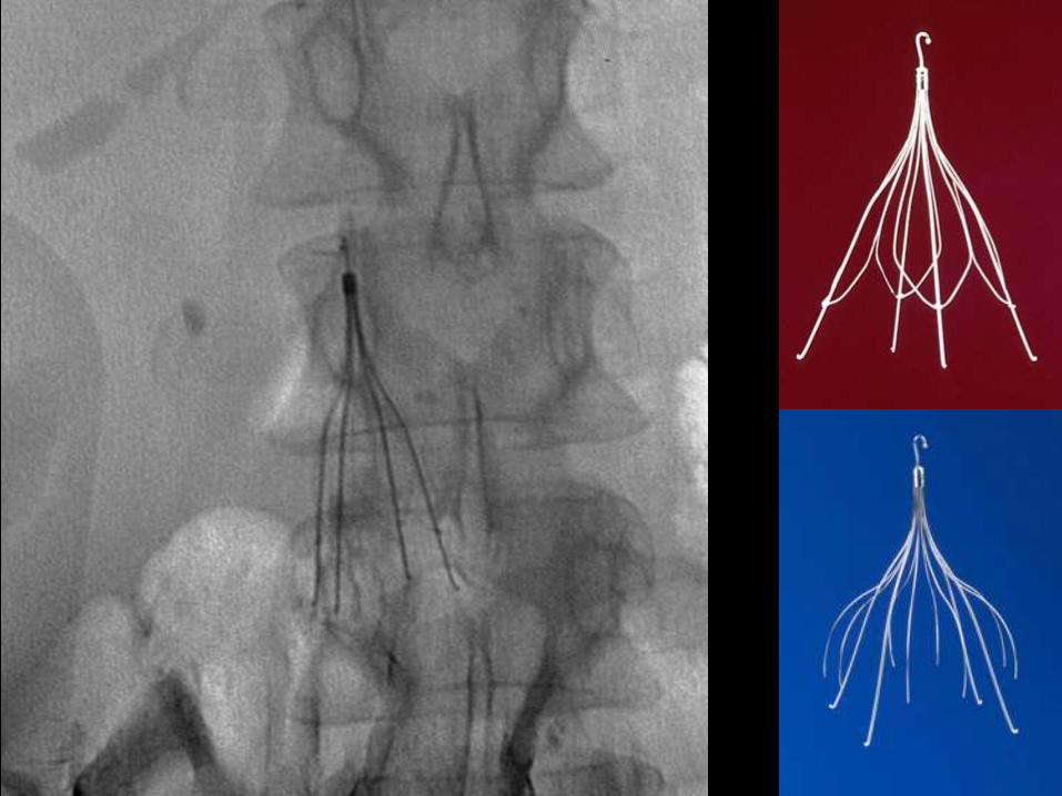
• Gunther Tulip
– 28mm Cava
– Jugular Removal
– 10 days
– Reported 2 to 126 days
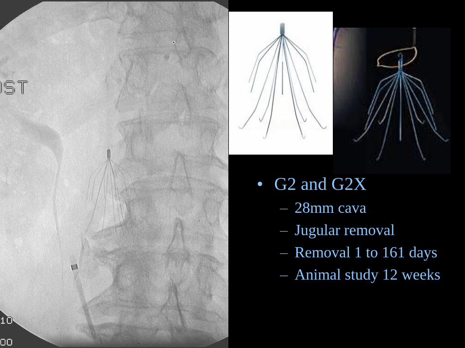
• G2 and G2X
– 28mm cava
– Jugular removal
– Removal 1 to 161 days
– Animal study 12 weeks
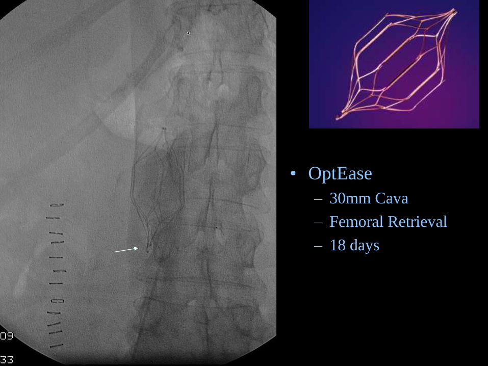
• OptEase
– 30mm Cava
– Femoral Retrieval
– 18 days
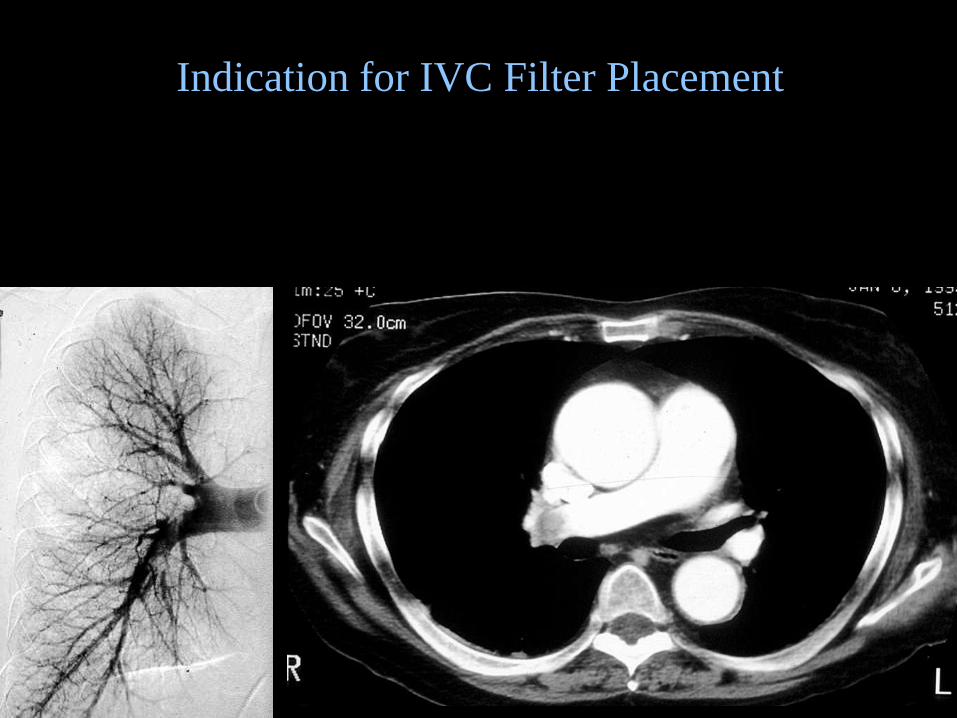
Indication for IVC Filter Placement

To Quote Tony Smith
Where are all the Bodies?
Course Objectives
• DVT Incidence and Etiology
• Imaging.
• Anticoagulation
• Systemic Thrombolysis
• Catheter Directed Thrombolysis
• To Filter or Not to Filter? That is the question.
• Mechanical Thrombectomy Devices.
• Venous Intervention.
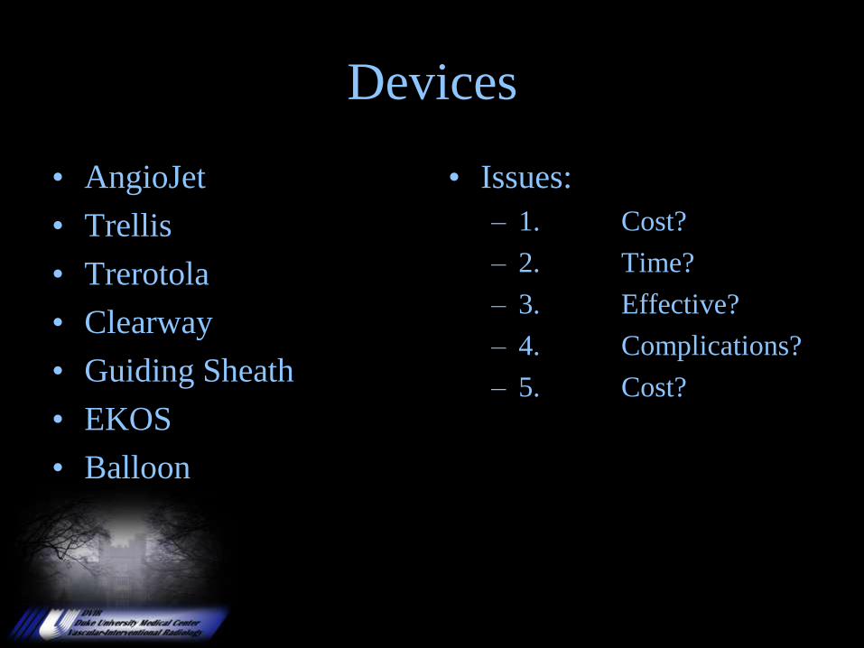
Devices
• AngioJet
• Trellis
• Trerotola
• Clearway
• Guiding Sheath
• EKOS
• Balloon
• Issues:
– 1. Cost?
– 2. Time?
– 3. Effective?
– 4. Complications?
– 5. Cost?
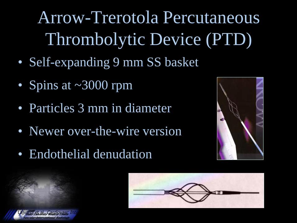
Lajvardi et al. Cardiovasc Intervent Radiol. 1995;18:172–178.
Arrow-Trerotola Percutaneous
Thrombolytic Device (PTD)
• Self-expanding 9 mm SS basket
• Spins at ~3000 rpm
• Particles 3 mm in diameter
• Newer over-the-wire version
• Endothelial denudation
Pharmacological
and
Mechanical
Thrombolysis
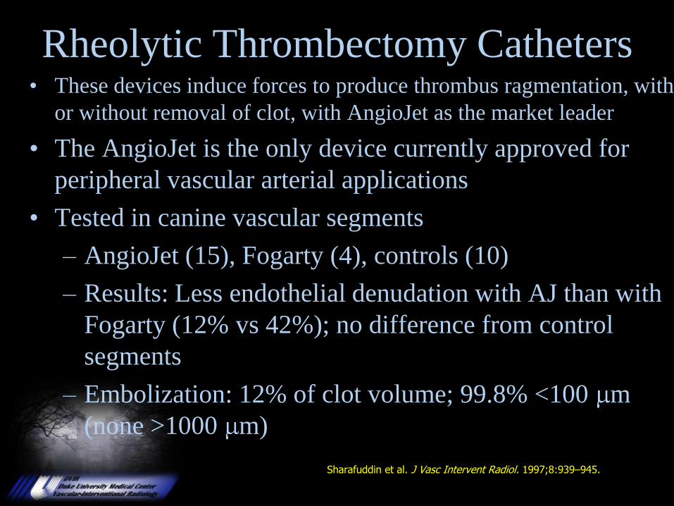
Sharafuddin et al. J Vasc Intervent Radiol. 1997;8:939–945.
Rheolytic Thrombectomy Catheters• These devices induce forces to produce thrombus ragmentation, with
or without removal of clot, with AngioJet as the market leader
• The AngioJet is the only device currently approved for
peripheral vascular arterial applications
• Tested in canine vascular segments
– AngioJet (15), Fogarty (4), controls (10)
– Results: Less endothelial denudation with AJ than with
Fogarty (12% vs 42%); no difference from control
segments
– Embolization: 12% of clot volume; 99.8% <100 m
(none >1000 m)
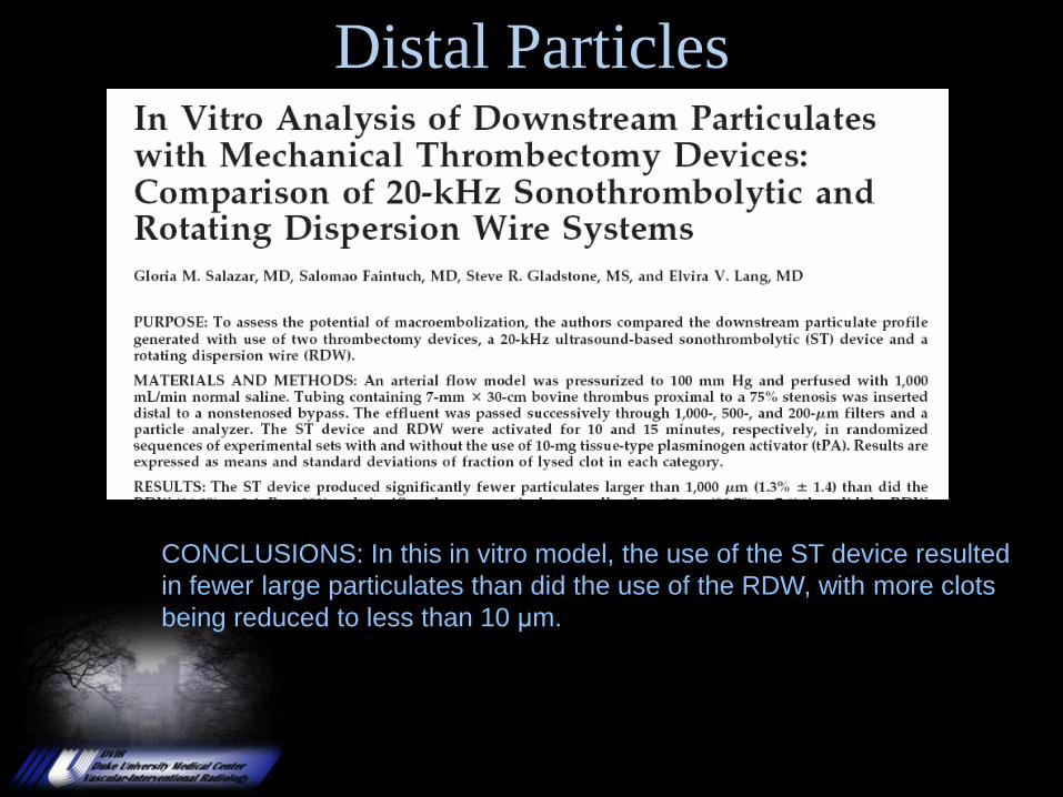
Distal Particles
CONCLUSIONS: In this in vitro model, the use of the ST device resulted
in fewer large particulates than did the use of the RDW, with more clots
being reduced to less than 10 μm.
Clinical Experience: Mechanical
Thrombectomy for DVT
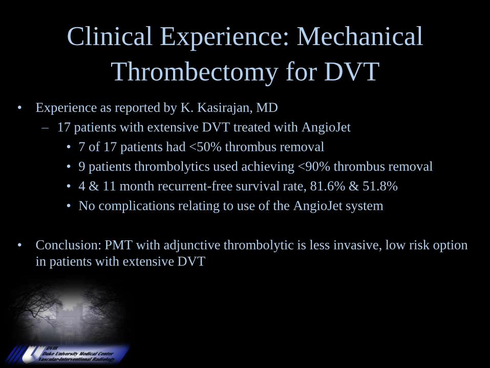
• Experience as reported by K. Kasirajan, MD
– 17 patients with extensive DVT treated with AngioJet
• 7 of 17 patients had <50% thrombus removal
• 9 patients thrombolytics used achieving <90% thrombus removal
• 4 & 11 month recurrent-free survival rate, 81.6% & 51.8%
• No complications relating to use of the AngioJet system
• Conclusion: PMT with adjunctive thrombolytic is less invasive, low risk option
in patients with extensive DVT
Kasirajan K, Gray B, Ouriel K, Jvasc Inter Radiol 2001 Feb;12(2):179-85
Clinical Experience: Mechanical
Thrombectomy for DVT
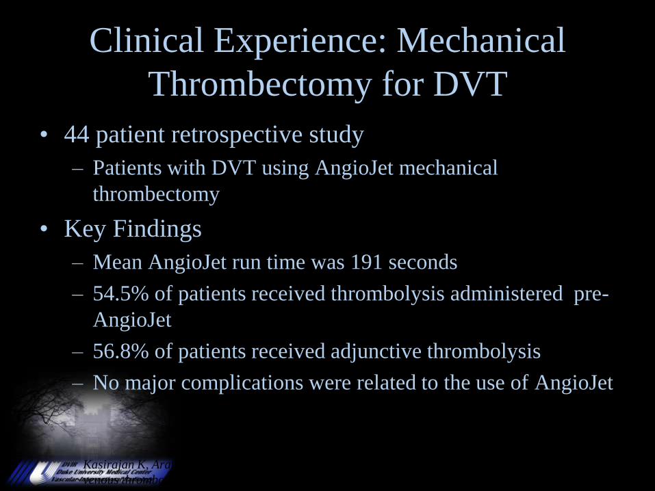
• 44 patient retrospective study
– Patients with DVT using AngioJet mechanical
thrombectomy
• Key Findings
– Mean AngioJet run time was 191 seconds
– 54.5% of patients received thrombolysis administered pre-
AngioJet
– 56.8% of patients received adjunctive thrombolysis
– No major complications were related to the use of AngioJet
Kasirajan K, Arata M, Swischuk S, Hunter D, Cazenave C, Rheolytic thrombectomy for management of
venous thrombosis: Results of a multicenter venous registry. J Vasc Interven Radiol 2003: 14: S16
Clinical Experience: Mechanical
Thrombectomy for DVT
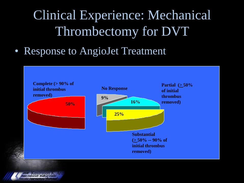
Complete (> 90% of
initial thrombus
removed)
Substantial
(> 50% -- 90% of
initial thrombus
removed)
Partial (> 50%
of initial
thrombus
removed)
No Response
• Response to AngioJet Treatment
Kasirajan K, Arata M, Swischuk S, Hunter D, Cazenave C, Rheolytic thrombectomy for management of
venous thrombosis: Results of a multicenter venous registry. J Vasc Interven Radiol 2003: 14: S16
9%16%
25%
50%
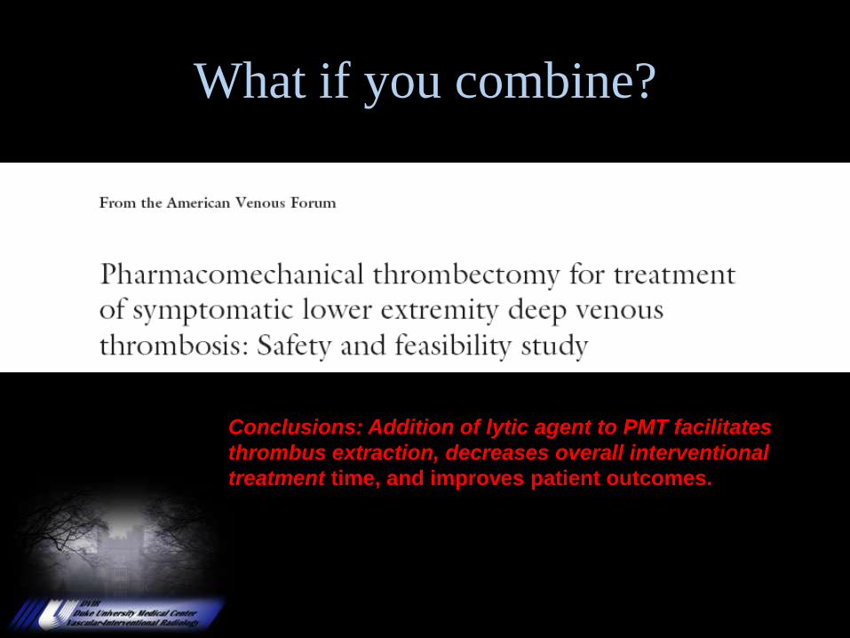
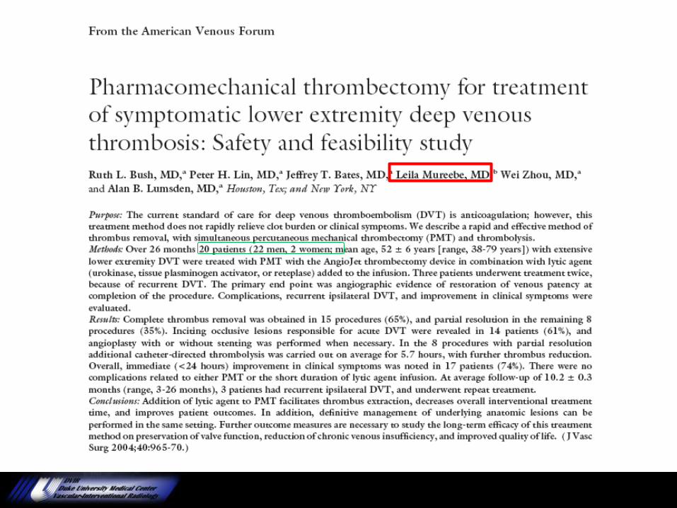
What if you combine?
Conclusions: Addition of lytic agent to PMT facilitates
thrombus extraction, decreases overall interventional
treatment time, and improves patient outcomes.
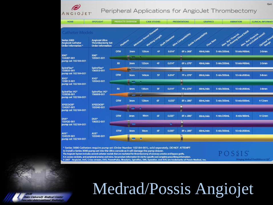
Medrad/Possis Angiojet
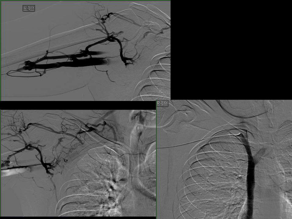
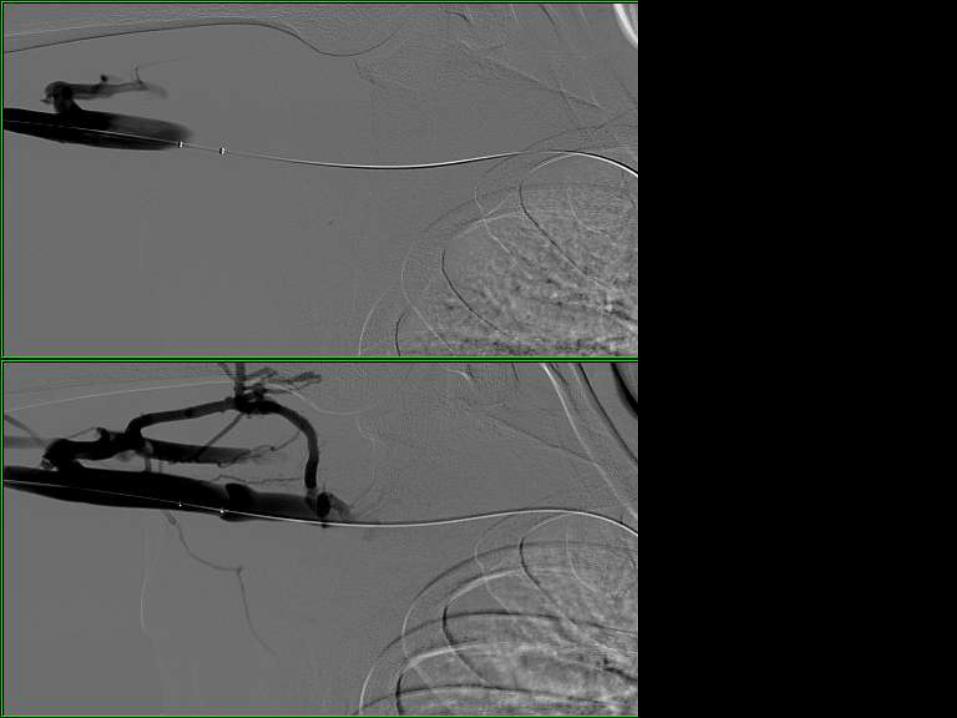
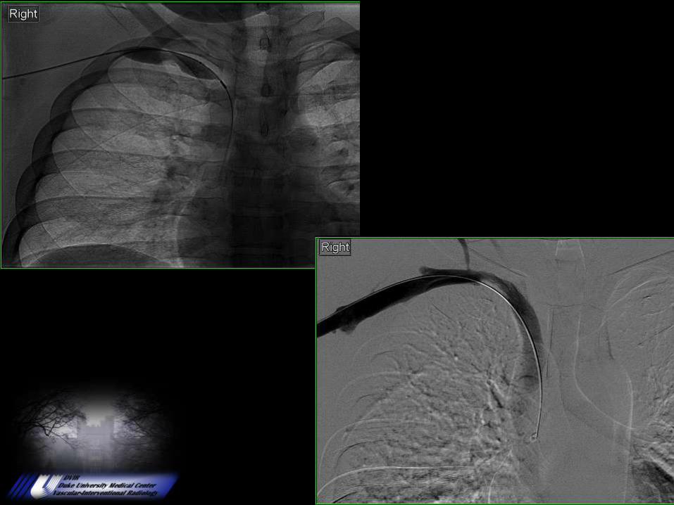
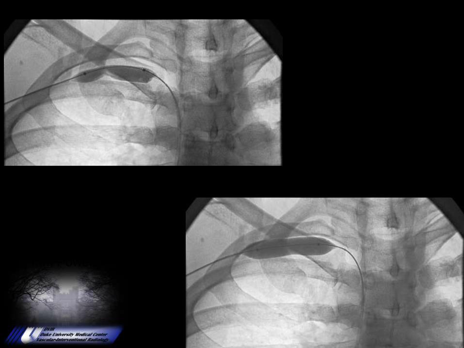
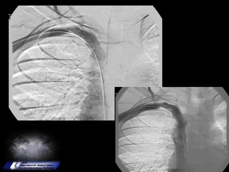
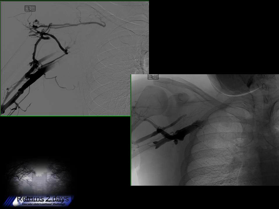
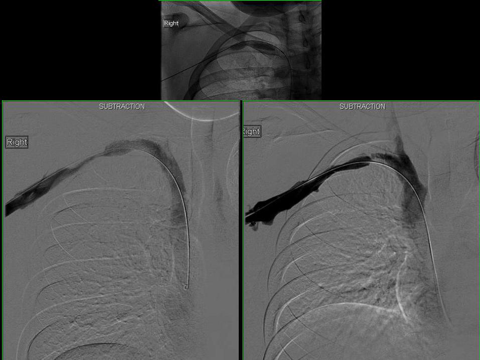
HPI
• 42 yo female
• Developed PE s/p hysterectomy for fibroids
• Heparin started
• Patient developed pelvic hematoma
• Bard G2 filter placed
Clinical Course
• Patient presented 2 wks later with SOB, abd
pain, and leg weakness
Clinical Course
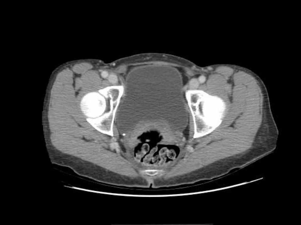
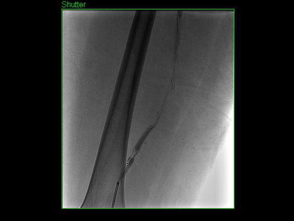
• Transferred to Duke for thrombolysis
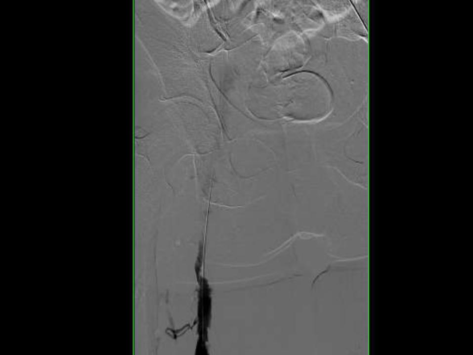
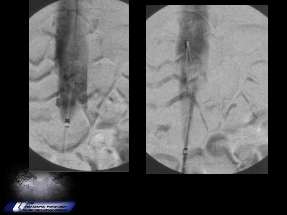
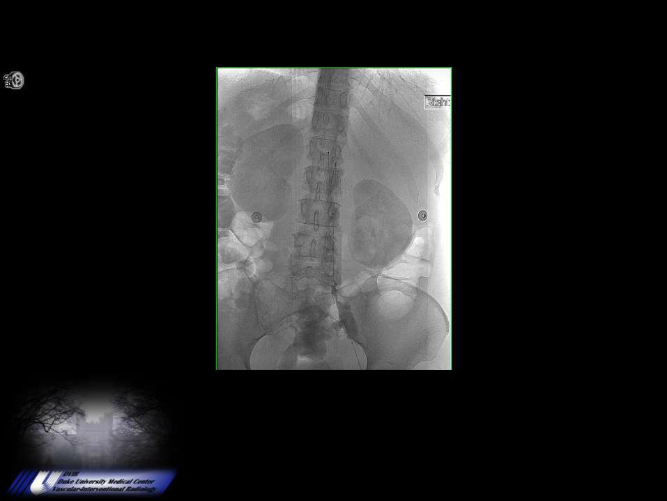
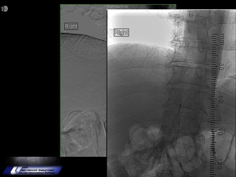
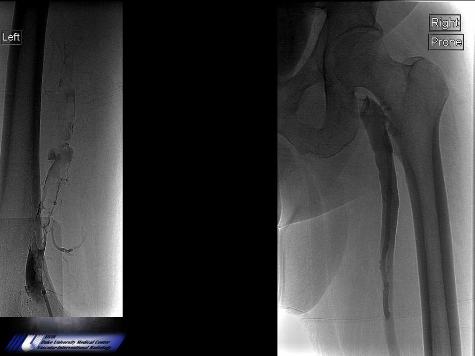
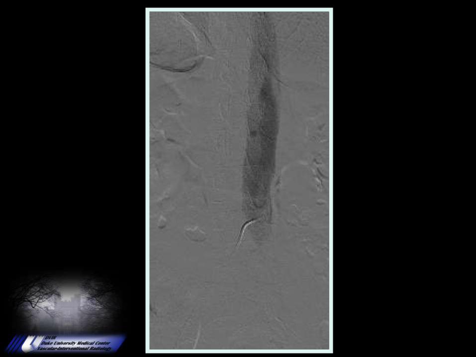
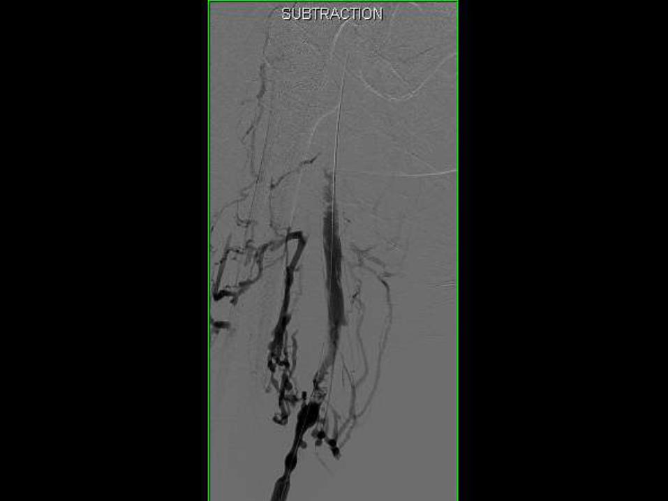
Lysis Check #1
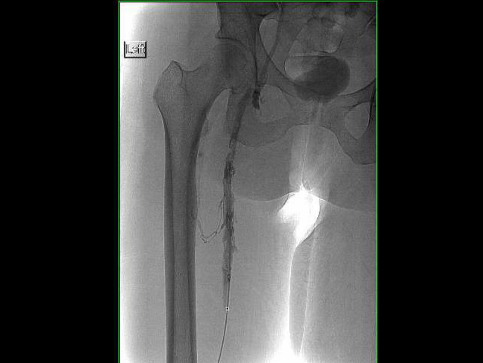
Lysis Check #1

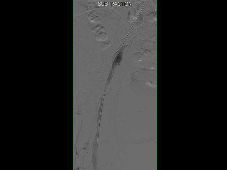
Lysis Check #4

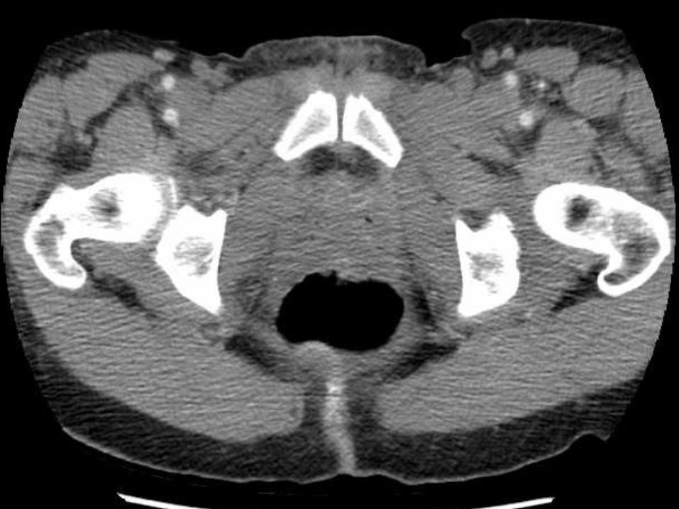
Follow up CT
3 weeks later
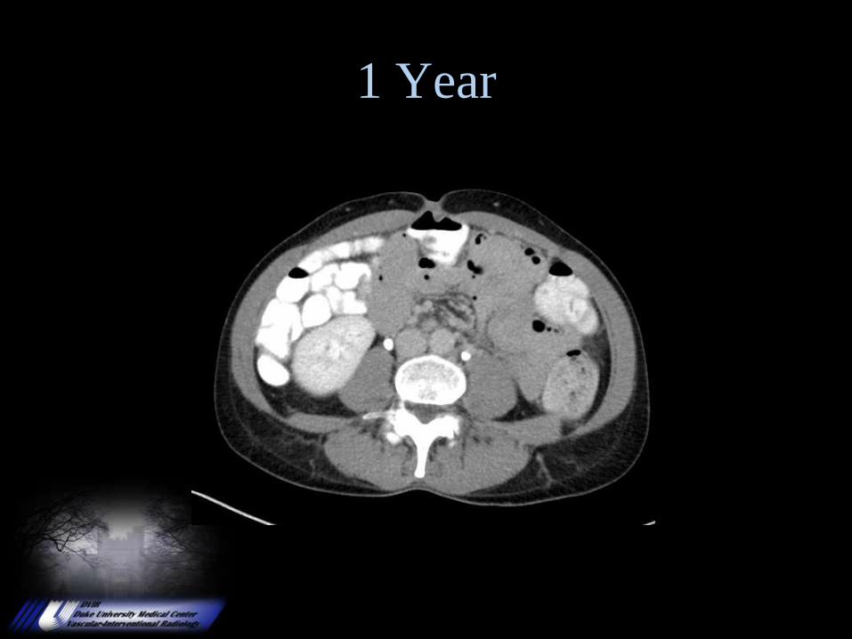
1 Year
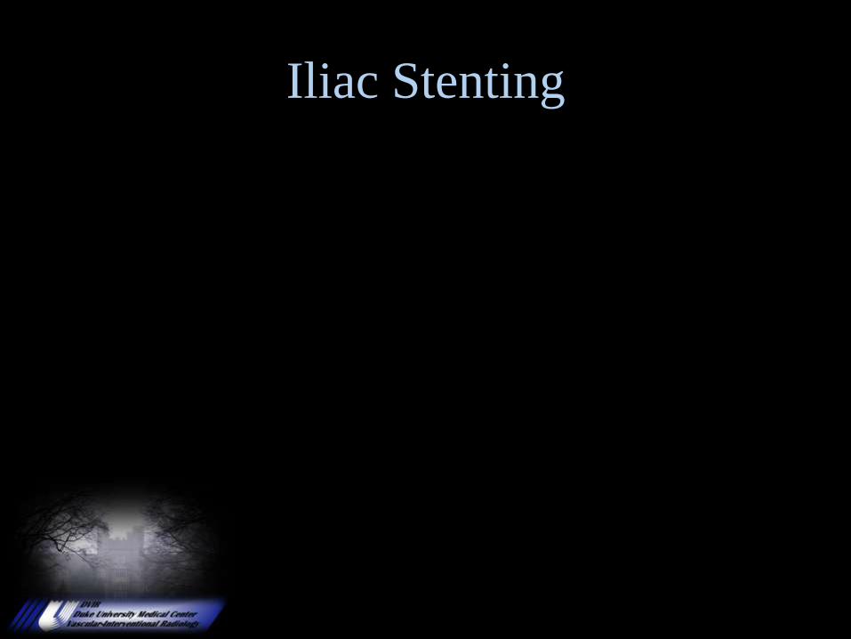
Iliac Stenting


Course Objectives
• DVT Incidence and Etiology
• Imaging.
• Anticoagulation
• Systemic Thrombolysis
• Catheter Directed Thrombolysis
• To Filter or Not to Filter? That is the question.
• Mechanical Thrombectomy Devices.
• Venous Intervention.
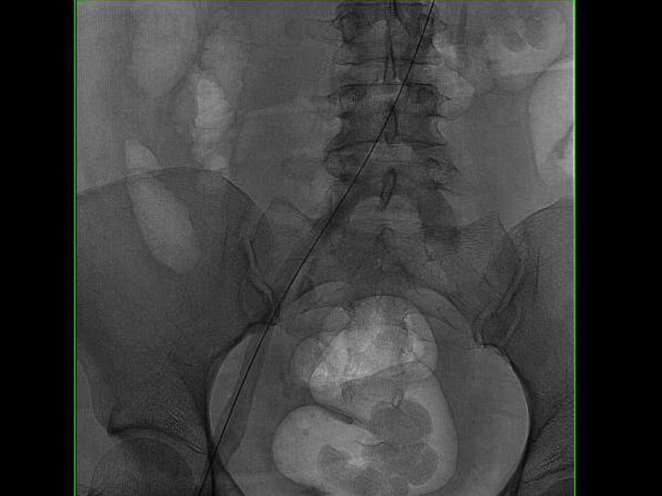
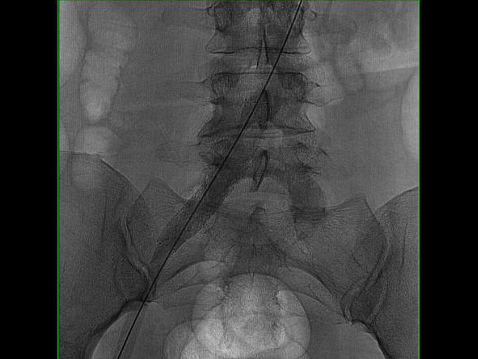
Initial PresentationHPI: 48 yo male with no significant medical history presents
with 3 day history of low back pain. Patient initially noticed a
pop and onset of pain while hanging a tire 3 days prior. Pain
was better following day, but became night before admission.
Additionally, pain in the medial right thigh. Used tramadol and
aleve with some relief. Noticed swelling in both legs day of
admission, worse on sitting.
PMH: obesity.
Surgeries: none.
FH: non contributory
SH: no tobacco, no alcohol, no drugs.
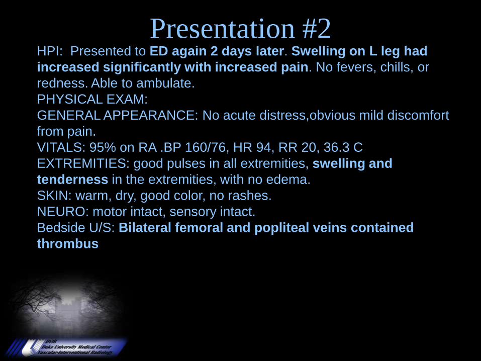
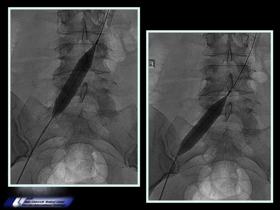
Presentation #2HPI: Presented to ED again 2 days later. Swelling on L leg had
increased significantly with increased pain. No fevers, chills, or
redness. Able to ambulate.
PHYSICAL EXAM:
GENERAL APPEARANCE: No acute distress,obvious mild discomfort
from pain.
VITALS: 95% on RA .BP 160/76, HR 94, RR 20, 36.3 C
EXTREMITIES: good pulses in all extremities, swelling and
tenderness in the extremities, with no edema.
SKIN: warm, dry, good color, no rashes.
NEURO: motor intact, sensory intact.
Bedside U/S: Bilateral femoral and popliteal veins contained
thrombus
Ultrasound
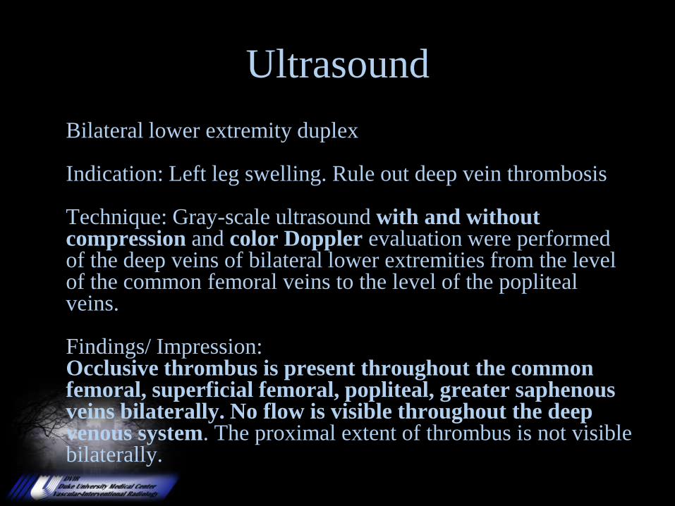
Bilateral lower extremity duplex
Indication: Left leg swelling. Rule out deep vein thrombosis
Technique: Gray-scale ultrasound with and without compression and color Doppler evaluation were performed of the deep veins of bilateral lower extremities from the level of the common femoral veins to the level of the popliteal veins.
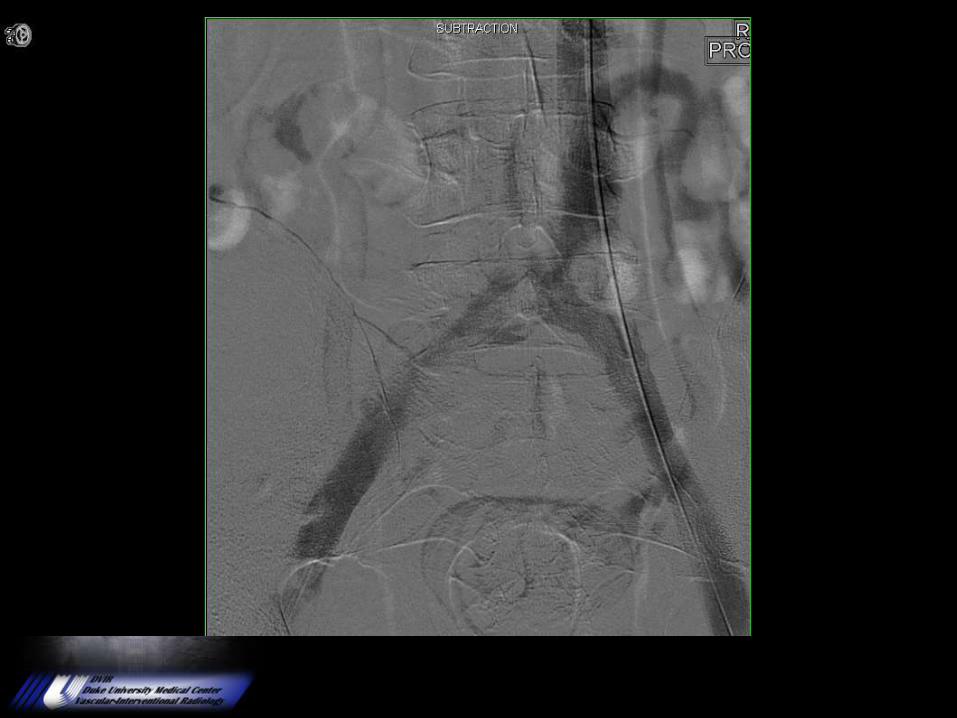
Findings/ Impression:Occlusive thrombus is present throughout the common femoral, superficial femoral, popliteal, greater saphenous veins bilaterally. No flow is visible throughout the deep venous system. The proximal extent of thrombus is not visible bilaterally.
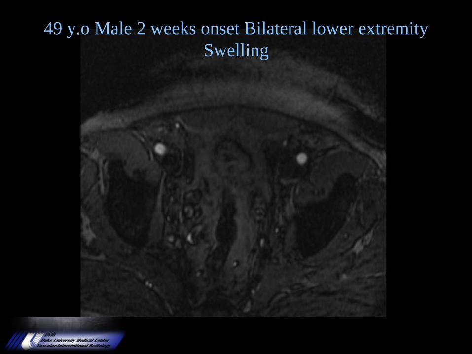
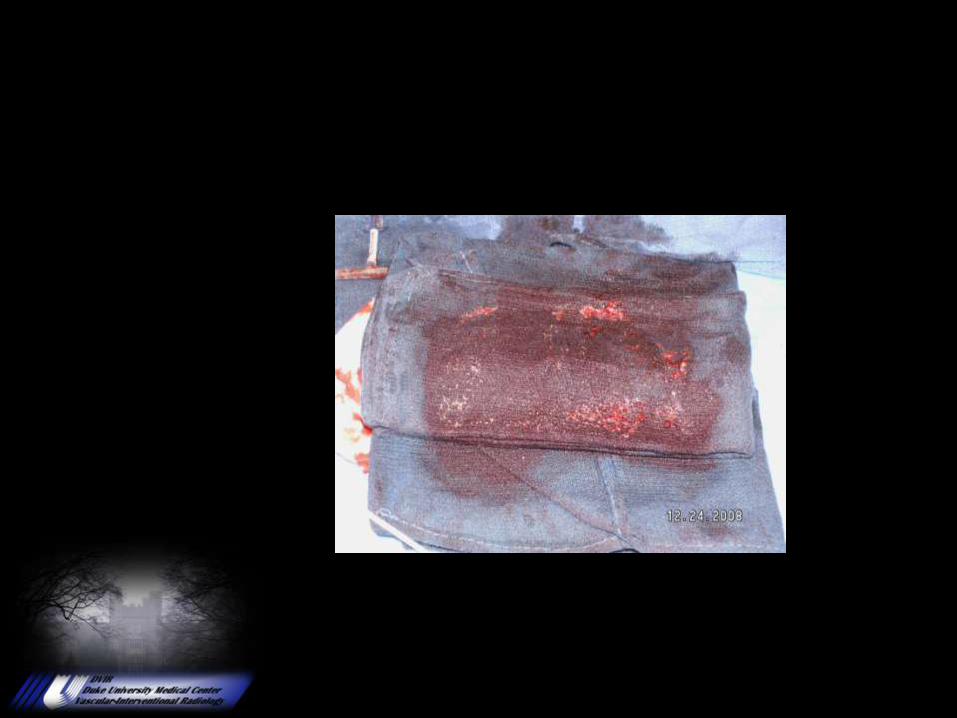
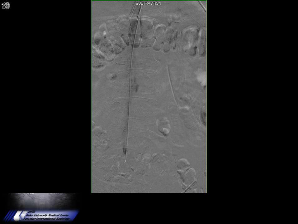
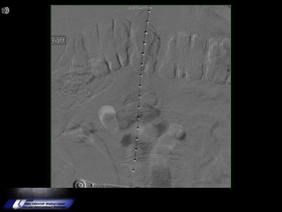
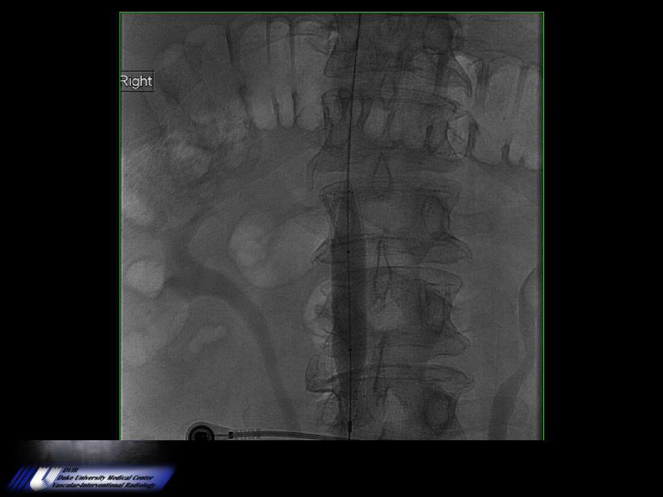
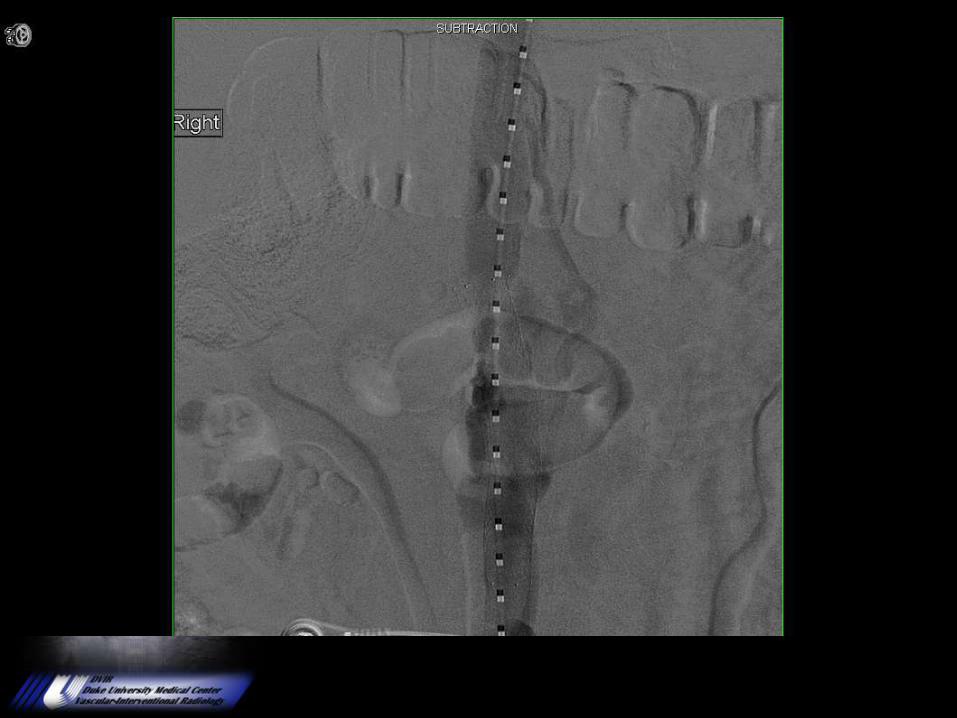
49 y.o Male 2 weeks onset Bilateral lower extremity
Swelling
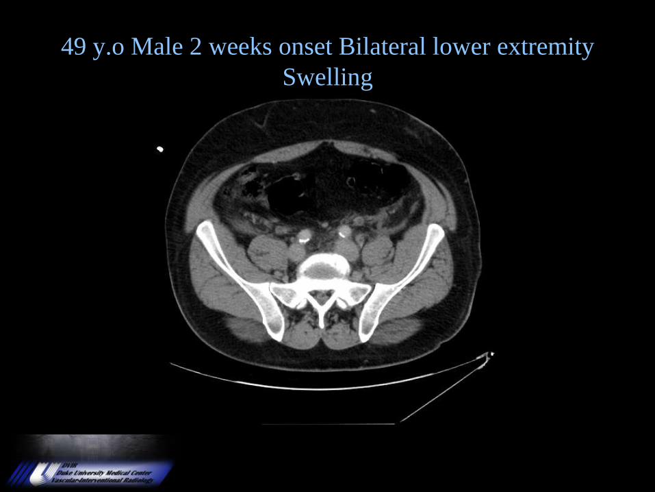
49 y.o Male 2 weeks onset Bilateral lower extremity
Swelling
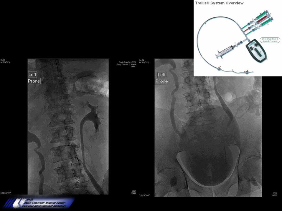
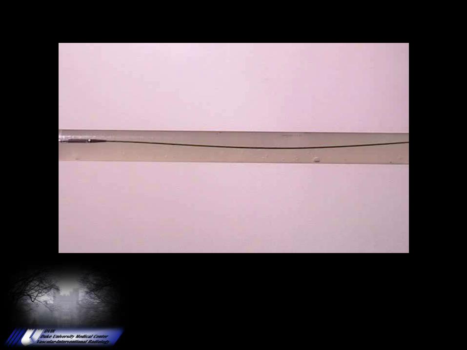
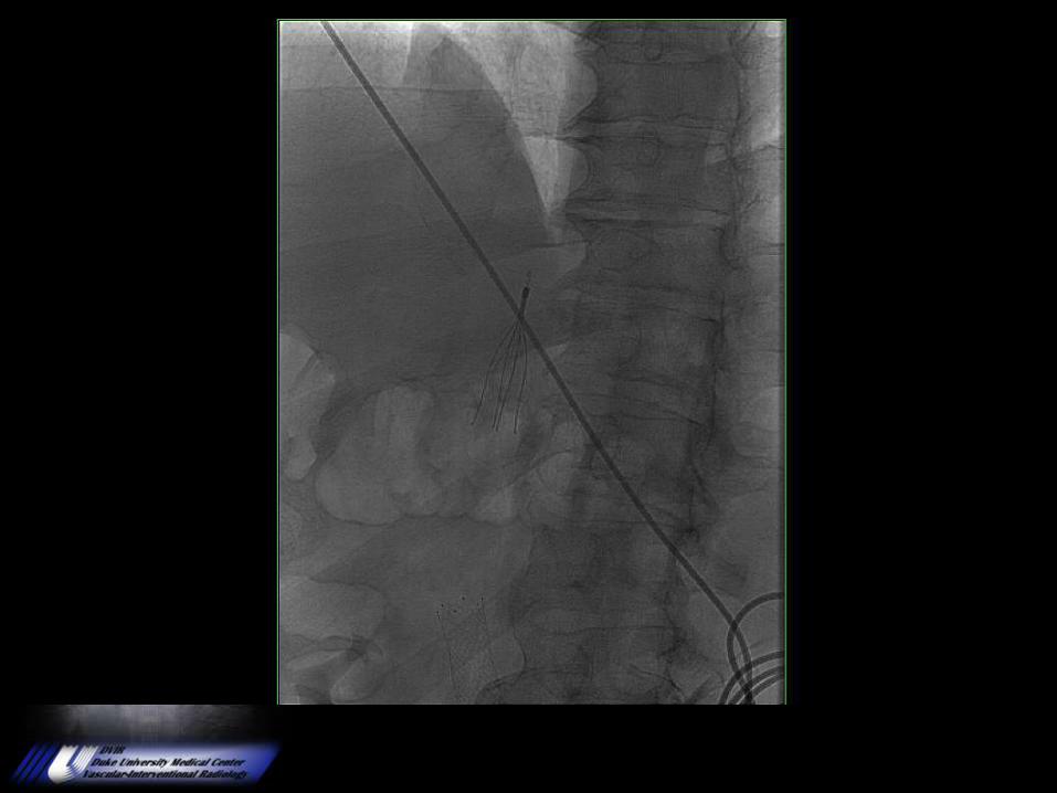
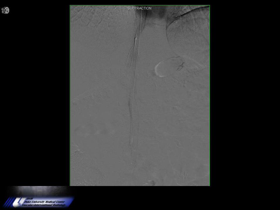
Trellis Catheter
http://www.bacchus-vascular.com/products/trellis/index.html
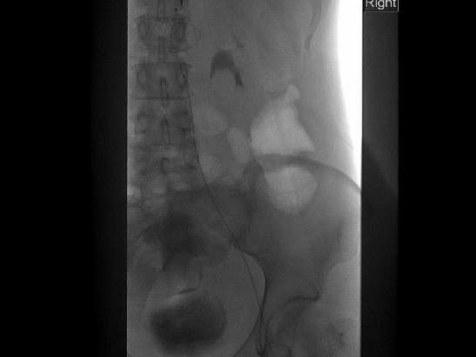
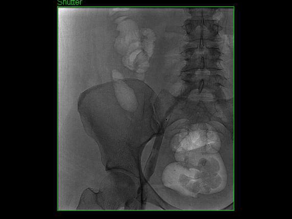
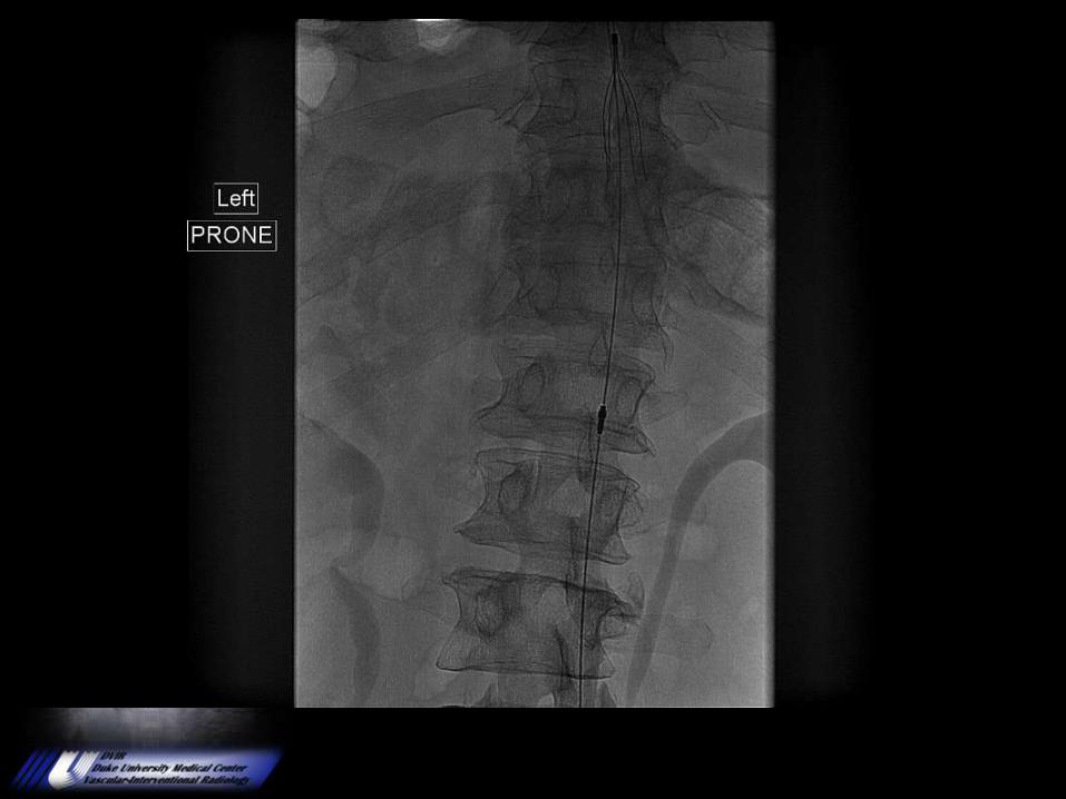
12 mm gradient across the stenosis in the IVC just below the renal
veins. Narrowing confirmed with intravascular ultrasound.
14 mm Stent angioplastied to 14 mm.
2 mm Gradient following stenting.
Celect Filter replaced and scheduled for removal after
surgery or biopsies complete
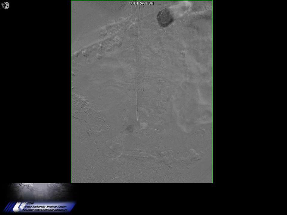
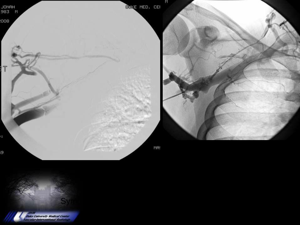
12/1/2008 Cavagram
This is his cavagram on 12/1/2008. He is symptom free.
Ultrasound of the legs is recommended prior to stopping Coumadin.
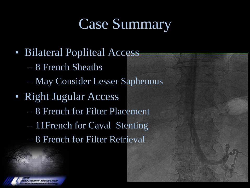
Case Summary
• Bilateral Popliteal Access
– 8 French Sheaths
– May Consider Lesser Saphenous
• Right Jugular Access
– 8 French for Filter Placement
– 11French for Caval Stenting
– 8 French for Filter Retrieval
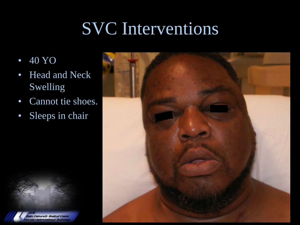
SVC Interventions
• 40 YO
• Head and Neck
Swelling
• Cannot tie shoes.
• Sleeps in chair
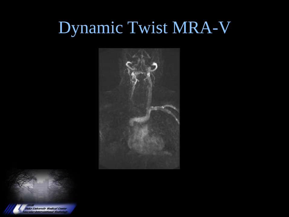
Dynamic Twist MRA-V
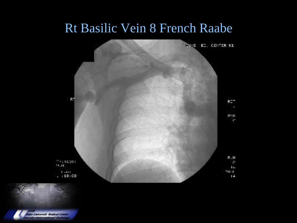
Rt Basilic Vein 8 French Raabe
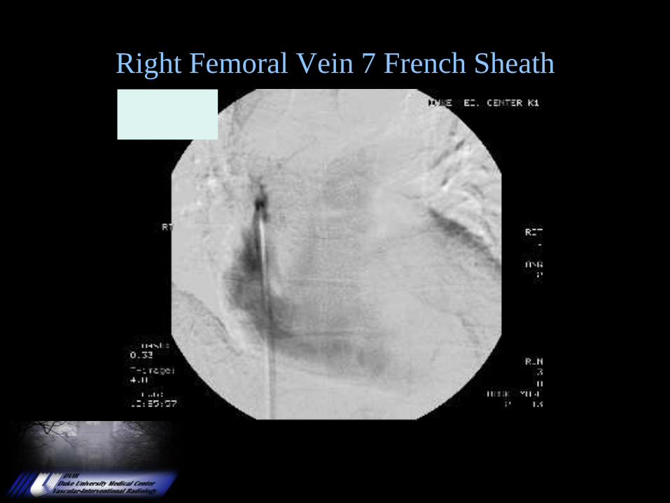
Right Femoral Vein 7 French Sheath
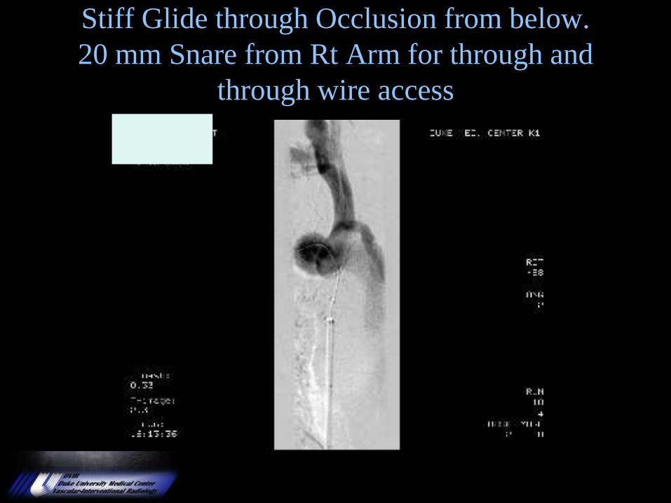
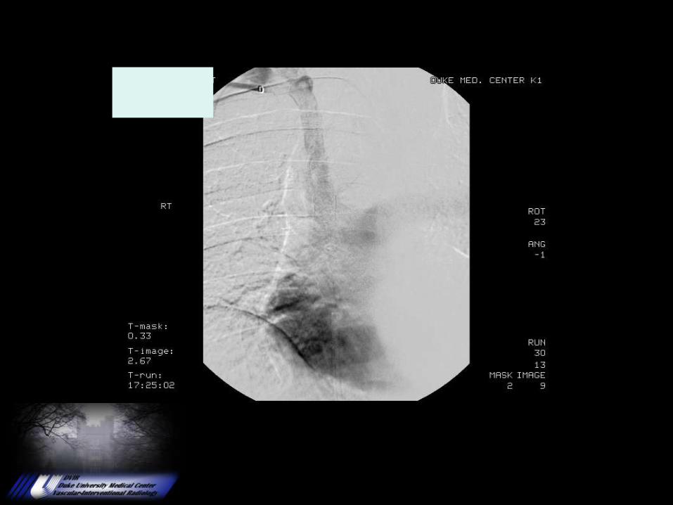
Stiff Glide through Occlusion from below.
20 mm Snare from Rt Arm for through and
through wire access
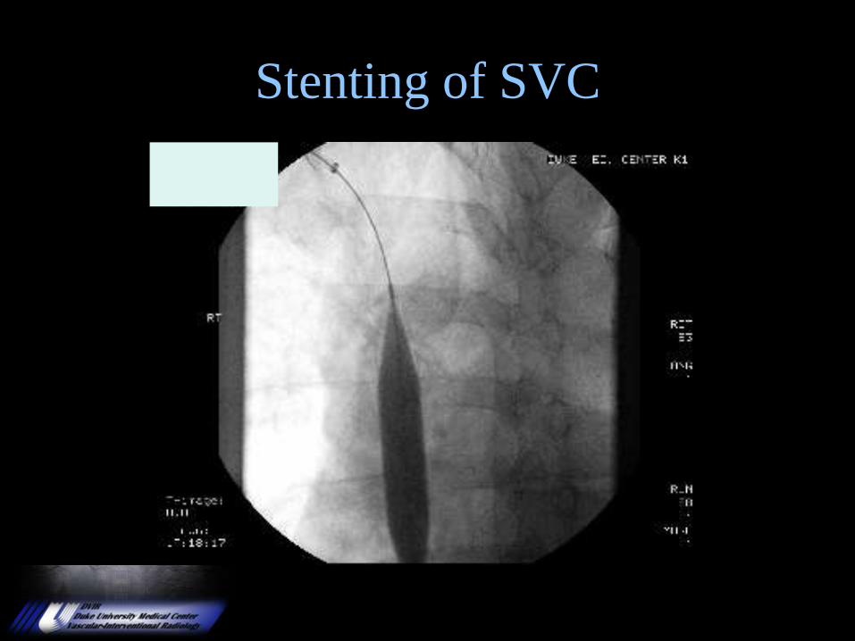
Stenting of SVC
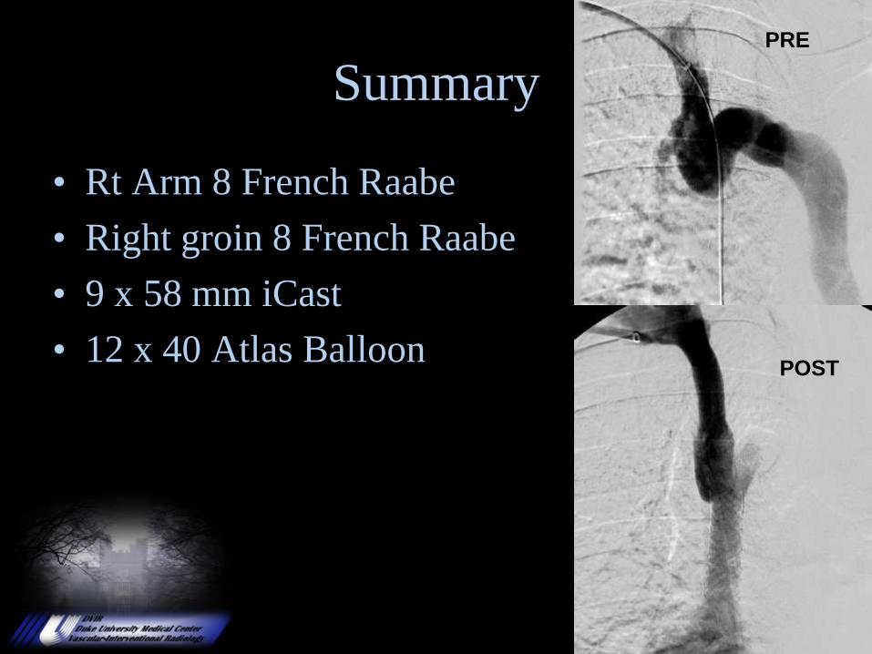
Summary
• Rt Arm 8 French Raabe
• Right groin 8 French Raabe
• 9 x 58 mm iCast
• 12 x 40 Atlas Balloon
PRE
POST
• Acute ( Less than 10 days )
• Iliofemoral DVT
• Patient less than 60 y.o.
• No HTN
• Image confirmation of extent
• Symptomatic
• Phlegmasia Cerulea Dolens
Ideal Patient Selection

Course Objectives
• DVT Incidence and Etiology
• Imaging.
• Anticoagulation
• Systemic Thrombolysis
• Catheter Directed Thrombolysis
• To Filter or Not to Filter? That is the question.
• Mechanical Thrombectomy Devices.
• Venous Intervention.


Venous Thrombosis
Thrombolysis• HISTORY OF PRESENT ILLNESS: The patient is a 79-year-old female
who felt low back pain and hematuria in 6/03. Reportedly, laboratory studies and
urine examination were negative by her primary care physician and her symptoms resolved spontaneously. In early 7/03, she developed new left lower extremity edema which was initially evaluated with ultrasound, which was negative for DVT. Later in 7/03, she developed left lower quadrant pain which was initially evaluated with KUB, which
reportedly showed LS spine degenerative changes; however, a follow-up abdominal CT scan showed a left lower quadrant mass measuring 8.5 x 6 cm encasing the left ureter and causing blockage as well as encasing the left common iliac artery and vein. She underwent fine needle aspiration of the retroperitoneal mass in
Virginia, 7/25/03, with the subsequent diagnosis of high grade malignant
neuroendocrine neoplasm. On 8/1/03, she underwent placement of a left ureteral
stent. She was found to have exacerbation of left lower extremity swelling and on US demonstrated Iliofemoral DVT.
• Future
– Combination Mechanical and Chemical
• TNK
• 24 hour floor based therapy
• Outpatient management of anticoagulation
• Need for randomized studies
• Direct Thrombin Inhibitors
Venous Thrombosis
Thrombolysis
Course Objectives

“Tying is Like Kissing Your Sister”
Paul “Bear” Bryant
Catheter Directed Thrombolysis
Abraham Thomas, MSIV
10/8/2008
Basic Radiology Clerkship
24 yo male with 1 day history of
right arm swelling
Works as a chef and a lifeguard
Swelling increased during the day
pre
Post
Angiojet
12 mm Conquest balloon
Symptoms improved then worsened 2 days later
12 mm PowerFlex Extreme
Returns 2 days later (4 days post initial Rx) with ↑ symptoms
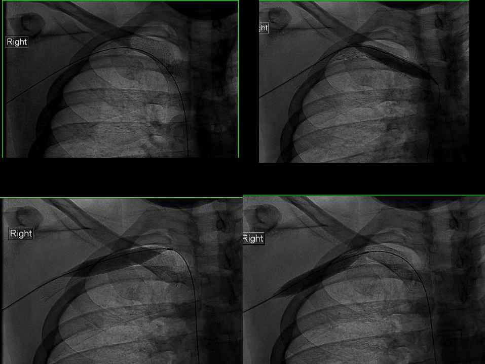
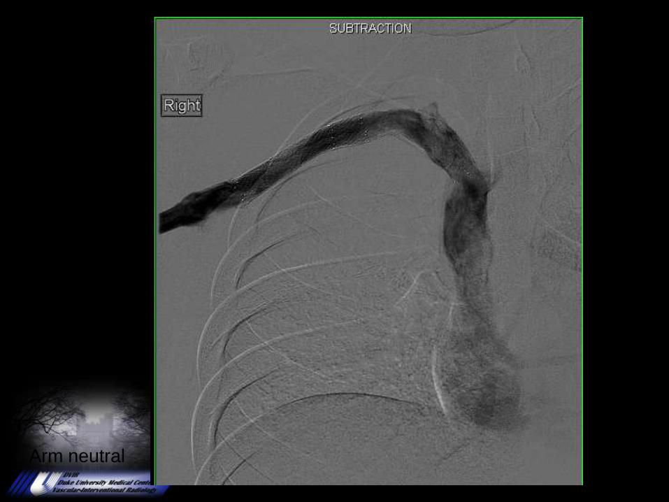
Arm neutral
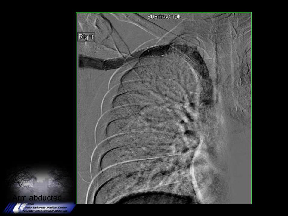
Arm abducted
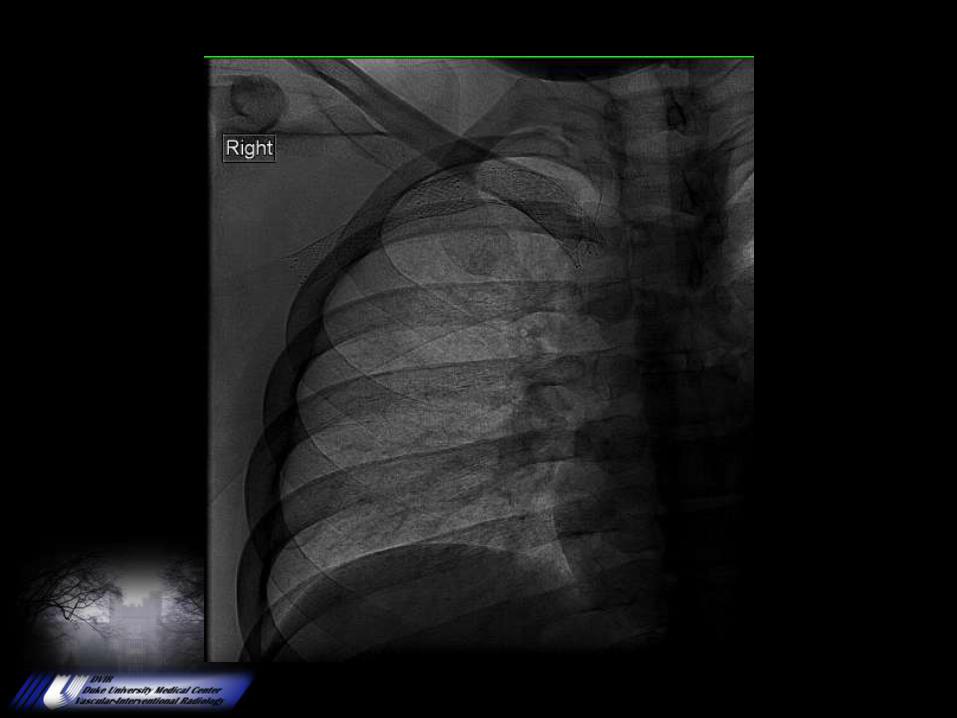
3 weeks post, mild ↑ symptoms
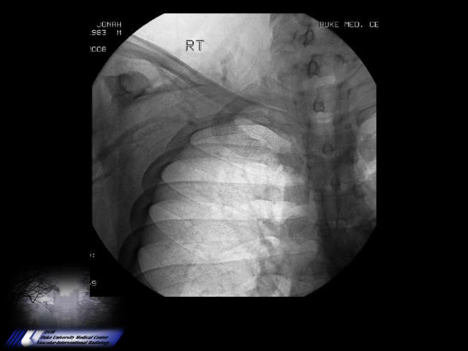
Case Presentation:Deep Venous Thrombolysis
Jason Harris, MD
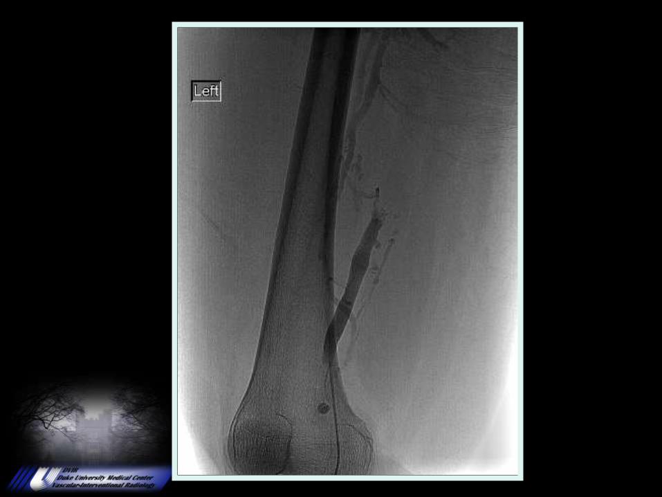
Presentation
• 50 year old female w/o sig PMH presents with
pain and swelling from LLE DVT after EVLT
for varicose veins.
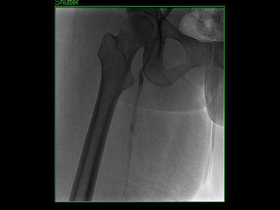

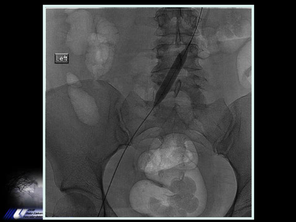


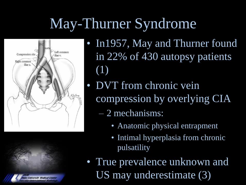
May-Thurner Syndrome
• In1957, May and Thurner found
in 22% of 430 autopsy patients
(1)
• DVT from chronic vein
compression by overlying CIA
– 2 mechanisms:
• Anatomic physical entrapment
• Intimal hyperplasia from chronic
pulsatility
• True prevalence unknown and
US may underestimate (3)
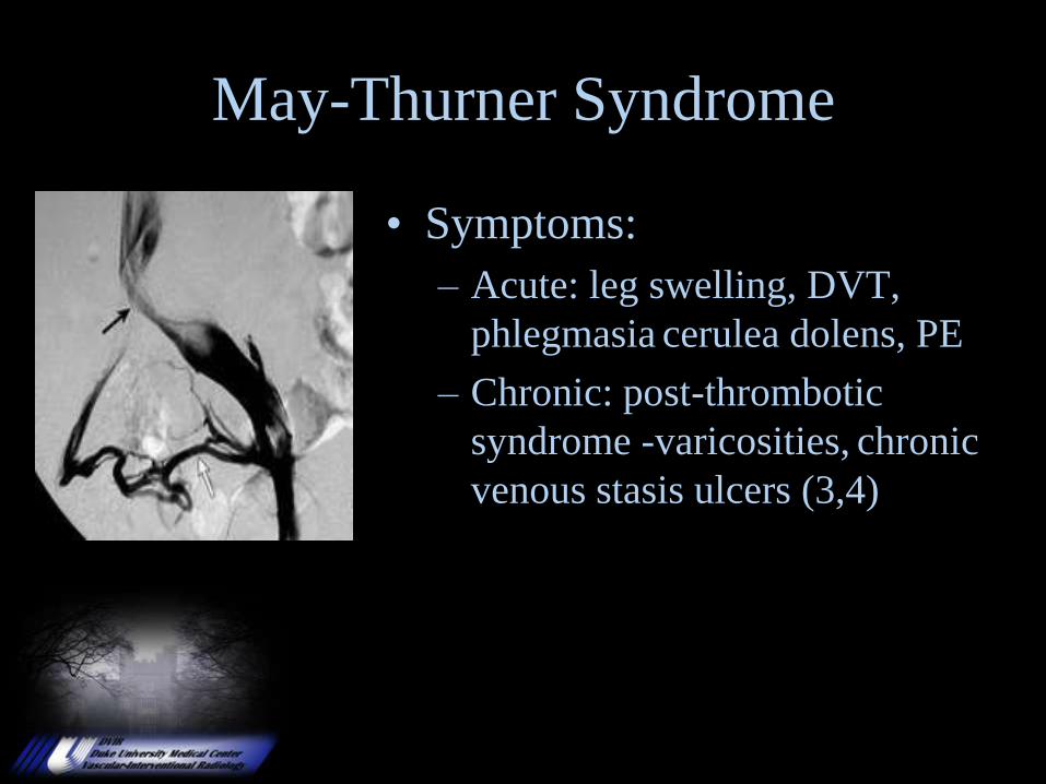
May-Thurner Syndrome
• Symptoms:
– Acute: leg swelling, DVT,
phlegmasia cerulea dolens, PE
– Chronic: post-thrombotic
syndrome -varicosities, chronic
venous stasis ulcers (3,4)
May-Thurner Syndrome
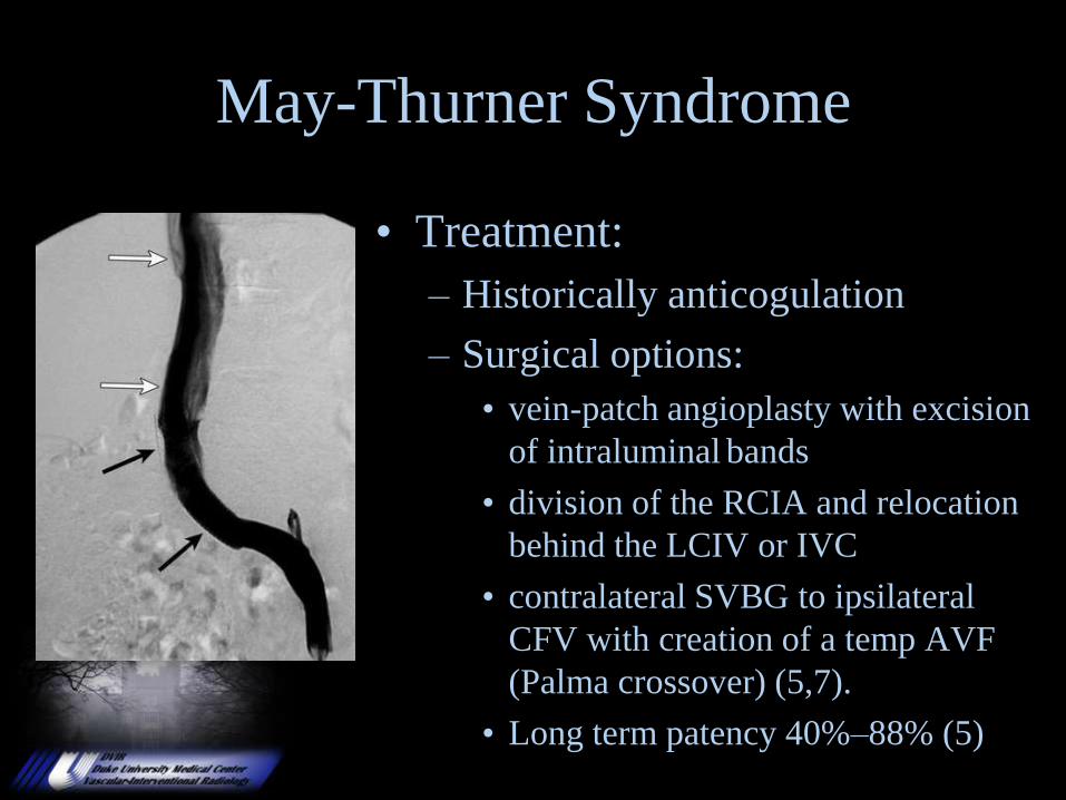
• Treatment:
– Historically anticogulation
– Surgical options:
• vein-patch angioplasty with excision
of intraluminal bands
• division of the RCIA and relocation
behind the LCIV or IVC
• contralateral SVBG to ipsilateral
CFV with creation of a temp AVF
(Palma crossover) (5,7).
• Long term patency 40%–88% (5)
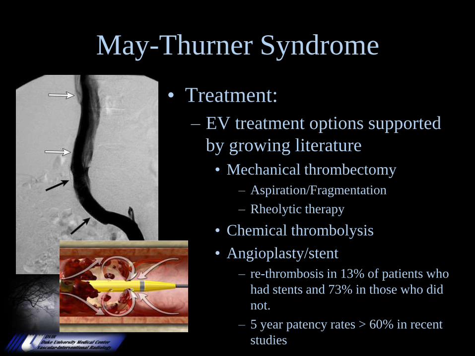
May-Thurner Syndrome
• Treatment:
– EV treatment options supported
by growing literature
• Mechanical thrombectomy
– Aspiration/Fragmentation
– Rheolytic therapy
• Chemical thrombolysis
• Angioplasty/stent
– re-thrombosis in 13% of patients who
had stents and 73% in those who did
not.
– 5 year patency rates > 60% in recent
studies
References1. May R, Thurner J. The cause of the predominantly sinistral occurrence of thrombosis of the pelvic veins.
Angiology 1957; 8:419-427.
2. Cockett FB, Thomas ML. The iliac compression syndrome. Br J Surg 1965; 52:816-821.
3. Taheri SA, Williams J, Powell S, et al. Iliocaval compression syndrome. Am J Surg 1987; 154:169-172.
4. Heniford BT, Senler SO, Olsofka JM, Carrillo EH, Bergamini TM. May-Thurner syndrome: management by
endovascular surgical techniques. Ann Vasc Surg 1998; 12:482-486.
5. Patel NH, Stookey KR, Ketcham DB, Cragg AH. Endovascular management of acute extensive iliofemoral
deep venous thrombosis caused by May-Thurner syndrome. J Vasc Interv Radiol 2000; 11:1297-1302.
6. Ehrich WE, Krumbhaar EB. A frequent obstructive anomaly of the mouth of the left common iliac vein. Am
Heart J 1943; 26:737-750.
7. Alimi YS, DiMauro P, Fabre D, Juhan C. Iliac vein reconstructions to treat acute and chronic venous
occlusive disease. J Vasc Surg 1997; 25:673-681.
8. Baron HC, Shams J, Wayne M. Iliac vein compression syndrome: a new method of treatment. Am Surg 2000;
66:653-655
9. Berger A, Jaffe JW. Iliac compression syndrome treated with stent placement. J Vasc Surg 1995; 21:510-514.
10. Buelens C, Vandenbosch G, Stockx L, et al. Cockett syndrome: initial results with percutaneous treatment in
6 patients. J Belge Radiol 1996; 79:132-135.
11. Cardiovasc Intervent Radiol. 2006 Jul-Aug;29(4):571-5. Percutaneous treatment of deep vein thrombosis in
May-Thurner syndrome.


![Current Drug Therapy€¦ · of acute DVT and PE is 9.0% and 30.1% in the first 3 months respectively [2]. Without adequate treatment, upto 50% of the patients with DVT may develop](https://img.pdfslide.us/doc/110x75/6041608628a049533e7d569f/current-drug-therapy-of-acute-dvt-and-pe-is-90-and-301-in-the-first-3-months.jpg)


![Tinzaparin Prescribing Advice Dosage Patient Weight Once Dailygeneralpracticemedicine.org/Tinzaparin_Dosage_Treatment[1].pdf · DVT/PE Treatment 20,000 IU/mLDVT/PE Treatment 20,000](https://img.pdfslide.us/doc/110x75/5f20f6c451c27966a818e5e8/tinzaparin-prescribing-advice-dosage-patient-weight-once-dailygen-1pdf-dvtpe.jpg)

![Treatment of venous thromboembolism: the single-drug …...for the initial and 6 -month treatment of patients with acute DVT [6]. Indeed, during the 6 -mo nth treatment period recurrent](https://img.pdfslide.us/doc/110x75/600adbc1be4f9950135beee5/treatment-of-venous-thromboembolism-the-single-drug-for-the-initial-and-6-month.jpg)