Embed Size (px)
Citation preview

P1: nrm/ayv P2: rbaP1: nrm/ayv P2: rba
Journal of Computational Neuroscience 04-Yisha December 11, 1996 14:26
Journal of Computational Neuroscience, 4, 57–77 (1997)c© 1997 Kluwer Academic Publishers, Boston. Manufactured in The Netherlands.
Traveling Waves and the Processing of Weakly Tuned Inputsin a Cortical Network Module
RANI BEN-YISHAIRacah Institute of Physics and Center for Neural Computation, Hebrew University, Jerusalem 91904, Israel
DAVID HANSELCentre de Physique Theorique, UPR014-CNRS, Ecole Polytechnique, 91128 Palaiseau, France
HAIM SOMPOLINSKYRacah Institute of Physics and Center for Neural Computation, Hebrew University, Jerusalem 91904, Israel; and
Luscent Technologies-Bell Laboratories, Murray Hill, NJ 07974 USA
Received February 6, 1996; Revised July 12, 1996; Accepted August 2, 1996
Action Editor: J. Rinzel
Abstract. Recent studies have shown that local cortical feedback can have an important effect on the response ofneurons in primary visual cortex to the orientation of visual stimuli. In this work, we study the role of the corticalfeedback in shaping the spatiotemporal patterns of activity in cortex. Two questions are addressed: one, what are thelimitations on the ability of cortical neurons to lock their activity torotatingoriented stimuli within a single receptivefield? Two, can the local architecture of visual cortex lead to the generation ofspontaneoustraveling pulses of acti-vity? We study these issues analytically by a population-dynamic model of a hypercolumn in visual cortex. The orderparameter that describes the macroscopic behavior of the network is the time-dependentpopulation vectorof thenetwork. We first study the network dynamics under the influence of a weakly tuned input that slowly rotates withinthe receptive field. We show that if the cortical interactions have strong spatial modulation, the network generatesa sharply tuned activity profile that propagates across the hypercolumn in a path that is completely locked to thestimulus rotation. The resultant rotating population vector maintains a constant angular lag relative to the stimulus,the magnitude of which grows with the stimulus rotation frequency. Beyond a critical frequency the population vectordoes not lock to the stimulus but executes a quasi-periodic motion with an average frequency that is smaller than thatof the stimulus. In the second part we consider the stable intrinsic state of the cortex under the influence ofisotropicstimulation. We show that if the local inhibitory feedback is sufficiently strong, the network does not settle into astationary state but develops spontaneous traveling pulses of activity. Unlike recent models of wave propagation incortical networks, the connectivity pattern in our model is spatially symmetric, hence thedirectionof propagationof these waves is arbitrary. The interaction of these waves with an external-oriented stimulus is studied. It is shownthat the system can lock to a weakly tuned rotating stimulus if the stimulus frequency is close to the frequency of theintrinsic wave.
Keywords: orientation selectivity, primary visual cortex, population vector

P1: nrm/ayv P2: rbaP1: nrm/ayv P2: rba
Journal of Computational Neuroscience 04-Yisha December 11, 1996 14:26
58 Ben-Yishai, Hansel and Sompolinsky
1. Introduction
The role of the local cortical interactions in shapingthe response properties of cortical neurons to sensorystimuli has been a matter of debate. According to theclassical model of Hubel and Wiesel (1962) (HW) thereceptive field (RF) properties of simple cells in pri-mary visual cortex is a reflection of the feedforwardafferents from the lateral geniculate nucleus (LGN). Inparticular, according to the HW model the preferredorientation (PO) of a simple cell originates from thegeometrical alignment of the small, circular RFs of theLGN neurons that project to it. Recently, an experimentin ferret has shown a significant correlation between theRF alignment of the LGN afferents to a site and the POof cortical cells in the same site (Chapman et al., 1991).In some cases, however, the LGN RFs were not aligned;no overlap between the LGN RFs and that of the cor-tical cells and no correlation between the sharpness oforientation tuning of cortical cells and the sharpness ofalignment of the observed LGN RFs have been found.More recent experiments in cat visual cortex have founda high correlation between the subfield organization ofthe RFs of simple cells and the RFs of LGN X-cellsthat are “functionally connected” to them (Reid andAlonso, 1995). In addition, recent intracellular record-ings showed substantial orientation tuning of the synap-tic inputs from the LGN to cells in cat primary visualcortex, generated by drifting gratings (Ferster et al.,1996). On the other hand, the blockage of extracellularinhibition in cortex caused substantial deterioration ofthe orientation tuning, suggesting that cortical circuitryplays an important role in shaping the relatively sharporientation tuning in cortex. (Sillito, 1977; Tsumotoet al., 1979; Sillito et al., 1980; Ferster and Koch, 1987;Hata et al., 1988; Nelson et al., 1994). Furthermore, re-cent intracellular measurements indicate that the directLGN input to cells in the input layer 4 constitutes onlya fraction of the total, orientation-selective excitatoryinput to these cells (Ahmed et al., 1994; Douglas et al.,1995; Pei et al., 1994), suggesting that massive corti-cal excitatory feedback in addition to cortical inhibitionmay be important in shaping orientation selectivity.
Several recent models that are based on plausi-ble assumptions about the local cortical connectionshave shown that these connections are capable ofplaying a central role in generating the sharp selec-tivity of cortical neurons to the orientation of visualstimuli (Worgotter and Koch, 1991; Ben-Yishai et al.,1995a; Somers et al., 1995; Hansel and Sompolinsky,1996a; Vidyasagar et al., 1996) and their direction
of movement (Suarez et al., 1995; Maex and Orban,1992). These models focused primarily on the responseof cortical neurons to the appearance of a stimulus witha fixed orientation or direction. Responses to time-dependent, time-oriented stimuli have received littleattention, both experimentally and theoretically.
In this work we broaden the scope of previous theo-retical studies to the spatiotemporal domain. We studyhow a spatial pattern of activity that is generated bya cooperative mechanism in the cortex interacts withan external input whose features change with time.Specifically, our working hypothesis is that the pri-mary visual cortex encodes the orientation of a localedge by the distributed response profile of the orienta-tion columns whose receptive field is stimulated by theedge. We further assume that the local cortical networkreceivesweakly tuned input from the LGNand gener-ates a sharply tuned response due to the combination ofmassive excitatory feedback and inhibition from withinthe network (Ben-Yishai et al., 1995a; Somers et al.,1995; Hansel and Sompolinsky, 1996a). The funda-mental assumption of this work is that the underlyingcortical circuitry is capable of generating sharply tunedresponses even for stimuli that generate only weaklytuned inputs to the cortex. Such stimuli can alwaysbe constructed by controlling the spatial extent and ge-ometry of a visual stimulus, in such a way that evena substantial alignment of the LGN RFs will generateonly weakly tuned inputs to the cortex. It is importantto emphasize that this assumption does not rule out thepossibility that certain visual stimuli, such as gratingswith appropriate spatial frequencies, generate LGN in-puts to the cortical cells that are substantially tuned tothe orientation of the stimuli.
These assumptions raise these questions: How doesthe population activity profile evolve in time in the pres-ence of a rotating stimulus? Does it lock to the stimulusmotion in frequency only or also in phase? For whatrange of rotation frequencies does this locking occur?
Another issue addressed in this article is the ap-pearance of traveling pulses of activity in neuronalnetworks. In recent years, traveling bursts of activityhave been observed in cortex (Gutnick et al., 1982;Chagnac-Amitai and Connors, 1989; Wadman andGutnick, 1993), superior colliculus (Munoz et al.,1991), thalamus (Kim et al., 1995) and hippocampus(Miles et al., 1988; Traub et al., 1993). Spatiotem-poral oscillations of neuronal activity in olfactory sys-tems have been extensively studied (Freeman, 1975;Laurent and Naraghi, 1994; Kleinfeld et al., 1994;Li and Hopfield, 1989). Neither the mechanisms that
P1: nrm/ayv P2: rbaP1: nrm/ayv P2: rba
Journal of Computational Neuroscience 04-Yisha December 11, 1996 14:26
Traveling Waves and the Processing of Weakly Tuned Inputs 59
generate these waves nor their functional relevance arewell understood. Several models have investigatedinternal cooperative mechanisms for the spontaneousgeneration of traveling waves in neuronal networks(Idiart and Abbott, 1993; Ermentrout and McLeod,1993; Golomb et al., 1996). In addition, moving pulsesof activity have been implicated in the coding of motortrajectories. Examples are the Moving Hill hypothe-sis regarding the control of eye movement in the supe-rior colliculus (Guitton, 1992), and the time-dependentpopulation vector hypothesis regarding the coding ofarm movement in motor cortex (Georgopoulos et al.,1988; Georgopoulos, 1995; Schwartz, 1993; Lukashinet al., 1995). Here we study the appearance of trav-eling waves in the context of a neuronal network thatcodes for an angle. We show that a cortical hyper-column, activated by an isotropic input, can exhibita stable spatiotemporal pattern consisting of a profileof activity that moves across the different orientationcolumns. We then study the interaction of this intrinsicwave with a rotating external input.
The architecture and dynamics of our model is pre-sented in Section 2. In Section 3 we study the stationarystates of the system. The response to a time-dependent(rotating) input is investigated in Section 4. In Section 5we study parameter regimes where the intrinsic stablestates consist of traveling waves. The properties ofthese waves and their response to a rotating input isstudied. In Section 6 we discuss our results and theirfunctional consequences.
2. The Model
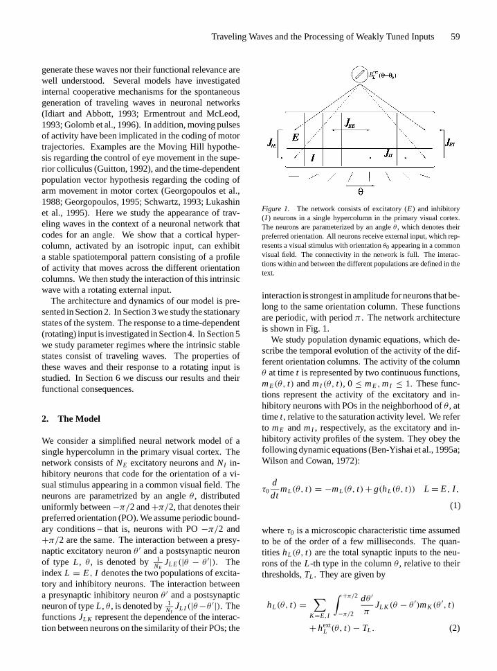
We consider a simplified neural network model of asingle hypercolumn in the primary visual cortex. Thenetwork consists ofNE excitatory neurons andNI in-hibitory neurons that code for the orientation of a vi-sual stimulus appearing in a common visual field. Theneurons are parametrized by an angleθ , distributeduniformly between−π/2 and+π/2, that denotes theirpreferred orientation (PO). We assume periodic bound-ary conditions – that is, neurons with PO−π/2 and+π/2 are the same. The interaction between a presy-naptic excitatory neuronθ ′ and a postsynaptic neuronof type L, θ , is denoted by 1
NEJL E(|θ − θ ′|). The
indexL = E, I denotes the two populations of excita-tory and inhibitory neurons. The interaction betweena presynaptic inhibitory neuronθ ′ and a postsynapticneuron of typeL, θ , is denoted by1
NIJL I (|θ−θ ′|). The
functionsJL K represent the dependence of the interac-tion between neurons on the similarity of their POs; the
Figure 1. The network consists of excitatory (E) and inhibitory(I ) neurons in a single hypercolumn in the primary visual cortex.The neurons are parameterized by an angleθ , which denotes theirpreferred orientation. All neurons receive external input, which rep-resents a visual stimulus with orientationθ0 appearing in a commonvisual field. The connectivity in the network is full. The interac-tions within and between the different populations are defined in thetext.
interaction is strongest in amplitude for neurons that be-long to the same orientation column. These functionsare periodic, with periodπ . The network architectureis shown in Fig. 1.
We study population dynamic equations, which de-scribe the temporal evolution of the activity of the dif-ferent orientation columns. The activity of the columnθ at timet is represented by two continuous functions,mE(θ, t) andmI (θ, t), 0≤ mE,mI ≤ 1. These func-tions represent the activity of the excitatory and in-hibitory neurons with POs in the neighborhood ofθ , attime t , relative to the saturation activity level. We referto mE andmI , respectively, as the excitatory and in-hibitory activity profiles of the system. They obey thefollowing dynamic equations (Ben-Yishai et al., 1995a;Wilson and Cowan, 1972):
τ0d
dtmL(θ, t) = −mL(θ, t)+ g(hL(θ, t)) L = E, I ,
(1)
whereτ0 is a microscopic characteristic time assumedto be of the order of a few milliseconds. The quan-tities hL(θ, t) are the total synaptic inputs to the neu-rons of theL-th type in the columnθ , relative to theirthresholds,TL . They are given by
hL(θ, t) =∑
K=E,I
∫ +π/2−π/2
dθ ′
πJL K (θ − θ ′)mK (θ
′, t)
+ hextL (θ, t)− TL . (2)

P1: nrm/ayv P2: rbaP1: nrm/ayv P2: rba
Journal of Computational Neuroscience 04-Yisha December 11, 1996 14:26
60 Ben-Yishai, Hansel and Sompolinsky
The quantitieshextL (θ, t) represent the inputs from the
LGN to theL-th population in the cortical columns. Wechoose the following functional forms for the corticalinteractions
JL E(θ) = JL E0 + JL E
2 cos 2θ, JL E0 ≥ JL E
2 ≥ 0
JL I (θ) = −JL I0 − JL I
2 cos 2θ, JL I0 ≥ JL I
2 ≥ 0.(3)
This choice implies that both excitatory and inhibitoryinteractions are maximal for neurons with similar POs,in agreement with experiment (see Georgopoulos et al.,1993). Substituting Eqs. (3) in Eq. (2), the synapticinputs can be written as
hL(θ, t) =JL E
0 mE0 − JL I
0 mI0 + JL E
2 mE2 cos(2(θ − ψE))
− JL I2 mI
2 cos(2(θ − ψI ))+ hextL (θ, t)− TL , (4)
wheremL0 , mL
2 , andψL are defined in terms of thefollowing order parameters:
mL0 (t) =
∫ +π/2−π/2
dθ
πmL(θ, t) (5)
and
PL(t) =∫ +π/2−π/2
dθ
πmL(θ, t)e
2i θ = mL2 (t)e
2iψL (t).
(6)
The first-order parametermL0 measures the activity of
the two types of neurons averaged over the entire net-work. The second-order parameterPL measures thedegree of the spatial modulation in the activity profiles.It is a complex number that represents a vector in two-dimensions. This vector is thepopulation vectorofthe system, evaluated by summing unit vectors pointedin the POs of the neurons, weighted by their instan-taneous activities (Georgopoulos et al., 1988; Seungand Sompolinsky, 1993). The angleψL denotes theorientation of the population vector andmL
2 denotesits length – that is, the strength of the angular modu-lation of the population. From a functional point ofview it is useful to considerψE(t) as a population cod-ing of the stimulus orientation. By integrating Eq. (1)overθ , with and without a factore2i θ , one reduces theequations to solving first-order nonlinear differentialequations formL
0 (t) andPL(t).Finally, we use a semilinear gain function –g(h) =
0 for h ≤ 0, g(h) = h for h ≥ 0, andg(h) = 1 for
h ≥ 1. The particular functional forms of the interac-tions and the gain-function were chosen for the sakeof simplicity. More general forms yield a qualitativelysimilar behavior.
3. Stationary State
We first study the case where the system settles in astationary state, in the presence of a time-independentstimulus,hext
L (θ, t) = hextL (θ). The stationary profile is
determined by the self-consistent equations
mL(θ) = g(hL(θ)). (7)
We model the stimulus by
hextL (θ) = CL(1− ε + ε cos(2(θ − θ0)))
0≤ ε ≤ 0.5. (8)
This choice represents a tuned input originating froma visual stimulus that is oriented at angleθ0. The para-meterCL denotes the maximal amplitude of the ex-ternal input from LGN to theLth population. It thusrepresents the overall strength of the afferent LGN in-put. It is assumed to be of the form
CL = CλL , (9)
whereC denotes the contrast of the stimulus, andλL
represents the transfer function from the LGN to thecortex, which may be different for excitatory and in-hibitory neurons. The parameterε denotes the angularanisotropy of the LGN input to the cortical hypercol-umn (Hubel and Wiesel, 1962; Chapman et al., 1991;Reid and Alonso, 1995). In the limitε = 0.5 the exter-nal input to neurons with PO orthogonal toθ0 is zero.This corresponds, in our parametrization, to maximallytuned inputs. A more general model ofhext
L (θ) will in-clude higher harmonics and thus will be able to incor-porate narrower inputs. As stated in the Introduction,we focus here on weakly tuned inputs and hence willassume thatε ¿ 1. In the limitε = 0 the external inputto all neurons in the same population is identical. It isimportant to note that the value ofε is determined bothby the geometric features of the visual stimulus, as wellas by the geometric organization of the LGN afferentsto individual cortical neurons (see Introduction).
3.1. Homogeneous State
The rotational symmetry of the network implies thatwhenε = 0 there is always a homogeneous solution,

P1: nrm/ayv P2: rbaP1: nrm/ayv P2: rba
Journal of Computational Neuroscience 04-Yisha December 11, 1996 14:26
Traveling Waves and the Processing of Weakly Tuned Inputs 61
mL(θ, t) = mL0 , which agrees with the naive expec-
tation that orientation selectivity disappears when theexternal input is isotropic. In this state,PL are zero.The mean activity levels of all the neurons in the twopopulations respectively can be written as
mL = χL(CL − TL). (10)
The prefactorsχL are the linear response coefficientsof the system, or simply the gain of the system. Theyare given by
χE = 1− κ K
(K − J−1)J I E0
χI = 1− κ−1 J
(K−1− J)JE I0
,
(11)
where
κ = CI − TI
CE − TE(12)
and
K = JE I0
/(J I I
0 +1); J = J I E
0
/(JE E
0 −1). (13)
Note that the effect of the cortical interactions on thelevel of activity of the excitatory neurons is twofold.The excitatory feedbackJE E
0 tends to amplify the res-ponse, whereas the inhibitory interactionJ I E
0 tends tosuppress it. Whether the net effect is an enhancement ofthe activity or the converse depends on the assumed val-ues of these parameters (compare with Douglas et al.,1995).
The above equations are valid of course only forparameters such thatmL > 0. Depending on the sys-tem parameters, there can be homogeneous states (withnonzero activity) even for subthreshold inputs.
3.2. Inhomogeneous State
3.2.1. Afferent Mechanism. When the input to thecortical network is anisotropic – that is,ε > 0 – theactivity profile is, of course, nonuniform, and sometuning to orientation will develop. Solving the self-consistent equations for the profile is relatively simplein the case of the semilinear gain function. We willconsider here the case of sharp tuning, where for eachstimulus orientation a fraction of the populations re-mains inactive. Such a state, which occurs for a widerange of parameters, has the profile
mL(θ) = HL (cos 2(θ − ψL)− cos(2θL))
|θ − ψL | < θL , (14)
andmL(θ) = 0 otherwise. Equations for the ampli-tudes,HL , the tuning widths,θL , and the angles,ψL ,are derived by substituting Eq. (14) in Eqs. (5) and (6),and solving the resultant self-consistent equations. Forthe population angles they yield
ψL = θ0 (15)
as expected. The details are presented in Appendix A.The sharpness of the tuning depends on the corti-
cal interactions. If their angular modulation is weakrelative to the external input, the system mimics thesimple Hubel-Wiesel afferent mechanism of orienta-tion selectivity (Hubel and Wiesel, 1962). In this case,to obtain sharp tuningε needs to be large. Alternatively,C has to be small so that the population is always onlyslightly above the threshold. Examples are shown inFigs. 2A–F, which display the results of numerical in-tegration of the population dynamic equations, Eq. (1).Here and in the rest of the paper the numerical resultsare obtained using numerical integration with fourth-order Runge-Kutta method and an angular resolutionof 1 deg. The results of Figs. 2A–F correspond tothe case where the spatial modulation of the excitatoryfeedback is roughly the same as that of the inhibitoryone; hence the net modulated feedback is weak. Con-sequently, when the input tuning is weak andC is largethe tuning is broad, as shown in Figs. 2A, B. Sharp tun-ing is obtained whenC is close to threshold (Figs. 2C,D) or ε is large (Figs. 2E, F). Figures 2A, C, and Edisplay the activity profilesmE andmI . Figures 2B,D, and F display the relationship between the externalinputs (solid and dashed lines) and the cortical feed-back. The latter is represented by an effective thresholdTeff(θ) (dotted line) defined asTeff = hext
L (θ)− hL(θ).Hence,mL(θ) = g(hext
L (θ) − Teff(θ)). Note that forsimplicity we have chosen here parameters such thatthe effective threshold is the same for the excitatoryand inhibitory populations.
3.2.2. Marginal Phase. If the spatial modulation ofthe cortical interactions is strong, sharp tuning maybe obtained even for a weakly tuned stimulus. Inthe extreme case, the homogeneous state is unsta-ble even forε = 0. Instead, the stable solution is aninhomogeneous state with a profile as described above,with an arbitrary location of the peak. This solu-tion represents spontaneous generation of orientationselectivity. Thus, in this regime there is a continuum ofpossible stable states, all represented by identical activ-ity profiles but with different locations of their peaks.

P1: nrm/ayv P2: rbaP1: nrm/ayv P2: rba
Journal of Computational Neuroscience 04-Yisha December 11, 1996 14:26
62 Ben-Yishai, Hansel and Sompolinsky
Figure 2. Orientation tuning in the case where the modulation of the cortical interactions is weak, A–F, and in the marginal phase, G–H. A,C, E, G: Solid and dashed lines are the excitatory and inhibitory activity profiles,mE(θ) andmI (θ), respectively. B, D, F, H: Solid and dashedlines are the inputs to the excitatory and inhibitory populations,hext
E (θ) andhextI (θ), respectively. Dotted line is the effective thresholdTeff(θ);
A–B: ε = 0.1, CE = 0.15 andCI = 0.14. All neurons are active,θE = θI = 90◦; C–D: ε = 0.1, CE = 0.11 andCI = 0.108. Only neuronswith θ close toθ0 = 0◦ are above their threshold. Sharp tuning is achieved via smallC; E–F: ε = 0.5, CE = 0.15 andCI = 0.14. Sharptuning is achieved via largeε. Model parameters for A–F areTE = TI = 0.1, JE E
0 = J I E0 = 13, JE E
2 = J I E2 = 9, JE I
0 = J I I0 = 18,
JE I2 = J I I
2 = 9; G–H: ε = 0.1, CE = 0.15 andCI = 0.14. Model parameters are as in A–F, except for the modulation of the excitatoryinteractions,JE E
2 = J I E2 = 12.5.
This solution is called a marginal phase because thereare no barriers between the different fixed points ofthe dynamics. The overall amplitude of the populationresponse,HL , in any one of these states, is determinedby the input contrastC, but the width of the activityprofiles is determined by the cortical interactions. The
width depends also on therelative inputs to the twopopulations – namely,κ, Eq. (12) – but this depen-dence is rather weak.
In reality, the location of the peak of the activity is notarbitrary but is determined by the orientation of the ex-ternal input. Therefore, in a realistic implementation of

P1: nrm/ayv P2: rbaP1: nrm/ayv P2: rba
Journal of Computational Neuroscience 04-Yisha December 11, 1996 14:26
Traveling Waves and the Processing of Weakly Tuned Inputs 63
the last scenario,ε is assumed to be nonzero but small.Sinceε is small, the main effect of the anisotropy ofthe input is to select among the continuum of possiblestates that state in which the location of the peak in theactivity matches the orientation of the stimulus, but itwill not have much effect on the width of the activityprofile. The equations of the marginal phase are givenin Appendix A. Its properties were discussed in detailin Ben-Yishai et al. (1995a). An example of a tuningcurve in this regime is shown in Fig. 2G. Here the an-gular modulation of the excitatory interactions is largerthan the inhibitory ones so that the net modulation ofthe cortical feedback is large and the system is in themarginal phase.
4. Response to Rotating Stimulus
We now consider an external stimulus in the form of ashort-oriented object that rotates within a single recep-tive field. This is modeled by a time-dependent externalangle – that is,θ0 = θ0(t), (see Fig. 3). If the systemencodes the instantaneous stimulus orientation by thepopulation vector, then the population activity profileshould be able to follow the rotation of the stimulus.This raises the following questions: Can the popula-tion activity profile lock to the input? If so, what is therange of input angular velocities for which such lockingoccurs? For a stimulus that varies on time scales com-parable to single-cell time constants, the answers to theabove questions may depend strongly on the details of
Figure 3. A stimulus with a time-dependent orientationθ0(t) ispresented in a common receptive field.
the single-cell microscopic dynamics. However, whenthe temporal variation of the stimulus is slow, and inaddition the direct coupling of the population profileto the rotating stimulus is relatively weak, cortical co-operative effects may be the dominant factor in deter-mining the locking properties. We therefore focus hereon the case of a weakly tuned(ε ¿ 1) and slow time-dependent(θ0 = O(ε)) input.
We first consider the case where the cortical interac-tions are such that the orientation tuning is dominatedby the input. In this case, whenε is small, the spa-tial modulation of the population profile induced bythe input is small and represents essentially the linearresponse of the system toε. When the stimulus ro-tates, this linear response is locked to the stimulus witha constant phase shift that is proportional roughly toθ0τ0. Thus in this case we expect that the system willlock to the stimulus but that this locking will involveonly a small component of the system activity, rela-tive to the DC component. An example is shown inFig. 4A. As mentioned above, this will be the case upto high stimulus frequencies where the nonlinearity ofthe single-cell dynamics may prevent complete lockingto the stimulus.
The situation is qualitatively different in the param-eter regime of the marginal phase. Here the tuning ofthe network may be sharp, and hence the response tothe stimulus is highly nonlinear. Fortunately, in thisregime the dynamics can be studied analytically usingthe approximation of phase dynamics, which becomesexact in the limit ofε, θ0→ 0. In this limit, the rotationof the stimulus generates a motion of the activityprofiles but does not change their shape. Hence thestate of the system is given by
mL(θ, t) = ML(θ − ψL(t)), (16)
whereML(θ) are the steady-state activity profiles ofthe two populations centered atθ = 0 for ε = 0. Interms of the order parameters defined above, Eq. (16)means thatmL
0 andmL2 are the same as in the station-
ary state atε = 0. However, the population anglesψL(t), which, as described above, denote the positionat timet of the peaks of the profiles of activity, are nowtime-dependent. The degree of locking to the rotatingstimulus is measured by the phase-shifts1L , defined as
1L(t) = ψL(t)− θ0(t). (17)
Because we assume now that the modulation of theinput is small and it is only slowly rotating, the rateof change of1L is also small, and in addition the

P1: nrm/ayv P2: rbaP1: nrm/ayv P2: rba
Journal of Computational Neuroscience 04-Yisha December 11, 1996 14:26
64 Ben-Yishai, Hansel and Sompolinsky
Figure 4. Locking of the activity profiles to a rotating external input in the Hubel-Wiesel mechanism (A) and in the marginal phase (B).Solid and dashed lines are the excitatory and inhibitory activity profiles,mE(θ, t) andmI (θ, t), respectively. Dotted and dot-dashed lines arethe external input to the excitatory and inhibitory populations,hext
E (θ, t) andhextI (θ, t), respectively; A: the angular velocity of the stimulus is
ω = 0.4 rad/τ0, whereτ0 is the neuronal time constant, andε = 0.1. The activity profiles have a small component which locks to the stimuluswith a constant phase shift of∼ 20◦. Model parameters as in Fig. 2A; B:ω = 0.15 rad/τ0 andε = 0.05. Model parameters are:TE = TI = 0.1,CE = 0.15,CI = 0.025,κ = −1.5, JE E
0 = J I E0 = 13, JE E
2 = J I E2 = 12.5, JE I
0 = 20, JE I2 = 9, J I I
0 = 17, J I I2 = 6.
difference1I −1E is small,1I −1E = O(ε), but thephase shifts1L themselves are in general large. Theangular velocity of1 ≡ 1E ≈ 1I is given by thefollowing phase equation (see Appendix C):
d1
dt= −dθ0
dt+ ωc sin(21), (18)
whereωc > 0 is a characteristic frequency that is pro-portional toε/τ0 with a proportionality constant thatdepends on other system parameters.
The degree of locking of the network population tothe external stimulus depends of course on the nature
of the time-dependence ofθ0. Below we consider thesimple case of a stimulus rotating with aconstantan-gular velocity:
dθ0
dt= ω. (19)
Substituting Eq. (19) in Eq. (18) one observes that forω < ωc, Eq. (18) has a stable fixed point
1 = 1
2arcsin(ω/ωc). (20)

P1: nrm/ayv P2: rbaP1: nrm/ayv P2: rba
Journal of Computational Neuroscience 04-Yisha December 11, 1996 14:26
Traveling Waves and the Processing of Weakly Tuned Inputs 65
Figure 5. Response to a rotating stimulus in the marginal phase. A–C: Dotted line is the orientation of the visual stimulus,θ0(t) = ωt , as afunction of time. Solid and dashed lines are the angles of the excitatory population vector,ψE(t), and the inhibitory one,ψI (t), respectively;A: complete locking of the activity profile to a visual stimulus rotating with frequencyω = 0.15 rad/τ0. The activity profile follows thestimulus with a constant phase lag; B: partial locking in the caseω = 0.175 rad/τ0; C: no locking in the caseω = 0.3 rad/τ0. D–F: Thedistribution of relative phases,ρ(1); D: complete locking—ρ(1) is a delta function; E: partial locking—ρ(1) has a pronounced peak at 55◦;F: no locking—ρ(1) is essentially flat. Model parameters (except forω) are as in Fig. 4B. For these parameterωc = 0.173 rad/τ0.
It corresponds to a state in which the activity profileis locked to the stimulus and follows it with a constantphase lag. Forω→ ωc the phase lag between the exci-tatory population and the stimulus reachesπ
4 . It shouldbe emphasized that here, unlike in the previous case,the locking is strong in that it involves the motion of asharply tuned population profile, as shown in Fig. 4B.
The angles of the population vectors and the stimulusangle in such a case are shown in Fig. 5A. The resultsof Fig. 5 are obtained by numerical integration of thefull population dynamics, Eq. (1), with a rotating input.
Forω > ωc the fixed point of Eq. (18) disappears,hence the activity profile is not frequency locked to therotating stimulus. In this regime, the solution of the

P1: nrm/ayv P2: rbaP1: nrm/ayv P2: rba
Journal of Computational Neuroscience 04-Yisha December 11, 1996 14:26
66 Ben-Yishai, Hansel and Sompolinsky
phase equation yields
1(t) = arctan
{ωc
ω+ Äω
tan(Ä(t − τ))}, (21)
where
Ä =√ω2− ω2
c (22)
andτ is determined by the initial condition1(0) =10. The phase1 is periodic in time with a periodT = 2π
Ä. Thus, the rotation of the population vector is
quasi-periodic, withψL(t) = ωt −1(t). The averagefrequency of the population vector rotation isω − Ä,which is slower than the stimulus frequency,ω. An in-teresting feature ofψL(t) is its nonmonotonicity. Eachtime it encounters the stimulus, its reverses its sign andexecutes a small-amplitude oscillation. The frequencyof such encounters isÄ. The behavior ofψL(t) in thisfrequency regime is shown in Fig. 5B for a value ofω
close toωc, and in Fig. 5C for a higher frequency. Theyare in a good qualitative agreement with the predictionsof the above phase equations, which are based on thelimit of small ε andω.
The above results imply that forω > ωc the relativephase between the system and the stimulus is not fixedbut depends both on time and on the initial conditions.Nevertheless, it can be seen from Fig. 5B that nearωc
the system spends considerable amount of time in aroughly fixed phase relative to stimulus. Hence thereis partial locking to the stimulus. Asω increases, thispartial locking weakens and eventually disappears. Toquantitatively assess this property it is useful to con-sider the distribution of relative phases evaluated byaveraging1(t) over time. From Eq. (18) it is evidentthat
ρ(1) = A
ω − ωc sin(21), ω > ωc. (23)
Thus, the approximate phase model predicts that thedistribution has a peak at1 = π
4 , which is narrow nearωc and broadens asω increases. Figures 5E and F showthe numerically evaluated distribution of1 for the pa-rameters of Figs. 5B and C. For comparison we presentin Fig. 5D the distribution in the locked regime, whereit is simply a delta function centered at the fixed pointvalue, Eq. (20). The predicted gradual broadening ofthe phase distribution asω increases is evident, al-though, in contrast to the approximate phase equations,
Figure 6. Phase diagram for locking to a rotating stimulus, in themarginal phase. The shaded area is the parameter regime where aphase locked solution is stable. The phase lag between the stimulusorientation and the population vector angle for a givenε continuouslyincreases across this area (not shown), and reaches its maximumat ω = ωc(ε). Points A–C indicate values ofω and ε used inFigs. 5A–C, respectively. Model parameters are as in Fig. 4B.
the actual peak of the distribution is at angles greaterthan π
4 .Figure 6 displays the full phase diagram for lock-
ing to a rotating stimulus in the plane of stimulusparameters,ε-ω. The border of the shaded area repre-sentsωc(ε), which is the transition line from locked tounlocked states. This phase diagram has been evalu-ated numerically from the full dynamic equations. Forsmall ε, ωc(ε) is proportional toε, and the evaluatedconstant phase lags forω < ωc are in agreement withEq. (20).
5. Intrinsic Propagation of Activity Profile
5.1. Isotropic Input
So far we have considered parameters where, for a staticstimulus, the system reaches a stationary state. Canour network generate anintrinsicstate of a propagatingactivity profile? We have found that there are parameterregimes for which the stable solution is a traveling waveof the form of a propagating pulse of neuronal activity,even forε = 0. This solution is of the form
mL(θ, t) = ML(θ − ω0t + φL). (24)
The two population activities propagate with the sameangular velocity, denoted byω0, the inhibitory popu-lation lagging after the excitatory one with a constant

P1: nrm/ayv P2: rbaP1: nrm/ayv P2: rba
Journal of Computational Neuroscience 04-Yisha December 11, 1996 14:26
Traveling Waves and the Processing of Weakly Tuned Inputs 67
phase shift. The magnitude ofω0 as well as the shapeof the profile are determined by the system parameters,as described in Appendix B. However, the sign ofω0 –that is, the direction of the propagation of the activityprofile – is arbitrary, since the underlying interactionsare completely symmetric with respect to changing thespatial directions. Thus, this propagating wave exhibitsa spontaneous breaking of the left/right symmetry ofour system. An example of the propagating pulse isshown in Fig. 7.
How does the existence of the propagating solutiondepend on the model parameters? We first study thedependence on the contrast relative to thresholds,CL − TL . Throughout this work we assume that theexcitatory population receives an inputCE which isabove threshold. Otherwise a resting state for these
Figure 7. Intrinsic propagating waves. Solid and dashed lines arethe excitatory and inhibitory activity profiles,mE(θ, t) andmI (θ, t),respectively, at five subsequent times separated by 2τ0. The intrinsicfrequency isω0 ∼ 0.245 rad/τ0. The peak in the inhibitory activityprofile, lags after the peak in the excitatory activity profile with aconstant phase shift. Parameters are as in Fig. 4B, except that hereκ = 0 andε = 0. For these parametersκc = −0.58. Bar is 0.1.
neurons will be stable. Beyond this condition, the char-acter of the solution depends onCL − TL only throughthe single parameterκ, Eq. (12). For small values ofκ the stable solution is the stationary one. Whenκ isbigger than a critical valueκc, the stationary solutionloses stability to the wave solution. The reason for thisinstability is the fact that increasingκ increases thecomponent of the local inhibitory feedback relative tothe excitatory one. As the inhibitory feedback becomeslarge, it destabilizes the stationary state and generatesinstead a propagating wave. This instability is also sig-naled by theκ-dependence ofωc, which marks the crit-ical frequency for locking in the marginal phase. Theratioωc/ε diverges asκ approachesκc from below, in-dicating the emergence of a spontaneously generatedpropagating wave. The angular velocity of the intrinsicwave,ω0, increases withκ. For large values ofκ, thestable solution becomes a homogeneous solution forwhich the excitatory population is quiescent and onlythe inhibitory neurons are active. The dependence ofω0 on κ is shown in Fig. 8. Forκ < κc = −0.58, thesystem reaches a stable stationary state withω0 = 0.For κ > κc this solution loses stability to a wave so-lution with ω0 > 0. As κ increases so doω0, θE andθI , while the mean activitiesmE
0 andmI0 decrease. The
plateau for high values ofκ indicates a solution with awide inhibitory activity profile,θI = 90◦, where theintrinsic frequencyω0 is independent onκ. For val-ues ofκ larger than 0.9 the stable solution is a ho-mogeneous one, where only the inhibitory populationis active. The appearance of intrinsic waves depends
Figure 8. The intrinsic frequencyω0 as a function ofκ, the relativeinput to the two populations. Parameters, except forκ, are as inFig. 4B.

P1: nrm/ayv P2: rbaP1: nrm/ayv P2: rba
Journal of Computational Neuroscience 04-Yisha December 11, 1996 14:26
68 Ben-Yishai, Hansel and Sompolinsky
Figure 9. The intrinsic frequencyω0 for different values ofκ and JE I2 . The planeκ − JE I
2 is divided into three regions. Stationary region:κ < 0.9 and smallJE I
2 (darkly shaded); Wave region:κ < 0.9 and largeJE I2 (not shaded); Homogeneous region:κ > 0.9 (lightly shaded).
Parameters, except forκ andJE I2 , are as in Fig. 4B. The solid curve is the same as in Fig. 8.
also on the parameters of the interactionsJL K (θ). Onesuch parameter isJE I
2 (see Eq. (3)). It represents theamplitude of the spatial modulation of the inhibitoryfeedback onto the excitatory neurons. As mentionedabove, increasing this feedback does not allow the ac-tivity in any local regime to persist. On the other hand,the excitatory input and feedback prevents the decay ofthe over all activity. Consequently, the instantaneousactivity is locally suppressed but propagates to a nearbylocation. We show in Fig. 9 the phase diagram in theκ − JE I
2 plane. The vertical axis denotes the magni-tude of the intrinsic angular velocity,ω0. For largeJE I
2there is a critical value ofκ, above which the station-ary solution,ω0 = 0, loses stability to a wave solution,ω0 > 0. For large values ofκ the stable solution isa homogeneous one, where only the inhibitory popu-lation is active. The wave solution does not exist forsmall JE I
2 , and in this case asκ increases the stablestate changes from a stationary solution directly to thehomogeneous one.
5.2. Response to Rotating Stimulus
How does the existence of an intrinsic wave of the typedescribed above affect the ability of the system to lockto a rotating stimulus? We expect that the system willtend to lock to a stimulus that rotates with a frequencythat is near the intrinsic frequencyω0 but will not do sowhen the two frequencies are far apart. This is indeedborne out by a solution of the system dynamics in thepresence of a weakly tuned stimulus that rotates withfrequencyω whereω − ω0 = O(ε). Assuming once
again a state of the form of Eq. (16), the dynamics ofthe phases can be described by
d1L
dt= −dθ0
dt+ ω0+ δω + ωc sin(2(1L − βL)),
(25)
whereωc is proportional toε. Likewise, δω repre-sents an orderε correction to the intrinsic frequencydue to the external stimulus. The phasesβL are time-independent and denote the constant phase shifts ofthe population profiles relative to the fieldhL(θ, t)for ε = 0 (for the derivation of this equation seeAppendix C). In the case of a stimulus with con-stant frequencyω, the predictions of the phase equa-tion is similar to that of the previous section. For|ω − ω0− δω| < ωc the system is phase locked to thestimulus. The phase shifts of the populations relative tothe stimulus varies fromβL− π
4 toβL+ π4 (note thatβL
is always smaller thanπ4 ). Figure 10 displays the full-phase diagram that shows the regime in theε-ω planewhere the phase locked solution is stable. The param-eters are the same as that of Fig. 6, except forκ whichis aboveκc. Comparing the two phase-diagrams, weconclude that the intrinsic waves shifted the frequencyregime where locking occurs to higher values.
It should be noted that the above analysis of theeffect of a rotating stimulus assumes that the averagefrequency of the system’s profile has the same sign asthat of the stimulus. In general, the bistability in thedirection of rotation that exists atε = 0 persists alsofor nonzero but small values ofε. Thus, dependingon the initial conditions and the values ofε andω, the

P1: nrm/ayv P2: rbaP1: nrm/ayv P2: rba
Journal of Computational Neuroscience 04-Yisha December 11, 1996 14:26
Traveling Waves and the Processing of Weakly Tuned Inputs 69
Figure 10. Phase diagram for locking to a rotating stimulus, inthe intrinsic wave regime. The shaded area is the parameter regimewhere a phase locked solution is stable. Model parameters, except forε, are as in Fig. 7. For these parameters the intrinsic stable solutionthat is, (forε = 0) is a wave withω0 ∼ 0.245 rad/τ0.
system may settle in a state of a wave that propagatesin opposite direction to the stimulus. This state is notincluded in Fig. 10.
6. Discussion
6.1. Summary
In this article we have explored the behavior of anetwork model of a cortical hypercolumn in a regime ofparameters where the orientation tuning is dominatedby the modulation of the cortical horizontal connec-tions. In contrast to earlier studies (Ben-Yishai et al.,1995a; Somers et al., 1995; Hansel and Sompolinsky,1996a) we have focused here on the spatiotemporal do-main. Specifically, we have examined scenarios wherethe stable state of the system is a moving hill of activity,rather than a stationary hill. We first studied the con-ditions under which the population activity profile willfollow a visual stimulus that rotates around its centerwithin a single receptive field. Our analysis predictsthat for low-stimulus rotation frequencies the systemwill respond with a sharply tuned profile of activity,which will follow the stimulus with a constant phaselag. For stimulus frequencies above a critical frequencyωc, the population activity profile will lag in frequencyand will execute a quasi-periodic motion. The secondscenario for a moving profile in our model is the spon-taneous appearance of propagating waves due to thedestabilization of the stationary state.
6.2. The Basic Assumptions of the Work
Our results are based on the central assumptions that(1) the anisotropy of the input from the LGN to thecortex induced by the stimulus is weakly tuned (thatε
of Eq. (8) is small), and (2) even in such conditions, thecortical responses are sharply tuned, due to the localcortical interactions. In our modelε is a global pa-rameter. In reality, there is apparently a substantialvariation in the degree of tuning of the afferent input todifferent cortical sites (Chapman et al., 1991), whichmakes it hard to compare directly to the model. Asstated in the Introduction, one way of satisfying thecondition of weakly tuned input is by controlling theanisotropy of the visual stimulus itself. Thus, an im-portant test of the hypothesis of cortical participationin orientation tuning would be to measure the widthof the tuning as a function of the stimulus anisotropy,such as by varying the aspect ratio of the visual stimu-lus. According to our hypothesis, a weakly anisotropicstimulus will generate a sharply tuned response withan amplitude that varies smoothly with its contrast. Itshould be noted, however, that in reality for very smallanisotropy the spontaneously generated profile of ac-tivity will wander across the hypercolumn because ofthe noise in the neuronal activity, which has been ne-glected in our mean-field equations. Such a wanderingwill occur also in the absence of noise if the sponta-neously generated pattern is a traveling wave. In thiscase, a minimal positive value ofε is needed in order to“pin” the activity profile to the stimulus orientation asis seen in Fig. 10. Hence, for very smallε theaveragelevel of activity will be substantially reduced.
We have also assumed in our present analysis thatthe input has a relatively broad shape, even for largeε
(see Eq. (8)). This assumption is made primarilyfor convenience of analysis. A more general form forhext
L (θ) including a narrower profile will not changequalitatively our present results as long as the overallspatial modulation of the input is small. This modu-lation can be parametrized by say, the total power ofits nonzero Fourier-components relative to the zero-thcomponent. The above is valid as long ashext
L (θ)has thegeneral shape assumed here – namely, it is symmetricaround a unique maximum.
Throughout this work we have assumed that the ac-tivation of the excitatory population by the stimulus issuprathreshold – thatCE > TE. If the excitatory popu-lation has subthreshold activation, then the state whereall the excitatory population is quiescent is a stable

P1: nrm/ayv P2: rbaP1: nrm/ayv P2: rba
Journal of Computational Neuroscience 04-Yisha December 11, 1996 14:26
70 Ben-Yishai, Hansel and Sompolinsky
state, which is of course not interesting. On the otherhand, the inhibitory population may be active (due tothe excitatory input from the network) even if the stim-ulus is subthreshold. Hence, the parameterκ, Eq. (12)was allowed to assume negative as well as positive val-ues; see the phase diagram of Fig. 9. The precise rangeof κ where the different transitions occur depends onthe other model parameters; hence our model does nottightly constrain its value. At present, it is hard to es-timate the value ofκ from experiment, since relativelylittle is known about the distribution of thresholds andcontrast gain of inhibitory neurons in primary visualcortex. Models that rely on feedforward inhibition incortex, for example, direction selectivity, may favor theassumption of suprathreshold activation of inhibitoryneurons, perhaps even stronger than that of excitatoryneurons. On the other hand, recent models of “sur-round effects” on RFs argue in favor of inhibitory neu-rons having a considerable less activation by stimulusthan the excitatory ones (Stemmler et al., 1995; Somerset al., 1996).
In our previous work (Ben-Yishai et al., 1995a),we have focused on a network with stable stationarystates; hence it was sufficient to consider a parame-ter regime where the inhibitory and excitatory popula-tions are identical, in which case the network can bedescribed in terms of a single activity profile. In thisreduced network intrinsic waves cannot exist due tothe symmetry of the connection matrix. Here we haveconsidered a more general architecture in which thetwo populations are distinct. In this case the connectionmatrix is not symmetric and time-dependent attractorscan exist.
Previously, we have pointed out that the corticalmechanism for orientation tuning explains the exper-imentally observed separability of the coding of thestimulus orientation and contrast. In the model we haveinvestigated, changing the stimulus contrast (abovethreshold) changes the overall amplitude of the tun-ing curve but does not change its width, which wasdetermined by the spatial modulation of the corticalinteractions. The present work shows that the sepa-rability of contrast and orientation dimensions is car-ried over to the spatiotemporal domain. However, dueto the presence of two nonequivalent populations, theeffect of changing the contrast is more subtle. If thetwo populations receive the same activationCL = Cand have the same thresholdsTL = T , then althoughtheir connectivity profiles may be different, changingCwill result in an overall scaling of the activity profiles
without changing either the shapes of the profile northeir motion. In the case of differentCL or TL , thenstrictly speaking, invariance of shape or motion isachieved only if bothCL andTL are scaled by the samefactor. Thus, in general a change in the stimulus con-trast alone will modify therelativelevels of input to thetwo populations,κ, and hence in principle may changethe statics or the dynamics of the profile. In fact, wehave found thatκ affects, albeit weakly, the width ofthe tuning curve.
Our dynamic model is similar to dynamic equa-tions used previously to model the behavior of largeneuronal populations (Wilson and Cowan, 1972;Freeman, 1975). It ignores the highly nonlineardynamics of spikes, the temporal fluctuations in theactivity are averaged out, and synaptic inputs aremodeled as currents. These simplifications are justi-fied because we focus here on dynamical processesthat are slow compared to the single-cell time con-stant and are dominated by smooth cooperative ef-fects. In the case studied in Section 4 the restrictionto slow dynamics was explicitly assumed, since ourphase model was limited toωτ0 ∝ ε ¿ 1. However,the above considerations imply that our theory of theintrinsic wave, Section 5, is justified only when the in-trinsic frequencyω0 is small – that is, that the systemis near the bifurcation pointκ = κc (see Fig. 8).
6.3. Locking to Rotating Stimulus
The existence of distinct regimes of frequency andphase locking in our system is similar to that observedin low-dimensional oscillatory systems that are forcedby an oscillatory external force (Glass and Mackey,1988; Knight, 1972). In general, the temporal relationbetween the oscillator and the force may be complex.Here we have focused on parameter regimes wherethe stable spatiotemporal patterns are relatively sim-ple. They consist of an activity profile that is time-independent in a reference frame that moves with it. Ingeneral, the shape of the profile depends on the stimu-lus parameters such asω andε. However, whenε andthe difference betweenω and the intrinsic frequencyare both small, the shape of the profile is essentiallythe same as that without an oriented stimulus. Thisleads to a relatively simple-phase dynamic equationsthat describe the evolution in time of the locii of theactivity profiles, Eqs. (18) and (25). Phase modelshave been applied successfully also in several neuronal

P1: nrm/ayv P2: rbaP1: nrm/ayv P2: rba
Journal of Computational Neuroscience 04-Yisha December 11, 1996 14:26
Traveling Waves and the Processing of Weakly Tuned Inputs 71
models. Examples are models of the high-frequencyγ
oscillations in cat visual cortex (Schuster and Wagner,1990a, 1990b; Sompolinsky et al., 1991; Grannan et al.,1993) and the propagating waves in lamprey (Williamset al., 1990). Phase models have been also used to in-vestigate theoretically the conditions (cell and synapticproperties) under which oscillating neurons can syn-chronize their activities in small and large neuronalsystems (Hansel et al., 1995; Van Vreeswijk et al.,1995). These studies considered the locking of oscil-lating neurons or neuronal populations, by weak cou-pling. In contrast, in the case studied in Section 4, thenetwork did not oscillate at all in the absence of thestimulus but approached a stationary profile. Lock-ing to the rotating stimulus was, nevertheless, possibledue to the marginality (forε = 0) of the stationaryphase – namely, the existence of a continuum of at-tractors with identical spatial profiles.
6.4. Mechanism for the Appearanceof Intrinsic Waves
A main finding of our work is that intrinsic rotation ofthe activity profile can occur in a simple model of ahypercolumn even if the lateral connections are sym-metric with respect to spatial direction. Thus, whereasthe magnitude of the rotation frequency is determineduniquely by the network parameters, the sign of therotation (that is, clockwise or counterclockwise) is ar-bitrary and is selected by the initial conditions of thenetwork. In contrast, other network models (Lukashinet al., 1995) for generating moving-activity profilesin similar hypercolumn architectures rely on spatialasymmetry in the connections, which “push” the activ-ity in the direction of increasing excitatory connections(or decreasing inhibitory ones). An interesting conse-quence of the bistability of the network in this para-meter regime is that even in the presence of an externalrotating input, the network may exhibit a propagatingwave in a direction that is opposite to the direction ofrotation of the stimulus.
Our results regarding the intrinsic waves suggest thatapplication of nonoriented stimulation will induce cor-tical activity that may include moving “spots” acrossdifferent orientation columns. Previous numerical sim-ulations of large neural networks with excitatory andinhibitory feedback connections have revealed com-plex spatiotemporal patterns of activation including“hotspots” that execute a “random walk motion” acrossthe network (see e.g., Usher et al., 1994). Our results
imply that the motion of these spots will have a signifi-cant component that will periodically activate differentorientation columns.
In the present model, the waves appear due to theexistence of strong local inhibitory feedback. A “spot”of activity at one location is inhibited by this nega-tive feedback, but the level of excitation is sufficientlystrong to prevent the decay of the overall activity. Thesystem is forced to move the spot of activity to a nearbylocation (either to the right or to the left of the presentone). This mechanism requires a spatially modulatedinhibition, and will not work in – for example, a net-work with global inhibition. In a recent work (Hanseland Sompolinsky, 1996b), we have found that neuronaladaptation, not included in the present model, pro-vides another mechanism for the generation of intrinsicwaves in a cortical module with an architecture similarto the present one. In this case, waves appear even ifthe inhibition is global and serves only to control theoverall level of activity.
From a dynamic point of view the mechanismfor generating waves in our model is similar to thewell-known phenomenon of spontaneous appearanceof propagating pulses in one-dimensional reaction-diffusion systems (Rinzel and Keller, 1973; Murray,1989; Cross and Hohenberg, 1993). Our system issimpler in that the interactions, although spatially mod-ulated, are long-ranged; hence the waves can be de-scribed by nonlinear equations of few order parametersrather than by nonlinear differential equations, whichare necessary in physical systems governed by short-range interactions. The onset of traveling fronts andpulses in one-dimensional neural networks has beenalso investigated by Idiart and Abbott (1993). In theirmodel the mechanism for traveling waves involved ex-plicit delays in the interactions. In addition, their modelhas open boundary conditions and therefore the spa-tiotemporal patterns are transient. In contrast, in ourmodel the effective delayed feedback is generated bythe inhibitory neurons and the spatiotemporal patternsare attractors of the dynamics.
6.5. Functional Implications
We now discuss some possible functional implicationsof the network dynamic behavior. A simple ansatz isthat the system encodes information about a stimulusorientationθ0(t) simply in the value of the phase of theexcitatory population vectorψE(t − τd), where theτd
represents an appropriate constant delay time. Then,

P1: nrm/ayv P2: rbaP1: nrm/ayv P2: rba
Journal of Computational Neuroscience 04-Yisha December 11, 1996 14:26
72 Ben-Yishai, Hansel and Sompolinsky
our results concerning locking to a rotating stimulusin the marginal phase imply that for low frequenciesthe system is able to faithfully transmit the informationabout a time-dependent external angle, even when thesignal to noise ratio of the stimulus orientation is small.In this context, a possible advantage of the existenceof intrinsic waves in an appropriate parameter regimeis that they shift the range of frequencies for whichlocking to a rotating stimulus occurs to higher val-ues – namely, to a band of frequencies near the in-trinsic frequency of the system. For frequencies out-side the regime where full locking occurs the amountof lost information about the stimulus is representedby the width of the distribution of the difference be-tween the instantaneous angles of the population vectorand the stimulus, see Fig. 5. Additional possibilitiesexist if the information about the stimulus is extractedin more complex manners. For instance, fast intrin-sic waves can encode the position of a slowly movingstimulus using an appropriate periodic resetting signal,as was suggested recently for the temporal coding ofplace in the hippocampus (Tsodyks et al., 1995).
6.6. Experimental Aspects
Electrophysiological measurements in visual cortexcan test the theoretical predictions about the responseof the system to a rotating visual stimulus. It should benoted however that in reality there is an inherent delaybetween the visual stimulus and the resultant input tothe cortex from the LGN, which is not incorporated inour model. Thus, only predictions about therelativephasesbetween the stimulus and the population vectorare meaningful. The response of neurons to rotatingvisual patterns have been measured in extrastriatal vi-sual areas, in particular in area MST (Tanaka and Saito,1989; Tanaka et al., 1989). These experiments focusprimarily on the selectivity to the direction of rotation.Indeed, it was found that some neurons respond selec-tively to clockwise or counterclockwise rotations. Weare not aware of measurements of responses of neuronsin V1 to rotating stimuli within their receptive fields. Anatural way of observing the motion of the activity pro-file in a hypercolumn is by means of thepopulation vec-tor. In fact, it is well known that constructing the popu-lation vector yields a plausible mechanism forreadingout the angle information from a population of noisy,broadly tuned neurons. Under certain conditions, thepopulation vector is asymptotically the optimal angleestimator (Seung and Sompolinsky, 1993; Salinas and
Abbott, 1994). It is interesting that the population vec-tor is in fact the natural time-dependent order parameterin our theory. The population vector of the neuronalresponses in V1 can be measured experimentally bysampling single unit responses time-locked to a rotat-ing stimulus. Alternatively, optical imaging techniques(Grinvald et al., 1984; Orbach et al., 1985) may be usedto construct the population vector from the large-scalespatiotemporal pattern of electrical activity evoked bya rotating stimulus.
Visual perception is another potential testing groundof our predictions. Previously, we have considered theresponse of the system to an abrupt switching of thestimulus orientation (Ben-Yishai et al., 1995a). Wehave shown that in the marginal phase the populationprofile travels slowly from its original position to thenew location, in a manner that mimics the response toa slowly rotating stimulus. Thisvirtual rotation maybe related to the perceptual phenomenon ofapparentrotational motionwhen subjects are presented with al-ternating visual patterns (Finke and Shepard, 1984).The present work deals with the case of a stimulus thatchanges smoothly its orientation. Thus, it might be pos-sible to test our predictions regarding the dependenceof the locking to external stimulus on the stimulus fre-quencyω and anisotropyε, by psychophysical mea-surements of the perception of rotating stimuli and itsdependence on such parameters.
6.7. Concluding Remarks
In this work we have focused on a network model ofthe local cortical circuits in the primary visual cortex.However, many of the issues and the results of our workare relevant to the behavior of local neuronal networksin other systems as well. A closely related system is theassembly of neurons that are selective to the directionof voluntary arm movements in the motor cortex. Theapplication of our approach to the coding of movementtrajectories in the motor cortex is currently being stud-ied (Ben-Yishai et al., 1995b). The representation ofeye-movement in the superior-colliculus is another areawhere our approach can be used. Recent studies of thewaves of electrical activity in the procerebral lobe ofthe terrestrial molluscLimaxindicate that odor signalsmodulate primarily the spatial distribution of phases ofthe underlying activity oscillations, and to a lesser ex-tent their frequency (Kleinfeld et al., 1994). This maybe analogous to the effect of an external stimulus onthe intrinsic propagating waves in our network.

P1: nrm/ayv P2: rbaP1: nrm/ayv P2: rba
Journal of Computational Neuroscience 04-Yisha December 11, 1996 14:26
Traveling Waves and the Processing of Weakly Tuned Inputs 73
Appendix A
For a stimulus with a time-dependent orientation, thegeneral equations are
hextL (θ, t) = CL [1− ε + ε cos(2(θ − θ0(t)))]
0≤ ε ≤ 0.5. (26)
The total inputs to the two populations are
hL(θ, t) = h0L(t)+ HL(t) cos(2(θ − αL(t))) (27)
h0L(t) = JL E
0 mE0 − JL I
0 mI0 + CL(1− ε)− TL
(28)
andHL(t) andαL(t) are defined by
HL(t)e2iαL (t)
= εCLe2i θ0(t) + JL E2 mE
2 e2iψE − JL I2 mI
2e2iψI . (29)
hL(θ, t) can be written as
hL(θ, t) = HL(t) [cos(2(θ −αL(t)))− cos(2θL(t))],
(30)
where the tuning width anglesθL are defined by thecondition
hL(αL(t)± θL(t)) = 0 (31)
yielding
cos(2θL(t)) = −h0L(t)
HL(t). (32)
Note that 2θL is the width of the region where the totalinput to the neurons is above threshold. The order pa-rametersmL
0 , mL2 , andψL are defined in Eqs. (5) and
(6). Integrating Eq. (1) overθ , with and without a factore2i θ , yields
τ0d
dtmL
0 = −mL0 + HL f L
0 (θL(t)) (33)
τ0d
dtmL
2
= −mL2 + HL(t) f2(θL(t)) cos(2(αL(t)− ψL(t)))
(34)
and
2mL2 τ0
d
dtψL = HL f2(θL(t)) sin(2(αL(t)− ψL(t)))
(35)
where
f0(x) = 1
π
(sin(2x)− 2x cos(2x)
)(36)
f2(x) = 1
π
(x − 1
4sin(4x)
). (37)
In the following we write f Ln = fn(θL).
We now give the details of the mean-field equationsthat determine the profiles of activity of the two pop-ulations in the stationary inhomogeneous state for astationary stimulation—that is,θ0 is time independentand the stationary profile is given by Eq. (7). In thiscase,
hL(θ) = h0L + HL cos(2(θ − αL)) (38)
h0L = JL E
0 mE0 − JL I
0 mI0 + CL(1− ε)− TL (39)
αL = ψL = θ0, (40)
which here we take asθ0 = 0◦, and
HL = εCL + JL E2 mE
2 − JL I2 mI
2. (41)
To evaluate the order parametersmL0 andmL
2 we notethat the population profiles are given by
mL(θ) ={
HL (cos(2θ)− cos(2θL)) |θ | < θL
0 |θ | > θL
(42)
The order parameters are
mL0 = HL f L
0 L = E, I (43)
mL2 = HL f L
2 L = E, I (44)
Substituting Eqs. (43) and (44) into Eq. (41) yieldslinear equations forHL for givenθL . To complete thesolution we have to determine the values ofθL . To dothis we use Eq. (32) and obtain a nonlinear equationfor θL .
In the limit ε = 0, Eq. (41) are homogeneous lin-ear equations inHL . The condition that they have a

P1: nrm/ayv P2: rbaP1: nrm/ayv P2: rba
Journal of Computational Neuroscience 04-Yisha December 11, 1996 14:26
74 Ben-Yishai, Hansel and Sompolinsky
non-trivial solution – that is, one withHL 6= 0, is(J I I
2 f I2 + 1
)(JE E
2 f E2 − 1
)− J I E2 JE I
2 f E2 f I
2 = 0.(45)
This equation determines a relation between the twoanglesθL . Depending on the values of the interac-tion parameters,JK L
2 , it may not have a solution. Inthat case the only stationary solution isHL = 0 – thatis, mL(θ) is constant. On the other hand, there is aconsiderable range of parameters for which a solu-tion to Eq. (45) exists. In such a case, Eq. (41) de-termine only the ratio betweenHE and HI . How-ever, now, the two equations Eq. (32) together withEq. (45) determine both the angles and the overallmagnitude ofHL . This completes the solution of themarginal phase. In this state, the angleψ = ψE = ψI
is arbitrary, reflecting the rotational invariance of thesystem.
Appendix B
In this Appendix, we sketch the derivation of the equa-tions that determine the conditions of locking to a ro-tating stimulus and the properties of the locked state.
We assume a stimulus of the form
hextL (θ, t) = CL(1− ε + ε cos(2(θ − ωt))) (46)
and look for a solution of the form
mL(θ, t) = ML(θ − ωt −1L), (47)
where1L = ψL(t) − ωt is time-independent. Thisangle measures the angular shift between the motion ofthe population profile and the orientation of the rotatingstimulus. Substituting this equation in (1) leads to a pairof differential equations forML(θ):
−ωτ0d
dθML(θ) = −ML(θ)+ g(hL(θ)), (48)
wherehL(θ) is given by
hL(θ) = h0L + HL cos(2(θ − γL − ωt)) (49)
HLe2i γL = εCL + JL E2 mE
2 e2i1E − JL I2 mI
2e2i1I
(50)
(see Eq. (29)). Note that hereγL is the time-indepen-dent angle that corresponds to the peak of the total input
relative toωt , related toαL(t) by γL = αL(t) − ωt .Fourier transforming Eq. (48) one derives the followingequations(L = E, I ) for the order parameters
mL0 = HL f L
0 (51)
mL2 =
HL√1+ 4ω2
f L2 (52)
tan 2(γL −1L) = 2ω, (53)
where f L0 and f L
2 are defined in Eqs. (36) and (37), re-spectively. Substituting (51) and (52) in Eq. (50), leadsto a system of four equations determiningHL andγL ,in terms of1L andθL . The angles1L are determinedby Eq. (53) andθL by Eq. (32), which completes theevaluation of the order parameters. For a givenε asolution to this system of equations exists in a rangeof values ofω that defines the range of the rota-tion frequency of the stimulus to which the activ-ity of the network can lock. Finally, the completeactivity profiles are evaluated by solving Eq. (48),yielding
ML(θ) =aLeθ/ω θ − γL < −θL
bLeθ/ω + HL√1+4ω2 cos(2θ)
− HL cos(2θL) |θ − γL | ≤ θL
aLe(θ−π)/ω θ − γL > θL .
(54)
The condition thatML is continuous atθ = γL ± θL
determines the coefficientsaL andbL . Note thatML
is always strictly positive. This is unlike the case ofω = 0 for which the activity of the neurons is 0 for|θ | > θL .
In the limit ε = 0 the four linear equations derivedfrom (50) are homogeneous. The condition that thissystem has a nontrivial solution forHL corresponds tothree determinantal equations that relate the synapticstrengths to the anglesγL and1L . Equations (50) yielda fourth equation for the ratioHI
HE. Since forε = 0 the
origin of the angles can be chosen arbitrarily we will fix1E = 0. Therefore one has a system of eight coupledequations, to determine the seven unknownsθL , γL ,1I , andHL . In general there is no solution to this setof equations except for a particular value ofω. Thisvalue is the frequencyω0 of the spontaneous travelingpulse.

P1: nrm/ayv P2: rbaP1: nrm/ayv P2: rba
Journal of Computational Neuroscience 04-Yisha December 11, 1996 14:26
Traveling Waves and the Processing of Weakly Tuned Inputs 75
Appendix C
In this Appendix we derive the phase equation Eq. (18).We assume thatε is small and that the parametersand the contrastsCL are such that the network wouldbe in a stationary marginal state ifε = 0. We alsoassume that the angular velocity of the stimulus issmall – that is,
dθ0
dt= O(ε). (55)
In this case, the derivative with respect to time ofall order parameters are of orderε. In addition, thefollowing ansatz is made (to leading order inε):
ψE(t)− ψI (t) = ε ψ(t) (56)
αL(t)− ψL(t) = ε α(t) (57)
ψL(t)− θ0(t) = 1(t). (58)
ψ(t), α(t), and1(t) are functions of time, of order1, with time derivatives which are of orderε. Substi-tuting this Ansatz in Eq. (29) we obtain the followingequations:
HE(t)e2i εα = εCEe−2i1 + JE E
2 mE2 − JE I
2 mI2e−2i εψ
(59)
HI (t)e2i εα = εCI e
−2i1 + J I E2 mE
2 e2i εψ − J I I2 mI
2.
(60)
Taking the imaginary part of these equations and ex-panding the resulting equations to orderε we obtain,
εα(t) = ωc sin(21(t)), (61)
where,
ωc = ε(1+ J I I
2 f I2
)CE − JE I
2 f I2 CI
2HE(JE E
2 f E2 − J I I
2 f I2 − 2
) . (62)
Finally, from Eq. (35) we obtain to leading order
dψL
dt= εα(t), (63)
where we have used Eq. (34) withdmL2
dt = 0. Hence,using the definition of1(t) we obtain
d1
dt= −dθ0
dt+ ωc sin(21). (64)
A similar method can be applied in the intrinsic waveregime to derive Eq. (25).
Acknowledgments
We thank D. Golomb, P.C. Hohenberg, D. Kleinfeld,and D.W. Tank for helpful discussions. D.H. acknowl-edges the very warm hospitality of the Racah Instituteof Physics and the Center for Neural Computation ofthe Hebrew University. We thank the anonymous ref-erees for very helpful comments. We acknowledgethe hospitality of the Marine Biological Laboratory,Woods Hole, during the summer of 1995, where partof the work was concluded. D.H. was supported in partby the PICS-CNRS (PIR Cognisciences and M.A.E):“Dynamique neuronale et codage de l’information:Aspects physiques et biologiques”.
References
Ahmed B, Douglas RJ, Martin KAC, Nelson JC (1994) Map of thesynapses formed with the dendrites of spiny stellate neurons in catvisual cortex.J. Comp. Neurol.341:16–24.
Barlow HB, Levick WR (1965) The mechanism of directionally se-lective units in rabbit’s retina.J. Physiol.178:477–504.
Ben-Yishai R, Hansel D, Sompolinsky H (1995b) Traveling wavesand coding of movement in a cortical network module. Proceed-ings of the Annual Meeting of the Israel Society for Neurosciences.Israel J. Med. Sci.31:772.
Ben-Yishai R, Lev Bar-Or R, Sompolinsky H (1995a) Theory oforientation tuning in visual cortex.Proc. Natl. Acad. Sci. (USA)92:3844–3848.
Celebrini S, Thorpe S, Trotter Y, Imbert M (1993) Dynamics of ori-entation coding in area V1 of the awake primate.Visual Neurosci.10:811–825.
Chapman B, Zahs KR, Stryker MP (1991) Relation of corticalcell orientation selectivity to alignment of receptive fields of thegeniculocortical afferents that arborize within a single orientationcolumn in Ferret visual cortex.J. Neurosci.11:1347–1358.
Cross MC, Hohenberg P (1993) Pattern formation outside of equi-librium. Rev. Mod. Phys.65:851–1112.
Chagnac-Amitai Y, Connors BW (1989) Horizontal spread of syn-chronized activity in neocortex and its control by GABA-mediatedinhibition. J. Neurophysiol.61:747–758.
Dinse HR, Kruger K, Best J (1990) A temporal structure of corticalinformation processing.Concepts in Neurosci.1:199–238.
Douglas RJ, Koch C, Mahowald M, Martin K, Suarez H (1995)Recurrent excitation in neocortical circuits.Science269:981–985.
Ermentrout GB, Mc Leod JB (1993) Existence and uniqueness oftraveling waves for a neuronal network.Proc. Royal Society ofEdinburgh123A:461–478.
Ferster D (1986) Orientation selectivity of synaptic potentials in neu-rons of cat primary visual cortex.J. Neurosci.6:1284–1301.

P1: nrm/ayv P2: rbaP1: nrm/ayv P2: rba
Journal of Computational Neuroscience 04-Yisha December 11, 1996 14:26
76 Ben-Yishai, Hansel and Sompolinsky
Ferster D, Chung S, Wheat H (1996) Orientation selectivity ofthalamic input to simple cells of cat visual cortex.Science380:249–252.
Ferster D, Koch C (1987) Neuronal connections underlying orienta-tion selectivity in cat visual cortex.Trends in Neurosci.10:487–492.
Finke RA, Shepard RN (1984) Visual functions of mental imagery.In: KR Boff, L Kaufman, JP Thomas, eds. Handbook of Percep-tion and Human Performance, Wiley, New York, vol. 2.
Freeman W (1975) Mass Action in the Brain. Academic, New York.Georgopoulos AP (1995) Current issues in directional motor control.
Trends in Neurosci.18:506–510.Georgopoulos AP, Kettner RE, Schwartz AB (1988) Primate mo-
tor cortex and free arm movements to visual targets in three-dimensional space. II. Coding of the direction of movement bya neuronal population.J. Neurosci.8:2928–2937.
Georgopoulos AP, Taira M, Lukashin A (1993) Cognitive neurophys-iology of the motor cortex.Science260:47–52.
Glass L, Mackey MC (1988) From Clocks to Chaos: The Rhythmsof Life. Princeton University Press, Princeton, NJ.
Golomb D, Wang XJ, Rinzel J (1996) Propagation of spindle wavesin a thalamic slice model.J. Neurophysiol.75:750–769.
Grannan ER, Kleinfeld D, Sompolinsky H (1993) Stimulus-dependent synchronization of neuronal assemblies.Neural Comp.5:550–569.
Grinvald A, Anglister L, Freeman JA, Hildesheim R, Manker A(1984) Real-time optical imaging of naturally evoked electricalactivity in the intact frog brain.Nature308:848–850.
Guitton D (1992) Control of eye-head coordination during orientinggaze shifts.Trends in Neurosci.15(5):174–179.
Gutnick MJ, Connors BW, Prince DA (1982) Mechanisms ofneocortical epileptogenesis in vitro.J. Neurophysiol48:1321–1335.
Hansel D, Mato G, Meunier C (1995) Synchronization in excitatoryneural networks.Neural Comp.7:307–338.
Hansel D, Sompolinsky H (1996a) Chaos and synchrony in a modelof a hypercolumn in visual cortex.J. Comput. Neurosci.3:7–34.
Hansel D, Sompolinsky H (1996b) The role of adaptation in spa-tiotemporal dynamics of cortical neural networks. In preparation.
Hata Y, Tsumoto T, Sato H, Hagihara K, Tamura H (1988) Inhibitioncontributes to orientation selectivity in visual cortex of cat.Nature335:815–817.
Hubel DH, Wiesel TN (1962) Receptive fields, binocular interactionand functional architecture in the cat’s visual cortex.J. Physiol.Lond.160:106–154.
Idiart MAP, Abbott LF (1993) Propagation of excitation in neuralnetwork models.Network4:285–294.
Kim U, Bal T, McCormick DA (1995) Spindle waves are propa-gating synchronized oscillations in the ferret LGNd in vitro.J.Neurophysiol.74:1301–1323.
Kleinfeld D, Delaney KR, Fee MS, Flores JA, Tank TW,Gelperin A (1994) Dynamics of propagating waves in the olfac-tory network of a terrestrial mollusc: An electrical and opticalstudy.J. Neurophysiol.72:1402.
Knight BW (1972) Dynamics of encoding in a population of neuronsJ. Gen. Physiol.56:734–767.
Kuramoto Y (1984) Chemical Oscillations, Waves and Turbulence.Springer-Verlag, New York.
Laurent G, Naraghi M (1994) Odorant-induced oscillations in themushroom bodies of the locust.J. Neurosci.14:2993–3004.
Li Z, Hopfield JJ (1989) Modeling the olfactory bulb and its neuraloscillatory processings.Biol. Cybern.61:379–392.
Lukashin AV, Georgopoulos AP (1993) A dynamical neural networkmodel of motor cortical activity during movement: Populationcoding of movement trajectories.Biol. Cyber.69:517–524.
Lukashin AV, Wilcox GL, Georgopoulos AP (1995) Modeling ofdirectional operations in the motor cortex: a noisy network ofspiking neurons is trained to generate neural-vector trajectories.Preprint.
Maex R, Orban GA (1992) A model circuit for cortical temporallow-pass filtering.Neural Comput.4:932–945.
Martin KAC (1988) From single cells to simple circuits in the cere-bral cortex.Q. J. Exp. Physiol.73:637–702.
Miles R, Traub RD, Wong RKS (1988) Spread of synchronizationfiring in longitudinal slices from the CA3 region of the hippocam-pus.J. Neurophysiol.60:1481–1496.
Munoz D, Pelisson D, Guitton D (1991) Movement of neural activityon the superior colliculus motor map during gaze shifts.Science251:1358–1360.
Murray JD (1989) Mathematical Biology. Springer, Berlin.Nelson S, Toth L, Sheth B, Sur M (1994) Orientation selectivity of
cortical neurons during intra-cellular blockade of inhibition.Sci-ence265:774–777.
Orbach HS, Cohen LB, Grinvald A (1985) Optical mapping of elec-trical activity in rat somatosensory and visual cortex.J. Neurosci.5:1886–1895.
Orban GA (1984) Neuronal operations in the visual cortex. Springer,Berlin.
Pei X, Vidyasagar TR, Volgushev M, Creutzfeldt OD (1994) Recep-tive field analysis and orientation selectivity of postsynaptic poten-tials of simple cells in cat visual cortex.J. Neurosci.14:7130–7140.
Reid RC, Alonso JM (1995) Specificity of monosynaptic connectionsfrom thalamus to visual cortex.Nature378:281–284.
Rinzel J, Keller JB (1973) Traveling wave solutions of a nerve con-duction equation.Biophys. J.13:1313–1337.
Salinas E, Abbott LF (1994) Vector reconstruction from firing rates.J. Comput. Neurosci.1:89–107.
Schuster HG, Wagner P (1990a) A model for neuronal oscillationsin the visual cortex. I. Mean field theory and the derivation of thephase equations.Biol. Cybern64:77–82.
Schuster HG, Wagner P (1990b) A model for neuronal oscillationsin the visual cortex. II. Phase description of the feature dependentsynchronization.Biol. Cybern64:83–85.
Schwartz AB (1993) Motor cortical activity during drawing move-ments: Population representation during sinusoid tracing.J. Neu-rophysiol.70:28–36.
Seung HS, Sompolinsky H (1993) Simple models for reading neu-ronal population codes.Proc. Natl. Acad. Sci.90:10749–10753.
Shevelev A, Novikova RV, Lazareva NA, Tikhomirov AS, SharaevGA (1995) Sensitivity to cross-like figures in the cat striate neu-rons.Neurosci.69:51–57.
Sillito AM (1977) Inhibitory process underlying the directionalspecificity of simple, complex and hypercomplex cells in cat’svisual cortex.J. Physiol.271:699–720.
Sillito AM, Kemp JA, Milson JA, Berardi N (1980) A re-evaluationof the mechanisms underlying simple cell orientation selectivity.Brain Res.194:517–520.
Somers D, Nelson S, Sur M (1995) An emergent model of orien-tation selectivity in cat visual cortical simple cells.J. Neurosci.15:5448–5465.

P1: nrm/ayv P2: rbaP1: nrm/ayv P2: rba
Journal of Computational Neuroscience 04-Yisha December 11, 1996 14:26
Traveling Waves and the Processing of Weakly Tuned Inputs 77
Somers D, Toth LJ, Todorov E, Chenchal-Rao S, Kim D, NelsonSB, Siapas AG, Sur M (1996) Variable gain control in local cor-tical circuitry supports context-dependent modulation of long-range connections. In: J Sirosh, R Miikkulainen, eds. Lateral In-teractions in the Cortex. University of Texas, Austin.
Sompolinsky H, Golomb D, Kleinfeld D (1991) Cooperative dynam-ics of visual processing.Phys. Rev.A43:6990–7011.
Stemmler M, Usher M, Niebur E (1995) Lateral interactions inprimary visual cortex: A model bridging physiology and psy-chophysics.Science269:1877–1880.
Suarez H, Koch C, Douglas R (1995) Modeling direction selectivityof simple cells in striate visual cortex within the framework of thecanonical microcircuit.J. Neurosci.15:6700–6719.
Tanaka K, Fukuda Y, Saito H (1989) Underlying mechanisms of theresponse specificity of expansion/contraction, and rotation cells inthe dorsal part of the medial superior temporal area of the macaquemonkey.J. Neurophysiol.62:642–656.
Tanaka K, Saito H (1989) Analysis of motion of the visual field di-rection, expansion/contraction, and rotation cells clustered in thedorsal part of the medial superior temporal area of the macaquemonkey.J. Neurophysiol.62:626–641.
Traub RD, Jefferys JGR, Miles R (1993) Analysis of the propaga-tion of disinhibition-induced after-discharges along the guinea-pighippocampal slice in vitro.J. Physiol. (Lond.)472:267–287.
Tsodyks MV, Skaggs EW, Sejnowski TJ, McNaughton BL (1995)Population dynamics and theta rythm phase precession ofhippocampal place cell firing: A spiking neuron model. Inpreparation.
Tsodyks M, Sejnowski TJ (1995) Associative memory and hip-pocampal place cells.Int. J. Neural Sys.7 (Supp.).
Tsumoto T, Eckart W, Creutzfeldt OD (1979) Modification of orien-tation selectivity of cat visual cortex neurons by removal of GABAmediated inhibition.Exp. Brain Res.34:351–363.
Usher M, Stemmler M, Koch C, Olami Z (1994) Network amplifica-tion of local fluctuations causes high spike rate variability, fractalfiring patterns and oscillatory local field potentials.Neural Comp.6:795–836.
Van Vreeswijk C, Abbott LF, Ermentrout GB (1995) Inhibition,not excitation synchronizes coupled neurons.J. Comp. Neurosci.1:313–321.
Vidyasagar TR, Pei X, Volgushev M (1996) Multiple mechanismsunderlying the orientation selectivity of visual cortical neurones.Trends Neurosci.19:272–277.
Wadman WJ, Gutnick MJ (1993) Non-uniform propagation ofepileptiform discharge in brain slices of rat neocortex.Neuro-science52:255–262.
Wiesel TN, Gilbert CD (1989) Neural mechanisms of visual per-ception. In D Lam Man-Kit, CD Gilbert, Houston, eds. NeuralMechanisms of Visual Perception, Gulf Publishing Company 2:7–33.
Williams TL, Sigvardt KA, Koppell N, Ermentrout GB, Remler MP(1990) Forcing of coupled nonlinear oscillators: Studies of inter-segmental coordination in the lamprey locomotor central patterngenerator.J. Neurophysiol.64:862–871.
Wilson HR, Cowan JD (1972) Excitatory and inhibitory interactionsin localized populations of model neurons.Biophys. J.12:1–24.
Worgotter F, Koch C (1991) A detailed model of the primary visualpathway in the cat: Comparison of afferent excitatory and intra-cortical inhibitory connection schemes for orientation selectivity.J. Neurosci.11:1959–1979.