Embed Size (px)
Citation preview
TRAUMA EMERGENCIES SPEARFISH EMERGENCY AMBULANCE SERVICE, INC.
Revision; April 2016
Trauma Emergencies Table of Contents
- 1 - Spearfish Emergency Ambulance Service, Inc.
Contents Multiple Trauma Overview ............................................................................................................. 1
Assessment Priorities ...................................................................................................................... 2 Scene Size Up ............................................................................................................................... 2
Primary Survey / Initial Assessment ............................................................................................ 2
History Taking Assessment .......................................................................................................... 2
Secondary Assessment ................................................................................................................ 2
American College of Emergency Physicians - Indicators of Significant Injury ............................. 3
General Treatment Priorities .......................................................................................................... 4
Abdominal Trauma ......................................................................................................................... 5
Amputation ..................................................................................................................................... 5
Bleeding (External) .......................................................................................................................... 6
Bleeding (Internal) .......................................................................................................................... 6
Burns ............................................................................................................................................... 7 Chemical Burn - Treatment: ........................................................................................................ 7
Electrical Burn - Treatment: ........................................................................................................ 7
Parkland Formula for fluid replacement ..................................................................................... 8
Rule of Nines ................................................................................................................................ 8
Chest Injuries .................................................................................................................................. 9
Extremity Injuries .......................................................................................................................... 10
Face and Neck Trauma .................................................................................................................. 11
Head Trauma ................................................................................................................................. 12
Spinal / Suspected Neurological Trauma ...................................................................................... 13 Spinal Immobilization Exclusion Criteria ................................................................................... 14
Sexual assault ................................................................................................................................ 15
Shock / Hypoperfusion .................................................................................................................. 15
TRAUMA ALERT CRITERIA ............................................................................................................. 16
Trauma Arrest ............................................................................................................................... 17 Comparison of Pneumothorax, Hemothorax and Pericardial Tamponade ............................... 17
MULTIPLE TRAUMA OVERVIEW Assessment and management of trauma in the field has changed considerably in the past several years. We now understand that there are patients who cannot tolerate a full assessment before life-saving interventions are needed. Likewise, splinting, bandaging, and even the detailed assessment are luxuries, which may need to be bypassed in the critical patient. The need for blood, x-rays, operating room, and even specialized treatment centers makes time and in-hospital resources critical elements in resuscitation.
For the special category of severely injured patients, the enlightened “load and go” phrase defines rapid extrication, stabilization and care for these patients.
Trauma Emergencies Table of Contents
- 2 - Spearfish Emergency Ambulance Service, Inc.
ASSESSMENT PRIORITIES SCENE SIZE UP • Take body substance isolation precautions (BSI). • Determine if the scene is safe for you, the patient, your team members, and bystanders. • If the scene is not safe, make it safe. • Determine the mechanism of injury or nature of illness.
o Cause, precipitating factors, weapons used, trajectories and forces involved to patient o For Vehicular trauma: condition of vehicle, windshield, steering wheel, compartment intrusion, type and
use of seatbelts. Specific description of mechanism, i.e., auto-pole, rollover, auto-ped, etc. o Helmet use, if motorcycle or bicycle. o Other factors such as drugs, alcohol, medications, diseases, pregnancy
• Determine the total number of patients. • If there are too many patients for one unit to handle, call for additional help and begin triage. • Request other resources (fire, rescue, law enforcement) and activate ALS as needed. • If trauma patient, stabilize cervical spine.
PRIMARY SURVEY / INITIAL ASSESSMENT • Form a general impression of the patient. • Presence of life-threatening injuries or signs/symptoms. • Patient's age and sex. • Assess level of consciousness. • Assess the airway.
o Open the airway as needed with the appropriate technique; head tilt-chin lift for medical patients; jaw thrust for trauma patients.
o Consider suctioning and the use of airway adjuncts; i.e., oropharyngeal or nasopharyngeal airways. o Assess adequacy of breathing. o Administer oxygen and assess ventilations as needed.
• Assess circulatory status. o Assess rate and strength of radial and carotid pulses. o Initiate CPR and use of the AED as appropriate. o Assess skin color, condition, and temperature. o Assess for and control major bleeding.
• Identify priority patients and make transport decision. • Focused History and Physical Exam
o Trauma patients. (See to Trauma Protocol) o Medical patients. (See to Medical Protocol)
HISTORY TAKING ASSESSMENT • If minor mechanism of injury and minor injuries
o Assess injuries based on chief complaint. o Assess baseline vital signs. o Assess the SAMPLE history.
SECONDARY ASSESSMENT On low priority patients, this is done on the scene. On high priority patients, this is done in the ambulance enroute to the hospital. • Assess the head and face for DCAP-BTLS. • Check for Battle’s sign and raccoon eyes. • Check ears, nose, and mouth for drainage of blood/fluids. • Assess the eyes, checking pupils for size, equality, and reactivity.
Trauma Emergencies Table of Contents
- 3 - Spearfish Emergency Ambulance Service, Inc.
• Assess the neck for DCAP-BTLS and jugular vein distention and tracheal deviation. • Assess the chest for DCAP-BTLS and paradoxical motion. Auscultate lung sounds. • Assess the abdomen for DCAP-BTLS and rigidity/distention, pulsatile masses • Assess the pelvis for DCAP-BTLS and stability. (If previous complaint of pelvic pain, do not compress.) • Assess the genitalia/perineum as needed. • Assess each extremity for DCAP-BTLS and CMS (circulation, movement, and sensation). • Logroll the patient and assess the posterior of the body for DCAP-BTLS. If the patient is on a backboard, do
not assess unless suspicions of further trauma to the back exist.
Critical Injuries: Any trauma patient with one or more of the following conditions must be treated as a “load and go”. • Difficulty with respiration • Difficulty with circulation (shock, major bleeding) • Decreased level of consciousness
Even in the non-critical patient with significant injury, “stabilization in the field” does not occur. With major injuries, the very most you can do is to buy time. If the initial bolus of fluids results in improved vital signs, do not become complacent. This patient frequently needs blood and an operating room to truly “stabilize” the traumatic process. Safe and Rapid transport is still of the highest priority.
Serial vital signs and observations of neurologic status in the field are critical.
The trauma patient is probably the greatest risk to the rescuer for exposure to “bodily fluids.” Observe Universal Precautions!
AMERICAN COLLEGE OF EMERGENCY PHYSICIANS - INDICATORS OF SIGNIFICANT INJURY
Multi-system Blunt or Penetrating Trauma with Unstable Vital Signs: • Hemodynamic Compromise • Respiratory Compromise • Altered Mentation
Anatomical Injury
• Penetrating injury of head, neck, torso, groin • Combination of burns >20% or involving face, airway, hands, feet, or genitalia • Amputation above wrist or ankle • Paralysis • Flail Chest • Two or more obvious proximal long bone fractures (upper arm or thigh) • Open or suspected depressed skull fracture • Unstable pelvis or suspected pelvic fracture
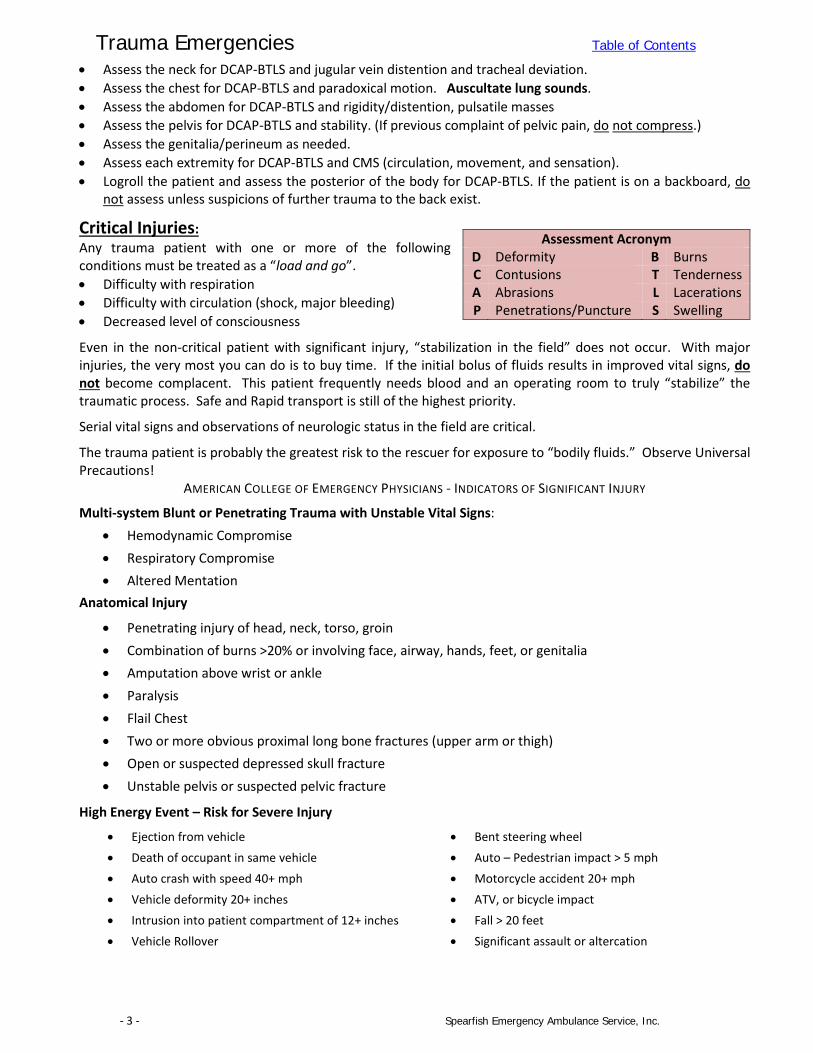
Assessment Acronym D Deformity B Burns C Contusions T Tenderness A Abrasions L Lacerations P Penetrations/Puncture S Swelling
High Energy Event – Risk for Severe Injury
• Ejection from vehicle • Death of occupant in same vehicle • Auto crash with speed 40+ mph • Vehicle deformity 20+ inches • Intrusion into patient compartment of 12+ inches • Vehicle Rollover
• Bent steering wheel • Auto – Pedestrian impact > 5 mph • Motorcycle accident 20+ mph • ATV, or bicycle impact • Fall > 20 feet • Significant assault or altercation

Trauma Emergencies Table of Contents
- 4 - Spearfish Emergency Ambulance Service, Inc.
GENERAL TREATMENT PRIORITIES
EMT:
− Management of Airway and Breathing with adjuncts, O2 and BVM with C-spine immobilization.
− Oxygen may be used in certain situations on ‘High Flow’ while less urgent patient condition only require an SpO2 > 94%
− Protect airway with Combi-tube if necessary.
− Circulation, with control of major bleeding.
− Transport decision;
• If patient unstable, transport immediately. Treat en-route.
• If patient stable, assess for potentially life threatening injuries and treat accordingly.
− Manage injuries per specific protocol.
Ongoing Assessment:
- This assessment is done every 15 minutes for stable patients, and every 5 minutes for unstable patients.
Repeat Initial Assessment.
Re-assess vital signs.
Check interventions
EMT-I85 & Advanced EMT:
− IV, NS, preferably with 2 large bore needles. Run fluids to maintain B/P of 100 systolic.
− Consider Cardiac Monitor.
Paramedic:
− If patient is unconscious, protect the airway endotracheally.
− Follow guidelines for chest decompression if needed.
− Consider medication for pain control if bleeding is NOT suspected in the head, Chest, or Abdomen. Call Medical Control for guidelines. (Pain control may be safe for isolated extremity injuries).
Trauma Emergencies Table of Contents
- 5 - Spearfish Emergency Ambulance Service, Inc.
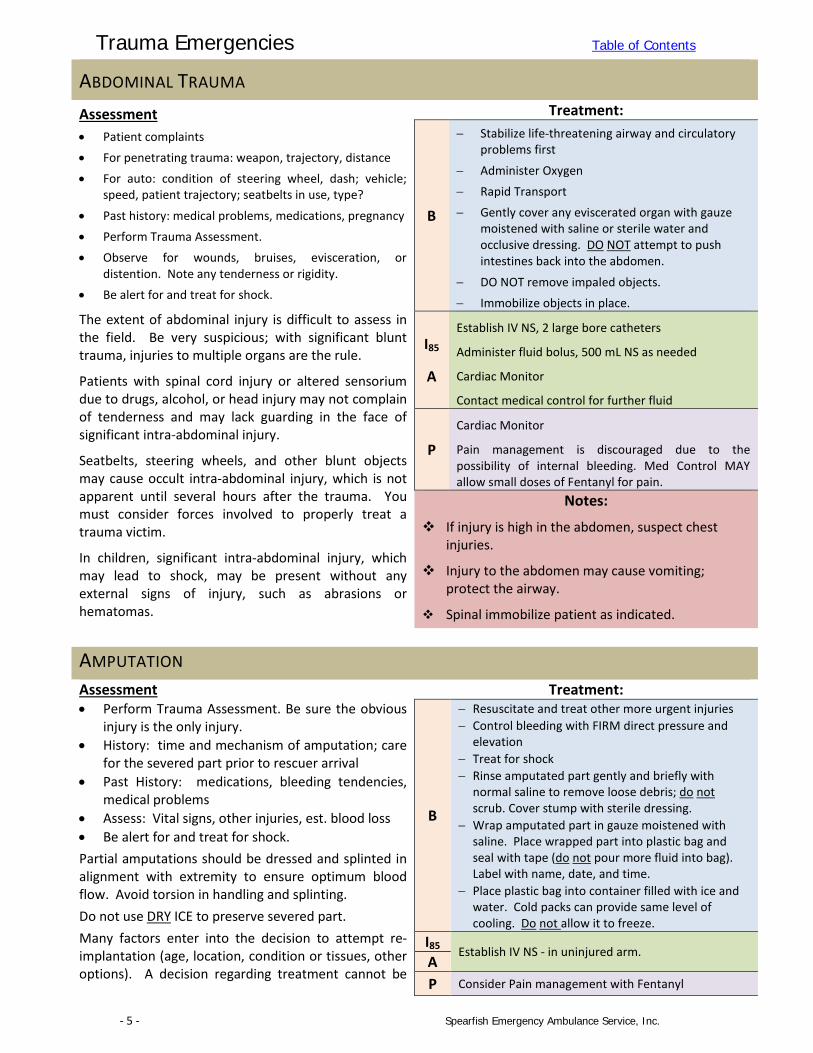
ABDOMINAL TRAUMA
Assessment • Patient complaints • For penetrating trauma: weapon, trajectory, distance • For auto: condition of steering wheel, dash; vehicle;
speed, patient trajectory; seatbelts in use, type?
• Past history: medical problems, medications, pregnancy • Perform Trauma Assessment. • Observe for wounds, bruises, evisceration, or
distention. Note any tenderness or rigidity. • Be alert for and treat for shock.
The extent of abdominal injury is difficult to assess in the field. Be very suspicious; with significant blunt trauma, injuries to multiple organs are the rule.
Patients with spinal cord injury or altered sensorium due to drugs, alcohol, or head injury may not complain of tenderness and may lack guarding in the face of significant intra-abdominal injury.
Seatbelts, steering wheels, and other blunt objects may cause occult intra-abdominal injury, which is not apparent until several hours after the trauma. You must consider forces involved to properly treat a trauma victim.
In children, significant intra-abdominal injury, which may lead to shock, may be present without any external signs of injury, such as abrasions or hematomas.
Treatment:
B
− Stabilize life-threatening airway and circulatory problems first
− Administer Oxygen − Rapid Transport − Gently cover any eviscerated organ with gauze
moistened with saline or sterile water and occlusive dressing. DO NOT attempt to push intestines back into the abdomen.
− DO NOT remove impaled objects. − Immobilize objects in place.
I85
A
Establish IV NS, 2 large bore catheters
Administer fluid bolus, 500 mL NS as needed
Cardiac Monitor
Contact medical control for further fluid
P
Cardiac Monitor
Pain management is discouraged due to the possibility of internal bleeding. Med Control MAY allow small doses of Fentanyl for pain.
Notes:
If injury is high in the abdomen, suspect chest injuries.
Injury to the abdomen may cause vomiting; protect the airway.
Spinal immobilize patient as indicated.
AMPUTATION Assessment • Perform Trauma Assessment. Be sure the obvious
injury is the only injury. • History: time and mechanism of amputation; care
for the severed part prior to rescuer arrival • Past History: medications, bleeding tendencies,
medical problems • Assess: Vital signs, other injuries, est. blood loss • Be alert for and treat for shock. Partial amputations should be dressed and splinted in alignment with extremity to ensure optimum blood flow. Avoid torsion in handling and splinting. Do not use DRY ICE to preserve severed part. Many factors enter into the decision to attempt re-implantation (age, location, condition or tissues, other options). A decision regarding treatment cannot be
Treatment:
B
− Resuscitate and treat other more urgent injuries − Control bleeding with FIRM direct pressure and
elevation − Treat for shock − Rinse amputated part gently and briefly with
normal saline to remove loose debris; do not scrub. Cover stump with sterile dressing.
− Wrap amputated part in gauze moistened with saline. Place wrapped part into plastic bag and seal with tape (do not pour more fluid into bag). Label with name, date, and time.
− Place plastic bag into container filled with ice and water. Cold packs can provide same level of cooling. Do not allow it to freeze.
I85 Establish IV NS - in uninjured arm.
A P Consider Pain management with Fentanyl
Trauma Emergencies Table of Contents
- 6 - Spearfish Emergency Ambulance Service, Inc.
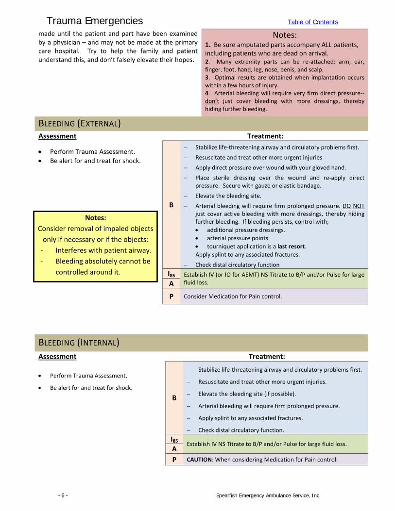
made until the patient and part have been examined by a physician – and may not be made at the primary care hospital. Try to help the family and patient understand this, and don’t falsely elevate their hopes.
Notes: 1. Be sure amputated parts accompany ALL patients, including patients who are dead on arrival. 2. Many extremity parts can be re-attached: arm, ear, finger, foot, hand, leg, nose, penis, and scalp. 3. Optimal results are obtained when implantation occurs within a few hours of injury. 4. Arterial bleeding will require very firm direct pressure--don’t just cover bleeding with more dressings, thereby hiding further bleeding.
BLEEDING (EXTERNAL) Assessment
• Perform Trauma Assessment. • Be alert for and treat for shock.
Treatment:
B
− Stabilize life-threatening airway and circulatory problems first. − Resuscitate and treat other more urgent injuries − Apply direct pressure over wound with your gloved hand. − Place sterile dressing over the wound and re-apply direct
pressure. Secure with gauze or elastic bandage. − Elevate the bleeding site. − Arterial bleeding will require firm prolonged pressure. DO NOT
just cover active bleeding with more dressings, thereby hiding further bleeding. If bleeding persists, control with; • additional pressure dressings. • arterial pressure points. • tourniquet application is a last resort.
− Apply splint to any associated fractures.
− Check distal circulatory function I85 Establish IV (or IO for AEMT) NS Titrate to B/P and/or Pulse for large
fluid loss. A
P Consider Medication for Pain control.
BLEEDING (INTERNAL) Assessment
• Perform Trauma Assessment.
• Be alert for and treat for shock.
Treatment:
B
− Stabilize life-threatening airway and circulatory problems first.
− Resuscitate and treat other more urgent injuries.
− Elevate the bleeding site (if possible).
− Arterial bleeding will require firm prolonged pressure.
− Apply splint to any associated fractures.
− Check distal circulatory function. I85
Establish IV NS Titrate to B/P and/or Pulse for large fluid loss. A P CAUTION: When considering Medication for Pain control.
Notes: Consider removal of impaled objects
only if necessary or if the objects: - Interferes with patient airway. - Bleeding absolutely cannot be
controlled around it.
Trauma Emergencies Table of Contents
- 7 - Spearfish Emergency Ambulance Service, Inc.
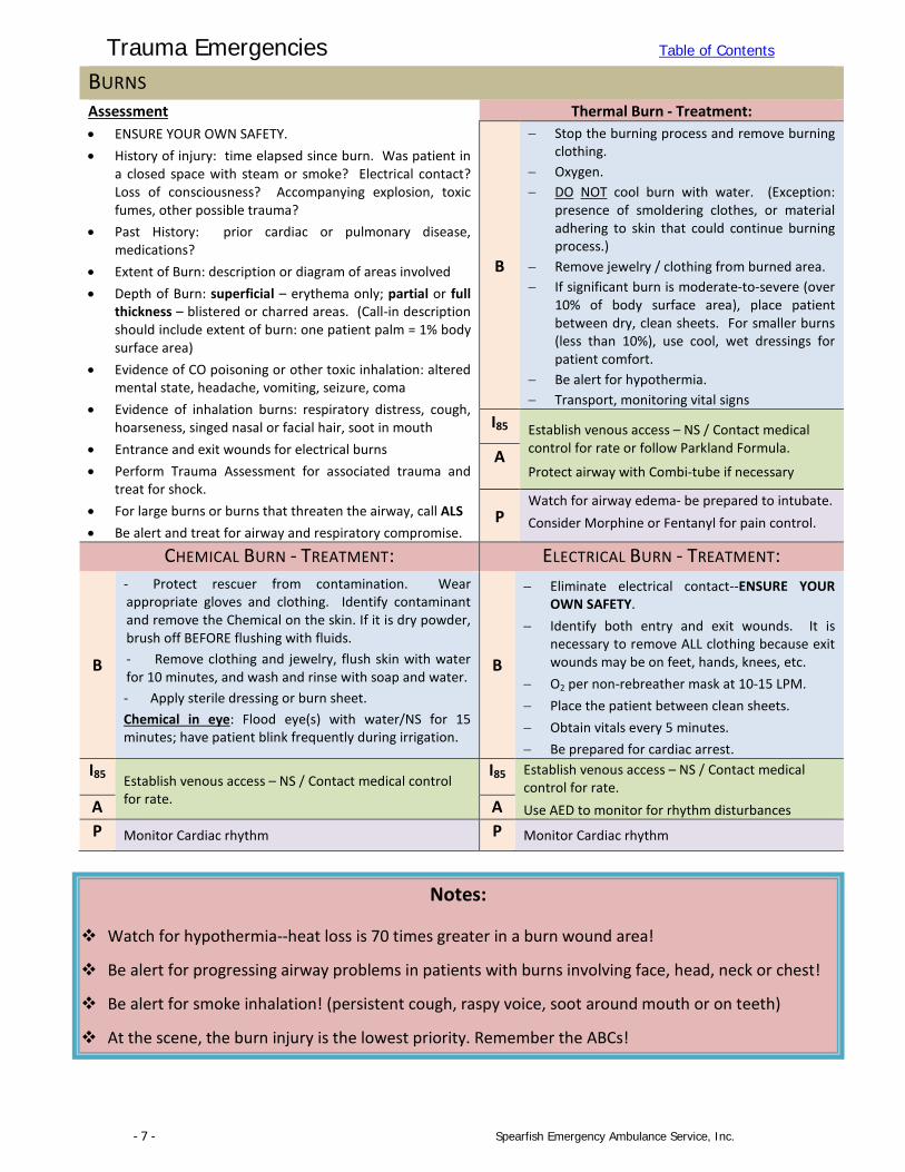
BURNS Assessment • ENSURE YOUR OWN SAFETY. • History of injury: time elapsed since burn. Was patient in
a closed space with steam or smoke? Electrical contact? Loss of consciousness? Accompanying explosion, toxic fumes, other possible trauma?
• Past History: prior cardiac or pulmonary disease, medications?
• Extent of Burn: description or diagram of areas involved • Depth of Burn: superficial – erythema only; partial or full
thickness – blistered or charred areas. (Call-in description should include extent of burn: one patient palm = 1% body surface area)
• Evidence of CO poisoning or other toxic inhalation: altered mental state, headache, vomiting, seizure, coma
• Evidence of inhalation burns: respiratory distress, cough, hoarseness, singed nasal or facial hair, soot in mouth
• Entrance and exit wounds for electrical burns • Perform Trauma Assessment for associated trauma and
treat for shock. • For large burns or burns that threaten the airway, call ALS • Be alert and treat for airway and respiratory compromise.
Thermal Burn - Treatment:
B
− Stop the burning process and remove burning clothing.
− Oxygen. − DO NOT cool burn with water. (Exception:
presence of smoldering clothes, or material adhering to skin that could continue burning process.)
− Remove jewelry / clothing from burned area. − If significant burn is moderate-to-severe (over
10% of body surface area), place patient between dry, clean sheets. For smaller burns (less than 10%), use cool, wet dressings for patient comfort.
− Be alert for hypothermia. − Transport, monitoring vital signs
I85 Establish venous access – NS / Contact medical control for rate or follow Parkland Formula.
Protect airway with Combi-tube if necessary A
P Watch for airway edema- be prepared to intubate. Consider Morphine or Fentanyl for pain control.
CHEMICAL BURN - TREATMENT: ELECTRICAL BURN - TREATMENT:
B
- Protect rescuer from contamination. Wear appropriate gloves and clothing. Identify contaminant and remove the Chemical on the skin. If it is dry powder, brush off BEFORE flushing with fluids. - Remove clothing and jewelry, flush skin with water for 10 minutes, and wash and rinse with soap and water. - Apply sterile dressing or burn sheet. Chemical in eye: Flood eye(s) with water/NS for 15 minutes; have patient blink frequently during irrigation.
B
− Eliminate electrical contact--ENSURE YOUR OWN SAFETY.
− Identify both entry and exit wounds. It is necessary to remove ALL clothing because exit wounds may be on feet, hands, knees, etc.
− O2 per non-rebreather mask at 10-15 LPM. − Place the patient between clean sheets. − Obtain vitals every 5 minutes. − Be prepared for cardiac arrest.
I85 Establish venous access – NS / Contact medical control for rate.
I85 Establish venous access – NS / Contact medical control for rate. Use AED to monitor for rhythm disturbances A A
P Monitor Cardiac rhythm P Monitor Cardiac rhythm
Notes:
Watch for hypothermia--heat loss is 70 times greater in a burn wound area!
Be alert for progressing airway problems in patients with burns involving face, head, neck or chest!
Be alert for smoke inhalation! (persistent cough, raspy voice, soot around mouth or on teeth)
At the scene, the burn injury is the lowest priority. Remember the ABCs!
Trauma Emergencies Table of Contents
- 8 - Spearfish Emergency Ambulance Service, Inc.
Specific Precautions Leave blisters intact when possible Suspect airway burns in any facial burns or burns received in closed spaces. Edema may become severe, but
not be immediately apparent. Avoid unnecessary trauma to airway. Humidified oxygen is useful if available. Assume carbon monoxide poisoning in all closed space burns. Treatment is 100% Oxygen continued for
several hours. In addition, other toxic products of combustion are more commonly encountered than realized. Contact medical control for special instructions if other toxic inhalations are suspected.
Consider suicide attempt as cause of burn, and child abuse in pediatric burns. Lightning injuries can cause prolonged respiratory arrest. Prompt, continuous respiratory assistance
(sometimes for hours to days) can result in full recovery. Field decontamination of chemical exposures has been shown to significantly reduce extent of burn. It is
rare to encounter a chemical, which is not properly decontaminated by copious water. Unless a specific contraindication is known, do not waste time before initiating treatment to find out the specific culprit.
EMS personnel should not participate in decontamination unless trained and equipped to do so.
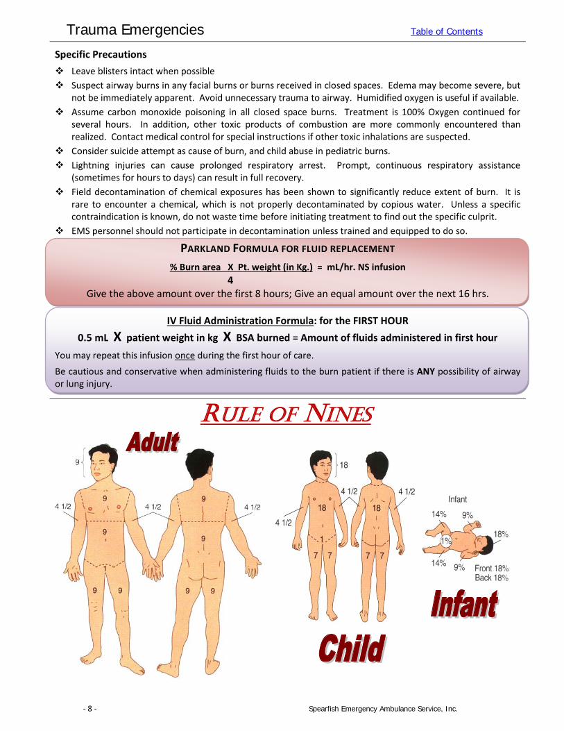
PARKLAND FORMULA FOR FLUID REPLACEMENT % Burn area X Pt. weight (in Kg.) = mL/hr. NS infusion
4 Give the above amount over the first 8 hours; Give an equal amount over the next 16 hrs.
IV Fluid Administration Formula: for the FIRST HOUR
0.5 mL X patient weight in kg X BSA burned = Amount of fluids administered in first hour You may repeat this infusion once during the first hour of care. Be cautious and conservative when administering fluids to the burn patient if there is ANY possibility of airway or lung injury.
RULE OF NINES
Trauma Emergencies Table of Contents
- 9 - Spearfish Emergency Ambulance Service, Inc.
CHEST INJURIES
Assessment • Perform Trauma Assessment. • Patient complaints: chest pain (type), respiratory
distress, neck pain, other areas of injury. • Note mechanism: amount of force involved (particularly
deceleration), speed of impact, seatbelt use/type; Penetrating trauma; size of object, caliber of bullet, trajectory.
• Observe: wounds, air leaks, chest wall movement, neck veins.
• Palpate: tenderness, crepitus, tracheal position, tenderness on sternal compression, pulse pressure.
• Auscultate: breath sounds, heart sounds (quality). • Surroundings: vehicle, steering wheel condition. • Be alert for and treat for shock. • Transport immediately with stabilization en-route to
hospital. • Assess Neurologic status.
Special Considerations
Hypovolemia secondary to major injury is common.
Underlying respiratory conditions should also be considered as causes of respiratory distress.
Specific Precautions
Chest trauma is treated with difficulty in the field and prolonged treatment is not indicated if significant injury is suspected. If patient is critical, transport rapidly and avoid treatment of non-emergent problems at the scene. Penetrating injury particularly should receive immediate transport with minimal intervention in the field.
Consider medical causes of respiratory distress such as asthma, pulmonary edema or COPD that have either caused trauma or been aggravated by it.
Chest injuries sufficient to cause respiratory distress are commonly associated with significant blood loss. Look for Hypovolemia.
Myocardial contusion can occur, particularly with anterior chest wall injury, as from a steering wheel. Pain is similar to myocardial infarct pain. Monitor the patient and treat arrhythmias as in a medical patient, but think first of hypoxia and Hypovolemia as potential causes of arrhythmias.
Don’t forget to check the back for injuries, especially the patient in shock, where a cause is not evident (check the back, axillary region, and base of neck).
Significant intra-thoracic injuries can exist without any external signs of injury.
Treatment:
B
− Airway management – with C-spine control. Assist breathing if patient is apneic or respirations depressed
− Oxygen 10-15 L/min. by mask
− Treat open chest wounds with Vaseline-gauze occlusive dressing or plastic wrap to allow air to escape but not enter the chest
− Stabilize rib fractures or flail segments with direct pressure, then bulky dressing secured to chest wall
− Control bleeding with direct pressure − Impaled object should be stabilized in place with
an occlusive dressing
− Place patient in position of comfort. Consider spinal immobilization if appropriate; or if possible, place patient on injured side.
− Monitor vital signs
− Re-assess neurologic status
I85 Establish large bore IV NS with Blood tubing if necessary. If hypotensive - bolus 250cc-500cc fluid challenge or as directed by Physician. Reassess Vital signs Monitor Cardiac rhythm A
P
Monitor cardiac rhythm
Needle thoracostomy to ventilate chest if signs of tension pneumothorax, NOTIFY MEDICAL CONTROL, so they can prepare for a chest tube upon patient arrival in ED.
Lidocaine if PVC’s > 6/min, multifocal or runs are present with cardiac or severe chest wall contusion; give 1 mg/kg bolus; if unsuccessful, repeat 0.5 mg/kg IV bolus after 8 to10 min.;
Relative contraindications - any heart block, bradycardia, atrial fibrillation, atrial flutter, hypotension, patient >70 years old.
Myocardial contusion can cause pain similar to that of myocardial infarction; hypoxemia & hypovolemia should be considered first as potential causes of arrhythmia.
If Patient goes into cardiac arrest, follow specific ACLS algorithm.
Trauma Emergencies Table of Contents
- 10 - Spearfish Emergency Ambulance Service, Inc.
EXTREMITY INJURIES
Assessment • Perform Trauma Assessment or History and Physical
Exam. Note: areas of pain or limited movement.
• Be alert for and treat for shock. • Note mechanism of injury: direction of forces, if known. • Vital signs • Observe: localized swelling, discoloration, angulation,
lacerations, exposed bone fragments, loss of function, guarding
• Palpate: tenderness, crepitus, instability, quality or distal pulses, sensation
• Note estimated blood loss at scene Check circulation, movement, and sensation distal to the injury before and after splinting.
Notes: Pelvic and femur fractures can cause severe
hemorrhage; anticipate and treat for shock!
Failure to immobilize fractures can do greater harm than the original injury.
When using air splints, avoid over-inflation, which can result in circulatory or neurological compromise.
Observe for changes when moving from cold to warm temperatures.
If other injuries are present, the long spine board will
provide adequate total body splinting
Treatment:
B
− Treat airway, breathing, and hypotension FIRST − Immobilize cervical spine when appropriate.
Examine for additional injuries to head, face, chest, and abdomen; treat problems with higher priority first.
− If patient unstable, transport rapidly, treat life-threatening problems en-route. Splint patient by securing to long board to minimize movement.
− Do not allow the obvious fracture to obscure other assessment findings.
− Protect the injury from excessive movement. Immobilize one joint above and below suspected injury
− DO NOT realign dislocation/fracture in the field unless circulation is compromised. Realignment of fracture may be necessary to facilitate splinting, correct a circulatory compromise or neurological deficit. However, careful assessment before and after manipulation and minimal movement of injury site is critical.
− Fractures involving joint (or within 3 inches of a joint) should be splinted in the position found.
− Elevate and apply cold packs to injury when practical. Do not apply cold packs directly on skin.
I85 Establish IV NS for long bone, pelvic or multiple Fractures A
P Consider medication for pain control.
Trauma Emergencies Table of Contents
- 11 - Spearfish Emergency Ambulance Service, Inc.
FACE AND NECK TRAUMA
Assessment DO NOT HYPEREXTEND NECK. • Perform Trauma Assessment. • Note mechanism of injury: impact to steering wheel,
windshield or other objects, clothes-line type injury to face or neck
• Assume cervical spine injury to be present in all trauma patients.
• Patient complaints: areas of pain; trouble with vision, hearing; neck pain; abnormal bite pattern; short of breath.
• Perform neurological exam assessing level of consciousness. A - Patient is alert V - Patient responds to voice stimuli P - Patient responds to painful stimuli U - Patient is unresponsive
• Obtain history (i.e., helmet or seat belt use) and level of consciousness since injury.
• Note cerebrospinal fluid from ears, nose, and mouth. • Airway: jaw or tongue instability, loose teeth, vomitus or
blood in airway, other evidence of impairment or obstruction
• Injury to eye: lid laceration, blood anterior to pupil, abnormal pupil, abnormal globe position
Check for associated injuries.
Specific Precautions: Fracture of the larynx should be suspected in patients
with respiratory distress, abnormal voice, and history of direct blow to neck from steering wheel, rope, fence wire, etc. Both intubation and cricothyrotomy may be unsuccessful in the patient with a fractured larynx, and attempts may precipitate respiratory arrest. Transport rapidly for definitive treatment if you suspect this potentially lethal injury. Do not attempt intubation or cricothyrotomy unless the patient is in severe respiratory distress.
Airway obstruction is the primary cause of death in persons sustaining head and face trauma. Meticulous attention to suctioning, and stabilization of tongue and mandible may be the most important treatment rendered.
Remember that the apex of the lung extends into the lower neck and may be injured in penetrating injuries of the lower neck, resulting in pneumothorax or hemothorax.
Do not be concerned with contact lens removal in the field.
Treatment:
B
− Secure airway maintaining C-Spine control. Support breathing and administer 100% oxygen via non-rebreather mask. If ventilation assistance is needed, use bag-valve- mask with reservoir (100% O2) at 24 times per minute. (Remember to have suction immediately available).
− Realign neck to a neutral, in-line position unless resistance is met. Manually stabilize head, neck and spine until secured on appropriate device.
− Open airway using jaw thrust, use finger sweep to remove oral foreign bodies.
− Note evidence of laryngeal injury and transport immediately if any signs present.
− Stop hemorrhage; Check circulation. Treat for shock.
− Continuously monitor vital signs and record neurological status.
o EYE Injuries: Cover both eyes with protective shield or cup – avoid pressure or direct contact to eye.
o DO NOT attempt to stop free drainage from ears. Cover lightly with dressing to avoid contamination.
o Bring avulsed teeth with you. Keep moist in saline-soaked gauze
I85 Protect airway with Combitube if necessary
Establish IV NS. Titrate to B/P A
P
Airway obstruction is the primary cause of death from facial and neck trauma. Meticulous attention is imperative. If intubation cannot be performed due to severe facial injury, attempt to manage with suctioning and bag-valve-mask.
If necessary, consider cricothyrotomy (see Advanced Airway Management: Cricothyrotomy protocol)
Fracture of larynx should be suspected in patients with respiratory distress, abnormal voice, and a history of a direct blow to the neck. Transport patient with this potentially lethal injury immediately
Protect airway Endotracheally if unresponsive.
Cardiac Monitor
Trauma Emergencies Table of Contents
- 12 - Spearfish Emergency Ambulance Service, Inc.
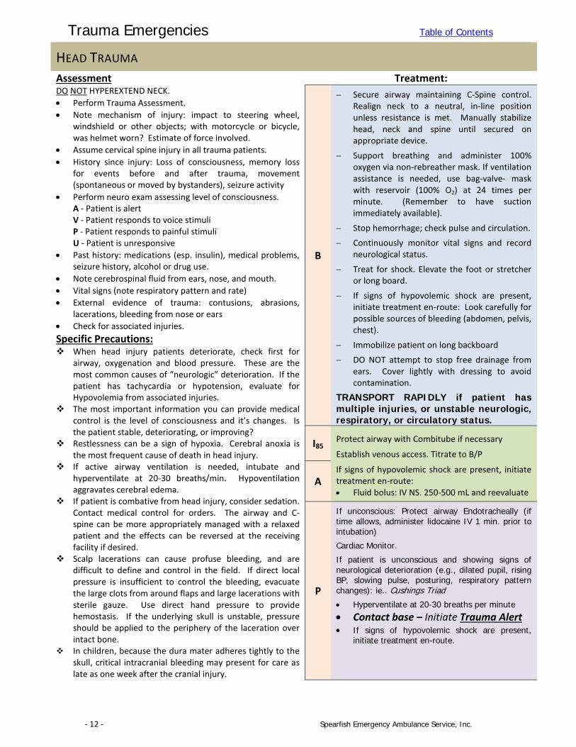
HEAD TRAUMA Assessment DO NOT HYPEREXTEND NECK. • Perform Trauma Assessment. • Note mechanism of injury: impact to steering wheel,
windshield or other objects; with motorcycle or bicycle, was helmet worn? Estimate of force involved.
• Assume cervical spine injury in all trauma patients. • History since injury: Loss of consciousness, memory loss
for events before and after trauma, movement (spontaneous or moved by bystanders), seizure activity
• Perform neuro exam assessing level of consciousness. A - Patient is alert V - Patient responds to voice stimuli P - Patient responds to painful stimuli U - Patient is unresponsive
• Past history: medications (esp. insulin), medical problems, seizure history, alcohol or drug use.
• Note cerebrospinal fluid from ears, nose, and mouth. • Vital signs (note respiratory pattern and rate) • External evidence of trauma: contusions, abrasions,
lacerations, bleeding from nose or ears • Check for associated injuries. Specific Precautions: When head injury patients deteriorate, check first for
airway, oxygenation and blood pressure. These are the most common causes of “neurologic” deterioration. If the patient has tachycardia or hypotension, evaluate for Hypovolemia from associated injuries.
The most important information you can provide medical control is the level of consciousness and it’s changes. Is the patient stable, deteriorating, or improving?
Restlessness can be a sign of hypoxia. Cerebral anoxia is the most frequent cause of death in head injury.
If active airway ventilation is needed, intubate and hyperventilate at 20-30 breaths/min. Hypoventilation aggravates cerebral edema.
If patient is combative from head injury, consider sedation. Contact medical control for orders. The airway and C-spine can be more appropriately managed with a relaxed patient and the effects can be reversed at the receiving facility if desired.
Scalp lacerations can cause profuse bleeding, and are difficult to define and control in the field. If direct local pressure is insufficient to control the bleeding, evacuate the large clots from around flaps and large lacerations with sterile gauze. Use direct hand pressure to provide hemostasis. If the underlying skull is unstable, pressure should be applied to the periphery of the laceration over intact bone.
In children, because the dura mater adheres tightly to the skull, critical intracranial bleeding may present for care as late as one week after the cranial injury.
Treatment:
B
− Secure airway maintaining C-Spine control. Realign neck to a neutral, in-line position unless resistance is met. Manually stabilize head, neck and spine until secured on appropriate device.
− Support breathing and administer 100% oxygen via non-rebreather mask. If ventilation assistance is needed, use bag-valve- mask with reservoir (100% O2) at 24 times per minute. (Remember to have suction immediately available).
− Stop hemorrhage; check pulse and circulation. − Continuously monitor vital signs and record
neurological status.
− Treat for shock. Elevate the foot or stretcher or long board.
− If signs of hypovolemic shock are present, initiate treatment en-route: Look carefully for possible sources of bleeding (abdomen, pelvis, chest).
− Immobilize patient on long backboard − DO NOT attempt to stop free drainage from
ears. Cover lightly with dressing to avoid contamination.
TRANSPORT RAPIDLY if patient has multiple injuries, or unstable neurologic, respiratory, or circulatory status.
I85 Protect airway with Combitube if necessary
Establish venous access. Titrate to B/P
If signs of hypovolemic shock are present, initiate treatment en-route: • Fluid bolus: IV NS. 250-500 mL and reevaluate
A
P
If unconscious: Protect airway Endotracheally (if time allows, administer lidocaine IV 1 min. prior to intubation)
Cardiac Monitor.
If patient is unconscious and showing signs of neurological deterioration (e.g., dilated pupil, rising BP, slowing pulse, posturing, respiratory pattern changes): ie.. Cushings Triad
• Hyperventilate at 20-30 breaths per minute • Contact base – Initiate Trauma Alert • If signs of hypovolemic shock are present,
initiate treatment en-route.
Trauma Emergencies Table of Contents
- 13 - Spearfish Emergency Ambulance Service, Inc.
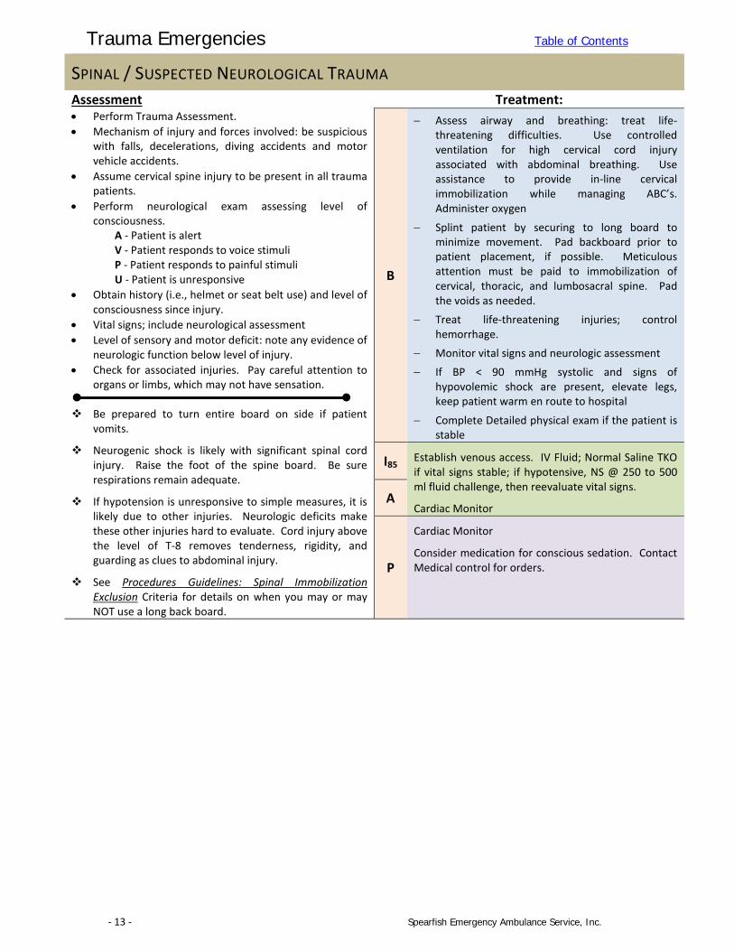
SPINAL / SUSPECTED NEUROLOGICAL TRAUMA Assessment • Perform Trauma Assessment. • Mechanism of injury and forces involved: be suspicious
with falls, decelerations, diving accidents and motor vehicle accidents.
• Assume cervical spine injury to be present in all trauma patients.
• Perform neurological exam assessing level of consciousness.
A - Patient is alert V - Patient responds to voice stimuli P - Patient responds to painful stimuli U - Patient is unresponsive
• Obtain history (i.e., helmet or seat belt use) and level of consciousness since injury.
• Vital signs; include neurological assessment • Level of sensory and motor deficit: note any evidence of
neurologic function below level of injury. • Check for associated injuries. Pay careful attention to
organs or limbs, which may not have sensation.
Be prepared to turn entire board on side if patient vomits.
Neurogenic shock is likely with significant spinal cord injury. Raise the foot of the spine board. Be sure respirations remain adequate.
If hypotension is unresponsive to simple measures, it is likely due to other injuries. Neurologic deficits make these other injuries hard to evaluate. Cord injury above the level of T-8 removes tenderness, rigidity, and guarding as clues to abdominal injury.
See Procedures Guidelines: Spinal Immobilization Exclusion Criteria for details on when you may or may NOT use a long back board.
Treatment:
B
− Assess airway and breathing: treat life-threatening difficulties. Use controlled ventilation for high cervical cord injury associated with abdominal breathing. Use assistance to provide in-line cervical immobilization while managing ABC’s. Administer oxygen
− Splint patient by securing to long board to minimize movement. Pad backboard prior to patient placement, if possible. Meticulous attention must be paid to immobilization of cervical, thoracic, and lumbosacral spine. Pad the voids as needed.
− Treat life-threatening injuries; control hemorrhage.
− Monitor vital signs and neurologic assessment − If BP < 90 mmHg systolic and signs of
hypovolemic shock are present, elevate legs, keep patient warm en route to hospital
− Complete Detailed physical exam if the patient is stable
I85 Establish venous access. IV Fluid; Normal Saline TKO if vital signs stable; if hypotensive, NS @ 250 to 500 ml fluid challenge, then reevaluate vital signs.
Cardiac Monitor A
P
Cardiac Monitor
Consider medication for conscious sedation. Contact Medical control for orders.
Trauma Emergencies Table of Contents
- 14 - Spearfish Emergency Ambulance Service, Inc.
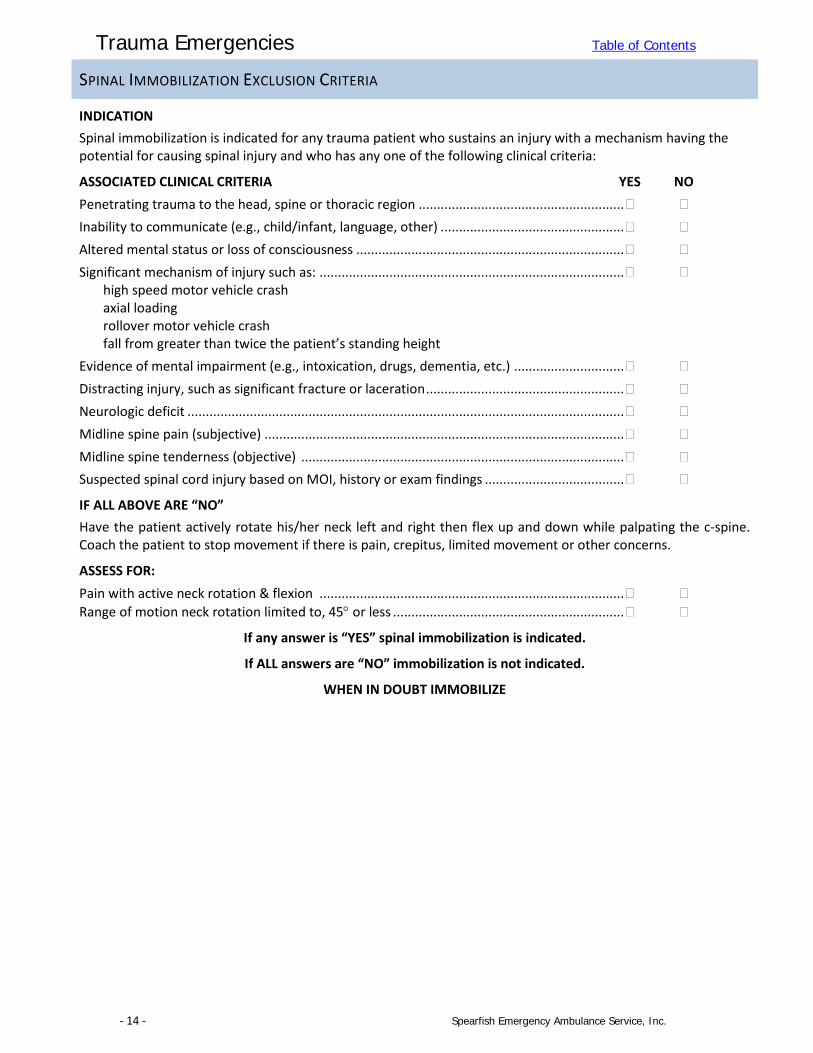
SPINAL IMMOBILIZATION EXCLUSION CRITERIA
INDICATION Spinal immobilization is indicated for any trauma patient who sustains an injury with a mechanism having the potential for causing spinal injury and who has any one of the following clinical criteria:
ASSOCIATED CLINICAL CRITERIA YES NO Penetrating trauma to the head, spine or thoracic region ........................................................ � � Inability to communicate (e.g., child/infant, language, other) .................................................. � � Altered mental status or loss of consciousness ......................................................................... � � Significant mechanism of injury such as: ................................................................................... � �
high speed motor vehicle crash axial loading rollover motor vehicle crash fall from greater than twice the patient’s standing height
Evidence of mental impairment (e.g., intoxication, drugs, dementia, etc.) .............................. � � Distracting injury, such as significant fracture or laceration ...................................................... � � Neurologic deficit ....................................................................................................................... � � Midline spine pain (subjective) .................................................................................................. � � Midline spine tenderness (objective) ........................................................................................ � � Suspected spinal cord injury based on MOI, history or exam findings ...................................... � �
IF ALL ABOVE ARE “NO” Have the patient actively rotate his/her neck left and right then flex up and down while palpating the c-spine. Coach the patient to stop movement if there is pain, crepitus, limited movement or other concerns.
ASSESS FOR: Pain with active neck rotation & flexion ................................................................................... � � Range of motion neck rotation limited to, 45° or less ............................................................... � �
If any answer is “YES” spinal immobilization is indicated.
If ALL answers are “NO” immobilization is not indicated.
WHEN IN DOUBT IMMOBILIZE
Trauma Emergencies Table of Contents
- 15 - Spearfish Emergency Ambulance Service, Inc.
SEXUAL ASSAULT Assessment • Protect the scene and preserve evidence in cooperation
with law enforcement.
• Clothing is to be placed in a PAPER bag. • Crewmembers of the same sex may relate better with
the patient in time of emotional crisis.
• Do not allow patient to bathe, douche, brush teeth, defecate, urinate, or change clothes.
• Notify law enforcement. Notes:
This is a highly emotional and volatile situation; be sure yo
findings and treatment are clearly documented on the
Patient Care Report.
Do not let emotions and anxiety cause you to miss underlying injuries and to take priority over crime scene preservation.
Treatment:
B
− Stabilize life-threatening airway and circulatory problems first
− Resuscitate and treat other more urgent injuries − Reassure patient and provide emotional support.
− Administer O2 if indicated. − Be alert for and treat for shock. − Treat other injuries as indicated. See specific
protocols.
− Transfer to a hospital capable of handling sexual assault.
I85 Establish IV NS if Indicated
A
P Pain Control as needed for other injuries
SHOCK / HYPOPERFUSION Assessment • Perform Trauma Assessment. • Early shock:
o Restlessness and anxiety o Tachycardia, > 110 BPM o Pale, cool, wet skin o Narrowed pulse pressure o Nausea and vomiting o Weakness
• Late shock. The above signs and symptoms plus:
o Tachypnea o Diaphoresis o Decreased level of consciousness o Cyanosis o Hypotension < 90 systolic
Treatment:
B
− Control profuse external bleeding.
− Immobilize the spine.
− O2 by non-rebreather mask at 10-15 LPM.
− Elevate lower extremities 8-12 inches
− Maintain body Temperature
− Consider use of PASG to maintain blood pressure.
− Transport as soon as efficiently possible.
− Repeat Ongoing Assessment every 5 minutes.
I85 Establish venous access. IV Fluid; Normal Saline TKO if vital signs stable; if hypotensive, NS @ 250 to 500 ml fluid challenge, then reevaluate vital signs.
Monitor Cardiac rhythm A
P
Aggressively treat signs of shock especially with evidence of mechanisms that could produce bleeding
Notes
Most field treatments for shock should be done in the ambulance on the way to the
hospital.
Trauma Emergencies Table of Contents
- 16 - Spearfish Emergency Ambulance Service, Inc.
TRAUMA ALERT CRITERIA Rationale: The morbidity and mortality of the seriously injured trauma patient can be reduced by decreasing the amount of time between when the injury occurred and when the patient receives definitive, in-hospital evaluation and treatment. Pre-hospital providers can first: identify in the field, those seriously injured trauma patients that will fit Trauma Alert criteria. Secondly, notification should be made to Spearfish Regional Hospital or other facility that a potential Trauma Alert patient has been encountered. Lastly these patients should receive rapid transport to the hospital; with as short of scene times as possible and most pre-hospital treatment performed en-route. Trauma Alerts should be “called” in the field as soon as possible. When a patient is encountered that meets Trauma Alert criteria the Paramedic (or other crew member) will contact the Hospital ER and advise that you have a patient that meets Trauma Alert criteria. This should be followed by a short report detailing the patient’s condition and the nature of their injuries. Trauma Alert criteria found should specifically be included in this short report. A more detailed report should be given later en-route to the hospital as time allows. A Trauma Alert will be called for a patient involved in a trauma event who demonstrates any of the following
physiologic or anatomic absolutes: Glasgow Coma Scale of < 10 Blood pressure < 90 (age specific hypotension for children) Pulse > 120 (age specific tachycardia for children) Respirations <10 or > 29, or airway obstruction or respiratory compromise requiring use of advanced airway
(age specific for children) Penetrating injury to chest, abdomen, head, neck Limb paralysis (associated with trauma) Flail chest Amputation proximal to wrist or ankle Partial Thickness burns of total body surface area greater than 10% Severe burns involving face, airway, hands, feet, genitalia, or major joints
Strong degree of suspicion should be used for the following patients, but this does not constitute an automatic categorization of a severe trauma patient:
o Pelvic fractures o Falls from 2 times the height of the patient o Patients involved in high energy MVC’s or MCC’s o Death of an occupant in the same compartment o Auto-Pedestrian or Auto-bicycle with impact of greater than 5MPH o Pedestrian that was thrown or run-over o Significant recreational vehicle or farm equipment incident o Significant injury associated with a large animal o Patients age <13 or > 55 o Pregnancy o Chronic medical illness
*A trauma alert can be called under the discretion of the provider at any time* QA Parameters:
EMS crews MUST document the Trauma Alert activation in your PCR. This meets the hospital’s Trauma Center requirements for field activation. Spearfish EMS will review the outcome and care of all patients that meet field criteria for Trauma Alerts that were treated and transported.
Trauma Emergencies Table of Contents
- 17 - Spearfish Emergency Ambulance Service, Inc.
TRAUMA ARREST
Assessment • Call ALS Immediately • Assess LOC and ABC’s; look for life threatening
injuries, control major bleeds. • Time of arrest • Mechanism: blunt vs. penetrating • Signs of irreversible death (decapitation,
dependent lividity, etc.) • Utilize appropriate body substance isolation. • Vital signs • Evidence of massive external blood loss • Evidence of massive blunt head, thorax or
abdominal trauma Victims of blunt trauma arrest without vital signs at the scene after initiation of ALS have a mortality rate approaching 100%.
Trauma arrests secondary to penetrating injuries to the trunk can be resuscitated and saved. There is a higher rate of survival in victims of low velocity penetrating injuries versus victims of high velocity injuries.
Treatment:
B
− Initiate basic life support − Establish and maintain open airway, using c-spine
precautions, modified jaw thrust.
− Suction airway if needed. − If unable to clear airway or unable to ventilate due
to airway trauma, transport immediately.
− Ventilate initially with 100% oxygen utilizing a bag valve mask and an oral airway.
− Perform CPR until AED is available. Follow AED Protocol.
I85 Hyperventilate & insert Combi-Tube as per protocol.
Establish IV NS. Consider fluid bolus of 20cc/kg.
Contact medical control for further fluid rates /amounts A
P
Hyperventilate/ secure airway with endotracheal tube.
Analyze rhythm and treat according to ACLS guidelines.
Hypoxemia and hypovolemia should be considered as potential causes of arrhythmia.
Consider field pronouncement (see Resuscitation and Field Pronouncement Guidelines protocol) for the following:
• Signs of irreversible death • ALS has been unavailable for at least 20 minutes
from the time EMS personnel initiate on-scene assessment and there is no return of vital signs or signs of life
COMPARISON OF PNEUMOTHORAX, HEMOTHORAX AND PERICARDIAL TAMPONADE
Signs/Symptoms Tension Pneumothorax Hemothorax Pericardial Tamponade
Presenting Symptoms Difficulty breathing and then shock
Shock, then difficulty breathing
Narrowing pulse pressure, then shock
Neck Veins Distended Flat Distended
Breath Sounds Decreased or absent on side of injury
Decreased or absent on side of injury Bilateral and clear
Percussion of Chest Hyper-resonant Dull Normal Resonance
Tracheal Deviation Away from side of injury Usually not present Not present

![[XLS] · Web viewCOMPUTER MATHEMATICAL FOUNDATIONS DCAP-202 DCAP-203 RDBMS DCAP-204 PROGRAMMING IN C DCAP-205 LAB-II (C LANGUAGE) DIPLOMA ENGINEERING - (CIVIL) DCEG-101 COMMUNICATION](https://img.pdfslide.us/doc/110x75/5af04d757f8b9ac62b8e7880/xls-viewcomputer-mathematical-foundations-dcap-202-dcap-203-rdbms-dcap-204-programming.jpg)