Embed Size (px)
Citation preview
This document is privileged and confidential Quality Committee or Peer Review work product under Hospital Committee Privilege contained in the TEXAS HEALTH AND SAFETY CODE §§ 161.031 & 161.032, or Medical Peer Review under the Medical Practice Act, TEXAS OCCUPATIONS CODE, § § 151.001 et. seq & 160.007.; and the Medical Peer Review immunity provided by federal law, the Health Care Quality Improvement Act, 42. U.S.C. 11101, et. seq.
1
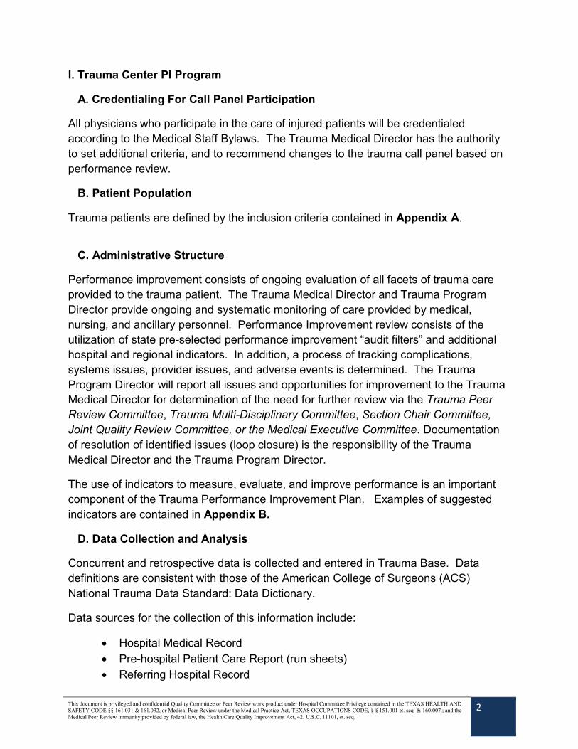
Trauma Center Performance Improvement and Patient Safety Plan
Mission and Goals of the Trauma PI Program
Mission:
Memorial Hermann South West (MHSW) Trauma Services is dedicated to providing comprehensive quality health care for victims of trauma, and community service through education, public awareness of trauma prevention, and regional trauma networking to improve trauma outcomes.
Goal:
The Trauma Program Performance Improvement and Patient Safety (PIPS) Plan is designed to ensure efficient, cost effective, quality patient care that is facilitated by continuous, systematic and objective data analysis and multidisciplinary peer review to identify opportunities to improve patient safety through all phases of trauma care. The ultimate goal is to reduce mortality and morbidity in the trauma patient population.
This document is privileged and confidential Quality Committee or Peer Review work product under Hospital Committee Privilege contained in the TEXAS HEALTH AND SAFETY CODE §§ 161.031 & 161.032, or Medical Peer Review under the Medical Practice Act, TEXAS OCCUPATIONS CODE, § § 151.001 et. seq & 160.007.; and the Medical Peer Review immunity provided by federal law, the Health Care Quality Improvement Act, 42. U.S.C. 11101, et. seq.
2
I. Trauma Center PI Program
A. Credentialing For Call Panel Participation
All physicians who participate in the care of injured patients will be credentialed according to the Medical Staff Bylaws. The Trauma Medical Director has the authority to set additional criteria, and to recommend changes to the trauma call panel based on performance review.
B. Patient Population
Trauma patients are defined by the inclusion criteria contained in Appendix A.
C. Administrative Structure
Performance improvement consists of ongoing evaluation of all facets of trauma care provided to the trauma patient. The Trauma Medical Director and Trauma Program Director provide ongoing and systematic monitoring of care provided by medical, nursing, and ancillary personnel. Performance Improvement review consists of the utilization of state pre-selected performance improvement “audit filters” and additional hospital and regional indicators. In addition, a process of tracking complications, systems issues, provider issues, and adverse events is determined. The Trauma Program Director will report all issues and opportunities for improvement to the Trauma Medical Director for determination of the need for further review via the Trauma Peer Review Committee, Trauma Multi-Disciplinary Committee, Section Chair Committee, Joint Quality Review Committee, or the Medical Executive Committee. Documentation of resolution of identified issues (loop closure) is the responsibility of the Trauma Medical Director and the Trauma Program Director.
The use of indicators to measure, evaluate, and improve performance is an important component of the Trauma Performance Improvement Plan. Examples of suggested indicators are contained in Appendix B.
D. Data Collection and Analysis
Concurrent and retrospective data is collected and entered in Trauma Base. Data definitions are consistent with those of the American College of Surgeons (ACS) National Trauma Data Standard: Data Dictionary.
Data sources for the collection of this information include:
• Hospital Medical Record • Pre-hospital Patient Care Report (run sheets) • Referring Hospital Record
This document is privileged and confidential Quality Committee or Peer Review work product under Hospital Committee Privilege contained in the TEXAS HEALTH AND SAFETY CODE §§ 161.031 & 161.032, or Medical Peer Review under the Medical Practice Act, TEXAS OCCUPATIONS CODE, § § 151.001 et. seq & 160.007.; and the Medical Peer Review immunity provided by federal law, the Health Care Quality Improvement Act, 42. U.S.C. 11101, et. seq.
3
• Medical Examiner Reports
E. Performance Improvement Process
1. Primary Review The Trauma Program Director or designee will do the initial case review of
all trauma patients. Appropriate clinical care without provider or system issues identified will need no further review.
2. Second Level of Review
Opportunities for improvement in the system or provider and sentinel events are referred to the Trauma Medical Director (TMD). The Trauma Medical Director and the Trauma Program Director will perform the second level of review. Further analysis of the case and issue(s) identified will occur. Those cases in which a simple action plan, such as trending of the issue, targeted education, provider counseling or discussion is the only corrective action identified need not proceed to the next level of review. Deaths, significant adverse events and cases involving more than one service or provider with opportunities for improvement should be elevated to the Third Level of Review.
Trauma PI issues will be documented in “Trauma Base”. This program tracks all patient care issues, serves as a reference for all PI activity, and assures proper documentation and loop closure by tracking all aspects of the case review to include:
• Clinical summary,
• Trauma Medical Director review,
• Judgment of committee,
• Corrective actions,
• Re-evaluation and loop closure date.
• Referral to Section Chair Committee, Joint Quality Review Committee, or the Medical Executive Committee for further review and PI with feedback to Trauma Services within defined time limits.
3. Third Level of Review Tertiary Review will occur at the committee level. Cases for tertiary review
may be referred to the Trauma Peer Review Committee, Trauma Multi-Disciplinary Committee, Section Chair Committee, Joint Quality Review Committee, or the Medical Executive Committee.
4. Purpose of the Meetings
This document is privileged and confidential Quality Committee or Peer Review work product under Hospital Committee Privilege contained in the TEXAS HEALTH AND SAFETY CODE §§ 161.031 & 161.032, or Medical Peer Review under the Medical Practice Act, TEXAS OCCUPATIONS CODE, § § 151.001 et. seq & 160.007.; and the Medical Peer Review immunity provided by federal law, the Health Care Quality Improvement Act, 42. U.S.C. 11101, et. seq.
4
a) Process Improvement-issues identified in the review that deal with the system of care in the facility are appropriate to discuss in this venue. These include, but are not limited to, issues such as:
i. Creation and/or Clarification of Trauma Activation Criteria ii. Creation of pathways and protocols iii. Process for reviewing interdepartmental activities related to
trauma iv. Determination of additional requirements for service on the
trauma call panel v. Review key metric values related to trauma (e.g. call volume,
transfer referrals, etc.)
These issues deal more with the system of care and not an individual provider. It is important to have representation from all hospital and pre-hospital stakeholders (representatives) at this meeting
b) Provider Peer Review-issues identified in the review that deal with specific cases and provider issues that arise. These include, but are not limited to, issues such as :
i. Timeliness of response to a high level activation ii. Appropriateness of evaluation and treatment iii. Appropriateness of admission or transfer iv. Trauma Death
c) A judgment will be rendered by the committee with regards to the
appropriateness of the issue referred for further review and on all mortality being reviewed according to the following metrics: • Survival with Opportunity for Improvement(OFI) in the care • Unanticipated Mortality with OFI • Anticipated Mortality with OFI • Mortality without OFI
Further recommendations for performance improvement based on tertiary
review will be made to the relevant hospital committees who, with the trauma program, are responsible for resolution of identified issues or loop closure.
5. Performance Improvement Action Plan
All corrective action planning and implementation will be overseen by the Trauma Medical Director and Trauma Program Director. Possible corrective actions may include, but are not limited to, the following:
• Education • Trending of Issue • Policy or Guideline Development/Revision • Counseling • Referral
This document is privileged and confidential Quality Committee or Peer Review work product under Hospital Committee Privilege contained in the TEXAS HEALTH AND SAFETY CODE §§ 161.031 & 161.032, or Medical Peer Review under the Medical Practice Act, TEXAS OCCUPATIONS CODE, § § 151.001 et. seq & 160.007.; and the Medical Peer Review immunity provided by federal law, the Health Care Quality Improvement Act, 42. U.S.C. 11101, et. seq.
5
• Peer Review • Focused Audit • Resource Enhancement • Privilege Action
6. Loop Closure and Re-Evaluation
An essential component in Performance Improvement is demonstrating that a corrective action has the desired effect. The outcome of any action plan will be monitored for expected change and re-evaluated accordingly so that the PI loop can be closed. No issue will be considered as “closed” until the re-evaluation process has been complete and it demonstrates a measure of performance that has been deemed acceptable. Documentation should include the following aspects of follow-up and re-evaluation:
• Time Frame for Re-evaluation • Documentation of Findings • Results of Re-monitoring
7. Integration into the Hospital Performance Improvement
Trauma Performance Improvement issue reports are prepared in summary format of problem identification and resolution. These reports are then integrated into the Hospital Quality Department through reporting of committee meeting minutes.
This document is privileged and confidential Quality Committee or Peer Review work product under Hospital Committee Privilege contained in the TEXAS HEALTH AND SAFETY CODE §§ 161.031 & 161.032, or Medical Peer Review under the Medical Practice Act, TEXAS OCCUPATIONS CODE, § § 151.001 et. seq & 160.007.; and the Medical Peer Review immunity provided by federal law, the Health Care Quality Improvement Act, 42. U.S.C. 11101, et. seq.
6
MHSW Guidelines Table of Contents
Cervical Spine Clearance in Obtunded Patients ................................................................................................. 7
Cervical Spine Evaluation ...................................................................................................................................... 8
Evaluation of Genitourinary Trauma .................................................................................................................10
Post Splenectomy Vaccination ...........................................................................................................................15
Resuscitative Endovascular Balloon Occlusion of the Aorta ..........................................................................17
Management of Rib Fractures ............................................................................................................................20 Screening for Blunt Cerebrovascular Injury ......................................................................................................24
Specialty Service Consultations ..........................................................................................................................28
Stress Ulcer Prophylaxis ......................................................................................................................................34
STICU Ventilator Weaning and Extubation Protocol .......................................................................................38
Venous Thromboembolism Prophylaxis ...........................................................................................................41 Trauma Team Activation, Roles, Responsibilities, and Resuscitation ...........................................................44
This document is privileged and confidential Quality Committee or Peer Review work product under Hospital Committee Privilege contained in the TEXAS HEALTH AND SAFETY CODE §§ 161.031 & 161.032, or Medical Peer Review under the Medical Practice Act, TEXAS OCCUPATIONS CODE, § § 151.001 et. seq & 160.007.; and the Medical Peer Review immunity provided by federal law, the Health Care Quality Improvement Act, 42. U.S.C. 11101, et. seq.
7
Memorial Hermann Southwest Hospital Clinical Guideline
GUIDELINE TITLE: Cervical Spine Clearance in Obtunded Patients PUBLICATION DATE: 01/15/18 VERSION: 1 GUIDELINE PURPOSE: To identify a method of clearing the cervical spine in the obtunded trauma patient to prevent decubitus ulcers, and not miss any clinically significant injuries. SCOPE: This applies to Trauma Services at Memorial Hermann Southwest Hospital. DEFINITION(S): PARAMETERS:
The C collar can be removed in the unconscious or obtunded patients once the following criteria have been met:
• The radiologist has dictated a final report of the CT scan of the cervical spine • This final report has no cervical spine fracture or acute abnormality • A tertiary survey has been completed and documented
The C collar should remain in place and the spine service consulted if ANY of the following criteria are present:
• Any signs of focal neurologic deficit on physical exam • Any acute abnormal findings on CT scan of the cervical spine • Please change to Miami J collar within 12 hours
References
1. Hennessy D, Widder S, Zygun D, Hurlbert RJ, Burrowes P, Kortbeek JB. Cervical spine clearance in obtunded blunt trauma patients: a prospective study. J Trauma. 2010;68(3):576–582. doi:10.1097/TA.0b013e3181cf7e55.
2. Raza M, Elkhodair S, Zaheer A, Yousaf S. Safe cervical spine clearance in adult obtunded blunt trauma patients on the basis of a normal multidetector CT scan--a meta-analysis and cohort study. Injury. 2013;44(11):1589–1595. doi:10.1016/j.injury.2013.06.005.
3. Ackland H, Cooper J, Malham G, Kossmann T. Factors predicting cervical collar-related decubitus ulceration in major trauma patients. Spine. 2007;32(4): 423-428.
This document is privileged and confidential Quality Committee or Peer Review work product under Hospital Committee Privilege contained in the TEXAS HEALTH AND SAFETY CODE §§ 161.031 & 161.032, or Medical Peer Review under the Medical Practice Act, TEXAS OCCUPATIONS CODE, § § 151.001 et. seq & 160.007.; and the Medical Peer Review immunity provided by federal law, the Health Care Quality Improvement Act, 42. U.S.C. 11101, et. seq.
8
Memorial Hermann Southwest Hospital Clinical Guideline
GUIDELINE TITLE: Cervical Spine Evaluation PUBLICATION DATE: 01/15/18 VERSION: 1 GUIDELINE PURPOSE: To optimize the cervical spine evaluation in adult trauma patients SCOPE: This applies to Trauma Services at Memorial Hermann Southwest Hospital. DEFINITION(S): PARAMETERS:
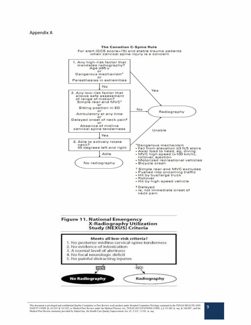
1) Criteria for radiographic (CT C-Spine) evaluation of cervical spine on a patient arriving to EC:
• Age >65 • Paresthesias in extremities/neurologic deficits • Altered mental status/intoxication • Distracting injury
2) The patient must be awake, alert, and not distracted in order to properly examine the
cervical spine. If unable to do this, proceed to radiographic evaluation. If the patient is alert and cooperative and exhibits no midline bony tenderness to palpation, next, passively rotate the patients head to right and left. If there is absence of midline cervical tenderness, the patient is to lift their head off the bed and touch their chin to their chest. If able to perform all these maneuvers, the collar can be removed.
If a patient is obtunded/persistently altered, the c-collar can be removed if an attending radiologist has posted a final negative acute read of a CT C-Spine. The collar should remain in place if ANY of the following are present: any signs of neurologic deficit on exam, or abnormalities on CT scan. If any abnormality is present on CT C-Spine, and the Philadelphia collar has been on >12 hours, order a Miami-J and proceed to MRI C-Spine without contrast and/or spine consult.
This document is privileged and confidential Quality Committee or Peer Review work product under Hospital Committee Privilege contained in the TEXAS HEALTH AND SAFETY CODE §§ 161.031 & 161.032, or Medical Peer Review under the Medical Practice Act, TEXAS OCCUPATIONS CODE, § § 151.001 et. seq & 160.007.; and the Medical Peer Review immunity provided by federal law, the Health Care Quality Improvement Act, 42. U.S.C. 11101, et. seq.
9
Appendix A
This document is privileged and confidential Quality Committee or Peer Review work product under Hospital Committee Privilege contained in the TEXAS HEALTH AND SAFETY CODE §§ 161.031 & 161.032, or Medical Peer Review under the Medical Practice Act, TEXAS OCCUPATIONS CODE, § § 151.001 et. seq & 160.007.; and the Medical Peer Review immunity provided by federal law, the Health Care Quality Improvement Act, 42. U.S.C. 11101, et. seq.
10
Memorial Hermann Southwest Hospital Clinical Guideline
GUIDELINE TITLE: Evaluation of Genitourinary Trauma PUBLICATION DATE: 01/15/18 VERSION: 1 GUIDELINE PURPOSE: To guide the work up of genitourinary trauma SCOPE: The urinary tract may be damaged by a variety of blunt and penetrating mechanisms. The presence of gross hematuria in the trauma patient mandates evaluation for genitourinary injury. This includes evaluation of the kidneys, bladder, and urethra. The purpose of this guideline is to provide guidance for the evaluation of genitourinary trauma. DEFINITION(S):
• Gross hematuria o Blood in the urine that can be seen as a change in the color of the urine.
• Microscopic hematuria o Urine that appears normal in color but has tested positive for blood on
microscopic examination. PARAMETERS: EVALUATION FOR RENAL INJURY:
1. Evaluation for the presence of blunt solid organ injury (including renal injury) is initially dictated by the hemodynamic status of the patient. a. In hemodynamically stable patients with gross hematuria, abdominal computed
tomography with intravenous contrast and immediate and delayed imaging is the radiologic gold standard for the evaluation of renal parenchymal injury and should be performed in hemodynamically stable patients with gross hematuria1, 2.
b. Hemodynamically unstable patients with gross hematuria should proceed to the operating room for exploratory laparotomy, especially if additional intra-abdominal injuries are suspected. A one shot IVP can be considered intra-operatively to evaluate the functional status of the kidneys1, 2. Use of IVP for determination of renal function should only be utilized in hemodynamically stable patients that have been adequately resuscitated.
• When ordering CT contrast studies, it is the ordering physician’s discretion whether or not to wait for the creatinine results.
2. The presence of microscopic hematuria does not mandate performance of CT to evaluate for renal injuries. However, CT imaging to rule out renal injury should be
This document is privileged and confidential Quality Committee or Peer Review work product under Hospital Committee Privilege contained in the TEXAS HEALTH AND SAFETY CODE §§ 161.031 & 161.032, or Medical Peer Review under the Medical Practice Act, TEXAS OCCUPATIONS CODE, § § 151.001 et. seq & 160.007.; and the Medical Peer Review immunity provided by federal law, the Health Care Quality Improvement Act, 42. U.S.C. 11101, et. seq.
11
undertaken in patients with major associated injuries, flank ecchymosis, and/or rapid deceleration injuries.
3. If there is no mechanism to suggest intra-abdominal injury then no further diagnostic studies are needed.
EVALUATION FOR URETERAL INJURIES: There are no classic clinical symptoms and signs of ureteral injury.
1. Injury to the ureters should be suspected in all cases of penetrating abdominal injury, and in cases of blunt deceleration trauma in which the kidney and renal pelvis can be torn away from the ureter.
2. Abdominal and pelvic CT imaging with IV contrast with both immediate and delayed imaging is the recommended diagnostic study for evaluation of ureteral trauma.
3. If CT scan cannot be performed, a one shot intravenous pyelogram (IVP) can be performed. If the patient is undergoing laparotomy, direct visualization of the ureters should be performed to evaluate for injury. The technique consists of a bolus intravenous injection of 2 ml/kg radiographic contrast (Omnipaque 350) followed by a single plain film taken after 10 minutes. This study provides important information for decision-making in the critical time of urgent laparotomy, and documents the presence of a functioning contralateral kidney.
EVALUATION FOR BLADDER INJURIES: Bladder injuries can be divided into extra peritoneal (60%), and intraperitoneal (30%). Simultaneous extra peritoneal and intraperitoneal injuries occur in 10% of all traumatic bladder injuries3. About 70–97% of patients with bladder rupture from blunt trauma have associated pelvic fractures4. The two most common sign and symptoms are gross hematuria (82%–100%) and abdominal tenderness (62%) 4. Other findings may include the inability to void, bruises over the suprapubic region, and abdominal distension. Extravasation of urine may result in swelling in the perineum, scrotum, thighs, and anterior abdominal wall.
1. The combination of pelvic fracture and gross hematuria constitutes an absolute indication for immediate cystography in blunt trauma patients2–4.
2. All patients with gross hematuria and a pelvic ring fracture should undergo radiologic examination of the bladder.
a. Conventional cystography is the preferred screening method for the evaluation of both intraperitoneal and extra peritoneal bladder injury.
b. CT Cystography with installation of 350 ml of contrast agent into the bladder is also an accepted diagnostic study for the evaluation of bladder injury.
3. The presence of microscopic hematuria is only a relative indication of injury. In patients with microscopic hematuria, imaging should be reserved for those with anterior rami fractures (straddle fracture) or severe pelvic ring disruption.
4. The presence of pelvic fluid in patients with pelvic fractures other than acetabular fractures should prompt cystography to evaluate for bladder injury.
This document is privileged and confidential Quality Committee or Peer Review work product under Hospital Committee Privilege contained in the TEXAS HEALTH AND SAFETY CODE §§ 161.031 & 161.032, or Medical Peer Review under the Medical Practice Act, TEXAS OCCUPATIONS CODE, § § 151.001 et. seq & 160.007.; and the Medical Peer Review immunity provided by federal law, the Health Care Quality Improvement Act, 42. U.S.C. 11101, et. seq.
12
5. Microscopic hematuria with isolated acetabular fracture or minimally displaced pelvic ring fractures is not an indication for cystography.
EVALUATION FOR URETHRAL TRAUMA IN THE MALE Blood at the meatus is present in 37–93% of patients with posterior urethral injury and at least 75% of patients with anterior urethral trauma2. The presence of blood at the meatus should preclude any attempts at urethral instrumentation, until the entire urethra is adequately imaged.
1. Retrograde urethrogram (RUG) is considered to be the gold standard diagnostic test for the evaluation of urethral injury. Evaluation for urethral injuries is recommended for the following patients:
a. Presence of blood at the urethral meatus b. “High-riding” prostate on rectal examination c. Gross hematuria d. Penetrating trauma to the penis or perineum e. Displaced fracture of the anterior pelvic ring (>10 mm displacement) f. Inability to void in the setting of pelvic trauma g. Unable to pass urethral catheter
2. In the event that a Foley catheter has been inserted prior to urethral evaluation (in a patient with concern for urethral trauma) a pericatheter retrograde urethrogram should be performed in a non- emergent fashion to identify a potential missed urethral injury.
a. This is done by injecting contrast via a 3 French catheter or angiocatheter held in the fossa navicularis to distend the urethra and prevent contrast leak from the meatus.
RETROGRADE URETHROGRAM (RUG) INSTRUCTIONS: Where to Perform:
• RUG is optimally performed in a fluoroscopy room • In urgent situations, RUG may be performed in the trauma room using digital
radiography (DR) equipment o A member of the ER and/or trauma team will place the urethral catheter and
inject the contrast o One or more members of the Emergency Radiology team will be present to assist
with timing the radiographic exposures and real-time interpretation of the images
Procedure: West modification of Sandler procedure
1. The external meatus is prepared in a standard sterile fashion. 2. Use an 8-F pediatric Foley catheter. 3. The catheter, with both the irrigating syringe and inflating (saline solution) syringe
attached, should be flushed before use. 4. Apply a very thin coat of water soluble lubricant to the tip and balloon of the catheter.
Very thin means a barely visible coating – less than 0.1 ml.
This document is privileged and confidential Quality Committee or Peer Review work product under Hospital Committee Privilege contained in the TEXAS HEALTH AND SAFETY CODE §§ 161.031 & 161.032, or Medical Peer Review under the Medical Practice Act, TEXAS OCCUPATIONS CODE, § § 151.001 et. seq & 160.007.; and the Medical Peer Review immunity provided by federal law, the Health Care Quality Improvement Act, 42. U.S.C. 11101, et. seq.
13
5. Insert the catheter approximately 2.0 – 2.5 cm into the penis so that the balloon portion of the catheter is seated in the fossa navicularis of the penile urethra. The balloon should be aligned with the corona of the glans penis.
6. The patient should be reassured about the discomfort that is experienced during balloon inflation.
7. The balloon is inflated with 0.5 - 1.0 mL of saline solution while the port is held with the free hand to partially inflate the balloon. Watch the patient grimace to judge when to stop inflating. A properly inflated catheter should remain in place when gentle traction is applied to gently stretch the penis.
8. If possible, the patient is rolled in a supine 45° oblique position. The penis should be gently pulled laterally over the proximal thigh using moderate traction on the catheter.
9. 5 ml of Omnipaque-300 is injected and the first radiograph is made. This first radiograph low volume radiograph may depict massive urethral disruption.
10. If no contrast extravasation is seen on the initial radiograph, an additional 15–25 mL of Omnipaque-300 is injected so that the anterior urethra is filled.
• Commonly, spasm of the external urethral sphincter will be encountered, which prevents filling of the deep bulbar, membranous, and prostatic urethras.
• Slow, gentle pressure is usually needed to overcome this resistance. • The pressure on the injection syringe often noticeably diminishes as the
external sphincter relaxes and the expressions on the patient’s face changes. • When these events occur, the physician performing the injection says “shoot”
for the second radiograph. 11. Timing of the second radiograph is important.
• The technologist should start the x-ray tube rotor when the higher volume injection commences and push the exposure button when the injecting physician says “shoot.”
12. If the second radiograph shows neither contrast extravasation nor filling of the posterior urethra, a third radiograph may be made during the injection of an additional 25 ml of Omnipaque-300 while the patient is told to “bear down” and try to forcibly urinate against the contrast stream.
• This maneuver sometimes relaxes the recalcitrant external sphincter. • Maximum 50 ml Omnipaque-300.
13. If the posterior urethra is still not filled after 50 ml injection, consider getting a voiding radiograph after the urinary bladder has been filled with contrast from the CT.
• For this radiograph, the technologist starts the rotor when the patient begins to urinate into a urinal.
• The technologist then asks the patient to squeeze his penis to interrupt the urine stream and shoots radiograph as the urine stream stops.
This document is privileged and confidential Quality Committee or Peer Review work product under Hospital Committee Privilege contained in the TEXAS HEALTH AND SAFETY CODE §§ 161.031 & 161.032, or Medical Peer Review under the Medical Practice Act, TEXAS OCCUPATIONS CODE, § § 151.001 et. seq & 160.007.; and the Medical Peer Review immunity provided by federal law, the Health Care Quality Improvement Act, 42. U.S.C. 11101, et. seq.
14
REFERENCES:
1. Morey AF, Brandes S, Dugi DD, et al. Urotrauma: AUA guideline. J Urol. 2014;192(2):327-335. doi:10.1016/j.juro.2014.05.004.
2. Lynch TH, Martínez-Piñeiro L, Plas E, et al. EAU guidelines on urological trauma. Eur Urol. 2005;47(1):1-15. doi:10.1016/j.eururo.2004.07.028.
3. Brandes S, Borrelli J. Pelvic fracture and associated urologic injuries. World J Surg. 2001;25(12):1578-1587.
4. Morey AF, Iverson AJ, Swan A, et al. Bladder rupture after blunt trauma: guidelines for diagnostic imaging. J Trauma. 2001;51(4):683-686.
5. Kawashima A, Sandler CM, Wasserman NF, LeRoy AJ, King BF Jr, Goldman SM. 6. Imaging of urethral disease: a pictorial review. Radiographics. 2004 Oct;24 Suppl 1:S195-
216. Review. PubMed PMID: 15486241. 7. http://www.acr.org/SecondaryMainMenuCategories/quality_safety/app_criteria/pdf/Ex
pertPanelonUrolog icImaging/SuspectedLowerUrinaryTractTraumaDoc19.aspx 8. http://emedicine.medscape.com/article/1893948-overview#a1
This document is privileged and confidential Quality Committee or Peer Review work product under Hospital Committee Privilege contained in the TEXAS HEALTH AND SAFETY CODE §§ 161.031 & 161.032, or Medical Peer Review under the Medical Practice Act, TEXAS OCCUPATIONS CODE, § § 151.001 et. seq & 160.007.; and the Medical Peer Review immunity provided by federal law, the Health Care Quality Improvement Act, 42. U.S.C. 11101, et. seq.
15
Memorial Hermann Southwest Hospital Clinical Guideline
GUIDELINE TITLE: Post Splenectomy Vaccination PUBLICATION DATE: 01/15/18 VERSION: 1 GUIDELINE PURPOSE: To delineate timing of post-splenectomy vaccines SCOPE: This applies to Trauma Services at Memorial Hermann Southwest Hospital. DEFINITION(S): Injured patients will be categorized and given an activation level based on criteria in this guideline and roles and responsibilities of each trauma team member will be defined. PARAMETERS:
• All patients status post-splenectomy • All patients with <50% perfused spleen
Vaccines:
1. Pneumococcal vaccine, PCV13 (Prevnar-13) - 0.5mL IM 2. Hemophilus influenzae vaccine (HiB) - 0.5mL IM 3. Meningococcal vaccine, MenACWY (Menactra/Menveo) - 0.5mL IM
FOR NON-ICU PATIENTS: Vaccinations should be administered the day prior to discharge. 1,2
FOR ICU PATIENTS: Vaccinations should be administered upon discharge from the ICU.3, 4
All patient charts should be labeled with either “Asplenic” or “S/p Splenectomy” to identify patients that require vaccinations. In addition, “Asplenic” or “S/p Splenectomy” will be added to the trauma service patient list for those patients requiring vaccination.
• Follow-up Plan
Education: All patients with splenectomy need to be informed of their operation, the risk and signs/symptoms of developing Overwhelming Post-Splenectomy Infection (OPSI) via
This document is privileged and confidential Quality Committee or Peer Review work product under Hospital Committee Privilege contained in the TEXAS HEALTH AND SAFETY CODE §§ 161.031 & 161.032, or Medical Peer Review under the Medical Practice Act, TEXAS OCCUPATIONS CODE, § § 151.001 et. seq & 160.007.; and the Medical Peer Review immunity provided by federal law, the Health Care Quality Improvement Act, 42. U.S.C. 11101, et. seq.
16
physician to patient discussion. Eight weeks after doses given prior to discharge, patient will need a second set of vaccinations: one dose of MenACWY (Menactra/Menveo) and one dose of pneumococcal PPSV23 (Pneumovax). Revaccination of Menactra/Menveo is recommended every 5 years. Revaccination of Pneumovax is recommended in 5 years and again after the age of 65 if at least 5 years has elapsed since their previous dose of PPSV23 (Pneumovax). Patients should be instructed to follow-up with their primary care physician for this assessment. 5
Asplenic Patient Database: To identify those patients’ that underwent Splenectomy, when they
received their vaccinations, and when they need to follow up for revaccination
Use of Medical Alert Bracelets: For emergency medical providers to identify patients in the community that are asplenic.
• References:
1 Shatz DV, Romero-Steiner S, Elie CM, et al. Antibody responses in postsplenectomy trauma patients receiving the 23- valent pneumococcal polysaccharide vaccine at 14 versus 28 days postoperatively. J Trauma 2002; 53(6): 1037-42. 2 Shatz DV. Vaccination practices among North American trauma surgeons in Splenectomy after trauma. J Trauma 2002; 53: 950-956. 3 Taylor MD, Genuit T, Napolitano LM. Overwhelming postsplenectomy sepsis and trauma: Time to consider revaccination? J Trauma 2005: 59: 1482-1485 4 Shatz DV, Schinsky M, Pais L, et al. Immune responses of splenectomized trauma patients to the 23-valent pneumococcal polysaccharide vaccine at 1 versus 7 versus 14 days after Splenectomy (RCT). J Trauma 1998; 44: 760-765. 5 Epidemiology and prevention of vaccine-preventable diseases. The Pink Book, 13th ed. Chapters 14 and 17, US Department of Health and Human Services Center for Disease Control and Prevention; April 2015.
This document is privileged and confidential Quality Committee or Peer Review work product under Hospital Committee Privilege contained in the TEXAS HEALTH AND SAFETY CODE §§ 161.031 & 161.032, or Medical Peer Review under the Medical Practice Act, TEXAS OCCUPATIONS CODE, § § 151.001 et. seq & 160.007.; and the Medical Peer Review immunity provided by federal law, the Health Care Quality Improvement Act, 42. U.S.C. 11101, et. seq.
17
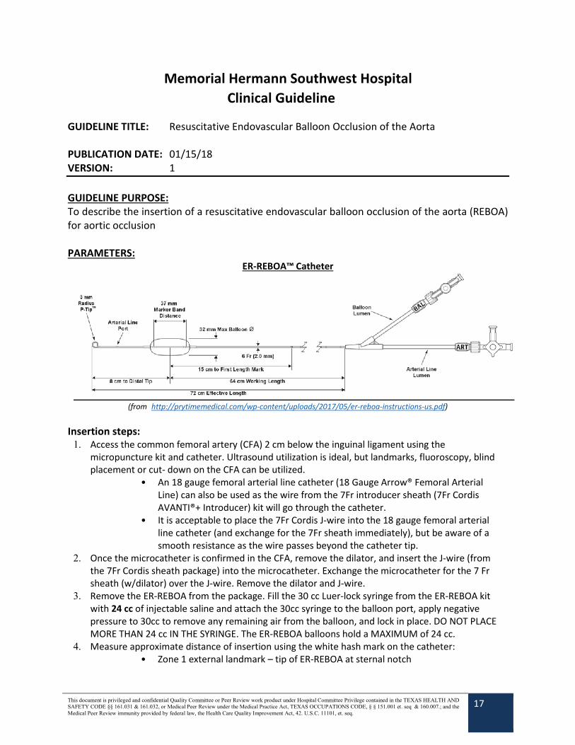
Memorial Hermann Southwest Hospital Clinical Guideline
GUIDELINE TITLE: Resuscitative Endovascular Balloon Occlusion of the Aorta PUBLICATION DATE: 01/15/18 VERSION: 1 GUIDELINE PURPOSE: To describe the insertion of a resuscitative endovascular balloon occlusion of the aorta (REBOA) for aortic occlusion PARAMETERS:
ER-REBOA™ Catheter
(from http://prytimemedical.com/wp-content/uploads/2017/05/er-reboa-instructions-us.pdf) Insertion steps:
1. Access the common femoral artery (CFA) 2 cm below the inguinal ligament using the micropuncture kit and catheter. Ultrasound utilization is ideal, but landmarks, fluoroscopy, blind placement or cut- down on the CFA can be utilized.
• An 18 gauge femoral arterial line catheter (18 Gauge Arrow® Femoral Arterial Line) can also be used as the wire from the 7Fr introducer sheath (7Fr Cordis AVANTI®+ Introducer) kit will go through the catheter.
• It is acceptable to place the 7Fr Cordis J-wire into the 18 gauge femoral arterial line catheter (and exchange for the 7Fr sheath immediately), but be aware of a smooth resistance as the wire passes beyond the catheter tip.
2. Once the microcatheter is confirmed in the CFA, remove the dilator, and insert the J-wire (from the 7Fr Cordis sheath package) into the microcatheter. Exchange the microcatheter for the 7 Fr sheath (w/dilator) over the J-wire. Remove the dilator and J-wire.
3. Remove the ER-REBOA from the package. Fill the 30 cc Luer-lock syringe from the ER-REBOA kit with 24 cc of injectable saline and attach the 30cc syringe to the balloon port, apply negative pressure to 30cc to remove any remaining air from the balloon, and lock in place. DO NOT PLACE MORE THAN 24 cc IN THE SYRINGE. The ER-REBOA balloons hold a MAXIMUM of 24 cc.
4. Measure approximate distance of insertion using the white hash mark on the catheter: • Zone 1 external landmark – tip of ER-REBOA at sternal notch
This document is privileged and confidential Quality Committee or Peer Review work product under Hospital Committee Privilege contained in the TEXAS HEALTH AND SAFETY CODE §§ 161.031 & 161.032, or Medical Peer Review under the Medical Practice Act, TEXAS OCCUPATIONS CODE, § § 151.001 et. seq & 160.007.; and the Medical Peer Review immunity provided by federal law, the Health Care Quality Improvement Act, 42. U.S.C. 11101, et. seq.
18
• Zone 3 external landmark – tip of ER-REBOA at xiphoid 5. Advance the orange peel away sheath over the balloon and P-tip. Insert the orange sheath tip
into the 7Fr sheath to pop open the valve (barely 1cm). Insert the catheter through the peel-away sheath and 7Fr sheath to the desired distance. Retract or peel the orange sheath away in order to visualize the catheter markings. A chest or abdominal x-ray MUST BE obtained to confirm device placement prior to balloon inflation. While waiting for x-ray, attach the A-line port (flush optional) to the transducer to obtain a systemic arterial pressure before the balloon is inflated.
6. Once the catheter is confirmed in the desired location (2 radiopaque markers located at each end of the balloon will be visible on x-ray), hold the catheter at its insertion site into the sheath DURING and AFTER inflation (especially at Zone 1).
• Failure to secure the catheter during and after inflation may result in balloon migration and possible aortic intimal injury.
7. Inflate the balloon until an increase in the patient’s blood pressure is seen or there is loss of pulse in the contralateral femoral artery. The balloon holds a max 24cc of saline and over inflation should be avoided. Once inflated to the appropriate volume lock in place.
• Average balloon fill for Zone 1: 15 cc (unpublished data) • Average balloon fill for Zone 3: 11 cc (unpublished data)
8. Secure the catheter to the sheath, and sheath to the patient. Additional x-rays are optional but encouraged if time permits.
9. Once the need for the catheter has passed, deflate the balloon by attaching an empty syringe, retracting to 30cc, and lock in place. A few seconds is required to remove all fluid and air from the balloon and catheter. Disconnect the A-line transducer from the A-line port and lock.
10. Remove the catheter from the sheath. 11. Flush the 7Fr sheath with saline. 12. When coagulation parameters are improved/corrected and patient has stabilized, remove the
sheath from the groin and apply manual compression for 30 minutes. No closure device has been found to be more effective than CORRECTLY APPLIED manual compression. The patient must be supine (no hip/knee flexion) for 6 hours after compression is completed.
13. A duplex arterial ultrasound of the arterial access site should be obtained 48 hours after sheath removal to assess for pseudoanuerysm formation or thrombus.
This document is privileged and confidential Quality Committee or Peer Review work product under Hospital Committee Privilege contained in the TEXAS HEALTH AND SAFETY CODE §§ 161.031 & 161.032, or Medical Peer Review under the Medical Practice Act, TEXAS OCCUPATIONS CODE, § § 151.001 et. seq & 160.007.; and the Medical Peer Review immunity provided by federal law, the Health Care Quality Improvement Act, 42. U.S.C. 11101, et. seq.
19
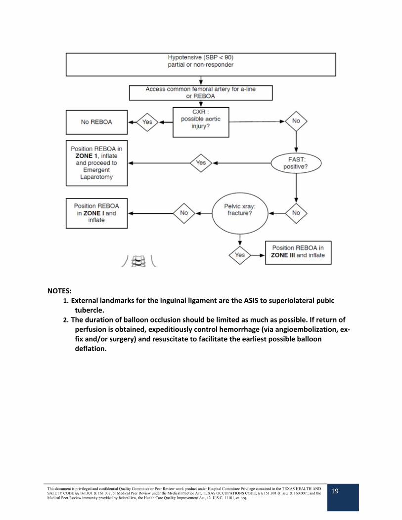
NOTES: 1. External landmarks for the inguinal ligament are the ASIS to superiolateral pubic
tubercle. 2. The duration of balloon occlusion should be limited as much as possible. If return of
perfusion is obtained, expeditiously control hemorrhage (via angioembolization, ex-fix and/or surgery) and resuscitate to facilitate the earliest possible balloon deflation.
This document is privileged and confidential Quality Committee or Peer Review work product under Hospital Committee Privilege contained in the TEXAS HEALTH AND SAFETY CODE §§ 161.031 & 161.032, or Medical Peer Review under the Medical Practice Act, TEXAS OCCUPATIONS CODE, § § 151.001 et. seq & 160.007.; and the Medical Peer Review immunity provided by federal law, the Health Care Quality Improvement Act, 42. U.S.C. 11101, et. seq.
20
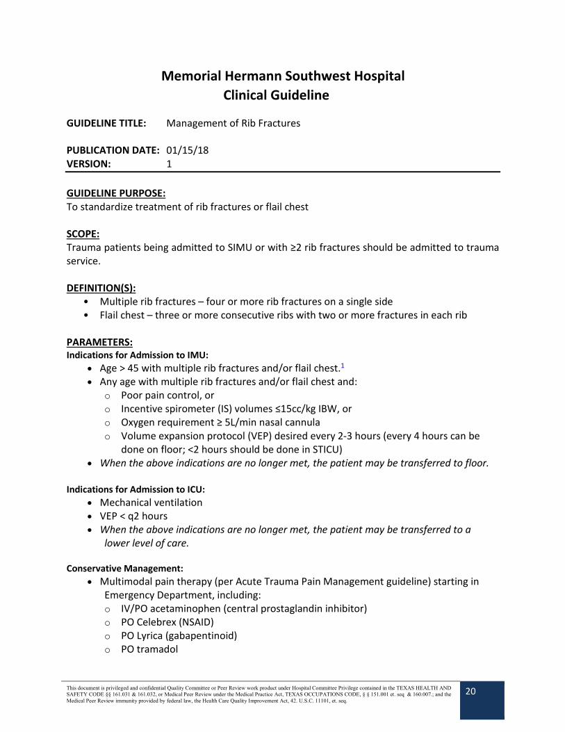
Memorial Hermann Southwest Hospital Clinical Guideline
GUIDELINE TITLE: Management of Rib Fractures PUBLICATION DATE: 01/15/18 VERSION: 1 GUIDELINE PURPOSE: To standardize treatment of rib fractures or flail chest SCOPE: Trauma patients being admitted to SIMU or with ≥2 rib fractures should be admitted to trauma service. DEFINITION(S):
• Multiple rib fractures – four or more rib fractures on a single side • Flail chest – three or more consecutive ribs with two or more fractures in each rib
PARAMETERS: Indications for Admission to IMU:
• Age > 45 with multiple rib fractures and/or flail chest.1 • Any age with multiple rib fractures and/or flail chest and:
o Poor pain control, or o Incentive spirometer (IS) volumes ≤15cc/kg IBW, or o Oxygen requirement ≥ 5L/min nasal cannula o Volume expansion protocol (VEP) desired every 2-3 hours (every 4 hours can be
done on floor; <2 hours should be done in STICU) • When the above indications are no longer met, the patient may be transferred to floor.
Indications for Admission to ICU:
• Mechanical ventilation • VEP < q2 hours • When the above indications are no longer met, the patient may be transferred to a
lower level of care. Conservative Management:
• Multimodal pain therapy (per Acute Trauma Pain Management guideline) starting in Emergency Department, including: o IV/PO acetaminophen (central prostaglandin inhibitor) o PO Celebrex (NSAID) o PO Lyrica (gabapentinoid) o PO tramadol
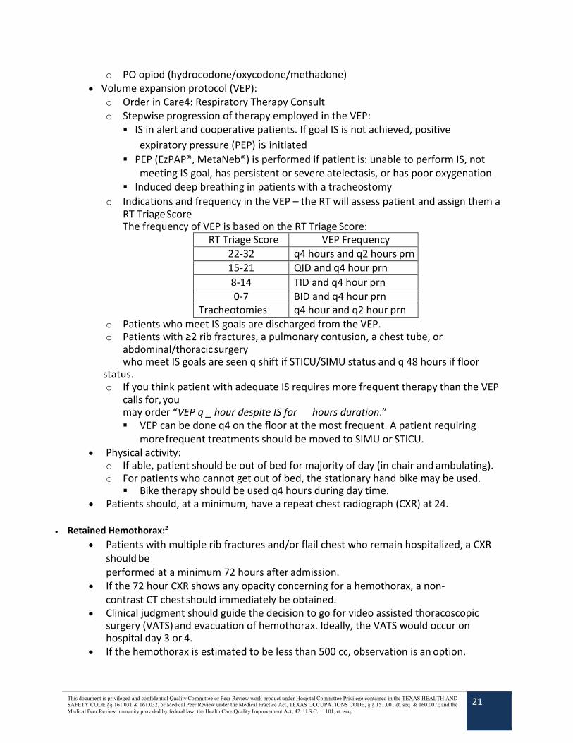
This document is privileged and confidential Quality Committee or Peer Review work product under Hospital Committee Privilege contained in the TEXAS HEALTH AND SAFETY CODE §§ 161.031 & 161.032, or Medical Peer Review under the Medical Practice Act, TEXAS OCCUPATIONS CODE, § § 151.001 et. seq & 160.007.; and the Medical Peer Review immunity provided by federal law, the Health Care Quality Improvement Act, 42. U.S.C. 11101, et. seq.
21
o PO opiod (hydrocodone/oxycodone/methadone) • Volume expansion protocol (VEP):
o Order in Care4: Respiratory Therapy Consult o Stepwise progression of therapy employed in the VEP: IS in alert and cooperative patients. If goal IS is not achieved, positive
expiratory pressure (PEP) is initiated PEP (EzPAP®, MetaNeb®) is performed if patient is: unable to perform IS, not
meeting IS goal, has persistent or severe atelectasis, or has poor oxygenation Induced deep breathing in patients with a tracheostomy
o Indications and frequency in the VEP – the RT will assess patient and assign them a RT Triage Score The frequency of VEP is based on the RT Triage Score:
RT Triage Score VEP Frequency 22-32 q4 hours and q2 hours prn 15-21 QID and q4 hour prn 8-14 TID and q4 hour prn 0-7 BID and q4 hour prn
Tracheotomies q4 hour and q2 hour prn o Patients who meet IS goals are discharged from the VEP. o Patients with ≥2 rib fractures, a pulmonary contusion, a chest tube, or
abdominal/thoracic surgery who meet IS goals are seen q shift if STICU/SIMU status and q 48 hours if floor
status. o If you think patient with adequate IS requires more frequent therapy than the VEP
calls for, you may order “VEP q _ hour despite IS for hours duration.” VEP can be done q4 on the floor at the most frequent. A patient requiring
more frequent treatments should be moved to SIMU or STICU. • Physical activity:
o If able, patient should be out of bed for majority of day (in chair and ambulating). o For patients who cannot get out of bed, the stationary hand bike may be used. Bike therapy should be used q4 hours during day time.
• Patients should, at a minimum, have a repeat chest radiograph (CXR) at 24.
• Retained Hemothorax:2
• Patients with multiple rib fractures and/or flail chest who remain hospitalized, a CXR should be performed at a minimum 72 hours after admission.
• If the 72 hour CXR shows any opacity concerning for a hemothorax, a non-contrast CT chest should immediately be obtained.
• Clinical judgment should guide the decision to go for video assisted thoracoscopic surgery (VATS) and evacuation of hemothorax. Ideally, the VATS would occur on hospital day 3 or 4.
• If the hemothorax is estimated to be less than 500 cc, observation is an option.
This document is privileged and confidential Quality Committee or Peer Review work product under Hospital Committee Privilege contained in the TEXAS HEALTH AND SAFETY CODE §§ 161.031 & 161.032, or Medical Peer Review under the Medical Practice Act, TEXAS OCCUPATIONS CODE, § § 151.001 et. seq & 160.007.; and the Medical Peer Review immunity provided by federal law, the Health Care Quality Improvement Act, 42. U.S.C. 11101, et. seq.
22
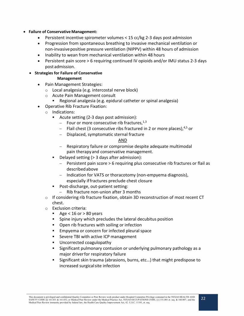
• Failure of Conservative Management: • Persistent incentive spirometer volumes < 15 cc/kg 2-3 days post admission • Progression from spontaneous breathing to invasive mechanical ventilation or
non-invasive positive pressure ventilation (NIPPV) within 48 hours of admission • Inability to wean from mechanical ventilation within 48 hours • Persistent pain score > 6 requiring continued IV opioids and/or IMU status 2-3 days
post admission. • Strategies for Failure of Conservative
Management • Pain Management Strategies:
o Local analgesia (e.g. intercostal nerve block) o Acute Pain Management consult Regional analgesia (e.g. epidural catheter or spinal analgesia)
• Operative Rib Fracture Fixation: o Indications: Acute setting (2-3 days post admission):
− Four or more consecutive rib fractures,1,3
− Flail chest (3 consecutive ribs fractured in 2 or more places),4,5 or − Displaced, symptomatic sternal fracture
AND − Respiratory failure or compromise despite adequate multimodal
pain therapy and conservative management. Delayed setting (> 3 days after admission):
− Persistent pain score > 6 requiring plus consecutive rib fractures or flail as described above
− Indication for VATS or thoracotomy (non-empyema diagnosis), especially if fractures preclude chest closure
Post-discharge, out-patient setting: − Rib fracture non-union after 3 months
o If considering rib fracture fixation, obtain 3D reconstruction of most recent CT chest.
o Exclusion criteria: Age < 16 or > 80 years Spine injury which precludes the lateral decubitus position Open rib fractures with soiling or infection Empyema or concern for infected pleural space Severe TBI with active ICP management Uncorrected coagulopathy Significant pulmonary contusion or underlying pulmonary pathology as a
major driver for respiratory failure Significant skin trauma (abrasions, burns, etc…) that might predispose to
increased surgical site infection
This document is privileged and confidential Quality Committee or Peer Review work product under Hospital Committee Privilege contained in the TEXAS HEALTH AND SAFETY CODE §§ 161.031 & 161.032, or Medical Peer Review under the Medical Practice Act, TEXAS OCCUPATIONS CODE, § § 151.001 et. seq & 160.007.; and the Medical Peer Review immunity provided by federal law, the Health Care Quality Improvement Act, 42. U.S.C. 11101, et. seq.
23
References:
1 Holcomb JB, McMullin NR, Kozar RA, Lygas MH, Moore FA. Morbidity from Rib Fractures Increases after Age 45. J Am Coll Surg. Apr 2003;196(4):549-55.
2 Meyer DM, Jessen ME, Wait MA, Estrera AS. Early Evacuation of Traumatic Retained Hemothoraces using Thoracoscopy: a Prospective, Randomized Trial. Ann Thorac Surg. Nov 1997;64(5):1396-400. 3 Bulger EM, Arneson MA, Mock CN, Jurkovich GJ. Rib Fractures in the Elderly. J Trauma. Jun 2000;48(6):1040-46. 4 Doben AR, Eriksson EA, Denlinger CE, Leon SM, Couillard DJ, Fakhry SM, Minshall CT. Surgical Rib Fixation fo Flail Chest Deformity Improves Liberation from Mechanical Ventilation. J Crit Care. Feb 2014;29(1):139-43. 5 Marasco SF, Davies AR, Cooper J, Varma D, Bennett V, Nevill R, Lee G, Bailey M, Fitzgerald M. Prospective Randomized Controlled Trial of Operative Rib Fixation in Traumatic Flail Chest. J Am Coll Surg. May 2013;216(5):924-32.
This document is privileged and confidential Quality Committee or Peer Review work product under Hospital Committee Privilege contained in the TEXAS HEALTH AND SAFETY CODE §§ 161.031 & 161.032, or Medical Peer Review under the Medical Practice Act, TEXAS OCCUPATIONS CODE, § § 151.001 et. seq & 160.007.; and the Medical Peer Review immunity provided by federal law, the Health Care Quality Improvement Act, 42. U.S.C. 11101, et. seq.
24
Memorial Hermann Southwest Hospital Clinical Guideline
GUIDELINE TITLE: Screening for Blunt Cerebrovascular Injury PUBLICATION DATE: 01/15/18 VERSION: 1 GUIDELINE PURPOSE: To identify patients to screen for blunt cerebrovascular injury (BCVI) SCOPE: While BCVI occurs in only 0.5 to 1.2%1, 2 of blunt trauma patients, the complications of missed injury resulting in stroke are devastating. A clinically latent period ranging from 10 -72 hours provides a short window of opportunity to make the diagnosis and initiate treatment (anti-thrombotic therapy or anticoagulation) prior to the onset of neurologic damage. Treatment is inexpensive and effective, shown to decrease the stroke rate from 21 to 0.5%3. While cerebral angiogram remains the gold standard for diagnosis of BCVI4, our institution utilizes multi-slice CTA secondary to immediate availability and improved CT technology. The clinical challenge is to identify patients at high risk of BCVI to make a prompt diagnosis and initiate treatment. Treatment of BCVI with other injuries contradicting immediate anti-platelet/anti- coagulation is controversial and is currently being studied at this institution. DEFINITION(S): PARAMETERS: The following injury patterns resulting from high energy transfer mechanism (including flexion/extension injuries) place the patient at high risk for BCVI and are indications for CTA neck5:
• Complex facial fractures (LeFort II or III) • Mandible fracture • Basilar skull fracture or occipital condyle fracture • Cervical vertebral body or transverse foramen fracture at any level (C1-7) • Any fracture at level C1-C3 • Cervical subluxation or ligamentous injury at any level • Severe traumatic brain injury (TBI) with GCS < 6 • Neurological exam incongruous with head CT • Near hanging with anoxic brain injury • Seatbelt or other clothesline-type injury with significant swelling, pain, or AMS • Combined TBI and major thoracic injury • Scalp degloving injury • Thoracic vascular injury
This document is privileged and confidential Quality Committee or Peer Review work product under Hospital Committee Privilege contained in the TEXAS HEALTH AND SAFETY CODE §§ 161.031 & 161.032, or Medical Peer Review under the Medical Practice Act, TEXAS OCCUPATIONS CODE, § § 151.001 et. seq & 160.007.; and the Medical Peer Review immunity provided by federal law, the Health Care Quality Improvement Act, 42. U.S.C. 11101, et. seq.
25
The following signs and symptoms of BCVI are indications for CTA neck:
• Potential arterial hemorrhage from neck/nose/mouth • Cervical bruit in patient < 50 years of age • Cervical hematoma • Focal neurologic defect: TIA, hemiparesis, vertebrobasilar symptoms, Horner’s
Syndrome • Neurologic deficit inconsistent with head CT • Stroke on CT or MRI
Diagnosis:
• Screening CTA neck should be performed no later than 6 hours from time of ED arrival. • The CTA neck should be performed at the time of the original diagnostic CT scan for
blunt trauma once the above risk factors are identified. • If the need for CTA neck is decided after the original IV contrast CT scan, discussion with
the responsible attending should occur in patients at high risk for contrast induced nephropathy.
o For these high risk patients, a 1 liter bolus of LR should be given prior to repeat CTA neck.
• If the patient is unable to get a CTA neck in a timely fashion, consider starting non-enteric coated aspirin 325 mg daily in the patient with no contraindication to therapy (TBI, SCI, solid organ injury) prior to confirming the diagnosis.
• If clinical suspicion of BCVI remains high despite a negative CTA neck, please consult the Neurosurgery Vascular service for cerebral angiogram and start non-enteric coated aspirin 325 mg daily immediately in the patient with no contraindication to therapy (TBI, SCI, solid organ injury).
• Consider angiogram if CTA neck is positive for injury and patient has a contraindication to aspirin (active peptic ulcer, documented aspirin allergy, hemophilia, von Willebrand’s desease).
Please notify the trauma neurosurgery team once the diagnosis of BCVI is made
at one of the following numbers
Treatment: • Isolated BCVI without neurologic symptoms: non-enteric coated Aspirin 325 mg daily
o start immediately after diagnosis • Isolated BCVI with neurologic symptoms: Heparin drip
o no bolus, goal PTT 40-50 o start immediately after diagnosis o please use heparin weight based protocol orders for blunt carotid or vertebral
artery injury MPP in Care4 • BCVI with traumatic brain injury or spinal cord injury
o start non-enteric coated aspirin or heparin after cleared by Neurosurgery
This document is privileged and confidential Quality Committee or Peer Review work product under Hospital Committee Privilege contained in the TEXAS HEALTH AND SAFETY CODE §§ 161.031 & 161.032, or Medical Peer Review under the Medical Practice Act, TEXAS OCCUPATIONS CODE, § § 151.001 et. seq & 160.007.; and the Medical Peer Review immunity provided by federal law, the Health Care Quality Improvement Act, 42. U.S.C. 11101, et. seq.
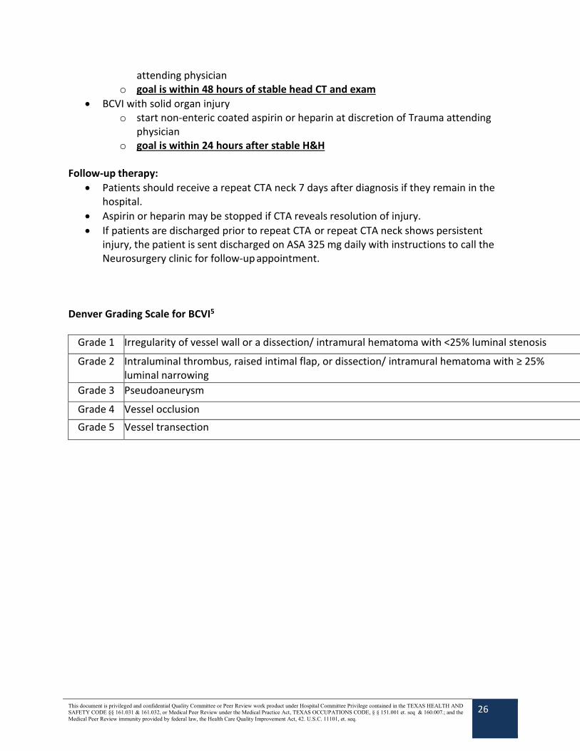
26
attending physician o goal is within 48 hours of stable head CT and exam
• BCVI with solid organ injury o start non-enteric coated aspirin or heparin at discretion of Trauma attending
physician o goal is within 24 hours after stable H&H
Follow-up therapy: • Patients should receive a repeat CTA neck 7 days after diagnosis if they remain in the
hospital. • Aspirin or heparin may be stopped if CTA reveals resolution of injury. • If patients are discharged prior to repeat CTA or repeat CTA neck shows persistent
injury, the patient is sent discharged on ASA 325 mg daily with instructions to call the Neurosurgery clinic for follow-up appointment.
Denver Grading Scale for BCVI5
Grade 1 Irregularity of vessel wall or a dissection/ intramural hematoma with <25% luminal stenosis
Grade 2 Intraluminal thrombus, raised intimal flap, or dissection/ intramural hematoma with ≥ 25% luminal narrowing
Grade 3 Pseudoaneurysm
Grade 4 Vessel occlusion Grade 5 Vessel transection
This document is privileged and confidential Quality Committee or Peer Review work product under Hospital Committee Privilege contained in the TEXAS HEALTH AND SAFETY CODE §§ 161.031 & 161.032, or Medical Peer Review under the Medical Practice Act, TEXAS OCCUPATIONS CODE, § § 151.001 et. seq & 160.007.; and the Medical Peer Review immunity provided by federal law, the Health Care Quality Improvement Act, 42. U.S.C. 11101, et. seq.
27
References: 1. Miller PR, Fabian TC, Croce MA, Cagiannos C. Prospective screening for blunt
cerebrovascular injuries: analysis of diagnostic modalities and outcomes. Annals of …. 2002.
2. Stein DM, Boswell S, Sliker CW, Lui FY, Scalea TM. Blunt Cerebrovascular Injuries: Does Treatment Always Matter? The Journal of Trauma: Injury, Infection, and Critical Care. 2009;66(1):132–144. doi:10.1097/TA.0b013e318142d146.
3. Cothren CC, Biffl WL, Moore EE, Kashuk JL, Johnson JL. Treatment for blunt cerebrovascular injuries: equivalence of anticoagulation and antiplatelet agents. Arch Surg. 2009;144(7):685–690. doi:10.1001/archsurg.2009.111.
4. Paulus EM, Fabian TC, Savage SA, et al. Blunt cerebrovascular injury screening with 64-channel multidetector computed tomography: More slices finally cut it. J Trauma Acute Care Surg. 2014;76(2):279–285. doi:10.1097/TA.0000000000000101.
5. Burlew CC, Biffl WL, Moore EE, Barnett CC, Johnson JL, Bensard DD. Blunt cerebrovascular injuries: redefining screening criteria in the era of noninvasive diagnosis. J Trauma Acute Care Surg. 2012;72(2):330–5– discussion 336–7– quiz 539. doi:10.1097/TA.0b013e31823de8a0.
6. Biffl WL, Cothren CC, Moore EE, et al. Western Trauma Association Critical Decisions in Trauma: Screening for and Treatment of Blunt Cerebrovascular Injuries. The Journal of Trauma: Injury, Infection, and Critical Care. 2009;67(6):1150–1153. doi:10.1097/TA.0b013e3181c1c1d6.
7. Cothren CC, Moore EE, Biffl WL, et al. Anticoagulation Is the Gold Standard Therapy for Blunt Carotid Injuries to Reduce Stroke Rate. Arch Surg. 2004;139(5):540–546. doi:10.1001/archsurg.139.5.540.
8. Emmett KP, Fabian TC, DiCocco JM, Zarzaur BL, Croce MA. Improving the screening criteria for blunt cerebrovascular injury: the appropriate role for computed tomography angiography. J Trauma. 2011;70(5):1058–63– discussion 1063–5. doi:10.1097/TA.0b013e318213f849.
9. Bruns BR, Tesoriero R, Kufera J, et al. Blunt cerebrovascular injury screening guidelines. Journal of Trauma and Acute Care Surgery. 2014;76(3):691–695. doi:10.1097/TA.0b013e3182ab1b4d.
This document is privileged and confidential Quality Committee or Peer Review work product under Hospital Committee Privilege contained in the TEXAS HEALTH AND SAFETY CODE §§ 161.031 & 161.032, or Medical Peer Review under the Medical Practice Act, TEXAS OCCUPATIONS CODE, § § 151.001 et. seq & 160.007.; and the Medical Peer Review immunity provided by federal law, the Health Care Quality Improvement Act, 42. U.S.C. 11101, et. seq.
28
Memorial Hermann Southwest Hospital Clinical Guideline
GUIDELINE TITLE: Specialty Service Consultations PUBLICATION DATE: 01/15/18 VERSION: 1 GUIDELINE PURPOSE: To clarify service assignment, admission criteria, and rotation schedule of specialty services and certain injuries SCOPE:
Traumatic Brain Injury ............................................................................................................................................29
Trauma Evaluation of Patients with Extremity and/or Pelvic Injuries ....................................................................30
Orthopedic Surgery Treatment & Transfer Policy for Orthopedic Emergencies ...................................................31
Ground Level Falls/”Found Down” (in Patients ≥65 Years) ....................................................................................31
Drowning/Near Drowning ......................................................................................................................................32
Spinal Cord Injury ....................................................................................................................................................33
DEFINITION(S): Surgical Trauma Intensive Care Unit (STICU) Nuero Trauma Intensive Care Unit (NTICU) PARAMETERS:
This document is privileged and confidential Quality Committee or Peer Review work product under Hospital Committee Privilege contained in the TEXAS HEALTH AND SAFETY CODE §§ 161.031 & 161.032, or Medical Peer Review under the Medical Practice Act, TEXAS OCCUPATIONS CODE, § § 151.001 et. seq & 160.007.; and the Medical Peer Review immunity provided by federal law, the Health Care Quality Improvement Act, 42. U.S.C. 11101, et. seq.
29
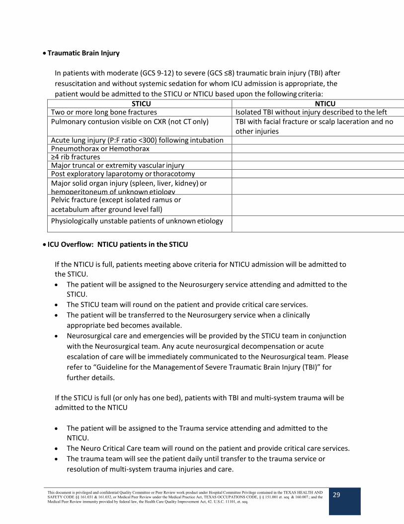
• Traumatic Brain Injury
In patients with moderate (GCS 9-12) to severe (GCS ≤8) traumatic brain injury (TBI) after resuscitation and without systemic sedation for whom ICU admission is appropriate, the patient would be admitted to the STICU or NTICU based upon the following criteria:
STICU NTICU Two or more long bone fractures Isolated TBI without injury described to the left Pulmonary contusion visible on CXR (not CT only) TBI with facial fracture or scalp laceration and no
other injuries Acute lung injury (P:F ratio <300) following intubation Pneumothorax or Hemothorax ≥4 rib fractures Major truncal or extremity vascular injury Post exploratory laparotomy or thoracotomy Major solid organ injury (spleen, liver, kidney) or hemoperitoneum of unknown etiology
Pelvic fracture (except isolated ramus or acetabulum after ground level fall)
Physiologically unstable patients of unknown etiology
• ICU Overflow: NTICU patients in the STICU
If the NTICU is full, patients meeting above criteria for NTICU admission will be admitted to the STICU. • The patient will be assigned to the Neurosurgery service attending and admitted to the
STICU. • The STICU team will round on the patient and provide critical care services. • The patient will be transferred to the Neurosurgery service when a clinically
appropriate bed becomes available. • Neurosurgical care and emergencies will be provided by the STICU team in conjunction
with the Neurosurgical team. Any acute neurosurgical decompensation or acute escalation of care will be immediately communicated to the Neurosurgical team. Please refer to “Guideline for the Management of Severe Traumatic Brain Injury (TBI)” for further details.
If the STICU is full (or only has one bed), patients with TBI and multi-system trauma will be admitted to the NTICU • The patient will be assigned to the Trauma service attending and admitted to the
NTICU. • The Neuro Critical Care team will round on the patient and provide critical care services. • The trauma team will see the patient daily until transfer to the trauma service or
resolution of multi-system trauma injuries and care.
This document is privileged and confidential Quality Committee or Peer Review work product under Hospital Committee Privilege contained in the TEXAS HEALTH AND SAFETY CODE §§ 161.031 & 161.032, or Medical Peer Review under the Medical Practice Act, TEXAS OCCUPATIONS CODE, § § 151.001 et. seq & 160.007.; and the Medical Peer Review immunity provided by federal law, the Health Care Quality Improvement Act, 42. U.S.C. 11101, et. seq.
30
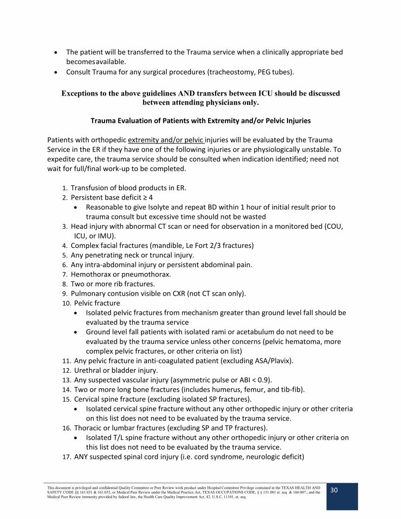
• The patient will be transferred to the Trauma service when a clinically appropriate bed becomes available.
• Consult Trauma for any surgical procedures (tracheostomy, PEG tubes).
Exceptions to the above guidelines AND transfers between ICU should be discussed between attending physicians only.
Trauma Evaluation of Patients with Extremity and/or Pelvic Injuries
Patients with orthopedic extremity and/or pelvic injuries will be evaluated by the Trauma Service in the ER if they have one of the following injuries or are physiologically unstable. To expedite care, the trauma service should be consulted when indication identified; need not wait for full/final work-up to be completed.
1. Transfusion of blood products in ER. 2. Persistent base deficit ≥ 4
• Reasonable to give Isolyte and repeat BD within 1 hour of initial result prior to trauma consult but excessive time should not be wasted
3. Head injury with abnormal CT scan or need for observation in a monitored bed (COU, ICU, or IMU).
4. Complex facial fractures (mandible, Le Fort 2/3 fractures) 5. Any penetrating neck or truncal injury. 6. Any intra-abdominal injury or persistent abdominal pain. 7. Hemothorax or pneumothorax. 8. Two or more rib fractures. 9. Pulmonary contusion visible on CXR (not CT scan only). 10. Pelvic fracture
• Isolated pelvic fractures from mechanism greater than ground level fall should be evaluated by the trauma service
• Ground level fall patients with isolated rami or acetabulum do not need to be evaluated by the trauma service unless other concerns (pelvic hematoma, more complex pelvic fractures, or other criteria on list)
11. Any pelvic fracture in anti-coagulated patient (excluding ASA/Plavix). 12. Urethral or bladder injury. 13. Any suspected vascular injury (asymmetric pulse or ABI < 0.9). 14. Two or more long bone fractures (includes humerus, femur, and tib-fib). 15. Cervical spine fracture (excluding isolated SP fractures).
• Isolated cervical spine fracture without any other orthopedic injury or other criteria on this list does not need to be evaluated by the trauma service.
16. Thoracic or lumbar fractures (excluding SP and TP fractures). • Isolated T/L spine fracture without any other orthopedic injury or other criteria on
this list does not need to be evaluated by the trauma service. 17. ANY suspected spinal cord injury (i.e. cord syndrome, neurologic deficit)
This document is privileged and confidential Quality Committee or Peer Review work product under Hospital Committee Privilege contained in the TEXAS HEALTH AND SAFETY CODE §§ 161.031 & 161.032, or Medical Peer Review under the Medical Practice Act, TEXAS OCCUPATIONS CODE, § § 151.001 et. seq & 160.007.; and the Medical Peer Review immunity provided by federal law, the Health Care Quality Improvement Act, 42. U.S.C. 11101, et. seq.
31
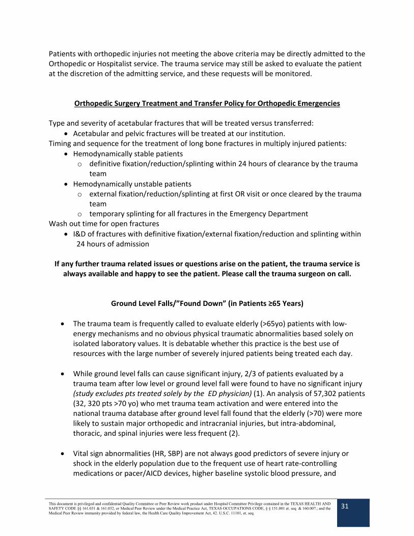
Patients with orthopedic injuries not meeting the above criteria may be directly admitted to the Orthopedic or Hospitalist service. The trauma service may still be asked to evaluate the patient at the discretion of the admitting service, and these requests will be monitored.
Orthopedic Surgery Treatment and Transfer Policy for Orthopedic Emergencies Type and severity of acetabular fractures that will be treated versus transferred:
• Acetabular and pelvic fractures will be treated at our institution. Timing and sequence for the treatment of long bone fractures in multiply injured patients:
• Hemodynamically stable patients o definitive fixation/reduction/splinting within 24 hours of clearance by the trauma
team • Hemodynamically unstable patients
o external fixation/reduction/splinting at first OR visit or once cleared by the trauma team
o temporary splinting for all fractures in the Emergency Department Wash out time for open fractures
• I&D of fractures with definitive fixation/external fixation/reduction and splinting within 24 hours of admission
If any further trauma related issues or questions arise on the patient, the trauma service is
always available and happy to see the patient. Please call the trauma surgeon on call.
Ground Level Falls/”Found Down” (in Patients ≥65 Years)
• The trauma team is frequently called to evaluate elderly (>65yo) patients with low-energy mechanisms and no obvious physical traumatic abnormalities based solely on isolated laboratory values. It is debatable whether this practice is the best use of resources with the large number of severely injured patients being treated each day.
• While ground level falls can cause significant injury, 2/3 of patients evaluated by a
trauma team after low level or ground level fall were found to have no significant injury (study excludes pts treated solely by the ED physician) (1). An analysis of 57,302 patients (32, 320 pts >70 yo) who met trauma team activation and were entered into the national trauma database after ground level fall found that the elderly (>70) were more likely to sustain major orthopedic and intracranial injuries, but intra-abdominal, thoracic, and spinal injuries were less frequent (2).
• Vital sign abnormalities (HR, SBP) are not always good predictors of severe injury or
shock in the elderly population due to the frequent use of heart rate-controlling medications or pacer/AICD devices, higher baseline systolic blood pressure, and
This document is privileged and confidential Quality Committee or Peer Review work product under Hospital Committee Privilege contained in the TEXAS HEALTH AND SAFETY CODE §§ 161.031 & 161.032, or Medical Peer Review under the Medical Practice Act, TEXAS OCCUPATIONS CODE, § § 151.001 et. seq & 160.007.; and the Medical Peer Review immunity provided by federal law, the Health Care Quality Improvement Act, 42. U.S.C. 11101, et. seq.
32
diminished sympathetic response (3). Several studies have demonstrated increasing mortality with increasing lactate and base deficit in the elderly population after blunt trauma (4, 5). These studies are limited because they include only patients that meet criteria for trauma team activation, were admitted to the trauma service, and frequently exclude fall from standing or ‘found down’ patients.
• Base deficit and lactate are important screening tools to identify normotensive elderly
patients who may have suffered significant injury. Base deficit more negative than -4 or lactate greater than 2.5 mmol/L have been shown to correlate with increased mortality in elderly bluntly injured trauma patients (5), but it is unclear if this can be applied to those patients with low energy mechanisms such as ground level falls. Base deficit and lactate should continue to be included in the initial laboratory data for patients >65yo after ground level fall.
Guideline:
• In addition to injury specific evaluation, all patients should receive CXR, Pelvic X-ray, FAST. Labs should continue to include base deficit, lactate, chem 7, CBC.
• If the BD is > -4 or the lactate is > 2.5 and there is no identified cause (i.e., serious injury, source of infection, toxins) the patient should be resuscitated with IV fluids and the labs repeated. A trauma consultation should be considered if the BD and lactate are persistently abnormal.
Drowning/Near Drowning
• Initial evaluation will be per usual Emergency Medicine (EM) standard of care. • A trauma activation is needed only if mechanism criteria are met for a trauma activation
(i.e. see criteria for level 2 trauma activation) • Intubation by itself WITHOUT a traumatic mechanism to the drowning (i.e. not fall from
height, not an MVC into water, not a boating accident, etc.) DOES NOT need a trauma activation.
o However, patients with a level 2 trauma activation who require intubation (either for drowning related issues or otherwise) should be upgraded to a level 1 per our typical process for level 2 trauma activations.
• Radiographic evaluation of an intubated patient with suspicion of trauma will routinely include a CT scan of the head and cervical spine in addition to other imaging studies at the discretion of the EM faculty.
• Patients without traumatic injuries can be admitted to the appropriate medical services (18 and over to the adult services, 17 and younger to the pediatric services)
• If traumatic injuries are identified, then appropriate consultation with trauma service (age 16 and over) or pediatric trauma service (age 15 and younger) will be required and patient should be admitted to appropriate service per usual standard of care
This document is privileged and confidential Quality Committee or Peer Review work product under Hospital Committee Privilege contained in the TEXAS HEALTH AND SAFETY CODE §§ 161.031 & 161.032, or Medical Peer Review under the Medical Practice Act, TEXAS OCCUPATIONS CODE, § § 151.001 et. seq & 160.007.; and the Medical Peer Review immunity provided by federal law, the Health Care Quality Improvement Act, 42. U.S.C. 11101, et. seq.
33
Spinal Cord Injury
• All spinal cord injury patients will be assessed by the Trauma Service • If an isolated injury, the patient may be admitted to the appropriate spine service • If the patient requires ICU admission, patients admitted to Neurosurgery will go to the
STICU References: 1. Velmahos. “Insignificant” Mechanism of injury: Not to be taken lightly. J Am Coll Surg
2000 2. Konstantinos Spaniolas. Ground level falls are associated with significant mortality in
elderly patients. J trauma 2010 3. Zarzaur. Identifying life-threatening shock in the older injured patient: an analysis of the
national trauma data bank 4. Callaway. Serum lactate and BD as predictors of mortality in normotensive elderly blunt
trauma pts. J trauma 2009 5. Davis. Base deficit in the elderly: a marker of severe injury and death. J trauma 1998
This document is privileged and confidential Quality Committee or Peer Review work product under Hospital Committee Privilege contained in the TEXAS HEALTH AND SAFETY CODE §§ 161.031 & 161.032, or Medical Peer Review under the Medical Practice Act, TEXAS OCCUPATIONS CODE, § § 151.001 et. seq & 160.007.; and the Medical Peer Review immunity provided by federal law, the Health Care Quality Improvement Act, 42. U.S.C. 11101, et. seq.
34
Memorial Hermann Southwest Hospital Clinical Guideline
GUIDELINE TITLE: Stress Ulcer Prophylaxis PUBLICATION DATE: 01/15/18 VERSION: 1 GUIDELINE PURPOSE: Assist in identification of patients who may benefit from stress ulcer prophylaxis SCOPE: Trauma Service line patients PARAMETERS: Recommendations:
Stress Ulcer Prophylaxis is indicated for select patients (Grade Level of Quality – moderate; USPSTF strength of recommendation – C [the intervention is recommended selectively based upon professional judgement and patient preferences. There is at least moderate certainty that the net benefit is small]).
Stress ulcer prophylaxis should be given with the following conditions: 1, 2
• Mechanical Ventilation • Disease associated coagulopathy • Major burn injury >30% TBSA
• Stress Ulcer Prophylaxis Agent Choice:
1. Famotidine (H2 blocker): a. Dosing:
• 20mg IV q 12H – in patients with no gastric/enteral access • 20mg PO/NGT q 12H– in patients with gastric feeds/gastric access only
b. In patients on TPN, famotidine can be added to the TPN bag daily 2. Proton Pump Inhibitors
a. Dosing: • Pantoprazole 40mg IV q24 hours • Lansoprazole 30mg suspension NGT q 24 hours
b. Limit use to: • Patients with overt and clinically significant GI bleeding • PPI use as outpatient
Initiation: At the onset of risk factors.
Duration of Treatment: Until risk factors resolve
This document is privileged and confidential Quality Committee or Peer Review work product under Hospital Committee Privilege contained in the TEXAS HEALTH AND SAFETY CODE §§ 161.031 & 161.032, or Medical Peer Review under the Medical Practice Act, TEXAS OCCUPATIONS CODE, § § 151.001 et. seq & 160.007.; and the Medical Peer Review immunity provided by federal law, the Health Care Quality Improvement Act, 42. U.S.C. 11101, et. seq.
35
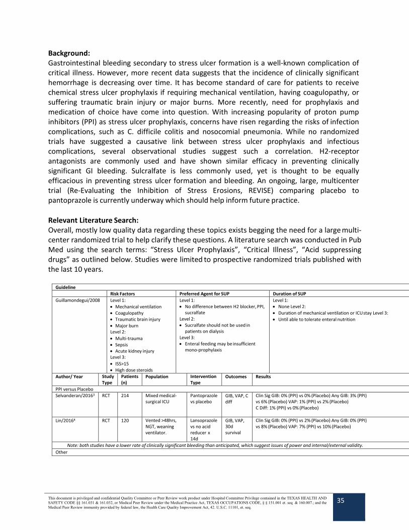
Background: Gastrointestinal bleeding secondary to stress ulcer formation is a well-known complication of critical illness. However, more recent data suggests that the incidence of clinically significant hemorrhage is decreasing over time. It has become standard of care for patients to receive chemical stress ulcer prophylaxis if requiring mechanical ventilation, having coagulopathy, or suffering traumatic brain injury or major burns. More recently, need for prophylaxis and medication of choice have come into question. With increasing popularity of proton pump inhibitors (PPI) as stress ulcer prophylaxis, concerns have risen regarding the risks of infection complications, such as C. difficile colitis and nosocomial pneumonia. While no randomized trials have suggested a causative link between stress ulcer prophylaxis and infectious complications, several observational studies suggest such a correlation. H2-receptor antagonists are commonly used and have shown similar efficacy in preventing clinically significant GI bleeding. Sulcralfate is less commonly used, yet is thought to be equally efficacious in preventing stress ulcer formation and bleeding. An ongoing, large, multicenter trial (Re-Evaluating the Inhibition of Stress Erosions, REVISE) comparing placebo to pantoprazole is currently underway which should help inform future practice. Relevant Literature Search: Overall, mostly low quality data regarding these topics exists begging the need for a large multi-center randomized trial to help clarify these questions. A literature search was conducted in Pub Med using the search terms: “Stress Ulcer Prophylaxis”, “Critical Illness”, “Acid suppressing drugs” as outlined below. Studies were limited to prospective randomized trials published with the last 10 years.
Guideline
Risk Factors Preferred Agent for SUP Duration of SUP Guillamondegui/2008 Level 1:
• Mechanical ventilation • Coagulopathy • Traumatic brain injury • Major burn Level 2: • Multi-trauma • Sepsis • Acute kidney injury Level 3: • ISS>15 • High dose steroids
Level 1: • No difference between H2 blocker, PPI,
sucralfate Level 2: • Sucralfate should not be used in
patients on dialysis Level 3: • Enteral feeding may be insufficient
mono-prophylaxis
Level 1: • None Level 2: • Duration of mechanical ventilation or ICU stay Level 3: • Until able to tolerate enteral nutrition
Author/ Year Study Type
Patients (n)
Population Intervention Type
Outcomes Results
PPI versus Placebo Selvanderan/20163 RCT 214 Mixed medical-
surgical ICU Pantoprazole vs placebo
GIB, VAP, C diff
Clin Sig GIB: 0% (PPI) vs 0% (Placebo) Any GIB: 3% (PPI) vs 6% (Placebo) VAP: 1% (PPI) vs 2% (Placebo) C Diff: 1% (PPI) vs 0% (Placebo)
Lin/20164 RCT 120 Vented >48hrs, NGT, weaning ventilator.
Lansoprazole vs no acid reducer x 14d
GIB, VAP, 30d survival
Clin Sig GIB: 0% (PPI) vs 2% (Placebo) Any GIB: 0% (PPI) vs 8% (Placebo) VAP: 7% (PPI) vs 10% (Placebo)
Note: both studies have a lower rate of clinically significant bleeding than anticipated, which suggest issues of power and internal/external validity. Other
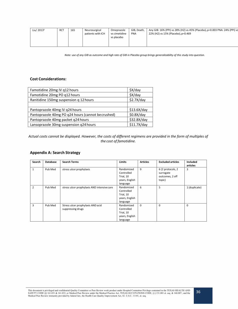
This document is privileged and confidential Quality Committee or Peer Review work product under Hospital Committee Privilege contained in the TEXAS HEALTH AND SAFETY CODE §§ 161.031 & 161.032, or Medical Peer Review under the Medical Practice Act, TEXAS OCCUPATIONS CODE, § § 151.001 et. seq & 160.007.; and the Medical Peer Review immunity provided by federal law, the Health Care Quality Improvement Act, 42. U.S.C. 11101, et. seq.
36
Liu/ 20135 RCT 165 Neurosurgical patients with ICH
Omeprazole vs cimetidine vs placebo
GIB, Death, PNA
Any GIB: 16% (PPI) vs 28% (H2) vs 45% (Placebo), p=0.003 PNA: 24% (PPI) vs 22% (H2) vs 15% (Placebo), p=0.469
Cost Considerations:
Famotidine 20mg IV q12 hours $X/day Famotidine 20mg PO q12 hours $X/day Ranitidine 150mg suspension q 12 hours $2.7X/day
Pantoprazole 40mg IV q24 hours $13.6X/day Pantoprazole 40mg PO q24 hours (cannot be crushed) $0.8X/day Pantoprazole 40mg packet q24 hours $32.8X/day Lansoprazole 30mg suspension q24 hours $11.7X/day
Actual costs cannot be displayed. However, the costs of different regimens are provided in the form of multiples of
the cost of famotidine.
Appendix A: Search Strategy
Search Database Search Terms Limits Articles Excluded articles Included articles
1 Pub Med stress ulcer prophylaxis Randomized Controlled Trial, 10 years, English language
9 6 (2 protocols, 2 surrogate outcomes, 2 off topic)
3
2 Pub Med stress ulcer prophylaxis AND intensive care Randomized Controlled Trial, 10 years, English language
6 5 1 (duplicate)
3 Pub Med Stress ulcer prophylaxis AND acid suppressing drugs
Randomized Controlled Trial, 10 years, English language
0 0 0
Note: use of any GIB as outcome and high rate of GIB in Placebo group brings generalizability of this study into question.
This document is privileged and confidential Quality Committee or Peer Review work product under Hospital Committee Privilege contained in the TEXAS HEALTH AND SAFETY CODE §§ 161.031 & 161.032, or Medical Peer Review under the Medical Practice Act, TEXAS OCCUPATIONS CODE, § § 151.001 et. seq & 160.007.; and the Medical Peer Review immunity provided by federal law, the Health Care Quality Improvement Act, 42. U.S.C. 11101, et. seq.
37
References:
1 Cook, D.J., Fuller, H.D., Guyatt, G.H., et al. Risk factors for gastrointestinal bleeding in critically ill patients. Canadian Critical Care Trials Groups. New England Journal of Medicine. 1994; 330(6): 377-381
2 Guillamondegui, O., Gunter, O.L., Bonadies, J.A. et al. Practice management guidelines for stress ulcer prophylaxis. EAST Practice Management Guidelines Committee. 2008: Accessed online at: http://www.east.org/tpg/archive/html/stressulcer.html
3 Selvanderan, Shane P., Matthew J. Summers, Mark E. Finnis, Mark P. Plummer, Yasmine Ali Abdelhamid, Michael B. Anderson, Marianne J. Chapman, Christopher K. Rayner, and Adam M. Deane. "Pantoprazole or Placebo for Stress Ulcer Prophylaxis (POP-UP)." Critical Care Medicine 44.10 (2016): 1842-850
4 Lin, Chien-Chu, et al. “Stress ulcer prophylaxis in patients being weaned from the ventilator in a respiratory care center: A randomized control trial.” Journal of the Formosan Medical Association, vol. 115, no. 1, 2016, pp. 19–24
5 Liu, Bo-Lin, Bing Li, Xiang Zhang, Zhou Fei, Shi-Jie Hu, Wei Lin, Da-Kuan Gao, and Li Zhang. "A randomized controlled study comparing omeprazole and cimetidine for the prophylaxis of stress-related upper gastrointestinal bleeding in patients with intracerebral hemorrhage." Journal of Neurosurgery 118.1 (2013): 115-20
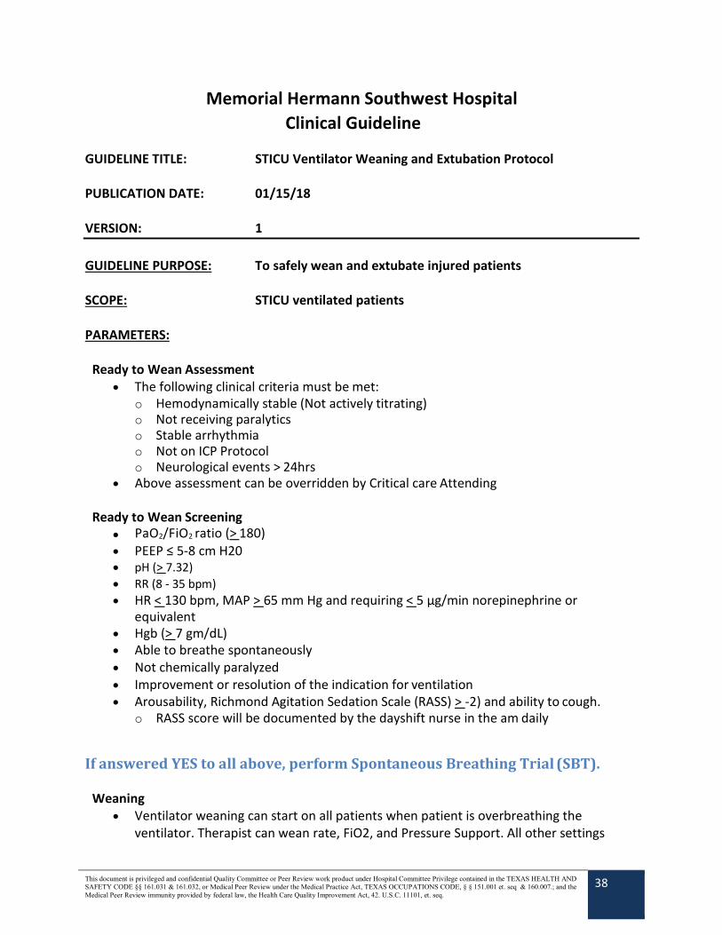
This document is privileged and confidential Quality Committee or Peer Review work product under Hospital Committee Privilege contained in the TEXAS HEALTH AND SAFETY CODE §§ 161.031 & 161.032, or Medical Peer Review under the Medical Practice Act, TEXAS OCCUPATIONS CODE, § § 151.001 et. seq & 160.007.; and the Medical Peer Review immunity provided by federal law, the Health Care Quality Improvement Act, 42. U.S.C. 11101, et. seq.
38
Memorial Hermann Southwest Hospital Clinical Guideline
GUIDELINE TITLE: STICU Ventilator Weaning and Extubation Protocol PUBLICATION DATE: 01/15/18 VERSION: 1 GUIDELINE PURPOSE: To safely wean and extubate injured patients SCOPE: STICU ventilated patients PARAMETERS:
Ready to Wean Assessment • The following clinical criteria must be met:
o Hemodynamically stable (Not actively titrating) o Not receiving paralytics o Stable arrhythmia o Not on ICP Protocol o Neurological events > 24hrs
• Above assessment can be overridden by Critical care Attending
Ready to Wean Screening • PaO2/FiO2 ratio (> 180) • PEEP ≤ 5-8 cm H20 • pH (> 7.32) • RR (8 - 35 bpm) • HR < 130 bpm, MAP > 65 mm Hg and requiring < 5 µg/min norepinephrine or
equivalent • Hgb (> 7 gm/dL) • Able to breathe spontaneously • Not chemically paralyzed • Improvement or resolution of the indication for ventilation • Arousability, Richmond Agitation Sedation Scale (RASS) > -2) and ability to cough.
o RASS score will be documented by the dayshift nurse in the am daily
If answered YES to all above, perform Spontaneous Breathing Trial (SBT).
Weaning • Ventilator weaning can start on all patients when patient is overbreathing the
ventilator. Therapist can wean rate, FiO2, and Pressure Support. All other settings
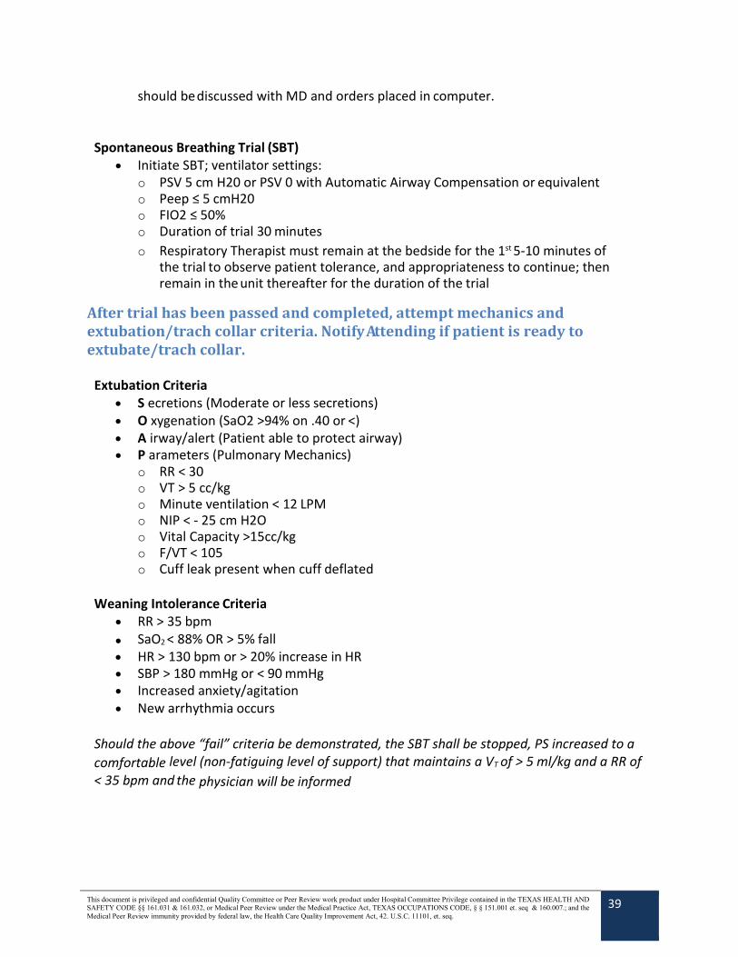
This document is privileged and confidential Quality Committee or Peer Review work product under Hospital Committee Privilege contained in the TEXAS HEALTH AND SAFETY CODE §§ 161.031 & 161.032, or Medical Peer Review under the Medical Practice Act, TEXAS OCCUPATIONS CODE, § § 151.001 et. seq & 160.007.; and the Medical Peer Review immunity provided by federal law, the Health Care Quality Improvement Act, 42. U.S.C. 11101, et. seq.
39
should be discussed with MD and orders placed in computer.
Spontaneous Breathing Trial (SBT)
• Initiate SBT; ventilator settings: o PSV 5 cm H20 or PSV 0 with Automatic Airway Compensation or equivalent o Peep ≤ 5 cmH20 o FIO2 ≤ 50% o Duration of trial 30 minutes o Respiratory Therapist must remain at the bedside for the 1st 5-10 minutes of
the trial to observe patient tolerance, and appropriateness to continue; then remain in the unit thereafter for the duration of the trial
After trial has been passed and completed, attempt mechanics and extubation/trach collar criteria. Notify Attending if patient is ready to extubate/trach collar.
Extubation Criteria • S ecretions (Moderate or less secretions) • O xygenation (SaO2 >94% on .40 or <) • A irway/alert (Patient able to protect airway) • P arameters (Pulmonary Mechanics)
o RR < 30 o VT > 5 cc/kg o Minute ventilation < 12 LPM o NIP < - 25 cm H2O o Vital Capacity >15cc/kg o F/VT < 105 o Cuff leak present when cuff deflated
Weaning Intolerance Criteria
• RR > 35 bpm • SaO2 < 88% OR > 5% fall • HR > 130 bpm or > 20% increase in HR • SBP > 180 mmHg or < 90 mmHg • Increased anxiety/agitation • New arrhythmia occurs
Should the above “fail” criteria be demonstrated, the SBT shall be stopped, PS increased to a comfortable level (non-fatiguing level of support) that maintains a VT of > 5 ml/kg and a RR of < 35 bpm and the physician will be informed
This document is privileged and confidential Quality Committee or Peer Review work product under Hospital Committee Privilege contained in the TEXAS HEALTH AND SAFETY CODE §§ 161.031 & 161.032, or Medical Peer Review under the Medical Practice Act, TEXAS OCCUPATIONS CODE, § § 151.001 et. seq & 160.007.; and the Medical Peer Review immunity provided by federal law, the Health Care Quality Improvement Act, 42. U.S.C. 11101, et. seq.
40
References: • Timothy D Girard, John P Kress, Barry D Fuchs, Jason W W Thomason, William D
Schweickert, Brenda T Pun, Darren B Taichman, Jan G Dunn,. (2008). Effi cacy and safety of a paired sedation and ventilator weaning protocol for mechanically ventilated patients in intensive care (Awakening and Breathing Controlled trial): a randomised controlled tri. Lancet. 371, 126-134.
• Tanios, MA, et al. A randomized, controlled trial of the role of weaning predictors in clinical decision-making. Critical Care Medicine. 2006; 34(10). 2676-7
• Cohen, JD et al. Extubation outcome following a spontaneous breathing trial with automatic tube compensation versus continuous positive airway pressure. Critical Care Medicine. 2006 ; 34(3) 682-6
• Haberthur, C. et al. Extubation after breathing trials with automatic tube compensation, T-tube or pressure support ventilation. Acta Anesthesiology Scanda. 2002 46(8) 973-9
• Perren, A. et al. Protocol-directed weaning from mechanical ventilation: clinical outcome in patients randomized for a 30 min or 120 min trial with pressure support ventilation. Intensive Care Medicine. 2002 28(8). 1058-63.
• MacIntyre, N. et al. Evidence-based guidelines for weaning and discontinuing ventilatory
support. Chest 120(6) 2001 375s-95s. • Marelich, GP et al. Protocol weaning of mechanical ventilation in medical and surgical
patients by respiratory care practitioners and nurses. Chest 2000 118 459-67.
• Esteban, A. et al. Effect of spontaneous breathing trial duration on outcome of attempts to discontinue mechanical ventilation: the Spanish Lung Failure Group. Am J Respiratory Crit Care Med 1999; 159: 512-18
• Kollef, MH. Et al. A randomized, controlled trial of protocol-directed versus physician-directed
weaning from mechanical ventilation. Critical Care Medicine. 1997 25; 567-74 • Estaban, A et al. Extubation outcome after spontaneous breathing trials with t-tube or
pressure support ventilation. The Spanish Lung Failure Collaboration Group. Am J Respir Crit Care. 1997 156; 459-65
• Ely, WE, et al. Effect on the duration of mechanical ventilation of identifying patients capable of
breathing spontaneously. N. Eng J Med 1996; 335; 1864-69 • Estaban, A, et al. A comparison of four methods of weaning patients from mechanical
ventilation: The Spanish Lung Failure Collaborative Group. N Eng J Med. 1995; 332; 345-50 • Brochard, L. et al. Comparison of three methods of gradual withdrawal from ventilatory
support during weaning from mechanical ventilation. Am J Respir Crit Care Med 1994; 150; 896-903.
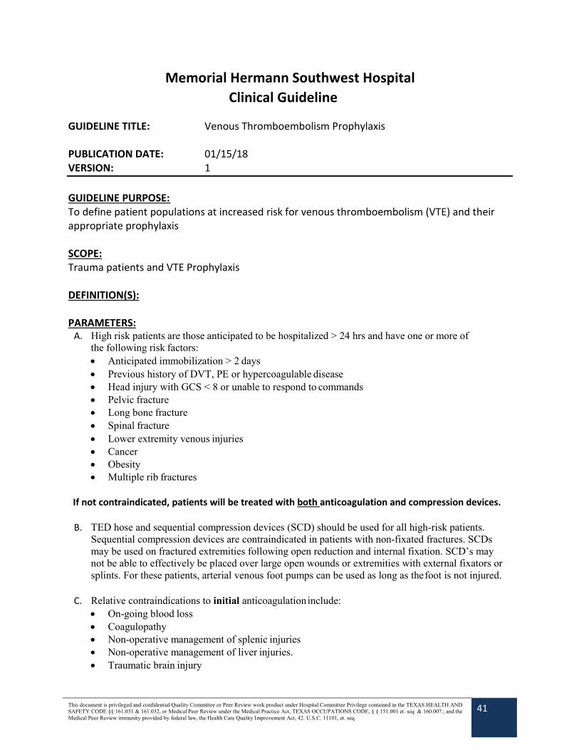
This document is privileged and confidential Quality Committee or Peer Review work product under Hospital Committee Privilege contained in the TEXAS HEALTH AND SAFETY CODE §§ 161.031 & 161.032, or Medical Peer Review under the Medical Practice Act, TEXAS OCCUPATIONS CODE, § § 151.001 et. seq & 160.007.; and the Medical Peer Review immunity provided by federal law, the Health Care Quality Improvement Act, 42. U.S.C. 11101, et. seq.
41
Memorial Hermann Southwest Hospital Clinical Guideline
GUIDELINE TITLE: Venous Thromboembolism Prophylaxis PUBLICATION DATE: 01/15/18 VERSION: 1 GUIDELINE PURPOSE: To define patient populations at increased risk for venous thromboembolism (VTE) and their appropriate prophylaxis SCOPE: Trauma patients and VTE Prophylaxis DEFINITION(S): PARAMETERS:
A. High risk patients are those anticipated to be hospitalized > 24 hrs and have one or more of the following risk factors: • Anticipated immobilization > 2 days • Previous history of DVT, PE or hypercoagulable disease • Head injury with GCS < 8 or unable to respond to commands • Pelvic fracture • Long bone fracture • Spinal fracture • Lower extremity venous injuries • Cancer • Obesity • Multiple rib fractures
If not contraindicated, patients will be treated with both anticoagulation and compression devices.
B. TED hose and sequential compression devices (SCD) should be used for all high-risk patients. Sequential compression devices are contraindicated in patients with non-fixated fractures. SCDs may be used on fractured extremities following open reduction and internal fixation. SCD’s may not be able to effectively be placed over large open wounds or extremities with external fixators or splints. For these patients, arterial venous foot pumps can be used as long as the foot is not injured.
C. Relative contraindications to initial anticoagulation include:
• On-going blood loss • Coagulopathy • Non-operative management of splenic injuries • Non-operative management of liver injuries. • Traumatic brain injury
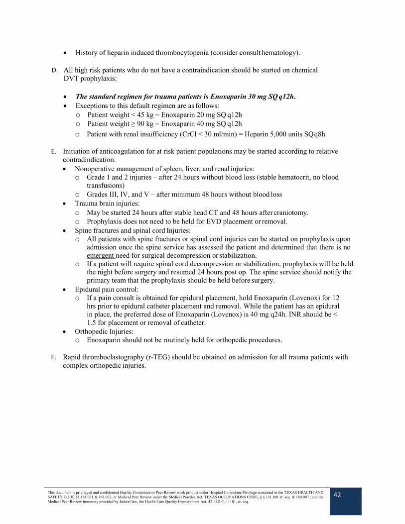
This document is privileged and confidential Quality Committee or Peer Review work product under Hospital Committee Privilege contained in the TEXAS HEALTH AND SAFETY CODE §§ 161.031 & 161.032, or Medical Peer Review under the Medical Practice Act, TEXAS OCCUPATIONS CODE, § § 151.001 et. seq & 160.007.; and the Medical Peer Review immunity provided by federal law, the Health Care Quality Improvement Act, 42. U.S.C. 11101, et. seq.
42
• History of heparin induced thrombocytopenia (consider consult hematology).
D. All high risk patients who do not have a contraindication should be started on chemical DVT prophylaxis:
• The standard regimen for trauma patients is Enoxaparin 30 mg SQ q12h. • Exceptions to this default regimen are as follows:
o Patient weight < 45 kg = Enoxaparin 20 mg SQ q12h o Patient weight ≥ 90 kg = Enoxaparin 40 mg SQ q12h o Patient with renal insufficiency (CrCl < 30 ml/min) = Heparin 5,000 units SQ q8h
E. Initiation of anticoagulation for at risk patient populations may be started according to relative
contradindication: • Nonoperative management of spleen, liver, and renal injuries:
o Grade 1 and 2 injuries – after 24 hours without blood loss (stable hematocrit, no blood transfusions)
o Grades III, IV, and V – after minimum 48 hours without blood loss • Trauma brain injuries:
o May be started 24 hours after stable head CT and 48 hours after craniotomy. o Prophylaxis does not need to be held for EVD placement or removal.
• Spine fractures and spinal cord Injuries: o All patients with spine fractures or spinal cord injuries can be started on prophylaxis upon
admission once the spine service has assessed the patient and determined that there is no emergent need for surgical decompression or stabilization.
o If a patient will require spinal cord decompression or stabilization, prophylaxis will be held the night before surgery and resumed 24 hours post op. The spine service should notify the primary team that the prophylaxis should be held before surgery.
• Epidural pain control: o If a pain consult is obtained for epidural placement, hold Enoxaparin (Lovenox) for 12
hrs prior to epidural catheter placement and removal. While the patient has an epidural in place, the preferred dose of Enoxaparin (Lovenox) is 40 mg q24h. INR should be < 1.5 for placement or removal of catheter.
• Orthopedic Injuries: o Enoxaparin should not be routinely held for orthopedic procedures.
F. Rapid thromboelastography (r-TEG) should be obtained on admission for all trauma patients with
complex orthopedic injuries.
This document is privileged and confidential Quality Committee or Peer Review work product under Hospital Committee Privilege contained in the TEXAS HEALTH AND SAFETY CODE §§ 161.031 & 161.032, or Medical Peer Review under the Medical Practice Act, TEXAS OCCUPATIONS CODE, § § 151.001 et. seq & 160.007.; and the Medical Peer Review immunity provided by federal law, the Health Care Quality Improvement Act, 42. U.S.C. 11101, et. seq.
43
References Cohen, D. B., Rinker, C., & Wilberger, J. E. (2006). Traumatic Brain Injury in Anticoagulated Patients.
Journal of Trauma and Acute Care Surgery, 60(3), 553. doi:10.1097/01.ta.0000196542.54344.05 Fang, M. C. (2004). Advanced Age, Anticoagulation Intensity, and Risk for Intracranial Hemorrhage
among Patients Taking Warfarin for Atrial Fibrillation. Annals of Internal Medicine, 141(10), 745–9. doi:10.7326/0003-4819-141-10-200411160-00005
Franko, J., Kish, K. J., O’Connell, B. G., Subramanian, S., & Yuschak, J. V. (2006). Advanced Age and Preinjury Warfarin Anticoagulation Increase the Risk of Mortality After Head Trauma. Journal of Trauma and Acute Care Surgery, 61(1), 107. doi:10.1097/01.ta.0000224220.89528.fc
Hylek, E. M. (2009). Risk Factors for Intracranial Hemorrhage in Outpatients Taking Warfarin. Annals of Internal Medicine, 1–6.
Peck, K. A., Calvo, R. Y., Schechter, M. S., Sise, C. B., Kahl, J. E., Shackford, M. C., et al. (2014). The impact of preinjury anticoagulants and prescription antiplatelet agents on outcomes in older patients with traumatic brain injury. The Journal of Trauma and Acute Care Surgery, 76(2), 431–436. doi:10.1097/TA.0000000000000107
Reynolds, F. D., Dietz, P. A., Higgins, D., & Whitaker, T. S. (2003). Time to Deterioration of the Elderly, Anticoagulated, Minor Head Injury Patient Who Presents without Evidence of Neurologic Abnormality. Journal of Trauma and Acute Care Surgery, 54(3), 492.
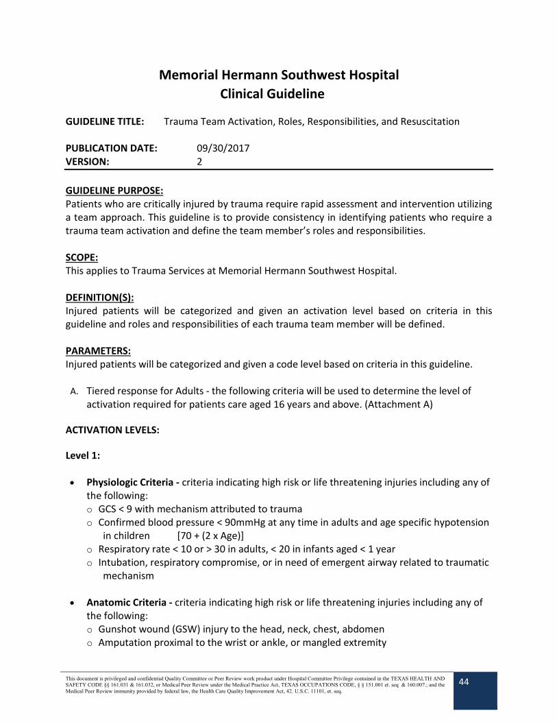
This document is privileged and confidential Quality Committee or Peer Review work product under Hospital Committee Privilege contained in the TEXAS HEALTH AND SAFETY CODE §§ 161.031 & 161.032, or Medical Peer Review under the Medical Practice Act, TEXAS OCCUPATIONS CODE, § § 151.001 et. seq & 160.007.; and the Medical Peer Review immunity provided by federal law, the Health Care Quality Improvement Act, 42. U.S.C. 11101, et. seq.
44
Memorial Hermann Southwest Hospital Clinical Guideline
GUIDELINE TITLE: Trauma Team Activation, Roles, Responsibilities, and Resuscitation PUBLICATION DATE: 09/30/2017 VERSION: 2 GUIDELINE PURPOSE: Patients who are critically injured by trauma require rapid assessment and intervention utilizing a team approach. This guideline is to provide consistency in identifying patients who require a trauma team activation and define the team member’s roles and responsibilities. SCOPE: This applies to Trauma Services at Memorial Hermann Southwest Hospital. DEFINITION(S): Injured patients will be categorized and given an activation level based on criteria in this guideline and roles and responsibilities of each trauma team member will be defined. PARAMETERS: Injured patients will be categorized and given a code level based on criteria in this guideline.
A. Tiered response for Adults - the following criteria will be used to determine the level of activation required for patients care aged 16 years and above. (Attachment A)
ACTIVATION LEVELS: Level 1: • Physiologic Criteria - criteria indicating high risk or life threatening injuries including any of
the following: o GCS < 9 with mechanism attributed to trauma o Confirmed blood pressure < 90mmHg at any time in adults and age specific hypotension
in children [70 + (2 x Age)] o Respiratory rate < 10 or > 30 in adults, < 20 in infants aged < 1 year o Intubation, respiratory compromise, or in need of emergent airway related to traumatic
mechanism • Anatomic Criteria - criteria indicating high risk or life threatening injuries including any of
the following: o Gunshot wound (GSW) injury to the head, neck, chest, abdomen o Amputation proximal to the wrist or ankle, or mangled extremity
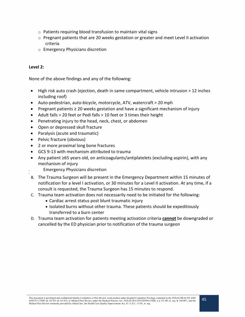
This document is privileged and confidential Quality Committee or Peer Review work product under Hospital Committee Privilege contained in the TEXAS HEALTH AND SAFETY CODE §§ 161.031 & 161.032, or Medical Peer Review under the Medical Practice Act, TEXAS OCCUPATIONS CODE, § § 151.001 et. seq & 160.007.; and the Medical Peer Review immunity provided by federal law, the Health Care Quality Improvement Act, 42. U.S.C. 11101, et. seq.
45
o Patients requiring blood transfusion to maintain vital signs o Pregnant patients that are 20 weeks gestation or greater and meet Level II activation
criteria o Emergency Physicians discretion
Level 2: None of the above findings and any of the following:
• High risk auto crash (ejection, death in same compartment, vehicle intrusion > 12 inches
including roof) • Auto-pedestrian, auto-bicycle, motorcycle, ATV, watercraft > 20 mph • Pregnant patients ≥ 20 weeks gestation and have a significant mechanism of injury • Adult falls > 20 feet or Pedi falls > 10 feet or 3 times their height • Penetrating injury to the head, neck, chest, or abdomen • Open or depressed skull fracture • Paralysis (acute and traumatic) • Pelvic fracture (obvious) • 2 or more proximal long bone fractures • GCS 9-13 with mechanism attributed to trauma • Any patient ≥65 years old, on anticoagulants/antiplatelets (excluding aspirin), with any
mechanism of injury • Emergency Physicians discretion
•
B. The Trauma Surgeon will be present in the Emergency Department within 15 minutes of notification for a level I activation, or 30 minutes for a Level II activation. At any time, if a consult is requested, the Trauma Surgeon has 15 minutes to respond.
C. Trauma team activation does not necessarily need to be initiated for the following: • Cardiac arrest status post blunt traumatic injury • Isolated burns without other trauma. These patients should be expeditiously
transferred to a burn center D. Trauma team activation for patients meeting activation criteria cannot be downgraded or
cancelled by the ED physician prior to notification of the trauma surgeon
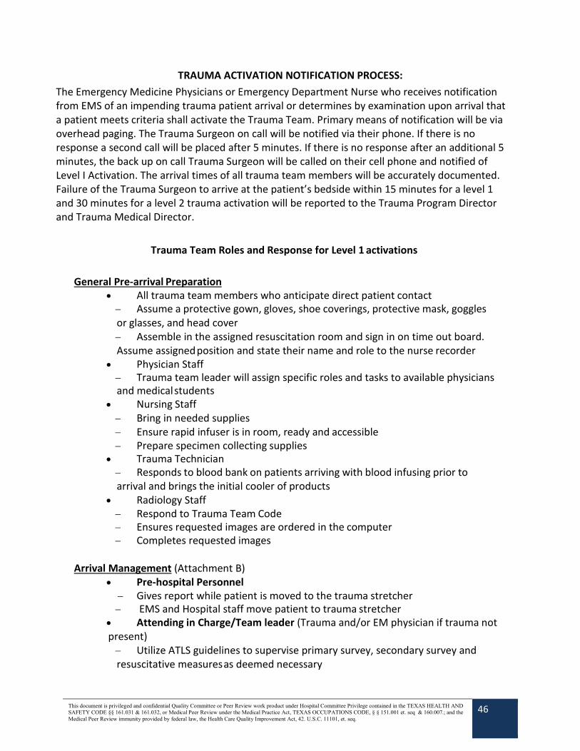
This document is privileged and confidential Quality Committee or Peer Review work product under Hospital Committee Privilege contained in the TEXAS HEALTH AND SAFETY CODE §§ 161.031 & 161.032, or Medical Peer Review under the Medical Practice Act, TEXAS OCCUPATIONS CODE, § § 151.001 et. seq & 160.007.; and the Medical Peer Review immunity provided by federal law, the Health Care Quality Improvement Act, 42. U.S.C. 11101, et. seq.
46
TRAUMA ACTIVATION NOTIFICATION PROCESS: The Emergency Medicine Physicians or Emergency Department Nurse who receives notification from EMS of an impending trauma patient arrival or determines by examination upon arrival that a patient meets criteria shall activate the Trauma Team. Primary means of notification will be via overhead paging. The Trauma Surgeon on call will be notified via their phone. If there is no response a second call will be placed after 5 minutes. If there is no response after an additional 5 minutes, the back up on call Trauma Surgeon will be called on their cell phone and notified of Level I Activation. The arrival times of all trauma team members will be accurately documented. Failure of the Trauma Surgeon to arrive at the patient’s bedside within 15 minutes for a level 1 and 30 minutes for a level 2 trauma activation will be reported to the Trauma Program Director and Trauma Medical Director.
Trauma Team Roles and Response for Level 1 activations
General Pre-arrival Preparation • All trauma team members who anticipate direct patient contact − Assume a protective gown, gloves, shoe coverings, protective mask, goggles or glasses, and head cover − Assemble in the assigned resuscitation room and sign in on time out board. Assume assigned position and state their name and role to the nurse recorder
• Physician Staff − Trauma team leader will assign specific roles and tasks to available physicians and medical students
• Nursing Staff − Bring in needed supplies − Ensure rapid infuser is in room, ready and accessible − Prepare specimen collecting supplies
• Trauma Technician − Responds to blood bank on patients arriving with blood infusing prior to arrival and brings the initial cooler of products
• Radiology Staff − Respond to Trauma Team Code − Ensures requested images are ordered in the computer − Completes requested images
Arrival Management (Attachment B)
• Pre-hospital Personnel − Gives report while patient is moved to the trauma stretcher − EMS and Hospital staff move patient to trauma stretcher
• Attending in Charge/Team leader (Trauma and/or EM physician if trauma not present) − Utilize ATLS guidelines to supervise primary survey, secondary survey and resuscitative measures as deemed necessary
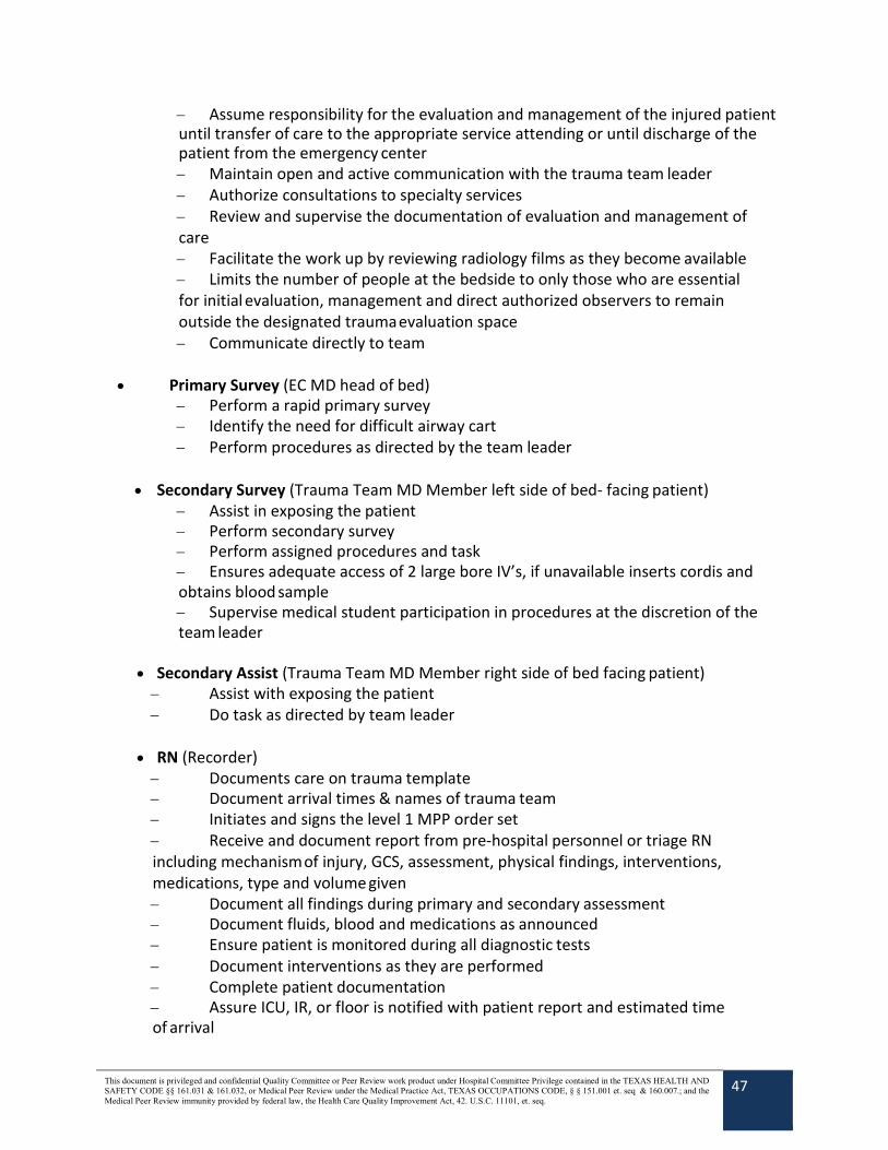
This document is privileged and confidential Quality Committee or Peer Review work product under Hospital Committee Privilege contained in the TEXAS HEALTH AND SAFETY CODE §§ 161.031 & 161.032, or Medical Peer Review under the Medical Practice Act, TEXAS OCCUPATIONS CODE, § § 151.001 et. seq & 160.007.; and the Medical Peer Review immunity provided by federal law, the Health Care Quality Improvement Act, 42. U.S.C. 11101, et. seq.
47
− Assume responsibility for the evaluation and management of the injured patient until transfer of care to the appropriate service attending or until discharge of the patient from the emergency center − Maintain open and active communication with the trauma team leader − Authorize consultations to specialty services − Review and supervise the documentation of evaluation and management of care − Facilitate the work up by reviewing radiology films as they become available − Limits the number of people at the bedside to only those who are essential for initial evaluation, management and direct authorized observers to remain outside the designated trauma evaluation space − Communicate directly to team
• Primary Survey (EC MD head of bed)
− Perform a rapid primary survey − Identify the need for difficult airway cart − Perform procedures as directed by the team leader
• Secondary Survey (Trauma Team MD Member left side of bed- facing patient) − Assist in exposing the patient − Perform secondary survey − Perform assigned procedures and task − Ensures adequate access of 2 large bore IV’s, if unavailable inserts cordis and obtains blood sample − Supervise medical student participation in procedures at the discretion of the team leader
• Secondary Assist (Trauma Team MD Member right side of bed facing patient) − Assist with exposing the patient − Do task as directed by team leader
• RN (Recorder) − Documents care on trauma template − Document arrival times & names of trauma team − Initiates and signs the level 1 MPP order set − Receive and document report from pre-hospital personnel or triage RN including mechanism of injury, GCS, assessment, physical findings, interventions, medications, type and volume given − Document all findings during primary and secondary assessment − Document fluids, blood and medications as announced − Ensure patient is monitored during all diagnostic tests − Document interventions as they are performed − Complete patient documentation − Assure ICU, IR, or floor is notified with patient report and estimated time of arrival
This document is privileged and confidential Quality Committee or Peer Review work product under Hospital Committee Privilege contained in the TEXAS HEALTH AND SAFETY CODE §§ 161.031 & 161.032, or Medical Peer Review under the Medical Practice Act, TEXAS OCCUPATIONS CODE, § § 151.001 et. seq & 160.007.; and the Medical Peer Review immunity provided by federal law, the Health Care Quality Improvement Act, 42. U.S.C. 11101, et. seq.
48
− Document vital signs and neurologic status as clinically indicated and with any significant changes
• RN (Circulating RN) − Obtains initial B/P manually and ensures collection of vital signs including a temperature in the initial set − Obtain large bore peripheral IV access and blood samples − Ensure patient remains covered with warm blankets after secondary survey and resuscitative measures are completed − Ensures recorder RN aware of all interventions and procedures performed − Administer medications ordered by the trauma team leader − Announce amount of crystalloid and blood products infused − Ensures allergy and blood band are on the patient − Prepares patient for immediate critical transport
• Trauma Technician − Announces the initial cooler of products arrival − Ensure that previous patient is discharged from monitor − Ensure the new patient is admitted to monitor and sets B/P to repeat Q 3 minutes. Also calls out first set of electronic V/S including temperature to recording nurse. − Place monitor leads on patient − Ensure patient remains covered with warm blankets after secondary survey and resuscitation measures − Deliver blood sample to lab and to blood bank and announces “Trauma team activation level 1”
• Patient Access − Register patient − Confirms Life Flight ID number − Documents patients valuables on inventory list and places a copy of the inventory list with patient chart
• Charge RN − Ensures and coordinates availability of beds − Assist to facilitate a smooth and prompt transfer our of EC − When hospital beds are not promptly available, provide ongoing communication to the trauma team leader, attending, and operations administrator regarding problems and/or progress in obtaining a bed
• Radiology Tech − Obtain images requested by trauma team leader − Assist the team leader or assigned member in arranging for all subsequent radiological studies
This document is privileged and confidential Quality Committee or Peer Review work product under Hospital Committee Privilege contained in the TEXAS HEALTH AND SAFETY CODE §§ 161.031 & 161.032, or Medical Peer Review under the Medical Practice Act, TEXAS OCCUPATIONS CODE, § § 151.001 et. seq & 160.007.; and the Medical Peer Review immunity provided by federal law, the Health Care Quality Improvement Act, 42. U.S.C. 11101, et. seq.
49
− Maintain open and active communication with the trauma team
• Radiologist − Be available to evaluate x-rays and CT scans as soon as they are processed and provide a prompt report in PACS − Notify trauma team leader of any changes in the final report
• Respiratory Therapy − Assist with airway control, prepare equipment, assist with intubation, and maintain a secured airway − Obtain arterial blood gases and communicate results to team leader − Ensure oxygen and ventilation equipment is available for transport of the patient − Maintain open and active communication with the trauma team
• Chaplain − Assume the role of the liaison with the family/visitors. − Contact any individuals per patient request − Maintain open and active communication with the trauma team − Provide spiritual and emotional support for patient, family, and staff as needed
• Security Officer − Monitor, reinforce, and assist ED in limiting attendance to only essential care personnel. − Allow family members to room after clearance has been given by physician or charge nurse − Will be informed of potential forensic situations − Assume the liaison role with law enforcement − Ensure the safety of team members and patients − Secure patient belongings − Secure helipad area as needed − Maintain open and active communication with the trauma team
• OR Nurse/OB Charge Nurse/Anesthesia (level 1 activations) − Assess possible surgical needs of patient and inform the surgical department − Maintain open and active communication with the trauma team
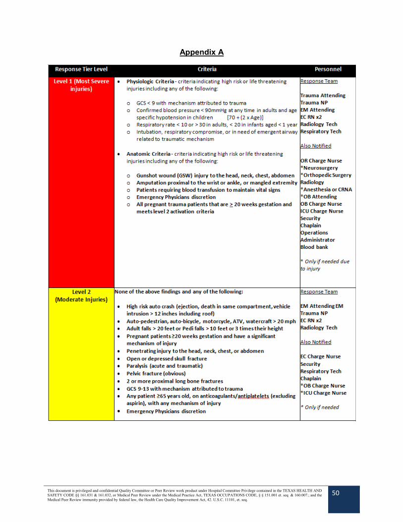
This document is privileged and confidential Quality Committee or Peer Review work product under Hospital Committee Privilege contained in the TEXAS HEALTH AND SAFETY CODE §§ 161.031 & 161.032, or Medical Peer Review under the Medical Practice Act, TEXAS OCCUPATIONS CODE, § § 151.001 et. seq & 160.007.; and the Medical Peer Review immunity provided by federal law, the Health Care Quality Improvement Act, 42. U.S.C. 11101, et. seq.
50
Appendix A
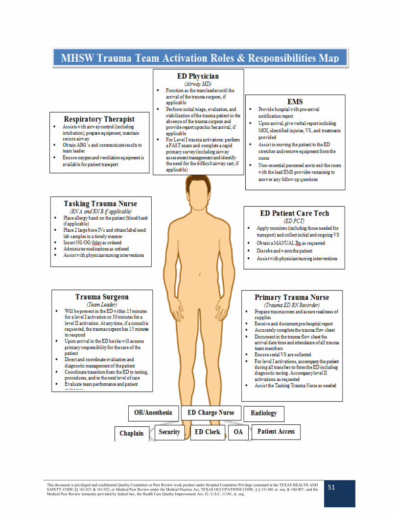
This document is privileged and confidential Quality Committee or Peer Review work product under Hospital Committee Privilege contained in the TEXAS HEALTH AND SAFETY CODE §§ 161.031 & 161.032, or Medical Peer Review under the Medical Practice Act, TEXAS OCCUPATIONS CODE, § § 151.001 et. seq & 160.007.; and the Medical Peer Review immunity provided by federal law, the Health Care Quality Improvement Act, 42. U.S.C. 11101, et. seq.
51
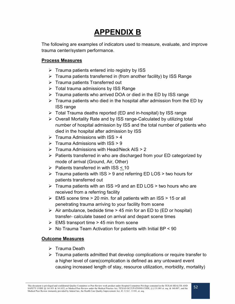
This document is privileged and confidential Quality Committee or Peer Review work product under Hospital Committee Privilege contained in the TEXAS HEALTH AND SAFETY CODE §§ 161.031 & 161.032, or Medical Peer Review under the Medical Practice Act, TEXAS OCCUPATIONS CODE, § § 151.001 et. seq & 160.007.; and the Medical Peer Review immunity provided by federal law, the Health Care Quality Improvement Act, 42. U.S.C. 11101, et. seq.
52
APPENDIX B
The following are examples of indicators used to measure, evaluate, and improve trauma center/system performance.
Process Measures
Trauma patients entered into registry by ISS Trauma patients transferred in (from another facility) by ISS Range Trauma patients Transferred out Total trauma admissions by ISS Range Trauma patients who arrived DOA or died in the ED by ISS range Trauma patients who died in the hospital after admission from the ED by
ISS range Total Trauma deaths reported (ED and in-hospital) by ISS range Overall Mortality Rate and by ISS range-Calculated by utilizing total
number of hospital admission by ISS and the total number of patients who died in the hospital after admission by ISS
Trauma Admissions with ISS > 4 Trauma Admissions with ISS > 9 Trauma Admissions with Head/Neck AIS > 2 Patients transferred in who are discharged from your ED categorized by
mode of arrival (Ground, Air, Other) Patients transferred in with ISS < 10 Trauma patients with ISS > 9 and referring ED LOS > two hours for
patients transferred out Trauma patients with an ISS >9 and an ED LOS > two hours who are
received from a referring facility EMS scene time > 20 min. for all patients with an ISS > 15 or all
penetrating trauma arriving to your facility from scene Air ambulance, bedside time > 45 min for an ED to (ED or hospital)
transfer- calculate based on arrival and depart scene times EMS transport time > 45 min from scene No Trauma Team Activation for patients with Initial BP < 90
Outcome Measures
Trauma Death Trauma patients admitted that develop complications or require transfer to
a higher level of care(complication is defined as any untoward event causing increased length of stay, resource utilization, morbidity, mortality)

This document is privileged and confidential Quality Committee or Peer Review work product under Hospital Committee Privilege contained in the TEXAS HEALTH AND SAFETY CODE §§ 161.031 & 161.032, or Medical Peer Review under the Medical Practice Act, TEXAS OCCUPATIONS CODE, § § 151.001 et. seq & 160.007.; and the Medical Peer Review immunity provided by federal law, the Health Care Quality Improvement Act, 42. U.S.C. 11101, et. seq.
53
Additional Optional Indicators:
EMS run report not left at hospital by EMS personnel Infusion of more than 40ml/kg crystalloid within 2 hours in a pediatric
patient with normal vital signs Readmission or return to the ED within 72 hours
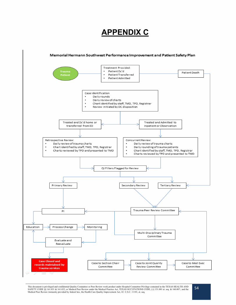
This document is privileged and confidential Quality Committee or Peer Review work product under Hospital Committee Privilege contained in the TEXAS HEALTH AND SAFETY CODE §§ 161.031 & 161.032, or Medical Peer Review under the Medical Practice Act, TEXAS OCCUPATIONS CODE, § § 151.001 et. seq & 160.007.; and the Medical Peer Review immunity provided by federal law, the Health Care Quality Improvement Act, 42. U.S.C. 11101, et. seq.
54
APPENDIX C

This document is privileged and confidential Quality Committee or Peer Review work product under Hospital Committee Privilege contained in the TEXAS HEALTH AND SAFETY CODE §§ 161.031 & 161.032, or Medical Peer Review under the Medical Practice Act, TEXAS OCCUPATIONS CODE, § § 151.001 et. seq & 160.007.; and the Medical Peer Review immunity provided by federal law, the Health Care Quality Improvement Act, 42. U.S.C. 11101, et. seq.
55