Embed Size (px)
Citation preview
HAL Id: hal-02564102https://hal.archives-ouvertes.fr/hal-02564102
Submitted on 5 May 2020
HAL is a multi-disciplinary open accessarchive for the deposit and dissemination of sci-entific research documents, whether they are pub-lished or not. The documents may come fromteaching and research institutions in France orabroad, or from public or private research centers.
L’archive ouverte pluridisciplinaire HAL, estdestinée au dépôt et à la diffusion de documentsscientifiques de niveau recherche, publiés ou non,émanant des établissements d’enseignement et derecherche français ou étrangers, des laboratoirespublics ou privés.
Transverse translunate fracture–dislocation: a rareinjury
Sabri Mahjoub, Bertrand Dunet, Patricia Thoreux, Alain-Charles Masquelet
To cite this version:Sabri Mahjoub, Bertrand Dunet, Patricia Thoreux, Alain-Charles Masquelet. Transverse translunatefracture–dislocation: a rare injury. Hand Surgery and Rehabilitation, Elsevier, 2016, 35 (3), pp.220-224. �10.1016/j.hansur.2016.02.008�. �hal-02564102�
Transverse translunate fracture–dislocation: A rare injury
Fracture-luxation translunaire transversale : une lésion exceptionnelle
S. Mahjoub a,c, B. Dunet b,c,*, P. Thoreux a, A.C. Masquelet aa Service de chirurgie orthopédique et traumatologique, hôpital Avicenne, 125, rue de Stalingrad, 93000 Bobigny, France
b Unité membre supérieur, service de chirurgie orthopédique et traumatologique, hôpital Pellegrin, place Amélie-Raba-Léon, 33076 Bordeaux cedex, France c Service de chirurgie orthopédique et traumatologique, hôpital de Libourne, 112, rue de la Marne, 33505 Libourne, France
Abstract
Perilunate fracture–dislocation is rare. We report the case of a 24-year-old male who fell from his motorcycle and presented with a transverse lunate fracture with perilunate ligament damage. The initial diagnosis based on X-rays was confirmed by CT scan. A dorsal approach was used to obtain good reduction, double screw fixation and ligament reinsertion protected by temporary K-wires. To the best of our knowledge, this is the first case of transverse lunate fracture within perilunate fracture–dislocation. The patient returned to normal activities after 6 months.
Keywords: Carpus; Lunate; Dislocation; Fracture; Ligament
Résumé
Les fractures-luxations périlunaires du carpe sont rares. Nous rapportons le cas d’un homme de 24 ans, droitier qui, dans les suites d’une chute par accident de la voie publique en moto, a présenté une fracture-luxation translunaire avec atteinte ligamentaire périlunaire. Le diagnostic, suspecté sur les radiographies initiales, a été confirmé par un scanner. Un abord postérieur a été réalisé pour permettre une réduction de la fracture fixée par un double vissage enfoui, associée à une réinsertion ligamentaire et brochage temporaire en protection. Il s’agit, à notre connaissance, du premier cas rapporté de fracture transversale du lunatum dans le cadre d’une fracture-luxation périlunaire. Le patient a pu reprendre ses activités dans un délai de 6 mois.
Mots clés : Carpe ; Lunatum ; Luxation ; Fracture ; Ligament
1. Introduction
Perilunate fracture–dislocations of the carpal bones are rare[1–3]. We will describe the first case involving a transversefracture of the lunate with complex perilunate ligamentdamage.
2. Case report
This was a young man of 24 years who suffered trauma toboth wrists following a motorcycle accident in September 2009.The initial examination found painful swelling of both wristsassociated with total functional disability and pain duringpassive motion of the fingers with no sensory or motorneurological deficit, vascular damage or skin disorder.
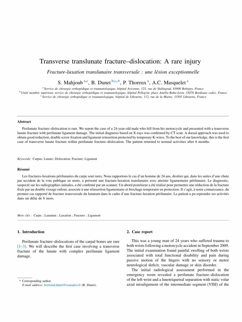
The initial radiological assessment performed in theemergency room revealed a perilunate fracture–dislocationof the left wrist and a lunotriquetral separation with static volaraxial misalignment of the intermediate segment (VISI) of the
* Corresponding author.E-mail address: [email protected] (B. Dunet).
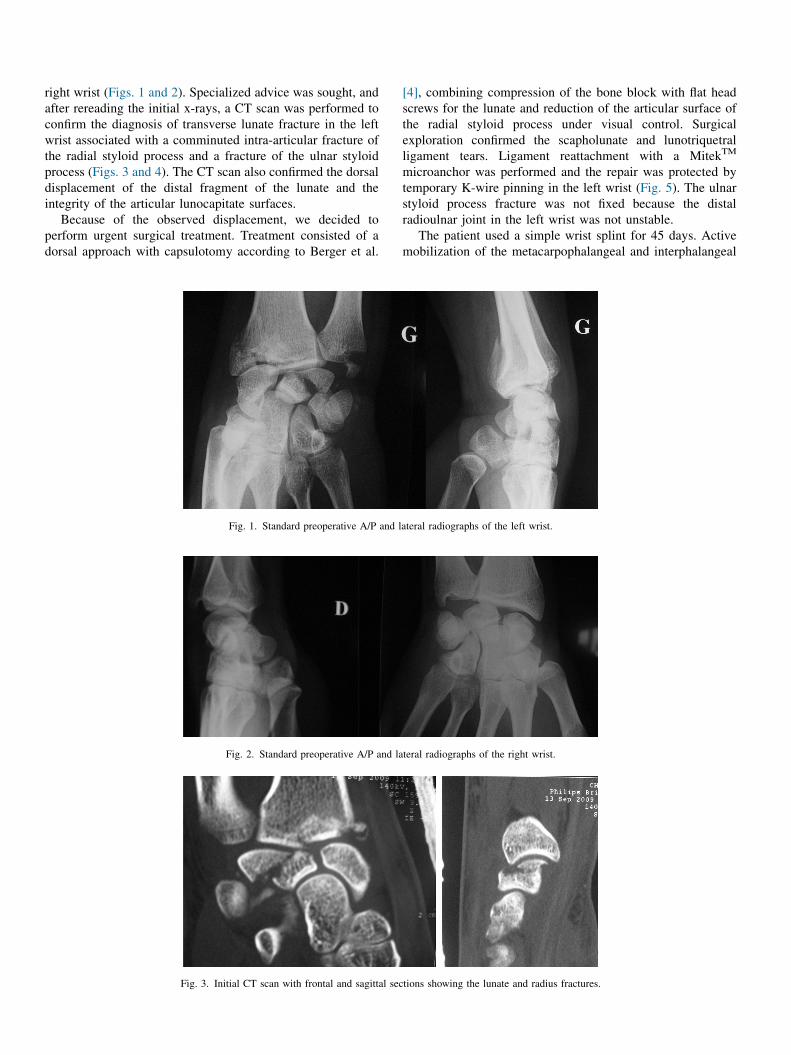
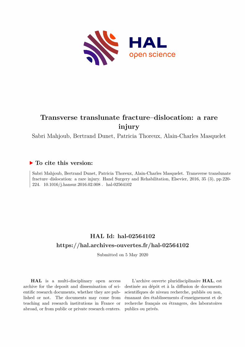
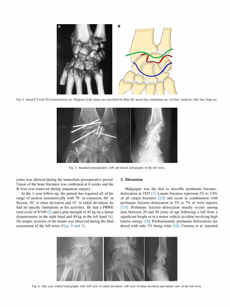
right wrist (Figs. 1 and 2). Specialized advice was sought, andafter rereading the initial x-rays, a CT scan was performed toconfirm the diagnosis of transverse lunate fracture in the leftwrist associated with a comminuted intra-articular fracture ofthe radial styloid process and a fracture of the ulnar styloidprocess (Figs. 3 and 4). The CT scan also confirmed the dorsaldisplacement of the distal fragment of the lunate and theintegrity of the articular lunocapitate surfaces.
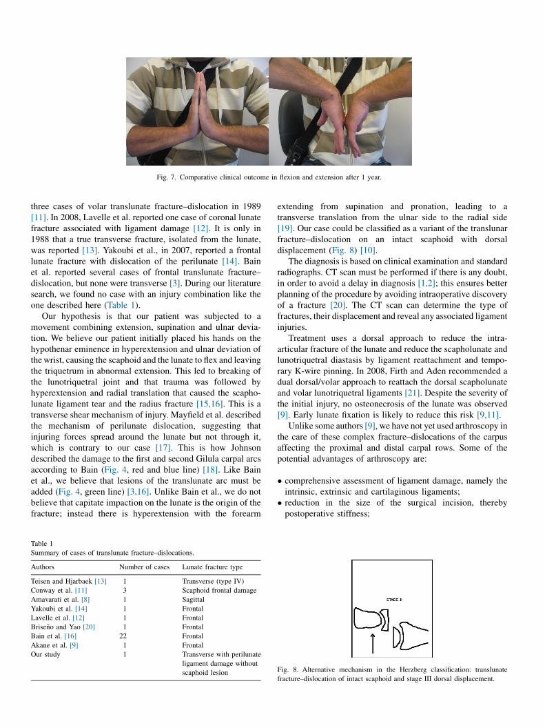
Because of the observed displacement, we decided toperform urgent surgical treatment. Treatment consisted of adorsal approach with capsulotomy according to Berger et al.
[4], combining compression of the bone block with flat headscrews for the lunate and reduction of the articular surface ofthe radial styloid process under visual control. Surgicalexploration confirmed the scapholunate and lunotriquetralligament tears. Ligament reattachment with a MitekTM
microanchor was performed and the repair was protected bytemporary K-wire pinning in the left wrist (Fig. 5). The ulnarstyloid process fracture was not fixed because the distalradioulnar joint in the left wrist was not unstable.
The patient used a simple wrist splint for 45 days. Activemobilization of the metacarpophalangeal and interphalangeal
Fig. 1. Standard preoperative A/P and lateral radiographs of the left wrist.
Fig. 3. Initial CT scan with frontal and sagittal sections showing the lunate and radius fractures.
Fig. 2. Standard preoperative A/P and lateral radiographs of the right wrist.
joints was allowed during the immediate postoperative period.Union of the bone fractures was confirmed at 6 weeks and theK-wire was removed during outpatient surgery.
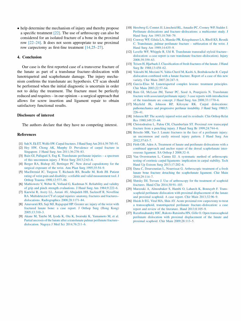
At the 1-year follow-up, the patient has regained all of hisrange of motion symmetrically with 708 in extension, 608 inflexion, 308 in ulnar deviation and 158 in radial deviation; hehad no specific limitations in his activities. He had a PRWEtotal score of 8/100 [5] and a grip strength of 45 kg on a Jamardynamometer in the right hand and 40 kg in the left hand [6].No aseptic necrosis of the lunate was observed during the finalassessment of the left wrist (Figs. 6 and 7).
3. Discussion
Malgaigne was the first to describe perilunate fracture–
dislocation in 1855 [7]. Lunate fractures represent 2% to 3.9%of all carpal fractures [2,8] and occur in combination withperilunate fracture–dislocation in 5% to 7% of wrist injuries[7,9]. Perilunate fracture–dislocation usually occurs amongmen between 20 and 30 years of age following a fall from asignificant height or in a motor vehicle accident involving highkinetic energy [10]. Predominantly, perilunate dislocations aredorsal with only 3% being volar [10]. Conway et al. reported
Fig. 5. Standard intraoperative A/P and lateral radiographs of the left wrist.
Fig. 4. Initial CT with 3D reconstruction (A). Diagram of the injury arcs described by Bain (B): green line: translunate arc; red line: small arc; blue line: large arc.
Fig. 6. One year control radiographs with A/P view of radial deviation, A/P view of ulnar deviation and lateral view of the left wrist.
three cases of volar translunate fracture–dislocation in 1989[11]. In 2008, Lavelle et al. reported one case of coronal lunatefracture associated with ligament damage [12]. It is only in1988 that a true transverse fracture, isolated from the lunate,was reported [13]. Yakoubi et al., in 2007, reported a frontallunate fracture with dislocation of the perilunate [14]. Bainet al. reported several cases of frontal translunate fracture–
dislocation, but none were transverse [3]. During our literaturesearch, we found no case with an injury combination like theone described here (Table 1).
Our hypothesis is that our patient was subjected to amovement combining extension, supination and ulnar devia-tion. We believe our patient initially placed his hands on thehypothenar eminence in hyperextension and ulnar deviation ofthe wrist, causing the scaphoid and the lunate to flex and leavingthe triquetrum in abnormal extension. This led to breaking ofthe lunotriquetral joint and that trauma was followed byhyperextension and radial translation that caused the scapho-lunate ligament tear and the radius fracture [15,16]. This is atransverse shear mechanism of injury. Mayfield et al. describedthe mechanism of perilunate dislocation, suggesting thatinjuring forces spread around the lunate but not through it,which is contrary to our case [17]. This is how Johnsondescribed the damage to the first and second Gilula carpal arcsaccording to Bain (Fig. 4, red and blue line) [18]. Like Bainet al., we believe that lesions of the translunate arc must beadded (Fig. 4, green line) [3,16]. Unlike Bain et al., we do notbelieve that capitate impaction on the lunate is the origin of thefracture; instead there is hyperextension with the forearm
extending from supination and pronation, leading to atransverse translation from the ulnar side to the radial side[19]. Our case could be classified as a variant of the translunarfracture–dislocation on an intact scaphoid with dorsaldisplacement (Fig. 8) [10].
The diagnosis is based on clinical examination and standardradiographs. CT scan must be performed if there is any doubt,in order to avoid a delay in diagnosis [1,2]; this ensures betterplanning of the procedure by avoiding intraoperative discoveryof a fracture [20]. The CT scan can determine the type offractures, their displacement and reveal any associated ligamentinjuries.
Treatment uses a dorsal approach to reduce the intra-articular fracture of the lunate and reduce the scapholunate andlunotriquetral diastasis by ligament reattachment and tempo-rary K-wire pinning. In 2008, Firth and Aden recommended adual dorsal/volar approach to reattach the dorsal scapholunateand volar lunotriquetral ligaments [21]. Despite the severity ofthe initial injury, no osteonecrosis of the lunate was observed[9]. Early lunate fixation is likely to reduce this risk [9,11].
Unlike some authors [9], we have not yet used arthroscopy inthe care of these complex fracture–dislocations of the carpusaffecting the proximal and distal carpal rows. Some of thepotential advantages of arthroscopy are:
� comprehensive assessment of ligament damage, namely theintrinsic, extrinsic and cartilaginous ligaments;� reduction in the size of the surgical incision, thereby
postoperative stiffness;
Fig. 7. Comparative clinical outcome in flexion and extension after 1 year.
Table 1Summary of cases of translunate fracture–dislocations.
Authors Number of cases Lunate fracture type
Teisen and Hjarbaek [13] 1 Transverse (type IV)Conway et al. [11] 3 Scaphoid frontal damageAmavarati et al. [8] 1 SagittalYakoubi et al. [14] 1 FrontalLavelle et al. [12] 1 FrontalBriseño and Yao [20] 1 FrontalBain et al. [16] 22 FrontalAkane et al. [9] 1 FrontalOur study 1 Transverse with perilunate
ligament damage withoutscaphoid lesion Fig. 8. Alternative mechanism in the Herzberg classification: translunate
fracture–dislocation of intact scaphoid and stage III dorsal displacement.
� help determine the mechanism of injury and thereby proposea specific treatment [22]. The use of arthroscopy can also beconsidered for an isolated fracture of a bone in the proximalrow [22–24]. It does not seem appropriate to use proximalrow carpectomy as first-line treatment [14,25–27].
4. Conclusion
Our case is the first reported case of a transverse fracture ofthe lunate as part of a translunar fracture–dislocation withlunotriquetral and scapholunate damage. The injury mecha-nism confirms the translunate arc hypothesis. CT scan shouldbe performed when the initial diagnostic is uncertain in ordernot to delay the treatment. The fracture must be perfectlyreduced and requires – in our opinion – a surgical approach thatallows for screw insertion and ligament repair to obtainsatisfactory functional results.
Disclosure of interest
The authors declare that they have no competing interest.
References
[1] Suh N, Ek ET, Wolfe SW. Carpal fractures. J Hand Surg Am 2014;39:785–91.[2] Hey HW, Chong AK, Murphy D. Prevalence of carpal fracture in
Singapore. J Hand Surg Am 2011;36:278–83.[3] Bain GI, Pallapati S, Eng K. Translunate perilunate injuries – a spectrum
of this uncommon injury. J Wrist Surg 2013;2:63–8.[4] Berger RA, Bishop AT, Bettinger PC. New dorsal capsulotomy for the
surgical exposure of the wrist. Ann Plast Surg 1995;35:54–9.[5] MacDermid JC, Turgeon T, Richards RS, Beadle M, Roth JH. Patient
rating of wrist pain and disability: a reliable and valid measurement tool. JOrthop Trauma 1998;12:577–86.
[6] Mathiowetz V, Weber K, Volland G, Kashman N. Reliability and validityof grip and pinch strength evaluations. J Hand Surg Am 1984;9:222–6.
[7] Kaewlai R, Avery LL, Asrani AV, Abujudeh HH, Sacknoff R, NovellineRA. Multidetector CT of carpal injuries: anatomy, fractures and fractures–
dislocations. Radiographics 2008;28:1171–84.[8] Amavarati RS, Saji MJ, Rajagopal HP. Greater arc injury of the wrist with
fractured lunate bone: a case report. J Orthop Surg (Hong Kong)2005;13:310–3.
[9] Akane M, Tatebe M, Iyoda K, Ota K, Iwatsuki K, Yamamoto M, et al.Partial necrosis of the lunate after a translunate palmar perilunate fracture–
dislocation. Nagoya J Med Sci 2014;76:211–6.
[10] Herzberg G, Comtet JJ, Linscheid RL, Amadio PC, Cooney WP, Stalder J.Perilunate dislocations and fracture–dislocations: a multicenter study. JHand Surg Am 1993;18:768–79.
[11] Conway WF, Gilula LA, Manske PR, Kriegshauser LA, Rholl KS, ResnikC. Translunate, palmar perilunate fracture – subluxation of the wrist. JHand Surg Am 1989;14:635–9.
[12] Lavelle WF, Whipple R, Uhl R. Translunate transradial styloid fracture–
dislocation: a case report (a rare translunate fracture–dislocation). Injury2008;39:359–63.
[13] Teisen H, Hjarbaek J. Classification of fresh fractures of the lunate. J HandSurg Br 1988;13:458–62.
[14] Yakoubi M, Meziani N, Yahia Cherif M, Kasbi A, Benbakouche R. Carpaldislocation combined with a lunate fracture. Report of a case of this newvariety. Chir Main 2007;26:247–9.
[15] Garcia-Elias M. Lunotriquetral complex lesions: treatment principles.Chir Main 2003;22:57–64.
[16] Bain GI, McLean JM, Turner PC, Sood A, Pourgiezis N. Translunatefracture with associated perilunate injury: 3 case reports with introductionof the translunate arc concept. J Hand Surg Am 2008;33:1770–6.
[17] Mayfield JK, Johnson RP, Kilcoyne RK. Carpal dislocations:pathomechanics and progressive perilunar instability. J Hand Surg 1980;5:226–41.
[18] Johnson RP. The acutely injured wrist and its residuals. Clin Orthop RelatRes 1980;149:33–44.
[19] Christodoulou L, Palou CH, Chamberlain ST. Proximal row transcarpalfracture from a punching injury. J Hand Surg Br 1999;24:744–6.
[20] Briseño MR, Yao J. Lunate fractures in the face of a perilunate injury:an uncommon and easily missed injury pattern. J Hand Surg Am2012;37:63–7.
[21] Firth GB, Aden A. Treatment of lunate and perilunate dislocations with acombined approach and anchor repair of the dorsal scapholunate inter-osseous ligament. SA Orthop J 2008;32–8.
[22] Van Overstraeten L, Camus EJ. A systematic method of arthroscopictesting of extrinsic carpal ligaments: implication in carpal stability. TechHand Up Extrem Surg 2013;17:202–6.
[23] Dana C, Doursounian L, Nourrissat G. Arthroscopic treatment of a freshlunate bone fracture detaching the scapholunate ligament. Chir Main2010;29:114–7.
[24] Slutsky DJ, Trevare J. Use of arthroscopy for the treatment of scaphoidfractures. Hand Clin 2014;30:91–103.
[25] Marzouki A, Almoubaker S, Hambi O, Laharch K, Boutayeb F. Trans-scaphoid perilunate dislocation with proximal displacement of the lunateand proximal scaphoid. A case report. Chir Main 2013;32:96–9.
[26] Huish Jr EG, Vital MA, Shin AY. Acute proximal row carpectomy to treata transscaphoid, transtriquetral perilunate fracture–dislocation: a casereport and review of the literature. Hand 2013;8:105–9.
[27] Razafimahandry HJC, Rakoto-Ratsimba HN, Gille O. Open transscaphoidperilunate dislocation with proximal displacement of the lunate andproximal scaphoid. Chir Main 2009;28:113–5.