Embed Size (px)
Citation preview
Traffic 2009; 10: 1414–1428 © 2009 John Wiley & Sons A/S
doi:10.1111/j.1600-0854.2009.00968.x
Transportin Mediates Nuclear Entry of DNAin Vertebrate Systems
Aurelie Lachish-Zalait1,†, Corine K. Lau2,†,
Boris Fichtman2, Ella Zimmerman1,
Amnon Harel3, Michelle R. Gaylord2,
Douglass J. Forbes2,∗ and Michael Elbaum1,∗
1Department of Materials and Interfaces, WeizmannInstitute of Science, Rehovot 76100, Israel2Section of Cell and Developmental Biology, Division ofBiological Sciences, University of California-San Diego, LaJolla, California 920933Department of Biology, Technion - Israel Institute ofTechnology, Haifa 32000, Israel*Corresponding authors: Douglass J. Forbes,[email protected] and Michael Elbaum,[email protected]†These authors contributed equally to this work.
Delivery of DNA to the cell nucleus is an essential
step in many types of viral infection, transfection,
gene transfer by the plant pathogen Agrobacterium
tumefaciens and in strategies for gene therapy. Thus,
the mechanism by which DNA crosses the nuclear
pore complex (NPC) is of great interest. Using nuclei
reconstituted in vitro in Xenopus egg extracts, we
previously studied DNA passage through the nuclear
pores using a single-molecule approach based on optical
tweezers. Fluorescently labeled DNA molecules were also
seen to accumulate within nuclei. Here we find that this
import of DNA relies on a soluble protein receptor of
the importin family. To identify this receptor, we used
different pathway-specific cargoes in competition studies
as well as pathway-specific dominant negative inhibitors
derived from the nucleoporin Nup153. We found that
inhibition of the receptor transportin suppresses DNA
import. In contrast, inhibition of importin β has little effect
on the nuclear accumulation of DNA. The dependence
on transportin was fully confirmed in assays using
permeabilized HeLa cells and a mammalian cell extract.
We conclude that the nuclear import of DNA observed in
these different vertebrate systems is largely mediated by
the receptor transportin. We further report that histones,
a known cargo of transportin, can act as an adaptor for
the binding of transportin to DNA.
Key words: DNA import, gene delivery, histones, importin
β, nuclear import, nuclear pores, permeabilized HeLa
cells, transportin, Xenopus
Received 19 February 2008, revised and accepted
for publication 21 July 2009, uncorrected manuscript
published online 27 July 2009
Many viruses have developed specific strategies topromote the nuclear uptake of their genomes by thehost (1,2). The addition of exogenous oligonucleotides tocells has separately been used for both viral- and non-viral-based gene therapy (3,4). In some studies, genetictransformation by the added DNA can take place on a timescale faster than cell division, implying that DNA importacross the interphase nuclear envelope must occur invivo (5). Indeed, a large number of studies have goneon to demonstrate that DNA entry through the nuclearpore occurs and can be enhanced by artificially couplingthe DNA to peptides containing nuclear localizationsignals (NLSs), implying translocation across the nuclearpore (6–14).
The existence of endogenous free non-chromosomal DNAin vivo has also been described (15–17). Some of thesestudies involved cytoplasmic DNA. Interestingly, it wasreported that human lymphocytes could be immortalizedby fusion with enucleated cytoplasts derived fromestablished tumor cell lines, including mouse L929 andErlich ascites cells (18–20). The active agent responsiblefor immortalization was shown to be extrachromosomalcytoplasmic DNA (20). Transfecting this same DNAsimilarly caused immortalization. We presume thattransformation of non-dividing cells could well requirenuclear import of the DNA. For all these reasons, DNAentry to the nucleus deserves a close examination.
Nuclear pores are large protein complexes each comprisedof ∼30 different proteins present in multiple copies,embedded in the double-membrane nuclear envelope ofcells from yeast to humans. The massive nuclear poremediates essentially all trafficking of proteins and RNAsbetween the nucleus and cytoplasm (21–27). The basicstructure of the nuclear pore consists of an eightfoldsymmetric central scaffold, eight cytoplasmic filamentsand a nuclear basket (28–30). Approximately one third ofthe nuclear pore proteins contain phenylalanine-glycine orFG repeats (31), which have been shown to interact withsoluble transport receptors.
Nuclear transport receptors in large part derive froma single protein family, the importin β or karyopherinβ superfamily (22,23,32–37). These receptors recognizespecific NLSs or nuclear export signals (NESs) on proteinor RNA cargoes, and then ferry those molecules across thenuclear pore (38). Upon entering the nucleus, interactionof the receptor with the small GTPase Ran in its GTP-bound form leads to accumulation of the cargo there(’nuclear import’) or alternatively depletion from thenucleus (’nuclear export’) (37,39–41).
1414 www.traffic.dk

Transportin and Nuclear Entry of DNA
Best known among the transport receptors is importinβ, the founding member of the importin β superfam-ily (42–45). Importin β (or karyopherin β1), through itsadaptor importin α (46), recognizes the so termed canoni-cal or classical NLS, which is characterized generally as ashort peptide containing basic amino acids (47–50). A sec-ond major receptor, transportin (or karyopherin β2A) bindsdirectly to different cargoes, such as the heterogeneousnuclear ribonucleoprotein A1 (hnRNP A1). HnRNP A1contains a 38-amino acid, glycine-rich NLS signal, specif-ically referred to in many studies as the M9 NLS (51,52).Recently, one study emanating from a crystal structure ofthe M9 peptide bound to transportin revealed a generalset of rules for transportin NLSs (53). Transportin NLSsare 20–30 residues in length, have an intrinsic structuraldisorder and an overall basic character, with a looselyconserved N-terminal hydrophobic or basic motif and aC-terminal R-X(2−5) PY motif (53). Transportin mediatesthe import of many RNA binding and/or processing pro-teins, such as hnRNP A1, as well as DNA transcriptionfactors (53). A large number of other importin β familyreceptors are known, but the signals that are recognizedby these receptors have not been defined. However,it appears that many importin β family members carryhistones into the nucleus (54,55). Notably, importin β/αhas the weakest ability to transport histones, whereastransportin has the strongest (56).
Two in vitro systems have been widely and successfullyused for 30 years to define the parameters of nuclearprotein transport. In one, mammalian cultured cells aretreated with digitonin to permeabilize their plasma mem-branes, while leaving their nuclear membranes intact (57).In a second, nuclei reconstituted in vitro in Xenopusegg extracts have provided a highly valuable system forstudying both nuclear assembly and nucleocytoplasmictransport (58–61).
In previous work using reconstituted nuclei, we visuallyfollowed the nuclear uptake of DNA at the single-moleculelevel using optical tweezers (9). We found that individualmolecules of long bacteriophage DNA attached to a latexbead were gradually and continuously drawn into thenucleus, in a manner that could be precisely measuredand the conditions for nuclear uptake tested (9). DNAentry was gradual, consistent with a picture of serpentinepassage through the nuclear pore. We found no evidencefor involvement of an ATPase motor in moving the DNAthrough the pore channel, as has been characterized, e.g.,in the mechanism of DNA packaging by bacteriophage φ
29 capsids (62) and in the DNA translocation machinery inBacillus subtilis (63). The proteomic analyses of isolatednuclear pores from yeast and mammals revealed noATPases or motors in the core nuclear pore complex (NPC)structure (29,30,64). However, the DEAD-box helicaseDBP5, a known ATPase, is found strongly associatedwith the cytoplasmic filaments of the nuclear pores and isrequired for mRNA export (65,66).
In the present work, we have sought to analyze the roleof nucleocytoplasmic transport receptors in DNA entryinto the nucleus. Using sets of specific inhibitors andcompetitors, we have found that transportin mediatesDNA import in Xenopus reconstituted nuclei. In addition,transportin also mediates nuclear entry of DNA in awell-characterized mammalian permeabilized cell assay.Moreover, biochemical analysis reveals that histones canact as a strong adaptor protein for transportin recognitionof DNA.
Results
A modified Xenopus egg extract promotes
transport-competent nuclei with long-term stability
Nuclei formed in vitro using Xenopus egg extract allowone to focus on DNA interactions with the nuclearpore at the nuclear envelope surface. In this in vitroenvironment, molecular access of DNA to the nuclearenvelope is relatively direct. In addition, the extractprovides a complete set of soluble constituents needed tosupport nucleocytoplasmic trafficking (9,40,58,59,67–69),nuclear and nuclear pore assembly (70–76), and mitoticspindle assembly (77–80).
As a model system for analyzing the nuclear import ofDNA substrates, we first tested a classical high-speedfractionated Xenopus egg extract. In this protocol, acrude, low-speed extract of Xenopus eggs is furtherseparated into a membrane fraction and a fractioncontaining soluble cytosolic components. These fractionsare then combined with demembranated sperm chromatinto form nuclei (59). We observed, however, that thesenuclei were often surrounded by a hazy cloud ofmembranes. While the cloud was difficult to observewithout differential interference contrast imaging, andprotein transport assays were not apparently affected,the associated membranes tended to trap the larger DNAsubstrates and hindered their access to the nuclear poresand envelope. Surprisingly, unfractionated crude extractsproduced far less of this membranous barrier surroundingthe nuclei. This led us to develop a modified protocol fora clarified crude (CCr) extract, which yielded a consistent,efficient nuclear assembly even after fourfold dilutioninto extract buffer (40). Moreover, protein accumulationinto such nuclei was robust to the prolonged incubationrequired for DNA import. The nuclear envelope and nuclearpores of CCr nuclei were examined in detail, using high-resolution field-emission scanning electron microscopy(FESEM). This analysis revealed well-developed nucleiwith continuous membranes bearing large numbers ofnuclear pores (Figure 1) that were essentially identical tothe nuclear envelopes formed in classically fractionatedextracts (59).
DNA accumulates in reconstituted nuclei
To examine and compare the DNA import capability ofthe reconstituted nuclei, a set of fluorescently labeled
Traffic 2009; 10: 1414–1428 1415
Lachish-Zalait et al.
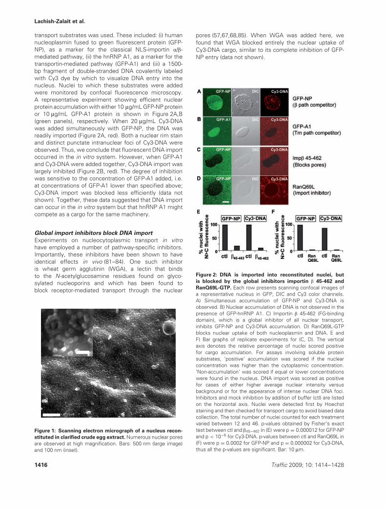
transport substrates was used. These included: (i) humannucleoplasmin fused to green fluorescent protein (GFP-NP), as a marker for the classical NLS-importin α/β-mediated pathway, (ii) the hnRNP A1, as a marker for thetransportin-mediated pathway (GFP-A1) and (iii) a 1500-bp fragment of double-stranded DNA covalently labeledwith Cy3 dye by which to visualize DNA entry into thenucleus. Nuclei to which these substrates were addedwere monitored by confocal fluorescence microscopy.A representative experiment showing efficient nuclearprotein accumulation with either 10 μg/mL GFP-NP proteinor 10 μg/mL GFP-A1 protein is shown in Figure 2A,B(green panels), respectively. When 20 μg/mL Cy3-DNAwas added simultaneously with GFP-NP, the DNA wasreadily imported (Figure 2A, red). Both a nuclear rim stainand distinct punctate intranuclear foci of Cy3-DNA wereobserved. Thus, we conclude that fluorescent DNA importoccurred in the in vitro system. However, when GFP-A1and Cy3-DNA were added together, Cy3-DNA import waslargely inhibited (Figure 2B, red). The degree of inhibitionwas sensitive to the concentration of GFP-A1 added, i.e.at concentrations of GFP-A1 lower than specified above;Cy3-DNA import was blocked less efficiently (data notshown). Together, these data suggested that DNA importcan occur in the in vitro system but that hnRNP A1 mightcompete as a cargo for the same machinery.
Global import inhibitors block DNA import
Experiments on nucleocytoplasmic transport in vitrohave employed a number of pathway-specific inhibitors.Importantly, these inhibitors have been shown to haveidentical effects in vivo (81–84). One such inhibitoris wheat germ agglutinin (WGA), a lectin that bindsto the N-acetylglucosamine residues found on glyco-sylated nucleoporins and which has been found toblock receptor-mediated transport through the nuclear
Figure 1: Scanning electron micrograph of a nucleus recon-
stituted in clarified crude egg extract. Numerous nuclear poresare observed at high magnification. Bars: 500 nm (large image)and 100 nm (inset).
pores (57,67,68,85). When WGA was added here, wefound that WGA blocked entirely the nuclear uptake ofCy3-DNA cargo, similar to its complete inhibition of GFP-NP entry (data not shown).
Figure 2: DNA is imported into reconstituted nuclei, but
is blocked by the global inhibitors importin β 45-462 and
RanQ69L-GTP. Each row presents scanning confocal images ofa representative nucleus in GFP, DIC and Cy3 color channels.A) Simultaneous accumulation of GFP-NP and Cy3-DNA isobserved. B) Nuclear accumulation of DNA is not observed in thepresence of GFP-hnRNP A1. C) Importin β 45-462 (FG-bindingdomain), which is a global inhibitor of all nuclear transport,inhibits GFP-NP and Cy3-DNA accumulation. D) RanQ69L-GTPblocks nuclear uptake of both nucleoplasmin and DNA. E andF) Bar graphs of replicate experiments for (C, D). The verticalaxis denotes the relative percentage of nuclei scored positivefor cargo accumulation. For assays involving soluble proteinsubstrates, ’positive’ accumulation was scored if the nuclearconcentration was higher than the cytoplasmic concentration.’Non-accumulation’ was scored if equal or lower concentrationswere found in the nucleus. DNA import was scored as positivefor cases of either higher average nuclear intensity versusbackground or for the appearance of intense nuclear DNA foci.Inhibitors and mock inhibition by addition of buffer (ctl) are listedon the horizontal axis. Nuclei were detected first by Hoechststaining and then checked for transport cargo to avoid biased datacollection. The total number of nuclei counted for each treatmentvaried between 12 and 46. p-values obtained by Fisher’s exacttest between ctl and β45−462 in (E) were p = 0.000012 for GFP-NPand p < 10−6 for Cy3-DNA. p-values between ctl and RanQ69L in(F) were p = 0.0002 for GFP-NP and p = 0.000002 for Cy3-DNA,thus all the p-values are significant. Bar: 10 μm.
1416 Traffic 2009; 10: 1414–1428

Transportin and Nuclear Entry of DNA
Another established dominant negative inhibitor ofnucleocytoplasmic transport is a truncated form ofimportin β, termed here Imp β 45-462, which containsamino acids 45-462. Imp β 45-462 binds irreversibly toFG nucleoporins (Nups) in the pore (86). Indeed, whenadded to nuclei Imp β 45-462 has been seen by atomicforce microscopy to create a physical plug, or mound, atthe central channel region of the pore through such FG-Nup binding (87). Here, when Imp β 45-462 was added toreconstituted nuclei prior to the addition of GFP-NP andDNA substrates, the nuclei were seen to exclude GFP-NP, indicating successful inhibition of transport (Figure 2C,GFP-NP). The import of DNA was also strongly inhibitedby Imp β 45-462, which prevented even the binding ofDNA to pores at the nuclear rim (Figure 2C, Cy3-DNA).These results are quantitated in Figure 2E.
Importin β family transport receptors are regulated intheir action by the small GTPase Ran (23). Indeed,interrupting the GTP-GDP cycle of Ran is an effectivemeans for inhibiting receptor-mediated nuclear transport.Thus, RanQ69L, a mutant of Ran incapable of hydrolyzingGTP, blocks transport by irreversibly binding to andsequestering the receptors (88). We asked whetherRanQ69L-GTP blocks DNA import into nuclei. If so,this would be a strong indication for the involvementof a Ran-regulated, receptor-mediated pathway forDNA import. RanQ69L-GTP was added to assembled,reconstituted nuclei and fluorescent transport substrateswere introduced 15 min later. The inhibitory competenceof RanQ69L-GTP was verified by the block of GFP-NPaccumulation in the nuclei (Figure 2D, GFP-NP). Similarly,Cy3-DNA was also excluded from nuclei after RanQ69Laddition. Cy3-DNA decorated the nuclear envelope witha rim of adsorbed DNA molecules, but no intranuclearDNA was observed (Figure 2D, Cy3-DNA), indicating thatDNA import could well employ a Ran-regulated transportreceptor. The inhibition of DNA import by RanQ69L-GTPis quantitated in Figure 2F. We concluded that the Cy3-DNA import is sensitive to global inhibitors of proteinnucleocytoplasmic transport, indicating the involvementof protein receptors in the nuclear import of DNA.
Inhibition of transportin leads to a block in DNA
import
To further test the hypothesis that nuclear import ofexogenous DNA is mediated by a protein receptor, weperformed import assays in the presence of solubleinhibitors to the importin β- and transportin-mediatedpathways of protein nucleocytoplasmic transport. Theseinhibitors are based on fragments of Nup153, a vertebratenucleoporin located in the nuclear pore basket thatserves as a major binding site for several importinfamily receptors (89–92). Different domains of Nup153act as binding sites for distinct transport receptors (93).A fragment of Xenopus Nup153 encompassing nineFG repeats binds the importin α/β heterodimer. Whenbound to this fragment, importin α/β can still bind itsNLS cargo, but cannot bind to FG-repeat Nups within
the pore. This Nup153-FG fragment thus sequestersimportin α/β and their cargoes. In this way, Nup153-FG has been shown to act as a dominant negativecompetitive inhibitor to classical NLS nuclear importwhen added to permeabilized cell assays (93). A moreamino terminal fragment of Xenopus Nup153 deficientin FG repeats, termed Nup153-N’, has been shown tobind transportin and, in doing so, sequesters transportinaway from the nuclear pores (93). The FG and N’fragments of Nup153 therefore function as pathway-specific dominant negative inhibitors of protein transportby the importin α/β- and transportin-mediated pathways,respectively (93). This pathway-specific inhibition wasconfirmed here, as a control, in the permeabilizedHeLa cell assay where it was originally described(Figure 5C).
The effects of the Nup153 inhibitory fragments in theXenopus system were tested and we determined theoptimal concentrations needed to inhibit specificallyeither the importin β (Nup153-FG) or the transportin(Nup153-N’) protein transport pathways (data not shown).These same optimal concentrations, 40 μM Nup153-FG and 30 μM Nup153-N’, were then assessed for aneffect on DNA import into Xenopus reconstituted nuclei(Figure 3A–C,F,G). Specifically, nuclei were reconstituted,then Nup153-FG and Nup153-N’ at the optimizedconcentrations were added and the reconstituted nucleiwere allowed to incubate for 20 min. At this time, thetransport substrates were introduced, and accumulationin the nuclei was assessed after 20 min. As expected,Nup153-FG significantly reduced the percentage of nucleiaccumulating the importin β cargo GFP-NP (Figure 3A,F,GFP-NP). It did not reduce the percentage of nucleiaccumulating the transportin cargo GFP-A1 (Figure 3F).In contrast, Nup153-N’ blocked the accumulation ofGFP-A1 (Figure 3B,G, GFP-A1), but not that of GFP-NP(Figure 3C,G, GFP-NP).
When Cy3-DNA was added to nuclei in the presenceof the Nup153-FG β-inhibitor, fluorescent DNA foci wereobserved in nuclei (Figure 3A, red). Nuclear DNA fociand rim association, observed here and in Figure 2A,is a hallmark of DNA import in mammalian cells[see (7) and references therein as well as Figures 4and 5 below]. The data for replicate experiments aregraphed in Figure 3F. In contrast, when the transportininhibitor Nup153-N’ was added, no fluorescent DNA fociwere observed in the nuclei (Figure 3B,C, red). GFP-nucleoplasmin accumulation was not impacted in thesame nucleus (Figure 3B, green), while GFP-hnRNP A1uptake was blocked (Figure 3C, green). The data forreplicate experiments with Nup153-N’ are graphed inFigure 3G.
Rim binding to nuclear pores has been recognized asan interim step in protein transport and is known to bedependent on the specific receptor involved (57,68,94).When Nup153-FG was added, Cy3-DNA molecules, in
Traffic 2009; 10: 1414–1428 1417

Lachish-Zalait et al.
Figure 3: Multiple inhibitors of the transportin-mediated import pathway block DNA import. Each row shows scanning confocalimages of a representative nucleus in GFP, DIC and Cy3 color channels. A) The importin β pathway inhibitor Nup153-FG effectivelyblocked the entry of nucleoplasmin. However, DNA uptake took place under these conditions, as observed from the punctate intranuclearfoci that are typical of DNA import. B) The transportin pathway inhibitor Nup153-N’ inhibited both GFP-A1 accumulation and DNA uptake.C) Nup153-N’ fragment again inhibited DNA import, while nucleoplasmin accumulated in the nucleus. D) DNA and nucleoplasmin, animportin β cargo, accumulated simultaneously in the nucleus, indicating a lack of competition between these substrates for transportfactors. E) GFP-M9, a transportin cargo, competed effectively with DNA, preventing its nuclear import. F–H) Replicate measurementswere scored and statistical tests performed as shown in Figure 2E,F. p-values between ctl and Nup153-FG in (F) were p = 0.33 forGFP-A1, p = 0.000147 for GFP-NP and p = 0.37 for Cy3-DNA import. This indicates significant inhibition by Nup153-FG of GFP-NPonly. p-values between ctl and Nup153-N’ in (G) were p = 0.008 for GFP-A1, p = 1.000 for GFP-NP and p = 0.002 for Cy3-DNA import.This confirms significant inhibition by Nup153-N’ of GFP-A1 and Cy3-DNA, but not of GFP-NP. For (H), p = 0.000003, confirming thesuppression of DNA import by GFP-M9. Bar: 10 μm.
addition to being observed inside the nucleus, were alsoobserved bound to the nuclear envelope, indicating thatthe binding step in Cy3-DNA import occurs in the presenceof importin β inhibition. However, when Nup153-N’ wasadded, there was no Cy3-DNA fluorescence at the nuclearrim (Figure 3B,C), consistent with a lack of DNA importupon transportin inhibition. Quantification is shown inFigure 3F,G. Taken together, these data strongly implicatetransportin as a receptor for the entry of DNA into thenucleus.
To further test the involvement of transportin in DNAimport, we also performed a formal cargo competition
assay. Even when a large excess of the importin β cargoGFP-NP (40 μM) was added, it was found to have noeffect on Cy3-DNA import (Figure 3D,H). With respect totransportin, hnRNP A1 is an excellent cargo for importassays (Figure 2B) but is not ideal for competition, asit becomes insoluble at the high molar concentrationsneeded for completely effective competition assays. Wetherefore used a GFP-M9 fusion protein, which containsthe M9 NLS region of hnRNP A1. GFP-M9 has been usedas a transportin cargo in many studies, and is soluble atvery high concentrations. In control experiments, a highexcess of GFP-M9 (40 μM) was observed not to affectthe importin β-mediated import of SV40-NLS-HSA, but to
1418 Traffic 2009; 10: 1414–1428

Transportin and Nuclear Entry of DNA
block the transportin-mediated import of MBP-M3, an M9-containing fusion protein (Figure S1). Importantly, 40 μM
GFP-M9 completely blocked the import of Cy3-DNA intoin vitro assembled nuclei (Figure 3E,H). We conclude fromthese multiple pathway-specific inhibitors that transportinis a significant receptor for DNA import into the nucleusin this Xenopus system.
DNA import in permeabilized HeLa cells shows a
similar dependence on the transportin pathway
To further test this conclusion, we performed DNAimport analysis using permeabilized HeLa cells. Notably,these cells have previously been shown to be com-petent for both protein and DNA import into thenucleus (6,7,57,95,96). The permeabilized HeLa cell assayhas been widely used to characterize the mechanism ofnucleocytoplasmic protein transport (57,97–101). Condi-tions for DNA import in HeLa cells included: (i) a shorterDNA substrate (400 bp), as a longer fragment often didnot enter the nucleus in a reasonable period of time, (ii)rabbit reticulocyte lysate as a source of cytosol, (iii) lowerconcentrations of the inhibitors to suit the more dilutereticulocyte lysate cytosol and (iv) a higher temperature(37◦C) to mimic mammalian cellular conditions.
When rhodamine-labeled double-stranded 400 bp DNAfragments were added to permeabilized HeLa cells andincubated for 90 min at 37◦C, distinct DNA foci wereobserved in the nuclei (Figure 4A, control). Consistentwith translocation taking place through the nuclear pores,no nuclear DNA foci were observed if the reaction wascarried out in the absence of an ATP regenerating systemor in the absence of cytosolic factors (Figure S2A,B), orwhen the inhibitor WGA was present (data not shown).It should be noted that under both import-competent andimport-inhibited conditions, DNA was also seen in thecytoplasm; this has been previously observed (3,102) andis thought to be DNA entrapped by endoplasmic reticulum(ER) and cytoskeletal barriers. We next evaluated DNAimport by quantitating the number of fluorescent nuclearDNA foci observed in the presence or absence of specifictransport pathway inhibitors.
When a substrate competition assay was carried out inpermeabilized HeLa cells with an importin β cargo, wefound that excess GFP-NP did not significantly affect thenumber of observed nuclear DNA foci (Figure 4A, GFP-NP, 3 and 8 μM). In contrast, when a competition assaywas carried out with a transportin cargo, we found thatexcess GFP-hnRNP A1 completely abolished DNA importinto HeLa cell nuclei; no nuclear DNA foci were observed(Figure 4A, GFP-A1, 3 and 8 μM). Quantitation is shown inFigure 4B. These results strongly reinforce the conclusionreached in the Xenopus experiments that DNA enters thenucleus via a transportin-mediated pathway.
To assess DNA import in the presence of pathway-specificinhibitors, Nup153-FG was added at a concentrationsufficient to inhibit importin β-mediated protein transport
Figure 4: Excess transportin cargo, but not importin β
cargo, blocks DNA import in permeabilized HeLa cells. CX-Rhodamine-labeled DNA fragments of 400 bp were added topermeabilized HeLa cells in transport mixture, with or withoutcompeting protein substrates. Cells were incubated at 37◦C for90 min and fixed for confocal microscopy. A) DNA imported intothe nucleus was observed as distinct nuclear foci (control). DNAimport was observed in the presence of excess of the importinα/β substrate, GFP-NP (GFP-NP, 3 or 8 μM), but not when excessGFP-A1, a transportin substrate, was present (GFP-A1, 3 or 8 μM).All images shown were projected from five z-sections at 0.5 μmspacing through the middle of the nuclei. Images were preparedusing IMAGEJ software (http://rsb.info.nih.gov/ij/). B) The numberof nuclear DNA foci in 12 nuclei per condition was counted, thehighest and lowest numbers were dropped, and the average andstandard deviation were plotted. Bar: 10 μm.
(2 μM). No effect was seen on DNA import intoHeLa nuclei (Figure 5A, 153-FG, 2 μM), although someinhibition of DNA import was observed at higherconcentrations of Nup153-FG (Figure 5A, 153-FG, 8 μM).However, addition of Nup153-N’, the dominant negativetransportin inhibitor, reduced DNA import dramatically atall tested concentrations (Figure 5A, 153-N’, 2 and 8 μM).Quantitation of these results is shown in Figure 5B.
Traffic 2009; 10: 1414–1428 1419

Lachish-Zalait et al.
Figure 5: A dominant negative inhibitor of the transportin
pathway blocks DNA import in permeabilized HeLa cells.
Permeabilized HeLa cell assays were performed and imageswere processed as in Figure 4. A) In the presence of the importinβ inhibitor Nup153-FG, a decrease in distinct nuclear foci of DNAwas observed, but only at high concentration of inhibitor (153-FG,8 μM). In the presence of the transportin inhibitor Nup153-N’, DNAimport was suppressed at both high and low concentrations (153-N’, 2 and 8 μM). B) The number of nuclear DNA foci was countedfor each inhibitor. The average number of punctate nuclear foci pernucleus and the standard deviation are shown, as in Figure 4. p-values comparing DNA foci for 153-FG and 153-N’ inhibitors at thesame concentrations (2 and 8 μM, respectively) were p < 0.005,indicating a significant difference in their effectiveness. C) Toconfirm specificity of the inhibition in this assay, Nup153-FG orNup153-N’ was added at a final concentration of 25 μM, whileGFP-NP or GFP-M9 was present at a final concentration of 3 or4 μM, respectively. Images shown are single planes through thez-axis. Bar: 10 μm.
Thus, DNA import is negatively impacted when thetransportin pathway is compromised in permeabilizedHeLa cells, just as it is in Xenopus reconstituted nuclei.
Histones serve as an adaptor between transportin
and DNA
As transportin has no known DNA-binding domain,adaptors that bear both a DNA-binding motif anda transportin-recognizable NLS domain might mediatetransportin-facilitated nuclear import of DNA. Histones areDNA-binding proteins where the N-terminal domains ofeach core histone as well as globular portions of the H2A,H2B, H3 and H4 histones can serve as NLSs (56,103).Despite their small size, core histones do not enter thenucleus by diffusion. The nuclear import of core histoneshas been found to be mediated by at least five nucleartransport receptors, specifically Imp β, transportin, Imp5,Imp7 and Imp9 (55). This study also showed that Impβ and Imp7 bind with relatively low affinity to all fourcore histones, Imp9 binds with high affinity to H2B,Imp5 binds with high affinity to H2B, H3 and H4, whiletransportin binds with high affinity to H2A, H2B and H3.This suggests that Imp5 and transportin might be the mostpotent transport receptors for core histones. Of its threehistone cargoes, transportin binds H2A with the highestaffinity and in a RanGTP-dependent manner. Strikingly,when Imp α and Imp β were tested in a permeabilizedcell assay together, they were unable to import anyof the four core histones (104), suggesting that in vivoImp β may play a minor role in the import of histones.Interestingly, in agreement with Muhlhausser et al. (55),transportin showed the highest capacity amongst theimport receptors tested to import core histones intothe nucleus (104). Taking these studies together, wehypothesized that core histones might be good candidatesfor adaptors between transportin and DNA for DNA import.
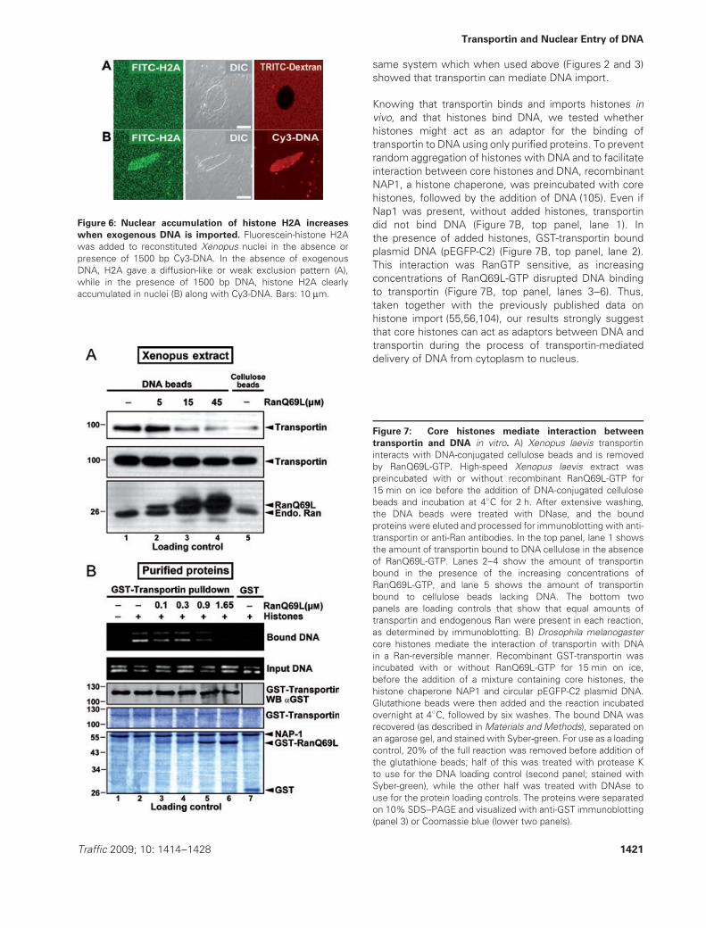
First, the effect of exogenous DNA was tested on histoneentry into reconstituted Xenopus nuclei. Recombinanthuman histone H2A was labeled with fluorescein andadded to Xenopus nuclei in the presence or absence of1500 bp linear Cy3-DNA. In the absence of exogenousDNA, H2A (14 kDa) equilibrated between the nucleus andcytoplasm or appeared slightly excluded from the nucleus(Figure 6A). However, in the presence of 1500 bp Cy3-DNA, histone H2A clearly accumulated to a higher extentin the nuclei along with the Cy3-DNA (Figure 6B). Weconclude that the nuclear accumulation of histone H2Acan be enhanced by the addition of exogenous DNA.
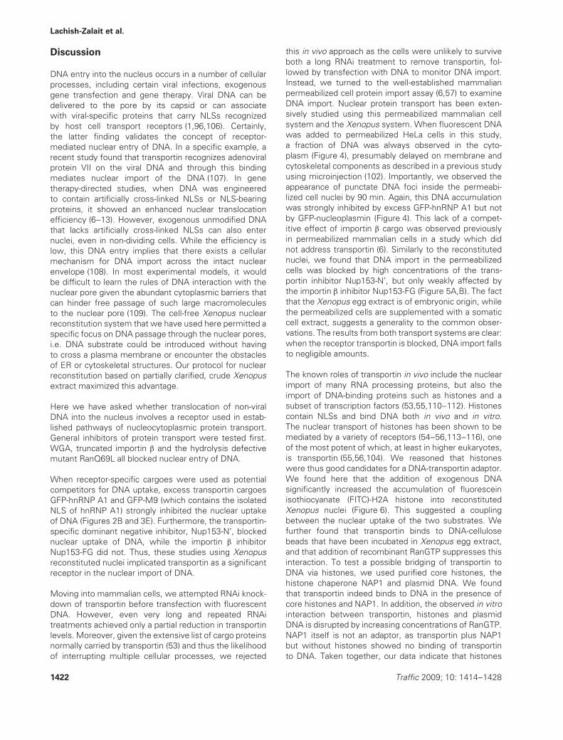
We next set out to test whether transportin binds to DNAin extract. For this, we incubated DNA-cellulose beadsor unmodified cellulose beads with high-speed Xenopusegg extract in the presence or absence of increasingconcentrations of RanQ69L-GTP. As shown in Figure 7A,transportin bound to DNA cellulose (top panel, lane 1), butwas increasingly removed in a RanGTP-dependent manner(top panel, lanes 2–4). This showed that transportin indeedcan bind to DNA in the presence of Xenopus extract, the
1420 Traffic 2009; 10: 1414–1428
Transportin and Nuclear Entry of DNA
Figure 6: Nuclear accumulation of histone H2A increases
when exogenous DNA is imported. Fluorescein-histone H2Awas added to reconstituted Xenopus nuclei in the absence orpresence of 1500 bp Cy3-DNA. In the absence of exogenousDNA, H2A gave a diffusion-like or weak exclusion pattern (A),while in the presence of 1500 bp DNA, histone H2A clearlyaccumulated in nuclei (B) along with Cy3-DNA. Bars: 10 μm.
same system which when used above (Figures 2 and 3)showed that transportin can mediate DNA import.
Knowing that transportin binds and imports histones invivo, and that histones bind DNA, we tested whetherhistones might act as an adaptor for the binding oftransportin to DNA using only purified proteins. To preventrandom aggregation of histones with DNA and to facilitateinteraction between core histones and DNA, recombinantNAP1, a histone chaperone, was preincubated with corehistones, followed by the addition of DNA (105). Even ifNap1 was present, without added histones, transportindid not bind DNA (Figure 7B, top panel, lane 1). Inthe presence of added histones, GST-transportin boundplasmid DNA (pEGFP-C2) (Figure 7B, top panel, lane 2).This interaction was RanGTP sensitive, as increasingconcentrations of RanQ69L-GTP disrupted DNA bindingto transportin (Figure 7B, top panel, lanes 3–6). Thus,taken together with the previously published data onhistone import (55,56,104), our results strongly suggestthat core histones can act as adaptors between DNA andtransportin during the process of transportin-mediateddelivery of DNA from cytoplasm to nucleus.
Figure 7: Core histones mediate interaction between
transportin and DNA in vitro. A) Xenopus laevis transportininteracts with DNA-conjugated cellulose beads and is removedby RanQ69L-GTP. High-speed Xenopus laevis extract waspreincubated with or without recombinant RanQ69L-GTP for15 min on ice before the addition of DNA-conjugated cellulosebeads and incubation at 4◦C for 2 h. After extensive washing,the DNA beads were treated with DNase, and the boundproteins were eluted and processed for immunoblotting with anti-transportin or anti-Ran antibodies. In the top panel, lane 1 showsthe amount of transportin bound to DNA cellulose in the absenceof RanQ69L-GTP. Lanes 2–4 show the amount of transportinbound in the presence of the increasing concentrations ofRanQ69L-GTP, and lane 5 shows the amount of transportinbound to cellulose beads lacking DNA. The bottom twopanels are loading controls that show that equal amounts oftransportin and endogenous Ran were present in each reaction,as determined by immunoblotting. B) Drosophila melanogastercore histones mediate the interaction of transportin with DNAin a Ran-reversible manner. Recombinant GST-transportin wasincubated with or without RanQ69L-GTP for 15 min on ice,before the addition of a mixture containing core histones, thehistone chaperone NAP1 and circular pEGFP-C2 plasmid DNA.Glutathione beads were then added and the reaction incubatedovernight at 4◦C, followed by six washes. The bound DNA wasrecovered (as described in Materials and Methods), separated onan agarose gel, and stained with Syber-green. For use as a loadingcontrol, 20% of the full reaction was removed before addition ofthe glutathione beads; half of this was treated with protease Kto use for the DNA loading control (second panel; stained withSyber-green), while the other half was treated with DNAse touse for the protein loading controls. The proteins were separatedon 10% SDS–PAGE and visualized with anti-GST immunoblotting(panel 3) or Coomassie blue (lower two panels).
Traffic 2009; 10: 1414–1428 1421
Lachish-Zalait et al.
Discussion
DNA entry into the nucleus occurs in a number of cellularprocesses, including certain viral infections, exogenousgene transfection and gene therapy. Viral DNA can bedelivered to the pore by its capsid or can associatewith viral-specific proteins that carry NLSs recognizedby host cell transport receptors (1,96,106). Certainly,the latter finding validates the concept of receptor-mediated nuclear entry of DNA. In a specific example, arecent study found that transportin recognizes adenoviralprotein VII on the viral DNA and through this bindingmediates nuclear import of the DNA (107). In genetherapy-directed studies, when DNA was engineeredto contain artificially cross-linked NLSs or NLS-bearingproteins, it showed an enhanced nuclear translocationefficiency (6–13). However, exogenous unmodified DNAthat lacks artificially cross-linked NLSs can also enternuclei, even in non-dividing cells. While the efficiency islow, this DNA entry implies that there exists a cellularmechanism for DNA import across the intact nuclearenvelope (108). In most experimental models, it wouldbe difficult to learn the rules of DNA interaction with thenuclear pore given the abundant cytoplasmic barriers thatcan hinder free passage of such large macromoleculesto the nuclear pore (109). The cell-free Xenopus nuclearreconstitution system that we have used here permitted aspecific focus on DNA passage through the nuclear pores,i.e. DNA substrate could be introduced without havingto cross a plasma membrane or encounter the obstaclesof ER or cytoskeletal structures. Our protocol for nuclearreconstitution based on partially clarified, crude Xenopusextract maximized this advantage.
Here we have asked whether translocation of non-viralDNA into the nucleus involves a receptor used in estab-lished pathways of nucleocytoplasmic protein transport.General inhibitors of protein transport were tested first.WGA, truncated importin β and the hydrolysis defectivemutant RanQ69L all blocked nuclear entry of DNA.
When receptor-specific cargoes were used as potentialcompetitors for DNA uptake, excess transportin cargoesGFP-hnRNP A1 and GFP-M9 (which contains the isolatedNLS of hnRNP A1) strongly inhibited the nuclear uptakeof DNA (Figures 2B and 3E). Furthermore, the transportin-specific dominant negative inhibitor, Nup153-N’, blockednuclear uptake of DNA, while the importin β inhibitorNup153-FG did not. Thus, these studies using Xenopusreconstituted nuclei implicated transportin as a significantreceptor in the nuclear import of DNA.
Moving into mammalian cells, we attempted RNAi knock-down of transportin before transfection with fluorescentDNA. However, even very long and repeated RNAitreatments achieved only a partial reduction in transportinlevels. Moreover, given the extensive list of cargo proteinsnormally carried by transportin (53) and thus the likelihoodof interrupting multiple cellular processes, we rejected
this in vivo approach as the cells were unlikely to surviveboth a long RNAi treatment to remove transportin, fol-lowed by transfection with DNA to monitor DNA import.Instead, we turned to the well-established mammalianpermeabilized cell protein import assay (6,57) to examineDNA import. Nuclear protein transport has been exten-sively studied using this permeabilized mammalian cellsystem and the Xenopus system. When fluorescent DNAwas added to permeabilized HeLa cells in this study,a fraction of DNA was always observed in the cyto-plasm (Figure 4), presumably delayed on membrane andcytoskeletal components as described in a previous studyusing microinjection (102). Importantly, we observed theappearance of punctate DNA foci inside the permeabi-lized cell nuclei by 90 min. Again, this DNA accumulationwas strongly inhibited by excess GFP-hnRNP A1 but notby GFP-nucleoplasmin (Figure 4). This lack of a compet-itive effect of importin β cargo was observed previouslyin permeabilized mammalian cells in a study which didnot address transportin (6). Similarly to the reconstitutednuclei, we found that DNA import in the permeabilizedcells was blocked by high concentrations of the trans-portin inhibitor Nup153-N’, but only weakly affected bythe importin β inhibitor Nup153-FG (Figure 5A,B). The factthat the Xenopus egg extract is of embryonic origin, whilethe permeabilized cells are supplemented with a somaticcell extract, suggests a generality to the common obser-vations. The results from both transport systems are clear:when the receptor transportin is blocked, DNA import fallsto negligible amounts.
The known roles of transportin in vivo include the nuclearimport of many RNA processing proteins, but also theimport of DNA-binding proteins such as histones and asubset of transcription factors (53,55,110–112). Histonescontain NLSs and bind DNA both in vivo and in vitro.The nuclear transport of histones has been shown to bemediated by a variety of receptors (54–56,113–116), oneof the most potent of which, at least in higher eukaryotes,is transportin (55,56,104). We reasoned that histoneswere thus good candidates for a DNA-transportin adaptor.We found here that the addition of exogenous DNAsignificantly increased the accumulation of fluoresceinisothiocyanate (FITC)-H2A histone into reconstitutedXenopus nuclei (Figure 6). This suggested a couplingbetween the nuclear uptake of the two substrates. Wefurther found that transportin binds to DNA-cellulosebeads that have been incubated in Xenopus egg extract,and that addition of recombinant RanGTP suppresses thisinteraction. To test a possible bridging of transportin toDNA via histones, we used purified core histones, thehistone chaperone NAP1 and plasmid DNA. We foundthat transportin indeed binds to DNA in the presence ofcore histones and NAP1. In addition, the observed in vitrointeraction between transportin, histones and plasmidDNA is disrupted by increasing concentrations of RanGTP.NAP1 itself is not an adaptor, as transportin plus NAP1but without histones showed no binding of transportinto DNA. Taken together, our data indicate that histones
1422 Traffic 2009; 10: 1414–1428
Transportin and Nuclear Entry of DNA
can indeed mediate the interaction between transportinand DNA, and argue further that this interaction couldfacilitate the transportin-mediated import of DNA that weobserve. We cannot formally rule out that other DNA-binding adaptors might also be used to recruit transportinto DNA. However, the core histones are such an abundantcargo for transportin (55,56) that they may likely suffice asthe major type of adaptor.
Might other import receptors also play a role in DNAimport? It will only be possible to determine this onceNLSs and inhibitors are identified that are specific for eachof those receptors. However, it must be emphasized thatbecause we find that inhibition of the transportin pathwayso strongly impaired DNA import, it would appear thattransportin is the most prominent in DNA import.
As mentioned earlier, a recent study found that transportinbinds to adenoviral terminal protein VII and mediates theimport of adenoviral DNA (107). Our finding that non-viral DNA reaches the nucleus via transportin suggestsperhaps that adenovirus co-opted and then optimized anexisting cellular pathway. This would further argue that thereceptor transportin can be considered a potential targetfor modulation in both viral infection and non-viral genedelivery.
Transport of nucleic acids through the nuclear pore is wellknown: e.g. mRNAs, tRNAs, snRNAs and snRNPs are alltransported through the pore. Clearly, one wishes to knowin what circumstances DNA import occurs in vivo. DNAtransfection occurs from yeast to humans, as we knowfrom the laboratory. It may also be common in vivo asnecrosis and injury likely lead to a source of extracellularDNA for transfection and potential import.
Also relevant to the overall discussion, natural instances ofnon-chromosomal DNA circles and fragments have beenreported in a wide variety of systems (117). In yeast, ERCs(extrachromosomal ribosomal circular DNAs) are producedby homologous recombination between adjacent rDNAgenes. Such circles accumulate in the nucleus as themother cell ages (118). In this case, however, the ERCsare not released into the cytoplasm, as yeast has a closedmitosis, and thus the ERCs would not be a substrate forDNA import. In multidrug-resistant mammalian cells, theMDR1, MDR2 and EGFR genes are often amplified to alsogive rise to extrachromosomal circular copies, which thenrecombine with one another to become the larger circularDNAs referred to as ’double-minutes’ (119,120). Thesedouble-minute circles are, however, tethered in somemanner to the chromosomes and do not for the most partexist as cytoplasmic DNAs (121). Multiple other organismshave also been found to have extrachromosomal DNAs,which have been implicated in genomic plasticity (122).Intriguingly, however, cases of cytoplasmic DNAs havealso been found in mouse L929 cell lines and Erlichascites tumor cells. These DNAs, when added to non-proliferating B cells, immortalized the B cells (18–20).
Presumably, this immortalization would require DNAimport because the non-proliferating B cells would notundergo mitotic nuclear breakdown. Interestingly, thetransfer of mitochondrial and chloroplast DNA to thenuclear genome over evolutionary time might also haveoccurred through DNA import (123–125).
In summary, we have shown that DNA entry intovertebrate cell nuclei is mediated by transportin andthat histones can serve as an adaptor for the binding oftransportin to DNA. Our results, together with the abovenon-chromosomal DNA studies, show that there is richfuture ground for a closer look at natural DNA entry intothe nucleus.
Materials and Methods
Xenopus laevis clarified crude egg extractGray-amber crude extract (unfractionated cytosol and membranes) ofXenopus laevis eggs was prepared as in (59). It was then transferredto 2 mL tubes for a clarifying spin (34 000x g or 20 000 rpm, 15 min,4◦C) in a TLS-55 rotor (Beckman Optima TL UltraCentrifuge). CCr extractwas collected by puncturing tubes just above the black pigment layerand collecting the upper solution. Sucrose (2 M) was added to a finalconcentration of 0.2 M as a cryoprotectant. Aliquots were frozen in liquidN2 and stored at −80◦C.
In vitro reconstitution of nuclei from clarified crude
extractNuclei were assembled in CCr extract that had been diluted three- tofourfold with extract buffer (HEPES-NaOH pH 7.4 10 mM; KCl 50 mM;MgCl2 2.5 mM; sucrose 250 mM; BSA 20 mg/mL) and supplementedwith an ATP regenerating system (final concentrations: ATP 1 mM;phosphocreatine 10 mM; creatine phosphokinase 50 μg/mL) by additionof 2000 units/μL demembranated sperm chromatin prepared as in (126).Experiments were performed immediately after nuclear assembly.However, we noted that the reconstituted nuclei could be held at 4◦Cfor up to 24 h and still retain nuclear transport activity when restored toroom temperature. Thus, these nuclei formed in CCr extract are unusuallyrobust.
Transport assays in reconstituted nucleiFully assembled nuclei were supplemented with an ATP regeneratingsystem (as above) and with the relevant inhibitor or, alternatively, withbuffer as a control. WGA was added at 1 mg/mL, importin β 45-462 at10 μM, RanQ69L at 20 μM, Nup153-FG at 40 μM and Nup153-N’ at 30 μM.Following a 20–30 min incubation, fluorescent substrates were added:GFP-NP (10 μg/mL) or GFP-hnRNP A1 (10 μg/mL) as markers for classicalNLS or M9 NLS-mediated accumulation, respectively, and 1500 bp Cy3-DNA (20 μg/mL) as the DNA import substrate. In cases where only onesubstrate was tested, fluorescently labeled dextran [tetramethylrhodamineisothiocyanate (TRITC) or FITC-dextran, Sigma-Aldrich] was added as anexclusion marker for intact nuclei. For cargo competition assays, 40 μM
GFP-M9 or GFP-NP was used. Following a 20–30 min incubation at 20◦C,samples were fixed and chromatin was stained with an ultraviolet (UV)-excited DNA dye (Hoechst 33258; 2 μg/mL in 5 mM HEPES, pH 7.4; 50 mM
sucrose; 2.5% paraformaldehyde) to assist in initial visual identification ofthe nuclei by epifluorescence. This eliminated a possible bias in scoring oftransport-inhibited nuclei that otherwise might not have been identified inthe transport assay itself.
Two-channel images were recorded on an Olympus Fluoview confocalmicroscope with PlanApo 60×/1.4 oil immersion objective. GFP is excited
Traffic 2009; 10: 1414–1428 1423
Lachish-Zalait et al.
at 488 nm and Cy3 at 568 nm in this microscope, ensuring a goodseparation of the emission channels. All results were confirmed usingunfixed specimens.
Transport substratesFor assays using Xenopus egg extracts, fluorescent DNA was preparedfrom 1500-bp fragments replicated by polymerase chain reaction (PCR)from a pEGFP plasmid (Enhanced Green Fluorescent Protein; BD Bio-sciences Clontech), using the primers: 5′-CTCTAGAATTCCAACTGAGCG-3′and 5′-GAACGAAAACTCACGTTAAGGG-3′. Fragments were then purifiedand covalently labeled with Cy3 Label IT reagent (Mirus Bio Corporation)at 37◦C for 3 h. Covalent labeling avoided leakage of dye to chromosomalDNA. Excess dye was removed by purifying DNA over size exclusion mini-spin columns (Mirus Bio Corporation). The final concentration of labeledDNA was 0.1 mg/mL.
Fluorescent DNA used in the permeabilized HeLa cell assaywas generated as a PCR fragment of 400 bp using EGFPprimers 5′-CCCGAGCTCATGGTGAGCAAGGGCGA GGAG-3′ and 5′-CAGCTCGATGCGGTTCACC-3′ and pEGFP-N2 (BD Biosciences Clontech)as template. The PCR fragment was purified using QIAquick Gel ExtractionKit (Qiagen). Five micrograms of DNA was labeled using CX-RhodamineLabel IT Tracker Kit (Mirus Bio Corporation), according to manufacturer’sinstructions.
6xHis-tagged recombinant GFP-NP and GFP-hnRNP A1 (plasmid obtainedfrom M. Michael, Harvard University) were expressed in BL21 cells (0.5 mM
isopropyl-beta-D-thiogalactopyranoside (IPTG), 3 h in a shaking incubator,36◦C) and purified by fast protein liquid chromatography (FPLC) over Ni2+-nitrilotriacetic acid (NTA) resin columns in native conditions (binding buffer:20 mM Tris, pH 8, 500 mM NaCl, 20 mM imidazole; elution buffer: 20 mM
Tris, pH 8, 500 mM NaCl, 400 mM imidazole). FPLC was operated in gradientelution mode and both proteins were eluted at 150–200 mM imidazole.Buffer was exchanged to PBS (8 g/L NaCl, 0.2 g/L KCl, 0.14 g/L Na2HPO4,0.24 g/L KH2PO4) by dialysis in SnakeSkin dialysis bags (Pierce/ThermoFisher Scientific Inc), glycerol was added to 5%, and aliquots were frozen inliquid N2. Alternately, 6xHis-tagged GFP-NP and GFP-A1 were expressedin BL21 cells with 0.1 mM IPTG at 16◦C overnight, and purified usingNi2+-NTA resin columns with the same binding buffer and elution buffer(20 mM Tris, pH 8, 500 mM NaCl, 1 M imidazole). Proteins were dialyzed inPBS + 5% glycerol, and aliquots were frozen in liquid N2.
Fluorescently labeled histone H2AHistone H2A (Roche) (0.5 mg/mL in 0.1 M sodium carbonate buffer, pH 9)was mixed with 5 mL of a 4 mg/mL FITC (Sigma-Aldrich) in DMSO at alow dye-protein molar ratio of 2:1. After overnight incubation at 4◦C, thereaction was stopped by addition of NH4Cl to 50 mM. Excess free dye wasremoved by dialysis against PBS.
Transport inhibitorsImportin β 45-462, which contains the FG nucleoporin-binding domain,but lacks the RanGTP and importin α binding domains of importin β (86),was expressed and purified as described in (73). RanQ69L in pET28A wasexpressed, purified and loaded with GTP essentially as described in (86).
6xHis-tagged recombinant Nup153-FG and Nup153-N’ (93) were expressedin BL21 cells and purified over Ni2+-NTA resin columns under denaturingconditions (binding buffer: 20 mM Tris, pH 8, 500 mM NaCl, 6 M guani-dinium, 20 mM imidazole; washing buffer: 20 mM Tris, pH 8, 500 mM NaCl,6 M urea, 20 mM imidazole; elution buffer: 20 mM Tris, pH 8, 500 mM
NaCl, 6 M urea, 400 mM imidazole). FPLC was operated in gradient elutionmode and both proteins were eluted at 150–200 mM imidazole. Bufferwas exchanged to PBS by dialysis in 10 kDa MW cutoff SnakeSkin dialysisbags (Pierce/Thermo Fisher Scientific Inc), glycerol was added to 5% andaliquots were frozen in liquid N2. Alternatively, 6xHis-tagged Nup153-FGand Nup153-N’ were expressed in BL21 cells with 0.1 mM IPTG at 16◦C
overnight, and purified using Ni2+-NTA resin columns with binding buffer(20 mM Tris, pH 8, 500 mM NaCl, 20 mM imidazole) and elution buffer(20 mM Tris, pH 8, 500 mM NaCl, 1 M imidazole). Proteins were dialyzed inPBS + 5% glycerol, and aliquots were frozen in liquid N2.
6xHis-tagged GFP-M9 was cloned from the GFP-hnRNP A1 plas-mid as follows: the GFP-hnRNP A1 gene was sequenced andprimers were designed to fit the end of GFP and beginning ofM9 sequences, with an insertion coding for TEV cleavage sitein the middle (CTATACAAAGAATTCCACGAAAACCTGTATTTTCAGGGAG-GATATAACGA CTTTGGC and GCCAAAGTCGTTATATCCTCCCTGAAAAT-ACAGGTTTTCGT GGAATTCTTTGTATAG). A major part of the hnRNP A1gene (927 bp) was then deleted by deletion mutation amplification withPfu Turbo DNA polymerase (Stratagene), leaving the C-terminal M9 regionlinked to 6xHis-tagged GFP through a TEV cleavage site. The parentalplasmid was digested by DpnI restriction enzyme (Stratagene) leaving thetruncated plasmids to be transformed into XL1B cells. Deletion mutationwas verified by sequencing. GFP-M9 protein was expressed in BL21 cells.Purification was performed by FPLC over Ni2+-NTA resin columns in nativeconditions, as described above.
Permeabilized HeLa cell transport assayUntreated rabbit reticulocyte lysate (Promega) was used as a source ofcytosolic proteins. Lysate was dialyzed versus transport buffer [20 mM
HEPES, pH 7.3, 110 mM potassium acetate, 5 mM sodium acetate, 2 mM
magnesium acetate, 1 mM EGTA, 2 mM DTT, 1 mg/mL aprotinin, 1 mg/mLleupeptin and 0.1 mM phenylmethylsulphonyl fluoride (PMSF)] and storedin aliquots at −80◦C. Permeabilized HeLa cell import assay was performedsimilarly to (57). HeLa cells were grown on coverslips, rinsed once withcold PBS then cold transport buffer, permeabilized with 40 μg/mL digitonin(high purity grade, Calbiochem/EMD Chemicals Inc) in transport buffer for3–5 min on ice, followed by two rinses in transport buffer. Four microlitersof rabbit reticulocyte lysate, 1 μL energy regenerating system (1 mM ATP,5 mM phosphocreatine and 75 μg/mL creatine phosphokinase), 0.5 μL CX-Rhodamine-labeled DNA, and/or fluorescent protein substrates (GFP-NP orGFP-A1 or GFP-M9; final concentration at 3 or 8 or 4 μM, respectively) and/ortransport receptor inhibitors (Nup153-FG or Nup153-N’; final concentrationat 2 or 8 μM) were mixed and added to coverslips in a 40 μL reaction andincubated at 37◦C for 90 min. The samples were then rinsed in transportbuffer, fixed in 4% formaldehyde for 10 min at 23◦C, rinsed 2× in PBS,and mounted on microscope slides with Vectashield (Vector Laboratories)with 4′-6-Diamidino-2-phenylindole (DAPI) for confocal microscopy.
Permeabilized HeLa cells were visualized with an Axiovert 200Mmicroscope (Carl Zeiss) at a magnification of 40× using an oil objective(Carl Zeiss) with a 1.3 numerical aperture. Images were recorded usinga Coolsnap HQ (Photometrics) camera and Metavue software (MolecularDevices Corporation).
Field-emission scanning electron microscopySamples were prepared essentially as described in (127). Fully assemblednuclei (4–10 μL) were adhered onto 5 × 5 mm2 silicon chips by gentlysuspending in 1 mL extract buffer, and then spinning them down onto thechips in a swinging bucket rotor at 1000 × g for 10 min at 4◦C. Siliconchips with adherent nuclei were then transferred into fix buffer (80 mM
PIPES-KOH, pH 6.8; 1 mM MgCl2; 150 mM sucrose; 2% paraformaldehyde;0.25% glutaraldehyde) for 10 min at room temperature, followed by twogentle washes in 0.2 M cacodylate and postfixation in 0.2 M cacodylatewith 1% OsO4. Samples were stained with 1% uranyl acetate, dehydratedin ethanol, and critical point dried from ultra-dry CO2 (BAL-TEC CPD030), sputter coated with 3.4 nm chromium (EMITECH K575X), andexamined using a field emission scanning electron microscope (FEI, XL30ESEM FEG).
1424 Traffic 2009; 10: 1414–1428

Transportin and Nuclear Entry of DNA
Statistical testsTransport assays using reconstituted nuclei were scored in a binary fashion.For assays involving soluble protein substrates, ’positive’ accumulationwas scored if the nuclear concentration was higher than the cytoplasmic,whereas equal or lower concentrations were scored as non-accumulating.DNA import was scored positive both in cases of higher average nuclearintensity versus background, and for the appearance of intense nuclearfoci. For the data in Figures 2 and 3, characterized by binary choice,statistical significance was determined using Fisher’s exact test with one-tailed probability (http://www.danielsoper.com/statcalc/calc29.aspx). Forthe data in Figures 4 and 5 where a continuum of values for the numberof DNA foci within nuclei was possible, the Student’s t-test available fromMicrosoft Excel software was used.
DNA-cellulose bead pulldown of transportinTo determine whether transportin present in the Xenopus extract can binddirectly or indirectly to DNA, DNA-conjugated cellulose beads were used.DNA-cellulose beads (Sigma-Aldrich, D8515) and microgranular cellulose(Sigma-Aldrich, C6413) were blocked with 50 mg/mL BSA in egg lysisbuffer with sucrose (ELBS) (HEPES 10 mM pH 7.8; KCl 50 mM; MgCl22.5 mM; sucrose 250 mM). High-speed Xenopus extract was pre-clearedwith blocked microgranular cellulose for 1 h at 4◦C. Pre-cleared Xenopusextract (28 μL) was supplemented with either buffer (ELBS) or increasingconcentrations of RanQ69-LGTP (2 μL total supplement) for 15 min onice. The reaction (30 μL) was diluted with 250 μL ELBS+ (ELBS; EDTA0.2 mM; DTT 1 mM; Triton-X-100 0.01%; protease inhibitors), mixed with5 μL (bed volume) of beads and incubated for 2 h at 4◦C. The beads werewashed 3× with ELBS+, twice with ELBS, followed by treatment withDNAse (100 μg/mL in PBS; DTT 10 mM; Triton-X-100 1%; MgCl2 10 mM)for 30 min on ice. Proteins were eluted with 5× Laemmli sample buffer,subjected to gel electrophoresis and immunoblotting with anti-transportinand anti-Ran (BD Transduction Laboratories) antibodies.
GST-transportin pulldown of DNATo determine whether transportin could bind directly to DNA or could bindindirectly to DNA in the presence of histones, a glutathione S-transferase(GST)-transportin pulldown was used. Glutathione beads were blockedwith 20 mg/mL BSA and 20 mM dNTPs for 1 h at 4◦C. First, Drosophilamelanogaster recombinant NAP1 (4.5 μg) plus (lanes 2–7) or minus (lane1) purified D. melanogaster core histones (1.5 μg) in 20 μL of ELBS +0.1% BSA were incubated for 20 min at room temperature. Then, 0.5 μgof circular plasmid DNA (pEGFP-C2) was added. At this time, a mix ofGST-hTransportin (5 μg) and increasing concentrations of RanQ69L-GTP(0–16.5 μg) in 20 μL ELBS + 0.1% BSA (preincubated for 15 min on ice)was added to each of the tubes above. Immediately 20% of each reactionwas taken, 10% for the DNA loading control and 10% for protein loadingcontrols. ELBS (163 μL) and blocked glutathione beads (5 μL) were thenadded to each reaction, giving a final concentration of 0.2 μM transportinand 0.1, 0.3, 0.9, 1.65 μM RanQ69L-GTP, before incubation overnight at4◦C with shaking. Beads were washed with washing buffer (ELBS +0.1% BSA + 0.01% Tween) 3×. Next, three washes were performedwith increasing concentrations of KCl (100, 150 and 200 mM) in washingbuffer. After each wash, the beads were collected by gravity on ice.For the experimental test (top panel) proteins bound to the beads wereremoved by digestion with protease K, before extraction of the DNA withphenol/chloroform, separation on a 0.8% agarose gel, and visualizationwith Syber-green.
Acknowledgments
The authors thank Dr M. Michael (Harvard University) for use of theGFP-nucleoplasmin and GFP-hnRNP A1 plasmids and Dr Y. M. Chook(University of Texas Southwestern Medical Center) for the very kind giftof MBP-M3 protein. In addition, we are grateful to Drs Debra Urwin andJim Kadonaga (UCSD) for the gift of Drosophila core histones and the
histone chaperone NAP1. This work was supported in part by a grant fromthe United States–Israel Binational Science Foundation to M. E. and D. F.,by an NIH RO1 grant (GM-R01-33279) to D. F. and by the Gerhard M. J.Schmidt Minerva Center for Supramolecular Architecture at the WeizmannInstitute, Israel. This research is made possible in part by and the historicgenerosity of the Harold Perlman Family.
Supporting Information
Additional Supporting Information may be found in the online version ofthis article:
Figure S1: Excess GFP-M9 inhibits transportin-mediated, but not
importin β-mediated transport. Excess GFP-M9 (40 μM) was added toa Xenopus nuclear reconstitution assay 30 min after the start of thereaction. A) TRITC-labeled classical NLS import substrate SV40-NLS-HSAor (B) Alexa-568-labeled transportin substrate MBP-M3 was added 30 minafter the addition of the excess GFP-M9. The nuclei were incubated foranother 30 min before fixation. Bar: 10 μm.
Figure S2: DNA import in permeabilized HeLa cells is dependent on
energy and cytosolic factors. Permeabilized cell assays were performedand analyzed as in Figures 4 and 5, except the transport mixture didnot contain (A) an energy regenerating system or (B) a rabbit reticulocytelysate. GFP-A1 was added at 0.7 μM. GFP is shown in green, DNA stainingin blue and CX-Rhodamine-labeled DNA import is shown in red. DNAimport images were projected from five z-sections through the middle ofthe nuclei at 0.5 μm apart using IMAGEJ software (http://rsb.info.nih.gov/ij/).Bar: 10 μm.
Please note: Wiley-Blackwell are not responsible for the content orfunctionality of any supporting materials supplied by the authors.Any queries (other than missing material) should be directed to thecorresponding author for the article.
References
1. Kasamatsu H, Nakanishi A. How do animal DNA viruses get to thenucleus? Annu Rev Microbiol 1998;52:627–686.
2. Marsh M, Helenius A. Virus entry: open sesame. Cell 2006;124:729–740.
3. Lechardeur D, Lukacs GL. Nucleocytoplasmic transport of plasmidDNA: a perilous journey from the cytoplasm to the nucleus. HumGene Ther 2006;17:882–889.
4. Pouton CW, Wagstaff KM, Roth DM, Moseley GW, JansDA. Targeted delivery to the nucleus. Adv Drug Deliv Rev2007;59:698–717.
5. Dowty ME, Williams P, Zhang G, Hagstrom JE, Wolff JA. PlasmidDNA entry into postmitotic nuclei of primary rat myotubes. Proc NatlAcad Sci U S A 1995;92:4572–4576.
6. Hagstrom JE, Ludtke JJ, Bassik MC, Sebestyen MG, Adam SA,Wolff JA. Nuclear import of DNA in digitonin-permeabilized cells. JCell Sci 1997;110 (Pt 18):2323–2331.
7. Ludtke JJ, Zhang G, Sebestyen MG, Wolff JA. A nuclear localizationsignal can enhance both the nuclear transport and expression of 1 kbDNA. J Cell Sci 1999;112 (Pt 12):2033–2041.
8. Mesika A, Kiss V, Brumfeld V, Ghosh G, Reich Z. Enhanced intracel-lular mobility and nuclear accumulation of DNA plasmids associatedwith a karyophilic protein. Hum Gene Ther 2005;16:200–208.
9. Salman H, Zbaida D, Rabin Y, Chatenay D, Elbaum M. Kinetics andmechanism of DNA uptake into the cell nucleus. Proc Natl Acad SciU S A 2001;98:7247–7252.
10. Subramanian A, Ranganathan P, Diamond SL. Nuclear targetingpeptide scaffolds for lipofection of nondividing mammalian cells. NatBiotechnol 1999;17:873–877.
11. Zanta MA, Belguise-Valladier P, Behr JP. Gene delivery: a singlenuclear localization signal peptide is sufficient to carry DNA to thecell nucleus. Proc Natl Acad Sci U S A 1999;96:91–96.
Traffic 2009; 10: 1414–1428 1425
Lachish-Zalait et al.
12. van der Aa MA, Mastrobattista E, Oosting RS, Hennink WE, KoningGA, Crommelin DJ. The nuclear pore complex: the gateway tosuccessful nonviral gene delivery. Pharm Res 2006;23:447–459.
13. Wagstaff KM, Jans DA. Nucleocytoplasmic transport of DNA:enhancing non-viral gene transfer. Biochem J 2007;406:185–202.
14. Shimizu N, Kamezaki F, Shigematsu S. Tracking of microinjectedDNA in live cells reveals the intracellular behavior and elimina-tion of extrachromosomal genetic material. Nucleic Acids Res2005;33:6296–6307.
15. Cohen S, Menut S, Mechali M. Regulated formation of extrachro-mosomal circular DNA molecules during development in Xenopuslaevis. Mol Cell Biol 1999;19:6682–6689.
16. Cohen S, Mechali M. Formation of extrachromosomal circles fromtelomeric DNA in Xenopus laevis. EMBO Rep 2002;3:1168–1174.
17. Cohen S, Agmon N, Yacobi K, Mislovati M, Segal D. Evidence forrolling circle replication of tandem genes in Drosophila. Nucleic AcidsRes 2005;33:4519–26.
18. Abken H, Jungfer H, Albert WH, Willecke K. Immortalization ofhuman lymphocytes by fusion with cytoplasts of transformed mouseL cells. J Cell Biol 1986;103:795–805.
19. Abken H, Butzler C, Willecke K. Immortalization of humanlymphocytes by transfection with DNA from mouse L929 cytoplasts.Proc Natl Acad Sci U S A 1988;85:468–472.
20. Abken H, Hegger R, Butzler C, Willecke K. Short DNA sequencesfrom the cytoplasm of mouse tumor cells induce immortalizationof human lymphocytes in vitro. Proc Natl Acad Sci U S A1993;90:6518–6522.
21. Cook A, Bono F, Jinek M, Conti E. Structural biology ofnucleocytoplasmic transport. Annu Rev Biochem 2007;76:647–671.
22. Pemberton LF, Paschal BM. Mechanisms of receptor-mediatednuclear import and nuclear export. Traffic 2005;6:187–198.
23. Gorlich D, Kutay U. Transport between the cell nucleus and thecytoplasm. Annu Rev Cell Dev Biol 1999;15:607–660.
24. Strom AC, Weis K. Importin-beta-like nuclear transport receptors.Genome Biol 2001;2:REVIEWS3008.
25. Kau TR, Way JC, Silver PA. Nuclear transport and cancer: frommechanism to intervention. Nat Rev Cancer 2004;4:106–117.
26. Fahrenkrog B, Koser J, Aebi U. The nuclear pore complex: a jack ofall trades? Trends Biochem Sci 2004;29:175–182.
27. Peters R. Translocation through the nuclear pore complex: selectivityand speed by reduction-of-dimensionality. Traffic 2005;6:421–427.
28. Lim RY, Aebi U, Fahrenkrog B. Towards reconciling structureand function in the nuclear pore complex. Histochem Cell Biol2008;129:105–116.
29. Rout MP, Aitchison JD, Suprapto A, Hjertaas K, Zhao Y, Chait BT. Theyeast nuclear pore complex: composition, architecture, and transportmechanism. J Cell Biol 2000;148:635–651.
30. Cronshaw JM, Krutchinsky AN, Zhang W, Chait BT, Matunis MJ.Proteomic analysis of the mammalian nuclear pore complex. J CellBiol 2002;158:915–927.
31. Allen NP, Huang L, Burlingame A, Rexach M. Proteomicanalysis of nucleoporin interacting proteins. J Biol Chem2001;276:29268–29274.
32. Pante N, Aebi U. Toward the molecular dissection of protein importinto nuclei. Curr Opin Cell Biol 1996;8:397–406.
33. Stewart M. Molecular mechanism of the nuclear protein importcycle. Nat Rev Mol Cell Biol 2007;8:195–208.
34. Suntharalingam M, Wente SR. Peering through the pore: nuclearpore complex structure, assembly, and function. Dev Cell2003;4:775–789.
35. Tran EJ, Wente SR. Dynamic nuclear pore complexes: life on theedge. Cell 2006;125:1041–1053.
36. Macara IG. Transport into and out of the nucleus. Microbiol Mol BiolRev 2001;65:570–594, table of contents.
37. Weis K. Regulating access to the genome: nucleocytoplasmictransport throughout the cell cycle. Cell 2003;112:441–451.
38. Yang W, Gelles J, Musser SM. Imaging of single-moleculetranslocation through nuclear pore complexes. Proc Natl Acad Sci US A 2004;101:12887–2892.
39. Macara IG. Why FRET about Ran? Dev Cell 2002;2:379–380.40. Kopito RB, Elbaum M. Reversibility in nucleocytoplasmic transport.
Proc Natl Acad Sci U S A 2007;104:12743–12748.
41. Kopito RB, Elbaum M. Nucleocytoplasmic transport: a thermody-namic mechanism. Hum Front Sci Program J 2009;3:130–141.
42. Gorlich D, Kostka S, Kraft R, Dingwall C, Laskey RA, Hartmann E,Prehn S. Two different subunits of importin cooperate to recognizenuclear localization signals and bind them to the nuclear envelope.Curr Biol 1995;5:383–392.
43. Imamoto N, Shimamoto T, Kose S, Takao T, Tachibana T, MatsubaeM, Sekimoto T, Shimonishi Y, Yoneda Y. The nuclear pore-targetingcomplex binds to nuclear pores after association with a karyophile.FEBS Lett 1995;368:415–419.
44. Radu A, Blobel G, Moore MS. Identification of a protein complexthat is required for nuclear protein import and mediates docking ofimport substrate to distinct nucleoporins. Proc Natl Acad Sci U S A1995;92:1769–1773.
45. Chi NC, Adam EJ, Adam SA. Sequence and characterizationof cytoplasmic nuclear protein import factor p97. J Cell Biol1995;130:265–274.
46. Lange A, Mills RE, Lange CJ, Stewart M, Devine SE, CorbettAH. Classical nuclear localization signals: definition, function, andinteraction with importin alpha. J Biol Chem 2007;282:5101–5105.
47. Conti E, Uy M, Leighton L, Blobel G, Kuriyan J. Crystallographicanalysis of the recognition of a nuclear localization signal by thenuclear import factor karyopherin alpha. Cell 1998;94:193–204.
48. Kalderon D, Richardson WD, Markham AF, Smith AE. Sequencerequirements for nuclear location of simian virus 40 large-T antigen.Nature 1984;311:33–38.
49. Lanford RE, Butel JS. Construction and characterization of anSV40 mutant defective in nuclear transport of T antigen. Cell1984;37:801–813.
50. Robbins J, Dilworth SM, Laskey RA, Dingwall C. Two interdependentbasic domains in nucleoplasmin nuclear targeting sequence:identification of a class of bipartite nuclear targeting sequence.Cell 1991;64:615–623.
51. Pollard VW, Michael WM, Nakielny S, Siomi MC, Wang F, DreyfussG. A novel receptor-mediated nuclear protein import pathway. Cell1996;86:985–994.
52. Nakielny S, Siomi MC, Siomi H, Michael WM, Pollard V, DreyfussG. Transportin: nuclear transport receptor of a novel nuclear proteinimport pathway. Exp Cell Res 1996;229:261–266.
53. Lee BJ, Cansizoglu AE, Suel KE, Louis TH, Zhang Z, Chook YM.Rules for nuclear localization sequence recognition by karyopherinbeta 2. Cell 2006;126:543–558.
54. Mosammaparast N, Guo Y, Shabanowitz J, Hunt DF, Pemberton LF.Pathways mediating the nuclear import of histones H3 and H4 inyeast. J Biol Chem 2002;277:862–868.
55. Muhlhausser P, Muller EC, Otto A, Kutay U. Multiple pathwayscontribute to nuclear import of core histones. EMBO Rep2001;2:690–696.
56. Baake M, Bauerle M, Doenecke D, Albig W. Core histones and linkerhistones are imported into the nucleus by different pathways. Eur JCell Biol 2001;80:669–677.
57. Adam SA, Marr RS, Gerace L. Nuclear protein import in permeabilizedmammalian cells requires soluble cytoplasmic factors. J Cell Biol1990;111:807–816.
58. Newmeyer DD, Finlay DR, Forbes DJ. In vitro transport of afluorescent nuclear protein and exclusion of non-nuclear proteins. JCell Biol 1986;103:2091–2102.
59. Newmeyer DD, Wilson KL. Egg extracts for nuclear import andnuclear assembly reactions. Methods Cell Biol 1991;36:607–634.
60. Newport J. Nuclear reconstitution in vitro: stages of assembly aroundprotein-free DNA. Cell 1987;48:205–217.
61. Smythe C, Newport JW. Systems for the study of nuclear assembly,DNA replication, and nuclear breakdown in Xenopus laevis eggextracts. Methods Cell Biol 1991;35:449–468.
62. Smith DE, Tans SJ, Smith SB, Grimes S, Anderson DL, BustamanteC. The bacteriophage straight phi29 portal motor can package DNAagainst a large internal force. Nature 2001;413:748–752.
63. Maier B, Chen I, Dubnau D, Sheetz MP. DNA transport into Bacillussubtilis requires proton motive force to generate large molecularforces. Nat Struct Mol Biol 2004;11:643–649.
64. Allen NP, Patel SS, Huang L, Chalkley RJ, Burlingame A, LutzmannM, Hurt EC, Rexach M. Deciphering networks of protein interactionsat the nuclear pore complex. Mol Cell Proteomics 2002;1:930–946.
1426 Traffic 2009; 10: 1414–1428
Transportin and Nuclear Entry of DNA
65. Snay-Hodge CA, Colot HV, Goldstein AL, Cole CN. Dbp5p/Rat8p is ayeast nuclear pore-associated DEAD-box protein essential for RNAexport. Embo J 1998;17:2663–2676.
66. Tseng SS, Weaver PL, Liu Y, Hitomi M, Tartakoff AM, Chang TH.Dbp5p, a cytosolic RNA helicase, is required for poly(A)+ RNA export.Embo J 1998;17:2651–2662.
67. Finlay DR, Forbes DJ. Reconstitution of biochemically altered nuclearpores: transport can be eliminated and restored. Cell 1990;60:17–29.
68. Newmeyer DD, Forbes DJ. Nuclear import can be separated intodistinct steps in vitro: nuclear pore binding and translocation. Cell1988;52:641–653.
69. Walther TC, Pickersgill HS, Cordes VC, Goldberg MW, Allen TD,Mattaj IW, Fornerod M. The cytoplasmic filaments of the nuclearpore complex are dispensable for selective nuclear protein import.J Cell Biol 2002;158:63–77.
70. Drummond SP, Wilson KL. Interference with the cytoplasmic tail ofgp210 disrupts ‘‘close apposition’’ of nuclear membranes and blocksnuclear pore dilation. J Cell Biol 2002;158:53–62.
71. Gant TM, Goldberg MW, Allen TD. Nuclear envelope and nuclearpore assembly: analysis of assembly intermediates by electronmicroscopy. Curr Opin Cell Biol 1998;10:409–415.
72. Goldberg MW, Wiese C, Allen TD, Wilson KL. Dimples, pores, star-rings, and thin rings on growing nuclear envelopes: evidence forstructural intermediates in nuclear pore complex assembly. J CellSci 1997;110 (Pt 4):409–420.
73. Harel A, Chan RC, Lachish-Zalait A, Zimmerman E, Elbaum M, ForbesDJ. Importin beta negatively regulates nuclear membrane fusion andnuclear pore complex assembly. Mol Biol Cell 2003;14:4387–4396.
74. Harel A, Orjalo AV, Vincent T, Lachish-Zalait A, Vasu S, Shah S,Zimmerman E, Elbaum M, Forbes DJ. Removal of a single poresubcomplex results in vertebrate nuclei devoid of nuclear pores. MolCell 2003;11:853–864.
75. Macaulay C, Forbes DJ. Assembly of the nuclear pore: biochemicallydistinct steps revealed with NEM, GTP gamma S, and BAPTA. J CellBiol 1996;132:5–20.
76. Zhang C, Goldberg MW, Moore WJ, Allen TD, Clarke PR.Concentration of Ran on chromatin induces decondensation, nuclearenvelope formation and nuclear pore complex assembly. Eur J CellBiol 2002;81:623–633.
77. Heald R, Tournebize R, Habermann A, Karsenti E, Hyman A. Spindleassembly in Xenopus egg extracts: respective roles of centrosomesand microtubule self-organization. J Cell Biol 1997;138:615–628.
78. Zhang C, Hughes M, Clarke PR. Ran-GTP stabilises microtubuleasters and inhibits nuclear assembly in Xenopus egg extracts. J CellSci 1999;112 (Pt 14):2453–2461.
79. Kalab P, Pu RT, Dasso M. The ran GTPase regulates mitotic spindleassembly. Curr Biol 1999;9:481–484.
80. Orjalo AV, Arnaoutov A, Shen Z, Boyarchuk Y, Zeitlin SG, FontouraB, Briggs S, Dasso M, Forbes DJ. The Nup107-160 nucleoporincomplex is required for correct bipolar spindle assembly. Mol BiolCell 2006;17:3806–3818.
81. Pruschy M, Ju Y, Spitz L, Carafoli E, Goldfarb DS. Facilitatednuclear transport of calmodulin in tissue culture cells. J Cell Biol1994;127:1527–1536.
82. Yoneda Y, Imamoto-Sonobe N, Yamaizumi M, Uchida T. Reversibleinhibition of protein import into the nucleus by wheat germ agglutinininjected into cultured cells. Exp Cell Res 1987;173:586–595.
83. Dabauvalle MC, Schulz B, Scheer U, Peters R. Inhibition of nuclearaccumulation of karyophilic proteins in living cells by microinjectionof the lectin wheat germ agglutinin. Exp Cell Res 1988;174:291–296.
84. Schulz B, Peters R. Nucleocytoplasmic protein traffic in singlemammalian cells studied by fluorescence microphotolysis. BiochimBiophys Acta 1987;930:419–431.
85. Finlay DR, Newmeyer DD, Price TM, Forbes DJ. Inhibition of in vitronuclear transport by a lectin that binds to nuclear pores. J Cell Biol1987;104:189–200.
86. Kutay U, Izaurralde E, Bischoff FR, Mattaj IW, Gorlich D. Dominant-negative mutants of importin-beta block multiple pathways ofimport and export through the nuclear pore complex. Embo J1997;16:1153–1163.
87. Jaggi RD, Franco-Obregon A, Muhlhausser P, Thomas F, KutayU, Ensslin K. Modulation of nuclear pore topology by transportmodifiers. Biophys J 2003;84:665–670.
88. Hughes M, Zhang C, Avis JM, Hutchison CJ, Clarke PR. Therole of the ran GTPase in nuclear assembly and DNA replication:characterisation of the effects of Ran mutants. J Cell Sci 1998;111(Pt 20):3017–3026.
89. Nakielny S, Shaikh S, Burke B, Dreyfuss G. Nup153 is an M9-containing mobile nucleoporin with a novel Ran-binding domain.Embo J 1999;18:1982–1995.
90. Shah S, Tugendreich S, Forbes D. Major binding sites for the nuclearimport receptor are the internal nucleoporin Nup153 and the adjacentnuclear filament protein Tpr. J Cell Biol 1998;141:31–49.
91. Ullman KS, Shah S, Powers MA, Forbes DJ. The nucleoporin nup153plays a critical role in multiple types of nuclear export. Mol Biol Cell1999;10:649–664.
92. Walther TC, Fornerod M, Pickersgill H, Goldberg M, Allen TD, MattajIW. The nucleoporin Nup153 is required for nuclear pore basketformation, nuclear pore complex anchoring and import of a subsetof nuclear proteins. Embo J 2001;20:5703–5714.
93. Shah S, Forbes DJ. Separate nuclear import pathways convergeon the nucleoporin Nup153 and can be dissected with dominant-negative inhibitors. Curr Biol 1998;8:1376–1386.
94. Blow JJ, Laskey RA. Initiation of DNA replication in nuclei and purifiedDNA by a cell-free extract of Xenopus eggs. Cell 1986;47:577–587.
95. Wilson GL, Dean BS, Wang G, Dean DA. Nuclear importof plasmid DNA in digitonin-permeabilized cells requires bothcytoplasmic factors and specific DNA sequences. J Biol Chem1999;274:22025–22032.
96. Saphire AC, Guan T, Schirmer EC, Nemerow GR, Gerace L. Nuclearimport of adenovirus DNA in vitro involves the nuclear protein importpathway and hsc70. J Biol Chem 2000;275:4298–4304.
97. Moroianu J, Blobel G. Protein export from the nucleus requiresthe GTPase Ran and GTP hydrolysis. Proc Natl Acad Sci U S A1995;92:4318–4322.
98. Holaska JM, Paschal BM. A cytosolic activity distinct from crm1mediates nuclear export of protein kinase inhibitor in permeabilizedcells. Proc Natl Acad Sci U S A 1998;95:14739–14744.
99. Rosenblum JS, Pemberton LF, Bonifaci N, Blobel G. Nuclear importand the evolution of a multifunctional RNA-binding protein. J CellBiol 1998;143:887–899.
100. Chook YM, Jung A, Rosen MK, Blobel G. Uncoupling Kap-beta2 substrate dissociation and ran binding. Biochemistry2002;41:6955–6966.
101. Bayliss R, Leung SW, Baker RP, Quimby BB, Corbett AH, Stewart M.Structural basis for the interaction between NTF2 and nucleoporinFxFG repeats. Embo J 2002;21:2843–2853.
102. Lukacs GL, Haggie P, Seksek O, Lechardeur D, Freedman N,Verkman AS. Size-dependent DNA mobility in cytoplasm andnucleus. J Biol Chem 2000;275:1625–1629.
103. Mosammaparast N, Jackson KR, Guo Y, Brame CJ, ShabanowitzJ, Hunt DF, Pemberton LF. Nuclear import of histone H2A andH2B is mediated by a network of karyopherins. J Cell Biol2001;153:251–262.
104. Baake M, Doenecke D, Albig W. Characterisation of nuclearlocalisation signals of the four human core histones. J Cell Biochem2001;81:333–346.
105. Ito T, Bulger M, Kobayashi R, Kadonaga JT. Drosophila NAP-1 is a core histone chaperone that functions in ATP-facilitatedassembly of regularly spaced nucleosomal arrays. Mol Cell Biol1996;16:3112–3124.
106. Trotman LC, Mosberger N, Fornerod M, Stidwill RP, GreberUF. Import of adenovirus DNA involves the nuclear porecomplex receptor CAN/Nup214 and histone H1. Nat Cell Biol2001;3:1092–1100.
107. Hindley CE, Lawrence FJ, Matthews DA. A role for transportin inthe nuclear import of adenovirus core proteins and DNA. Traffic2007;8:1313–1322.
108. Herweijer H, Wolff JA. Progress and prospects: naked DNA genetransfer and therapy. Gene Ther 2003;10:453–458.
109. Luby-Phelps K. Cytoarchitecture and physical properties of cyto-plasm: volume, viscosity, diffusion, intracellular surface area. Int RevCytol 2000;192:189–221.
110. Suel KE, Gu H, Chook YM. Modular organization and combinatorialenergetics of proline-tyrosine nuclear localization signals. PLoS Biol2008;6:e137.
Traffic 2009; 10: 1414–1428 1427
Lachish-Zalait et al.
111. Mosammaparast N, Pemberton LF. Karyopherins: from nuclear-transport mediators to nuclear-function regulators. Trends Cell Biol2004;14:547–556.
112. Pinol-Roma S, Dreyfuss G. Shuttling of pre-mRNA binding proteinsbetween nucleus and cytoplasm. Nature 1992;355:730–732.
113. Johnson-Saliba M, Siddon NA, Clarkson MJ, Tremethick DJ, Jans DA.Distinct importin recognition properties of histones and chromatinassembly factors. FEBS Lett 2000;467:169–174.
114. Marcello M, Fischer R, Troster H, Trendelenburg MF, Sczakiel G.Selecting karyophilic DNA cis elements in Xenopus laevis oocytes; anew approach. Int J Dev Biol 2002;46:309–316.
115. Mosammaparast N, Del Rosario BC, Pemberton LF. Modulationof histone deposition by the karyopherin kap114. Mol Cell Biol2005;25:1764–1778.
116. Laigle A, Chinsky L, Turpin PY, Liquier J, Taillandier E. In vitrorecognition of DNA base pairs by histones in histone-DNA complexesand reconstituted core particles: an ultraviolet resonance Ramanstudy. Nucleic Acids Res 1982;10:7395–404.
117. Kuttler F, Mai S. Formation of non-random extrachromosomalelements during development, differentiation and oncogenesis.Semin Cancer Biol 2007;17:56–64.
118. Shcheprova Z, Baldi S, Frei SB, Gonnet G, Barral Y. A mechanismfor asymmetric segregation of age during yeast budding. Nature2008;454:728–734.
119. Schoenlein PV, Shen DW, Barrett JT, Pastan I, GottesmanMM. Double minute chromosomes carrying the human multidrugresistance 1 and 2 genes are generated from the dimerization ofsubmicroscopic circular DNAs in colchicine-selected KB carcinomacells. Mol Biol Cell 1992;3:507–520.
120. Vogt N, Lefevre SH, Apiou F, Dutrillaux AM, Cor A, Leuraud P,Poupon MF, Dutrillaux B, Debatisse M, Malfoy B. Molecular structure
of double-minute chromosomes bearing amplified copies of theepidermal growth factor receptor gene in gliomas. Proc Natl AcadSci U S A 2004;101:11368–11373.
121. Von Hoff DD, Forseth B, Clare CN, Hansen KL, VanDevanter D.Double minutes arise from circular extrachromosomal DNAintermediates which integrate into chromosomal sites in humanHL-60 leukemia cells. J Clin Invest 1990;85:1887–1895.
122. Cohen S, Regev A, Lavi S. Small polydispersed circular DNA(spcDNA) in human cells: association with genomic instability.Oncogene 1997;14:977–985.
123. Nakazono M, Hirai A. Frequent DNA transfer among mitochondrial,plastid and nuclear genomes of rice during evolution. In: Hirano HY,Sano Y, Hirai A, Sasaki T, editors. Biotechnology in Agriculture andForestry, Volume 62. New York: Springer Berlin Heidelberg; 2008,pp. 107–117.
124. Kim JH, Antunes A, Luo SJ, Menninger J, Nash WG, O’Brien SJ,Johnson WE. Evolutionary analysis of a large mtDNA translocation(numt) into the nuclear genome of the Panthera genus species.Gene 2006;366:292–302.
125. Martin W, Rujan T, Richly E, Hansen A, Cornelsen S, Lins T,Leister D, Stoebe B, Hasegawa M, Penny D. Evolutionary anal-ysis of Arabidopsis, cyanobacterial, and chloroplast genomesreveals plastid phylogeny and thousands of cyanobacterialgenes in the nucleus. Proc Natl Acad Sci U S A 2002;99:12246–12251.
126. Chan RC, Forbes DJ. In vitro study of nuclear assembly andnuclear import using Xenopus egg extracts. Methods Mol Biol2006;322:289–300.
127. Allen TD, Rutherford SA, Bennion GR, Wiese C, Riepert S, KiselevaE, Goldberg MW. Three-dimensional surface structure analysis ofthe nucleus. Methods Cell Biol 1998;53:125–138.
1428 Traffic 2009; 10: 1414–1428


![Survivin mediates the anti-apoptotic effect of -opioid ... · morphine or [D-Ala2, D-Leu5]-enkephalin acetate (DADLE)-treated DEPV group, no obvious DNA ladder formation was found](https://img.pdfslide.us/doc/110x75/5e395fe7603ee72cef70248f/survivin-mediates-the-anti-apoptotic-effect-of-opioid-morphine-or-d-ala2.jpg)