Embed Size (px)
Citation preview
1ournal of Tongji Medical University 10(1):27-30, 1990 27
Transmission Electron Microscopic Observation of CFU-Mix Culture in Normal Adults and Leukemia Patients
CHEN Yan (~$ ~ ) , WANG Bian-ming (X.#~n~]), LI Cbong-Yu ( ~ i _ ~ ) , YU Dong-jiao (~ ' r Instftute o I Hematology, Tongji Medical Universtty, Wuhan RUAN You-bing (~;~9]~) Department ol Ultrastructural Pathology, Tongji Medical University, Wuhan
Summary: By use of transmission electron microscope we demonstrated that the limiting dilution assay is an important method for investigating differentiation of hemopoietie cells in vit:'o. If va-ious growth factors were added into this eultuz'e system, the stem cells of no~-mal adults could differentiate into erythro- cyte, granulocyte, megakazyocyte and mae-'ophage, while leukemic progenitor cells could not. The colony forming cell in leukemia still had the nature of leukemic cell.
Key words, pluripotent hemopoietic progenitor cell, normal adult, leukemia, tTansmission electron mie'~-oscopy
H u m a n hemopoie t ic stem cell could not be detected in v i t ro up to now, but plur ipotent progeni tor cells ( C F U - M i x ) can reflect the level of human hemopo ie - tic cell The di f ferent ia t ion of hemopoie - tic cell could be studied ma in ly by o b - se rva t ion of its microscopic s t r u c t u r e monoclonal an t i body (McAb) technique and cy toh i s tochemica l s t a in ing methods. Because of the lack of McAb which could be used to differentiate va r ious cell lines and ceils in va r ious stages ~, t r ansmiss ion electron microscopy ( T R M ) came to be an impor tan t subs t i tu te m e - thod. Our s tudy on th is aspects is r e - ported.
M A T E R I A L S AND METHODS
Samples 2 normal adults and 5 cases of leukemia, including acute myelogenous leukemia Mz and M5 s u b - type,, acute lymphocy t i c leukemia L~ subtype, acute undifferentiated leukemia and chronic myelogenous leukemia were observed. All of them were untreated when the samples were collected.
Limiting dilution assay R P M I 1640, 15 o~ calf serum, 15 horse s e r u m 2 .5 a~ fetal muscular c o n - dit ioned media, 2.5 ~ P H A - t h y m o c y t i c condit ioned m e d i a 0 .2 U e ry th rop0 ie - t in /mic rowel l ( M i l i t a r y Academy of Medical Sciences, Bei j ing) , 5 • 10 -5 tool 2-mereaptoethanol , 0 .4 ~ L-g lu tamine were mixed, and the mar row cells were diluted f rom 4 • to 2 . 5 • a doubly
28
into 5 concentrations, then planted in flat bottom plate of 96 microwells The cells were cultured at 37~ in 5 o~ CO 2 for 12-14 days. The colonies were cal- culated with probability formulae.
TEM The colonies from 40-90 microwells were collected. They were fixed in suspension with 2.5 0fo phosphate buffered glutaraldehyde. The cells were washed and postfixed in 1 o~ osmium tetroxide, dehydrated with al- cohol and embedded in Epon 12 #. Ultra- thin sections stained with uranyl acetate and lead citrate were observed with EM10C TEM.
RESULTS
Normal adults 1. Erythrocytic series Po ly-
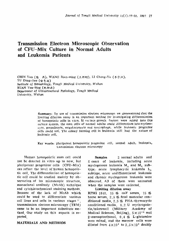
chromatic and orthochromatic normo- blasts were predominant. Nucleus/cyto- plasm ratio ~ 1. Heterochromatin was seen mainly in peripheral area of nucleus, and euchromatin was located chiefly in nuclear center Nucleoli were not found, but nuclear membrane could be observcd clearly. Polyribosomes were extremely numerous throughout the cytoplasm. Mitochondria were large and oval in shape but reduced in number ( f ig . l ) .
2. Granulocytic series The cells were found to be in all stages of development with myelocytes and meta- myelocytes predominating. Microvilli were located on the cellular membrane. The nuclei were horseshoe- or kidneysha- ped with much euchromatin, nucleoli were rarely seen Numerous round mi- tochondria were present. The Golgi zone was well developed containing centriole and vesicles Rough endoplasmic reticu- lure was welI developed. There were two different kinds of granules, namely azu- rophilic granules with homogeneous den- si ty surrounded by a typical unit mem- brane, and special granules with a distinct limiting membrane somewhat less dense than that of the large azurophilic gra- nules.
3. Megakaryocytic series
One type of the cell5 showed a large lobular nucleus surrounded by cytoplasm divided into distinct zoncs Demarcation membrane could be seen clearly. Another type had concave nucleus with prepon- derant euchromatin and numerous poly- podal prejections (fig.2). The irregular, "dark coloured" platelets with microtu- bules were decreased.
4. Macrophage series The cells were round or oval in shape and had matrix of finely dispersed cytoplas- mie protein and granules engulfing other substances.
Leukemia patients After cultured, the colonies still
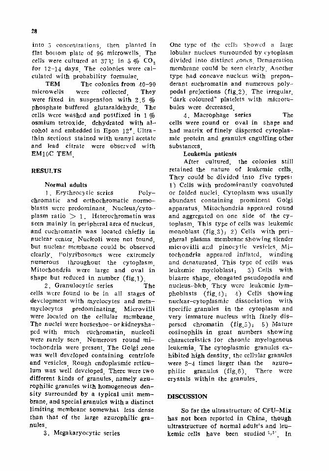
retained the nature of leukemic cells They could be divided into five types: 1) Cells with predominantly convoluted or folded nuclei. Cytoplasm was usually abundant containing prominent Golgi apparatus. Mitochondria appeared round and aggregated on one side of the cy- toplasm. This type of cells was leukemic monoblast (f ig.3); 2) Cells with peri- pheral plasma membrane showing slender micIovilli and pinocytic vesicles. Mi- tochondria appeared inflated, winding and denaturated. This type of cells was leukemic myeloblast; 3) Cells with bizarre shape, elongated pseudopodia and nucleus-bleb. They were leukemic lym- phoblasts (f ig.4); 4) Cells showing nuclear-cytoplasmic dissociation with specific granules in the cytoplasm and very immature nucleus with finely dis- perscd chromatin (f ig.5); 5) Mature eosinophi!s in great numbers showing characteristics for chronic myelogenous leukemia. The cytoplasmic granules ex- hibited high density, the cellular granules were 3-4 times larger than the azuro- philie granules (f ig.6) . There were crystals within the granules.
DISCUSSION
So far the ultrastructure of CFU-Mix has not been reported in China, though ultrastructure of normal adult 's and leu- kemic cells have been studied 2,~, In
Journal ol Tongji Medical University 10(1),27-30, 1990 29
Fig. I. Fig. 2. Fig.3. Fig.4.
No:real adu!l: polychromatie normob~ast.6300 • No:'mal adu!t: megaka:,-yocyte showing numerous po!y~oda! F:'o'ections, 3000x. Acute monocytic !eukemia: monoblast with fatty vesie!e in the cytop!a~m. 5000 x . Acute lymphocytic leukemia: iymphobla'_t with a b!eb in the cellular nucleus. 4000 X.
the classical semisolid culture system, methylcellulose and agar adhere to the colonies, and it is difficult to obtain sat isfactory pictures of TEM. In the past we successfully used l imit ing d i lu - tion assay to demonstrate that CFU-Mix in normal adults contain erythrocyte, granulocyte, megakaryocyte and macro- phage TM. Now we further proved by TEM
that CFU-Mix is an important method for the s tudy of differentiation of he- mopoietic cell in v i t ro Because m i - croenvironment of ceils growing in v i t ro is different from that in v i v o the cellular ultrastructure also differs from that of uncultered cells Before cultured, the normal cells differentiate from blast cell to mature cell, the development is
30
Fig.5.
Fig.6.
Acute mye!o~enous .~eukemia. Tke leukemie cell with development of eytoplasna and nucleus showing asynchronism. 4000 • Chronic mye!ogenous leukemia. Eosinophil with numerous crystals in the cytop- lasm. 5000• r
listed as fol lows ~ Nucleus: euch roma- t in is predominant , heterochromat in increases, euchromat in reduces obv ious ly , he terochromat in increases greatly. C y - toplasm: mi tochondr ia are large and abundant , then reduce in amount or disappear. Special elements lack, then appear and increase in amount . F i r s t cellular organelles development was poor, then well and then began to reduce. When the cells were cultured, their m o r - phology changed. The nucleus was often mature, but euchromat in remained p re - dominant . Mi tochondr ia could be seen clearly even in more mature stage w i t h - out increase of special elements. I t is suggested that there is strong prol i fera t ive capaci ty of the hemopoiet ic cell in v i t ro
Genetics and g lucose -6 -phospha te dehydrogenase (G6PD) isoenzyme studies in chronic myelogenous leukemia have demonstrated that the or ig in of the leu- kemic cell is of plur ipotent stem cell in nature. Whether in acute myelogenous leukemia there are defects in the stem cell or in the granulocyte-macrophage
progeni tor cell is unknown B: Though va r ious g rowth factors such as g r a n u - locytic s t imula t ing factor ( F M C M ) , e ry thropoie t in (EPO) and lymphocyte growth factor ( P H A - T C M ) have been used, the leukemic stem cell lost its potent ial ab i l i t y of di f ferent ia t ion. The di f ferent ia t ion of the granulocytes also did go along the normal path way , and the leukemic progeni tor cell remained at the blast cell stage.
R E F E R E N C E S z
1. F~ ,,~. ~ ~ & ~ . , I ~ , c e ~ P ~ [ ~ ) ~ ' ~ ' ~ r ; ~ 7 1 . . ~ - ; ' r 1987;10 200-3.
r 1981~ 2 : 6-9.
~,~ ~ , : ~ ; ~ a : 1981; 2 : 170-2.
1988~ 9 : 527-9.
# { f f @ ~ ; K ~ f ~ • 1985 : 117-8. 6 . Mcssne~'NA, et al.Eiology of acute myeloid
leukemia. ClJn l-lemato] 1986~ 15:641-67.


![[CANCER RESEARCH 27, 18-25,January 1967] Preliminary ...leukemia were obtained for light and electron microscopic evaluation. Fluid and cells from the pleural cavity in one case and](https://img.pdfslide.us/doc/110x75/6093b50b47e8b95f602c8acc/cancer-research-27-18-25january-1967-preliminary-leukemia-were-obtained.jpg)


![TEST REPORT - smartfiber AG · Sampie size: Preincubation C: ... log [cfU]IGC Oh - log [cfU]sample 72h X 100 log [cfu]IGC Oh ... Test specimen mean 3.12 x 10 x 100 99.96](https://img.pdfslide.us/doc/110x75/5adbb5be7f8b9a53618e4b7d/test-report-smartfiber-ag-size-preincubation-c-log-cfuigc-oh-log-cfusample.jpg)