Embed Size (px)
Citation preview
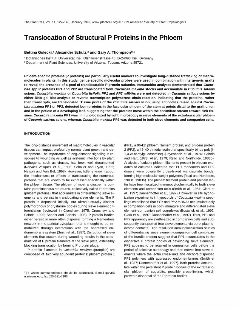
The Plant Cell, Vol. 11, 127–140, January 1999, www.plantcell.org © 1999 American Society of Plant Physiologists
Translocation of Structural P Proteins in the Phloem
Bettina Golecki,
a
Alexander Schulz,
a
and Gary A. Thompson
b,1
a
Botanisches Institut, Universität Kiel, Olshausenstrasse 40, D-24098 Kiel, Germany
b
Department of Plant Sciences, University of Arizona, Tucson, Arizona 85721
Phloem-specific proteins (P proteins) are particularly useful markers to investigate long-distance trafficking of macro-molecules in plants. In this study, genus-specific molecular probes were used in combination with intergeneric graftsto reveal the presence of a pool of translocatable P protein subunits. Immunoblot analyses demonstrated that
Cucur-bita
spp P proteins PP1 and PP2 are translocated from
Cucurbita maxima
stocks and accumulate in
Cucumis sativus
scions.
Cucurbita maxima
or
Cucurbita ficifolia PP1
and
PP2
mRNAs were not detected in
Cucumis sativus
scions byeither RNA gel blot analysis or reverse transcription–polymerase chain reaction, indicating that the proteins, ratherthan transcripts, are translocated. Tissue prints of the
Cucumis sativus
scion, using antibodies raised against
Cucur-bita maxima
PP1 or PP2, detected both proteins in the fascicular phloem of the stem at points distal to the graft unionand in the petiole of a developing leaf, suggesting that the proteins move within the assimilate stream toward sink tis-sues.
Cucurbita maxima
PP1 was immunolocalized by light microscopy in sieve elements of the extrafascicular phloemof
Cucumis sativus
scions, whereas
Cucurbita maxima
PP2 was detected in both sieve elements and companion cells.
INTRODUCTION
The long-distance movement of macromolecules in vasculartissues can impact profoundly normal plant growth and de-velopment. The importance of long-distance signaling in re-sponse to wounding as well as systemic infections by plantpathogens, such as viruses, has been well documented(Narváez-Vásquez et al., 1995; Schaller and Ryan, 1995;Nelson and Van Bel, 1998). However, little is known aboutthe mechanisms or effects of translocating the numerousproteins that are known to be expressed specifically withinthe phloem tissue. The phloem of most angiosperms con-tains proteinaceous structures, collectively called P proteins(phloem proteins), that accumulate in differentiating sieve el-ements and persist in translocating sieve elements. The Pprotein is deposited initially into ultrastructurally distinctpolymorphous or crystalline bodies during sieve element dif-ferentiation (reviewed in Cronshaw, 1975; Cronshaw andSabnis, 1990; Sabnis and Sabnis, 1995). P protein bodieseither persist or more often disperse, forming a filamentousnetwork in the parietal cytoplasm that is thought to be im-mobilized through interactions with the appressed en-domembrane system (Smith et al., 1987). Disruption of sieveelements that occurs during wounding results in the accu-mulation of P protein filaments at the sieve plate, ostensiblyblocking translocation by forming P protein plugs.
P protein filaments in
Cucurbita maxima
(pumpkin) arecomposed of two very abundant proteins: phloem protein 1
(PP1), a 96-kD phloem filament protein, and phloem protein2 (PP2), a 48-kD dimeric lectin that specifically binds poly(
b
-1,4-
N
-acetylglucosamine) (Beyenbach et al., 1974; Sabnisand Hart, 1978; Allen, 1979; Read and Northcote, 1983b).Analysis of soluble phloem filaments present in phloem exu-dates of cucurbits indicated that PP1 monomers and PP2dimers were covalently cross-linked via disulfide bonds,forming high molecular weight polymers (Read and Northcote,1983a, 1983b). The phloem filament protein and phloem lec-tin have been localized immunocytochemically to both sieveelements and companion cells (Smith et al., 1987; Clark etal., 1997; Dannenhoffer et al., 1997). However, in situ hybrid-ization experiments in hypocotyls of
Cucurbita maxima
seed-lings established that
PP1
and
PP2
mRNAs accumulate onlyin companion cells in both immature and differentiated sieveelement–companion cell complexes (Bostwick et al., 1992;Clark et al., 1997; Dannenhoffer et al., 1997). Thus, PP1 andPP2 apparently are synthesized in companion cells and sub-sequently transported into sieve elements via pore–plasmo-desma contacts. High-resolution immunolocalization studiesof differentiating sieve element–companion cell complexesof the bundle phloem suggest that PP1 accumulates in thedispersive P protein bodies of developing sieve elements;PP2 appears to be retained in companion cells before theperiod of selective autophagy and then moves into sieve el-ements where the lectin cross-links and anchors dispersedPP1 polymers with appressed endomembranes (Smith etal., 1987; Dannenhoffer et al., 1997). Both proteins accumu-late within the persistent P protein bodies of the extrafascic-ular phloem of cucurbits, possibly cross-linking, whichprevents dispersal of the P protein bodies.
1
To whom correspondence should be addressed. E-mail [email protected]; fax 520-621-7186.
128 The Plant Cell
In contrast to the incorporation of P proteins into poly-merized structures, several lines of evidence suggest theexistence of a pool of unpolymerized PP1 and PP2 subunitswithin sieve element–companion cell complexes. In theiranalysis of phloem filament structure, Read and Northcote(1983a) estimated that as much as 43% of PP1 and 18% ofPP2 were present as free monomers or dimers in phloemexudates of
Cucurbita maxima.
Alosi et al. (1988) questionedwhether P protein filament formation or stabilization by di-sulfide linkages is possible when the reducing environmentof the phloem sap is considered. The existence of a pool ofunpolymerized P protein subunits is supported further bythe apparent translocation of genus-specific P proteins ortheir precursors in intergeneric grafts between members ofthe Cucurbitaceae (Tiedemann and Carstens-Behrens, 1994;Golecki et al., 1998). These observations are possible be-cause SDS-PAGE profiles of phloem exudate proteins col-lected from different cucurbit genera show considerable sizeheterogeneity (Sabnis and Hart, 1976, 1979). Additional pro-teins with molecular weights typical of
Cucurbita
spp P pro-teins were observed in exudate samples collected from
Cucumis sativus
(cucumber) scions when grafted onto
Cu-curbita
spp stocks. Moreover, subsequent developmentalanalysis demonstrated that the appearance of the additionalproteins in
Cucumis sativus
scions was strongly correlatedto the establishment of intergeneric sieve element connec-tions in the graft union (Golecki et al., 1998).
P proteins share functional similarities among genera ofthe Cucurbitaceae but are sufficiently divergent with regardto their protein and nucleic acid sequences so that genus-specific probes can be used to determine their origin inintergeneric grafts (A.M. Clark and G.A. Thompson, unpub-lished results). In this study, we used the intergenericdivergence of PP1 and PP2 to demonstrate that these pro-teins are capable of long-distance movement in the phloemof grafted plants. Evidence is presented that
Cucurbita
sppPP1 and PP2 are translocated from
Cucurbita maxima
or
Cucurbita ficifolia
stocks to
Cucumis sativus
scions viaphloem bridges formed at the graft union. Our results alsodemonstrate that PP2 exits from sieve elements and accu-mulates in companion cells of the extrafascicular phloem ofthe
Cucumis sativus
scion. The implications for long-dis-tance movement of macromolecules and intercellular inter-actions between sieve elements and companion cells at adistance from the point of protein synthesis are discussed.
RESULTS
We have exploited the intergeneric divergence of the twomajor P proteins in cucurbits to determine whether thephloem filament protein PP1 and phloem lectin PP2 aretranslocated over long distances in the transport phloem. In-tergeneric approach grafts consisting of
Cucurbita maxima
or
Cucurbita ficifolia
stocks and
Cucumis sativus
scions
were used in this study (Figure 1A). We have shown previ-ously that additional proteins in phloem exudates of
Cucu-mis sativus
scions grafted to
Cucurbita
spp stocks weredetected easily within 9 to 11 days after grafting (Golecki etal., 1998). Figure 1B diagrammatically shows a grafted plantand the points at which samples were collected.
Cucurbita maxima
PP1 and PP2 Accumulate in
Cucumis sativus
Scions
To avoid cross-contamination of
Cucumis sativus
tissuewith
Cucurbita maxima
phloem sap, the first transverse cutacross the scion hypocotyl below the cotyledons alwaysseparated the
Cucumis sativus
scion from the
Cucurbita
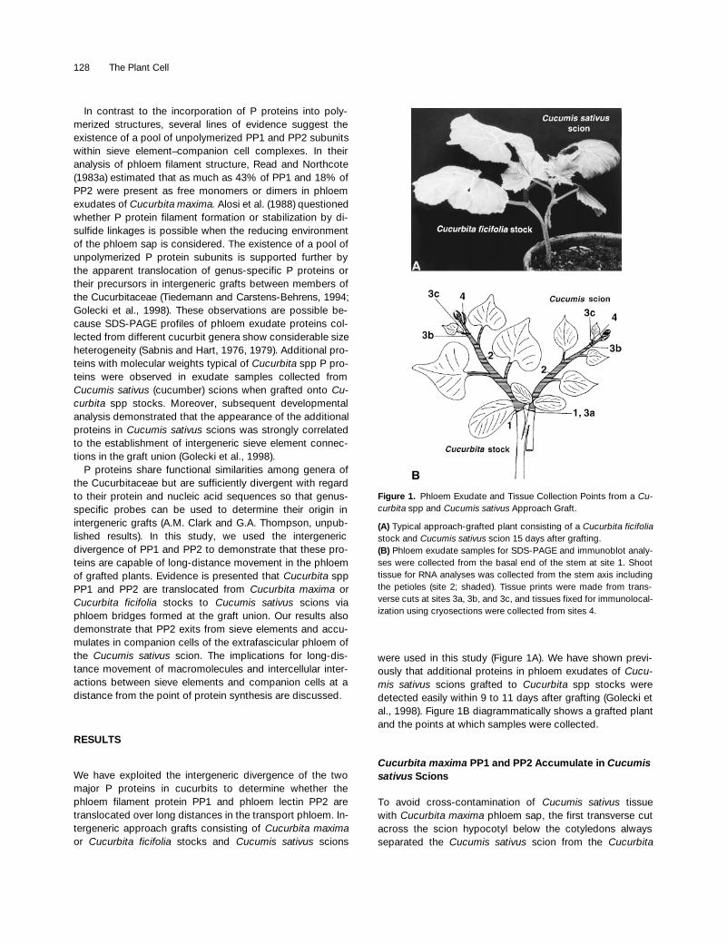
Figure 1. Phloem Exudate and Tissue Collection Points from a Cu-curbita spp and Cucumis sativus Approach Graft.
(A) Typical approach-grafted plant consisting of a Cucurbita ficifoliastock and Cucumis sativus scion 15 days after grafting.(B) Phloem exudate samples for SDS-PAGE and immunoblot analy-ses were collected from the basal end of the stem at site 1. Shoottissue for RNA analyses was collected from the stem axis includingthe petioles (site 2; shaded). Tissue prints were made from trans-verse cuts at sites 3a, 3b, and 3c, and tissues fixed for immunolocal-ization using cryosections were collected from sites 4.
P Protein Translocation in Intergeneric Grafts 129
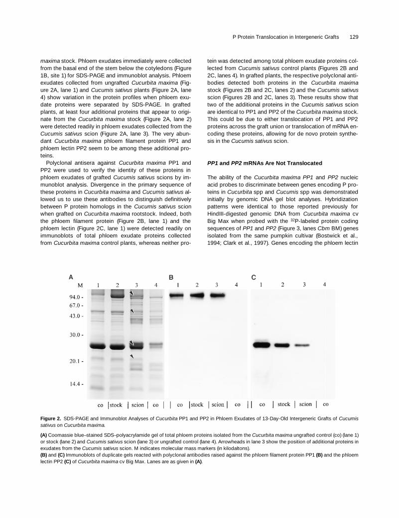
maxima
stock. Phloem exudates immediately were collectedfrom the basal end of the stem below the cotyledons (Figure1B, site 1) for SDS-PAGE and immunoblot analysis. Phloemexudates collected from ungrafted
Cucurbita maxima
(Fig-ure 2A, lane 1) and
Cucumis sativus
plants (Figure 2A, lane4) show variation in the protein profiles when phloem exu-date proteins were separated by SDS-PAGE. In graftedplants, at least four additional proteins that appear to origi-nate from the
Cucurbita maxima
stock (Figure 2A, lane 2)were detected readily in phloem exudates collected from the
Cucumis sativus
scion (Figure 2A, lane 3). The very abun-dant
Cucurbita maxima
phloem filament protein PP1 andphloem lectin PP2 seem to be among these additional pro-teins.
Polyclonal antisera against
Cucurbita maxima
PP1 andPP2 were used to verify the identity of these proteins inphloem exudates of grafted
Cucumis sativus
scions by im-munoblot analysis. Divergence in the primary sequence ofthese proteins in
Cucurbita maxima
and
Cucumis sativus
al-lowed us to use these antibodies to distinguish definitivelybetween P protein homologs in the
Cucumis sativus
scionwhen grafted on
Cucurbita maxima
rootstock. Indeed, boththe phloem filament protein (Figure 2B, lane 1) and thephloem lectin (Figure 2C, lane 1) were detected readily onimmunoblots of total phloem exudate proteins collectedfrom
Cucurbita maxima
control plants, whereas neither pro-
tein was detected among total phloem exudate proteins col-lected from
Cucumis sativus
control plants (Figures 2B and2C, lanes 4). In grafted plants, the respective polyclonal anti-bodies detected both proteins in the
Cucurbita maxima
stock (Figures 2B and 2C, lanes 2) and the
Cucumis sativus
scion (Figures 2B and 2C, lanes 3). These results show thattwo of the additional proteins in the
Cucumis sativus
scionare identical to PP1 and PP2 of the
Cucurbita maxima
stock.This could be due to either translocation of PP1 and PP2proteins across the graft union or translocation of mRNA en-coding these proteins, allowing for de novo protein synthe-sis in the
Cucumis sativus
scion.
PP1
and
PP2
mRNAs Are Not Translocated
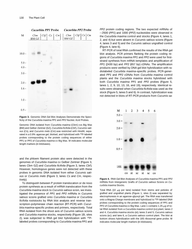
The ability of the
Cucurbita maxima PP1
and
PP2
nucleicacid probes to discriminate between genes encoding P pro-teins in
Cucurbita
spp and
Cucumis
spp was demonstratedinitially by genomic DNA gel blot analyses. Hybridizationpatterns were identical to those reported previously forHindIII-digested genomic DNA from
Cucurbita maxima
cvBig Max when probed with the
32
P-labeled protein codingsequences of
PP1
and
PP2
(Figure 3, lanes
Cbm
BM) genesisolated from the same pumpkin cultivar (Bostwick et al.,1994; Clark et al., 1997). Genes encoding the phloem lectin
Figure 2. SDS-PAGE and Immunoblot Analyses of Cucurbita PP1 and PP2 in Phloem Exudates of 13-Day-Old Intergeneric Grafts of Cucumissativus on Cucurbita maxima.
(A) Coomassie blue–stained SDS–polyacrylamide gel of total phloem proteins isolated from the Cucurbita maxima ungrafted control (co) (lane 1)or stock (lane 2) and Cucumis sativus scion (lane 3) or ungrafted control (lane 4). Arrowheads in lane 3 show the position of additional proteins inexudates from the Cucumis sativus scion. M indicates molecular mass markers (in kilodaltons).(B) and (C) Immunoblots of duplicate gels reacted with polyclonal antibodies raised against the phloem filament protein PP1 (B) and the phloemlectin PP2 (C) of Cucurbita maxima cv Big Max. Lanes are as given in (A).
130 The Plant Cell
and the phloem filament protein also were detected in thegenomes of
Cucurbita maxima
cv Gelber Zentner (Figure 3,lanes
Cbm
GZ) and
Cucurbita ficifolia
(Figure 3, lanes
Cbf
).However, homologous genes were not detected with theseprobes in genomic DNA isolated from either
Cucumis sati-vus
or
Cucumis melo
(Figure 3, lanes
Cs
and
Cm
, respec-tively).
To distinguish between P protein translocation or de novoprotein synthesis as a result of mRNA translocation from the
Cucurbita maxima
stock to
Cucumis sativus
scion, we inves-tigated the presence of
PP1
and
PP2
mRNAs in
Cucumissativus
scions grafted onto
Cucurbita maxima
or
Cucurbitaficifolia
rootstocks by RNA blot analysis and reverse tran-scription–polymerase chain reaction (RT-PCR) with
Cucur-bita maxima
–specific probes and primers, respectively. TotalRNA isolated from the shoot axis of
Cucumis sativus
scionsand
Cucurbita maxima
stocks, respectively (Figure 1B, sites2), was subjected to RNA gel blot hybridization with
32
P-labeled probes corresponding to
Cucurbita maxima PP1
and
PP2
protein coding regions. The two expected mRNAs of
z
2500 (
PP1
) and 1000 (PP2) nucleotides were observed inthe Cucurbita maxima control and stocks (Figure 4, lanes 1,2, and 4) but were absent in Cucumis sativus scions (Figure4, lanes 3 and 5) and the Cucumis sativus ungrafted control(Figure 4, lane 6).
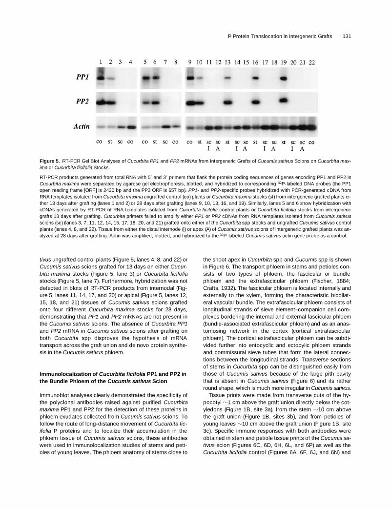
RT-PCR of total RNA confirmed the results of the RNA gelblot analysis. PCR primers flanking the protein coding re-gions of Cucurbita maxima PP1 and PP2 were used for first-strand synthesis from mRNA templates and amplification ofPP1 (2430 bp) and PP2 (657 bp) cDNAs. The amplificationproducts were verified by DNA gel blot hybridization with ra-diolabeled Cucurbita maxima–specific probes. PCR-gener-ated PP1 and PP2 cDNAs from Cucurbita maxima controlplants and the Cucurbita maxima stocks hybridized withboth Cucurbita maxima PP1 and PP2 probes (Figure 5,lanes 1, 2, 9, 10, 13, 16, and 19), respectively. Identical re-sults were obtained when Cucurbita ficifolia was used as thestock (Figure 5, lanes 5 and 6). In contrast, hybridization wasnot detected in blots of RT-PCR products from Cucumis sa-
Figure 3. Genomic DNA Gel Blot Analyses Demonstrate the Speci-ficity of the Cucurbita maxima PP1 and PP2 Nucleic Acid Probes.
Genomic DNA isolated from Cucurbita maxima (Cbm) cvs Big Max(BM) and Gelber Zentner (GZ), Cucurbita ficifolia (Cbf ), Cucumis sati-vus (Cs), and Cucumis melo (Cm) was restricted with HindIII, sepa-rated in a 0.9% agarose gel, blotted, and hybridized with 32P-labeledprobes corresponding to the protein coding sequences of eitherPP1 or PP2 of Cucurbita maxima cv Big Max. M indicates molecularlength markers (in kilobases).
Figure 4. RNA Gel Blot Analyses of Cucurbita maxima PP1 and PP2mRNAs from Intergeneric Grafts of Cucumis sativus Scions on Cu-curbita maxima Stocks.
Total RNA (10 mg per lane) isolated from stems and petioles ofgrafted and ungrafted plants (Figure 1, sites 2) was separated byelectrophoresis in an agarose–glyoxyl gel. The RNA was transferredonto a Magna Charge membrane and hybridized to 32P-labeled DNAprobes corresponding to the protein coding sequences of PP1 andPP2 of Cucurbita maxima cv Big Max. Lane 1 contains 1.25 mg of to-tal RNA isolated from a Cucurbita maxima control (co) plant; lanes 2and 4, Cucurbita maxima stocks (st); lanes 3 and 5, Cucumis sativusscions (sc); and lane 6, a Cucumis sativus control plant. The blot atbottom shows hybridization with the 18S ribosomal gene probe. Mindicates molecular length markers (in kilobases).
P Protein Translocation in Intergeneric Grafts 131
tivus ungrafted control plants (Figure 5, lanes 4, 8, and 22) orCucumis sativus scions grafted for 13 days on either Cucur-bita maxima stocks (Figure 5, lane 3) or Cucurbita ficifoliastocks (Figure 5, lane 7). Furthermore, hybridization was notdetected in blots of RT-PCR products from internodal (Fig-ure 5, lanes 11, 14, 17, and 20) or apical (Figure 5, lanes 12,15, 18, and 21) tissues of Cucumis sativus scions graftedonto four different Cucurbita maxima stocks for 28 days,demonstrating that PP1 and PP2 mRNAs are not present inthe Cucumis sativus scions. The absence of Cucurbita PP1and PP2 mRNA in Cucumis sativus scions after grafting onboth Cucurbita spp disproves the hypothesis of mRNAtransport across the graft union and de novo protein synthe-sis in the Cucumis sativus phloem.
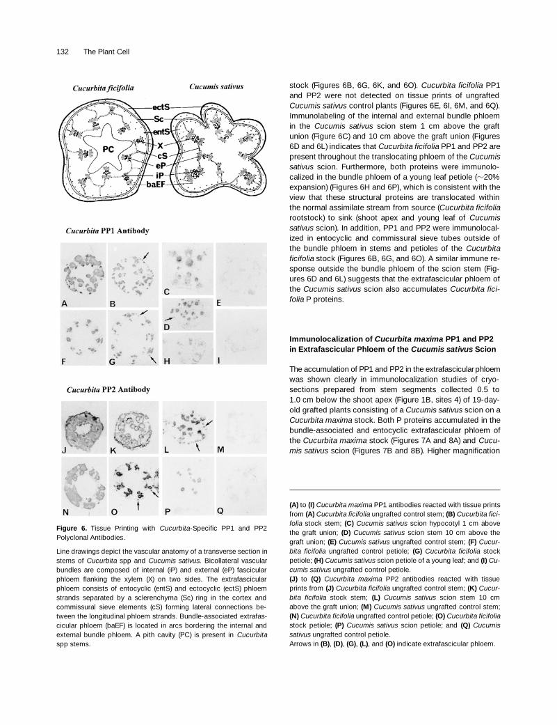
Immunolocalization of Cucurbita ficifolia PP1 and PP2 in the Bundle Phloem of the Cucumis sativus Scion
Immunoblot analyses clearly demonstrated the specificity ofthe polyclonal antibodies raised against purified Cucurbitamaxima PP1 and PP2 for the detection of these proteins inphloem exudates collected from Cucumis sativus scions. Tofollow the route of long-distance movement of Cucurbita fic-ifolia P proteins and to localize their accumulation in thephloem tissue of Cucumis sativus scions, these antibodieswere used in immunolocalization studies of stems and peti-oles of young leaves. The phloem anatomy of stems close to
the shoot apex in Cucurbita spp and Cucumis spp is shownin Figure 6. The transport phloem in stems and petioles con-sists of two types of phloem, the fascicular or bundlephloem and the extrafascicular phloem (Fischer, 1884;Crafts, 1932). The fascicular phloem is located internally andexternally to the xylem, forming the characteristic bicollat-eral vascular bundle. The extrafascicular phloem consists oflongitudinal strands of sieve element–companion cell com-plexes bordering the internal and external fascicular phloem(bundle-associated extrafascicular phloem) and as an anas-tomosing network in the cortex (cortical extrafascicularphloem). The cortical extrafascicular phloem can be subdi-vided further into entocyclic and ectocylic phloem strandsand commissural sieve tubes that form the lateral connec-tions between the longitudinal strands. Transverse sectionsof stems in Cucurbita spp can be distinguished easily fromthose of Cucumis sativus because of the large pith cavitythat is absent in Cucumis sativus (Figure 6) and its ratherround shape, which is much more irregular in Cucumis sativus.
Tissue prints were made from transverse cuts of the hy-pocotyl z1 cm above the graft union directly below the cot-yledons (Figure 1B, site 3a), from the stem z10 cm abovethe graft union (Figure 1B, sites 3b), and from petioles ofyoung leaves z10 cm above the graft union (Figure 1B, site3c). Specific immune responses with both antibodies wereobtained in stem and petiole tissue prints of the Cucumis sa-tivus scion (Figures 6C, 6D, 6H, 6L, and 6P) as well as theCucurbita ficifolia control (Figures 6A, 6F, 6J, and 6N) and
Figure 5. RT-PCR Gel Blot Analyses of Cucurbita PP1 and PP2 mRNAs from Intergeneric Grafts of Cucumis sativus Scions on Cucurbita max-ima or Cucurbita ficifolia Stocks.
RT-PCR products generated from total RNA with 59 and 39 primers that flank the protein coding sequences of genes encoding PP1 and PP2 inCucurbita maxima were separated by agarose gel electrophoresis, blotted, and hybridized to corresponding 32P-labeled DNA probes (the PP1open reading frame [ORF] is 2430 bp and the PP2 ORF is 657 bp). PP1- and PP2-specific probes hybridized with PCR-generated cDNA fromRNA templates isolated from Cucurbita maxima ungrafted control (co) plants or Cucurbita maxima stocks (st) from intergeneric grafted plants ei-ther 13 days after grafting (lanes 1 and 2) or 28 days after grafting (lanes 9, 10, 13, 16, and 19). Similarly, lanes 5 and 6 show hybridization withcDNAs generated by RT-PCR of RNA templates isolated from Cucurbita ficifolia control plants or Cucurbita ficifolia stocks from intergenericgrafts 13 days after grafting. Cucurbita primers failed to amplify either PP1 or PP2 cDNAs from RNA templates isolated from Cucumis sativusscions (sc) (lanes 3, 7, 11, 12, 14, 15, 17, 18, 20, and 21) grafted onto either of the Cucurbita spp stocks and ungrafted Cucumis sativus controlplants (lanes 4, 8, and 22). Tissue from either the distal internode (I) or apex (A) of Cucumis sativus scions of intergeneric grafted plants was an-alyzed at 28 days after grafting. Actin was amplified, blotted, and hybridized to the 32P-labeled Cucumis sativus actin gene probe as a control.
132 The Plant Cell
stock (Figures 6B, 6G, 6K, and 6O). Cucurbita ficifolia PP1and PP2 were not detected on tissue prints of ungraftedCucumis sativus control plants (Figures 6E, 6I, 6M, and 6Q).Immunolabeling of the internal and external bundle phloemin the Cucumis sativus scion stem 1 cm above the graftunion (Figure 6C) and 10 cm above the graft union (Figures6D and 6L) indicates that Cucurbita ficifolia PP1 and PP2 arepresent throughout the translocating phloem of the Cucumissativus scion. Furthermore, both proteins were immunolo-calized in the bundle phloem of a young leaf petiole (z20%expansion) (Figures 6H and 6P), which is consistent with theview that these structural proteins are translocated withinthe normal assimilate stream from source (Cucurbita ficifoliarootstock) to sink (shoot apex and young leaf of Cucumissativus scion). In addition, PP1 and PP2 were immunolocal-ized in entocyclic and commissural sieve tubes outside ofthe bundle phloem in stems and petioles of the Cucurbitaficifolia stock (Figures 6B, 6G, and 6O). A similar immune re-sponse outside the bundle phloem of the scion stem (Fig-ures 6D and 6L) suggests that the extrafascicular phloem ofthe Cucumis sativus scion also accumulates Cucurbita fici-folia P proteins.
Immunolocalization of Cucurbita maxima PP1 and PP2 in Extrafascicular Phloem of the Cucumis sativus Scion
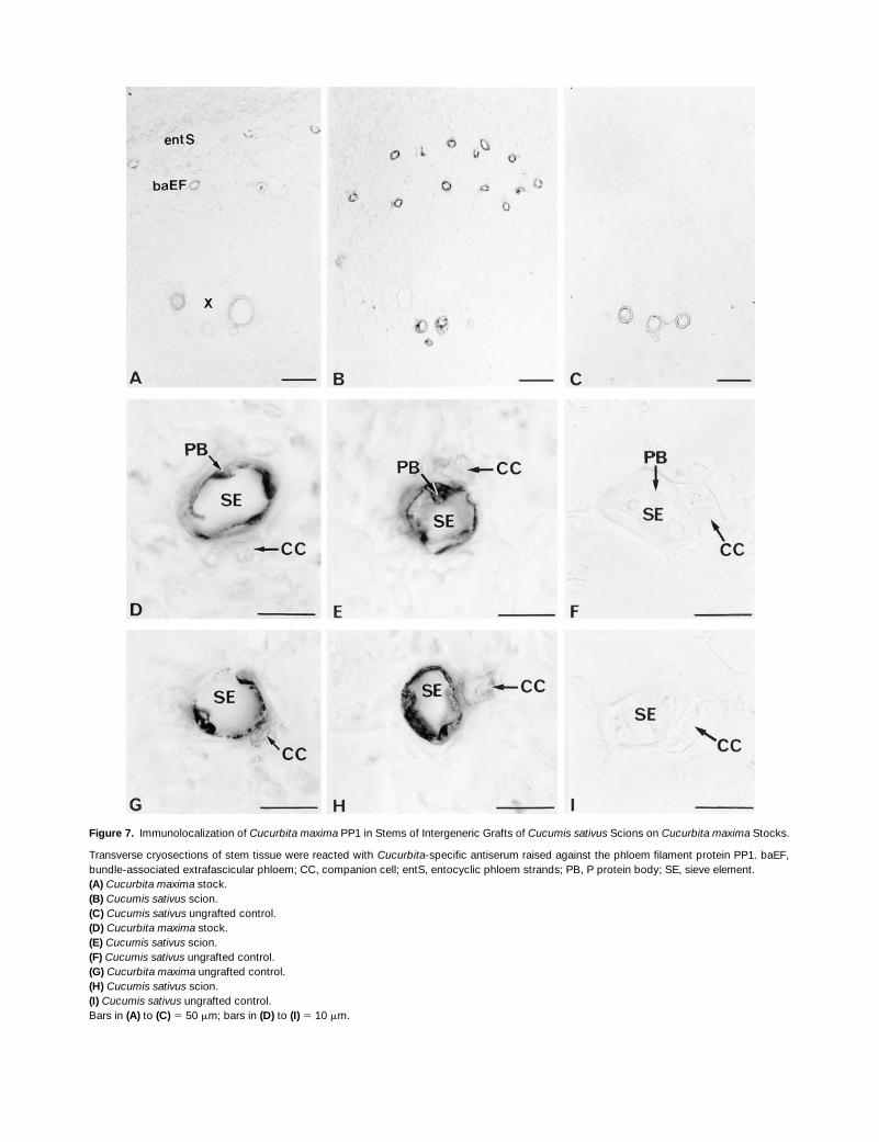
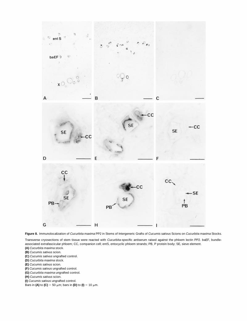
The accumulation of PP1 and PP2 in the extrafascicular phloemwas shown clearly in immunolocalization studies of cryo-sections prepared from stem segments collected 0.5 to1.0 cm below the shoot apex (Figure 1B, sites 4) of 19-day-old grafted plants consisting of a Cucumis sativus scion on aCucurbita maxima stock. Both P proteins accumulated in thebundle-associated and entocyclic extrafascicular phloem ofthe Cucurbita maxima stock (Figures 7A and 8A) and Cucu-mis sativus scion (Figures 7B and 8B). Higher magnification
Figure 6. Tissue Printing with Cucurbita-Specific PP1 and PP2Polyclonal Antibodies.
Line drawings depict the vascular anatomy of a transverse section instems of Cucurbita spp and Cucumis sativus. Bicollateral vascularbundles are composed of internal (iP) and external (eP) fascicularphloem flanking the xylem (X) on two sides. The extrafascicularphloem consists of entocyclic (entS) and ectocyclic (ectS) phloemstrands separated by a sclerenchyma (Sc) ring in the cortex andcommissural sieve elements (cS) forming lateral connections be-tween the longitudinal phloem strands. Bundle-associated extrafas-cicular phloem (baEF) is located in arcs bordering the internal andexternal bundle phloem. A pith cavity (PC) is present in Cucurbitaspp stems.
(A) to (I) Cucurbita maxima PP1 antibodies reacted with tissue printsfrom (A) Cucurbita ficifolia ungrafted control stem; (B) Cucurbita fici-folia stock stem; (C) Cucumis sativus scion hypocotyl 1 cm abovethe graft union; (D) Cucumis sativus scion stem 10 cm above thegraft union; (E) Cucumis sativus ungrafted control stem; (F) Cucur-bita ficifolia ungrafted control petiole; (G) Cucurbita ficifolia stockpetiole; (H) Cucumis sativus scion petiole of a young leaf; and (I) Cu-cumis sativus ungrafted control petiole.(J) to (Q) Cucurbita maxima PP2 antibodies reacted with tissueprints from (J) Cucurbita ficifolia ungrafted control stem; (K) Cucur-bita ficifolia stock stem; (L) Cucumis sativus scion stem 10 cmabove the graft union; (M) Cucumis sativus ungrafted control stem;(N) Cucurbita ficifolia ungrafted control petiole; (O) Cucurbita ficifoliastock petiole; (P) Cucumis sativus scion petiole; and (Q) Cucumissativus ungrafted control petiole.Arrows in (B), (D), (G), (L), and (O) indicate extrafascicular phloem.
P Protein Translocation in Intergeneric Grafts 133
Figure 7. Immunolocalization of Cucurbita maxima PP1 in Stems of Intergeneric Grafts of Cucumis sativus Scions on Cucurbita maxima Stocks.
Transverse cryosections of stem tissue were reacted with Cucurbita-specific antiserum raised against the phloem filament protein PP1. baEF,bundle-associated extrafascicular phloem; CC, companion cell; entS, entocyclic phloem strands; PB, P protein body; SE, sieve element.(A) Cucurbita maxima stock.(B) Cucumis sativus scion.(C) Cucumis sativus ungrafted control.(D) Cucurbita maxima stock.(E) Cucumis sativus scion.(F) Cucumis sativus ungrafted control.(G) Cucurbita maxima ungrafted control.(H) Cucumis sativus scion.(I) Cucumis sativus ungrafted control.Bars in (A) to (C) 5 50 mm; bars in (D) to (I) 5 10 mm.
134 The Plant Cell
Figure 8. Immunolocalization of Cucurbita maxima PP2 in Stems of Intergeneric Grafts of Cucumis sativus Scions on Cucurbita maxima Stocks.
Transverse cryosections of stem tissue were reacted with Cucurbita-specific antiserum raised against the phloem lectin PP2. baEF, bundle-associated extrafascicular phloem; CC, companion cell; entS, entocyclic phloem strands; PB, P protein body; SE, sieve element.(A) Cucurbita maxima stock.(B) Cucumis sativus scion.(C) Cucumis sativus ungrafted control.(D) Cucurbita maxima stock.(E) Cucumis sativus scion.(F) Cucumis sativus ungrafted control.(G) Cucurbita maxima ungrafted control.(H) Cucumis sativus scion.(I) Cucumis sativus ungrafted control.Bars in (A) to (C) 5 50 mm; bars in (D) to (I) 5 10 mm.
P Protein Translocation in Intergeneric Grafts 135
showed the detection of PP1 and PP2 in the sieve element–companion cell complexes of the Cucurbita maxima stock(Figures 7D and 8D), Cucurbita maxima ungrafted control(Figures 7G and 8G), and Cucumis sativus scions (Figures7E and 7H, and 8E and 8H). These immune responses werespecific for Cucurbita maxima PP1 and PP2 in the Cucumissativus scion because neither of the Cucurbita maxima Pproteins was detected in the phloem tissue and surroundingcells of the ungrafted Cucumis sativus control (Figures 7C,7F, and 7I, and 8C, 8F, and 8I). P proteins were not detectedin the immature fascicular phloem (Figures 7A and 7B, and8A and 8B). Nonspecific immunolabeling of the lignifiedwalls of xylem elements was observed (Figures 7A to 7C,and 8A to 8C).
Interestingly, high magnifications of the extrafascicularphloem in the Cucumis sativus scion showed differences inthe accumulation of the 96-kD phloem filament protein andthe 48-kD (24.5-kD subunits) dimeric phloem lectin within thesieve element–companion cell complex. Cucurbita maximaPP1 was localized with intense signals in parietal regionsand persistent P protein bodies of sieve elements and withvery weak signals in companion cells (Figures 7E and 7H). Insharp contrast, Cucurbita maxima PP2 was localized withintense signals in both cell types (Figures 8E and 8H). Theseobservations provide evidence that subsequent to long-dis-tance movement within sieve tubes, P proteins move fromsieve elements into companion cells via connecting pore–plasmodesma contacts.
DISCUSSION
P proteins were defined initially as ultrastructurally distinctproteinaceous filaments or aggregates that accumulatewithin differentiating sieve elements (Esau and Cronshaw,1967). The present observations on the long-distance move-ment of P proteins across a graft union are in contrast to thetraditional concept that these proteins form only immobi-lized polymeric structures within individual sieve tube mem-bers (Smith et al., 1987; Fisher et al., 1992). Immunoblotanalyses of Cucurbita maxima P proteins in phloem exudatefrom intergeneric grafts clearly illustrated that the phloem fil-ament protein PP1 and the phloem lectin PP2, both originat-ing from the stock, accumulated within the Cucumis sativusscion. In a previous study, it was confirmed that functionalsieve elements bridging the graft union must be establishedbefore additional vascular proteins in Cucumis sativus sci-ons could be observed by using SDS-PAGE at 10 days aftergrafting (Golecki et al., 1998). Furthermore, transport of car-boxyfluorescein from the stock to the scion verified thatnewly formed vascular bridges were functional at that time.Thus, at 13 days after grafting, we can exclude the possibil-ity that the polymerized P protein was dislodged from its pa-rietal position in sieve elements of the stock plant during thegrafting process. In addition, the method of exudate collec-
tion from the Cucumis sativus scion excluded any contami-nation with P protein from the stock at the time of sampling.Although Cucurbita maxima P proteins were identified eas-ily, their respective transcripts were undetectable in the Cu-cumis sativus scion, indicating that the proteins, rather thantheir mRNAs, move within the assimilate stream. This con-trasts with recent reports of mRNA trafficking between thecompanion cell and sieve element (Kühn et al., 1997) butsupports our previous findings that PP1 and PP2 mRNA ac-cumulates exclusively in companion cells (Bostwick et al.,1992; Clark et al., 1997; Dannenhoffer et al., 1997).
Transport Form of P Proteins
Transport of P proteins across the graft union suggests thatthey are in a mobile form incapable of blocking translocationwithin sieve elements. Although we cannot rule out the long-distance movement of small polymers, the most likely formof translocated P protein is either PP1 monomers or PP2dimers. In vivo labeling of soluble phloem proteins of wheat,rice, and castor bean indicated that between 100 and 200soluble polypeptides are present in the translocation stream(Fisher et al., 1992; Nakamura et al., 1993; Sakuth et al.,1993). Because these phloem-mobile proteins range in sizefrom 10 to 70 kD, the 96-kD phloem filament protein of cu-curbits is the largest phloem protein for which long-distancemovement has been demonstrated. Based on observationsof 35S-labeled phloem-mobile proteins in wheat, Fisher et al.(1992) presented a model for long-distance transport of pro-teins that essentially followed the normal assimilate source-to-sink translocation pathway. Phloem exudates collected atseveral points along the source-to-sink pathway in wheatshowed similar protein concentrations and compositions.Cucurbita ficifolia PP1 and PP2 were detected in tissueprints of the stems of Cucumis sativus scions near (1 cm)and at a distance from (10 cm) the graft union as well as inthe petioles of sink leaves. These data suggest that struc-tural P proteins also are translocated in the assimilatestream to sink tissues and provide direct evidence of spe-cific protein translocation in sieve elements.
Our previous studies of protein movement in intergenericgrafts also indicated that similar quantities of translocatedprotein from Cucurbita spp stocks are distributed through-out the Cucumis sativus scion (Golecki et al., 1998). Thus,the soluble P protein appears to be maintained at a steady-state level in the phloem throughout the entire vascular sys-tem. This would concur with previous reports of little or nodifferences in SDS-PAGE and isoelectric focusing proteinpatterns in phloem exudates collected from the followingcontrasting developmental states: 5-day-old seedlings andmature plants; young petioles and the oldest stem inter-nodes; dark-grown and light-grown seedlings; and vegeta-tive and flowering plants (Sabnis and Hart, 1976, 1978,1979; Smith et al., 1987).
136 The Plant Cell
The relationship between mobile P protein subunits andmicroscopic observations of proteinaceous structures insieve elements is unclear. The myriad of ultrastructural stud-ies that show the P protein deposited into filaments andbodies in differentiating and translocating sieve elementsstrongly support the presence of polymerized P proteinstructures. Recent confocal laser scanning imaging of func-tional sieve elements in intact fava bean leaves (Knoblauchand Van Bel, 1998) indicates that previous ultrastructuralobservations cannot simply be ignored as artifacts gener-ated during tissue preparation. However, the presence ofmobile P protein filaments in the sieve tube lumen in vivo isunlikely and would hinder assimilate translocation. Alosi etal. (1988) observed that pure phloem exudate does not poly-merize rapidly; they suggested that diluting the P proteinduring collection induces conformational changes, exposingsulfhydryl residues that can cross-link rapidly. Filaments indiluted samples analyzed to date are probably due to rapidoxidation of P protein subunits that occurs during exudatecollection. Thus, P proteins of cucurbits appear to bepresent in two forms: (1) individual PP1 and PP2 subunitsthat are translocatable and (2) polymerized filaments com-posed of PP1 and PP2 subunits that are immobilized withinindividual sieve tube members. The interrelationship of thetwo forms remains to be determined.
Intercellular Trafficking and Accumulation of P Proteins
Differential accumulation of Cucurbita maxima PP1 and PP2within sieve element–companion cell complexes of the ex-trafascicular phloem in Cucumis sativus scions raises sev-eral enigmatic questions regarding P protein trafficking andaccumulation. Cucurbita maxima PP2 was immunolocalizedin both sieve elements and companion cells (Figure 7),whereas Cucurbita maxima PP1 was limited primarily tosieve elements (Figure 8) in both the stock and scion. Twoscenarios can be envisioned to explain these data in maturesieve element–companion cell complexes: either PP2 readilymoves between the two cell types, while PP1 is physicallyretained within the sieve element, or both proteins trafficfrom sieve elements to companion cells, where PP1 is rap-idly degraded and PP2 accumulates, possibly to be recy-cled. Regulation of macromolecular trafficking within thesieve element–companion cell complex certainly involvesthe numerous symplasmic connections, or pore–plasmo-desma contacts, that form between sieve elements andcompanion cells (reviewed in Van Bel and Kempers, 1997).Although size exclusion limits (SELs) of plasmodesmata be-tween mesophyll cells range from 0.75 to 1.0 kD (Terry andRobards, 1987; Wolf et al., 1989; Robards and Lucas, 1990),the SELs of pore–plasmodesma contacts in translocatingsieve element–companion cell complexes are much larger.Microinjection of either companion cells or sieve elementswith a series of fluorescein isothiocyanate (FITC)–labeledconjugates demonstrated a minimum SEL of 3 kD for the
pore–plasmodesma contacts in the extrafascicular phloemof Cucurbita maxima and up to 25 kD in the fascicular phloemof fava bean (Kempers et al., 1993; Kempers and Van Bel,1997). Coincidentally, the Stokes radius of the globular PP2dimer (2.9 nm; Anantharam et al., 1986) is similar in size tothe 10-kD dextran conjugates (2.3 nm) that readily movedfrom sieve elements to companion cells in fava bean(Kempers and Van Bel, 1997). The increased SEL of pore–plasmodesma contacts shown by Kempers and Van Bel(1997) might allow unrestricted movement of Cucurbita sppPP2 dimers or monomers between sieve elements and com-panion cells in the Cucumis sativus scion, while limiting theconsiderably larger 96-kD phloem filament protein to thesieve element.
Cucurbita maxima PP1 and PP2 have the capacity to in-crease the SEL of plasmodesmata and mediate cell-to-cellmovement in parenchymatic tissue. Balachandran et al.(1997) demonstrated extensive cell-to-cell movement of thephloem lectin after microinjecting FITC-labeled PP2 into me-sophyll cells of C. maxima cotyledons. In coinjection studieswith dextrans, very low concentrations of PP2 directedmovement of 20-kD, but not 40-kD, FITC-dextrans. Microin-jection of mesophyll cells with an 80- to 100-kD fraction iso-lated from C. maxima phloem exudate composed primarilyof PP1 also allowed movement of 20-kD FITC-dextrans.Thus, both proteins could interact with pore–plasmodesmacontacts to increase the SEL and move from sieve elementsinto companion cells. Schobert et al. (1995) identified sev-eral chaperones in phloem exudates from castor bean seed-lings and speculated about their involvement in unfoldingproteins to facilitate symplasmic movement from compan-ion cells to sieve elements. Unidirectional movement of thelarge 96-kD phloem filament protein soon after its synthesisin companion cells (Clark et al., 1997) could be mediated bychaperones or other “movement-related” proteins in thecompanion cell that are either developmentally expressed orlacking in the assimilate stream. Alternatively, Fisher et al.(1992) hypothesized that phloem-associated proteins tar-geted for degradation in companion cells are selectively re-moved from sieve elements in the transport phloem.
Developmental and Functional Implications ofP Protein Translocation
Protein and mRNA localization patterns provide convincingevidence that PP1 and PP2 are synthesized in companioncells of differentiating sieve element–companion cell com-plexes within the transport phloem (Smith et al., 1987; Clarket al., 1997; Dannenhoffer et al., 1997). Based on ultrastruc-tural studies of developing vascular tissues, polymerizedforms of the P protein (i.e., tubules, bodies, or filaments) ap-pear to accumulate during differentiation. However, P pro-tein mRNA also can be detected readily in companion cellsof mature sieve element–companion cell complexes, espe-cially in the extrafascicular phloem (Bostwick et al., 1992;
P Protein Translocation in Intergeneric Grafts 137
Dannenhoffer et al., 1997). Furthermore, microautoradiogra-phy of in vivo–labeled proteins led Nuske and Eschrich(1976) to conclude that P proteins are synthesized continu-ally in the companion cells of mature metaphloem. In bundlephloem, in situ hybridization revealed that PP2 mRNA waseasier to detect in early stages of primary growth than later,suggesting that P protein synthesis in the bundle phloemwas of limited duration after the onset of translocation(Dannenhoffer et al., 1997). Given a finite developmentalperiod for the accumulation of the polymerized P protein,the continued synthesis of PP1 and PP2 in mature sieveelement–companion cell complexes could be the origin ofmobile P protein subunits, especially in the extrafascicularphloem where P protein mRNA is most often detected inmature sieve element–companion cell complexes.
Functional continuity of bundle and extrafascicular phloemwas demonstrated by immunodetection of Cucurbita sppPP1 and PP2 in the entocyclic phloem of young Cucumissativus scion stems (Figures 7 and 8). The anastomosingnetwork of extrafascicular sieve elements with the bundlephloem would allow a steady state level of mobile P proteinto be obtained throughout the vascular system. This modelsuggests that mobile P protein continually enters the assim-ilate stream throughout the translocation pathway ratherthan being loaded in source tissues. However, the fate ofmobile P protein in the translocation conduit seems to bedetermined by the direction and intensity of assimilate trans-port. Tiedemann and Carstens-Behrens (1994) showed byusing SDS-PAGE of vascular exudates from intergenericgrafts that Cucurbita ficifolia PP1 and PP2 appear in fruit ofthe Cucumis sativus scion, a strong sink organ. The mecha-nisms of P protein transport appear remarkably similar tothe long-distance movement of phloem-distributed virusesthat move to all sink tissues including young leaves (Robertset al., 1997; Santa Cruz et al., 1998).
In contrast, reports of viral movement out of the importingphloem of class III veins by cell-to-cell transport are notparalleled by P protein movement. In transport phloem ofstems, neither PP1 nor PP2 was detected outside the sieveelement–companion cell complexes by light or electron micros-copy immunocytochemistry (Clark et al., 1997; Dannenhofferet al., 1997), even though plasmodesmata link companioncells with neighboring parenchyma cells (Kempers et al.,1998). Although microinjection studies have shown that PP1and PP2 can gate plasmodesmata to mediate cell-to-cellmovement in nonvascular tissue (Balachandran et al., 1997),P proteins do not seem able to gate the plasmodesmatalinking companion cells with phloem parenchyma to movebeyond the sieve element–companion cell complex.
The functional role of P proteins and the interrelationshipbetween mobile P protein subunits and polymerized struc-tures remain unresolved. However, P protein depositionappears to be much more dynamic than originally thought,and the existence of translocatable P protein subunits sug-gests that P protein interactions can occur at a distancefrom the site of synthesis. It appears likely that polymerized
and unpolymerized P proteins exist in dynamic equilibriumwithin sieve elements, where the concentration of each is re-sponsive to physiological changes within the vascular sys-tem. Change in the redox state of the phloem sap has beenproposed as one mechanism to regulate gel–sol transitionsbetween P protein subunits and filaments (Alosi et al., 1988).Blockage of sieve tubes by P protein filaments plugging thesieve plate pores (Evert, 1982; Schulz, 1986) and oxidativecross-linking of P protein subunits at wound surfaces (Readand Northcote, 1983a) are consistent with the interpretationthat the redox state in the sieve tube sap plays a regulatoryrole in aggregating P protein filaments. Intergeneric graftingstudies also indicate that divergent P proteins from two gen-era can coexist without dramatic effects on the plant pheno-type. The ability to follow genus-specific P proteins in bothtransgenic and intergeneric grafted plants will providegreater insights into the functional significance of transport-ing these proteins throughout the plant.
METHODS
Plant Cultivation and Grafting
Seedlings of Cucumis sativus cv Hoffmanns Produkta, Cucurbitamaxima cv Gelber Zentner, and Cucurbita ficifolia cv Clevia weregrown and grafted by the technique described by Golecki et al.(1998). Cucumis sativus was used as the scion and Cucurbita max-ima or Cucurbita ficifolia as rootstocks. Cucurbita maxima cv BigMax and Cucumis melo cv Charentais were used only as ungraftedcontrol plants.
Isolation of Phloem Exudate Proteins
Phloem exudate samples were collected from scion and stock of 13-day-old Cucumis sativus and Cucurbita maxima approach grafts. Inall cases, the first transverse cut across the scion hypocotyl belowthe cotyledons separated the Cucumis sativus scion from the Cucur-bita maxima stock to avoid cross-contamination of Cucumis sativustissue with Cucurbita maxima phloem sap. Afterward, exudate fromthe scion and the stock was taken immediately from the basal end ofthe hypocotyl below the cotyledons and from a subsequent cut be-low the next leaf (Figure 1, site 1). For immunoblot analysis, phloemexudates were diluted 1:4 in extraction buffer (0.1 M Tris, pH 8.2,5 mM EDTA, and 20 mM DTT). Total protein concentration of eachsample was determined according to Lowry et al. (1951) or by theBradford assay (Bio-Rad), using BSA as a standard.
SDS-PAGE and Immunoblot Analysis
Reduced phloem exudate proteins were separated by SDS-PAGE in15% acrylamide gels (Laemmli, 1970). After electrophoresis, the gelswere either stained with Coomassie Brilliant Blue R 250 or the proteinswere transferred from the gel to Immobilon-P membrane (Millipore,
138 The Plant Cell
Bedford, MA) by electroblotting in a TransBlot apparatus (Bio-Rad)by using a Tris–glycine buffer (Towbin et al., 1979). Polyclonal anti-bodies against Cucurbita maxima PP1 and PP2 were raised in NewZealand white rabbits, and IgG was purified on protein A columns asdescribed by Dannenhoffer et al. (1997) and Clark et al. (1997). Blotswere incubated overnight with either Cucurbita maxima PP1(1:500,000 [v/v]) or PP2 (1:500,000 [v/v]) polyclonal antibodies. Alka-line phosphatase–conjugated goat anti–rabbit IgG (Jackson Immu-noResearch Laboratories, West Grove, PA) was diluted 1:10,000 forthe secondary antibody reaction. The immunoblotting procedure andthe detection with the chemiluminescent substrate were adaptedfrom the Tropix Western-Light kit protocol (Tropix, Bedford, MA).
RNA and DNA Isolation
Total RNA and genomic DNA were extracted from various organs byusing the method of Gustincich et al. (1991) as modified by Clark etal. (1997). RNA was isolated from hypocotyls, stems, and petioles ofindividual Cucumis sativus scions and their respective Cucurbita spprootstocks (Figure 1B, site 2), as well as from ungrafted Cucumis sppand Cucurbita spp control plants. Genomic DNA was isolated fromyoung leaves of Cucumis spp and Cucurbita spp control plants.
RNA Gel Blot Analysis
Ten micrograms of total RNA was electrophoresed in a 1.2% agar-ose–glyoxal gel. The Cucurbita maxima hypocotyl control samplewas intentionally underloaded (1.25 mg of total RNA) to allow the blotto be overexposed during autoradiography. RNA was transferredonto nylon membrane (Magna Charge; Micron Separations Inc.,Westboro, MA) and probed with 32P-labeled Cucurbita maxima PP1and PP2 DNA probes. DNA probes for all nucleic acid hybridizationswere generated by polymerase chain reaction (PCR) amplification ofthe protein coding sequences of their respective genes (seeBostwick et al. [1994] for PP2 primers; PP1 59 primer 59-GCGAAT-TCATGAGTTTTGCAG-39 and 39 primer 59-GCGCTCGAGTCAACA-CTTCCTTTGC-39) and labeled using the RadPrime DNA LabelingSystem (Gibco BRL). Hybridization and wash conditions were as de-scribed by the membrane manufacturer. Blots were prehybridized at658C in prehybridization solution (5 3 SSPE [1 3 SSPE is 0.18 MNaCl, 10 mM NaH2PO4, pH 7.7, and 1 mM EDTA], 50% formamide,0.1% SDS, 100 mg/mL denatured salmon sperm DNA, and 5 3
Denhardt’s solution [1 3 Denhardt’s solution is 0.02% Ficoll, 0.02%PVP, and 0.02% BSA]) and hybridized at 658C in hybridization solu-tion (5 3 SSPE, 10% dextran sulfate, 0.5% SDS, 100 mg/mL dena-tured salmon sperm DNA, and 5 3 Denhardt’s solution). Afterhybridization, blots were washed several times at 658C for 30 mineach in 2 3 SSPE and 0.5% SDS and rinsed with 2 3 SSPE. Afterautoradiography, the blot was stripped and rehybridized with a 32P-labeled 18S ribosomal gene probe, as previously described.
Reverse Transcription–PCR and Gel Blot Analysis of Reverse Transcription–PCR Products
Oligonucleotide primers (see RNA gel blot analysis) flanking the pro-tein coding sequences of Cucurbita maxima PP1 and PP2 genes
(Bostwick et al., 1992; Clark et al., 1997) were used for first-strandsynthesis and amplification of mRNA templates. Control reactionswere performed using the 59 primer (59-GGIACTGGAATGGTIAAGG-39) and 39 primer (59-GIGATCTCCTTGCTCATACTG-39) designedfrom the sequence of a pea actin cDNA (Genbank accession numberX67666). One microgram of total RNA was denatured for 3 min at658C and added to the reverse transcription reaction mix (final con-centrations: 1 mM 39 primer, 5 mM DTT, 1 mM each deoxynucleotidetriphosphate, 1 3 reverse transcriptase buffer [Gibco BRL], and 150units Moloney murine leukemia virus reverse transcriptase in a totalvolume of 20 mL). Samples were incubated at 378C for 60 min,heated to 958C for 5 min, and cooled to 108C for 15 min. The cDNAwas amplified by PCR as described by Bostwick et al. (1992). The re-verse transcription–PCR (RT-PCR) products were electrophoresedin a 1.0% agarose gel, transferred onto nitrocellulose membrane(Gibco BRL), and probed with either 32P-labeled Cucurbita maximaPP1 or PP2 DNA probes or a Cucumis sativus actin probe. Blots werehybridized overnight in 2 3 phosphate saline solution (Sambrook etal., 1989) at 658C and washed three times in a low-stringency solu-tion (2 3 SSC [1 3 SSC is 0.15 M NaCl and 0.015 M sodium citrate]and 0.1% SDS) and for 30 min in a high-stringency solution (0.1%SSC and 0.1% SDS) at 658C.
Genomic DNA Blot Analysis
Genomic DNA was digested to completion with the restriction endo-nuclease HindIII and electrophoresed in a 0.9% agarose gel. DNAwas transferred onto Optitran nitrocellulose membrane (Schleicher &Schuell). Blots were prehybridized at 658C in prehybridization solu-tion (6 3 SSC, 0.5% SDS, 100 mg/mL denatured salmon sperm DNA,and 5 3 Denhardt’s solution) and hybridized at 658C in prehybridiza-tion solution containing 32P-labeled Cucurbita maxima PP1 or PP2DNA probes. Blots were then washed under low-stringency (twice for5 min each in 7 3 SSPE and 0.1% SDS at room temperature andtwice for 5 min each in 1 3 SSPE and 0.5% SDS at 378C) and high-stringency (1 hr in 0.1 3 SSPE and 1% SDS at 658C) conditions, asdescribed by the membrane manufacturer.
Tissue Printing
PP1 and PP2 were immunolocalized on tissue prints of 17- and 15-day-old Cucumis sativus and Cucurbita ficifolia approach grafts.Corresponding ungrafted plants were used as controls. Tissue printswere obtained from transverse-cut hypocotyls of the Cucumis sati-vus scion (Figure 1B, site 3a), transverse-cut stems of the scion androotstock (z10 cm above the graft union; Figure 1B, sites 3b), andtransverse-cut petioles of young leaves (z10 cm above the graftunion; Figure 1B, sites 3c). Immediately after cutting, the exposedtissue was blotted on paper towel before printing on nitrocellulose(Protan BA 85; Schleicher & Schuell). Tissue prints were blocked for1 hr in Tris-buffered saline (TBS; 500 mM NaCl and 20 mM Tris, pH7.5) plus 3% dry milk for PP1 and 5% (w/v) dry milk for PP2 immu-nolocalization. Then, either PP1 (1:5000 [v/v]) or PP2 antibodies(1:2000 [v/v]) were added and incubated for 3 hr. Tissue prints werewashed with TBS and then incubated for 1 hr with 10-nm gold-labeled goat anti–rabbit IgGs (1:50 [v/v]; Amersham Life Science). Af-ter washing with TBS, TTBS (TBS with 0.15% [v/v] Tween 20), anddistilled water, the prints were incubated for 20 min with Intense M(Amersham Life Science) for silver enhancement.
P Protein Translocation in Intergeneric Grafts 139
Immunolocalization
Cucurbita maxima PP1 and PP2 were immunolocalized in 19-day-oldCucumis sativus and Cucurbita maxima approach grafts. Ungraftedplants were used as controls. Hand-cut stem sections (Figure 1B,sites 4) were postfixed at room temperature in 2% paraformalde-hyde–0.1 M cacodylate buffer, pH 7.3, for 30 min, washed twice inPBS (137 mM NaCl, 3 mM KCl, 10 mM Na2HPO4, and 2 mM KH2PO4,pH 7.2), covered with tissue-freezing medium (Jung, Leica Instru-ments GmbH, Nussloch, Germany), and frozen at 2308C for 30 to 60sec in the freeze station of a Reichert-Jung Frigocut 2800E cryotome(Leica Instruments). Transverse 14-mm sections were mounted onpoly-L-lysine–coated slides and incubated overnight (17 hr) with Cu-curbita maxima PP1 (1:2500 [v/v]) or PP2 (1:4000 [v/v]) polyclonalantibodies. Sections were then washed and incubated with goatanti–rabbit 10 nm gold (1:50 [v/v]; Amersham Life Science) for 1 hr,followed by subsequent washes in PBS and distilled water, and incu-bated for 15 to 25 min with Intense M for silver enhancement.
ACKNOWLEDGMENTS
This research was supported by Grant No. Schu 617/4-1 fromthe Deutsche Forschungsgemeinschaft, Germany; Grants No. IBN-9422615 and No. 9727626 from the National Science Foundation In-tegrative Plant Biology Program; and a Collaborative Research Grantfrom the North Atlantic Treaty Organization International ScientificExchange Programmes, Belgium.
Received May 13, 1998; accepted October 26, 1998.
REFERENCES
Allen, A.K. (1979). A lectin from the exudate of the fruit of the vege-table marrow (Cucurbita pepo) that has a specificity for a b-1,4–linked N-acetylglucosamine oligosaccharide. Biochem. J. 183,133–137.
Alosi, M.C., Melroy, D.L., and Park, R.B. (1988). The regulation ofgelation of phloem exudate from Cucurbita fruit by dilution, gluta-thione, and glutathione reductase. Plant Physiol. 86, 1089–1094.
Anantharam, V., Patanjali, S.R., Swamy, M.J., Sanadi, A.R.,Goldstein, I.J., and Surolia, A. (1986). Isolation, macromolecularproperties, and combining site of chito-oligosaccharide–specificlectin from the exudate of ridge gourd (Luffa acutangula). J. Biol.Chem. 261, 14621–14627.
Balachandran, S., Xiang, Y., Schobert, C., Thompson, G.A., andLucas, W.J. (1997). Phloem sap proteins from Cucurbita maximaand Ricinus communis have the capacity to traffic cell to cellthrough plasmodesmata. Proc. Natl. Acad. Sci. USA 94, 14150–14155.
Beyenbach, J., Weber, C., and Kleinig, H. (1974). Sieve-tube pro-teins from Cucurbita maxima. Planta 119, 113–124.
Bostwick, D.E., Dannenhoffer, J.M., Skaggs, M.I., Lister, R.M.,Larkins, B.A., and Thompson, G.A. (1992). Pumpkin phloem lec-
tin genes are specifically expressed in companion cells. Plant Cell4, 1539–1548.
Bostwick, D.E., Skaggs, M.I., and Thompson, G.A. (1994). Organi-zation and characterization of Cucurbita phloem lectin genes.Plant Mol. Biol. 26, 887–897.
Clark, A.M., Jacobsen, K.R., Dannenhoffer, J.M., Skaggs, M.I.,and Thompson, G.A. (1997). Molecular characterization of aphloem-specific gene encoding the filament protein, phloem pro-tein 1 (PP1), from Cucurbita maxima. Plant J. 12, 49–61.
Crafts, A.S. (1932). Phloem anatomy, exudation and transport oforganic nutrients in cucurbits. Plant Physiol. 7, 183–225.
Cronshaw, J. (1975). P-proteins. In Phloem Transport, S. Aronoff, J.Dainty, P.R. Gorham, L.M. Srivastava, and C.A. Swanson, eds(New York: Plenum Press), pp. 79–147.
Cronshaw, J., and Sabnis, D.D. (1990). Phloem proteins. In SieveElements, Comparative Structure, Induction and Development,H.-D. Behnke and R.D. Sjolund, eds (Berlin: Springer-Verlag), pp.257–283.
Dannenhoffer, J.M., Schulz, A., Skaggs, M.I., Bostwick, D.E., andThompson, G.A. (1997). Expression of the phloem lectin is devel-opmentally linked to vascular differentiation in cucurbits. Planta201, 405–414.
Esau, K., and Cronshaw, J. (1967). Tubular components in cells ofhealthy and tobacco mosaic virus–infected Nicotiana. Virology 33,26–35.
Evert, R.F. (1982). Sieve-tube structure in relation to function. Bio-science 32, 789–795.
Fischer, A. (1884). Untersuchungen über das Siebröhrensystem derCucurbitaceen. (Berlin: Gebrüder Borntraeger).
Fisher, D.B., Wu, Y., and Ku, M.S.B. (1992). Turnover of solubleproteins in the wheat sieve tube. Plant Physiol. 100, 1433–1441.
Golecki, B., Schulz, A., Carstens-Behrens, U., and Kollmann, R.(1998). Evidence for graft transmission of structural phloem pro-teins or their precursors in heterografts of Cucurbitaceae. Planta206, 630–640.
Gustincich, S., Manfioletti, G., del Sal, G., and Schneider, C.(1991). A fast method for high quality genomic DNA extractionfrom whole human blood. BioTechniques 11, 298–302.
Kempers, R., and Van Bel, A.J.E. (1997). Symplasmic connectionsbetween sieve elements and companion cell in the stem phloemof Vicia faba L. have a molecular exclusion limit of at least 10 kDa.Planta 201, 195–201.
Kempers, R., Prior, D.A.M., Van Bel, A.J.E., and Oparka, K.J.(1993). Plasmodesmata between sieve element and companioncell of extrafascicular stem phloem of Cucurbita maxima permitpassage of 3 kDa fluorescent probes. Plant J. 4, 567–575.
Kempers, R., Ammerlaan, A., and Van Bel, A.J.E. (1998). Symplas-mic constriction and ultrastructural features of the sieve element/companion cell complex in the transport phloem of apoplasmi-cally and symplasmically phloem-loading species. Plant Physiol.116, 271–278.
Knoblauch, M., and Van Bel, A.J.E. (1998). Sieve tubes in action.Plant Cell 10, 35–50.
Kühn, C., Franceschi, V.R., Schulz, A., Lemoine, R., and Frommer,W.B. (1997). Macromolecular trafficking indicated by localization
140 The Plant Cell
and turnover of sucrose transporters in enucleate sieve elements.Science 275, 1298–1300.
Laemmli, U.K. (1970). Cleavage of structural proteins during theassembly of the head of bacteriophage T4. Nature 227, 680–685.
Lowry, O.H., Rosebrough, N.J., Farr, A.L., and Randall, R.J.(1951). Protein measurement with the folin phenol reagent. J. Biol.Chem. 193, 265–275.
Nakamura, S., Hayashi, H., Mori, S., and Chino, M. (1993). Proteinphosphorylation in the sieve tubes of rice plants. Plant Cell Phys-iol. 34, 927–933.
Narváez-Vásquez, J., Pearce, G., Orozco-Cardenas, M.,Franceschi, V.R., and Ryan, C.A. (1995). Autoradiographic andbiochemical evidence for the systemic translocation of systeminin tomato plants. Planta 195, 593–600.
Nelson, R.S., and Van Bel, A.J.E. (1998). The mystery of virus traf-ficking into, through and out of vascular tissue. Progr. Bot. 59,476–533.
Nuske, J., and Eschrich, W. (1976). Synthesis of P-protein inmature phloem of Cucurbita maxima. Planta 132, 109–118.
Read, S.M., and Northcote, D.H. (1983a). Subunit structure andinteractions of the phloem proteins of Cucurbita maxima (pump-kin). Eur. J. Biochem. 134, 561–569.
Read, S.M., and Northcote, D.H. (1983b). Chemical and immuno-logical similarities between the phloem proteins of three genera ofCucurbitaceae. Planta 158, 119–127.
Robards, A.W., and Lucas, W.J. (1990). Plasmodesmata. Annu.Rev. Plant Physiol. Plant Mol. Biol. 41, 369–419.
Roberts, A.G., Santa Cruz, S., Roberts, I.M., Prior, D.A.M., Turgeon,R., and Oparka, K.J. (1997). Phloem unloading in sink leaves ofNicotiana benthamiana: Comparison of a fluorescent solute with afluorescent virus. Plant Cell 9, 1381–1396.
Sabnis, D.D., and Hart, J.W. (1976). A comparative analysis ofphloem exudate proteins from Cucumis melo, Cucumis sativusand Cucurbita maxima by polyacrylamide gel electrophoresis andisoelectric focusing. Planta 130, 211–218.
Sabnis, D.D., and Hart, J.W. (1978). The isolation and some proper-ties of a lectin (haemagglutinin) from Cucurbita phloem exudate.Planta 142, 97–101.
Sabnis, D.D., and Hart, J.W. (1979). Heterogeneity in phloem proteincomplements from different species: Consequences to hypothesesconcerned with P-protein functions. Planta 145, 459–466.
Sabnis, D.D., and Sabnis, H.M. (1995). Phloem proteins: Structure,biochemistry and function. In The Cambial Derivatives, M. Iqbal,ed (Berlin: Gebrüder Borntraeger), pp. 271–292.
Sakuth, T., Schobert, C., Pecsvaradi, A., Eichholz, A., Komor, E.,and Orlich, G. (1993). Specific proteins in the sieve-tube exudateof Ricinus communis L. seedlings: Separation, characterizationand in-vivo labeling. Planta 191, 207–213.
Sambrook, J., Fritsch, E.F., and Maniatis, T. (1989). MolecularCloning: A Laboratory Manual, 2nd ed. (Cold Spring Harbor, NY:Cold Spring Harbor Laboratory Press).
Santa Cruz, S., Roberts, A.G., Prior, D.A.M., Chapman, S., andOparka, K.J. (1998). Cell-to-cell transport and phloem-mediatedtransport of potato virus X: The role of virions. Plant Cell 10, 495–510.
Schaller, A., and Ryan, C.A. (1995). Systemin—A polypeptidedefense signal in plants. Bioessays 18, 27–33.
Schobert, C., Grossmann, P., Gottschalk, M., Komor, E.,Pecsvaradi, P., and Nieden, U.Z. (1995). Sieve-tube exudatefrom Ricinus communis L. seedlings contains ubiquitin and chap-erones. Planta 196, 205–210.
Schulz, A. (1986). Wound phloem in transition to bundle phloem inprimary roots of Pisum sativum L. II. The plasmic contact betweenwound-sieve tubes and regular phloem. Protoplasma 130, 27–40.
Smith, L.M., Sabnis, D.D., and Johnson, R.P.C. (1987). Immu-nochemical localisation of phloem lectin from Cucurbita maximausing peroxidase and colloidal-gold labels. Planta 170, 461–470.
Terry, B.R., and Robards, A.W. (1987). Hydrodynamic radius alonegoverns the mobility of molecules through plasmodesmata.Planta 171, 145–157.
Tiedemann, R., and Carstens-Behrens, U. (1994). Influence ofgrafting on the phloem protein patterns in Cucurbitaceae. I. Addi-tional phloem exudate proteins in Cucumis sativus grafted on twoCucurbita species. J. Plant Physiol. 143, 189–194.
Towbin, H., Staehelin, T., and Gordon, T. (1979). Electrophoretictransfer of proteins from polyacrylamide gels to nitrocellulosesheets. Proc. Natl. Acad. Sci. USA 76, 4350–4354.
Van Bel, A.J.E., and Kempers, R. (1997). The pore/plasmodesmunit: Key element in the interplay between sieve element andcompanion cell. Progr. Bot. 58, 278–291.
Wolf, S., Deom, C.M., Beachy, R.N., and Lucas, W.J. (1989).Movement protein of tobacco mosaic virus modifies plasmodes-matal size exclusion limit. Science 246, 377–379.
DOI 10.1105/tpc.11.1.127 1999;11;127-140Plant Cell
Bettina Golecki, Alexander Schulz and Gary A. ThompsonTranslocation of Structural P Proteins in the Phloem
This information is current as of July 29, 2018
References /content/11/1/127.full.html#ref-list-1
This article cites 42 articles, 15 of which can be accessed free at:
Permissions https://www.copyright.com/ccc/openurl.do?sid=pd_hw1532298X&issn=1532298X&WT.mc_id=pd_hw1532298X
eTOCs http://www.plantcell.org/cgi/alerts/ctmain
Sign up for eTOCs at:
CiteTrack Alerts http://www.plantcell.org/cgi/alerts/ctmain
Sign up for CiteTrack Alerts at:
Subscription Information http://www.aspb.org/publications/subscriptions.cfm
is available at:Plant Physiology and The Plant CellSubscription Information for
ADVANCING THE SCIENCE OF PLANT BIOLOGY © American Society of Plant Biologists