Embed Size (px)
Citation preview
Neurocomputing 44–46 (2002) 61–67www.elsevier.com/locate/neucom
Transitions between di erent synchronous #ringmodes using synaptic depression
Victoria Booth∗, Amitabha BoseDepartment of Mathematical Sciences, New Jersey Institute of Technology, University Heights, Newark,
NJ 07102-1982, USA
Abstract
Bistability of di erent synchronous #ring patterns arising in networks of pyramidal cells andinterneurons which include depressing synapses is demonstrated. The #ring modes di er in theirfrequencies, in the type of #ring pattern and in their degree of synchrony. The network elementsgoverning the frequency of each mode are identi#ed and ways to transition between modes arediscussed. c© 2002 Published by Elsevier Science B.V.
Keywords: Synaptic depression; Bistability; Pyramidal cell; Inhibition
1. Introduction
Networks of neurons that display di erent types of stable #ring patterns have theadvantage that they can code for di erent types of phenomena. For example, in regionCA3 of the hippocampus, pyramidal cell #ring during the theta rhythm may code fordi erent locations within known environments [4] and during sharp wave bursts mayalso participate in the transfer of information from the hippocampus to the entorhinalcortex [7]. The network #ring patterns may vary in the type of burst or spike pro#lesand may also vary in the degree of synchrony across the network [1]. It is importantto identify networks and the neural mechanisms within these networks that allow formultiple outputs.In previous work, we have analyzed di erent #ring patterns in model networks of
2-compartment Pinsky–Rinzel pyramidal neurons [5] coupled to excitable interneurons[1,2]. In [2], we showed how the burst pro#le of a repetitively #ring pyramidal cellcould be changed from a complex burst to bursts with 4, 3, 2 or 1 spike by varying the
∗ Corresponding author. Tel.: +1-973-596-5835; fax: +1-973-596-5591.E-mail addresses: [email protected] (V. Booth), [email protected] (A. Bose).
0925-2312/02/$ - see front matter c© 2002 Published by Elsevier Science B.V.PII: S0925 -2312(02)00350 -8

62 V. Booth, A. Bose /Neurocomputing 44–46 (2002) 61–67
P1
I I
I
gS D
P2
Excitation
S D
Excitation
Excitation
Excitation fast inhibition fast inhibition
1
c
exc
ginhc
delayed
ginhc
delayed
depressing
depressing
depressing
2
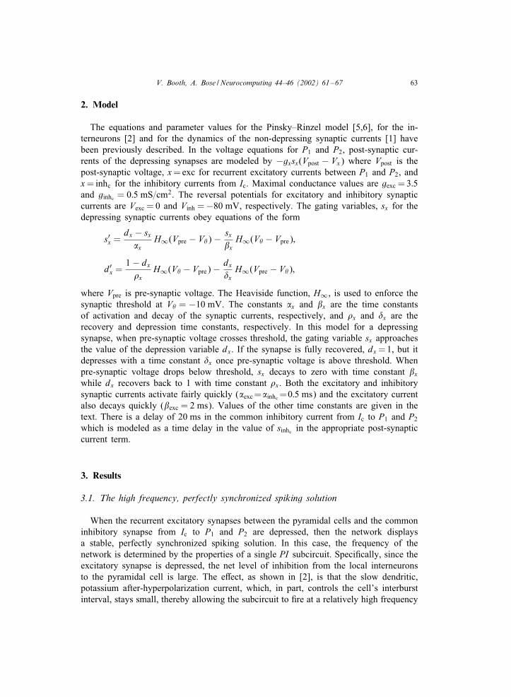
Fig. 1. Schematic of pyramidal cell—interneuron network with depressing synapses.
net level of synaptic inhibition arriving to the cell. In [1], we showed that di erent typesof synchrony patterns exist in these networks and can be modulated as the strength andtiming of synaptic inputs vary. In these papers, we studied the steady-state behavior ofsolutions for di erent but #xed values of the inhibitory and excitatory synaptic maximalconductances. We did not address how networks may transition between these patternsin a dynamic way.In di erent work, we have investigated how synaptic depression can be used to
create a switch between two distinct oscillatory modes [3]. In an ideal network of arepetitively #ring excitatory cell reciprocally connected to an inhibitory cell through adepressing synapse, we showed that there is a fast oscillatory mode whose frequency iscontrolled by the intrinsic properties of the cells in the network, and a slow oscillatorymode whose frequency is controlled by the synaptic properties present between cells.In this paper, we show how bistability of di erent synchronous solutions can arise
in networks of pyramidal cells and interneurons in which some of the synapses aredepressing. We consider a network consisting of two Pinsky–Rinzel pyramidal cells, P1and P2, with recurrent excitatory synapses between them, each reciprocally coupled toseparate “local” interneurons, I1 and I2, and both with reciprocal synaptic connectionsto a “common” interneuron, Ic (Fig. 1). The recurrent excitatory synaptic currents be-tween the pyramidal cells and the slowly decaying, inhibitory current from the commoninterneuron display depression. The two di erent synchronous solutions are (1) a spik-ing mode in which the #ring times of the pyramidal cells are perfectly synchronizedand (2) a “burst-envelope” synchronous mode [1] in which the pyramidal cells burstat the same time, but the burst pro#les are di erent. As in [3], the network frequenciesare di erent in the two modes as are the network elements controlling the frequency.In the spiking mode, frequency is high and is controlled by a combination of theintrinsic properties of the pyramidal cells and the strength of the inhibitory synapticcurrents from the local interneurons, I1 and I2. In the burst-envelope synchronous mode,burst frequency is lower and is controlled by the inhibitory synaptic currents from thecommon interneuron.
V. Booth, A. Bose /Neurocomputing 44–46 (2002) 61–67 63
2. Model
The equations and parameter values for the Pinsky–Rinzel model [5,6], for the in-terneurons [2] and for the dynamics of the non-depressing synaptic currents [1] havebeen previously described. In the voltage equations for P1 and P2, post-synaptic cur-rents of the depressing synapses are modeled by −gxsx(Vpost − Vx) where Vpost is thepost-synaptic voltage, x=exc for recurrent excitatory currents between P1 and P2, andx= inhc for the inhibitory currents from Ic. Maximal conductance values are gexc = 3:5and ginhc = 0:5 mS=cm2. The reversal potentials for excitatory and inhibitory synapticcurrents are Vexc = 0 and Vinh =−80 mV, respectively. The gating variables, sx for thedepressing synaptic currents obey equations of the form
s′x =dx − sxx
H∞(Vpre − V�)− sx xH∞(V� − Vpre);
d′x =1− dx�x
H∞(V� − Vpre)− dx�xH∞(Vpre − V�);
where Vpre is pre-synaptic voltage. The Heaviside function, H∞, is used to enforce thesynaptic threshold at V� = −10 mV. The constants x and x are the time constantsof activation and decay of the synaptic currents, respectively, and �x and �x are therecovery and depression time constants, respectively. In this model for a depressingsynapse, when pre-synaptic voltage crosses threshold, the gating variable sx approachesthe value of the depression variable dx. If the synapse is fully recovered, dx=1, but itdepresses with a time constant �x once pre-synaptic voltage is above threshold. Whenpre-synaptic voltage drops below threshold, sx decays to zero with time constant xwhile dx recovers back to 1 with time constant �x. Both the excitatory and inhibitorysynaptic currents activate fairly quickly (exc=inhc =0:5 ms) and the excitatory currentalso decays quickly ( exc = 2 ms). Values of the other time constants are given in thetext. There is a delay of 20 ms in the common inhibitory current from Ic to P1 and P2which is modeled as a time delay in the value of sinhc in the appropriate post-synapticcurrent term.
3. Results
3.1. The high frequency, perfectly synchronized spiking solution
When the recurrent excitatory synapses between the pyramidal cells and the commoninhibitory synapse from Ic to P1 and P2 are depressed, then the network displaysa stable, perfectly synchronized spiking solution. In this case, the frequency of thenetwork is determined by the properties of a single PI subcircuit. Speci#cally, since theexcitatory synapse is depressed, the net level of inhibition from the local interneuronsto the pyramidal cell is large. The e ect, as shown in [2], is that the slow dendritic,potassium after-hyperpolarization current, which, in part, controls the cell’s interburstinterval, stays small, thereby allowing the subcircuit to #re at a relatively high frequency
64 V. Booth, A. Bose /Neurocomputing 44–46 (2002) 61–67
0 1000 2000 3000
msec
0
0.5
1
d inh c
0
0.5
1
d exc
70
30
10
V1
70
30
10
V2
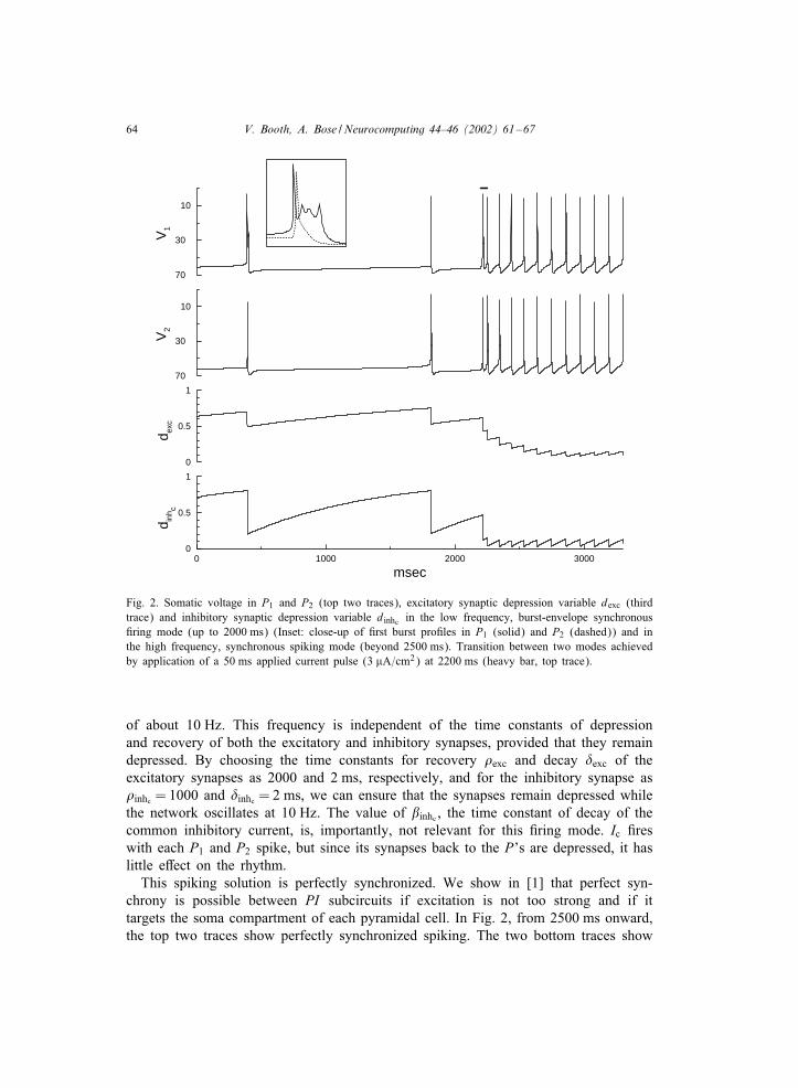
Fig. 2. Somatic voltage in P1 and P2 (top two traces), excitatory synaptic depression variable dexc (thirdtrace) and inhibitory synaptic depression variable dinhc in the low frequency, burst-envelope synchronous#ring mode (up to 2000 ms) (Inset: close-up of #rst burst pro#les in P1 (solid) and P2 (dashed)) and inthe high frequency, synchronous spiking mode (beyond 2500 ms). Transition between two modes achievedby application of a 50 ms applied current pulse (3 �A=cm2) at 2200 ms (heavy bar, top trace).
of about 10 Hz. This frequency is independent of the time constants of depressionand recovery of both the excitatory and inhibitory synapses, provided that they remaindepressed. By choosing the time constants for recovery �exc and decay �exc of theexcitatory synapses as 2000 and 2 ms, respectively, and for the inhibitory synapse as�inhc = 1000 and �inhc = 2 ms, we can ensure that the synapses remain depressed whilethe network oscillates at 10 Hz. The value of inhc , the time constant of decay of thecommon inhibitory current, is, importantly, not relevant for this #ring mode. Ic #reswith each P1 and P2 spike, but since its synapses back to the P’s are depressed, it haslittle e ect on the rhythm.This spiking solution is perfectly synchronized. We show in [1] that perfect syn-
chrony is possible between PI subcircuits if excitation is not too strong and if ittargets the soma compartment of each pyramidal cell. In Fig. 2, from 2500 ms onward,the top two traces show perfectly synchronized spiking. The two bottom traces show

V. Booth, A. Bose /Neurocomputing 44–46 (2002) 61–67 65
the excitatory depression variable (dexc) and the common inhibitory depression variable(dinhc) in a depressed state oscillating between 0 and 0.18.
3.2. The low frequency, burst-envelope synchronous solution
When the depressing synapses in the network are in a non-depressed state then burstfrequency is very low, about 0:6 Hz, and, while the bursts are nearly synchronized, theirpro#les are di erent. Speci#cally, one cell #res a complex burst and the other #res asingle spike (see inset in Fig. 2). In this #ring mode (Fig. 2 up to 2000 ms), the valuesof dexc and dinhc recover to above 0.7 at the time of burst initiation. The interburstinterval is suIciently long for them to recover back to this value by subsequent burst#ring. In contrast to the fast spiking mode, the length of the interburst interval, andthus burst frequency, is primarily controlled by inhc . We have chosen inhc = 5000 msto exaggerate the frequency di erence between the two #ring modes and thus highlightthe dependence on this parameter. The slowly decaying inhibition, which is delayed sothat it only acts during the interburst interval, slows the pyramidal cells’ approach to#ring threshold and thus dominates this low frequency solution.The existence and stability of the burst-envelope synchronous solution is discussed
in [1]. There, we show that although strong recurrent excitation between pyramidalcells causes them to #re at the same or nearly the same time, di erences in each cellin the timing of recurrent excitation and inhibition from the local interneurons allowsthe leading pyramidal cell to #re a complex burst, but causes the follower cell to #rea single spike (as in inset in Fig. 2). When the strength of the recurrent excitation de-creases, with local inhibition remaining strong, the burst pro#le of the leader pyramidalcell changes to bursts with 4, 3 or 2 spikes, and for low values of excitation, bothcells #re single spikes. Thus, the burst-envelope synchronous #ring and the perfectlysynchronized spiking in the two #ring modes are both stable #ring patterns of thisnetwork when the common interneuron is removed and the synapses do not depress.Including depression of the recurrent excitatory synapses between the pyramidal cellsallows the network to intrinsically transfer between these modes and including thecommon interneuron with its depressing synapse creates the bistable switch betweenthe modes.
3.3. Transitions between fast and slow rhythms
The two #ring patterns exist for a common set of parameter values thus implyingbistability of solutions. There are several ways that the network can transition betweenthese two modes. In Fig. 2, the transition from the slow bursting mode to the fastspiking mode is achieved by providing a short depolarizing current pulse to both ofthe pyramidal cells (heavy bar at 2200 ms). This pulse forces the cells to #re at a higherfrequency thereby depressing both the excitatory and common inhibitory synapses. Theswitch between modes could also have been activated by supplying the current pulseto just one of the pyramidal cells, as recurrent excitation would force the other cellto also #re at high frequency. Depolarizing the common interneuron, forcing it to #refaster, would also switch modes, but not as quickly as in Fig. 2.
66 V. Booth, A. Bose /Neurocomputing 44–46 (2002) 61–67
The reverse transition from the fast spiking mode to the slow bursting mode can beachieved by applying a suIciently long, hyperpolarizing pulse to both pyramidal cellsor to the common interneuron. In either case, Ic stops #ring and the inhibitory synapsehas a chance to recover. When the hyperpolarization is removed and Ic #res again, itproduces a strong, long lasting inhibitory current that keeps pyramidal cell frequencylow. We note that hyperpolarizing only one of the pyramidal cells would not activatethis transition since the other pyramidal cell would continue to stimulate Ic and theinhibitory synapse would not be able to recover.
4. Discussion
In this paper, we have shown how various synaptic mechanisms interact with in-trinsic mechanisms of two-compartment pyramidal cells to produce multiple, stablesynchronous #ring patterns. These solutions di er in their frequencies, in the type of#ring pattern and in their degree of synchrony. We have identi#ed the network ele-ments governing the frequency of each mode, and discussed ways to transition betweenthe two modes.These results may have implications for the action of neuromodulators on a network.
We have shown that the high frequency spiking mode is largely independent of thecommon interneuron Ic. Therefore, neuromodulators that target Ic or its synapse coulda ect pyramidal cell #ring without directly acting on the cells. For example, if thenetwork is in the fast mode and the common interneuron is hyperpolarized, pyramidalcell #ring will not change. Indeed, since the inhibitory synapse is depressed, its absencewill only minimally a ect the pyramidal cells. However, once Ic does #re, if its synapsehas suIciently recovered, it will immediately switch the network to the slow burstingmode.Our results also demonstrate how a single network can participate in di erent neural
coding activities. The di erent frequency #ring modes would have di erent e ects ondownstream neurons which react to changes in #ring rate. Moreover, the di erent typesof synchrony could also be utilized by downstream neurons that detect di erences in#ring times. In this way, a single network can have many di erent uses within largernetworks of neurons.
Acknowledgements
The authors were supported in part by a Grant from the National Science Foundation(DMS-9973230).
References
[1] V. Booth, A. Bose, Burst synchrony patterns in hippocampal pyramidal cell model networks. Network:Comput. Neural Systems, 2002, in press.
V. Booth, A. Bose /Neurocomputing 44–46 (2002) 61–67 67
[2] V. Booth, A. Bose, Neural mechanisms for generating rate and temporal codes in model CA3 pyramidalcells, J. Neurophysiol. 85 (2001) 2432–2445.
[3] A. Bose, Y. Manor, F. Nadim, Bistable osciallations arising from synaptic depression, SIAM J. Appl.Math. (2001), in press.
[4] J. O’Keefe, J. Dostrovsky, The hippocampus as a spatial map. Preliminary evidence from unit activityin the freely-moving rat, Brain Res. 34 (1971) 171–175.
[5] P.F. Pinsky, J. Rinzel, Intrinsic and network rhythmogenesis in a reduced Traub model for CA3 neurons,J. Comput. Neurosci. 1 (1994) 39–60.
[6] P.F. Pinsky, J. Rinzel, Erratum, J. Comput. Neurosci. 2 (1995) 275.[7] A. Ylinen, A. Bragin, Z. Nadasy, G. Jando, I. Szabo, A. Sik, G. Buzsaki, Sharp wave-associated
high-frequency oscillation (200 Hz) in the intact hippocampus: network and intracellular mechanism,J. Neurosci. 15 (1995) 30–46.
Amitabha Bose received his PhD from Brown University in Applied Mathematics. He currently is an assistantprofessor in the Department of Mathematical Sciences at the New Jersey Institute of Technology. His researchinterests are in applications of dynamical systems to neuronal networks and nonlinear waves.
Victoria Booth received her PhD in Applied Mathematics from Northwestern University. Following a post-doctoral fellowship at the Mathematical Research Branch at NIH, she joined the Department of MathematicalSciences at the New Jersey Institute of Technology as an assistant professor. Her research interests are inbiophysical modeling of neuronal networks.


![dxi - Imperial College Londonmjfield1/research/networks/sync/... · · 2015-10-10Asimple model for synchronous firing ofbiological oscillators based on Peskin’s ... [33], [48,](https://img.pdfslide.us/doc/110x75/5afa11df7f8b9abd588dcb37/dxi-imperial-college-mjfield1researchnetworkssync2015-10-10asimple-model.jpg)