Embed Size (px)
Citation preview
Transition to A level Biology to help you get ready
Welcome,
We are happy that you have chosen to study A-Level Biology. You have chosen a very desirable subject
for Universities and employers. The subject however, is very demanding and requires dedication. Please
use this booklet to ease your transition into A-Level Biology
The entry requirements for A level Biology :
Grade 6 in Biology and 6 in Chemistry or 66 in Combined Science GCSE.
Grade 6 GCSE maths.
The course you will be following is OCR A level Biology A (H420). The home page is:-
https://ocr.org.uk/qualifications/as-and-a-level/biology-a-h020-h420-from-2015/
Teaching the course:
Ms Birchnall (Head of Biology) [email protected]
Miss Evans (Head of Science) [email protected] k
If you need help, email us

Contents for transition work in A Level Biology
The material contained within this Biology pack is extended GCSE level to help prepare for the A Level Biology
course. Bring your completed work with you to your first lesson in September. (5 maths booklets plus this
booklet)
1. Get Inspired - Read a book and watch a film
2. Recap and Extend- Complete the activities and the questions. Use the suggested websites and the basic recap material to help you on the following topics-
1. Cells and cell division
2. Biological molecules
3. Exchange and Transport
4. Energy
5. DNA and the genetic code
6. Scientific Skills
Bill Bryson is a witty travel writer but has
done some serious research into the way the
body functions. The resulting book is a
fantastic read for anyone interested in the
human body and its workings, it also reveals
how we have learnt about how the body
works – and the stories he tells will make you
laugh and cringe!
Beautiful Boy 2018 (15) is a gripping and
gritty true story about a young man’s addiction to drugs and his father’s dedication to save his son. It undercovers some of the
biological effects of drug use and how the
body works to heal itself.
3. Maths booklets:
OCR tutorial maths booklets split into 5
sections.
Each section contains a tutorial, modelled
responses and practice questions for you to
complete.

Cells and Cell division
The cell is a unifying concept in biology, you will come across it many times during your two years of A level study.
Prokaryotic and eukaryotic cells can be distinguished on the basis of their structure and ultrastructure. In complex
multicellular organisms’ cells are organised into tissues, tissues into organs and organs into systems. During the cell cycle genetic information is copied and passed to daughter cells. Daughter cells formed during mitosis have identical
copies of genes while cells formed during meiosis are not genetically identical
http://www.s-cool.co.uk/a-level/biology/cells-and-organelles
And take a look at these videos:
https://www.youtube.com/watch?v=gcTuQpuJyD8
https://www.youtube.com/watch?v=L0k-enzoeOM
https://www.youtube.com/watch?v=qCLmR9-YY7o
Task- Produce a powerpoint/revision cards/diagram slide on the main structures of Plant cells, animal cells
and Prokaryote cells.
Microscopes;
The Light microscope allows you to
Prokaryotes;
view animal cells. It can magnify up to 1500 times.
Some organelles such as mitochondria, chloroplasts,
vacuoles, cell walls, cell membranes and nuclei are
visible. Staining makes these organelles visible.
Label and annotate the diagram
Eukaryote Animal Cell
The electron microscope; invented in 1950s it allows a
much higher magnification (500 000x) and better
resolution, allowing greater detail to be seen.
Electron microscopes allowed detailed ultrastructure
of the cell to be seen, such as ribosomes and the
inside of mitochondria and chloroplasts. Quick Questions;
Name 3 things visible with a light microscope in both animal
and plant cells Name 4 organelles that both plant and an animal cell have.
What is the calculation used to calculate the magnification of
an object? What is the function of the mitochondria?

Cell structure;
Nuclei: controls the cell function, containing the DNA
which is the coded information for the production of
proteins.
During cell division the chromosomes become shorter
and thicker and can be seen with a light microscope. The
chromosomes will then make a copy of themselves, one
copy for each cell produced during cytokinesis.
Nuclei have a double membrane called the nuclear
envelope. Mitochondria: can be seen with a light microscope, however, greater internal detail can be seen using an electron microscope. The mitochondria’s function is to carry out aerobic respiration. The energy released is used to form molecules of ATP. ATP is used in the cells to provide energy for muscular contractions, active transport as well as anabolic and catabolic reactions. Cell wall: the plant cell wall is made up of cellulose Molecules laid side by side to form microfibrils. These provides rigidity and support for the cell.
Questions; Name 2 molecules that make up the cell membrane. _______________________________________________ Describe the membranes of the mitochondria. _______________________________________________
What is the name of the molecule that provide energy to
the cell? _______________________________________ What term is used to describe water concentration? _______________________________________________
Cell structure;
Cell surface membrane: Found around every cell, it
allows the movement of substances into and out
of the cell. It is a partially permeable membrane
and will prevent certain substances from entering.
It is made up of a double layer called the
PHOSPHOLIPID BILAYER. These are molecules
closely packed together in a mosaic pattern. Within
the bilayer are large proteins which are also
responsible for transport and for cell recognition.
Transport into and out of cells
There are 4 modes of transport you need to be
aware of;
Diffusion; can be gas or liquid particles. They move
from an area of high concentration to an area of low
concentration down a concentration gradient. Small
molecules such as oxygen, water and carbon dioxide
can pass through the phospholipid bilayer.
Osmosis; occurs only with water. The water
particles move from an area of high water
concentration to an area of low water
concentration, down a concentration gradient,
across a partially permeable membrane. NO
ENERGY IS REQUIRED. You will be required to refer
to water potential in AS level not water
concentration.
Facilitated diffusion; Some particles are too large to
fit through the phospholipid bilayer and therefore
require a carrier protein to assist. The protein
carriers are within the bilayer and they change
shape when they come into contact with a specific
molecule (i.e. Glucose). NO ENERGY IS REQUIRED.
Active transport; This moves substances for an area
of low concentration to an area of high
concentration against a concentration gradient.
ENERGY IS NEEDED for this to occur. Specific carrier
proteins are also required these can be called
‘pumps’.

Genetics and cell division;
The DNA molecule contains thousands of genes along its length.
The DNA molecule is wound up into a chromosome. Each body
cell in a human contains 23 pairs of chromosomes (diploid
number), one from mother and one from father. These pair up
forming a homologous pair, both the same size
Meiosis; This cell division is responsible for the production of sex cells and introduces genetic variation. It results in
the formation of gametes containing half the original
genetic information (haploid number). This ensures that
during fertilisation, the embryo obtains two complete
sets of genetic information. In meiosis the cell undergoes 2 cellular divisions.
and containing the same genes (these genes can be different
alleles). A chromosome is often seen as an X shaped
molecule. The X shape is actually one chromosome attached
to an exact copy of itself (2
CHROMATIDS). They are joined
together by an attachment
called a centromere. In
preparation for cell division the
chromosome will make a copy of
itself. All damaged tissue and
cells are replaced by a process of
cell division called MITOSIS.
Mitosis is also seen in asexual
reproduction, the offspring are genetically identical to the parent.
Mitosis cell division;
Interphase; DNA molecules are
indistinct in the nucleus. They
replicate their DNA, attaching
at the centromeres.
Prophase; The DNA becomes supercoiled and compact and can now be seen under a light microscope. It has the X shape.
2) Metaphase I;
The homologous
pairs arrange
themselves
randomly along the
equator.
1) Prophase I; The
chromosomes condense and
the nuclear envelope breaks
down. Homologous
chromosomes pair up and
can cross over DNA
(Recombination).
3) Anaphase I; Spindle fibres
attach to the centromere
and pull the chromosomes
to opposites poles.
4) Telophase I; Two
nuclear envelopes surround
the divided genetic
information. Each contains
half the number of
chromosomes.
5) Prophase 2; New nucleus breaks down and chromosomes coil and condense.
6) Metaphase 2;
Spindle fibres
attach to the
centromeres.
Metaphase; the nuclear
membrane breaks down,
the chromosomes line up
along the equator of the cell
and spindle fibres,
produced by the centrioles,
attach to the chromosomes.
Anaphase; The spindle fibres pull the centromere apart
and the chromatids separate
and are dragged to the poles
of the cell.
Telophase; A nuclear
envelope forms around each set of chromatids and the
cytoplasm divides forming 2
genetically identical cells.
7) Anaphase 2; The spindle fibres start to drag
the chromatids to opposite sides of the cell. 8) Telophase 2; The nuclei’s start to reform and the
cytoplasm spits to form 4 haploid cells that are genetically
different to the parent cells.
Questions; Which cell division forms haploid cells? __________________ What happens during prophase? _______________________ __________________________________________________ What do centrioles do? ______________________________ __________________________________________________ Which organs produce haploid cells? ____________________ __________________________________________________ What happens in Telophase? __________________________
Biological Molecules
Biological molecules are often polymers and are based on a small number of chemical elements. In living organisms
carbohydrates, proteins, lipids, inorganic ions and water all have important roles and functions related to their properties. DNA
determines the structure of proteins, including enzymes. Enzymes catalyse the reactions that determine structures and
functions from cellular to whole-organism level. Enzymes are proteins with a mechanism of action and other properties
determined by their tertiary structure. ATP provides the immediate source of energy for biological processes.
Read the information on these websites (you could make more Cornell notes if you wish): http://www.s-cool.co.uk/a-level/biology/biological-molecules-and-enzymes http://www.bbc.co.uk/education/guides/zb739j6/revision And take a look at these videos: https://www.youtube.com/watch?v=H8WJ2KENlK0 http://ed.ted.com/lessons/activation-energy-kickstarting-chemical-reactions-vance-kite Task:
Krabbe disease occurs when a person doesn’t have a certain enzyme in their body. The disease effects the
nervous system. Write a letter to a sufferer of Krabbe disease to explain what an enzyme is. Your poster should:
Describe the structure of an enzyme
Explain what enzymes do inside the body Exchange and Transport
Organisms need to exchange substances selectively with their environment and this takes place at exchange surfaces.
Factors such as size or metabolic rate affect the requirements of organisms and this gives rise to adaptations such as
specialised exchange surfaces and mass transport systems. Substances are exchanged by passive or active transport
across exchange surfaces. The structure of the plasma membrane enables control of the passage of substances into and
out of cells Read the information on these websites: http://www.s-cool.co.uk/a-level/biology/gas-exchange http://www.s-cool.co.uk/a-level/biology/nutrition-and-digestion/revise-it/human-digestive-system And take a look at these videos: http://ed.ted.com/lessons/insights-into-cell-membranes-via-dish-detergent-ethan-perlstein http://ed.ted.com/lessons/what-do-the-lungs-do-emma-bryce Task:
Create a Venn diagram to either: compare exchange surfaces in mammals and fish or compare exchange surfaces in
the lungs and the intestines. Your poster should:
Describe diffusion, osmosis and active transport
Explain why oxygen and glucose need to be absorbed and waste products
removed. Compare and contrast your chosen focus.

Biological Molecules
Proteins;
Proteins are made of long chains of amino acids, up to several
hundred long. There are only 20 different amino acids and
the combination of these 20 produce a wide range of
complex proteins. Protein structures are held together with
strong bonds called PEPTIDE bonds. The order of the amino
acids determines the structure and how it works.
All amino acids have the same structure with one variation
on the R group. Contains; hydrogen, oxygen, nitrogen and carbon (some contain sulphur).
Protein structure;
The order of the amino acids forms the PRIMARY STRUCTURE.
The protein chain can then coil or fold into pleats which are
held together by weak hydrogen bonds to for the
SECONDARY STRUCTURE.
Enzymes have a further folding held together with stronger
disulphide bonds. This is the TERTIARY STRUCTURE. If the
structure is almost spherical it is called a globular protein.
Enzymes; Help to speed up biochemical reactions.
Metabolism is the sum of all the biochemical reactions
that occur per second and a single chain of these
reactions is called a metabolic pathway. Enzymes are biological catalysts and increase the rate
of reactions. Reactions that release energy need an input energy to start. The input energy is called the ACTIVATION ENERGY. Enzymes reduce the activation
energy.
Enzymes are proteins; enzymes are globular proteins
with a specific order of amino acids that determines
what the enzyme does.
Enzymes can be catabolic (break substrates down) or
anabolic (build substrates up). Enzymes have a specific
site into which the substrates can attach itself, this
attachment site is called the active site. The active site
is complementary to the shape of the substrate. Once
they attach together, they form the enzyme substrate
complex. The substrate then breaks bonds or makes
bonds (depending on the type of enzyme) and the
product leaves the active site. The active site is now
able to accept another substrate.
Denaturing enzymes; Enzymes have a specific tertiary
shape held in place by weak hydrogen bonds and
stronger disulphide bonds. These bonds can be broken
by an increase in temperature (kinetic energy) or a
change in pH (H+ in acid or OH- in alkali disrupt the
bonds).
Useful enzymes; Digestive enzymes are catabolic,
breaking down food into smaller molecules. Enzymes
are also needed in DNA replication, building up
molecules (DNA polymerase). Questions;
What types of bond hold together the secondary structure? ________________ The tertiary structure? ______________________ How many amino acids are there and what elements are found in them? _______________________________________________ Explain why denatured enzymes will not function. __________________________________________________________________ ___________________________________________________________________________________________________________ What is activation energy? ____________________________________________________________________________________
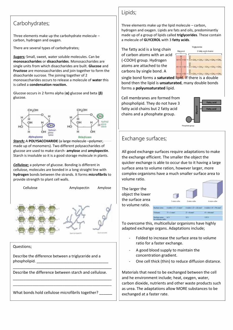
Carbohydrates;
Three elements make up the carbohydrate molecule – carbon, hydrogen and oxygen. There are several types of carbohydrates; Sugars; Small, sweet, water soluble molecules. Can be
monosaccharides or disaccharides. Monosaccharides are
single units from which disaccharides are built. Glucose and
Fructose are monosaccharides and join together to form the
disaccharide sucrose. The joining together of 2
monosaccharides occurs to release a molecule of water this
is called a condensation reaction. Glucose occurs in 2 forms alpha (α) glucose and beta (β) glucose.
Starch; A POLYSACCHARIDE (a large molecule –polymer,
made up of monomers). Two different polysaccharides of
glucose are used to make starch- amylose and amylopectin.
Starch is insoluble so it is a good storage molecule in plants.
Cellulose; a polymer of glucose. Bonding is different in
cellulose, molecules are bonded in a long straight line with
hydrogen bonds between the strands. It forms microfibrils to
provide strength to plant cell walls.
Cellulose Amylopectin Amylose
Questions; Describe the difference between a triglyceride and a
phospholipid. ___________________________________ ______________________________________________ Describe the difference between starch and cellulose. ______________________________________________ ______________________________________________ What bonds hold cellulose microfibrils together? ______
Lipids;
Three elements make up the lipid molecule – carbon,
hydrogen and oxygen. Lipids are fats and oils, predominantly
made up of a group of lipids called triglycerides. These contain
a molecule of GLYCEROL with 3 fatty acids.
The fatty acid is a long chain
of carbon atoms with an acid
(-COOH) group. Hydrogen
atoms are attached to the
carbons by single bond. A single bond forms a saturated lipid. If there is a double
bond then the lipid is unsaturated, many double bonds
forms a polyunsaturated lipid.
Cell membranes are formed from
phospholipid. They do not have 3
fatty acid chains but 2 fatty acid
chains and a phosphate group.
Exchange surfaces;
All good exchange surfaces require adaptations to make
the exchange efficient. The smaller the object the
quicker exchange is able to occur due to it having a large
surface area to volume ration, however larger, more
complex organisms have a much smaller surface area to
volume ratio. The larger the
object the lower
the surface area
to volume ratio.
To overcome this, multicellular organisms have highly
adapted exchange organs. Adaptations include;
- Folded to increase the surface area to volume
ratio for a faster exchange.
- A good blood supply to maintain the
concentration gradient. - One cell thick (thin) to reduce diffusion distance.
Materials that need to be exchanged between the cell
and he environment include; heat, oxygen, water,
carbon dioxide, nutrients and other waste products such
as urea. The adaptations allow MORE substances to be
exchanged at a faster rate.
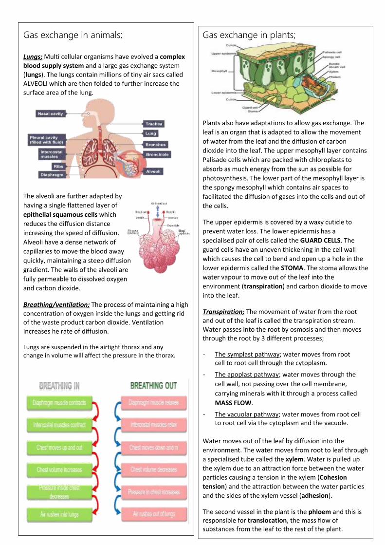
Gas exchange in animals;
Lungs; Multi cellular organisms have evolved a complex
blood supply system and a large gas exchange system
(lungs). The lungs contain millions of tiny air sacs called
ALVEOLI which are then folded to further increase the
surface area of the lung.
The alveoli are further adapted by
having a single flattened layer of
epithelial squamous cells which
reduces the diffusion distance
increasing the speed of diffusion.
Alveoli have a dense network of
capillaries to move the blood away
quickly, maintaining a steep diffusion
gradient. The walls of the alveoli are
fully permeable to dissolved oxygen
and carbon dioxide. Breathing/ventilation; The process of maintaining a high
concentration of oxygen inside the lungs and getting rid
of the waste product carbon dioxide. Ventilation
increases he rate of diffusion. Lungs are suspended in the airtight thorax and any
change in volume will affect the pressure in the thorax.
Gas exchange in plants;
Plants also have adaptations to allow gas exchange. The
leaf is an organ that is adapted to allow the movement
of water from the leaf and the diffusion of carbon
dioxide into the leaf. The upper mesophyll layer contains
Palisade cells which are packed with chloroplasts to
absorb as much energy from the sun as possible for
photosynthesis. The lower part of the mesophyll layer is
the spongy mesophyll which contains air spaces to
facilitated the diffusion of gases into the cells and out of
the cells.
The upper epidermis is covered by a waxy cuticle to
prevent water loss. The lower epidermis has a
specialised pair of cells called the GUARD CELLS. The
guard cells have an uneven thickening in the cell wall
which causes the cell to bend and open up a hole in the
lower epidermis called the STOMA. The stoma allows the
water vapour to move out of the leaf into the
environment (transpiration) and carbon dioxide to move
into the leaf.
Transpiration; The movement of water from the root
and out of the leaf is called the transpiration stream.
Water passes into the root by osmosis and then moves
through the root by 3 different processes;
- The symplast pathway; water moves from root
cell to root cell through the cytoplasm. - The apoplast pathway; water moves through the
cell wall, not passing over the cell membrane,
carrying minerals with it through a process called
MASS FLOW. - The vacuolar pathway; water moves from root cell
to root cell via the cytoplasm and the vacuole.
Water moves out of the leaf by diffusion into the
environment. The water moves from root to leaf through
a specialised tube called the xylem. Water is pulled up
the xylem due to an attraction force between the water
particles causing a tension in the xylem (Cohesion
tension) and the attraction between the water particles
and the sides of the xylem vessel (adhesion).
The second vessel in the plant is the phloem and this is
responsible for translocation, the mass flow of
substances from the leaf to the rest of the plant.

Other exchange surfaces;
Digestion; The human digestive system has 3
main functions;
- Mechanical breakdown of food - Chemical breakdown of food
- Absorption of digested food particles into
the blood stream.
The digestive system contains 3 types of enzyme;
- Carbohydrase enzymes for breaking down
complex carbohydrates into simple
sugars. These are found in the mouth
(amylase enzyme), the pancreas and the
small intestine.
- Protease enzymes break down proteins into
amino acids. These are found in the
stomach (protease enzyme requires a pH 2
which is provided by the hydrochloric acid),
the pancreas and the small intestine.
- Lipase enzymes breaks down lipids into fatty
acids and glycerol. These are found in the
pancreas and the small intestine.
Bile is made in the liver and stored in the gall
bladder.
1. Makes the digested food, leaving the
stomach, slightly alkali for enzymes to
work in.
2. It emulsifies the lipids, breaking them up
into small droplets to increase the
surface area for lipase to digest.
Other exchange surfaces: All of the digested food is now small enough to pass through
the wall of the small intestine into the blood stream.
As an exchange surface it displays the same characteristic
adaptations as the lung; Large surface area to volume ratio,
good blood supply and one cell thick.
Microvilli; the walls of the small intestine are highly folded into
villi, to increase the SA:Vol. ratio. However, this can be increase
further by each individual cell having further folds called
microvilli.
Questions; What are the 3 ways water moves through the root? _________ _____________________________________________________ _____________________________________________________ What are the 3 types of digestive enzymes, what do they break
down and where are they found?
What is the role of Bile? _________________________________ _____________________________________________________ How is the small intestine adapted to increase the rate of
diffusion of digested food products?
_______________________ _____________________________________________________ _____________________________________________________
Questions; What are the features that makes a surface better adapted for exchange?__________________________________________
What is transpiration? ________________________________ What is translocation? ________________________________
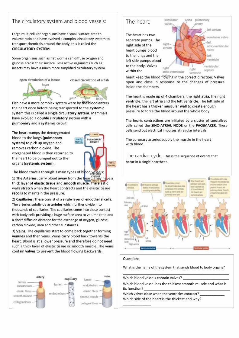
The circulatory system and blood vessels;
Large multicellular organisms have a small surface area to
volume ratio and have evolved a complex circulatory system to
transport chemicals around the body, this is called the CIRCULATORY SYSTEM.
Some organisms such as flat worms can diffuse oxygen and
glucose across their surface. Less active organisms such as
insects may have a much more simplified circulatory system.
Fish have a more complex system were by the blood enters
the heart once before being transported to the systemic
system this is called a single circulatory system. Mammals
have evolved a double circulatory system with a
pulmonary and a systemic circuit.
The heart pumps the deoxygenated
blood to the lungs (pulmonary
system) to pick up oxygen and
removes carbon dioxide. The
oxygenated blood is then returned to
the heart to be pumped out to the
organs (systemic system). The blood travels through 3 main types of blood vessels; 1) The Arteries; carry blood away from the heart. They have a
thick layer of elastic tissue and smooth muscle. The elastic
walls stretch when the heart contracts and the elastic tissue
recoils to maintain the pressure. 2) Capillaries; These consist of a single layer of endothelial cells.
The arteries subdivide arterioles which further divide into
thousands of capillaries. The capillaries come into close contact
with body cells providing a huge surface area to volume ratio and
a short diffusion distance for the exchange of oxygen, glucose,
carbon dioxide, urea and other substances. 3) Veins; The capillaries start to come back together forming
venules and then veins. Veins carry blood back towards the
heart. Blood is at a lower pressure and therefore do not need
such a thick layer of elastic tissue or smooth muscle. The veins
contain valves to prevent the blood flowing backwards.
The heart;
The heart has two
separate pumps. The
right side of the
heart pumps blood
to the lungs and the
left side pumps blood
to the body. Valves
within the
heart keep the blood flowing in the correct direction. Valves
open and close in response to the changes of pressure
inside the chambers.
The heart is made up of 4 chambers; the right atria, the right
ventricle, the left atria and the left ventricle. The left side of
the heart has a thicker muscular wall to create enough
pressure to force the blood around the whole body.
The hearts contractions are initiated by a cluster of specialised
cells called the SINO-ATRIAL NODE or the PACEMAKER. These
cells send out electrical impulses at regular intervals.
The coronary arteries supply the muscle in the heart
with blood.
The cardiac cycle; This is the sequence of events that occur in a single heartbeat.
Questions; What is the name of the system that sends blood to body organs? _____________________________________________________ Which blood vessels contain valves? _______________________ Which blood vessel has the thickest smooth muscle and what is
its function? ___________________________________________ Which valves close when the ventricles contract? _____________ Which side of the heart is the thickest and why?
______________

Energy for Biological Processes
In cellular respiration, glycolysis takes place in the
cytoplasm and the remaining steps in the mitochondria. ATP
synthesis is associated with the electron transfer chain in
the membranes of mitochondria and chloroplasts in
photosynthesis energy is transferred to ATP in the light-
dependent stage and the ATP is utilised during synthesis in
the light-independent stage. Read the information on these websites: http://www.bbc.co.uk/education/guides/zcxrd2p/revision http://www.s-cool.co.uk/a-level/biology/respiration And take a look at these videos: https://www.youtube.com/watch?v=00jbG_cfGuQ https://www.youtube.com/watch?v=2f7YwCtHcgk Task:
Produce an A3 annotated information poster that
illustrates the process of cellular respiration and
summarises the key points. Your poster should include: Both text and images Be visually stimulating Key words and definitions Clearly labelled diagrams Short explanations of key ideas or processes.
Respiration;
Aerobic respiration; This occurs in the mitochondria of cells. It
requires a number of small stages to break down glucose
(C6H12O6) to release a large amount of energy; adenosine
triphosphate (ATP). The first stage is a stage called
GLYCOLYSIS, this occurs in the cytoplasm and converts glucose
into two 3 carbon molecules called PYRUVATE. Pyruvate is
formed in both aerobic and anaerobic respiration, however in
aerobic respiration the pyruvate passes into the matrix of the
Mitochondria. Pyruvate then goes into the link reaction to
form acetyl CoA which then passes into to the Kreb cycle with
the oxidise products passing into oxidative phosphorylation to
form ATP and waste products carbon dioxide and water.
Anaerobic respiration; Respiration without oxygen. This form of respiration occurs without oxygen. Glucose is
converted into pyruvate, through the process of GLYCOLYSIS,
in the cytoplasm and is unable to pass into the mitochondria.
The process of glycolysis releases small amounts of energy and
over a short period of time it can keep the muscles working.
Anaerobic respiration in plants and yeast forms carbon dioxide
and alcohol. Anaerobic respiration in animals forms lactic acid.
The build-up of lactic acid in muscles must be broken down as
the formation of the acid alters the pH and affects enzymes in
the cells, slowing down reactions. As the lactate ions build up
in the muscles this causes pain called fatigue. The oxygen
required to convert the lactate ions back to pyruvate is called
the oxygen debt.
1. What is the name of the stage of respiration that is common to aerobic and anaerobic respiration? _____________________
2. What are the products of the first stage of respiration? ____________________________________________________
3. Name the remaining 3 stages of aerobic respiration. _____________________________________________________________________________________
4. Name the molecule that is produced and will supply energy to other parts of the body. ______________________________
5. Define the term anaerobic respiration- _____________________________________________________________________
6. Write a word equation for; a) anaerobic respiration in plants and yeast- ____________________________________________________ b) anaerobic respiration in mammals ____________________________________________________
7. What is the oxygen debt? ___________________________ 8. Why can a person not anaerobically respire for a long time? ____________________________________
DNA and the Genetic Code
In living organisms nucleic acids (DNA and RNA have important roles and functions related to their properties.
The sequence of bases in the DNA molecule determines the structure of proteins, including enzymes.
The double helix and its four bases store the information that is passed from generation to generation. The sequence of
the base pairs adenine, thymine, cytosine and guanine tell ribosomes in the cytoplasm how to construct amino acids into
polypeptides and produce every characteristic we see. DNA can mutate leading to diseases including cancer and
sometimes anomalies in the genetic code are passed from parents to babies in disease such as cystic fibrosis, or can be
developed in unborn foetuses such as Downs Syndrome. Read the information on these websites: http://www.s-cool.co.uk/a-level/biology/dna-and-genetic-code https://www.wisc-online.com/learn/general-education/anatomy-and-physiology1/ap1302/protein-synthesis And take a look at these videos: http://ed.ted.com/lessons/the-twisting-tale-of-dna-judith-hauck http://ed.ted.com/lessons/where-do-genes-come-from-carl-zimmer Task:
Produce a wall display. You might make a poster or do this using PowerPoint or similar. Your display should use
images, keywords and simple explanations to: Define gene, chromosome, DNA and base pair Describe the structure and function of DNA and RNA
Explain how DNA is copied in the body - Protein Synthesis done at TRIPLE HIGHER not covered in Combined
Outline some of the problems that occur with DNA replication and what the consequences of this might be.

DNA and protein synthesis;
DNA is a complex chemical, found in the nucleus of eukaryotes
and in the cytoplasm of prokaryotes. DNA is made up of; pentose
sugar, phosphate and nitrogenous bases forming a NUCLEOTIDE. There
are 4 different
nitrogenous bases; A= Adenine T=
Thymine C=
Cytosine G=
Guanine
Complementary
pair; A pairs with T
C pairs with G The bases pair up in
the formation stated above. They are held together by hydrogen bonds. The two
strands run in opposite directions causing the molecule to
spiral forming a DOUBLE HELIX. DNA controls the production of proteins. A section of DNA that
codes for a protein is called a gene. Proteins are made up of a
string of amino acids, each protein has a different number and
order of amino acids. The proteins also have different bonds
which holds the molecule in a unique shape which means all
proteins have a different function.
Protein synthesis; Protein synthesis occurs in the cytoplasm,
carried out by RIBOSOMES. When a protein is required then the
gene has to be copied producing a molecule called
messengerRNA (mRNA). mRNA is small enough to pass out of the
nucleus into the cytoplasm. mRNA is a template, containing
nucleotides and bases. The nucleotide on the mRNA will line up
with the complementary base. However, on RNA there is no
Thymine, RNA will have the base URACIL (U).
The mRNA passes
out of the nucleus carrying the code 3 for a protein. Once in
the cytoplasm the
mRNA binds to a
ribosome. Within the cytoplasm there is another molecule called transferRNA (tRNA). At one end, the anticodon is complementary to the mRNA. At the opposite end there are three unpaired bases which code for an amino acid. The amino acid is brought in to form a peptide bond with the amino acids brought in by the previous tRNA. This forms a polypeptide chain which will form hydrogen and disulfide bonds to form the unique protein.
Mutations: Mutations change the order of bases in the DNA.
Some bases may change to a different base (substitution),
some bases may be deleted and some bases may be added.
Mutations can cause the following; - Incorrect protein to be produces - No change in protein being made - Causes a harmful proteins/ no protein to be made
Questions; What are the components of a nucleotide. ________________ __________________________________________________ What are the names of the 4 nitrogenous bases? __________________________________________________ What type of bonds hold the 2 strands together? __________ What is the name of a section of DNA that codes for a
protein? ___________________________________________ What are proteins made from? _________________________ DNA is too big to leave the nucleus, what is the copy of the
gene called that enters the cytoplasm? __________________ What organelle will this molecule attach to? ______________ Which molecule has a complementary anticodon and brings in
the correct amino acid? __________________________

Challenge-
If you are aiming for the heights you could have a look at some of the following-
Looking forward to seeing you all in September-
Be safe! Ms Birchnall
Virology & • Explained: The Next Global Pandemic (20 mins) Global Health https://www.netflix.com/watch/81062202?trackId=13752289&tctx=0%2C3%2C0d0
3e68c-6321-41f2-9dfa-11f336ddc8ca-52560540%2C%2C
• FutureLearn course on Coronavirus: https://www.futurelearn.com/courses/covid19-novel-coronavirus
• The Life Scientific - viruses: https://www.bbc.co.uk/programmes/m0009b2t
Natural • Can Science Make Me Perfect? Selection & https://www.bbc.co.uk/iplayer/episode/b0b6q3qy/can-science-make-me-perfect-
Genetic with-alice-roberts
Modification • Explained: Designer DNA (20 mins) https://www.netflix.com/search?q=science&jbv=80216752&jbp=2&jbr=1
• Unnatural Selection (short series) https://www.netflix.com/watch/80208833?trackId=13752289&tctx=0%2C0%2C8bd
41505-055d-4d08-a8c9-e71150318bb2-44683054%2C%2C
• In Our Time: Neanderthals https://www.bbc.co.uk/programmes/b00sq1nv
• The Life Scientific: evolution of cancer https://www.bbc.co.uk/programmes/m0003ks6
Homeostasis • Interviews with researchers working on hormones: & Hormones https://endocrinepod.com/episodes/
• Open University course on diabetes: https://www.open.edu/openlearn/science-maths-technology/biology/living-
diabetes/content-section-3.1