Embed Size (px)
Citation preview

1521-0111/87/4/617–628$25.00 http://dx.doi.org/10.1124/mol.114.095695MOLECULAR PHARMACOLOGY Mol Pharmacol 87:617–628, April 2015Copyright ª 2015 by The American Society for Pharmacology and Experimental Therapeutics
Transient Receptor Potential Melastatin-3 (TRPM3)–InducedActivation of AP-1 Requires Ca21 Ions and the TranscriptionFactors c-Jun, ATF2, and Ternary Complex Factor s
Andrea Lesch, Xin Hui, Peter Lipp, and Gerald ThielDepartment of Medical Biochemistry and Molecular Biology (A.L., G.T.) and Department of Anatomy and Cell Biology, Universityof Saarland Medical Faculty, Homburg, Germany (X.H., P.L.)
Received September 2, 2014; accepted January 9, 2015
ABSTRACTThe steroid pregnenolone sulfate activates the transcription fac-tor activator protein-1 (AP-1) via stimulation of transient receptorpotential melastatin-3 (TRPM3) channels. Here, we show thatthe signaling pathway requires an influx of Ca21 ions into thecells and a rise in the intracellular Ca21 levels. The upregulationof AP-1 was attenuated in cells that overexpressed mitogenactivated protein kinase phosphatase–1, indicating that Ca21
ions prolong the signaling cascade via activation of mitogenactivated protein kinases. On the transcriptional level, expres-sion of a dominant-negative mutant of the basic region leucinezipper protein c-Jun, a major constituent of the AP-1 transcrip-tion factor complex, or expression of a c-Jun–specific shorthairpin RNA attenuated pregnenolone sulfate–induced AP-1activation. In addition, stimulation of TRPM3 channels increased
the transcriptional activation potential of the basic region leucinezipper protein ATF2. Inhibition of ATF2 target gene expressionvia expression of a dominant-negative mutant of ATF2 orexpression of an ATF2-specific short hairpin RNA interfered withTRPM3-mediated stimulation of AP-1. Moreover, we show thata dominant-negative mutant of the ternary complex factor (TCF)Elk-1 attenuated the upregulation of AP-1 following stimulationof TRPM3 channels. Thus, c-Jun, ATF2, and TCFs are requiredto connect the intracellular signaling cascade elicited by ac-tivation of TRPM3 channels with enhanced transcription ofAP-1–regulated genes. We conclude that pregnenolone sulfate–induced TRPM3 channel activation changes the gene expressionpattern of the cells by activating transcription of c-Jun-, ATF2-,and TCF-controlled genes.
IntroductionTransient receptor potential melastatin-3 (TRPM3) is a
typical transient receptor potential channel containing sixtransmembrane domains, a pore domain between the fifthand sixth transmembrane domains, and a cytosolic location ofboth the amino-terminus and the carboxy-terminus (Thiel et al.,2013). TRPM3 channels are expressed in various tissues, in-cluding the kidney, liver, ovary, brain, spinal cord, pituitarygland, vascular smoothmuscle, testis, and b-cells of the pancreas(Grimm et al., 2003; Lee et al., 2003; Oberwinkler et al., 2005;Wagner et al., 2008; Naylor et al., 2010).Several metabolites were described to function as activators
of TRPM3 channels, including the steroid pregnenolone sul-fate, lipid D-erythro-sphingosine, and L-type voltage-gatedCa21 channel inhibitor nifedipine (Grimm et al., 2003; Wagneret al., 2008; Naylor et al., 2010; Islam, 2011). A comprehensiveanalysis of different compounds revealed that pregnenolonesulfate is currently themost powerful known activator of TRPM3(Lesch et al., 2014). However, the concentration of pregnenolone
sulfate required to stimulate TRPM3 channels is in the mic-romolar range, suggesting that pregnenolone sulfate is not aphysiologic agonist of TRPM3 and may have only pharmaco-logical relevance.An analysis of TRPM3-deficient mice revealed that TRPM3
is a nociceptor channel involved in the detection of noxiousheat in the somatosensory system (Vriens et al., 2011). Themutant mice exhibited deficiencies in acute heat sensing andinflammatory heat hyperalgesia. It has also been proposedthat TRPM3 functions as an ionotropic steroid receptor inpancreatic b-cells responsible for modulating insulin bio-synthesis and secretion (Wagner et al., 2008). However,TRPM3-deficient mice did not show alterations in restingblood glucose levels (Vriens et al., 2011). Thus, TRPM3 mayplay no or only amarginal role in controlling insulin secretion.TRPM3 activation has frequently been monitored using
either intracellular Ca21 measurement with Ca21 indicatorsand/or a whole-cell patch clamp to measure the cationicmembrane current (Wagner et al., 2008; Majeed et al., 2010).Thus, the influx of Ca21 ions into the cells and the subsequentrise in intracellular Ca21 concentration was used as anindication for an activated TRPM3 channel. Recent data fromour laboratory showed that pregnenolone sulfate–inducedstimulation of endogenous TRPM3 channels in insulinoma
This work was supported by the University of Saarland.dx.doi.org/10.1124/mol.114.095695.s This article has supplemental material available at molpharm.aspetjournals.
org.
ABBREVIATIONS: bZIP, basic region leucine zipper; EGFP, enhanced green fluorescent protein; ERK, extracellular signal-regulated protein kinase;JNK, c-Jun N-terminal protein kinase; PKC, protein kinase C; shRNA, short hairpin RNA; SRE, serum response element; SRF, serum responsefactor; TCF, ternary complex factor; TRE, 12-O-tetradecanoylphorbol-13-acetate–responsive element.
617
http://molpharm.aspetjournals.org/content/suppl/2015/01/09/mol.114.095695.DC1Supplemental material to this article can be found at:
at ASPE
T Journals on A
pril 26, 2019m
olpharm.aspetjournals.org
Dow
nloaded from

cells activates a signal cascade that leads to an upregulationof gene transcription (Mayer et al., 2011; Müller et al., 2011).Pregnenolone sulfate also activates gene transcription inHEK293 engineered to express TRPM3 channels (Lesch et al.,2014).Recently, we showed that stimulation of TRPM3 channels
with pregnenolone sulfate upregulates activator protein-1(AP-1)–regulated gene transcription in insulinoma cells (Mülleret al., 2011). Moreover, TRPM3 stimulation enhances expres-sion of c-Fos and c-Jun, frequent constituents of the AP-1transcriptional complex, in this cell type as well as in pancreaticislets in a primary short-term culture (Müller et al., 2011). Here,we analyzed the signaling pathway leading to increasedtranscription of an AP-1–responsive reporter gene followingstimulation of TRPM3 channels. Originally, AP-1 was de-scribed as a heterodimer of c-Jun and c-Fos (Chiu et al., 1988;Curran and Franza, 1988). These basic region leucine zipper(bZIP) transcription factors dimerize via their leucine zipperdomains, which in turn bring together their basic domains tobind DNA in a sequence-specific manner. The current view isthat AP-1 is actually a group of several distinct homodimersor heterodimers composed of various members of the Fos, Jun,and ATF bZIP subfamilies. AP-1 functions in cells as a con-vergence point for many intracellular signaling cascades ini-tiated by cytokines, growth factors, hormones, and stressors,such as UV light. The biologic functions of AP-1 are tissuespecific and encompass the regulation of proliferation, transfor-mation, differentiation, and programmed cell death (Shaulianand Karin, 2002). The results of this study show that TRPM3-mediated activation of AP-1 requires the influx of Ca21 ionsinto the cells and arise in the intracellular Ca21 concentration.The activation of mitogen activated protein (MAP) kinases con-nects the TRPM3-triggered cytosolic signaling cascade withchanges of the gene expression pattern. In the nucleus, thebZIP proteins c-Jun and ATF2 and ternary complex factors(TCFs) are essential for TRPM3-induced upregulation of AP-1controlled gene transcription. Thus, these data connect TRPM3signaling with Ca21-regulated transcription of c-Jun-, ATF2-,and TCF-regulated genes.
Materials and MethodsCell Culture. HEK293 cells containing the human TRPM3 coding
region under the control of a tetracycline-regulated promoter werekindly provided by David Beech and Yasser Majeed, University ofLeeds, UK, and cultured as described (Naylor et al., 2008). TRPM3expression was induced by adding tetracycline (1 mg/ml, dissolved inwater; Sigma-Aldrich, St. Louis, MO) to the culture medium con-taining 0.05% fetal bovine serum for 24 hours prior to stimulationwith pregnenolone sulfate. Stimulation with pregnenolone sulfate(20 mM, dissolved in dimethylsulfoxide; Sigma-Aldrich) was perfor-med for 24 hours in Dulbecco’s modified Eagle’s medium containing0.05% fetal bovine serum.
Lentiviral Gene Transfer. The lentiviral transfer vectors pFUW-REST/Elk-1DC, pFUW-MEKK1D, pFUW-MKK6E, pFUWc-JunDN,pFUW-ATF2DN, and pFUW–MKP-1 have been described previously(Mayer et al., 2008, 2009; Mayer and Thiel, 2009; Rössler and Thiel,2009; Müller et al., 2010; Spohn et al., 2010; Thiel et al., 2012). TheGAL4 expression plasmid pFA2ATF2 was purchased from Stratagene(Amsterdam, The Netherlands). To generate a lentiviral vectorencoding GAL4-ATF2, we digested plasmid pFA2ATF2 with ClaI,filled inwith theKlenow fragment of DNA polymerase I, and recut withEcl136II. The fragment was cloned into the HpaI site of the lentiviraltransfer vector pFUW, thus generating the plasmid pFUW-GAL4-
ATF2. The viral particles were produced as previously described (Keimet al., 2012a). In brief, viral particles were produced by transienttransfection of 2.0 � 106 293T/17 cells using the calcium phosphatecoprecipitation technique. Three plasmids were transfected into thecells, which were plated on 60-mm plates: 6.6 mg of the transfer vector,5 mg of the pCMVDR8.91 packaging vector, and 2.3 mg of the plasmidpCMVG, encoding the vesicular stomatitis virus glycoprotein. Trans-fections were performed in the presence of 25 mM chloroquine. Viralsupernatants were harvested 60 hours after transfection, filteredthrough a 0.45-mm filter, and used to infect HEK293 cells in thepresence of 8 mg/ml polybrene at 37°C.
Lentiviral Expression of Short Hairpin RNAs. The lentiviralvector pLentiLox3.7 (pLL3.7) was purchased from American TypeCulture Collection (Manassas, VA). The lentiviral transfer vectorspLLTRPM3, used to knock down TRPM3 expression, and pLL3.7c-Jun, used to express a c-Jun–specific short hairpin RNA (shRNA)under the control of the U6 promoter, have been described (Mayeret al., 2011, Keim et al., 2012b). A lentiviral transfer vector (pLL3.7ATF2)expressing an ATF2-specific shRNA was designed as described (http://mcmanuslab.ucsf.edu/protocols/ll37stemloop_design.pdf) based on apublished sequence (Bhoumik et al., 2002).
Reporter Assays. The lentiviral transfer vectors pFWColl.luc,pFWColl.lucDTRE, and pFWUAS5Sp12luc have been described else-where (Rössler et al., 2008; Müller et al., 2010; Ekici et al., 2012).Cell extracts were prepared using reporter lysis buffer (Promega,Mannheim, Germany) and analyzed for luciferase activities as described(Thiel et al., 2000). Luciferase activity was normalized to the proteinconcentration.
Microscopy. Phase contrast and green fluorescent protein fluo-rescence microscopy was done with a Zeiss Axiovert 200Mmicroscopeequipped with the respective filter (Zeiss, Göttingen, Germany).
Ca21 Transient Acquisition and Analysis. For ratiometricFura-2–based Ca21 measurements, 293 cells containing a tetracycline-regulated TRPM3 expression unit were treated with or without tetra-cycline (1 mg/ml) 24 hours prior to the experiments. After loading withFura-2/AM and/or BAPTA-AM (both from Life Technologies, Darm-stadt, Germany), cells were bathed in Tyrode’s solution (135mMNaCl,5.4 mM KCl, 1.8 mM CaCl2, 10 mM glucose, 2 mM MgCl2, and10HEPES adjusted to pH 7.35), mounted on a microscope (uiMic; TILLPhotonics GmbH, Gräfelfing, Germany), and imaged through a 20� oil-immersion objective (UPLSAPO 20� oil 0.85; Olympus, Tokyo, Japan).Excitation was performed at alternating wavelengths of 340/380 nm(15-nm full width at half maximum with PolyChrome V; TILLPhotonics, Munich, Germany), while the emission (.510 nm) was imagedby a charge-coupled device camera (Retiga-2000R; Qimaging, Surrey,Canada) with 2 frames per second (image size: 400 � 300 pixels). Preg-nenolone sulfate was administered by a local solenoid-controlled gravity-driven perfusion system. Where mentioned, Ca21 free extracellularTyrode was generated by omitting additional Ca21 and adding 5 mMEGTA. The acquired images were analyzed using custom written mac-ros in ImageJ (W. Rasband, National Institutes of Health, Bethesda,MD). Images were background corrected, and region-of-interest fluores-cence over time data were collected and further processed into Fura-2ratios in Igor (Wavemetrics, LakeOswego, OR), whichwas also employedfor measuring the amplitude of the resulting Ca21 transients. The finalstatistical analysis was undertaken in Prism 6 software (GraphPadSoftware, La Jolla, CA).
Western Blots. Whole cell and nuclear extracts were prepared asdescribed (Kaufmann and Thiel, 2002). Proteins were separated bySDS-PAGE, blotted, and incubated with antibodies directed againstc-Jun (Santa Cruz, Heidelberg, Germany) or ATF2 (Santa Cruz). Theantibody directed against histone deacetylase-1 was used as a loadingcontrol as previously described (Spohn et al., 2010; Mayer et al., 2011).To detect FLAG-tagged proteins, we used the M2 monoclonal anti-body directed against the FLAG epitope (Sigma-Aldrich, Steinheim,Germany) at 1:3000 dilution. Immunoreactive bands were detectedvia enhanced chemiluminescence as described (Spohn et al., 2010;
618 Lesch et al.
at ASPE
T Journals on A
pril 26, 2019m
olpharm.aspetjournals.org
Dow
nloaded from
Mayer et al., 2011). Values are expressed as the mean 6 S.D. fromthree independent experiments (n 5 3).
Statistics. Statistical analysis was done by using the two-tailedStudent’s t test. Data shown are mean 6 S.D. from two to fourindependent experiments performed in quadruplicate. Statisticalprobability is expressed as *P , 0.05, **P , 0.01, and ***P , 0.001.Values were considered significant when P , 0.05.
ResultsRecently, we showed that stimulation of TRPM3 channels
with pregnenolone sulfate induces a signal cascade in in-sulinoma cells that leads to an activation of gene transcription(Mayer et al., 2011; Müller et al., 2011). Insulinoma cells alsoexpress L-type voltage-gated Ca21 channels that are, inaddition to TRPM3, involved in the regulation of pregneno-lone sulfate–induced gene expression (Mayer et al., 2011). Incontrast, in HEK293 cells engineered to express TRPM3channels, gene transcription is activated following stimula-tion of TRPM3 stimulation with pregnenolone sulfate, in-dependently of L-type voltage-gated Ca21 channels (Leschet al., 2014). Thus, TRPM3 functions in this heterologoussystem in the absence of L-type voltage-gated Ca21 channelsas a ligand-activated ionotropic receptor, leading to an influxof Ca21 into the cells following activation (Wagner et al., 2008;Majeed et al., 2010). To elucidate the signaling pathway con-necting TRPM3 stimulation with enhanced gene transcrip-tion of AP-1 responsive genes, we used this engineeredHEK293 cell line, in which expression of TRPM3 is inducedby adding tetracycline to the culture medium, as shownpreviously (Lesch et al., 2014). We thus avoided interferencebetween TRPM3 and L-type voltage-gated Ca21 channelsignaling.Pregnenolone Sulfate Triggers an Upregulation of
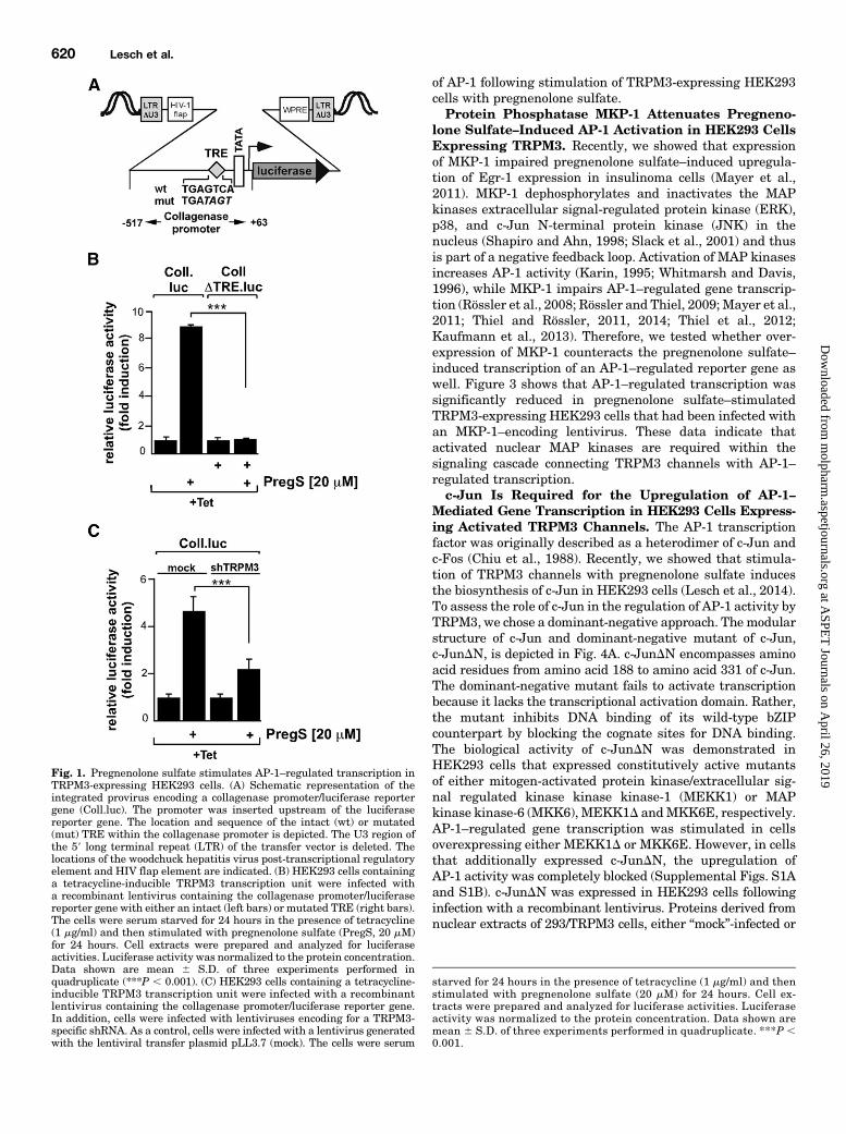
AP-1–Mediated Gene Transcription in HEK293 CellsExpressing TRPM3. To measure AP-1–regulated tran-scription, we used a collagenase promoter/luciferase reportergene. The collagenase promoter contains an AP-1 bindingsite in the proximal promoter region, also known as 12-O-tetradecanoylphorbol-13-acetate–responsive element (TRE).We and others have frequently used this promoter to monitorAP-1 activity (Angel et al., 1987; Steinmüller et al., 2001;Vries et al., 2001; Müller et al., 2010, 2011; Thiel and Rössler,2011, 2014; Thiel et al., 2012; Kaufmann et al., 2013). We usedlentiviral gene transfer to integrate the collagenase promoter/luciferase reporter gene into the genome of the cells. By thismeans, we were sure that the reporter gene was embeddedinto a nucleosomal context. In contrast, transient transfectionsof plasmids may result in incompletely organized reportergenes in comparison with cellular chromatin, having a pro-karyotic gene organization including a nonrestrictive transcrip-tional ground state. A schematic depiction of the integratedprovirus encoding the collagenase promoter/luciferase reportergene is depicted in Fig. 1A, including the sequence of the wild-type andmutated TRE. HEK293 cells containing a tetracycline-responsive TRPM3 expression cassette were infected witha lentivirus containing the collagenase promoter/luciferasereporter gene. Cells were treated with tetracycline to induceTRPM3 expression. Cells were serum starved for 24 hours andstimulated with pregnenolone sulfate for 24 hours. Figure 1B(left bars) shows that pregnenolone sulfate stimulation ofHEK293 cells expressing TRPM3 induced an upregulation of
reporter gene transcription. The AP-1 binding site within thecollagenase promoter encompasses the sequence 59-TGAGTCA-39. Mutation of the TRE to 59-TGATAGT-39 abolished the effectof TRPM3 activation upon AP-1–regulated gene transcription(Fig. 1B, right bars). This experiment indicates that TREfunctions as a pregnenolone sulfate–responsive element inHEK293 cells. Figure 1C shows that pregnenolone sulfate–stimulated collagenase promoter/luciferase reporter gene tran-scription was attenuated in cells expressing shRNAs directedagainst TRPM3, indicating that TRPM3 is required for stimulus-transcription coupling. This experiment supplements pre-viously published data that showed that the TRPM3 inhibitormefenamic acid blocked pregnenolone sulfate–induced AP-1activation in TRPM3-expressing HEK293 cells (Lesch et al.,2014).Essential Role of Extracellular and Intracellular
Ca21 Ions in Pregnenolone Sulfate–Induced Stimula-tion of AP-1 in HEK293 Cells Expressing TRPM3Channels. TRPM3 channels function as Ca21 permeablecation channels. The influx of Ca21 ions into the cells and thesubsequent rise in intracellular Ca21 concentration has beenfrequently used as an indication for an activated TRPM3channel. We confirmed that stimulation of HEK293 cellsexpressing TRPM3 with pregnenolone sulfate increasedintracellular Ca21 concentration. Cells with induced TRPM3expression (Tet1) or naïve cells (Tet2) were challenged withpregnenolone sulfate to activate TRPM3-mediated Ca21
influx. Figure 2A shows exemplified typical Fura-2 ratiotraces. Figure 2B depicts the statistical comparison (black andgray color, respectively). In TRPM3-expressing HEK293 cells,pregnenolone sulfate resulted in a substantially increasedCa21 response (black traces and black/gray bars). Preincuba-tion of the cells with BAPTA-AM to increase the intracellularCa21 buffer capacity significantly decreased the response topregnenolone sulfate in TRPM3-expressing cells and totallyabrogated the response in naïve cells (red traces in Fig. 2Aand red bars in Fig. 2B for statistical comparison). We founda similar behavior when diminishing extracellular Ca21 byaddition of EGTA (green traces in Fig. 2A and green bars inFig. 2B for statistical comparison).Recently, we showed that stimulation of Egr-1 expression
via TRPM3 channels in pregnenolone sulfate–treated insuli-noma cells required a rise in intracellular Ca21 (Mayer et al.,2011). Here, we assessed the role of extracellular and in-tracellular Ca21 ions in pregnenolone sulfate–induced acti-vation of AP-1–regulated gene transcription in HEK293 cellsexpressing a tetracycline-controlled TRPM3 expression unit.First, we tested whether extracellular Ca21 is required toupregulate AP-1 in pregnenolone sulfate–stimulated HEK293cells expressing TRPM3. Figure 2C shows that the activationof AP-1 following pregnenolone sulfate stimulation wascompletely blocked when the cells were cultured in a Ca21
-free medium containing EGTA. Thus, an influx of Ca21 ionsinto the cells is essential to connect pregnenolone sulfate stim-ulation with enhanced AP-1 activity. Second, we tested therequirement of elevated intracellular Ca21 levels for TRPM3-induced activation of AP-1–regulated gene transcription. Thepregnenolone sulfate–induced elevation of [Ca21]i was atten-uated by preincubation with BAPTA-AM. As a result, theupregulation of AP-1 was significantly reduced (Fig. 2D).Hence, an influx of Ca21 ions into the cells via TRPM3 and thesubsequent elevation of [Ca21]i is essential for the activation
TRPM3-Triggered AP-1 Activation 619
at ASPE
T Journals on A
pril 26, 2019m
olpharm.aspetjournals.org
Dow
nloaded from
of AP-1 following stimulation of TRPM3-expressing HEK293cells with pregnenolone sulfate.Protein Phosphatase MKP-1 Attenuates Pregneno-
lone Sulfate–Induced AP-1 Activation in HEK293 CellsExpressing TRPM3. Recently, we showed that expressionof MKP-1 impaired pregnenolone sulfate–induced upregula-tion of Egr-1 expression in insulinoma cells (Mayer et al.,2011). MKP-1 dephosphorylates and inactivates the MAPkinases extracellular signal-regulated protein kinase (ERK),p38, and c-Jun N-terminal protein kinase (JNK) in thenucleus (Shapiro and Ahn, 1998; Slack et al., 2001) and thusis part of a negative feedback loop. Activation of MAP kinasesincreases AP-1 activity (Karin, 1995; Whitmarsh and Davis,1996), while MKP-1 impairs AP-1–regulated gene transcrip-tion (Rössler et al., 2008; Rössler and Thiel, 2009;Mayer et al.,2011; Thiel and Rössler, 2011, 2014; Thiel et al., 2012;Kaufmann et al., 2013). Therefore, we tested whether over-expression of MKP-1 counteracts the pregnenolone sulfate–induced transcription of an AP-1–regulated reporter gene aswell. Figure 3 shows that AP-1–regulated transcription wassignificantly reduced in pregnenolone sulfate–stimulatedTRPM3-expressing HEK293 cells that had been infected withan MKP-1–encoding lentivirus. These data indicate thatactivated nuclear MAP kinases are required within thesignaling cascade connecting TRPM3 channels with AP-1–regulated transcription.c-Jun Is Required for the Upregulation of AP-1–
Mediated Gene Transcription in HEK293 Cells Express-ing Activated TRPM3 Channels. The AP-1 transcriptionfactor was originally described as a heterodimer of c-Jun andc-Fos (Chiu et al., 1988). Recently, we showed that stimula-tion of TRPM3 channels with pregnenolone sulfate inducesthe biosynthesis of c-Jun in HEK293 cells (Lesch et al., 2014).To assess the role of c-Jun in the regulation of AP-1 activity byTRPM3, we chose a dominant-negative approach. The modularstructure of c-Jun and dominant-negative mutant of c-Jun,c-JunDN, is depicted in Fig. 4A. c-JunDN encompasses aminoacid residues from amino acid 188 to amino acid 331 of c-Jun.The dominant-negative mutant fails to activate transcriptionbecause it lacks the transcriptional activation domain. Rather,the mutant inhibits DNA binding of its wild-type bZIPcounterpart by blocking the cognate sites for DNA binding.The biological activity of c-JunDN was demonstrated inHEK293 cells that expressed constitutively active mutantsof either mitogen-activated protein kinase/extracellular sig-nal regulated kinase kinase kinase-1 (MEKK1) or MAPkinase kinase-6 (MKK6), MEKK1D andMKK6E, respectively.AP-1–regulated gene transcription was stimulated in cellsoverexpressing either MEKK1D or MKK6E. However, in cellsthat additionally expressed c-JunDN, the upregulation ofAP-1 activity was completely blocked (Supplemental Figs. S1Aand S1B). c-JunDN was expressed in HEK293 cells followinginfection with a recombinant lentivirus. Proteins derived fromnuclear extracts of 293/TRPM3 cells, either “mock”-infected or
Fig. 1. Pregnenolone sulfate stimulates AP-1–regulated transcription inTRPM3-expressing HEK293 cells. (A) Schematic representation of theintegrated provirus encoding a collagenase promoter/luciferase reportergene (Coll.luc). The promoter was inserted upstream of the luciferasereporter gene. The location and sequence of the intact (wt) or mutated(mut) TRE within the collagenase promoter is depicted. The U3 region ofthe 59 long terminal repeat (LTR) of the transfer vector is deleted. Thelocations of the woodchuck hepatitis virus post-transcriptional regulatoryelement and HIV flap element are indicated. (B) HEK293 cells containinga tetracycline-inducible TRPM3 transcription unit were infected witha recombinant lentivirus containing the collagenase promoter/luciferasereporter gene with either an intact (left bars) or mutated TRE (right bars).The cells were serum starved for 24 hours in the presence of tetracycline(1 mg/ml) and then stimulated with pregnenolone sulfate (PregS, 20 mM)for 24 hours. Cell extracts were prepared and analyzed for luciferaseactivities. Luciferase activity was normalized to the protein concentration.Data shown are mean 6 S.D. of three experiments performed inquadruplicate (***P , 0.001). (C) HEK293 cells containing a tetracycline-inducible TRPM3 transcription unit were infected with a recombinantlentivirus containing the collagenase promoter/luciferase reporter gene.In addition, cells were infected with lentiviruses encoding for a TRPM3-specific shRNA. As a control, cells were infected with a lentivirus generatedwith the lentiviral transfer plasmid pLL3.7 (mock). The cells were serum
starved for 24 hours in the presence of tetracycline (1 mg/ml) and thenstimulated with pregnenolone sulfate (20 mM) for 24 hours. Cell ex-tracts were prepared and analyzed for luciferase activities. Luciferaseactivity was normalized to the protein concentration. Data shown aremean 6 S.D. of three experiments performed in quadruplicate. ***P ,0.001.
620 Lesch et al.
at ASPE
T Journals on A
pril 26, 2019m
olpharm.aspetjournals.org
Dow
nloaded from

infected with a lentivirus encoding c-JunDN, were fractionatedby SDS-PAGE. The mutant was identified by Western blotanalysis using an antibody that recognized the FLAG epitope(Fig. 4B). Next, we assessed the impact of c-JunDN on TRPM3-mediated stimulation of AP-1. The results show that expres-sion of c-JunDN attenuated AP-1–regulated gene transcriptionin pregnenolone sulfate–stimulated HEK293 cells thatexpressed TRPM3 channels (Fig. 4C), indicating that c-Junor a c-Jun dimerization partner is involved in the up-regulation of AP-1 activity in TRPM3-expressing HEK293cells that had been stimulated with pregnenolone sulfate.To confirm the previous results, we expressed a c-Jun–
specific shRNA in TRPM3-expressing HEK293 cells usinglentiviral gene transfer. As a control, cells were infected witha lentivirus that was generated using the lentiviral transfervector pLL3.7 (mock). To demonstrate the biological activity
of the c-Jun–specific shRNA, HEK293 cells were either mockinfected or infected with lentivirus-encoding shRNAs directedagainst c-Jun. Cell extracts were prepared and analyzed forc-Jun immunoreactivity. In HEK293 cells expressing a c-Jun–specific shRNA, expression of c-Jun was significantly reduced(Fig. 4D), confirming previous data (Freund et al., 2004; Keimet al., 2012b). The provirus expressing the c-Jun–specificshRNA contained a second transcription unit that encodedenhanced green fluorescent protein (EGFP) under the controlof the CMV promoter/enhancer. Expression of EGFPwas usedto measure the infection rate following lentiviral gene trans-fer. Supplemental Figure 2 shows that EGFP was expressedin almost all cells, indicating a high rate of infection. Next, weassessed the effect of the c-Jun–specific shRNA on pregnen-olone sulfate–induced stimulation of AP-1 in HEK293 cellsexpressing TRPM3. Figure 4E shows that the upregulation of
Fig. 2. Pregnenolone sulfate (PregS)–induced activation of AP-1 in TRPM3-expressing HEK293 cells requires Ca2+. (A and B) TRPM3 expressionincreases the amplitude of Ca2+ responses to pregnenolone sulfate stimulation, which was sensitive to both intracellular Ca2+ buffering and a decrease inextracellular Ca2+. Cells were loaded with Fura-2, and TRPM3 expression was either induced (Tet+) or not induced (Tet2) by the addition of tetracycline24 hours before the experiment. Typical Fura-2 ratio traces of individual cells with TRPM3 expression (solid thick lines) or without induction (dashedlines) are displayed (A). The statistical summary of a larger population of cells is depicted in (B). (C) HEK293 cells expressing TRPM3 were infected witha recombinant lentivirus containing a collagenase promoter/luciferase reporter gene (Coll.luc). The cells were serum starved for 24 hours in eitherDulbecco’s modified Eagle’s medium (DMEM) or a Ca2+-free medium that has been supplemented with EGTA (0.5 mM). Cells were stimulated withpregnenolone sulfate (20 mM) for 24 hours. Cell extracts were prepared and analyzed for luciferase activities. Luciferase activity was normalized to theprotein concentration. Data shown are mean 6 S.D. of three experiments performed in quadruplicate. ***P , 0.001. (D) HEK293 cells expressingTRPM3 were infected with a recombinant lentivirus containing a collagenase promoter/luciferase reporter gene. The cells were serum starved for24 hours in DMEM. Cells were preincubated with BAPTA-AM (20 mM) or vehicle for 3 hours. Cells were stimulated with pregnenolone sulfate (20 mM)for 24 hours in the presence or absence of BAPTA-AM. Cell extracts were prepared and analyzed for luciferase activities. Luciferase activity wasnormalized to the protein concentration. Data shown are mean 6 S.D. of four experiments performed in quadruplicate. **P , 0.01.
TRPM3-Triggered AP-1 Activation 621
at ASPE
T Journals on A
pril 26, 2019m
olpharm.aspetjournals.org
Dow
nloaded from

AP-1–regulated gene transcription by pregnenolone sulfatestimulation was almost completely blocked in the presence ofa c-Jun–specific shRNA, indicating that c-Jun is required toconnect the activation of the TRPM3 channels with enhancedAP-1 activity in the cells.Stimulation of TRPM3 Channels with Pregnenolone
Sulfate Induces an Upregulation of the Transcrip-tional Activation Potential of the bZIP Protein Acti-vating Transcription Factor 2. Activating transcriptionfactor 2 (ATF2) is a bZIP protein that constitutes, togetherwith bZIP proteins of the Fos and Jun families, the AP-1transcription factor complex. We therefore asked whetherATF2 is also involved in the signaling cascade connectingTRPM3 stimulation with AP-1–regulated gene transcription.First, we assessed the transcriptional activation potential ofATF2 using a GAL4-ATF2 fusion protein that contained thephosphorylation-dependent activation domain of ATF2(amino acids 1–96) and the DNA-binding domain of the yeasttranscription factor GAL4 (Fig. 5A). Since GAL4 does not bindto any known mammalian gene promoter element, interfer-ence by other transcriptional regulatory proteins was avoided.To measure the biological activities of the GAL4-ATF2 fusionprotein, we implanted a GAL4-responsive reporter gene intothe chromatin of the cells to ensure that the reporter gene ispacked into an ordered nucleosomal structure. Figure 5Bshows a schematic depiction of the integrated provirus,encoding the GAL4-responsive luciferase reporter gene.HEK293 cells containing a tetracycline-inducible TRPM3expression unit were infected with a lentivirus containing theGAL4-responsive luciferase reporter gene together witha lentivirus that encoded for GAL4-ATF2. The results,depicted in Fig. 5C, revealed that the transcriptionalactivation potential of ATF2 was significantly elevated inTRPM3-expressing HEK293 cells that had been stimulatedwith pregnenolone sulfate.
ATF2 Is Required for the Upregulation of AP-1–Mediated Gene Transcription in HEK293 Cells Express-ing Activated TRPM3 Channels. We assessed the role ofATF2 in the regulation of AP-1 activity in HEK293 cellsexpressing activated TRPM3 channels using a dominant-negative mutant of ATF2, ATF2DN. The modular structureof the mutant is depicted in Fig. 6A. ATF2DN lacks theN-terminal regulatory regions and transcriptional activationdomains but contains ATF2 residues from amino acid 138 toamino acid 389, including the bZIP domain. Expression ofATF2DNwas verified inHEK293 cells containing a tetracycline-responsive TRPM3 transcription unit that were infected withan ATF2DN-encoding lentivirus (Fig. 6B). To demonstrate thebiological activity of the ATF2 mutant, we showed that ex-pression of ATF2DNattenuated the stimulation of AP-1 in cellsexpressing constitutively active mutants of either MEKK1 orMKK6 (Supplemental Figs. S1A and S1B). Next, we assessedthe role of ATF2 in the TRPM3-induced signaling cascade.Figure 6C shows that expression of ATF2DN significantlyinterfered with the upregulation of AP-1–mediated genetranscription in HEK293 cells expressing stimulated TRPM3channels.These results were corroborated by expressing an ATF2-
specific shRNA. Figure 6D shows that the ATF2 levels weresignificantly reduced in HEK293 cells expressing this shRNA.The infection rate was controlled by assessing the expressionof EGFP in HEK293 cells infected with a lentivirus thatexpressed the ATF2-specific shRNA (Supplemental Fig. S2).To test the impact of ATF2 on the TRPM3-triggered signalingcascade, we expressed the ATF2-specific shRNA in TRPM3-expressing HEK293 cells using lentiviral gene transfer. Asa control, cells were infected with a lentivirus that was gen-erated with the lentiviral transfer vector pLL3.7 (mock).Figure 6E shows that the upregulation of AP-1–regulated genetranscription by pregnenolone sulfate stimulation was signifi-cantly reduced in the presence of the ATF2-specific shRNA,indicating that ATF2 is part of the signaling pathway thatconnects the activation of the TRPM3 channels with enhancedAP-1 activity in the cells.Suppression of Ternary Complex Factor Activity
Blocks the Activation of AP-1 in Pregnenolone Sulfate–Stimulated HEK293 Cells Expressing TRPM3. Expres-sion of a dominant-negative mutant of the ternary complexfactor Elk-1 attenuated the upregulation of the transcriptionfactor Egr-1 following stimulation of insulinoma cells withpregnenolone sulfate (Mayer et al., 2011), indicating that Elk-1or related ternary complex factors connect the transcription ofthe Egr-1 gene with the pregnenolone sulfate–induced in-tracellular signaling cascade elicited by the initial influx ofCa21. Recently, we showed that pregnenolone sulfate robustlyenhanced the transcriptional activation potential of Elk-1 inHEK293 cells expressing a tetracycline-inducible TRPM3 ex-pression unit (Lesch et al., 2014). TCFs are transcription factorsthat contact DNA and also bind to a dimer of the serum re-sponse factor (SRF), thus generating a ternary complex. TCFsconnect intracellular signaling cascades with transcriptionalactivation of serum response element (SRE)–responsive genes(Cahill et al., 1995; Shaw and Saxton, 2003). In addition, weshowed that TCFs are regulators of AP-1, most likely viacontrolling c-Fos expression (Müller et al., 2010; Thiel andRössler, 2011, 2014; Thiel et al., 2012; Kaufmann et al., 2013).Therefore, we assessed whether TCFs also regulate AP-1
Fig. 3. MAP kinases connect TRPM3 activation with stimulation of AP-1in HEK293 cells expressing TRPM3. HEK293 cells containing a tetracy-cline-inducible TRPM3 transcription unit were infected with a recombi-nant lentivirus containing the collagenase promoter/luciferase reportergene (Coll.luc). Cells were infected with a lentivirus encoding MKP-1. Thetransgenes were expressed under the control of the human ubiquitin-Cpromoter. As a control, cells were infected with a lentivirus generated withthe lentiviral transfer plasmid pFUW (mock). The cells were serumstarved for 24 hours in the presence of tetracycline (1 mg/ml) and thenstimulated with pregnenolone sulfate (PregS, 20 mM) for 24 hours. Cellextracts were prepared and analyzed for luciferase activities. Luciferaseactivity was normalized to the protein concentration. Data shown aremean 6 S.D. of three experiments performed in quadruplicate. ***P ,0.001.
622 Lesch et al.
at ASPE
T Journals on A
pril 26, 2019m
olpharm.aspetjournals.org
Dow
nloaded from

activity following stimulation of TRPM3 with pregnenolonesulfate. We expressed a dominant-negative mutant of the TCFElk-1, termed REST/Elk-1DC, to overcome the problemassociated with redundancy of functions between ternarycomplex factors (Cesari et al., 2004). The mutant retains theDNA binding and SRF interaction domains, but lacks theC-terminal activationdomainofElk-1.REST/Elk-1DCadditionally
contains theN-terminal repression domain of the transcriptionalrepressor REST, a FLAG epitope for immunologic detection anda nuclear localization signal. The modular structure of Elk-1 andREST/Elk-1DC is depicted in Fig. 7A. Expression of the Elk-1mutant was verified by infecting HEK293 cells with a REST/Elk-1DC–encoding lentivirus. The protein was identified byWestern blot analysis using antibodies targeting the FLAG
Fig. 4. The transcription factor c-Jun connects TRPM3 activation with enhanced AP-1–mediated gene transcription. (A) Modular structure of c-Jun andthe dominant-negative form c-JunDN. The dominant-negative mutant encompasses amino acid residues 188–331 of c-Jun, retaining the bZIPresponsible for DNA binding and dimerization, but lacking the NH2-terminal transcriptional activation domain. (B) Western blot analysis of HEK293cells containing a tetracycline-inducible TRPM3 transcription unit infected with a lentivirus encoding c-JunDN. As a control, mock-infected cells wereanalyzed. Western blots were probed with the antibody directed against the FLAG epitope. (C) HEK293 cells containing a tetracycline-inducible TRPM3transcription unit were infected with a lentivirus containing the collagenase promoter/luciferase reporter gene (Coll.luc). In addition, cells were infectedwith a lentivirus encoding the c-Jun mutant c-JunDN. As a control, cells were infected with lentiviral stocks prepared with the lentiviral transfer vectorpFUW (mock). The cells were serum starved for 24 hours in the presence of tetracycline (1 mg/ml) and then stimulated with pregnenolone sulfate (PregS,20 mM) for 24 hours. Cell extracts were prepared and analyzed for luciferase activities. Luciferase activity was normalized to the protein concentration.Data shown are mean6 S.D. of two experiments performed in quadruplicate (**P, 0.01). (D) HEK293 cells expressing a tetracycline-inducible TRPM3transcription unit were infected with a lentivirus that encoded for a c-Jun–specific shRNA. As a control, cells were infected with a lentivirus generatedwith the lentiviral transfer vector pLL3.7 (mock). Cells were incubated for 3 days, and nuclear extracts were prepared and subjected to Western blotanalysis using antibodies directed against c-Jun. The antibody directed against HDAC1 was used as a loading control. (E) HEK293 cells containinga tetracycline-inducible TRPM3 transcription unit were infected with lentiviruses containing the collagenase promoter/luciferase reporter gene. Inaddition, cells were infected with a lentivirus encoding a c-Jun–specific shRNA. As a control, cells were infected with a lentivirus generated with thelentiviral transfer vector pLL3.7 (mock). The cells were serum starved for 24 hours in the presence of tetracycline (1 mg/ml) and then stimulated withpregnenolone sulfate (20 mM) for 24 hours. Cell extracts were prepared and analyzed for luciferase activities. Luciferase activity was normalized to theprotein concentration. Data shown are mean 6 S.D. of three experiments performed in quadruplicate. ***P , 0.001.
TRPM3-Triggered AP-1 Activation 623
at ASPE
T Journals on A
pril 26, 2019m
olpharm.aspetjournals.org
Dow
nloaded from

epitope (Fig. 7B). Figure 7C shows that expression of REST/Elk-1DC almost completely prevented activation of AP-1–regulatedgene transcription in TRPM3-expressing HEK293 cells thathad been stimulated with pregnenolone sulfate. We concludethat TCF activation is essential for connecting TRPM3 ac-tivation with transcription of AP-1–controlled genes.
DiscussionThe steroid pregnenolone sulfate has been identified as
a ligand for TRPM3 channels, leading to an influx of Ca21 ionsinto pancreatic b-cells that express endogenous TRPM3channels or in HEK293 cells engineered to express TRPM3(Wagner et al., 2008). Recently, we showed that pregnenolonesulfate regulates gene transcription in insulinoma cells andpancreatic islets. In particular, we showed that pregnenolonesulfate stimulation triggers the expression of the zinc fingertranscription factor Egr-1 and the transcription of Egr-1–responsive target genes (Mayer et al., 2011). Furthermore, weshowed that transcription of AP-1– and CREB-regulatedgenes are also enhanced following stimulation of TRPM3 withpregnenolone sulfate (Müller et al., 2011). The objective of thisstudy was to investigate the intracellular signaling cascadeconnecting TRPM3 stimulation with an upregulation of AP-1–regulated gene transcription.Experiments performed with insulinoma cells revealed that
both TRPM3 channels and L-type voltage-dependent Ca21
channels are required to induce gene transcription followingapplication of pregnenolone sulfate to the cells (Mayer et al.,2011; Müller et al., 2011). Based on these data, we proposedthat stimulation of TRPM3 with pregnenolone sulfate inducesa depolarization of the plasma membrane of insulinoma cellsthat triggers the activation of L-type voltage-gated Ca21
channels. As a result, a further influx of Ca21 into the cellsoccurs, which initiates an intracellular signaling cascade,leading to changes in the gene expression pattern of the cells.In contrast to pancreatic b-cells, pregnenolone sulfate stimu-lation activates AP-1– and Egr-1–regulated gene transcriptionindependently of L-type voltage-gated Ca21 channels inHEK293cells that do not express L-type voltage-gated Ca21 channels andwere engineered to express TRPM3 (Lesch et al., 2014). Giventhe fact that stimulation of L-type voltage-gated Ca21 channelsactivates c-Jun (Cruzalegui et al., 1999), a major constituent ofthe AP-1 transcriptional complex, we have used the TRPM3-expressing HEK293 cells in this study as a cellular model toinvestigate TRPM3-induced AP-1 activation, avoiding inter-ference between TRPM3 and L-type voltage-gated Ca21
channels.Using pharmacological tools, we have shown that the influx
of Ca21 ions and the subsequent rise of the intracellular Ca21
concentration are essential to activate AP-1–regulated genetranscription in TRPM3-expressing HEK293 cells that hadbeen stimulated with pregnenolone sulfate. Elevation of theintracellular Ca21 concentration is known to activate proteinkinase C (PKC) and subsequently the ERK signaling pathway(Schönwasser et al., 1998; Mayer and Thiel; 2009; Mayeret al., 2011). Accordingly, pharmacological inhibition of PKCwith the compound bisindolylmaleimide III blocked thesignaling cascade that connected TRPM3 channel activationwith AP-1–regulated gene transcription. However, as bisin-dolylmaleimide III also inhibits the activity of other proteinkinases, a final proof that PKC connects activated TRPM3
Fig. 5. Upregulation of the transcriptional activation potential of ATF2following stimulation of TRPM3. (A) Schematic representation of themodularstructure of ATF2 and GAL4-ATF2. ATF2 contains a phosphorylation-responsive N-terminal activation domain, including the threonine residues69 and 71, which are phosphorylated by the protein kinases p38 MAPkinase and JNK. The GAL4-ATF2 fusion protein contains the N-terminalDNA-binding domain of the yeast transcription factor GAL4 fused to theN-terminal activation domain of human ATF2 encompassing amino acids1–96. (B) Schematic representation of the integrated provirus encoding aluciferase reporter gene under the control of theminimal promoter, consistingof two Sp1 binding sites, a TATA box, and an initiator element. Upstream ofthe minimal promoter, five GAL4 binding sites (upstream activating se-quence) were inserted. (C) HEK293 cells containing a tetracycline-inducibleTRPM3 transcription unit were double infected with a lentivirus containinga GAL4-responsive luciferase reporter gene and a lentivirus encodingGAL4-ATF2. Cells were treated for 24 hours with tetracycline to induceTRPM3 expression. Then, cells were serum starved for 24 hours and thenstimulated with pregnenolone sulfate (PregS, 20 mM) for 24 hours. Cellextracts were prepared and analyzed for luciferase activities. Luciferaseactivity was normalized to the protein concentration. Data shown are mean6 S.D. of three experiments performed in quadruplicate. ***P , 0.001.LTR, long terminal repeat.
624 Lesch et al.
at ASPE
T Journals on A
pril 26, 2019m
olpharm.aspetjournals.org
Dow
nloaded from

channels with the activation of the MAP kinase signalingcascade is still lacking.AP-1 activity is regulated by MAP kinases in different cell
types (Karin, 1995; Whitmarsh and Davis, 1996). Controlexperiments, depicted as Supplemental Figures in this study,corroborated previous observations that activation of JNK
and p38 results in enhanced AP-1 activity. In addition, AP-1 isactivated following stimulation of the ERK signaling pathway(Kaufmann et al., 2013). Thus, overexpression of MKP-1, anenzyme that dephosphorylates and inactivates MAP kinasesin the nucleus, attenuated pregnenolone sulfate–induced AP-1 activation. These data suggest that the phosphatase MKP-1
Fig. 6. The transcription factor ATF2 connects TRPM3 activation with enhanced AP-1–mediated gene transcription. (A) Modular structure of ATF2 ofthe rat and the dominant-negative form ATF2DN. (B) Western blot analysis of HEK293 cells containing a tetracycline-inducible TRPM3 transcriptionunit infected with a lentivirus encoding ATF2DN. As a control, mock-infected cells were analyzed. Western blots were probed with the antibody directedagainst the FLAG epitope. (C) HEK293 cells containing a tetracycline-inducible TRPM3 transcription unit were infected with lentiviruses containinga collagenase promoter/luciferase reporter gene (Coll.luc). In addition, cells were infected with a lentivirus encoding ATF2DN. As a control, cells wereinfected with lentiviral stocks prepared with the lentiviral transfer vector pFUW (mock). The cells were serum starved for 24 hours in the presence oftetracycline (1 mg/ml) and then stimulated with pregnenolone sulfate (PregS, 20 mM) for 24 hours. Cell extracts were prepared and analyzed forluciferase activities. Luciferase activity was normalized to the protein concentration. Data shown are mean 6 S.D. of three experiments performed inquadruplicate (**P, 0.01). (D) HEK293 cells expressing a tetracycline-inducible TRPM3 transcription unit were infected with a lentivirus that encodedfor an ATF2-specific shRNA. As a control, cells were infected with lentivirus generated with the lentiviral transfer vector pLL3.7 (mock). Cells wereincubated for 3 days, and nuclear extracts were prepared and subjected to Western blot analysis using antibodies directed against ATF2 or HDAC1(loading control). (E) HEK293 cells containing a tetracycline-inducible TRPM3 transcription unit were infected with lentiviruses containing thecollagenase promoter/luciferase reporter gene. In addition, cells were infected with a lentivirus encoding an ATF2-specific shRNA. As a control, cellswere infected with a lentivirus generated with the lentivial transfer vector pLL3.7 (mock). The cells were serum starved for 24 hours in the presence oftetracycline (1 mg/ml) and then stimulated with pregnenolone sulfate (20 mM) for 24 hours. Cell extracts were prepared and analyzed for luciferaseactivities. Luciferase activity was normalized to the protein concentration. Data shown are mean 6 S.D. of three experiments performed inquadruplicate. **P , 0.01.
TRPM3-Triggered AP-1 Activation 625
at ASPE
T Journals on A
pril 26, 2019m
olpharm.aspetjournals.org
Dow
nloaded from

functions as nuclear shut-off devices that interrupt the sig-naling cascades induced by pregnenolone sulfate stimulation.Furthermore, these experiments indicate that the nucleartranslocation of phosphorylated MAP kinases is required forTRPM3-regulated gene transcription. In insulinoma cells,activation of ERK is essential to trigger Egr-1 biosynthesisand an upregulation of Egr-1 activity following stimulationwith pregnenolone sulfate (Mayer et al., 2011). Moreover,pregnenolone sulfate stimulation induces a sustained activa-tion of ERK2 in the hippocampus (Chen et al., 2010). Ex-pression ofMKP-5, a nuclear phosphatase that dephosphorylatesthe protein kinases JNK and p38, reduced pregnenolone sulfate–triggered AP-1 activation (data not shown), suggesting that
JNK, p38, and ERK are involved in connecting TRPM3signaling with AP-1 activation. Future work will elucidatethe role of these MAP kinases and various PKC isoforms inthe upregulation of AP-1 following stimulation of TRPM3channels.Initially described as a heterodimer of c-Jun and c-Fos, today’s
view is that the AP-1 transcriptional complex may be composedof several distinct homodimers or heterodimers of variousmembers of the Fos, Jun, and ATF bZIP subfamilies. The datadescribed in this study show that the bZIP proteins c-Jun andATF2 are involved in the regulation of AP-1 in HEK293 cellsexpressing activated TRPM3 channels. Given the fact thatactivation of TRPM3 triggers an influx of Ca21 ions into thecells, these results implicate c-Jun and ATF2 as belongingto the Ca21 responsive transcription factors, together withCREB, Egr-1, c-Fos, and others (Sheng et al., 1991; Thielet al., 2010). c-Jun has been described as a Ca21-regulatedtranscriptional activator in AtT20 cells following activation ofL-type voltage-gated Ca21 channels (Cruzalegui et al., 1999).Interestingly, in this cellular system, Ca21-mediated activa-tion of c-Jun is independent of MAP kinase activation andrelies on the activation of Ca21/calmodulin-dependent proteinkinases. In pituitary gonadotrophs, c-Jun and ATF2 arephosphorylated and activated in response to stimulation ofGaq-coupled gonadotropin-releasing hormone receptors, in-volving elevated cytosolic Ca21 levels and activation of ERKand JNK (Mulvaney and Roberson, 2000; Xie et al., 2005;Mayer et al., 2008). Thus, elevation of the intracellular Ca21
concentration, either via stimulation of TRPM3 ion channelsor activation of Gaq-coupled receptors that triggers an influxof Ca21 from the endoplasmic reticulum into the cytosol, maybe sufficient to activate c-Jun and ATF2.The AP-1 transcriptional complex often includes the bZIP
protein c-Fos. Recently, we showed that stimulation of TRPM3channels in HEK293 cells with pregnenolone sulfate inducesthe biosynthesis of c-Fos (Lesch et al., 2014). Transcription ofc-Fos is controlled by extracellular signaling molecules thattarget different transcription factors bound to the c-Fospromoter. Stimulation of insulinoma cells with pregnenolonesulfate activates TCFs, such as Elk-1, which regulate, to-gether with a dimer of SRF, SRE-mediated transcription. Elk-1, a member of the Ets family of transcription factors, isa major nuclear substrate for the MAP kinases ERK, JNK,and p38 and is an essential component of the ternary complexthat binds to DNA and a dimer of SRF. In fact, the TCFs, aspart of the SRE-binding protein complex, integrate MAPkinase signaling pathways, resulting in a change of the geneexpression pattern (Cavigelli et al., 1995; Whitmarsh et al.,1995; Shaw and Saxton, 2003). Recently, we showed that thetranscriptional activation potential of Elk-1 is upregulated inHEK293 cells expressing activated TRPM3 channels (Leschet al., 2014). The c-Fos promoter contains a SRE, and themolecular biology of SRE-mediated gene transcription hasbeen elucidated in the analysis of the c-Fos gene (Cahill et al.,1995; Treisman, 1995). In this study, we proved the necessityof TCF activation within the TRPM3-induced signaling cas-cade by using a dominant-negative version of Elk-1 in loss-of-function experiments. These experiments revealed that TCFactivation is essential to connect TRPM3 stimulation withenhanced AP-1–mediated gene transcription in HEK293 cells.In summary, this study shows the influx of Ca21 ions via
TRPM3 and activation of MAP kinases are integral parts of
Fig. 7. Essential role of ternary complex factors for controlling AP-1activity in HEK293 cells expressing stimulated TRPM3 channels. (A)Schematic representation of the modular structure of Elk-1 and dominant-negative mutant REST/Elk-1DC. (B) Western blot analysis of mock-infected HEK293 cells or cells infected with a recombinant lentivirusencoding REST/Elk-1DC. (C) Expression of REST/Elk-1DC blocks preg-nenolone sulfate–induced upregulation of AP-1–mediated transcription inHEK293 cells expressing TRPM3. HEK293 cells containing a tetracycline-inducible TRPM3 transcription unit were infected with lentivirusescontaining a collagenase promoter/luciferase reporter gene (Coll.luc). Inaddition, cells were either mock infected or infected with a recombinantlentivirus encoding REST/Elk-1DC. The cells were serum starved for24 hours in the presence of tetracycline (1 mg/ml) and then stimulated withpregnenolone sulfate (PregS, 20 mM) for 24 hours. Cell extracts wereprepared and analyzed for luciferase activities. Luciferase activity wasnormalized to the protein concentration. Data shown are mean 6 S.D. ofthree experiments performed in quadruplicate. ***P , 0.001.
626 Lesch et al.
at ASPE
T Journals on A
pril 26, 2019m
olpharm.aspetjournals.org
Dow
nloaded from

the signaling cascade connecting pregnenolone sulfate stim-ulation with enhanced AP-1 activity. In the nucleus, AP-1activation is controlled by c-Jun, ATF2, and TCFs followingstimulation of TRPM3-expressing cells with pregnenolonesulfate. Thus, this study connects the TRPM3-induced sig-naling cascade with transcription of target genes controlled byc-Jun, ATF2, and TCF.
Acknowledgments
The authors thank David Beech and Yasser Majeed for theirgenerous gift of HEK293 cells containing a tetracycline-regulatedTRPM3 expression unit. We thank Silke Dahl, of the Department ofAnatomy and Cell Biology, University of Saarland, for her help inmicroscopy and Libby Guethlein and Oliver Rössler for criticalreading of the manuscript.
Authorship Contributions
Participated in research design: Thiel, Lesch.Conducted experiments: Lesch, Xin.Contributed new reagents and analytic tools: Thiel, Lipp.Performed data analysis: Lesch, Lipp, Thiel.Wrote or contributed to the writing of the manuscript: Thiel, Lesch,
Lipp.
References
Angel P, Baumann I, Stein B, Delius H, Rahmsdorf HJ, and Herrlich P (1987) 12-O-tetradecanoyl-phorbol-13-acetate induction of the human collagenase gene is me-diated by an inducible enhancer element located in the 59-flanking region. Mol CellBiol 7:2256–2266.
Bhoumik A, Huang T-G, Ivanov V, Gangi L, Qiao RF, Woo SLC, Chen S-H, and RonaiZ (2002) An ATF2-derived peptide sensitizes melanomas to apoptosis and inhibitstheir growth and metastasis. J Clin Invest 110:643–650.
Cahill MA, Janknecht R, and Nordheim A (1995) Signal uptake by the c-fos serumresponse element, in Inducible Gene Expression, (Baeuerle PA, ed) vol 2 pp 39–72,Birkhäuser, Boston.
Cavigelli M, Dolfi F, Claret F-X, and Karin M (1995) Induction of c-fos expressionthrough JNK-mediated TCF/Elk-1 phosphorylation. EMBO J 14:5957–5964.
Cesari F, Brecht S, Vintersten K, Vuong LG, Hofmann M, Klingel K, Schnorr J-J,Arsenian S, Schild H, and Herdegen T et al. (2004) Mice deficient for the etstranscription factor elk-1 show normal immune responses and mildly impairedneuronal gene activation. Mol Cell Biol 24:294–305.
Chen L, Cai W, Chen L, Zhou R, Furuya K, and Sokabe M (2010) Modulatory meta-plasticity induced by pregnenolone sulfate in the rat hippocampus: a leftward shiftin LTP/LTD-frequency curve. Hippocampus 20:499–512.
Chiu R, Boyle WJ, Meek J, Smeal T, Hunter T, and Karin M (1988) The c-Fos proteininteracts with c-Jun/AP-1 to stimulate transcription of AP-1 responsive genes. Cell54:541–552.
Cruzalegui FH, Hardingham GE, and Bading H (1999) c-Jun functions as a calcium-regulated transcriptional activator in the absence of JNK/SAPK1 activation.EMBO J 18:1335–1344.
Curran T and Franza BR Jr (1988) Fos and Jun: the AP-1 connection. Cell 55:395–397.
Ekici M, Keim A, Rössler OG, Hohl M, and Thiel G (2012) Chromatin structure andexpression of the AMPA receptor subunit Glur2 in human glioma cells: majorregulatory role of REST and Sp1. J Cell Biochem 113:528–543.
Freund A, Jolivel V, Durand S, Kersual N, Chalbos D, Chavey C, Vignon F,and Lazennec G (2004) Mechanisms underlying differential expression ofinterleukin-8 in breast cancer cells. Oncogene 23:6105–6114.
Grimm C, Kraft R, Sauerbruch S, Schultz G, and Harteneck C (2003) Molecular andfunctional characterization of the melastatin-related cation channel TRPM3. J BiolChem 278:21493–21501.
Islam MS (2011) TRP channels of islets. Adv Exp Med Biol 704:811–830.Karin M (1995) The regulation of AP-1 activity by mitogen-activated protein kinases.J Biol Chem 270:16483–16486.
Kaufmann A, Keim A, and Thiel G (2013) Regulation of immediate-early genetranscription following activation of Ga(q)-coupled designer receptors. J Cell Bio-chem 114:681–696.
Kaufmann K and Thiel G (2002) Epidermal growth factor and thrombin inducedproliferation of immortalized human keratinocytes is coupled to the synthesis ofEgr-1, a zinc finger transcriptional regulator. J Cell Biochem 85:381–391.
Keim A, Müller I, and Thiel G (2012a) Efficient genetic manipulation of 1321N1astrocytoma cells using lentiviral gene transfer. J Neurosci Methods 206:138–142.
Keim A, Rössler OG, Rothhaar TL, and Thiel G (2012b) Arsenite-induced apoptosis ofhuman neuroblastoma cells requires p53 but occurs independently of c-Jun. Neu-roscience 206:25–38.
Lee N, Chen J, Sun L, Wu S, Gray KR, Rich A, Huang M, Lin J-H, Feder JN,and Janovitz EB et al. (2003) Expression and characterization of human transientreceptor potential melastatin 3 (hTRPM3). J Biol Chem 278:20890–20897.
Lesch A, Rubil S, and Thiel G (2014) Activation and inhibition of transient re-ceptor potential TRPM3-induced gene transcription. Br J Pharmacol 171:2645–2658.
Majeed Y, Agarwal AK, Naylor J, Seymour VAL, Jiang S, Muraki K, Fishwick CWG,and Beech DJ (2010) Cis-isomerism and other chemical requirements of steroidalagonists and partial agonists acting at TRPM3 channels. Br J Pharmacol 161:430–441.
Mayer SI, Dexheimer V, Nishida E, Kitajima S, and Thiel G (2008) Expression of thetranscriptional repressor ATF3 in gonadotrophs is regulated by Egr-1, CREB, andATF2 after gonadotropin-releasing hormone receptor stimulation. Endocrinology149:6311–6325.
Mayer SI, Müller I, Mannebach S, Endo T, and Thiel G (2011) Signal transduction ofpregnenolone sulfate in insulinoma cells: activation of Egr-1 expression involvingTRPM3, voltage-gated calcium channels, ERK, and ternary complex factors. J BiolChem 286:10084–10096.
Mayer SI, Rössler OG, Endo T, Charnay P, and Thiel G (2009) Epidermal-growth-factor-induced proliferation of astrocytes requires Egr transcription factors. J CellSci 122:3340–3350.
Mayer SI and Thiel G (2009) Calcium influx into MIN6 insulinoma cells inducesexpression of Egr-1 involving extracellular signal-regulated protein kinase and thetranscription factors Elk-1 and CREB. Eur J Cell Biol 88:19–33.
Müller I, Endo T, and Thiel G (2010) Regulation of AP-1 activity in glucose-stimulated insulinoma cells. J Cell Biochem 110:1481–1494.
Müller I, Rössler OG, and Thiel G (2011) Pregnenolone sulfate activates basic regionleucine zipper transcription factors in insulinoma cells: role of voltage-gated Ca21
channels and transient receptor potential melastatin 3 channels. Mol Pharmacol80:1179–1189.
Mulvaney JM and Roberson MS (2000) Divergent signaling pathways requiringdiscrete calcium signals mediate concurrent activation of two mitogen-activatedprotein kinases by gonadotropin-releasing hormone. J Biol Chem 275:14182–14189.
Naylor J, Li J, Milligan CJ, Zeng F, Sukumar P, Hou B, Sedo A, Yuldasheva N,Majeed Y, and Beri D et al. (2010) Pregnenolone sulphate- and cholesterol-regulated TRPM3 channels coupled to vascular smooth muscle secretion andcontraction. Circ Res 106:1507–1515.
Naylor J, Milligan CJ, Zeng F, Jones C, and Beech DJ (2008) Production of a specificextracellular inhibitor of TRPM3 channels. Br J Pharmacol 155:567–573.
Oberwinkler J, Lis A, Giehl KM, Flockerzi V, and Philipp SE (2005) Alternativesplicing switches the divalent cation selectivity of TRPM3 channels. J Biol Chem280:22540–22548.
Rössler OG, Henss I, and Thiel G (2008) Transcriptional response to muscarinicacetylcholine receptor stimulation: regulation of Egr-1 biosynthesis by ERK, Elk-1,MKP-1, and calcineurin in carbachol-stimulated human neuroblastoma cells. ArchBiochem Biophys 470:93–102.
Rössler OG and Thiel G (2009) Thrombin induces Egr-1 expression in fibroblastsinvolving elevation of the intracellular Ca21 concentration, phosphorylation ofERK and activation of ternary complex factor. BMC Mol Biol 10:40.
Shapiro PS and Ahn NG (1998) Feedback regulation of Raf-1 and mitogen-activatedprotein kinase (MAP) kinase kinases 1 and 2 by MAP kinase phosphatase-1 (MKP-1). J Biol Chem 273:1788–1793.
Schönwasser DC, Marais RM, Marshall CJ, and Parker PJ (1998) Activation of themitogen-activated protein kinase/extracellular signal-regulated kinase pathway byconventional, novel, and atypical protein kinase C isotypes. Mol Cell Biol 18:790–798.
Shaulian E and Karin M (2002) AP-1 as a regulator of cell life and death. Nat CellBiol 4:E131–E136.
Shaw PE and Saxton J (2003) Ternary complex factors: prime nuclear targets formitogen-activated protein kinases. Int J Biochem Cell Biol 35:1210–1226.
Sheng M, Thompson MA, and Greenberg ME (1991) CREB: a Ca21-regulated tran-scription factor phosphorylated by calmodulin-dependent kinases. Science 252:1427–1430.
Slack DN, Seternes O-M, Gabrielsen M, and Keyse SM (2001) Distinct bindingdeterminants for ERK2/p38a and JNK map kinases mediate catalytic activationand substrate selectivity of map kinase phosphatase-1. J Biol Chem 276:16491–16500.
Spohn D, Rössler OG, Philipp SE, Raubuch M, Kitajima S, Griesemer D, Hoth M,and Thiel G (2010) Thapsigargin induces expression of activating transcriptionfactor 3 in human keratinocytes involving Ca21 ions and c-Jun N-terminal proteinkinase. Mol Pharmacol 78:865–876.
Steinmüller L, Cibelli G, Moll JR, Vinson C, and Thiel G (2001) Regulation andcomposition of activator protein 1 (AP-1) transcription factors controlling collage-nase and c-Jun promoter activities. Biochem J 360:599–607.
Thiel G, Kaufmann K, Magin A, Lietz M, Bach K, and Cramer M (2000) The humantranscriptional repressor protein NAB1: expression and biological activity. Bio-chim Biophys Acta 1493:289–301.
Thiel G, Lesch A, and Keim A (2012) Transcriptional response to calcium-sensingreceptor stimulation. Endocrinology 153:4716–4728.
Thiel G, Mayer SI, Müller I, Stefano L, and Rössler OG (2010) Egr-1-A Ca21-regu-lated transcription factor. Cell Calcium 47:397–403.
Thiel G, Müller I, and Rössler OG (2013) Signal transduction via TRPM3 channels inpancreatic b-cells. J Mol Endocrinol 50:R75–R83.
Thiel G and Rössler OG (2011) Immediate-early transcriptional response to angio-tensin II in human adrenocortical cells. Endocrinology 152:4211–4223.
Thiel G and Rössler OG (2014) Resveratrol stimulates AP-1-regulated gene tran-scription. Mol Nutr Food Res 58:1402–1413.
Treisman R (1995) Journey to the surface of the cell: Fos regulation and the SRE.EMBO J 14:4905–4913.
Vriens J, Owsianik G, Hofmann T, Philipp SE, Stab J, Chen X, Benoit M, Xue F,Janssens A, and Kerselaers S et al. (2011) TRPM3 is a nociceptor channel involvedin the detection of noxious heat. Neuron 70:482–494.
Vries RGJ, Prudenziati M, Zwartjes C, Verlaan M, Kalkhoven E, and Zantema A(2001) A specific lysine in c-Jun is required for transcriptional repression by E1Aand is acetylated by p300. EMBO J 20:6095–6103.
TRPM3-Triggered AP-1 Activation 627
at ASPE
T Journals on A
pril 26, 2019m
olpharm.aspetjournals.org
Dow
nloaded from

Wagner TFJ, Loch S, Lambert S, Straub I, Mannebach S, Mathar I, Düfer M, Lis A,Flockerzi V, and Philipp SE et al. (2008) Transient receptor potential M3 channelsare ionotropic steroid receptors in pancreatic beta cells. Nat Cell Biol 10:1421–1430.
Whitmarsh AJ and Davis RJ (1996) Transcription factor AP-1 regulation by mitogen-activated protein kinase signal transduction pathways. J Mol Med (Berl) 74:589–607.
Whitmarsh AJ, Shore P, Sharrocks AD, and Davis RJ (1995) Integration of MAPkinase signal transduction pathways at the serum response element. Science 269:403–407.
Xie J, Bliss SP, Nett TM, Ebersole BJ, Sealfon SC, and Roberson MS (2005) Tran-script profiling of immediate early genes reveals a unique role for activatingtranscription factor 3 in mediating activation of the glycoprotein hormone a-subunitpromoter by gonadotropin-releasing hormone. Mol Endocrinol 19:2624–2638.
Address correspondence to: Gerald Thiel, Department of Medical Bio-chemistry and Molecular Biology, University of Saarland Medical Faculty,Building 44, D-66421 Homburg, Germany. E-mail: [email protected]
628 Lesch et al.
at ASPE
T Journals on A
pril 26, 2019m
olpharm.aspetjournals.org
Dow
nloaded from