Embed Size (px)
Citation preview

1
Transient Phosphatidylinositol-3 Kinase Inhibition Protects Immature Primary Cortical
Neurons from Oxidative Toxicity via Suppression of Extracellular-Signal Regulated Kinase
Activation*
Levinthal, D.J.† and DeFranco, D.B.†‡¶
Center for Neuroscience† and Department of Pharmacology‡, University of Pittsburgh School of
Medicine, Pittsburgh, PA
RUNNING TITLE: PI3K crosstalk with ERK during oxidative stress
ADDRESS OF FIRST AUTHOR: David Levinthal, Center for Neuroscience, University of
Pittsburgh School of Medicine, W1316 BST, Pittsburgh, PA 15261; (412) 383-9564 (PHONE),
(412) 648-1945 (FAX); EMAIL: [email protected]
¶ To whom correspondence should be addressed: Dr. Donald B. DeFranco, Ph.D., Department
of Pharmacology, University of Pittsburgh School of Medicine, E1352 BST, Pittsburgh, PA
15261; (412) 624-4259 (PHONE), (412) 648-1945 (FAX); EMAIL: [email protected]
* We would like to thank Dr. Julie Pongrac for her excellent training and advice with the
primary neuronal cultures. Dr. Sophie Lee is also thanked for preliminary data demonstrating
the toxicity of LY294002 in HT22 cells. Work was supported by NIH grants R01 NS38319
(DD), NIH Predoctoral Institutional NRSA T32 NS07433 (DL), and an NIH Predoctoral
JBC Papers in Press. Published on January 8, 2004 as Manuscript M314261200
Copyright 2004 by The American Society for Biochemistry and Molecular Biology, Inc.
by guest on July 13, 2018http://w
ww
.jbc.org/D
ownloaded from

2
Individual NRSA F30 NS43824 (DL). The costs of publication of this article were defrayed in
part by the payment of page charges. This article must therefore be hereby marked
"advertisement" in accordance with 18 U.S.C. Section 1734 solely to indicate this fact.
KEYWORDS : PI3K, Akt, MEK, ERK, neuronal cell death, oxidative stress, primary cortical
neurons, U0126, LY294002, wortmannin
1 The abbreviations used are: AMPA, a-amino-3-hydroxy-5-methylisoxazole-4-propionate;
CHO, Chinese Hampster Ovary; DCF, 2',7'-dichlorofluorescein; DCFH-DA, 2',7'-
dichorofluorescin diacetate; ERK, extracellular signal-regulated kinase; GFAP, glial fibrillary
acidic protein; IGF-1, insulin- like growth factor-1; NGF, nerve growth factor; NMDA, N-methyl
D-aspartate; MAPK, mitogen-activated protein kinsase; MEK, mitogen-activated protein kinase
kinase; PI, propidium iodide; PI3K, phosphotidylinositol 3-kinase; ROS, reactive oxygen
species; SOCC, store-operated calcium channel; VDCC, voltage-dependent calcium channel.
SUMMARY Oxidative stress has been shown to underlie a diverse range of neuropathological conditions.
Glutamate-induced oxidative toxicity is a well-described model of oxidative stress-induced
neurodegeneration that relies upon the ability of extracellular glutamate to inhibit a glutamate /
cystine antiporter, which results in a depletion of intracellular cysteine and the blockade of
continued glutathione synthesis. Glutathione depletion leads to a gradual, toxic accumulation of
reactive oxygen species (ROS). We have previously determined that glutamate- induced
oxidative toxicity is accompanied by a robust increase in activation of the mitogen-activated
protein kinase (MAPK) member extracellular-signal regulated kinase (ERK) and that this
by guest on July 13, 2018http://w
ww
.jbc.org/D
ownloaded from
3
activation is essential for neuronal cell death. This study demonstrates that delayed ERK
activation is dependent upon the activity of phosphoinositol-3 kinase (PI3K) and that transient,
but not sustained, PI3K inhibition leads to significant protection of neurons from oxidative
stress-induced neurodegeneration. Furthermore, we show that transient PI3K inhibition prevents
the delayed activation of MEK-1, a direct activator of ERK, during oxidative stress. Thus this
study is the first to demonstrate a novel level of cross-talk between the PI3K and ERK pathways
in cultured immature cortical neuronal cultures that contributes to the unfolding of a cell death
program. The PI3K pathway, therefore, may serve opposing roles during the progression of
oxidative stress in neurons, acting at distinct kinetic phases to either promote or limit a slowly
developing program of cell death.
INTRODUCTION
Neuronal cell death due to oxidative stress links such diverse conditions as Parkinson’s
Disease, Alzheimer’s Disease, Amyotrophic Lateral Sclerosis, and stroke (1,2). Glutamate-
induced oxidative toxicity has become an excellent paradigm for studying the effects of
oxidative stress in primary immature neuronal culture (3-6). In this model, inhibition of a
glutamate / cystine antiporter deprives cells of essential precursors for glutathione synthesis. An
increased load of ROS1 results from this glutathione depletion (3,5) and activates intracellular
signaling events that engage an apoptotic-like cell death program (4).
The MAPK family member ERK is activated by a vast array of stimuli that impinge upon
the cell and acts on a diverse range of cellular targets (7-9). In neurons, ERK can function to
either support cell survival or promote cell death. ERK was originally implicated in neuronal
cell survival in differentiated PC12 cells that required neurotrophic factor support (10), and many
by guest on July 13, 2018http://w
ww
.jbc.org/D
ownloaded from
4
subsequent studies have confirmed its contribution to neuronal cell survival in other systems (11-
14). However, in many other models of neuronal cell death, ERK activation has been found to
be associated with cell death (6,15-21). The mechanisms that underlie such diametric effects of
ERK are unclear but could be based on differences in both the temporal and spatial pattern of
ERK activation induced by the various treatments (21).
The PI3K-Akt pathway has been found to consistently serve a pro-survival function in
neurons exposed to various apoptosis- inducing stimuli (22). These effects are thought to occur
via Akt-mediated inactivation of Bad (23), caspase-9 (24), or members of the Forkhead
transcription factor family (11), among others. In neurons, there is much evidence to support
significant crosstalk between the PI3K and ERK (25-29), with the potential for PI3K to act as a
required upstream activator of ERK.
We have previously demonstrated in immature primary cortical neurons that activation of
ERK is necessary for glutamate-induced oxidative toxicity (6). We now demonstrate that this
ERK activation is PI3K-dependent, and that transient, but not sustained, PI3K inhibition leads to
significant protection of neurons. Thus the PI3K pathway may serve opposing roles during the
progression of oxidative stress in neurons, acting at distinct kinetic phases to either promote or
limit a slowly developing program of cell death.
EXPERIMENTAL PROCEDURES
Primary Cortical Cultures Cortices from embryonic day 17 Spague-Dawley rat fetuses (Hilltop
Lab Animals, Scottdale, PA) were dissected and manually dissociated by repeated trituration
using fire-polished glass pipettes in Hanks Balanced Salt Solution (5.4 mM KCl, 0.3 mM
Na2HPO4, 0.4 mM KH2PO4, 4.2 mM NaHCO3, 137 mM NaCl, 5.6 mM D-glucose, pH 7.4)
by guest on July 13, 2018http://w
ww
.jbc.org/D
ownloaded from
5
without Ca2+ or Mg2+ (Invitrogen, Carlsbad, CA) followed by passage through a 40 µm cell
strainer (Becton Dickinson Labware, Franklin Lakes, NJ) to remove clumped cells. Cells were
counted and plated on 50 µg/ml poly-D-lysine treated culture plates at a density of ~2.1 x 10⁴
cells / cm2. Cell viability was routinely greater than 80% as assessed by uptake of trypan blue
dye upon plating. Cultures were maintained for 3-4 days in media (DMEM (Invitrogen), 10%
fetal calf serum (Hyclone, Logan, UT), 10% Ham’s F12 Nutrient Supplement (Invitrogen), 1.9
mM glutamine, 24 mM Hepes Buffer, and 4.5 mg/mL glucose) at 37ºC and 5% CO2. All
experiments were performed on DIV3-4 day old cultures to avoid the confounding effect of
functional ionotropic glutamate receptor expression, which is not present in immature cultures,
but which begins soon after this time period (30). Unless otherwise stated, all chemicals and
reagents used were purchased from Sigma Chemical Corporation, St. Louis, MO.
Cell Line Culture HT22 cells, a hippocampal cell line that is particularly sensitive to glutamate-
induced oxidative toxicity (31), were maintained in DMEM supplemented with 10% fetal calf
serum (Atlanta Biologicals, Norcross, GA), 100 Units Penicillin, and 100 µg/mL Streptomycin at
37ºC and 5% CO2.
Cell Viability Eighteen hours following the initiation of all treatments, cultures were incubated
for 10 minutes with 2 µL (1:1000 dilution) of a 6.25mg/mL solution of propidium iodide (PI) to
visualize dead or dying PI-positive cells. Cells were observed under an inverted fluorescence
microscope equipped with phase contrast optics (Nikon Eclipse TE200), and PI- labeled and
unlabeled cells were counted. The percentage of labeled cells in each field was then calculated
by guest on July 13, 2018http://w
ww
.jbc.org/D
ownloaded from
6
(approximately 200 cells per field at 400x). Three random fields were counted for each
condition in at least three separate cultures.
Western Blot Analysis Cells were treated as described, scraped and collected into phosphate-
buffered saline (PBS; 137 mM NaCl, 2.7 mM KCl, 4.3 mM Na2HPO4, 1.4 mM KH2PO4, pH
7.4), pelleted at 2-3 x 10³ rpm for 5 minutes, and lysed in Lysis Buffer (50mM Tris-Cl, pH 7.5,
2mM EDTA, 100 mM NaCl, 1% NP-40, 100µM NaVO4, 100 µM NaF, 2mM DTT)
supplemented with 5µL protease inhibitor cocktail per milliliter of lysis buffer. Total protein
concentrations were determined using the Bio-Rad™ Kit. Equivalent amounts of total protein
(either 20 or 30 µg) were separated by SDS-PAGE on 10% polyacrylamide gels and then
transferred to polyvinylidene fluoride (PVDF) membranes (Millipore, Bedford, MA).
Membranes were blocked with 5% dry milk in PBS / 0.1% v/v Tween-20 (PBST). Membranes
were incubated with primary antibodies (anti-phospho ERK, anti-total ERK, anti-phospho Akt,
and anti-total Akt all from Cell Signaling, Beverly, MA) overnight at 4ºC with 3% dry milk or
5% BSA, washed 3x 10 minutes with PBST, and then exposed to the appropriate horseradish-
peroxidase (HRP)-conjugated secondary antibody for one hour at room temperature. Membranes
were again washed 3x 10 minutes with PBST, and immunoreactive bands detected by enhanced
chemiluminescence (ECL, Amersham Biosciences, Piscataway, NJ) using standard x-ray film
(Kodak, Rochester, NY). Several different exposure times were used for each blot to ensure
linearity of band intensities.
MEK Kinase Activity Assay MEK kinase activity was determined via the ability of
immunoprecipitated MEK to phosphorylate purified, unphosphorylated GST-ERK2 protein
by guest on July 13, 2018http://w
ww
.jbc.org/D
ownloaded from

7
using 32P-labelled ATP. In brief, 400 µg of total lysate protein from primary immature cortical
cultures were pre-cleared with 50 µl of a 100 mg/mL stock of protein-A Sepharose beads
(Amersham Biosciences) for 1 hour at 4ºC on a rotating shaker. The supernatants were then
immunocomplexed with a non-specific rabbit IgG antibody (FITC-conjugated rabbit anti-sheep)
or a rabbit polyclonal antibody directed against the N-terminal region of MEK-1 (anti-MEK1
NT; Upstate, Lake Placid, NY) overnight at 4ºC on a rotating shaker. The immunocomplexes
were absorbed to 80 µL of the 100 mg/mL stock of Protein-A Sepharose beads for 2 hours at 4ºC
on a rotating shaker, and subsequently washed twice with ice-cold Lysis Buffer (see above). The
pelleted beads were resuspended in 50 µl Kinase Buffer (Lysis Buffer + 50mM MgCl2 and 100
µM ATP) supplemented with 10 µCi 32P-labelled ATP (Amersham) and 0.250 µg non-
phosphorylated GST-ERK2 (Upstate) per reaction. The kinase reaction was maintained at 37 ºC
for 25 minutes and terminated by pelleting the Sepharose beads, and adding 10 µL 6x Laemmli
Buffer to the individual supernatant fractions, which were then placed on ice. After boiling for
10 minutes, 20 µl of each of the samples was loaded onto a 10 % polyacrylamide gel and
electrophoresed at 150 V for 1 hour. Autoradiographic exposures of the gels were performed for
4-12 hours at -80ºC.
Indirect Immunofluorescence Cortical cells were plated directly onto 0.05 mg/mL poly- lysine
coated 12-well tissue culture plates. Cells were treated with or without 5 µM cytosine-
arabinoside (Ara-C) at DIV2. On DIV4, cells were washed twice with PBS, fixed in 4%
paraformaldehyde for 18 minutes, permeabilized with 0.1% Triton X-100 for 10 minutes, and
blocked for 1 hour with PBS supplemented with 10% goat serum (Invitrogen) at room
temperature. The fixed cells were incubated with anti-GFAP (1:500) in PBS with 10% goat
serum overnight at 4°C and washed three times with PBS before incubation with a fluorescent-
by guest on July 13, 2018http://w
ww
.jbc.org/D
ownloaded from
8
labelled secondary antibody (1:1000 anti-rabbit IgG conjugated to Alexa Fluor 488; Molecular
Probes, Eugene, OR) and the nuclear stain DAPI (1:1000). Images were viewed with a Zeiss
Axiophot inverted fluorescence microscope, and the number of glial cells was counted in three
random fields and calculated as a percentage of total cells.
Detection of intracellular ROS Intracellular ROS was measured as previously described (32).
Primary cortical cells were treated as described, and the dye DCFH-DA (50 µM) (Molecular
Probes, Eugene, OR) was added for 1 hour to establish a stable intracellular level of the probe.
DCFH-DA is taken up by cells, where it is converted by esterases to DCFH, and is in turn
oxidized to DCF in the presence of ROS. The extent of DCF fluorescence is therefore a direct
readout of intracellular oxidative stress. After one hour, cells were washed with HBSS, scraped
from their plates, and measured for DCF fluorescence intensity (excitation 475, emission 525) in
a fluorometer (Wallac Victor2, Perkin Elmer, Wellesley, MA). Cell counts from each sample
were used to normalize the DCF signal intensity. A 500 µM dose of H2O2 (2 hours) was used as
a positive control for the detection of oxidative stress. Data were expressed as the fold-change in
DCF signal intensity compared to control cells within each independent experiment.
Statistics One-way ANOVA was performed with Bonferroni’s post-hoc correction for multiple
comparisons. P values less than 0.05 were taken to be significant.
RESULTS
LY294002 Protects Primary Mixed Cortical Cultures from Glutamate-Induced Oxidative
Toxicity -- Primary immature cortical neuronal cultures (DIV 3-4) undergo an apoptotic cell
death within 24 hours of exposure to 5 mM glutamate (4). This glutamate- induced oxidative
toxicity has been extensively used as a model for oxidative stress in neurons (3-6). We have
by guest on July 13, 2018http://w
ww
.jbc.org/D
ownloaded from
9
previously determined that glutamate-induced oxidative toxicity can be abrogated in cortical
cultures by administration of the MEK inhibitor U0126 (10 µM) (6). Mixed cultures of
immature cortical cells (DIV3) were either left untreated, treated with 5 mM glutamate, or
treated with 5 mM glutamate administered with 10 µM U0126 for 24 hours. As shown in
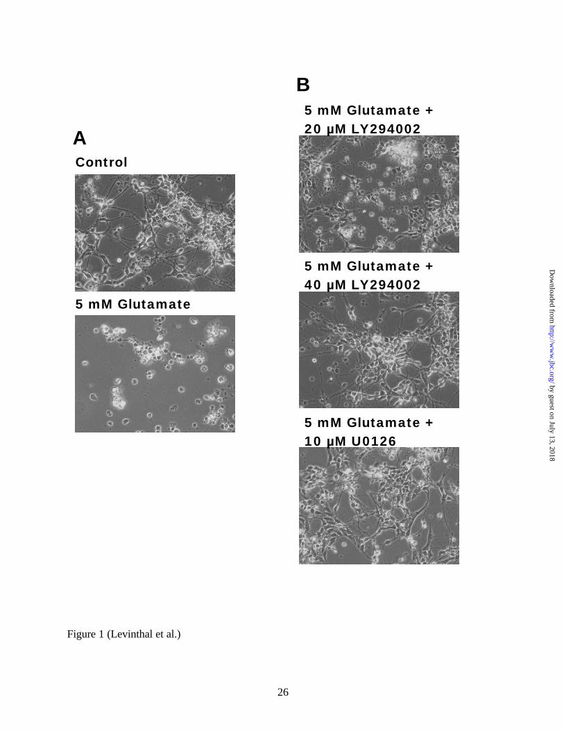
Figures 1A and 1B, U0126 protects these cells from glutamate- induced oxidative toxicity. To
examine the role of PI3K-Akt in glutamate-induced oxidative toxicity, we used various doses of
the specific PI3K inhibitor, LY294002. Because the PI3K-Akt signaling pathway has been so
reliably associated with neuroprotection in a wide variety of neurotoxicity models, we
anticipated that the administration of LY294002 would not be able to prevent glutamate- induced
oxidative toxicity. Surprisingly, LY294002 conferred significant neuroprotection against
glutamate- induced oxidative toxicity at doses ranging from 10 µM to 40µM (Figure 1B, 20 µM
and 40 µM doses shown).
To quantify the ability of these inhibitors to abrogate glutamate- induced oxidative toxicity,
we employed the PI-uptake assay, which utilizes the property of this fluorescent dye to be taken
up selectively by dying or dead cells, but not by viable cells. PI labeled and unlabeled cells were
counted in three random fields per experimental condition in at least three separate cultures 18
hours after the initial exposure to glutamate. At this time point, cells destined to die have begun
to retract their processes, to have shrunken cell bodies, and to display condensed nuclei. The
vast majority of cells treated with glutamate display all of these features within 20-24 hours and
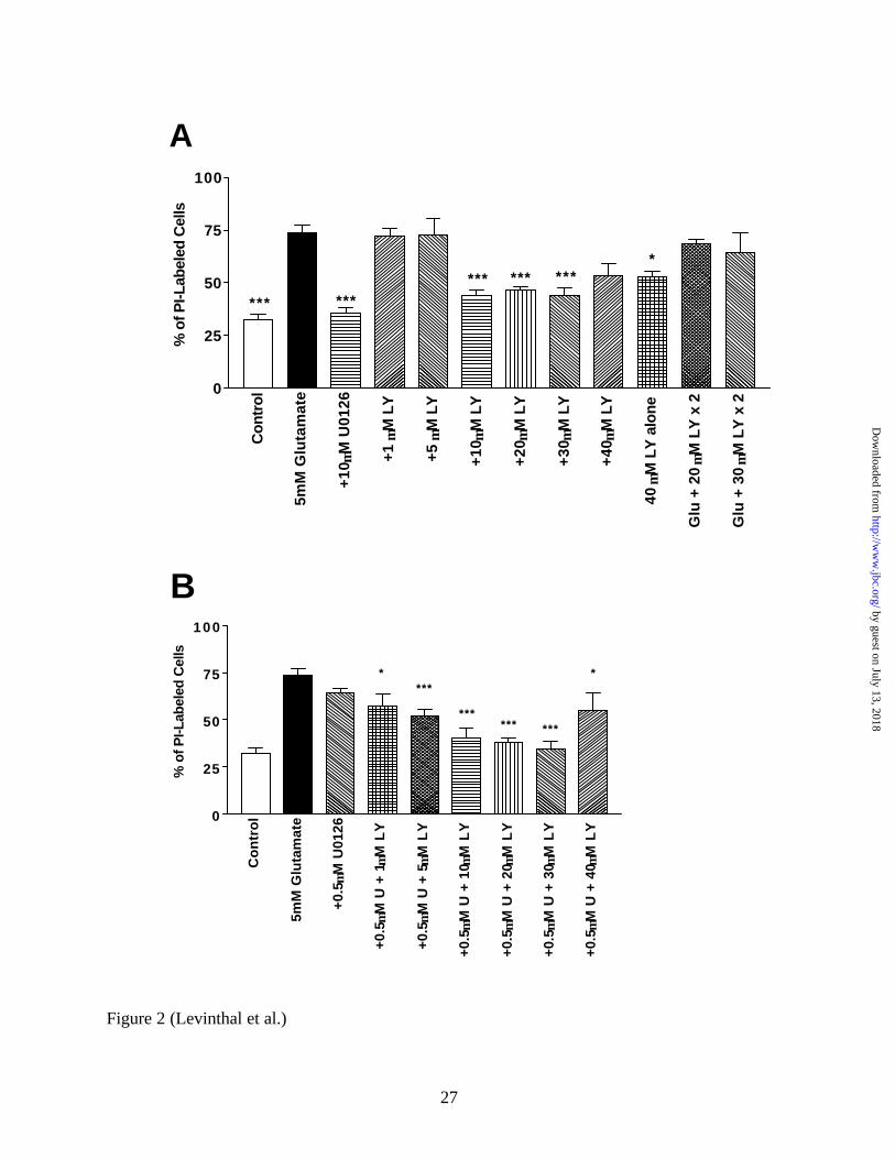
become detached from the plate soon after this time. Figure 2 demonstrates the dose
dependency for the neuroprotective effects of LY294002. Doses of LY294002 ranging from 1
µM to 5 µM did not abrogate glutamate-induced oxidative toxicity, while higher doses (e.g. 10-
30 µM) significantly protected cells (p < 0.001). The highest dose of LY294002 (e.g. 40 µM)
by guest on July 13, 2018http://w
ww
.jbc.org/D
ownloaded from
10
had a slightly less protective effect, which approached significance. The administration of 20
µM alone did not lead to cell death (Figure 4C), but 40 µM LY294002 alone exhibited some
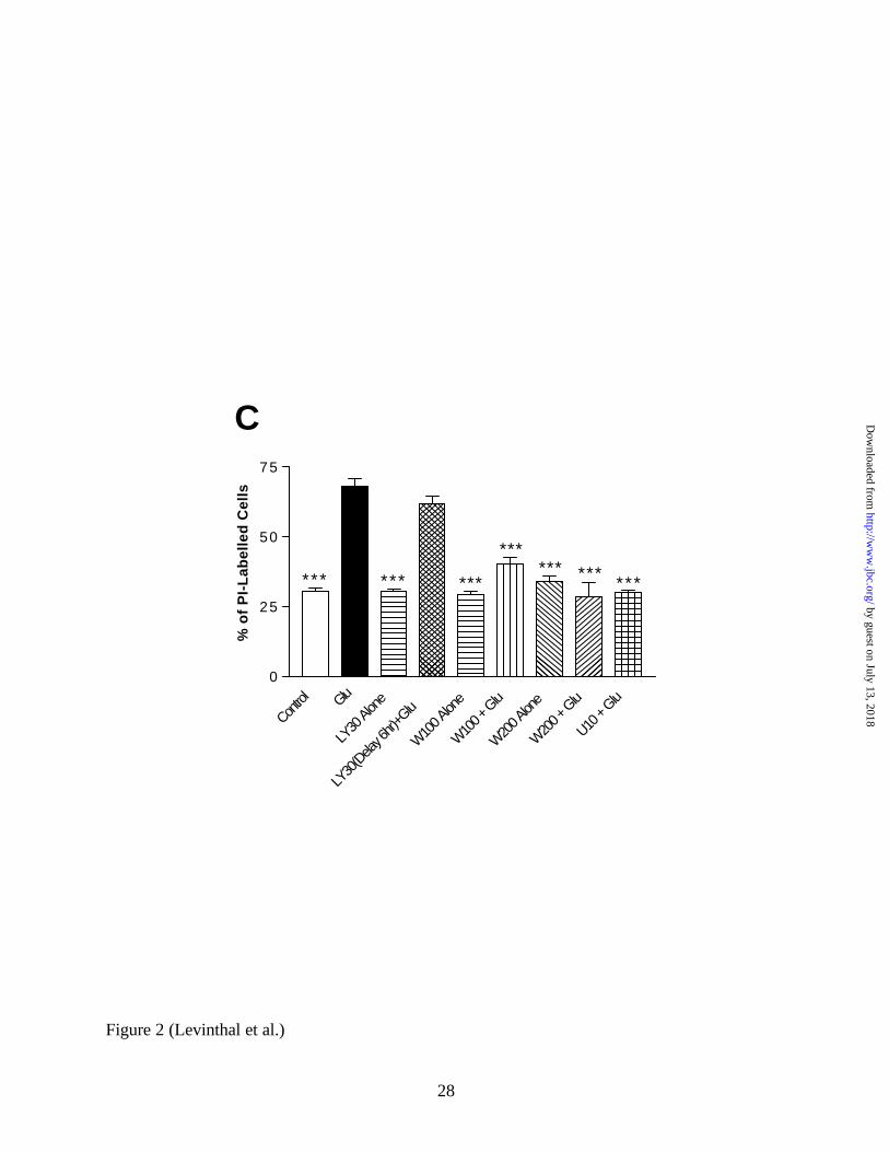
toxicity (Figure 2A). Wortmannin, which inhibits PI3K through a different mechanism than
LY294002 but is much less stable in aqueous solution (33), also protects against glutamate-
induced oxidative toxicity and was not toxic when administered alone (Figure 2C).
To examine if a subthreshold 0.5 µM U0126 dose (i.e. a dose that did not by itself protect
neurons from glutamate- induced oxidative toxicity; Figure 2B) could potentiate the
neuroprotective effect of LY294002, 0.5 µM U0126 and increasing doses of LY294002 (1 µM –
40 µM) were co-administered to cortical cultures with 5 mM glutamate. In the presence of this
subthreshold dose of U0126, the minimum dose of LY294002 that was found to be significantly
neuroprotective was 5 µM, a 2-fold lower dose than observed in the absense of U0126 (Figure
2B). However, the 1 µM LY294002 dose approached significance (p < 0.012 before the
stringent Bonferroni post-hoc correction for multiple comparisons) in its neuroprotective effect.
These results suggest that some crosstalk between the PI3K-Akt and MEK-ERK pathways may
be operating in these neurons undergoing oxidative stress.
The manner in which LY294002 was administered was critical for its neuroprotective effect.
For example, one bolus of inhibitor (40 µM) given at the initiation of glutamate treatment led to
some protection from toxicity (p < 0.034 before post-hoc correction; Figure 2A). However,
administering one 20 µM dose of LY294002 at the initiation of glutamate treatment and a
subsequent 20 µM dose 6 hours later completely abolished the neuroprotective effect of the drug
(Figure 2A). This phenomenon was also observed for an analagous 30 µM - 30 µM LY294002
dosing schedule (Figure 2A). The administration of two 20 µM doses of LY294002 six hours
apart by itself was not toxic to the cells (Figure 4C). Furthermore, delaying the administration of
by guest on July 13, 2018http://w
ww
.jbc.org/D
ownloaded from

11
LY294002 by 6 hours after the addition of glutamate abolished any protective effect of the
compound (Figure 2C). This evidence would suggest that some critical PI3K-dependent event
occurs within 6 hours, but not later, that is necessary for glutamate- induced oxidative toxicity.
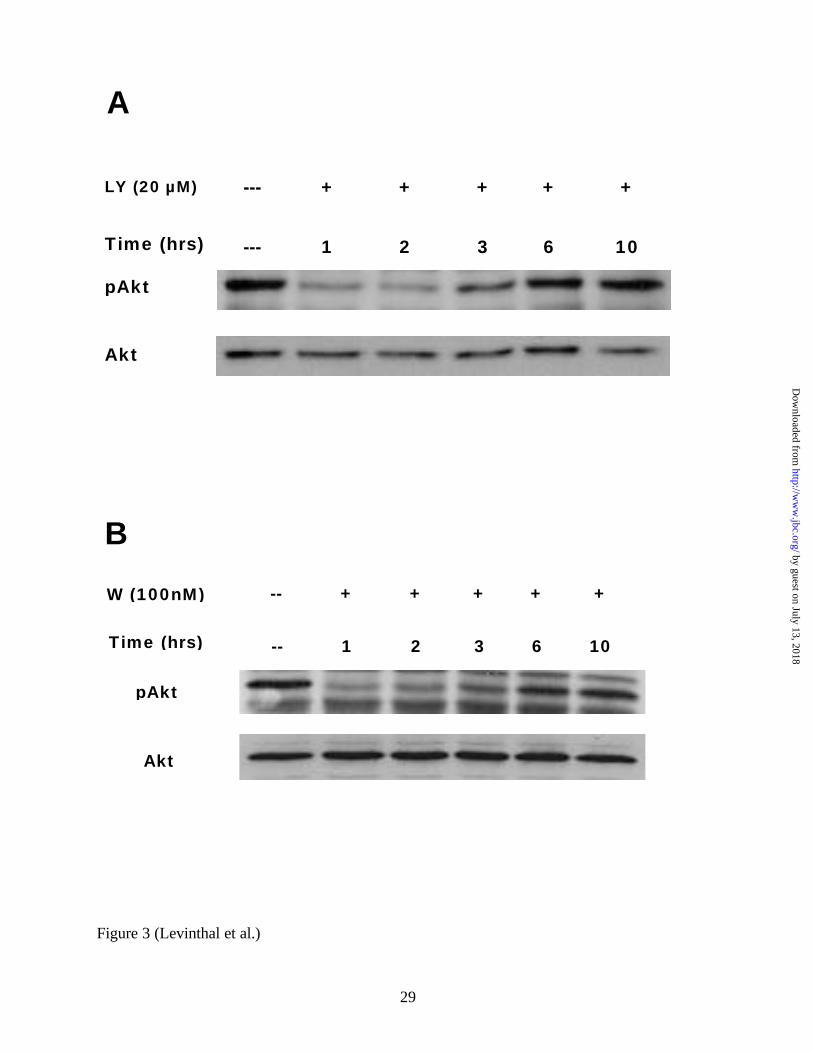
Both LY294002 and Wortmannin Only Transiently Inhibit PI3K in Mixed Cortical Cultures -- To
confirm that LY294002 and wortmannin were indeed inhibiting the PI3K-Akt pathway in our
cultures, we administered the drugs for various amounts of time and monitored levels of
phosphorylated Akt as an output measure of PI3K activity. As shown in Figure 3A, in primary
immature mixed cortical cultures, a single LY294002 treatment (20 µM) only transiently
inhibited Akt phosphorylation. This dose had conferred significant neuroprotection from
glutamate- induced oxidative toxicity (Figure 1B, Figure 2A). This was similarly observed in
wortmannin-treated cultures (Figure 3B). Consistently, phosphorylated Akt returned to baseline
levels within about 6 hours after the initial administration of both drugs. Thus, LY294002 and
wortmannin administration to mixed primary cortical cultures both inhibit PI3K transiently and
have no long term effect (i.e. beyond 6 hours) on the PI3K-Akt pathway.
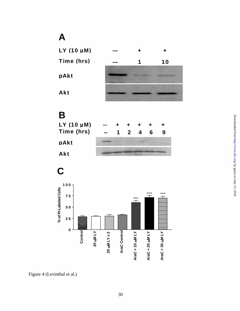
LY294002 Is Sequestered or Metabolized by Glial Cells in Mixed Cortical Cultures -- Because
LY294002 is known to be stable in aqueous solution, the apparent loss after 6 hours of its
efficacy to inhibit PI3K action on one of its downstream targets (i.e. Akt) would be consistent
with metabolism or sequestration of the compound. To further explore this phenomenon of
transient PI3K inhibition in our culture system, we asked whether the presence of glial elements
in the system was responsible for this apparent LY294002 inactivation. Our cultures typically
contain about 20-25% GFAP-positive cells at DIV 3-4 as determined by immmunocytochemical
by guest on July 13, 2018http://w
ww
.jbc.org/D
ownloaded from
12
staining (20.3% +/- 7.2 SEM). The anti-mitotic agent cytosine arabinoside (Ara-C) was added to
the mixed cultures, and LY294002 administered 2 days later, after the vast majority of glial cells
were eliminated. The efficacy of Ara-C in eliminating GFAP-positive cells was verified by
immunocytochemistry (i.e. fewer than 1% of cells were GFAP-positive). As shown in Figure
4A, a single administration of 20 µM LY294002 to these enriched neuronal cultures is very
effective in inhibiting PI3K activity for a prolonged period of time (i.e. 10 hours). This implies
that the transient inhibition of PI3K activity in mixed cultures given a single dose of LY294002
may be due to glial metabolism or sequestration of the drug. To further test this hypothesis, we
administered a lower dose of LY294002 (10 µM) to the hippocampal cell line, HT22. These
cultures are devoid of glial cells, and therefore we expected LY294002 to have a prolonged
effect in inhibiting PI3K. Indeed, as shown in Figure 4B, a single dose of LY294002 (10 µM)
was sufficient to inhibit PI3K for extended periods of time. Furthermore, low doses of
LY294002 are toxic to HT22 cells (data not shown), which confirms that prolonged maintenance
of PI3K activity plays an important role in neuronal cell survival.
To further confirm that the maintenance of PI3K activity is important to the survival of
primary cortical neurons, we measured the viability of cells in Ara-C-treated, neuron-enriched
cultures following treatment with LY294002. As shown in Figure 4C, 20µM LY294002,
administered in one single dose, or two doses six hours apart, to mixed cortical cultures shows no
toxic effect. However, in neuron-enriched cultures, single doses of LY294002 ranging from 10
µM to 30 µM showed significant neurotoxicity (Figure 4C, p < 0.001). Thus prolonged PI3K
inhibition alone is sufficient to undermine cell survival in our cultures.
by guest on July 13, 2018http://w
ww
.jbc.org/D
ownloaded from
13
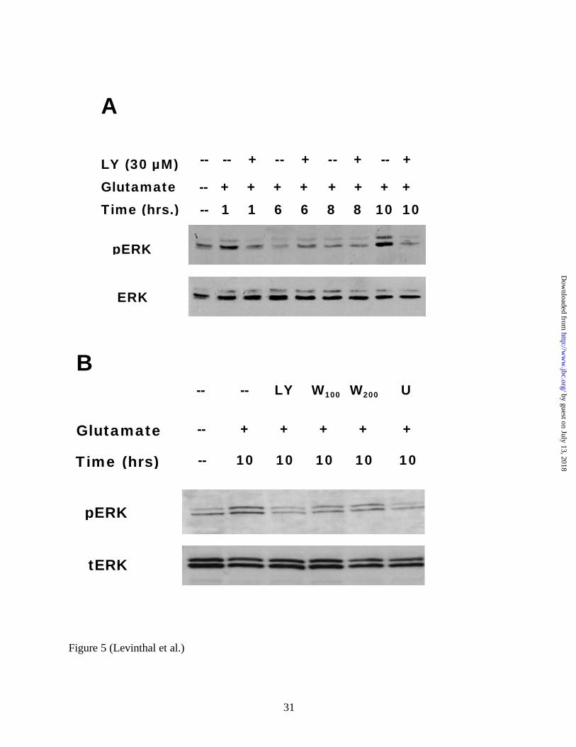
Transient, Early PI3K Inhibition Prevents the Development of a Delayed ERK Activation -- We
have previously determined that glutamate- induced oxidative toxicity is associated with a
delayed chronic activation of ERK1/2 and that this activation is necessary for neuronal cell death
(6) (Figures 1 and 2A). In cortical cultures, we typically observe this delayed activation at 10
hours or more after the initiation of glutamate treatment, which correlates with the times of
increasing oxidative stress in these cells (5) (Figure 7). We therefore asked whether LY294002
or wortmannin, which both protect these cultures from glutamate-induced oxidative toxicity
(Figures 1 and 2), interfere with the development of this delayed ERK1/2 activation. As shown
in Figure 5A, 30 µM LY294002, a dose that significantly protected cortical neurons, is effective
in preventing the delayed activation of ERK1/2. Furthermore, treatment with either 100 nM or
200 nM wortmannin showed attenuation of the ERK activation measured 10 hours after
glutamate administration (Figure 5B). Interestingly, a 6 hour delay in the administration of
LY294002 after glutamate, a protocol which failed to protect cortical neurons from oxidative
toxicity (Figure 2C), abolished the ability of the compound to prevent the later ERK activation
seen during oxidative toxicity (data not shown). Thus, only an early, transient inhibition of PI3K
activity in immature cortical neurons can inhibit the delayed activation of ERK that accompanies
glutamate- induced oxidative stress.
PI3K Inhibition Prevents the Delayed Activation of MEK-1 -- PI3K has been found to exert
either a stimulatory or inhibitory effect on ERK activation depending, in part, upon the identity
and strength of the applied extracellular stimulus (34-36). In cases where PI3K is necessary for
ERK activation, B-Raf appears to be a major conversion point for these two pathways, especially
in neurons (27). Because MEK-1 is the major target of all Raf isoforms, we used an
by guest on July 13, 2018http://w
ww
.jbc.org/D
ownloaded from
14
immunoprecipitation (IP)-kinase assay to monitor changes in MEK-1 activity in extracts
prepared from primary neurons treated with glutamate in the presence or absence of LY294002.
In this way, we could determine whether PI3K was acting at the level of ERK or on upstream
kinases such as Raf / MEK-1 during the development of the PI3K-dependent, delayed ERK
activation. As is shown in Figure 6, glutamate- induced oxidative toxicity in primary immature
cortical cells leads to the delayed recruitment of active MEK-1 at 10 hours, which is consistent
with the observed onset of ERK activation. This is result is not surprising given that the MEK-1
inhibitor U0126 was previously shown to prevent the development of ERK activation at this time
point (6). However, administration of LY294002 at the initiation of glutamate treatment
prevented the recruitment of MEK-1 activity at 10 hours, showing that the late recruitment of
MEK-1 is dependent upon early, but not late PI3K activity (Figure 6). Thus PI3K activity must
affect the ERK pathway at the level of MEK-1 or upstream of MEK-1 during glutamate- induced
oxidative toxicity, formally eliminating the possibility that PI3K activity influences ERK
phosphorylation independent of MEK-1.
Neither PI3K Inhibition nor MEK Inhibition Prevents Oxidative Stress in Primary Mixed
Cortical Cultures To test the hypothesis that the protective effects of PI3K inhibition or MEK
inhibition may be due to the prevention of oxidative stress in our cultures, we measured ROS
production using the dye DCF. As can be seen in Figure 7, a significant increase in DCF
fluorescence intensity is seen 10 hours, but not 4 hours, after the administration of glutamate. A
high dose of H2O2 was used as a positive control in the assay. Interestingly, neither LY294002
(30µM) nor U0126 (10µM) significantly effected the extent of oxidative stress measured 10
hours after glutamate administration. This implies that the loss of ERK activation at 10 hours
by guest on July 13, 2018http://w
ww
.jbc.org/D
ownloaded from

15
(via PI3K inhibition or direct MEK inhibition) can uncouple cells from oxidative toxicity despite
the ongoing presence of oxidative stress.
DISCUSSION
The PI3K-Akt pathway is well recognized for its ability to mediate neuronal protection from
a wide range of toxic insults and conditions (37). In many cases, specific inhibition of PI3K (e.g.
wortmannin, LY294002) has been instrumental in establishing the neuroprotective action of this
pathway (38). However, we report here that transient inhibition of the PI3K-Akt pathway can
protect primary neurons from oxidative toxicity. Thus, PI3K activity may be required for some
initial steps of a cell death program that unfolds when neurons are subjected to oxidative stress.
To our knowledge, this is the first demonstrated example of PI3K activity associated with cell
death. Importantly, a transient “window” of PI3K inhibition in mixed cortical cultures exposed
to LY294002 or wortmannin enabled us to distinguish between the short-term and long-term
requirements for PI3K activity in this neurotoxicity model. Transient PI3K inhibition is
apparently insufficient to undermine the long-term pro-survival function of this pathway. Since
neuron-enriched cultures of the same age treated with LY294002 do not exhibit a transient
inhibition of PI3K activity, it seems likely that glial cells in our cultures (~20-25%) are
responsible for the metabolism, inactivation, or sequestration of LY294002. While we have not
detected appreciable inactivation of LY294002 in enriched, mature astrocyte cultures (data not
shown), there are other glial cell types in our mixed cultures that could exhibit this activity.
Our results are consistent with the wealth of data supporting a neuroprotective role for the
PI3K pathway, including in an established cell culture model of oxidative toxicity (i.e. HT22
cells). We are able in our mixed cultures to generate prolonged inhibition of PI3K by simply
by guest on July 13, 2018http://w
ww
.jbc.org/D
ownloaded from
16
administering LY294002 in two boluses 6 hours apart. In this case where PI3K activity was
inhibited for prolonged periods of time (i.e. up to 12 hours), the neuroprotective effects of
LY294002 were abrogated. Furthermore, in neuron-enriched cultures, a single bolus of
LY294002 was toxic due to prolonged inhibition of the PI3K-Akt pathway (Figure 4C). This
finding is consonant with recent data showing that a single 20 µM dose of LY294002 is toxic to
neuron-enriched primary hippocampal cultures (39).
Transient inhibition of the PI3K-Akt pathway does not affect neuronal survival in other
models where prolonged inhibition would be predicted to lead to cellular demise. For example,
infusion of the PI3K inhibitor wortmannin, an unstable compound in aqueous solution, into the
amygdala of rats impaired their ability to learn in a fear-conditioning paradigm soon after the
administration of the drug, but did not affect their ability to be effectively re-trained in the same
paradigm several days later (28). This implies that the effects of wortmannin in vivo are
reversible and do not result in neuronal cell death, at least in the amygdala. Similarly,
LY294002 has been used in vivo in models of transient focal cerebral ischemia- or seizure-
induced neuronal toxicity, and in these studies LY294002 alone did not lead to increased
neuronal cell death in the hippocampus or the caudate / putamen (40,41), presumably due to the
surrounding glia. Our demonstration of transient PI3K inhibition by LY294002 in mixed
neuronal / glial cultures provides strong support for the notion that metabolism, inactivation, or
sequestration of LY294002 can have functional consequences for neuronal cell responses to a
cell death- inducing stimulus. This would be of great interest in the development of therapeutic
uses of these compounds in vivo, where surrounding glial elements would likely alter the
pharmacodynamics of these drugs in the central nervous system.
by guest on July 13, 2018http://w
ww
.jbc.org/D
ownloaded from
17
We have previously determined that glutamate- induced oxidative toxicity is associated
with the development of a delayed activation of ERK1/2, and that the MEK inhibitor prevents
this activation and is significantly neuroprotective (6,21). Interestingly, the ability of transient
PI3K inhibition to protect these cultures is associated with an abrogation of delayed MEK-1
activation (Figure 6) and ERK1/2 phosphorylation (Figure 5). This further establishes ERK1/2
activation in this model as a requisite event for eventual cellular demise. In addition, because of
the temporal disparity between the period of PI3K inhibition and the regulation of ERK1/2
activity, we can place PI3K activation upstream of the course of events that lead to MEK-1 and
ERK1/2 activation. Furthermore, the fact that neither PI3K nor MEK inhibition blocks oxidative
stress implies that somehow PI3K inhibition can uncouple ERK activation from oxidative stress
and that oxidative stress is not dependent upon ERK activation in this model (Figure 7).
Cross-talk between the PI3K-Akt and Raf-MEK-ERK pathways has been demonstrated
in an increasingly diverse set of circumstances and in different cell types. While this link was
first reported to be involved in the signaling of G-protein coupled receptors (42,43), PI3K
activity can be necessary for the recruitment of the ERK pathway in signaling events associated
with other types of receptors as well. For example, PI3K activity is required for in vitro ERK
activation in AMPA and NMDA receptor-mediated signaling events in striatal cultures (25,26)
and for the NGF-induced sustained activation of ERK during PC12 differentiation (27).
Furthermore, PI3K activation was shown to occur upstream of ERK activation in a rat model of
associative learning (28). Of great interest is the recent finding that PI3K positively contributes
to the activation of ERK1/2 in cortical cultures exposed to oxidative stress induced by hydrogen
peroxide (29). Thus there appears to exist a mechanism for functional coupling of the PI3K and
ERK pathways in neurons, with the potential for PI3K to act as a required upstream activator.
by guest on July 13, 2018http://w
ww
.jbc.org/D
ownloaded from

18
Contradictory effects of PI3K on the ERK pathway have been noted and were initially
attributed to cell- type specific signaling events that reflected a wide variety of direct and indirect
interactions between the two pathways (42-44). However, recent reports showing opposing
effects of PI3K on ERK activation within a single cell type in response to one signal reveal the
importance of signal intensity in dictating the eventual outcome of PI3K/ERK cross-talk (34-36).
Furthermore, EGF-induced ERK activation was found to be PI3K-dependent under conditions
where PI3K activity remained near basal levels (35). This previously described “permissive”
effect of the PI3K pathway on ERK activation (29,34,35,46) appears to operate in our neuronal
cultures as increased stimulation of PI3K activity in response to glutamate treatment is minimal
(data not shown). However, our neuronal cultures do exhibit a substantial baseline level of PI3K
activity (Figures 3A, 3B and 4A). Interestingly, our results are unique in that the coupling
between PI3K and ERK is restricted to a distinct kinetic phase during the progression of an
oxidative-stress induced cell death pathway. Thus, inhibition of basal PI3K activity (as assessed
by levels of phosphorylated Akt) at early times following the initiation of glutamate treatment
(i.e. within 4-6 hours) impacts ERK activation that is only apparent following an additional 4-6
hours.
We consider the observed coupling in our primary neuron cultures to reflect a permissive
effect of PI3K on ERK activation. Glutamate- induced oxidative toxicity in immature neurons
and in HT22 cells is driven by a delayed rise in intracellular ROS and Ca2+ (47). The rapid burst
and accumulation of ROS and Ca2+ that ultimately results from glutamate treatment are likely to
be proximal to the terminal execution phases of the cell death program that operates in these cells
(47). PI3K has been found to influence plasma membrane Ca2+ channel activity through
activated Akt and thereby contribute to neuronal cell survival in response to neuroprotective
by guest on July 13, 2018http://w
ww
.jbc.org/D
ownloaded from
19
factors such as IGF-1 (48). Furthermore, a VDCC-dependent form of long-term potentiation at
hippocampal CA1 region synapses was found to require PI3K activity, implying that PI3K
activity can directly regulate calcium entry into neurons (49). In addition, PI3K has been
demonstrated to be required for the activation of voltage- independent Ca2+ channels in a CHO
cell system (50) and VDCCs in vascular smooth muscle cells (51), non-selective cation channels
(NSCCs) (52), and for Ca2+ release from intracellular SOCCs (52). Furthermore, there is
increasing evidence showing that PI3K activity is not only necessary for increases in intracellular
Ca2+ but that this increase in Ca2+ is necessary for ERK activation (53). Interestingly, PI3K-
dependent increases in intracellular Ca2+ have been shown to be dissociated from increases in
phosphorylated Akt and may represent a point of divergence between action of PI3K on the Akt
versus the ERK pathway (52, 53). This is consistent with our findings that Akt activation (above
baseline) is minimal during the deve lopment of glutamate- induced oxidative toxicity (data not
shown). Thus, it is tempting to speculate that the permissive effects of PI3K on ERK-dependent
cell death in oxidatively stressed primary neurons might be due to effects of basal PI3K activity
on either plasma membrane Ca2+ channels, or intracellular store-operated channels. ROS
generated by the mitochondria contribute to Ca2+ influx and accumulation in glutamate-treated
HT22 cells and primary cortical neurons (5,47), and a Ca2+ channel response to ROS that
requires some direct or indirect priming action of the PI3K pathway may very well be the link
between the PI3K and ERK pathways in our system.
The identification of multiple intracellular signaling pathways that operate to promote
or limit neuronal cell death and the delineation of various levels of cross-talk between these
pathways both present unique challenges to the development of pharmacological neuroprotective
therapies. Our results suggest that depending upon the nature and identity of the coupled signal
by guest on July 13, 2018http://w
ww
.jbc.org/D
ownloaded from

20
transduction pathways, a temporal window may exist that allows for the transient inhibition of a
specific signal transduction pathway. Thus, rapid metabolism or sequestration of drugs targeted
to specific signaling pathways may prove beneficial particularly in cases where unique coupling
between signal transduction pathways occurs at distinct kinetic phases of a progressing cell death
program.
REFERENCES
1. Coyle, J. T., and Puttfarcken, P. (1993) Science 262, 689-695 2. Mattson, M. P., Duan, W., Pedersen, W. A., and Culmsee, C. (2001) Apoptosis 6, 69-81 3. Murphy, T. H., Schnaar, R. L., and Coyle, J. T. (1990) Faseb J 4, 1624-1633 4. Ratan, R. R., Murphy, T. H., and Baraban, J. M. (1994) J Neurochem 62, 376-379 5. Li, Y., Maher, P., and Schubert, D. (1997) Neuron 19, 453-463 6. Stanciu, M., Wang, Y., Kentor, R., Burke, N., Watkins, S., Kress, G., Reynolds, I., Klann, E.,
Angiolieri, M. R., Johnson, J. W., and DeFranco, D. B. (2000) J Biol Chem 275, 12200-12206
7. Davis, R. J. (1993) J Biol Chem 268, 14553-14556 8. Garrington, T. P., and Johnson, G. L. (1999) Curr Opin Cell Biol 11, 211-218 9. Grewal, S. S., York, R. D., and Stork, P. J. (1999) Curr Opin Neurobiol 9, 544-553 10. Xia, Z., Dickens, M., Raingeaud, J., Davis, R. J., and Greenberg, M. E. (1995) Science 270,
1326-1331 11. Bonni, A., Brunet, A., West, A. E., Datta, S. R., Takasu, M. A., and Greenberg, M. E. (1999)
Science 286, 1358-1362 12. Gonzalez-Zulueta, M., Feldman, A. B., Klesse, L. J., Kalb, R. G., Dillman, J. F., Parada, L.
F., Dawson, T. M., and Dawson, V. L. (2000) Proc Natl Acad Sci U S A 97, 436-441 13. Han, B. H., and Holtzman, D. M. (2000) J Neurosci 20, 5775-5781 14. Zhu, Y., Yang, G. Y., Ahlemeyer, B., Pang, L., Che, X. M., Culmsee, C., Klumpp, S., and
Krieglstein, J. (2002) J Neurosci 22, 3898-3909 15. Murray, B., Alessandrini, A., Cole, A. J., Yee, A. G., and Furshpan, E. J. (1998) Proc Natl
Acad Sci U S A 95, 11975-11980 16. Alessandrini, A., Namura, S., Moskowitz, M. A., and Bonventre, J. V. (1999) Proc Natl
Acad Sci U S A 96, 12866-12869 17. Namura, S., Iihara, K., Takami, S., Nagata, I., Kikuchi, H., Matsushita, K., Moskowitz, M.
A., Bonventre, J. V., and Alessandrini, A. (2001) Proc Natl Acad Sci U S A 98, 11569-11574 18. Du, S., McLaughlin, B., Pal, S., and Aizenman, E. (2002) J Neurosci 22, 7408-7416 19. Mori, T., Wang, X., Jung, J. C., Sumii, T., Singhal, A. B., Fini, M. E., Dixon, C. E.,
Alessandrini, A., and Lo, E. H. (2002) J Cereb Blood Flow Metab 22, 444-452 20. Noshita, N., Sugawara, T., Hayashi, T., Lewen, A., Omar, G., and Chan, P. H. (2002) J
Neurosci 22, 7923-7930 21. Stanciu, M., and DeFranco, D. B. (2002) J Biol Chem 277, 4010-4017
by guest on July 13, 2018http://w
ww
.jbc.org/D
ownloaded from

21
22. Datta, S. R., Brunet, A., and Greenberg, M. E. (1999) Genes Dev 13, 2905-2927 23. Datta, S. R., Dudek, H., Tao, X., Masters, S., Fu, H., Gotoh, Y., and Greenberg, M. E. (1997)
Cell 91, 231-241 24. Cardone, M. H., Roy, N., Stennicke, H. R., Salvesen, G. S., Franke, T. F., Stanbridge, E.,
Frisch, S., and Reed, J. C. (1998) Science 282, 1318-1321 25. Perkinton, M. S., Sihra, T. S., and Williams, R. J. (1999) J Neurosci 19, 5861-5874 26. Perkinton, M. S., Ip, J. K., Wood, G. L., Crossthwaite, A. J., and Williams, R. J. (2002) J
Neurochem 80, 239-254 27. York, R. D., Molliver, D. C., Grewal, S. S., Stenberg, P. E., McCleskey, E. W., and Stork, P.
J. (2000) Mol Cell Biol 20, 8069-8083 28. Lin, C. H., Yeh, S. H., Lu, K. T., Leu, T. H., Chang, W. C., and Gean, P. W. (2001) Neuron
31, 841-851 29. Crossthwaite, A. J., Hasan, S., and Williams, R. J. (2002) J Neurochem 80, 24-35 30. Mizuta, I., Katayama, M., Watanabe, M., Mishina, M., and Ishii, K. (1998) Cell Mol Life Sci,
54, 721-725. 31. Davis, J. B., and Maher, P. (1994) Brain Res 652, 169-173 32. Hsieh, H., Cheng, C., Wu, S., Chiu, J., Wung, B., and Wang, D. (1998) J Cell Physiology
175, 156-162 33. Walker, E. H., Pacold, M. E., Perisic, O., Stephens, L., Hawkins, P. T., Wymann, M. P., and
Williams, R. L. (2000) Mol Cell 6, 909-919 34. Duckworth, B. C., and Cantley, L. C. (1997) J Biol Chem 272, 27665-27670 35. Wennstrom, S., and Downward, J. (1999) Mol Cell Biol 19, 4279-4288 36. Moelling, K., Schad, K., Bosse, M., Zimmermann, S., and Schweneker, M. (2002) J Biol
Chem 277, 31099-31106 37. Brunet, A., Datta, S. R., and Greenberg, M. E. (2001) Curr Opin Neurobiol 11, 297-305 38. Zheng, W. H., Kar, S., and Quirion, R. (2002) Mol Pharmacol 62, 225-233 39. Luo, H., Hattori, H., Hossain, M., Hester, L., Huang, Y., Lee-Kwon, W., Donowitz, M.,
Nagata, E., and Snyder, S. (2003) PNAS 100, 11712-11717 40. Noshita, N., Lewen, A., Sugawara, T., and Chan, P. H. (2001) J Cereb Blood Flow Metab 21,
1442-1450 41. Henshall, D. C., Araki, T., Schindler, C. K., Lan, J. Q., Tiekoter, K. L., Taki, W., and Simon,
R. P. (2002) J Neurosci 22, 8458-8465 42. Hawes, B. E., Luttrell, L. M., van Biesen, T., and Lefkowitz, R. J. (1996) J Biol Chem 271,
12133-12136 43. Lopez-Ilasaca, M., Crespo, P., Pellici, P. G., Gutkind, J. S., and Wetzker, R. (1997) Science
275, 394-397 44. Zimmermann, S., and Moelling, K. (1999) Science 286, 1741-1744 45. Chaudhary, A., King, W. G., Mattaliano, M. D., Frost, J. A., Diaz, B., Morrison, D. K.,
Cobb, M. H., Marshall, M. S., and Brugge, J. S. (2000) Curr Biol 10, 551-554 46. Sutor, S. L., Vroman, B. T., Armstrong, E. A., Abraham, R. T., and Karnitz, L. M. (1999) J
Biol Chem 274, 7002-7010 47. Tan, S., Sagara, Y., Liu, Y., Maher, P., and Schubert, D. (1998) J Cell Biol 141, 1423-1432 48. Blair, L. A., Bence-Hanulec, K. K., Mehta, S., Franke, T., Kaplan, D., and Marshall, J.
(1999) J Neurosci 19, 1940-1951 49. Sanna, P. P., Cammalleri, M., Berton, F., Simpson, C., Lutjens, R., Bloom, F. E., and
Francesconi, W. (2002) J Neurosci 22, 3359-3365
by guest on July 13, 2018http://w
ww
.jbc.org/D
ownloaded from
22
50. Kawanabe, Y., Hashimoto, N., and Masaki, T. (2002) Mol Pharmacol 62, 756-761 51. Seki, T., Yokoshiki, H., Sunagawa, M., Nakamura, M., and Sperelakis, N. (1999) Pflugers
Arch 437, 317-323 52. Kawanabe, Y., Hashimoto, N., and Masaki, T. (2003) Mol Pharmacol 63, 808-813 53. Kansra, V., Groves, C., Gutierrez-Ramos, J., and Polakiewicz, R. (2001) J Biol Chem 276,
31831-31838
FIGURE LEGENDS
FIG 1. Representative phase-contrast images (400x magnification) demonstrating glutamate-
induced oxidative toxicity in primary cortical neurons and neuroprotection via PI3K or MEK-1
inhibition. A, Immature primary cortical cultures (DIV3) were either left untreated or treated
with 5 mM glutamate for 24 hours. B, Cultures were treated with glutamate plus 20 µM
LY294002, 40 µM LY294002, or 10 µM U0126. Both compounds led to significant
neuroprotection from glutamate-induced oxidative toxicity as assessed 24 hours after the
initiation of glutamate treatment.
FIG 2. The neuroprotective effect of LY294002 is dose-dependent, and concurrent inhibition of
both MEK and PI3K can potentiate neuroprotection from glutamate- induced oxidative toxicity.
A, Primary immature cortical cultures (DIV 3) were treated with 5 mM glutamate and increasing
single doses of LY294002 (1 µM to 40 µM), two doses (20 µM or 30 µM) of LY294002 each
administered six hours apart (i.e. one with the initial treatment of glutamate and another six
hours later), 10 µM U0126, or 40 µM LY294002 alone. Eighteen hours after initial treatment,
cells were incubated for 10 minutes with propidium iodide (PI) to visualize dead or dying PI-
positive cells using an inverted fluorescence microscope equipped with phase-contrast optics.
Both PI-positive and PI-negative cells were counted, and the percentage of PI-positive cells was
calculated for three random fields in at least three separate cultures. LY294002 exihibited
by guest on July 13, 2018http://w
ww
.jbc.org/D
ownloaded from
23
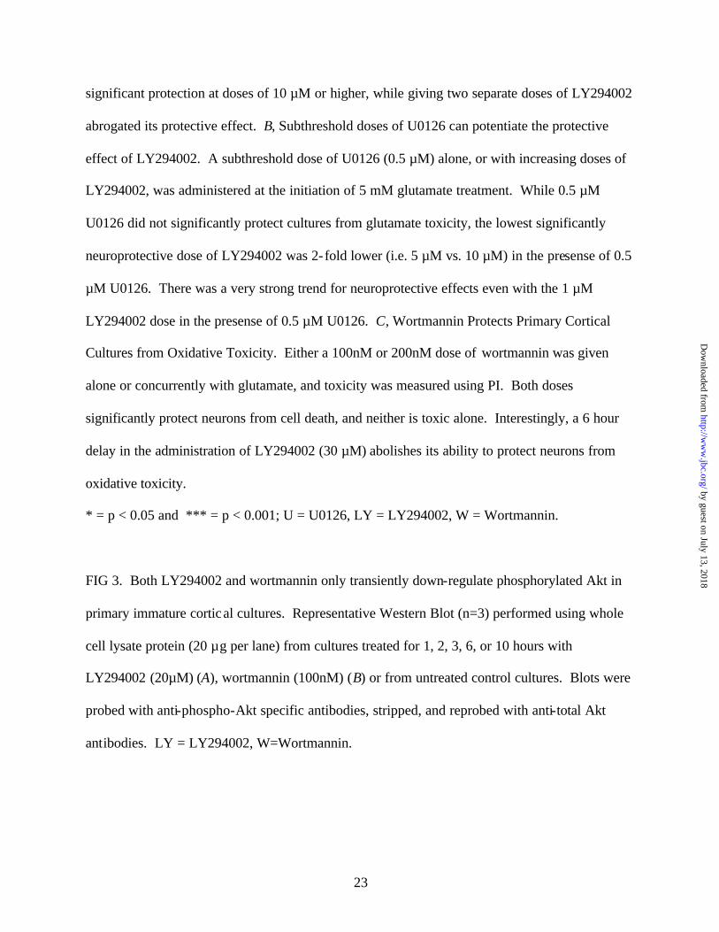
significant protection at doses of 10 µM or higher, while giving two separate doses of LY294002
abrogated its protective effect. B, Subthreshold doses of U0126 can potentiate the protective
effect of LY294002. A subthreshold dose of U0126 (0.5 µM) alone, or with increasing doses of
LY294002, was administered at the initiation of 5 mM glutamate treatment. While 0.5 µM
U0126 did not significantly protect cultures from glutamate toxicity, the lowest significantly
neuroprotective dose of LY294002 was 2-fold lower (i.e. 5 µM vs. 10 µM) in the presense of 0.5
µM U0126. There was a very strong trend for neuroprotective effects even with the 1 µM
LY294002 dose in the presense of 0.5 µM U0126. C, Wortmannin Protects Primary Cortical
Cultures from Oxidative Toxicity. Either a 100nM or 200nM dose of wortmannin was given
alone or concurrently with glutamate, and toxicity was measured using PI. Both doses
significantly protect neurons from cell death, and neither is toxic alone. Interestingly, a 6 hour
delay in the administration of LY294002 (30 µM) abolishes its ability to protect neurons from
oxidative toxicity.
* = p < 0.05 and *** = p < 0.001; U = U0126, LY = LY294002, W = Wortmannin.
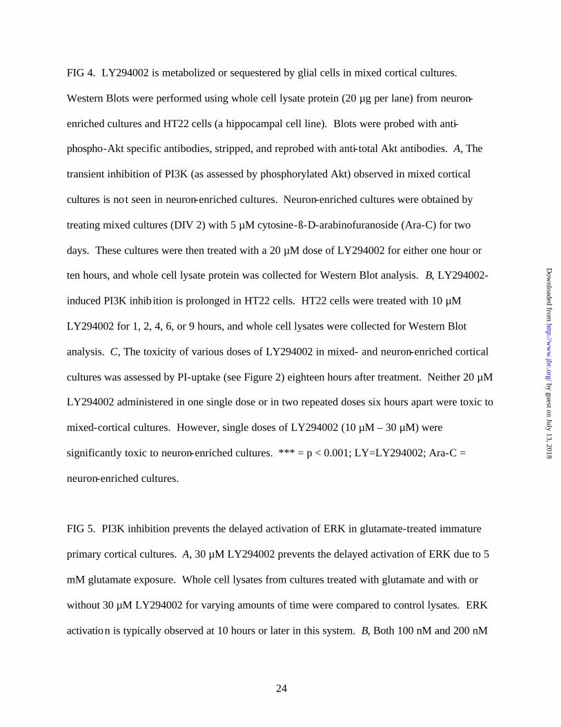
FIG 3. Both LY294002 and wortmannin only transiently down-regulate phosphorylated Akt in
primary immature cortical cultures. Representative Western Blot (n=3) performed using whole
cell lysate protein (20 µg per lane) from cultures treated for 1, 2, 3, 6, or 10 hours with
LY294002 (20µM) (A), wortmannin (100nM) (B) or from untreated control cultures. Blots were
probed with anti-phospho-Akt specific antibodies, stripped, and reprobed with anti-total Akt
antibodies. LY = LY294002, W=Wortmannin.
by guest on July 13, 2018http://w
ww
.jbc.org/D
ownloaded from
24
FIG 4. LY294002 is metabolized or sequestered by glial cells in mixed cortical cultures.
Western Blots were performed using whole cell lysate protein (20 µg per lane) from neuron-
enriched cultures and HT22 cells (a hippocampal cell line). Blots were probed with anti-
phospho-Akt specific antibodies, stripped, and reprobed with anti-total Akt antibodies. A, The
transient inhibition of PI3K (as assessed by phosphorylated Akt) observed in mixed cortical
cultures is not seen in neuron-enriched cultures. Neuron-enriched cultures were obtained by
treating mixed cultures (DIV 2) with 5 µM cytosine-ß-D-arabinofuranoside (Ara-C) for two
days. These cultures were then treated with a 20 µM dose of LY294002 for either one hour or
ten hours, and whole cell lysate protein was collected for Western Blot analysis. B, LY294002-
induced PI3K inhib ition is prolonged in HT22 cells. HT22 cells were treated with 10 µM
LY294002 for 1, 2, 4, 6, or 9 hours, and whole cell lysates were collected for Western Blot
analysis. C, The toxicity of various doses of LY294002 in mixed- and neuron-enriched cortical
cultures was assessed by PI-uptake (see Figure 2) eighteen hours after treatment. Neither 20 µM
LY294002 administered in one single dose or in two repeated doses six hours apart were toxic to
mixed-cortical cultures. However, single doses of LY294002 (10 µM – 30 µM) were
significantly toxic to neuron-enriched cultures. *** = p < 0.001; LY=LY294002; Ara-C =
neuron-enriched cultures.
FIG 5. PI3K inhibition prevents the delayed activation of ERK in glutamate-treated immature
primary cortical cultures. A, 30 µM LY294002 prevents the delayed activation of ERK due to 5
mM glutamate exposure. Whole cell lysates from cultures treated with glutamate and with or
without 30 µM LY294002 for varying amounts of time were compared to control lysates. ERK
activation is typically observed at 10 hours or later in this system. B, Both 100 nM and 200 nM
by guest on July 13, 2018http://w
ww
.jbc.org/D
ownloaded from
25
doses of wortmannin attenuate ERK activation at 10 hours. Blots were probed with anti-phospho
ERK antibodies, stripped, and reprobed with anti- total ERK antibodies (20 µg of total protein
were loaded per lane). A representative blot (n=4) is shown. LY=LY294002, W=Wortmannin.
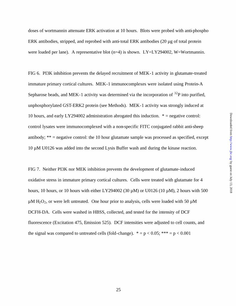
FIG 6. PI3K inhibition prevents the delayed recruitment of MEK-1 activity in glutamate-treated
immature primary cortical cultures. MEK-1 immunocomplexes were isolated using Protein-A
Sepharose beads, and MEK-1 activity was determined via the incorporation of 32P into purified,
unphosphorylated GST-ERK2 protein (see Methods). MEK-1 activity was strongly induced at
10 hours, and early LY294002 administration abrogated this induction. * = negative control:
control lysates were immunocomplexed with a non-specific FITC conjugated rabbit anti-sheep
antibody; ** = negative control: the 10 hour glutamate sample was processed as specified, except
10 µM U0126 was added into the second Lysis Buffer wash and during the kinase reaction.
FIG 7. Neither PI3K nor MEK inhibition prevents the development of glutamate-induced
oxidative stress in immature primary cortical cultures. Cells were treated with glutamate for 4
hours, 10 hours, or 10 hours with either LY294002 (30 µM) or U0126 (10 µM), 2 hours with 500
µM H2O2, or were left untreated. One hour prior to analysis, cells were loaded with 50 µM
DCFH-DA. Cells were washed in HBSS, collected, and tested for the intensity of DCF
fluorescence (Excitation 475, Emission 525). DCF intensities were adjusted to cell counts, and
the signal was compared to untreated cells (fold-change). * = p < 0.05; *** = p < 0.001
by guest on July 13, 2018http://w
ww
.jbc.org/D
ownloaded from
26
Figure 1 (Levinthal et al.)
B 5 mM Glutamate + 20 µM LY294002
5 mM Glutamate + 40 µM LY294002
5 mM Glutamate + 10 µM U0126
A Control
5 mM Glutamate
by guest on July 13, 2018http://w
ww
.jbc.org/D
ownloaded from
27
Figure 2 (Levinthal et al.)
A
Co
ntr
ol
5mM
Glu
tam
ate
+0.5
µM
U01
26
+0.5
µM
U +
1µ
M L
Y
+0.5
µM
U +
5µ
M L
Y
+0.5
µM
U +
10 µ
M L
Y
+0.5
µM
U +
20 µ
M L
Y
+0.5
µM
U +
30 µ
M L
Y
+0.5
µM
U +
40 µ
M L
Y
0
25
50
75
100
****
****** ***
*
% o
f PI-L
abel
ed C
ells
B
Con
trol
5mM
Glu
tam
ate
+10 µ
M U
0126
+1 µ
M L
Y
+5 µ
M L
Y
+10 µ
M L
Y
+20 µ
M L
Y
+30 µ
M L
Y
+40 µ
M L
Y
40 µ
M L
Y a
lone
Glu
+ 2
0 µ
M L
Y x
2
Glu
+ 3
0 µ
M L
Y x
2
0
25
50
75
100
*** ****** *** ***
*
% o
f PI-L
abel
ed C
ells
by guest on July 13, 2018http://w
ww
.jbc.org/D
ownloaded from
28
Figure 2 (Levinthal et al.)
C
Contro
l Glu
LY30
Alon
e
LY30
(Dela
y 6hr)
+Glu
W10
0 Alon
e
W10
0 + G
lu
W200 A
lone
W20
0 + G
lu
U10 +
Glu0
25
50
75
*** *** ***
****** *** ***
% o
f P
I-L
abel
led
Cel
ls
by guest on July 13, 2018http://w
ww
.jbc.org/D
ownloaded from
29
Figure 3 (Levinthal et al.)
--- 1 2 3 6 10
--- + + + + + LY (20 µM)
Time (hrs)
pAkt
Akt
-- 1 2 3 6 10
-- + + + + + W (100nM)
Time (hrs)
pAkt
Akt
B
A
by guest on July 13, 2018http://w
ww
.jbc.org/D
ownloaded from
30
Figure 4 (Levinthal et al.)
-- 1 2 4 6 9 -- + + + + +
pAkt
Akt
Time (hrs) LY (10 µM) B
pAkt
Akt
Time (hrs) --- 1 10
LY (10 µM) --- + +
A
C
Co
ntr
ol
20 µ
M L
Y
20 µ
M L
Y x
2
Ara
C C
on
tro
l
Ara
C +
10
µM
LY
Ara
C +
20
µM
LY
Ara
C +
30
µM
LY
0
2 5
5 0
7 5
1 0 0
***
* * * * * *
% o
f PI-L
abel
ed C
ells
by guest on July 13, 2018http://w
ww
.jbc.org/D
ownloaded from
31
Figure 5 (Levinthal et al.)
-- 1 1 6 6 8 8 10 10 Time (hrs.) Glutamate -- + + + + + + + + LY (30 µM) -- -- + -- + -- + -- +
pERK
ERK
-- 10 10 10 10 10 10
-- -- LY W100 W200 U LY*
Time (hrs)
pERK
tERK
-- + + + + + Glutamate
B
A
by guest on July 13, 2018http://w
ww
.jbc.org/D
ownloaded from

32
Figure 6 (Levinthal et al.)
Time (hrs)
Glutamate LY (30 µM)
IP
U (10 µM)
GST- ERK2
-- -- -- -- -- -- -- -- +**
-- -- -- -- -- -- + + +
-- -- + + + + + + +
--* + + + + + + + +
-- -- 1 1 6 6 10 10 10
by guest on July 13, 2018http://w
ww
.jbc.org/D
ownloaded from

33
Figure 7 (Levinthal et al.)
Contro
l
4 hr G
lu
10 hr
Glu
10 hr
Glu+
LY(30
µM)
10 hr
Glu+
U(10µM
)
H 2O2 (50
0µM)
0
1
2
3
* * *
***
Fold
Cha
nge
in D
CF
Fluo
resc
ence
Inte
nsity
by guest on July 13, 2018http://w
ww
.jbc.org/D
ownloaded from
David J. Levinthal and Donald B. DeFrancokinase activation
neurons from oxidative toxicity via suppression of extracellular-signal regulated Transient phosphatidylinositol-3 kinase inhibition protects immature primary cortical
published online January 8, 2004J. Biol. Chem.
10.1074/jbc.M314261200Access the most updated version of this article at doi:
Alerts:
When a correction for this article is posted•
When this article is cited•
to choose from all of JBC's e-mail alertsClick here
by guest on July 13, 2018http://w
ww
.jbc.org/D
ownloaded from