Embed Size (px)
Citation preview
Transient and sustained oxidative stress differentially activatethe JNK1/2 pathway and apoptotic phenotype in H9c2 cells
Anastasia Pechtelidou Æ Isidoros Beis ÆCatherine Gaitanaki
Received: 3 September 2007 / Accepted: 14 November 2007 / Published online: 4 December 2007
� Springer Science+Business Media, LLC. 2007
Abstract The aim of this study was to investigate the
activation of JNK1/2 signalling pathway and the respective
cellular phenotype of H9c2 cardiac myoblasts during two
distinct types of oxidative insult. We examined the dose-
and time-dependent activation of JNK1/2 pathway by
exogenous H2O2, both under transient and sustained stim-
ulation. At 2 h of either sustained or transient treatment,
maximal phosphorylation of c-Jun was observed, coinci-
dently with the activation of nuclear JNK1/2; under
sustained stress, these phosphorylation levels remained
elevated above basal for up to 6 h, whereas under transient
stress they declined to basal ones within 4 h of withdrawal.
Furthermore, the JNK1/2 selective inhibitor SP600125
abolished the c-jun phosphorylation induced by oxidative
stress. Our results using cell viability assays and light
microscopy revealed that sustained H2O2 stimulation sig-
nificantly and time-dependently decreased H9c2 viability,
in contrast to transient stimulation; SP600125 (10 lM)
abolished cell death induced by sustained as well as cell
survival induced by transient oxidative stress. Hoechst
staining showed an increase in DNA condensation during
sustained, but not during transient stimulation. Moreover,
from the antioxidants tested, catalase and superoxide dis-
mutase prevented oxidative stress-induced cell death. Flow
cytometry studies reconfirmed that sustained oxidative
stress induced apoptosis, whereas transient resulted in the
recovery of cardiac myoblasts within 24 h. We conclude
that in H9c2 myoblasts, sustained activation of JNK1/2
signalling pathway during oxidative stimulation is
followed by an apoptotic phenotype, while transient
JNK1/2 activation correlates well with cell survival, sug-
gesting a dual role of this signalling pathway in cell fate
determination.
Keywords H9c2 � JNKs - c-Jun � Apoptosis �Oxidative stress � Signal transduction � Cardiac myoblast
Abbreviations
7-AAD 7-Amino actinomycin D
AP-1 Activating protein-1
ASK1 Apoptosis signal-regulating kinase-1
BSA Bovine serum albumin
CAT Catalase
DMSO Dimethylsulfoxide
DTT Dithiothreitol
ECL Enhanced chemiluminescence
ERK Extracellular signal-regulated kinase
FBS Fetal bovine serum
HOX-1 Heme oxygenase-1
JNK c-Jun N-terminal kinase
MAPK Mitogen-activated protein kinase
MKK MAPK kinase
MKKK MAPK kinase kinase
MTT 3-(4.5-dimethyl-thiazol-2yl)-2.5-diphenyl
tetrazolium bromide
NFjB Nuclear factor jB
PAGE Polyacrylamide gel electrophoresis
P-PDA p-Phenylen-diamine
PMSF Phenyl methyl sulfonyl fluoride
PBS Phosphate buffered saline
PI Propidium iodide
ROS Reactive oxygen species
SOD Superoxide dismutase
A. Pechtelidou � I. Beis � C. Gaitanaki (&)
Department of Animal and Human Physiology, School
of Biology, Faculty of Sciences, University of Athens,
Panepistimioupolis, Athens 157 84, Greece
e-mail: [email protected]
123
Mol Cell Biochem (2008) 309:177–189
DOI 10.1007/s11010-007-9658-4
TBS Tris-buffered saline
TNF-a Tumor necrosis factor alpha
Trx Thioredoxin
Introduction
Oxidative stress has been defined as a disturbance in the
pro-oxidant/antioxidant balance, resulting in potential cell
damage. Aerobic organisms are continuously exposed to
reactive oxygen species (ROS), including superoxide anion
(O�2 ), hydroxyl ion (OH-) and hydrogen peroxide (H2O2)
[1]. During oxidative stress, an increase in intracellular
ROS leads to irreversible damage of various cellular
components, such as lipids, proteins and DNA [2].
Increasing evidence shows that cellular ROS act as sec-
ondary messengers in intracellular signalling cascades;
however, ROS can also induce cellular senescence and
apoptosis [3]. Apoptosis is a complex form of cell death,
characterized by cell shrinkage, chromatin condensation,
internucleosomal DNA fragmentation and formation of
apoptotic bodies [4].
A number of intracellular signal transduction pathways
are regulated by variations in cellular redox status, such as
the mitogen-activated protein kinase (MAPK) family
members and nuclear factor jB (NF-jB) [5]. ROS are
critical messengers participating in the regulation of
MAPK-dependent apoptotic pathway by different signal-
ling systems [6]. The three well-established subfamilies
that constitute the MAPK family (the extracellular
signal-regulated kinases-ERKs, the c-Jun N-terminal
kinases-JNKs and the p38-MAPK) [7] have been directly
implicated in regulating aspects of cardiac myocyte apop-
tosis [8]. JNKs appear to be pro-apoptotic and experimental
data suggests that JNK1/2 signalling is involved in apop-
tosis secondary to oxidative stress [9], tumor necrosis
factor alpha (TNF-a) [10] and in animal models of acute
myocardial infarction [11]. Nevertheless, other studies
have suggested a protective role of JNKs [12, 13], and the
duration of their induction may be crucial in mediating the
signalling decision [14–16].
Direct treatment of cells with oxidants such as H2O2 was
thought to exclusively cause necrosis, but more recent
studies have shown that ROS can trigger apoptosis under
certain circumstances [2, 3]. Many previous studies on the
induction of apoptosis by H2O2 actually employed a long-
term exposure paradigm, while in vivo, transient exposure
of cells to H2O2 is a frequently encountered situation, as in
the case of acute myocardial ischemia and reperfusion [17].
In the present study, H9c2 cardiac myoblasts—a clonal
cell line derived from embryonic rat heart ventricle [18]—
were used as an experimental model, since they have been
proven to be ideal for signal transduction studies [19, 20].
The aim of our study was to investigate the exact pattern of
JNK1/2 pathway activation during two types of oxidative
insult in H9c2 cells: sustained exposure to H2O2 and
transient exposure followed by a subsequent time of
stimulus withdrawal. In parallel, we aimed to examine the
respective cellular phenotype under these two experimental
conditions as well as whether JNK1/2 signalling pathway is
related with these distinguishable differences between the
two types of stress. As positive controls for cell death
evaluation, H9c2 myoblasts treated with either 10 mM
H2O2 or 400 lM CoCl2 were used. Cobalt chloride is a
well-known hypoxia mimetic agent [21] that has been
demonstrated to act as a pro-oxidant, inducing an increase
in the levels of ROS [22–24] in diverse cell types [25, 26].
Finally, we also examined the protective effects of three
known antioxidant substances, L-ascorbic acid, catalase and
superoxide dismutase (SOD), on the apoptotic cell death
induced by sustained oxidative stress.
Materials and methods
Materials
All chemicals were of analytical grade and purchased from
Sigma-Aldrich Co, Merck and Applichem. Hoechst 33258
was purchased from Molecular Probes (Invitrogen). Protein
concentration was determined using the Bradford protein
assay reagent from BioRad Laboratories; prestained pro-
tein markers were from New England Biolabs Inc.;
nitrocellulose from Schleicher & Schuell. Rabbit poly-
clonal antibodies against the dually phosphorylated
(Thr183/Tyr185) JNK1/2, the phosphorylated (Ser63)
c-Jun and total (phosphorylation state independent) c-Jun
were from Cell Signaling Technology; the antibody against
the actin N-terminus was from Sigma-Aldrich Co; the
secondary antibody (goat anti-rabbit HRP-conjugated) was
from DAKO; the enhanced chemiluminescence (ECL)
western blot reagent kit was from Amersham Biosciences;
the X-ray SuperRX (18 9 24 cm) was from Fuji Film.
Flow cytometry materials were obtained from Becton
Dickinson.
Cell culture
The H9c2 embryonic rat heart-derived cell line [18] was
obtained from the American Type Culture Collection
(CRL-1446) and cultured in growth medium comprising
DMEM (High glucose-PAA Laboratories GmbH) supple-
mented with 10% (v/v) of heat-inactivated fetal bovine
serum (FBS) (PAA Laboratories, GmbH), 100 U/ml
178 Mol Cell Biochem (2008) 309:177–189
123
penicillin and 0.1 mg/ml streptomycin (Gibco). Cells were
grown in an atmosphere of 95% O2/5% CO2 in a humidi-
fied incubator and passaged at a 1:4 ratio.
For all experiments, except for viability assays and
morphological examination, cells were plated at the
appropriate density 2 days before experimentation. For the
preparation of whole cell extracts and subcellular frac-
tionation, plating density was 3 9 105 cells/petri dish
(35 mm) and 1.2 9 106 cells/petri dish (100 mm),
respectively. For fluorescent nuclear staining, 5 9 103 cells
were plated onto slides in a 24-well plate. For viability
assays and microscopic examination, 104 cells were plated
in 96-well plates 24 h prior to experimentation.
Oxidative stress treatments
Cells were treated with H2O2 by addition of the oxidant to
the culture medium using two distinct protocols: a transient
5 min exposure to H2O2 followed by increasing times of
withdrawal (transient conditions) and a prolonged stimu-
lation for desired time periods (sustained conditions). For
the transient protocol, cells were washed with Ca++- Mg++-
free phosphate-buffered saline (PBS) prior to the addition
of fresh culture medium (withdrawal). Treatment with
antioxidants at appropriate concentrations (500 lM
L-ascorbic acid, 75 U/ml catalase and 30 U/ml SOD) or
with the JNK1/2 selective inhibitor SP600125 (2.5 and
10 lM) was performed 30 min prior to the addition of
H2O2.
Preparation of whole cell extracts and subcellular
fractionation
For whole cell extraction, cells were harvested in Buffer G
[50 mM Hepes, 2 mM EDTA, 20 mM NaF, 0.2 mM
Na3VO4, 20 mM b-glycerophosphate, 0.2 mM leupeptin,
5 mM dithiothreitol (DDT), 0.3 mM phenylmethylsulfonyl
fluoride (PMSF), 10 lM trans-epoxysuccinyl-L-leucylami-
do-(4-guanidino)butane (E64), 0.5% (v/v) Triton X-100]
and incubated on ice for 30 min. After centrifugation (4�C,
10 min, 10,000 g), samples were prepared for Western blot
analysis.
For preparation of nuclear fractions, cells were har-
vested into Buffer A [10 mM Hepes, 10 mM KCl, 0.1 mM
EGTA, 0.1 mM EDTA, 1.5 mM MgCl2, 10 mM NaF,
1 mM Na3VO4, 20 mM b-glycerophosphate, 2 lg/ml leu-
peptin, 1 mM DDT, 0.5 mM PMSF, 4 lg/ml aprotinin]
and incubated for 15 min on ice. Samples were centrifuged
(4�C, 5 min, 1,400 g), pellets were washed in Buffer A
containing 0.6% (v/v) Nonidet P40 and nuclear proteins
were extracted in Buffer B [20 mM Hepes, 0.4M NaCl,
1 mM EGTA, 0.1 mM EDTA, 1.5 mM MgCl2, 10 mM
NaF, 1 mM Na3VO4, 20 mM b-glycerophosphate, 2 lg/ml
leupeptin, 0.2 mM DDT, 0.5 mM PMSF, 4 lg/ml aproti-
nin] for 1 h, under stirring at 4�C, and obtained in the
supernatant by a final centrifugation (4�C, 10 min,
11,000 g).
For Western blot analysis, whole cellular or nuclear
extracts were boiled with 0.33 vol. of SDS sample buffer
[0.3M Tris–HCl, pH 6.8, 10% (w/v) SDS, 13% (v/v)
glycerol, 20% (v/v) b-mercaptoethanol, 0.2% (w/v) bro-
mophenol blue].
Western blot analysis
Proteins from whole cellular (20 lg) or nuclear extracts
(40 lg) were separated by SDS-PAGE on 10% polyacryl-
amide gels. Proteins were transferred electrophoretically
onto nitrocellulose membranes (0.45 lm). Nonspecific
sites were blocked by incubating membranes (30 min,
room temperature) in 5% (w/v) non-fat milk powder in
Tris-buffered saline containing 0.05% (v/v) Tween 20
(TBS-T). Blots were incubated with primary antibodies
against phospho-JNK1/2, b-actin, c-Jun, or phospho-c-Jun,
diluted in 5% (w/v) BSA in TBS-T (1:1,000 dilution, 16 h,
4�C). Membranes were washed in TBS-T and incubated
with HRP-linked antibodies diluted in 5% (w/v) non-fat
milk in TBS-T (1:5,000 dilution, 1 h, room temperature).
Blots were washed in TBS-T and bands were visualized
using an ECL kit according to the manufacturer’s instruc-
tions. Bands were semi-quantitatively analysed using
scanning densitometry.
Cell viability assays
Cell viability was measured using the MTT bioreduction
assay [27] in the absence or presence of various antioxi-
dants. Cells were treated with H2O2 for the desired time
periods (sustained or transient exposure). Four hours prior
to the end of treatment, 3-(4.5-dimethyl-thiazol-2yl)-2.5-
diphenyl tetrazolium bromide (MTT) substrate diluted in
sterile PBS was added in each chamber at a final concen-
tration of 0.4 mg/ml, and cells were kept in the incubator
(37�C). At end of the reaction, chamber growth medium
was discarded, blue crystals were dissolved in 100 ll of
0.1 M HCl in isopropanol, and the absorbance at 545 nm
was read using an ELISA plate-reader.
In another series of experiments, cell viability was
assessed by staining with propidium iodide (PI) which
penetrates damaged membranes and intercalates to nuclear
DNA. H9c2 cells were treated with 400 lM H2O2 for
either 24 h (sustained exposure) or for 5 min followed by
Mol Cell Biochem (2008) 309:177–189 179
123
24 h of withdrawal (transient exposure), both in the
absence or presence of the JNK1/2 selective inhibitor
SP600125 (10 lM). About 30 min prior the end of treat-
ments, propidium iodide was added at a dilution of 1:2,000.
Coverslips were washed 3 times in PBS, mounted and
observed under a Zeiss Axioplan fluorescence microscope
(209 magnification) equipped with an AxioCam MRc5.
Microscopic examination of cell death
In order to qualitatively examine cell death, cells were
exposed to H2O2 as above and observed under an optical
microscope (109 magnification). Photographs were taken
with a camera appropriately adjusted to the microscope
(NOVA IN834 Trinocular inverted microscope). A suitable
film for coloured slides (Kodak ektachrome 4009) was
used. As positive controls cells treated for 24 h with
10 mM H2O2 or 400 lM CoCl2 were used.
Fluorescent staining of nuclei
Cells were cultured onto cover slips in 24-well plate
chambers for 48 h prior to treatment with H2O2. After the
desired treatment, growth medium was removed and in
each chamber 300 ll of medium containing 10 lg/ml
Hoechst 33258 were added. Cover slips were incubated for
15 min, washed with PBS and covered with 300 ll of
mounting medium (P-PDA: p-phenylen-diamine). Cells
were observed (409 magnification) under a fluorescence
microscope (Zeiss-Axioplan). Photographs were taken
from all conditions with a camera appropriately adjusted to
the microscope, using a suitable film for coloured slides
(Kodak ektachrome 4009).
Flow cytometry
Annexin and 7-AAD (7-amino actinomycin D) staining
was performed in conjunction with flow cytometry to
confirm the above used methods of apoptosis detection on a
larger, unselected population of cells using the Annexin
V-PE Apoptosis Detection Kit. This assay takes advantage
of the binding by annexin to phosphatidyl-serine residues
exposed on the cell surface soon after induction of apop-
tosis before nuclear breakdown occurs (when 7-AAD
staining is present). H9c2 cells were plated on 60 mm
dishes for 2 days, at which time they were treated with
H2O2 as above. About 6 or 24 h later, cells were collected
by trypsinisation and suspended in PBS. All subsequent
steps were performed with the cells and reagents on ice.
Cells were pelleted and washed once more in binding
buffer (10 mM Hepes/NaOH, pH 7.4, 140 mM NaCl and
2.5 mM CaCl2) before the addition of 2.5 ll annexin V-PE
and 2.5 ll 7AAD and were incubated for 15 min in the
dark. Cells were assayed by flow cytometry using FAC-
SCaliburTM (BD) and CellQuest analysis software, with
results positive or negative for both 7AAD and annexin.
Viable cells do not stain with either reagent, early apoptotic
cells stain with annexin only, and either late apoptotic or
necrotic cells stain with both annexin and 7AAD. Because
we were mainly interested in the number of cells that were
clearly apoptotic, cells positive for annexin alone were
compared on the basis of treatment condition.
Statistical evaluation
Western blots were scanned using Adobe Photoshop 6.0
and bands were semi-quantified by Gel Analyzer 1.0.
JNK1/2 and c-Jun phosphorylation levels were normalized
against total protein levels and in untreated cells were set
as 1, while JNK1/2 and c-Jun activation in treated cells was
expressed as ‘‘fold’’ activation or increase over control
values. Statistical analysis of all results was conducted for
at least three independent experiments using GraphPad
Prism 4.0 and the Student’s t-test with 95% level of cer-
tainty (P-values \0.05 are considered to be significant).
Results of all experiments are presented as mean ± stan-
dard error for at least three independent experiments.
Results
As the first step in the present study, we made an effort to
examine the dose- and time-dependent phosphorylation
pattern of p54 and p46 JNKs during a sustained or a
transient stimulation by exogenous H2O2 in H9c2 cardiac
myoblasts.
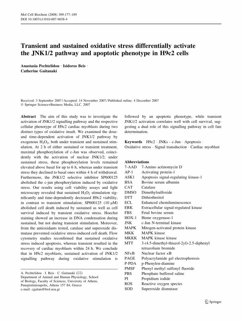
In order to assess the dose-dependent response of
JNK1/2, cardiac myoblasts were incubated with increasing
concentrations of H2O2 either continuously for 2 h (100–
1,000 lM), or transiently (40–1,000 lM) for 5 min fol-
lowed by 15 min of withdrawal. Under these different
experimental conditions, activation of p54 and p46 showed
different dose-dependent patterns. In particular, sustained
exposure to 200 lM of the oxidant resulted in a moderate
phosphorylation of both kinase isoforms, with a maximal
phosphorylation attained at 400 lM of H2O2 [7.64 ± 0.46-
fold (P \ 0.001) and 5.47 ± 0.44-fold relative to control
values for p54 and p46, respectively] (Fig. 1a top panel,
Fig. 1b). During the transient stimulation with 100 lM
H2O2, we observed a strong activation of p54 and p46
JNKs, with maximal phosphorylation detected at 400 lM
of the oxidant [7.95 ± 0.34-fold and 4.37 ± 0.52-fold
180 Mol Cell Biochem (2008) 309:177–189
123
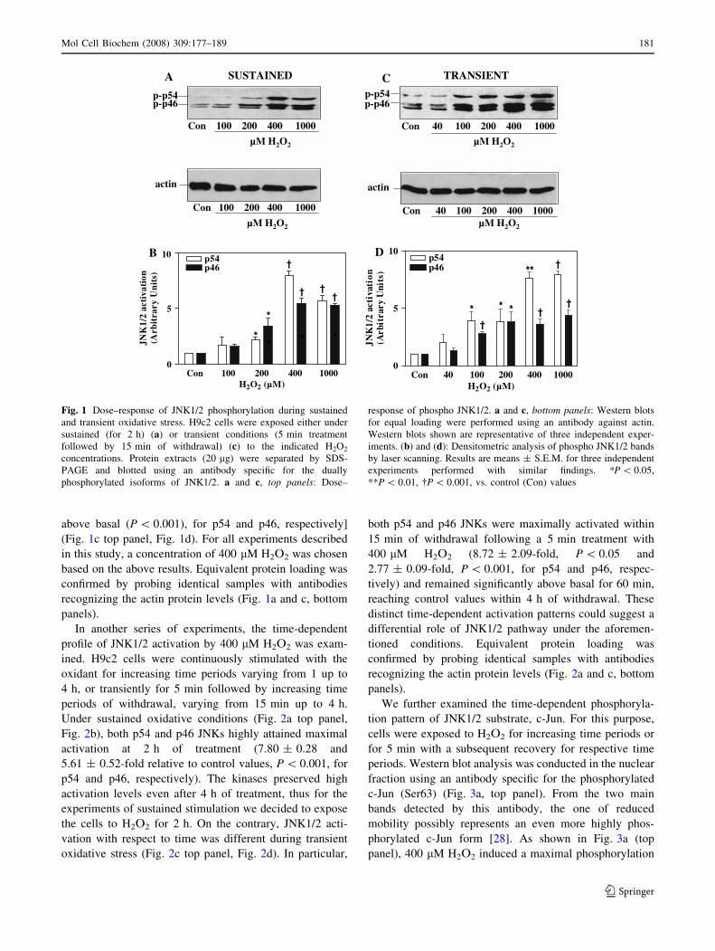
above basal (P \ 0.001), for p54 and p46, respectively]
(Fig. 1c top panel, Fig. 1d). For all experiments described
in this study, a concentration of 400 lM H2O2 was chosen
based on the above results. Equivalent protein loading was
confirmed by probing identical samples with antibodies
recognizing the actin protein levels (Fig. 1a and c, bottom
panels).
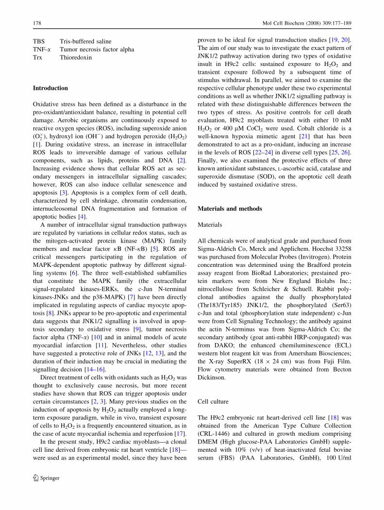
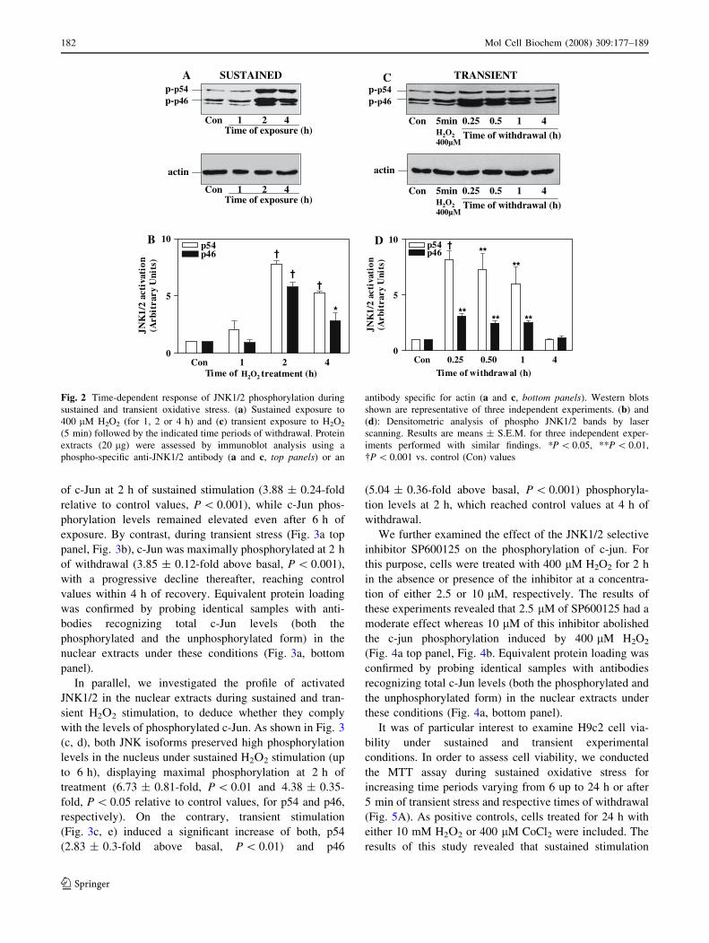
In another series of experiments, the time-dependent
profile of JNK1/2 activation by 400 lM H2O2 was exam-
ined. H9c2 cells were continuously stimulated with the
oxidant for increasing time periods varying from 1 up to
4 h, or transiently for 5 min followed by increasing time
periods of withdrawal, varying from 15 min up to 4 h.
Under sustained oxidative conditions (Fig. 2a top panel,
Fig. 2b), both p54 and p46 JNKs highly attained maximal
activation at 2 h of treatment (7.80 ± 0.28 and
5.61 ± 0.52-fold relative to control values, P \ 0.001, for
p54 and p46, respectively). The kinases preserved high
activation levels even after 4 h of treatment, thus for the
experiments of sustained stimulation we decided to expose
the cells to H2O2 for 2 h. On the contrary, JNK1/2 acti-
vation with respect to time was different during transient
oxidative stress (Fig. 2c top panel, Fig. 2d). In particular,
both p54 and p46 JNKs were maximally activated within
15 min of withdrawal following a 5 min treatment with
400 lM H2O2 (8.72 ± 2.09-fold, P \ 0.05 and
2.77 ± 0.09-fold, P \ 0.001, for p54 and p46, respec-
tively) and remained significantly above basal for 60 min,
reaching control values within 4 h of withdrawal. These
distinct time-dependent activation patterns could suggest a
differential role of JNK1/2 pathway under the aforemen-
tioned conditions. Equivalent protein loading was
confirmed by probing identical samples with antibodies
recognizing the actin protein levels (Fig. 2a and c, bottom
panels).
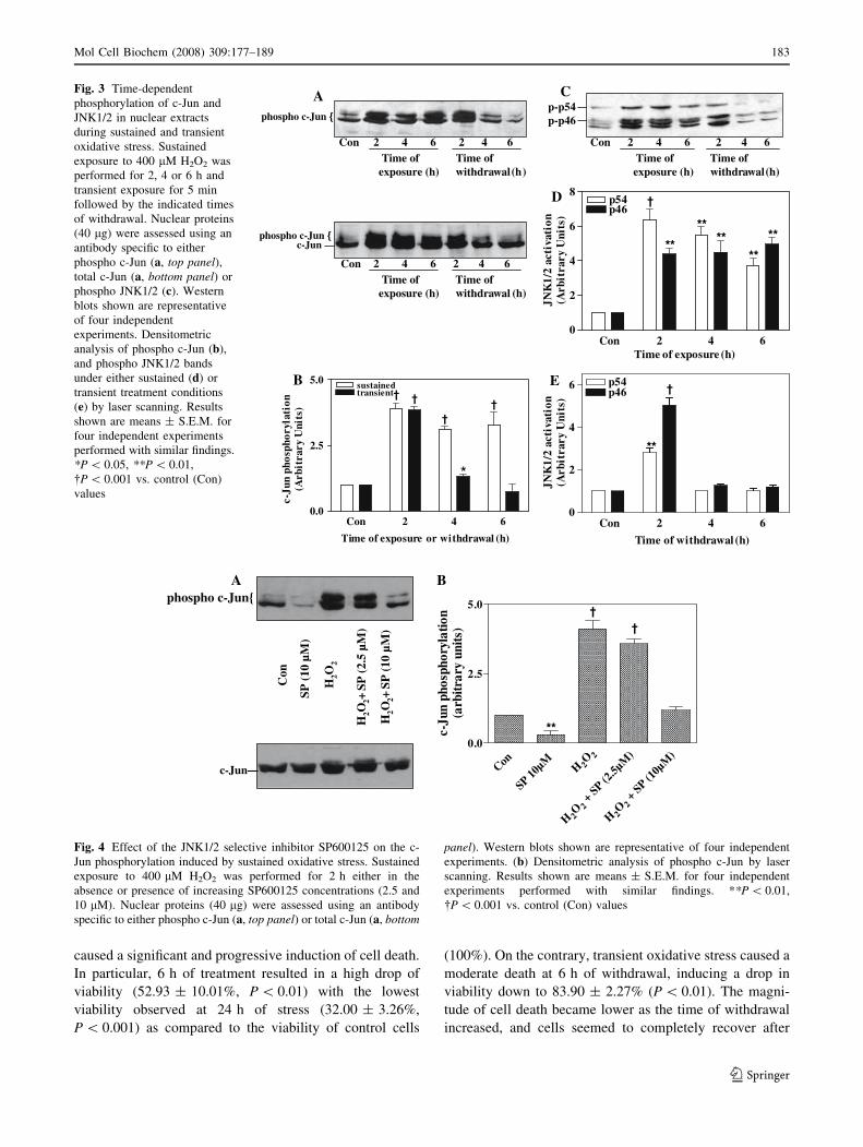
We further examined the time-dependent phosphoryla-
tion pattern of JNK1/2 substrate, c-Jun. For this purpose,
cells were exposed to H2O2 for increasing time periods or
for 5 min with a subsequent recovery for respective time
periods. Western blot analysis was conducted in the nuclear
fraction using an antibody specific for the phosphorylated
c-Jun (Ser63) (Fig. 3a, top panel). From the two main
bands detected by this antibody, the one of reduced
mobility possibly represents an even more highly phos-
phorylated c-Jun form [28]. As shown in Fig. 3a (top
panel), 400 lM H2O2 induced a maximal phosphorylation
001noC 002 004 0001
A DENIATSUS
HMµ 2O2
45p-p64p-p
C TNEISNART
noC 04 001 002 004 0001
HMµ 2O2
45p-p64p-p
001noC 002 004 0001
HMµ 2O2
nitca
04noC 001 004002 0001HMµ 2O2
nitca
noC 04 001 002 004 00010
5
105p 44p 6
*†
* *
**
†
†
†
H2O2 Mµ( )
JNK
1/2
acti
vati
on(A
rbit
rary
Uni
ts)
DB
noC 001 002 004 00010
5
10 45p64p
* ***
*
†
† ††
H2O2 µ( M)
JNK
1/2
acti
vati
on(A
rbit
rary
Un
its)
Fig. 1 Dose–response of JNK1/2 phosphorylation during sustained
and transient oxidative stress. H9c2 cells were exposed either under
sustained (for 2 h) (a) or transient conditions (5 min treatment
followed by 15 min of withdrawal) (c) to the indicated H2O2
concentrations. Protein extracts (20 lg) were separated by SDS-
PAGE and blotted using an antibody specific for the dually
phosphorylated isoforms of JNK1/2. a and c, top panels: Dose–
response of phospho JNK1/2. a and c, bottom panels: Western blots
for equal loading were performed using an antibody against actin.
Western blots shown are representative of three independent exper-
iments. (b) and (d): Densitometric analysis of phospho JNK1/2 bands
by laser scanning. Results are means ± S.E.M. for three independent
experiments performed with similar findings. *P \ 0.05,
**P \ 0.01, �P \ 0.001, vs. control (Con) values
Mol Cell Biochem (2008) 309:177–189 181
123
of c-Jun at 2 h of sustained stimulation (3.88 ± 0.24-fold
relative to control values, P \ 0.001), while c-Jun phos-
phorylation levels remained elevated even after 6 h of
exposure. By contrast, during transient stress (Fig. 3a top
panel, Fig. 3b), c-Jun was maximally phosphorylated at 2 h
of withdrawal (3.85 ± 0.12-fold above basal, P \ 0.001),
with a progressive decline thereafter, reaching control
values within 4 h of recovery. Equivalent protein loading
was confirmed by probing identical samples with anti-
bodies recognizing total c-Jun levels (both the
phosphorylated and the unphosphorylated form) in the
nuclear extracts under these conditions (Fig. 3a, bottom
panel).
In parallel, we investigated the profile of activated
JNK1/2 in the nuclear extracts during sustained and tran-
sient H2O2 stimulation, to deduce whether they comply
with the levels of phosphorylated c-Jun. As shown in Fig. 3
(c, d), both JNK isoforms preserved high phosphorylation
levels in the nucleus under sustained H2O2 stimulation (up
to 6 h), displaying maximal phosphorylation at 2 h of
treatment (6.73 ± 0.81-fold, P \ 0.01 and 4.38 ± 0.35-
fold, P \ 0.05 relative to control values, for p54 and p46,
respectively). On the contrary, transient stimulation
(Fig. 3c, e) induced a significant increase of both, p54
(2.83 ± 0.3-fold above basal, P \ 0.01) and p46
(5.04 ± 0.36-fold above basal, P \ 0.001) phosphoryla-
tion levels at 2 h, which reached control values at 4 h of
withdrawal.
We further examined the effect of the JNK1/2 selective
inhibitor SP600125 on the phosphorylation of c-jun. For
this purpose, cells were treated with 400 lM H2O2 for 2 h
in the absence or presence of the inhibitor at a concentra-
tion of either 2.5 or 10 lM, respectively. The results of
these experiments revealed that 2.5 lM of SP600125 had a
moderate effect whereas 10 lM of this inhibitor abolished
the c-jun phosphorylation induced by 400 lM H2O2
(Fig. 4a top panel, Fig. 4b. Equivalent protein loading was
confirmed by probing identical samples with antibodies
recognizing total c-Jun levels (both the phosphorylated and
the unphosphorylated form) in the nuclear extracts under
these conditions (Fig. 4a, bottom panel).
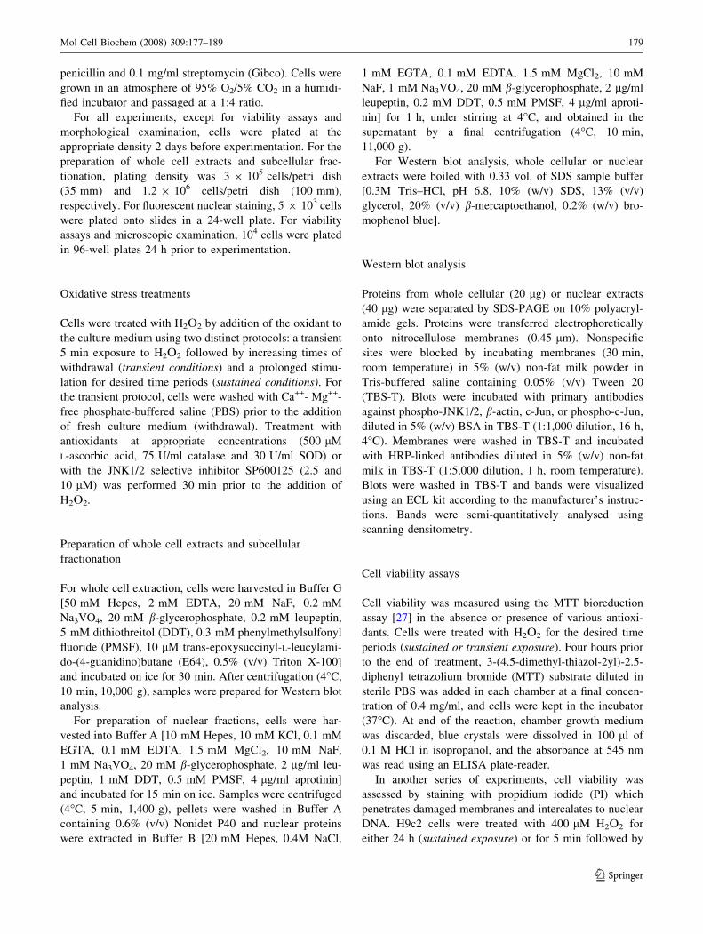
It was of particular interest to examine H9c2 cell via-
bility under sustained and transient experimental
conditions. In order to assess cell viability, we conducted
the MTT assay during sustained oxidative stress for
increasing time periods varying from 6 up to 24 h or after
5 min of transient stress and respective times of withdrawal
(Fig. 5A). As positive controls, cells treated for 24 h with
either 10 mM H2O2 or 400 lM CoCl2 were included. The
results of this study revealed that sustained stimulation
TNEISNARTCA DENIATSUS
1noC 2 4h(erusopxefoemiT )
p-p54p-p46
p-p54p-p46
nim5noC 415.052.0H2O2
Mµ004wardhtiwfoemiT )h(la
1noC 2 4h(erusopxefoemiT )
nitca
nim5noC 415.052.0
nitca
H2O2
H2O2
Mµ004wardhtiwfoemiT )h(la
B
noC 1 2 40
5
105p 44p 6
iT me of trea mt en h(t )
†
*
†
†
JNK
1/2
acti
vati
on(A
rbit
rary
Uni
ts)
noC 52.0 05.0 1 40
5
10 5p 44p 6
Time of wit rdh wa la (h)
**
†**
**
****
JNK
1/2
acti
vati
on(A
rbit
rary
Uni
ts)
D
Fig. 2 Time-dependent response of JNK1/2 phosphorylation during
sustained and transient oxidative stress. (a) Sustained exposure to
400 lM H2O2 (for 1, 2 or 4 h) and (c) transient exposure to H2O2
(5 min) followed by the indicated time periods of withdrawal. Protein
extracts (20 lg) were assessed by immunoblot analysis using a
phospho-specific anti-JNK1/2 antibody (a and c, top panels) or an
antibody specific for actin (a and c, bottom panels). Western blots
shown are representative of three independent experiments. (b) and
(d): Densitometric analysis of phospho JNK1/2 bands by laser
scanning. Results are means ± S.E.M. for three independent exper-
iments performed with similar findings. *P \ 0.05, **P \ 0.01,
�P \ 0.001 vs. control (Con) values
182 Mol Cell Biochem (2008) 309:177–189
123
caused a significant and progressive induction of cell death.
In particular, 6 h of treatment resulted in a high drop of
viability (52.93 ± 10.01%, P \ 0.01) with the lowest
viability observed at 24 h of stress (32.00 ± 3.26%,
P \ 0.001) as compared to the viability of control cells
(100%). On the contrary, transient oxidative stress caused a
moderate death at 6 h of withdrawal, inducing a drop in
viability down to 83.90 ± 2.27% (P \ 0.01). The magni-
tude of cell death became lower as the time of withdrawal
increased, and cells seemed to completely recover after
2Con 4 6426
45p-pC
foemiTh(erusopxe )
foemiTwi wardht )h(la
64p-p
2noC 4 6426foemiT
h(erusopxe )foemiT
wi wardht )h(la
Aohpsohp nuJ-c {
ohpsohp nuJ-c {nuJ-c
2Con 4 6426foemiT
h(erusopxe )foemiT
wi wardht )h(la
D
Con 2 4 60
2
4
6 5p 44p 6
Time of with rd wa la (h)
†
**
JNK
1/2
acti
vati
on(A
rbit
rary
Uni
ts)
Con 2 4 60
2
4
6
85p 44p 6
****
**
**
†
**
Time of xe po us (er h)
JNK
1/2
acti
vati
on(A
rbit
rary
Uni
ts)
B
noC 2 4 60.0
2.5
5.0 us stained† †
††
*
Time of exposure or with rd awal (h)
c-Ju
n ph
osph
oryl
atio
n(A
rbit
rary
Uni
ts)
rt ansientE
Fig. 3 Time-dependent
phosphorylation of c-Jun and
JNK1/2 in nuclear extracts
during sustained and transient
oxidative stress. Sustained
exposure to 400 lM H2O2 was
performed for 2, 4 or 6 h and
transient exposure for 5 min
followed by the indicated times
of withdrawal. Nuclear proteins
(40 lg) were assessed using an
antibody specific to either
phospho c-Jun (a, top panel),total c-Jun (a, bottom panel) or
phospho JNK1/2 (c). Western
blots shown are representative
of four independent
experiments. Densitometric
analysis of phospho c-Jun (b),
and phospho JNK1/2 bands
under either sustained (d) or
transient treatment conditions
(e) by laser scanning. Results
shown are means ± S.E.M. for
four independent experiments
performed with similar findings.
*P \ 0.05, **P \ 0.01,
�P \ 0.001 vs. control (Con)
values
A
noC µ01PS
M2O
2H
+
µ5.2(PS
M)
2O2H
+S
(P
µ01M
)
2O2H
.0 0
.2 5
.5 0
**c-Ju
n ph
osph
oryl
atio
n(a
rbitr
ary
units
) ††
nuJ-c —
Bohpsohp {nuJ-c
Con
H2O
2
SP (
10 µ
M)
H2O
+ 2SP
(2.
5 µM
)
H2O
+ 2SP
(10
µM
)
Fig. 4 Effect of the JNK1/2 selective inhibitor SP600125 on the c-
Jun phosphorylation induced by sustained oxidative stress. Sustained
exposure to 400 lM H2O2 was performed for 2 h either in the
absence or presence of increasing SP600125 concentrations (2.5 and
10 lM). Nuclear proteins (40 lg) were assessed using an antibody
specific to either phospho c-Jun (a, top panel) or total c-Jun (a, bottom
panel). Western blots shown are representative of four independent
experiments. (b) Densitometric analysis of phospho c-Jun by laser
scanning. Results shown are means ± S.E.M. for four independent
experiments performed with similar findings. **P \ 0.01,
�P \ 0.001 vs. control (Con) values
Mol Cell Biochem (2008) 309:177–189 183
123

24 h of withdrawal, displaying similar viability with con-
trol cells (99.10 ± 4.96%). This differential profile of
metabolically viable cells under the two distinct protocols
is consistent with the time-dependent activation pattern of
the JNK1/2 pathway demonstrated in Figs. 2 and 3.
By using the same assay, we next tested the efficacy of
three known antioxidant substances, L-ascorbic acid, cata-
lase and SOD, towards cells exposed continuously to H2O2
for 24 h (Fig. 5B). None of the antioxidants showed any
cytotoxic influence at the concentrations used (data not
shown). Pretreatment of H9c2 cells with L-ascorbic acid
exhibited no protective effect during sustained stimulation,
resulting in low percentages of viability (27.66 ± 2.33%,
P \ 0.001 compared to the control values). Pretreatment
with 30 U/ml SOD resulted in a significant increase of cell
viability (80.10 ± 5.50, P \ 0.001 compared to the values
observed under sustained oxidative exposure) whereas
catalase caused a complete cell viability recovery
(113.66 ± 8.97%, P \ 0.001 compared to the values
observed under sustained oxidative exposure).
In order to elucidate the possible role of JNK1/2 sig-
nalling pathway for cell survival or death, we assessed cell
viability under either sustained (24 h) or transient (5 min
followed by 24 h of withdrawal) oxidative stress in the
absence or presence of the selective JNK1/2 inhibitor
SP600125, using the propidium idodide staining assay. The
tnoC
rlo -h6
T S-h6 9hT- S-h9
T-42 h42S- M
m01(
)
2O2H
(04
µ0
)M
2CoC
l
0
52
05
57
001
**
† †
** **
† †C
ell
viab
ilit
y (%
)
A
C
B
lortnoC 2h4-S
2h4-S
+-L
SAC
42h-S
DOS+AC+S-h42
T
(
)M
m012O2H
0
52
05
57
001
† ††
†
#
Cel
l vi
abil
ity
(%)
#
(a)
(d) (e) (f)
(b) (c)
Fig. 5 Effect of sustained and transient oxidative stress on H9c2 cell
viability in the absence or presence of various antioxidants and
SP600125. (A) Percentage (%) of viable cells during sustained
exposure (row S) or transient exposure (row T) to 400 lM H2O2 for
the indicated time periods using the MTT method. As positive
controls, cells treated with 10 mM H2O2 or 400 lM CoCl2 were used.
Results shown are means ± S.E.M. for three independent experi-
ments performed with similar findings: ** P\0.01, � P\0.001. (B)
Percentage (%) of viable cells during sustained exposure (row S) to
400 lM H2O2 for 24 h in the absence or presence of 500 lM
L-ascorbic acid (L-ASC), 75 U/ml catalase (CAT) or 30 U/ml
SOD using the MTT method. Results shown are means ± S.E.M.
for three independent experiments performed with similar findings.
�P \ 0.001 vs. control (Con) values; # P \ 0.001 vs. values observed
after treatment of cells with 400 lM H2O2 for 24 h. (C) Cells were
grown on slides and exposed to 400 lM H2O2 either continuously for
24 h, or for 5 min followed by 24 h of withdrawal, both in the
absence or presence of the JNK1/2 selective inhibitor SP600125.
Viability of cells was assessed by fluorescence microscopy using the
propidium iodide staining of dead cells. (a) Control, (b) sustained
exposure, (c) transient exposure, (d) cells treated with 10 lM
SP600125 for 24 h, (e) sustained exposure in the presence of
10 lM SP600123, (f) transient exposure in the presence of 10 lM
SP600123. Representative photographs were taken with a camera
adjusted to a fluorescence microscope. Pictures are representative of
four independent experiments. Magnification (209)
184 Mol Cell Biochem (2008) 309:177–189
123
results of this study showed that cells exposed to 400 lM
H2O2 for 24 h were heavily stained by propidium iodide,
indicating an extensive cell death (Fig. 5Cb). Furthermore,
the JNK1/2 selective inhibitor SP600125 (10 lL) abol-
ished this propidium iodide staining under sustained
exposure (Fig. 5Ce). On the contrary, transient exposure to
oxidative stress resulted in cell survival (Fig. 5Cc) and
SP600125 (10 lM) induced a significant increase in pro-
pidium iodide staining, thus cell death (Fig. 5Cf). As
controls untreated cells or cells treated with the respective
inhibitor alone were included (Fig. 5Ca and 5Cd, respec-
tively). The above results reveal the possible involvement
of JNK1/2 signalling pathway in cell survival or death
during oxidative stress.
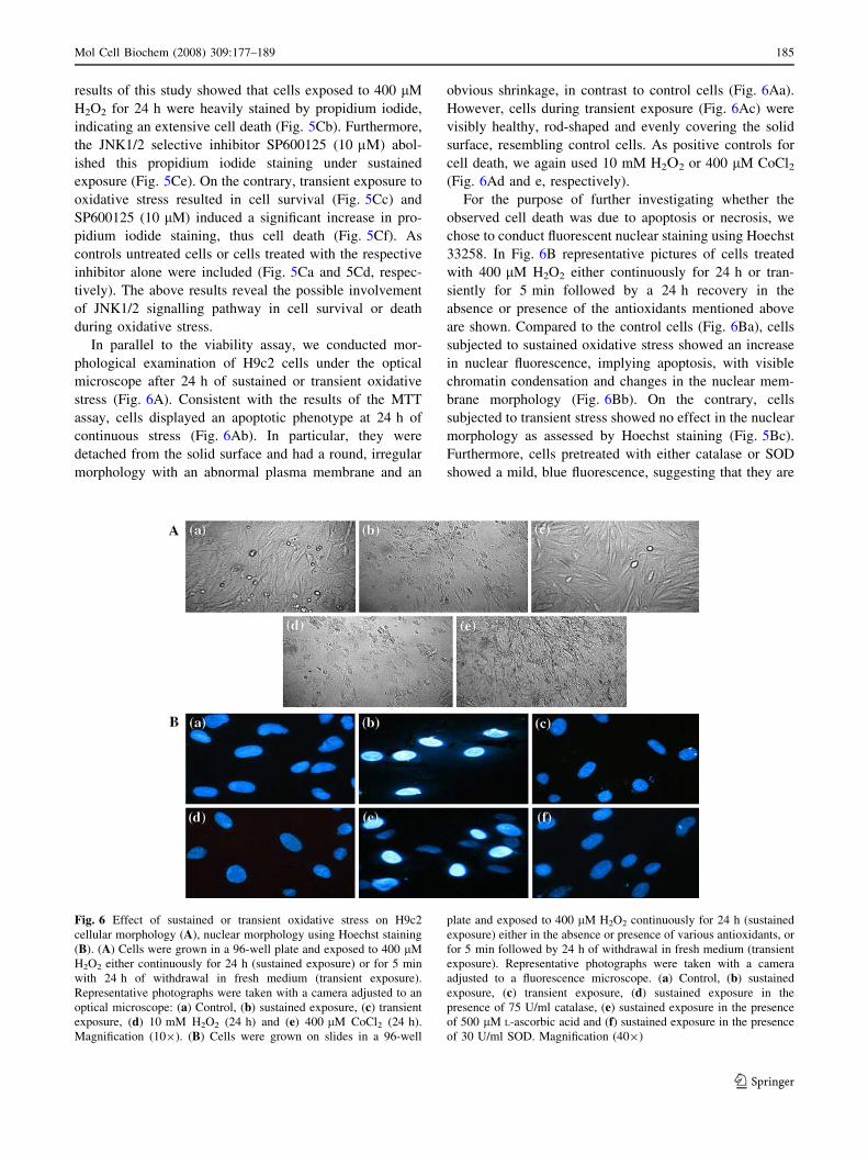
In parallel to the viability assay, we conducted mor-
phological examination of H9c2 cells under the optical
microscope after 24 h of sustained or transient oxidative
stress (Fig. 6A). Consistent with the results of the MTT
assay, cells displayed an apoptotic phenotype at 24 h of
continuous stress (Fig. 6Ab). In particular, they were
detached from the solid surface and had a round, irregular
morphology with an abnormal plasma membrane and an
obvious shrinkage, in contrast to control cells (Fig. 6Aa).
However, cells during transient exposure (Fig. 6Ac) were
visibly healthy, rod-shaped and evenly covering the solid
surface, resembling control cells. As positive controls for
cell death, we again used 10 mM H2O2 or 400 lM CoCl2(Fig. 6Ad and e, respectively).
For the purpose of further investigating whether the
observed cell death was due to apoptosis or necrosis, we
chose to conduct fluorescent nuclear staining using Hoechst
33258. In Fig. 6B representative pictures of cells treated
with 400 lM H2O2 either continuously for 24 h or tran-
siently for 5 min followed by a 24 h recovery in the
absence or presence of the antioxidants mentioned above
are shown. Compared to the control cells (Fig. 6Ba), cells
subjected to sustained oxidative stress showed an increase
in nuclear fluorescence, implying apoptosis, with visible
chromatin condensation and changes in the nuclear mem-
brane morphology (Fig. 6Bb). On the contrary, cells
subjected to transient stress showed no effect in the nuclear
morphology as assessed by Hoechst staining (Fig. 5Bc).
Furthermore, cells pretreated with either catalase or SOD
showed a mild, blue fluorescence, suggesting that they are
Fig. 6 Effect of sustained or transient oxidative stress on H9c2
cellular morphology (A), nuclear morphology using Hoechst staining
(B). (A) Cells were grown in a 96-well plate and exposed to 400 lM
H2O2 either continuously for 24 h (sustained exposure) or for 5 min
with 24 h of withdrawal in fresh medium (transient exposure).
Representative photographs were taken with a camera adjusted to an
optical microscope: (a) Control, (b) sustained exposure, (c) transient
exposure, (d) 10 mM H2O2 (24 h) and (e) 400 lM CoCl2 (24 h).
Magnification (109). (B) Cells were grown on slides in a 96-well
plate and exposed to 400 lM H2O2 continuously for 24 h (sustained
exposure) either in the absence or presence of various antioxidants, or
for 5 min followed by 24 h of withdrawal in fresh medium (transient
exposure). Representative photographs were taken with a camera
adjusted to a fluorescence microscope. (a) Control, (b) sustained
exposure, (c) transient exposure, (d) sustained exposure in the
presence of 75 U/ml catalase, (e) sustained exposure in the presence
of 500 lM L-ascorbic acid and (f) sustained exposure in the presence
of 30 U/ml SOD. Magnification (409)
Mol Cell Biochem (2008) 309:177–189 185
123
healthy viable cells (Fig. 6Bd and 6Bf, for catalase and
SOD, respectively). By contrast, L-ascorbic acid showed no
anti-apoptotic effect (Fig. 6Be). These results, taken toge-
ther with the viability assay ones and the morphological
examination analysed earlier, imply the induction of an
apoptotic process during sustained H2O2 stimulation,
which is not evident during transient stress. Since JNK1/2
follow a differential activation pattern under the two dis-
tinct experimental conditions used and SP600125 abolishes
the detrimental cell death after sustained exposure or the
beneficial effect of transient exposure, these results suggest
that prolonged JNK1/2 pathway activation is related with a
H2O2-induced cell death, whereas transient activation of
this signalling pathway is related with a H2O2-induced cell
survival.
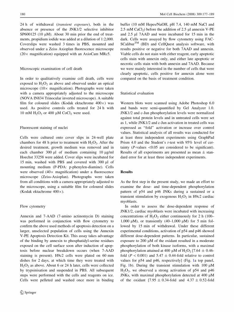
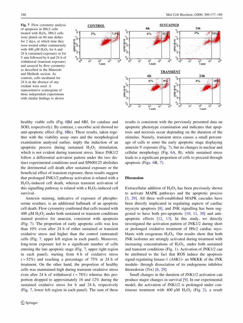
Annexin staining, indicative of exposure of phospho-
serine residues, is an additional hallmark of an apoptotic
cell death. Flow cytometry confirmed that cells treated with
400 lM H2O2 under both sustained or transient conditions
stained positive for annexin, consistent with apoptosis
(Fig. 7). The proportion of early apoptotic cells was less
than 10% even after 24 h of either sustained or transient
oxidative stress and higher than the control (untreated)
cells (Fig. 7, upper left region in each panel). Moreover,
long-term exposure led to a significant number of cells
entering the late apoptotic stage (Fig. 7, upper right region
in each panel), starting from 6 h of oxidative stress
(*52%) and reaching a percentage of 75% at 24 h of
treatment. On the other hand, the proportion of healthy
cells was maintained high during transient oxidative stress
even after 24 h of withdrawal (*78%) whereas this pro-
portion dropped to approximately 16 and 12% during the
sustained oxidative stress for 6 and 24 h, respectively
(Fig. 7, lower left region in each panel). The sum of these
results is consistent with the previously presented data on
apoptotic phenotype examination and indicates that apop-
tosis and necrosis occur depending on the duration of the
stimulus. Namely, transient stress causes a small percent-
age of cells to enter the early apoptotic stage displaying
annexin-V exposure (Fig. 7), but no changes in nuclear and
cellular morphology (Fig. 6A, B), while sustained stress
leads to a significant proportion of cells to proceed through
apoptosis (Figs. 6B, 7).
Discussion
Extracellular addition of H2O2 has been previously shown
to activate MAPK pathways and the apoptotic process
[3, 29]. All three well-established MAPK cascades have
been directly implicated in regulating aspects of cardiac
myocyte apoptosis [8], and JNK signalling has been sug-
gested to have both pro-apoptotic [10, 11, 30] and anti-
apoptotic effects [12, 13]. In this study, we directly
investigated the activation pattern of JNK1/2 during short
or prolonged oxidative treatment of H9c2 cardiac myo-
blasts with exogenous H2O2. Our results show that both
JNK isoforms are strongly activated during treatment with
increasing concentrations of H2O2, under both sustained
and transient conditions (Fig. 1). Activation of JNK1/2 can
be attributed to the fact that ROS induce the apoptosis
signal-regulating kinase-1 (ASK1)- an MKKK of the JNK
module- through dissociation of its endogenous inhibitor
thioredoxin (Trx) [6, 29].
Small changes in the duration of JNK1/2 activation can
produce major changes in survival [9]. In our experimental
model, the activation of JNK1/2 is prolonged under con-
tinuous treatment with 400 lM H2O2 (Fig. 2), a result
Fig. 7 Flow cytometry analysis
of apoptosis in H9c2 cells
treated with H2O2. H9c2 cells
were plated on 60 mm dishes
for 2 days, at which time they
were treated either continuously
with 400 lM H2O2 for 6 and
24 h (sustained exposure) or for
5 min followed by 6 and 24 h of
withdrawal (transient exposure)
and assayed by flow cytometry
as described in the Materials
and Methods section. As
controls, cells incubated for
24 h in the absence of any
oxidant were used. A
representative scattergram of
three independent experiments
with similar findings is shown
186 Mol Cell Biochem (2008) 309:177–189
123
consistent with previous reports [20, 31, 32]. By contrast,
short-term treatment causes an early activation of JNK1/2
during withdrawal, suggesting a transient activation of this
pathway that might serve a physiological role. Transient
JNK activity during oxidative treatment has been reported
previously [17], but to our knowledge our study is the first
to characterise the activation pattern of JNK1/2 pathway in
H9c2 cells under these two distinct types of oxidative
stimulation, revealing an interesting differential profile.
A growing body of evidence suggests that H2O2 can
modulate transcription factors, such as activating protein-1
(AP-1) and NF-jB [1]. The transcription factor c-Jun is a
basic component of AP-1 and the main substrate of JNK1/2
[7]. Our results demonstrate a respective time-dependent
pattern of c-Jun phosphorylation (Fig. 3), in support to the
differential profile of activated JNK1/2 in the whole cell
extract (Fig. 2) and the nuclear fraction (Fig. 3c, d) under
the same conditions. Furthermore, the JNK1/2 selective
inhibitor SP600125 abolishes the c-Jun phosphorylation
induced by oxidative stress (Fig. 4). The prolonged acti-
vation of c-Jun during sustained oxidative challenge could
have a physiological meaning in the cellular context, since
AP-1 is associated with a large number of apoptotic sce-
narios [33]. However, the precise role of AP-1 in response
to JNK activation is likely to be modified by the activity of
other transcription factors that interact with AP-1 on the
promoters of target genes [15]. By contrast, during tran-
sient oxidative stress both phosphorylated p54 and p46
JNKs were detected in the nucleus only at 2 h of with-
drawal (Fig. 3c, e), a pattern that largely resembles the
transient c-Jun phosphorylation (Fig. 3a, b) under such
conditions.
Nevertheless, it is not known whether the aforemen-
tioned nuclear phosphorylated isoforms represent a fraction
of activated JNK1/2 that translocates to the nucleus or
whether they are derived from phosphorylation in the
nucleus by specific MAPK kinases (MKKs). In resting
cells, JNK1/2 are localized in both the cytoplasm and the
nucleus [34], thus the nuclear isoforms we detected could
be indeed phosphorylated within the latter.
Subsequent studies to investigate the phenotype of H9c2
cells under the two distinct experimental protocols were in
accordance with the pattern of JNK1/2 activation men-
tioned previously. Sustained H2O2 treatment resulted in a
significant percentage of cell death and an apoptotic phe-
notype, as assessed by viability assays (Fig. 5A, C),
microscopic examination (Fig. 6A) and Hoechst 33258
staining (Fig. 6B). On the contrary, during transient stim-
ulation there was only a small and transient impact on cell
survival (Fig. 5A, C), and no obvious phenotypical chan-
ges as compared to control cells (Fig. 6A, B). In addition,
the abolishment of either cell death during sustained or cell
survival during transient stimulation by the JNK1/2
selective inhibitor SP600125 (Fig. 5C), provides evidence
for a direct link between this signalling pathway and cell
fate determination. Moreover, flow cytometry experiments
(Fig. 7) showed a small percentage of early apoptosis
during transient stimulation in contrast to substantial
number of late apoptotic cells during sustained oxidative
episode, in compliance with the above morphological data.
Our results comply with other studies that have reported a
significant decrease in cell viability during oxidative insults
[35, 36]. They are also consistent with previous reports on
increased Hoechst staining [13, 36, 37] or pathological
phenotype [17, 37, 38] of cardiac myocytes under different
types of sustained oxidative stress. From the antioxidants
tested, catalase and SOD were able to prevent the observed
cell death and apoptotic phenotype under sustained con-
ditions (Figs. 5B, 6B).
Overall, the results of the present study demonstrate that
JNK isoforms are strongly activated in H9c2 cardiac
myoblasts during both sustained and transient oxidative
stimulation. Nevertheless, their time-dependent profile is
different under these two distinct conditions of stress, in
consistence with the pattern of their phosphorylated sub-
strate, c-Jun. During the short-term oxidative episode,
transient activation of JNK1/2 correlates well with a sur-
vival phenotype. Nonetheless, during prolonged H2O2
treatment, sustained JNK1/2 activation is followed by a
pathological cellular phenotype with well-established
apoptotic hallmarks.
Our results suggest that stress-dependent JNK1/2 acti-
vation might be implicated in diverse cellular responses, by
signalling in favour of either survival or apoptosis [15].
The mechanisms of this dual role of JNK have been dis-
cussed in a number of reviews [6, 8, 15]. In addition, a
recent study in our laboratory reported a JNK and p38-
MAPK-dependent up-regulation of heme oxygenase-1
(HOX-1) in H9c2 cells treated with 200 lM H2O2, thus
implicating JNK in the mechanisms preserving cellular
homeostasis [39].
A critical factor determining cell fate is thought to be the
duration of JNK activation, with transient JNK activation
mediating a survival response and prolonged JNK activa-
tion contributing to apoptotic responses [14, 16].
Moreover, the dynamic balance between ERK and JNK
pathways is important in determining whether a cell sur-
vives or undergoes apoptosis [14, 40]. Therefore, it is likely
that the differences in sensitivity of various cell types to the
cytotoxic effects of H2O2 reflect the relative activation of
ERK and JNK [9]. A third explanation for the different
roles of JNK in apoptosis signalling is that the cellular
outcome may depend on the cross talk with other signal
transduction pathways (e.g. NF-jB and Akt). For example,
target genes of the anti-apoptotic NF-jB pathway may
contain JNK-responsive elements in their promoters
Mol Cell Biochem (2008) 309:177–189 187
123
(e.g. AP-1 sites), as has been shown for inhibitory of
apoptosis protein (cIAP) [41].
In conclusion, the present study demonstrates that
transient and sustained oxidative stress can cause differ-
ential JNK pathway activation, resulting in cell survival or
apoptosis, respectively, in accordance with previous studies
on UV-C [14] and TNF-a treatment [10, 42]. Clearly, a
greater understanding of the regulatory role of JNK1/2
pathway in apoptosis may reveal additional strategies for
treating heart failure, cardiomyopathy and acute ischemic
heart damage in humans, and Dr A. Gritzapis for his
assistance in FACS analyis.
Acknowledgements This study was funded by O.P.E.I.V.T. II. We
thank Vasilis Varvarigos for his kind assistance in fluorescence
microscopy examination, and Dr A. Gritzapis for his assistance in
FACS analysis.
References
1. Haddad JJ (2002) Antioxidant and prooxidant mechanisms in the
regulation of redox(y)-sensitive transcription factors. Cell Signal
14:879–897
2. Marczin N, El-Habashi N, Hoare GS, Bundy RE, Yacoub M
(2003) Antioxidants in myocardial ischaemia- reperfusion injury:
therapeutic potential and basic mechanisms. Arch Biochem
Biophys 420:222–236
3. Valko M, Leibfritz D, Moncol J, Cronin MTD, Mazur M, Telser J
(2007) Free radicals and antioxidants in normal physiological
functions and human disease. Int J Biochem Cell Biol 39:44–84
4. Kumar D, Jugdutt BI (2003) Apoptosis and oxidants in the heart.
J Lab Clin Med 142:288–297
5. Allen RG, Tresini M (2000) Oxidative stress and gene regulation.
Free Radic Biol Med 28:463–499
6. Sumbayev VV, Yasinska IM (2005) Regulation of MAP kinase-
dependent apoptotic pathway: implication of reactive oxygen and
nitrogen species. Arch Biochem Biophys 436:406–412
7. Kyriakis JM, Avruch J (2001) Mammalian MAPKs signal
transduction pathways activated by stress and inflammation.
Physiol Rev 81:807–869
8. Baines CP, Molkentin JD (2005) Stress signalling pathways that
modulate cardiac myocyte apoptosis. J Mol Cell Cardiol 38:47–62
9. Wang X, Martindale JL, Holbrook NJ (1998) The cellular
response to oxidative stress: influences of mitogen-activated
protein kinase signalling pathways on cell survival. Biochem J
333:291–300
10. Guo YL, Baysal K, Kang B, Yang LJ, Williamson JR (1998)
Correlation between sustained c-Jun N-terminal protein kinase
activation and apoptosis induced by tumor necrosis factor-a in rat
mesangial cells. J Biol Chem 273:4027–4034
11. Li GW, Zaheer A, Coppey L, Oskarsson HJ (1998) Activation of
JNK in the remote myocardium after large myocardial infarction
in rats. Biochem Biophys Res Commun 246:816–820
12. Dougherty CJ, Kubasiak LA, Prentice H, Andreka P, Bishopric
NH, Webster KA (2002) Activation of c-Jun N-terminal kinase
promotes survival of cardiac myocytes after oxidative stress.
Biochem J 362:561–571
13. Engelbrecht AM, Niesler C, Page C, Lochner A (1996) p38 and
JNK have distinct regulatory functions on the development of
apoptosis during simulated ishaemia and reperfusion in neonatal
cardiomyocytes. Basic Res Cardiol 99:338–350
14. Chen YR, Wang X, Templeton D, Davis RJ, Tan TH (1996) The
Role of c-jun N-terminal kinase (JNK) in apoptosis induced by
ultraviolet C and c radiation. J Biol Chem 271:31929–31936
15. Davis RJ (2000) Signal transduction by the JNK group of MAP
kinases. Cell 103:239–252
16. Lamb JA, Ventura JJ, Hess P, Flavell RA, Davis RJ (2003) JunD
mediates survival signaling by the JNK signal transduction
pathway. Mol Cell 11:1479–1489
17. Han H, Long H, Wang H, Wang J, Zhang Y, Wang Z (2004)
Progressive apoptotic cell death triggered by transient oxidative
insult in H9c2 rat ventricular cells: a novel pattern of apoptosis
and the mechanisms. Am J Physiol Heart Circ Physiol 286:
H2169–H2182
18. Kimes BW, Brandt BL (1976) Properties of a clonal muscle cell
line from rat heart. Exp Cell Res 98:367–381
19. Su C, Chong K, Edelstein K, Lille S, Khardori R, Lai C (1999)
Constitutive hsp70 attenuates hydrogen peroxide-induced mem-
brane lipid peroxidation. Biochem Biophys Res Commun
265:279–284
20. Turner NA, Xia F, Azhar G, Zhang X, Liu L, Wei JY (1998)
Oxidative stress induces DNA fragmentation and caspase acti-
vation via the JNK pathway in H9c2 cardiac muscle cells. J Mol
Cell Cardiol 30:1789–1801
21. Goldberg MA, Dunning SP Bunn HF (1988) Regulation of
erythropoietin gene: evidence that the oxygen sensor is a heme
protein. Science 242:1412–1415
22. Zou W, Yan M, Xu W, Huo H, Sun L, Zheng Z, Liu X (2001)
Cobalt chloride induces PC12 cells apoptosis through reactive
oxygen species and accompanied by AP-1 activation. J Neurosci
Res 64:646–653
23. Xi L, Taher M, Yin C, Salloum F, Kukreja RC (2004) Cobalt
chloride induces delayed cardiac preconditioning in mice through
selective activation of HIF-1a and AP-1 and iNOS signaling. Am
J Physiol Heart Circ Physiol 287:H2369–H2375
24. Kotake-Nara E, Saida K (2006) Endothelin-2/vasoactive intesti-
nal contractor: regulation of expression via reactive oxygen
species induced by CoCl2 and biological activities including
neurite outgrowth in PC12 cells. Scientific World J 6:176–186
25. Tomaro ML, Frydman J, Frydman RB (1991) Heme oxygenase
induction by CoCl2, Co-protoporphyrin IX, phenylhydrazine, and
diamide: evidence for oxidative stress involvement. Arch Bio-
chem Biophys 286:610–617
26. Chandel NS, McClintock DS, Feliciano CE, Wood TM, Melen-
dez JA, Rodriguez AM, Schumacker PT (2000) Reactive oxygen
species generated at mitochondrial complex III stabilize hypoxia-
inducible factor 1a during hypoxia. J Biol Chem 275:25130–
25138
27. Denizot F, Lang R (1986) Rapid colorimetric assay for cell
growth and survival. Modifications to the tetrazolium dye pro-
cedure giving improved sensitivity and reliability. J Immunol
Method 89:271–277
28. Clerk A, Kemp TJ, Harrison JG, Mullen AJ, Barton PJR, Sugden
PH (2002) Up-regulation of c-Jun mRNA in cardiac myocytes
requires the extracellular signal-regulated kinase cascade, but
c-Jun N-terminal kinases are required for efficient up-regulation
of c-Jun protein. Biochem J 368:101–110
29. Takeda K, Matsuzawa A, Nishitoh H, Ichijo H (2003) Roles of
MAPKKK ASK1 in stress-induced cell death. Cell Struct Funct
28:23–29
30. Aikawa R, Komuro I, Yamazaki T, Zou Y, Kudoh S, Tanaka M,
Shiojima I, Hiroi Y, Yazaki T (1997) Oxidative stress activates
extracellular signal-regulated kinases through Src and Ras in
cultured cardiac myocytes of neonatal rats. J Clin Invest
100:1813–1821
31. Hong F, Kwon SJ, Jhun BS, Kim SS, Ha J, Kim SJ, Sohn NW,
Kang C, Kang I (2001) IGF-1 protects H9c2 cardiac myoblasts
188 Mol Cell Biochem (2008) 309:177–189
123
from oxidative stress-induced apoptosis via PI3K and ERK
pathway. Life Sci 68:1095–1105
32. Mizukami Y, Okamura T, Miura T, Kimura M, Mogami K,
Todoroki-Ikeda N, Kobayashi S, Matsuzaki M (2001) Phos-
phorylation of proteins and apoptosis induced by JNK1 activation
in rat cardiomyocytes by H2O2 stimulation. Biochim Biophys
Acta 1540:213–220
33. Ameyar M, Wisniewska M, Weitzman JB (2003) A role for AP-1
in apoptosis: the case for and against. Biochimie 85:747–752
34. Whitmarsh AJ, Davis RJ (1996) Transcription factor AP-1 reg-
ulation by mitogen-activated protein kinase signal transduction
pathways. J Mol Med 74:589–607
35. Chen HW, Chien CT, Yu SL, Lee YT, Chen WJ (2002) Cyclo-
sporine A regulates oxidative stress-induced apoptosis in
cardiomyocytes: mechanisms via ROS generation, iNOS and
Hsp70. Brit J Pharmacol 137:771–781
36. Park C, So HS, Shin CH, Baek SH, Moon BS, Shin SH, Lee HS,
Lee DW, Park R (2003) Quercetin protects the hydrogen perox-
ide-induced apoptosis via inhibition of mitochondrial dysfunction
in H9c2 cardiomyoblast cells. Biochem Pharmacol 66:1287–1295
37. Yue TL, Wang C, Romanic A, Kikly K, Keller P, DeWolf WE,
Hart TK, Thomas HC, Storer B, Gu JL, Wang X, Feuerstein GZ
(1998) Staurosporine-induced apoptosis in cardiomyocytes:
a potential role for caspase-3. J Mol Cell Cardiol 30:495–507
38. Chen QM, Tu VC, Wu Y, Bahl JJ (2000) Hydrogen peroxide dose
dependent induction of cell death or hypertrophy in cardiomyo-
cytes. Arch Biochem Biophys 373:242–248
39. Aggeli IK, Gaitanaki C, Beis I (2006) Involvement of JNKs and
p38-MAPK/MSK1 pathways in H2O2-induced upregulation of
heme oxygenase-1 mRNA in H9c2 cells. Cell Signal 18:1801–
1812
40. Xia Z, Dickens M, Raingeaud J, Davis RJ, Greenberg ME (1995)
Opposing effects of ERK and JNK–p38 MAP kinases on apop-
tosis. Science 270:1326–1331
41. Hong SY, Yoon WH, Park JH, Kang SG, Ahn JH, Lee TH (2000)
Involvement of two NF-kappa B binding elements in tumor
necrosis factor alpha-, CD40-, and Epstein-Barr virus latent
membrane protein 1-mediated induction of the cellular inhibitor
of apoptosis protein 2 gene. J Biol Chem 275:8022–18028
42. Roulston A, Reinhard C, Amiri P, Williams LT (1998) Early
activation of c-Jun N-terminal kinase and p38 kinase regulate cell
survival in response to tumor necrosis factor-a. J Biol Chem
273:10232–10239
Mol Cell Biochem (2008) 309:177–189 189
123