Embed Size (px)
Citation preview
Transgenic maize lines with cell-type specific expressionof fluorescent proteins in plastidsAmir Sattarzadeh1, Jonathan Fuller2, Salvador Moguel3,†, Katia Wostrikoff2,‡, Shirley Sato3,Sarah Covshoff2,§, Tom Clemente3, Maureen Hanson1 and David B. Stern2,*
1Department of Molecular Biology and Genetics, Cornell University, Ithaca, NY, USA2Boyce Thompson Institute for Plant Research, Tower Rd., Ithaca, NY, USA3Department of Agronomy & Horticulture, Center for Biotechnology, Center for Plant Science Innovation, University of Nebraska-Lincoln, Lincoln, NE, USA
Received 4 April 2009;
revised 9 September 2009;
accepted 12 September 2009.
*Correspondence (Tel 254 1306; fax 607
254 6779; e-mail [email protected])
†Present address: Division of Science &
Mathematics, Union College, Lincoln, NE,
USA.
‡Present address: IBPC, 13 rue Pierre et
Marie Curie, Paris, France.
§Present address: Department of Plant
Sciences, University of Cambridge,
Cambridge, United Kingdom.
Accession numbers: GQ221700 (mzYFP),
GQ221701 (mzGFP), GQ221702 (mzBFP).
Keywords: fluorescent protein,
transgenic, chloroplast, bundle
sheath, mesophyll.
SummaryPlastid number and morphology vary dramatically between cell types and at
different developmental stages. Furthermore, in C4 plants such as maize,
chloroplast ultrastructure and biochemical functions are specialized in mesophyll
and bundle sheath cells, which differentiate acropetally from the proplastid form
in the leaf base. To develop visible markers for maize plastids, we have created a
series of stable transgenics expressing fluorescent proteins fused to either the
maize ubiquitin promoter, the mesophyll-specific phosphoenolpyruvate carboxylase
(PepC) promoter, or the bundle sheath-specific Rubisco small subunit 1 (RbcS)
promoter. Multiple independent events were examined and revealed that maize
codon-optimized versions of YFP and GFP were particularly well expressed, and
that expression was stably inherited. Plants carrying PepC promoter constructs
exhibit YFP expression in mesophyll plastids and the RbcS promoter mediated
expression in bundle sheath plastids. The PepC and RbcS promoter fusions also
proved useful for identifying plastids in organs such as epidermis, silks, roots and
trichomes. These tools will inform future plastid-related studies of wild-type and
mutant maize plants and provide material from which different plastid types may
be isolated.
Introduction
Maize has a long history as a model for plastid biology
and is of particular importance as a platform for studying
the biochemistry and developmental biology of C4 photo-
synthesis. The successive maturation of chloroplasts from
proplastids from leaf base to tip was described nearly
35 years ago (Leech et al., 1973) and the dimorphism, or
Kranz anatomy of maize leaf chloroplasts in mesophyll vs.
bundle sheath cells was noted several years later (Miranda
et al., 1981). The maize plastid gene expression apparatus
(Maier et al., 1995) and proteome (Majeran et al., 2005)
have also been scrutinized. Two mutants specifically defec-
tive in bundle sheath differentiation have been isolated
(Langdale and Kidner, 1994; Roth et al., 1996) and later
studied at the molecular level (Hall et al., 1998; Brutnell
et al., 1999). Despite these advances, the signals that
regulate cell type specification and plastid morphology are
still poorly understood.
A valuable tool for investigating organelle number and
morphology is labelling with fluorescent proteins (FPs).
Among numerous applications have been studies of mito-
chondrial division in yeast (e.g. Tieu et al., 2002), observa-
tions of plastids and plastid tubules (stromules) in higher
plants (Kohler et al., 1997a; Kohler and Hanson, 2000;
Kwok and Hanson, 2004; Hanson and Sattarzadeh, 2008),
and identification of mitochondria and mitochondrial
mutants in higher plants (Kohler et al., 1997b; Logan
et al., 2003). In the case of plastids, the fluorescent tag
has been expressed in the nucleus fused to a transit pep-
tide (Kohler et al., 1997b; Primavesi et al., 2008), fused to
the entire coding region of lipoxygenase 10 (Mohanty
ª 2009 The Authors112 Journal compilation ª 2010 Blackwell Publishing Ltd
Plant Biotechnology Journal (2010) 8, pp. 112–125 doi: 10.1111/j.1467-7652.2009.00463.x
et al., 2009), or expressed within the organelle following
insertion of a cassette into the chloroplast genome (Shiina
et al., 2000; Reed et al., 2001).
We commenced a project to test different fluorescent
tags for labelling of maize plastids in stable transformants
and also to develop lines that labelled specifically either
bundle sheath or mesophyll chloroplasts. Three new maize
codon-optimized fluorescent protein coding regions were
produced. We find that the promoter from a Rubisco small
subunit-encoding gene can drive bundle sheath-
specific expression and the PepC promoter results in expres-
sion in mesophyll cells. We were also able to visualize plast-
ids in many organs. These transgenic lines should be useful
to maize researchers studying various aspects of plastid
development and should faciliate fluorescence-activated cell
sorting of bundle sheath and mesophyll plastids.
Results
Development of transformation cassettes
Our initial expression cassettes were constructed with the
human codon-optimized green fluorescent protein (GFP)
that was shown to be effective for transient expression in
maize protoplasts (Chiu et al., 1996). Subsequently, coding
regions for three different variants of GFP were synthesized,
using preferred maize codons and incorporating appropri-
ate mutations with respect to the original jellyfish GFP
sequence. All three variants carry the Q80R mutation origi-
nally incorporated into most GFPs by accident, plus addi-
tional mutations known to enhance solubility or to cause
spectral shifts to yellow or blue (Cormack et al., 1996;
Crameri et al., 1996; Davis and Vierstra, 1998; Wachter
et al., 1998). The predicted protein sequences of mzGFP
(mz; modified zea) and mzBFP are identical to smGFP and
smBFP described by Davis and Vierstra (1998). The mzYFP
protein sequence is identical to the yellow fluorescent pro-
tein (YFP) for which a crystal structure was determined
(Wachter et al., 1998). These are detailed in Table 1.
Table 2 shows the series of chimeric genes used for sta-
ble transformation of the maize recipient genotype Hi II.
Our aim was to develop a cassette driving ubiquitous
expression of a chloroplast-targeted GFP on one hand,
and tissue-specific cassettes driving mesophyll or bundle
sheath chloroplast expression on the other hand. To deter-
mine the best promoter for strong general expression, we
tested the CaMV 35S and rice actin (Cao et al., 1992) pro-
moters, reasoning that the 35S promoter is widely used
for high expression and is known to function in trans-
formed maize callus (Fromm et al., 1986) and regenerated
plants (Gordon-Kamm et al., 1990). Although heterolo-
gous, the rice actin promoter was also initially tested, as it
is reputed to be stronger than the 35S promoter in mono-
cot transformation (discussed in Prakash et al., 2008). We
also created a cassette where GFP was driven by the Phos-
phoenolpyruvate Carboxylase (PepC) promoter; this gene
should express GFP only in mesophyll cells. Conversely, we
used the promoter of Rubisco small subunit (RbcS) to drive
GFP expression specifically in bundle sheath chloroplasts.
The cassettes were used to generate an initial set of
transgenic lines.
Analysis of transgenic plants containing
non-codon-optimized green fluorescent protein
A total of 26 independent events were analysed for the
first three cassettes shown in Table 2. In all cases, we
either did not observe fluorescence above the background
contributed by chlorophyll, or the signal was slightly above
Table 1 Predicted protein sequences of maize codon-optimized
fluorescent proteins
Fluorescent
protein Mutations with respect to jellyfish GFP
mzGFP Q80R M99S M153T V163A
mzYFP S65G V68L S72A Q80 T203Y
mzBFP Y66H Q80R F99S M153T V163A
Table 2 Maize transformation summary*
Plasmid
name Transit sequence from Cassette† Events‡
pPTN343 Maize cpRNA polymerase 35S ⁄ TP-GFP 7
pPTN372 Maize cpRNA polymerase OsActin ⁄ TP-GFP 14
pPTN442 Pea Rubisco SS PepC ⁄ TP-GFP 5
pPTN448 Pea Rubisco SS UBi1 ⁄ TP-mzBFP 12
pPTN458 Pea Rubisco SS Ubi1 ⁄ TP-mzGFP 16
pPTN469 Pea Rubisco SS Ubi1 ⁄ TP-mzYFP 18
pPTN512 Pea Rubisco SS PEPC ⁄ TP-mzYFP 7
pPTN533 Pea Rubisco SS RbcS1 ⁄ PsTP-mzYFP 21
pPTN629 Maize Rubisco SS RbcS1 ⁄ ZmTP-mzYFP 7
*Cassettes in bold are those for which experimental data are presented
here.
†35S; CaMV 35S promoter; TP, chloroplast transit peptide; OsActin, rice
actin promoter; SS, small subunit; GFP, human codon-optimized GFP; mzFP
coding regions, maize codon-optimized FPs as in Table 1.
‡Independent T0 events analysed by microscopy for fluorescent protein
expression.
ª 2009 The AuthorsJournal compilation ª 2010 Blackwell Publishing Ltd, Plant Biotechnology Journal, 8, 112–125
Fluorescent proteins in maize plastids 113
background in the cases of some of the transformants
from the constitutive promoter-driven cassettes (data not
shown). For these plants, subsequent RNA and immuno-
blot analysis showed low mRNA accumulation and very
low GFP accumulation, suggesting that the genes were
weakly transcribed or the mRNAs were unstable, and that
those RNAs that accumulated were poorly translated (data
not shown). Therefore, these transgenic lines were
abandoned.
Analysis of transformants expressing
codon-optimized cassettes
Because of the unsatisfactory results with the initial con-
structs, we utilized maize codon-optimized BFP, GFP and
YFP (Table 1) in subsequent experiments. We also
switched from the rice Actin promoter to the maize Poly-
ubiquitin-1 (Ubi1) promoter (Streatfield et al., 2004) for
cell type-independent expression. Plants expressing the
maize-optimized fluorescent proteins under the control of
the Ubi1 promoter displayed fluorescent protein signals
using gain settings where chlorophyll fluorescence was
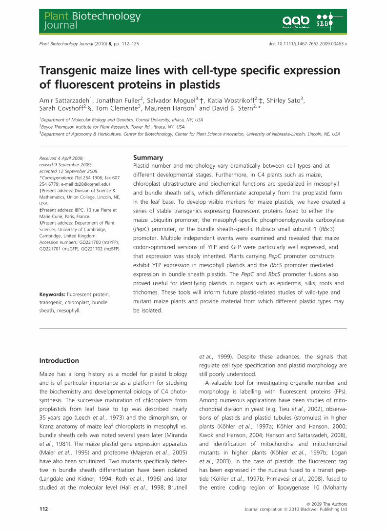
not observed in negative controls (Figure 1). When the
images were merged, the fluorescent protein signal colo-
calized with the chloroplast autofluorescence. In Figure 1,
small non-chlorophyll-containing epidermal plastids are vis-
ible in panels (b) and (c) because the images contain sig-
nals from both the leaf epidermal layer as well as
mesophyll tissue.
To develop archival lines, we screened multiple events
through the T2 generation (Table S1), seeking lines where
transgene expression was not silenced. DNA gel blot
7.7
3.52.7
1.9
0.9
UB
I
A63
2
RB
CS
PE
PC
Total protein
GFP
UBI1/TP-mzYFP
UBI1/TP-mzGFP
UBI1/TP-mzBFP
(a) (d)
(e)
(b)
(c)
Figure 1 Confocal microscopy images of primary maize transformants expressing the fluorescent protein constructs indicated above each panel.
The view is from the top of the leaf. For (a–c), the top row is an untransformed control, and left to right columns are fluorescent protein channel,
merged images, and chlorophyll autofluorescence. Constructs used for transformation were (a); pPTN448, event 480-1-1-1; (b) pPTN458, event
485-3-1-2; (c) pPTN469, event 487-1-1-2. (d) Total DNA from the indicated events was digested with BamHI and probed with the mzYFP coding
region, which hybridizes with all the fluorescent protein genes. DNA from an untransformed plant (variety Hi II) was a negative control. (e) Equal
amounts of total protein of leaves from an untransformed control (A632) and transgenic lines carrying the indicated promoters were analysed
using an anti-GFP antibody.
ª 2009 The AuthorsJournal compilation ª 2010 Blackwell Publishing Ltd, Plant Biotechnology Journal, 8, 112–125
Amir Sattarzadeh et al.114
analysis of the events ultimately selected is shown in Fig-
ure 1d, showing that each has a single insertion. There-
fore, these lines should be appropriate for introgression
and other standard manipulations. We noted that some
progeny expressing fluorescent proteins at high levels, par-
ticularly those with the ubiquitin promoter, sometimes
were more susceptible to stress and exhibited slow growth
rates (data not shown). Currently T2 lines, which segre-
gate for transgene expression, are being used for experi-
mental material and also to propagate subsequent
generations, where it is hoped that homozygous plants
with no growth defect can be obtained. The frequency of
transgene expression in the T2 generation, however, is
sufficient that among a dozen seedlings, some fluorescent
progeny were always readily identified for experimental
use.
To gain an idea of the overall expression levels of G ⁄ YFP
in the transgenic lines, total plant protein was analysed by
immunoblotting with anti-GFP antiserum, as shown in
Figure 1e. As expected, on a total protein basis, the Ubi1
promoter gave the highest expresion, with PepC driving
an intermediate level, and RbcS-driven expression being
slightly lower. It should be noted that because the number
of mesophyll cells vs. bundle sheath cells is higher per unit
volume, the results reflect not only promoter strength, but
also cell-type specificity and the prevalence of a given cell
type.
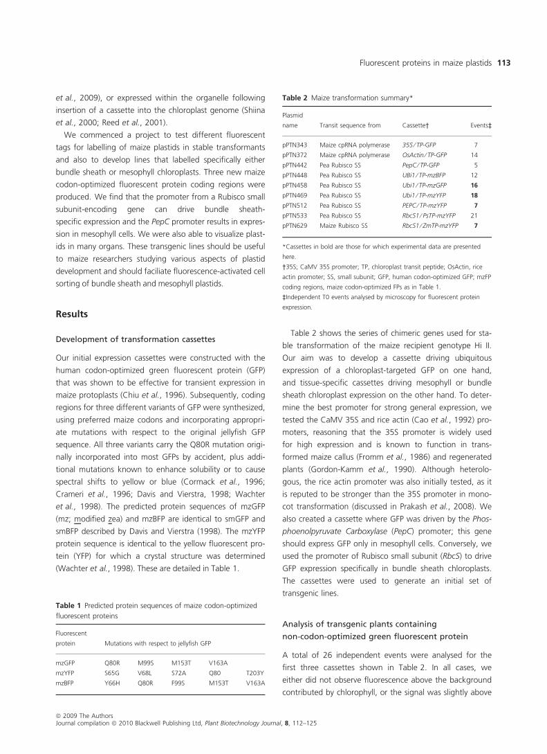
Examination of cell type specificity
Using confocal microscopy, we examined plants where the
transgene had been functionally inherited, (i.e. T1 genera-
tion and beyond) in more detail. Detailed Z-series images
of leaf cells from the plants producing RBCS ⁄ TP-mzYFP
are shown in Figure 2. RBCS ⁄ TP-mzYFP was observed in
the merged images not to colocalize with all chlorophyll
fluorescence, but instead to surround vascular tissue when
viewed in cross-section or longitudinal section. In the
RbcS-mzYFP
50 µ
50 µ 50 µ
DIC
50 µ
(a)
(d)
(h) (i) (j) (k)
(e) (f) (g)
(b) (c)
Figure 2 Localization of RBCS ⁄ TP-mzYFP in bundle sheath cells of maize leaves. (a–c) Leaf cross-section sliced manually. (a) Chlorophyll autofluo-
rescence; (b) merged chlorophyll and YFP signals; and (c) YFP fluorescence. (d) YFP fluorescence in leaf epidermal plastids (e–g) longitudinal
view of leaf: (e) chlorophyll autofluorescence; (f) merged chlorophyll, differential interference contrast (DIC) and YFP signals; and (g) YFP fluores-
cence; (h) YFP fluorescence in hypocotyl epidermis (i–k) close-up longitudinal view of leaf cells as in (d–f) with (l) DIC image to illustrate location of
vein. Each image represents a composite Z-series through mesophyll and bundle sheath cells from the surface of the leaf.
ª 2009 The AuthorsJournal compilation ª 2010 Blackwell Publishing Ltd, Plant Biotechnology Journal, 8, 112–125
Fluorescent proteins in maize plastids 115
differential interference contrast (DIC) image, the co-locali-
zation of the YFP signal cells near the vascular tissue is evi-
dent. Small YFP-labelled plastids seen in Figure 2g,h are
present in the epidermal layer. Thus, the RbcS promoter
appears to mediate bundle sheath-specific expression in
leaves. In contrast to the RBCS ⁄ TP-mzYFP transgenic
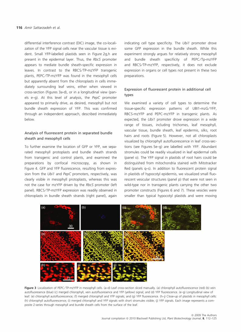
plants, PEPC ⁄ TP-mzYFP was found in the mesophyll cells
but apparently absent from the chloroplasts in cells imme-
diately surrounding leaf veins, either when viewed in
cross-section (Figures 3a-d), or in a longitudinal view (pan-
els e–g). At this level of analysis, the PepC promoter
appeared to primarily drive, as desired, mesophyll but not
bundle sheath expression of YFP. This was confirmed
through an independent approach, described immediately
below.
Analysis of fluorescent protein in separated bundle
sheath and mesophyll cells
To further examine the location of GFP or YFP, we sepa-
rated mesophyll protoplasts and bundle sheath strands
from transgenic and control plants, and examined the
preparations by confocal microscopy, as shown in
Figure 4. GFP and YFP fluorescence, resulting from expres-
sion from the Ubi1 and PepC promoters, respectively, was
clearly visible in mesophyll protoplasts, whereas this was
not the case for mzYFP driven by the RbcS promoter (left
panel). RBCS ⁄ TP-mzYFP expression was readily observed in
chloroplasts in bundle sheath strands (right panel), again
indicating cell type specificity. The Ubi1 promoter drove
some GFP expression in the bundle sheath. While this
experiment strongly argues for relatively strong mesophyll
and bundle sheath specificity of PEPC ⁄ Tp-mzYFP
and RBCS ⁄ TP-mzYFP, respectively, it does not exclude
expression in organs or cell types not present in these two
preparations.
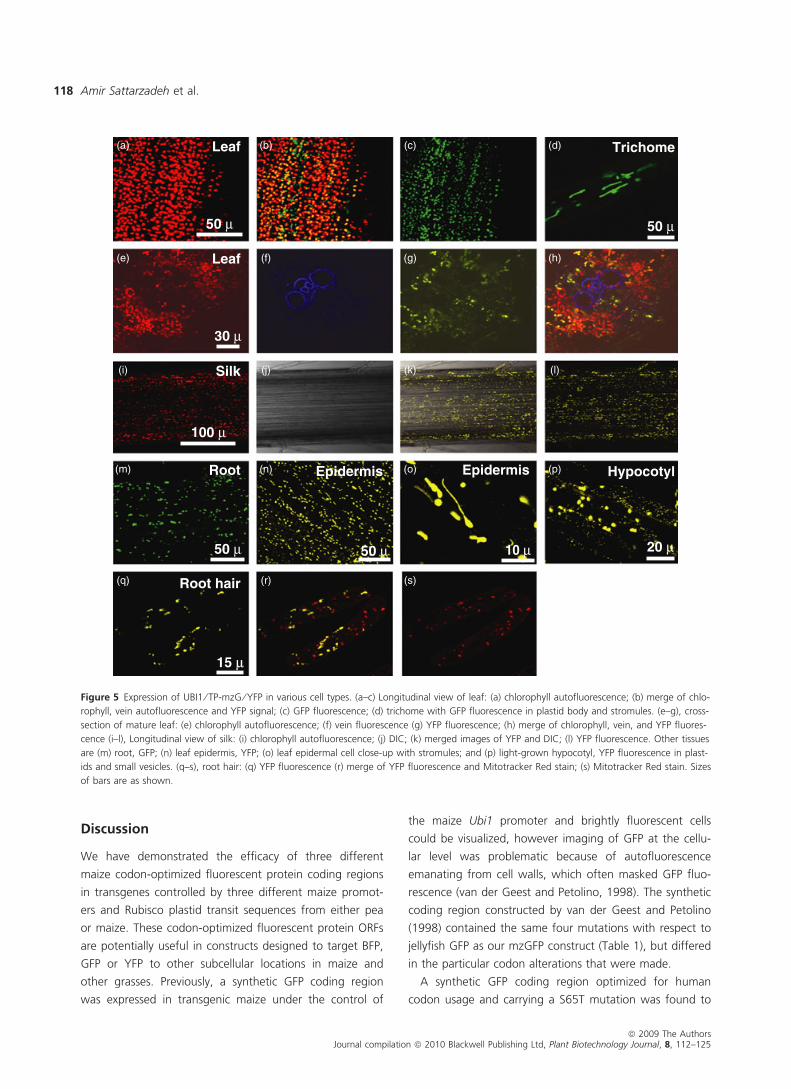
Expression of fluorescent protein in additional cell
types
We examined a variety of cell types to determine the
tissue-specific expression patterns of UBI1-mzG ⁄ YFP,
RBCS-mzYFP and PEPC-mzYFP in transgenic plants. As
expected, the Ubi1 promoter drove expression in a wide
range of tissues, including trichomes, leaf mesophyll,
vascular tissue, bundle sheath, leaf epidermis, silks, root
hairs and roots (Figure 5). However, not all chloroplasts
visualized by chlorophyll autofluorescence in leaf cross-sec-
tions (see Figures 5e–g) are labelled with YFP. Abundant
stromules could be readily visualized in leaf epidermal cells
(panel o). The YFP signal in plastids of root hairs could be
distinguished from mitochondria stained with Mitotracker
Red (panels q–s). In addition to fluorescent protein signal
in plastids of hypocotyl epidermis, we visualized small fluo-
rescent vesicular structures (panel p) that were not seen in
wild-type nor in transgenic plants carrying the other two
promoter constructs (Figures 6 and 7). These vesicles were
smaller than typical hypocotyl plastids and were moving
30
50
15
(a)
(e)
(h) (i) (j)
(f) (g)
(b) (c) (d)
Figure 3 Localization of PEPC ⁄ TP-mzYFP in mesophyll cells. (a–d) Leaf cross-section sliced manually. (a) chlorophyll autofluorescence (red) (b) vein
autofluoresence (blue) (c) merged chlorophyll, vein autofluoresence and YFP (yellow) signal; and (d) YFP fluorescence. (e–g) Longitudinal view of
leaf: (e) chlorophyll autofluorescence; (f) merged chlorophyll and YFP signals; and (g) YFP fluorescence. (h–j) Close-up of plastids in mesophyll cells:
(h) chlorophyll autofluorescence; (i) merged chlorophyll and YFP signals with short stromules visible; (j) YFP signals. Each image represents a com-
posite Z-series through mesophyll and bundle sheath cells from the surface of the leaf.
ª 2009 The AuthorsJournal compilation ª 2010 Blackwell Publishing Ltd, Plant Biotechnology Journal, 8, 112–125
Amir Sattarzadeh et al.116
more rapidly (Movie S1). Movies of tobacco hypocotyl
plastids are available for comparison (Kwok and Hanson,
2003).
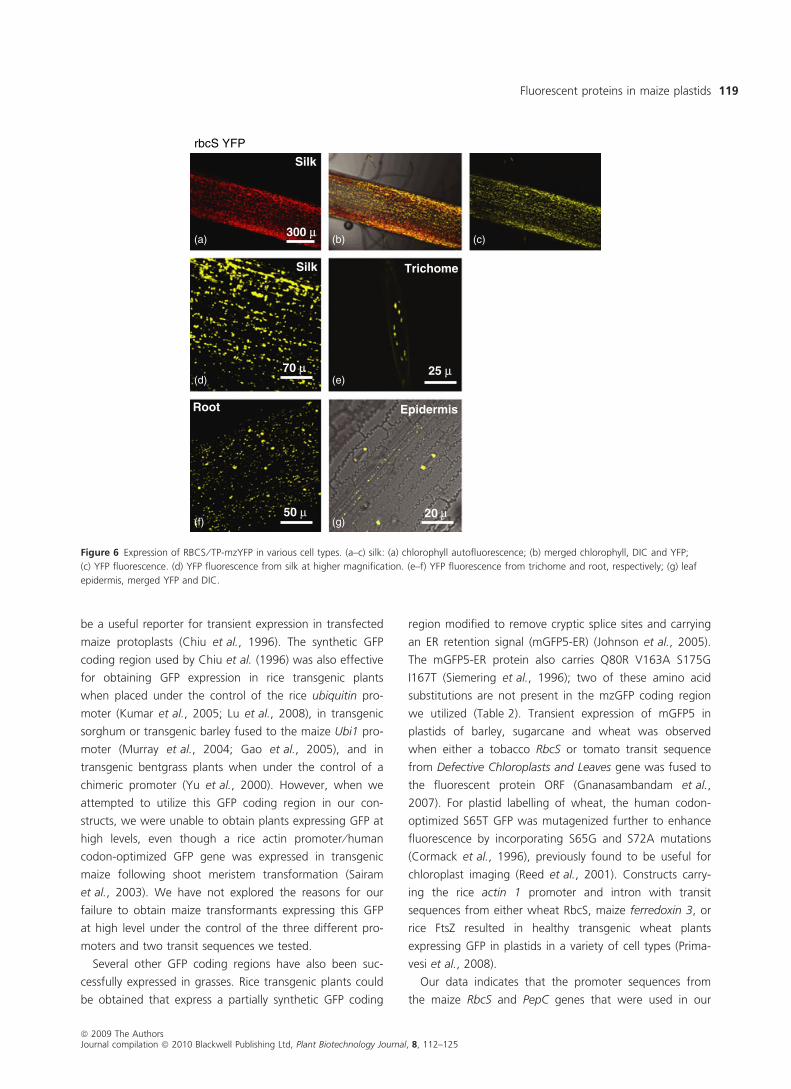
In addition to bundle sheath chloroplasts (Figure 2),
plants carrying the RbcS promoter contained YFP in plast-
ids of leaf epidermis, guard cells, trichomes, roots and silks
(Figure 6). The PEPC ⁄ TP-mzYFP plants exhibited YFP sig-
nals in plastids of trichomes, leaf epidermis, roots and root
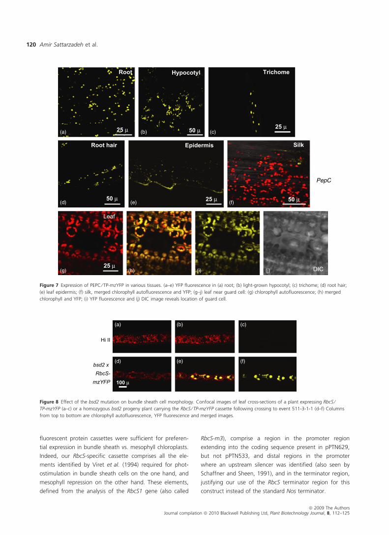
hairs (Figure 7). No PEPC ⁄ TP-mzYFP was evident in silks.
An example of a leaf region lacking YFP fluorescence in
guard cell plastids of PEPC ⁄ TP-mzYFP is shown in
Figure 7g–j, but we also sometimes did observe fluores-
cent plastids in stomatal cells (data not shown).
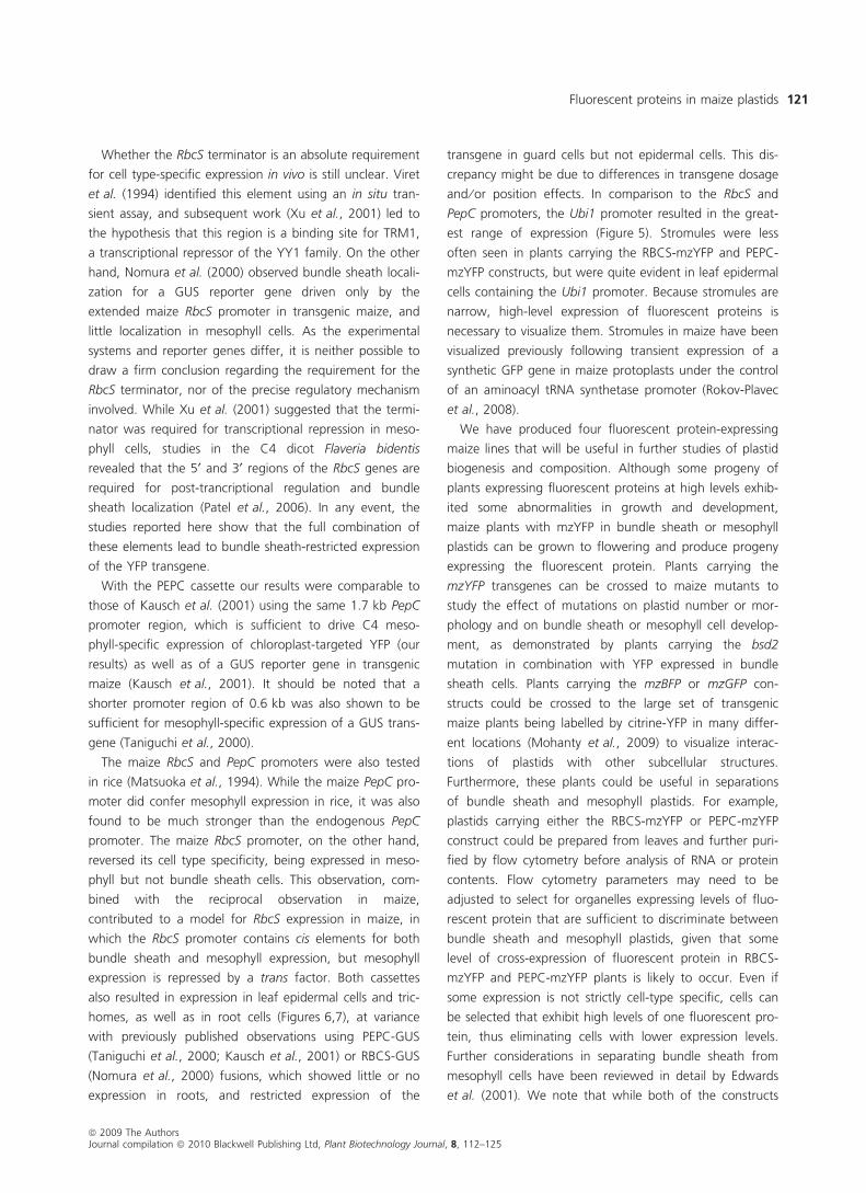
RbcS ⁄ TP-mzYFP expression in the bsd2 mutant
background
One possible use for organelle-labelled transgenic lines is
to examine the morphology of chloroplasts and other plas-
tid types in mutant backgrounds. Because expression of
the RbcS ⁄ TP-mzYFP construct appeared to be specific to
bundle sheath cells, we decided to use it as a test case by
crossing the transgene into the bundle sheath defective2
background (Brutnell et al., 1999). The bsd2 mutant is
pale-green and seedling-lethal because it does not accu-
mulate Rubisco, and exhibits abnormal chloroplast mor-
phology in light-exposed tissues that are biochemically C4
(Roth et al., 1996). The major gross morphological differ-
ence is that bundle sheath chloroplasts appear to be swol-
len; by electron microscopy, altered membrane structure
can be observed (Roth et al., 1996). Figure 8 compares
WT (panels a–c) and bsd2 ⁄ RBCS-YFP (panels d–f) in a
cross-section from a part of the leaf that would normally
exhibit Kranz anatomy. In the mutant background, the
bundle sheath cells were smaller in number and irregularly
shaped, in contrast to the regular shape and spacing
observed in a wild-type background (Figure 2). Thus, use
of the fluorescent protein allows visualization of cell
morphology in bsd2 which would normally require more
arduous techniques.
A632
Mesophyll protoplasts Bundle sheath strands
UBI
PepC
RbcS
0 µm 25
0 µm 25
0 µm 25
0 µm 25 0 µm 50 0 µm 50 0 µm 50
0 µm 500 µm 500 µm 50
0 µm 50 0 µm 50 0 µm 50
0 µm 50 0 µm 50 0 µm 50
(a)
(b)
(c)
(d)
(e)
(f)
(g)
(h)
Figure 4 Analysis of fluorescent protein expression in mesophyll protoplasts and bundle sheath strands prepared from leaves of (a,e) wild-type;
(b,f) UBI1 ⁄ TPmzYFP plants; (c,g) PEPC ⁄ TPmzYFP plants; and (d,h) RBCS ⁄ TPmzYFP plants. Each set of columns, left to right: chlorophyll fluores-
cence, merged images, and fluorescent proteins. In panel b, YFP flurorescence not overlapping with chlorophyll fluorescence may represent imma-
ture plastids lacking thylakoid membranes.
ª 2009 The AuthorsJournal compilation ª 2010 Blackwell Publishing Ltd, Plant Biotechnology Journal, 8, 112–125
Fluorescent proteins in maize plastids 117
Discussion
We have demonstrated the efficacy of three different
maize codon-optimized fluorescent protein coding regions
in transgenes controlled by three different maize promot-
ers and Rubisco plastid transit sequences from either pea
or maize. These codon-optimized fluorescent protein ORFs
are potentially useful in constructs designed to target BFP,
GFP or YFP to other subcellular locations in maize and
other grasses. Previously, a synthetic GFP coding region
was expressed in transgenic maize under the control of
the maize Ubi1 promoter and brightly fluorescent cells
could be visualized, however imaging of GFP at the cellu-
lar level was problematic because of autofluorescence
emanating from cell walls, which often masked GFP fluo-
rescence (van der Geest and Petolino, 1998). The synthetic
coding region constructed by van der Geest and Petolino
(1998) contained the same four mutations with respect to
jellyfish GFP as our mzGFP construct (Table 1), but differed
in the particular codon alterations that were made.
A synthetic GFP coding region optimized for human
codon usage and carrying a S65T mutation was found to
Leaf Trichome
50 µ50 µ
Leaf
30 µ
Silk
100 µ
Epidermis Epidermis
10 µ50 µ50 µ
Hypocotyl
20 µ
Root
Root hair
15 µ
(a) (b) (c) (d)
(e) (f) (g) (h)
(i) (j) (k) (l)
(m) (n) (o)
(q) (r) (s)
(p)
Figure 5 Expression of UBI1 ⁄ TP-mzG ⁄ YFP in various cell types. (a–c) Longitudinal view of leaf: (a) chlorophyll autofluorescence; (b) merge of chlo-
rophyll, vein autofluorescence and YFP signal; (c) GFP fluorescence; (d) trichome with GFP fluorescence in plastid body and stromules. (e–g), cross-
section of mature leaf: (e) chlorophyll autofluorescence; (f) vein fluorescence (g) YFP fluorescence; (h) merge of chlorophyll, vein, and YFP fluores-
cence (i–l), Longitudinal view of silk: (i) chlorophyll autofluorescence; (j) DIC; (k) merged images of YFP and DIC; (l) YFP fluorescence. Other tissues
are (m) root, GFP; (n) leaf epidermis, YFP; (o) leaf epidermal cell close-up with stromules; and (p) light-grown hypocotyl, YFP fluorescence in plast-
ids and small vesicles. (q–s), root hair: (q) YFP fluorescence (r) merge of YFP fluorescence and Mitotracker Red stain; (s) Mitotracker Red stain. Sizes
of bars are as shown.
ª 2009 The AuthorsJournal compilation ª 2010 Blackwell Publishing Ltd, Plant Biotechnology Journal, 8, 112–125
Amir Sattarzadeh et al.118
be a useful reporter for transient expression in transfected
maize protoplasts (Chiu et al., 1996). The synthetic GFP
coding region used by Chiu et al. (1996) was also effective
for obtaining GFP expression in rice transgenic plants
when placed under the control of the rice ubiquitin pro-
moter (Kumar et al., 2005; Lu et al., 2008), in transgenic
sorghum or transgenic barley fused to the maize Ubi1 pro-
moter (Murray et al., 2004; Gao et al., 2005), and in
transgenic bentgrass plants when under the control of a
chimeric promoter (Yu et al., 2000). However, when we
attempted to utilize this GFP coding region in our con-
structs, we were unable to obtain plants expressing GFP at
high levels, even though a rice actin promoter ⁄ human
codon-optimized GFP gene was expressed in transgenic
maize following shoot meristem transformation (Sairam
et al., 2003). We have not explored the reasons for our
failure to obtain maize transformants expressing this GFP
at high level under the control of the three different pro-
moters and two transit sequences we tested.
Several other GFP coding regions have also been suc-
cessfully expressed in grasses. Rice transgenic plants could
be obtained that express a partially synthetic GFP coding
region modified to remove cryptic splice sites and carrying
an ER retention signal (mGFP5-ER) (Johnson et al., 2005).
The mGFP5-ER protein also carries Q80R V163A S175G
I167T (Siemering et al., 1996); two of these amino acid
substitutions are not present in the mzGFP coding region
we utilized (Table 2). Transient expression of mGFP5 in
plastids of barley, sugarcane and wheat was observed
when either a tobacco RbcS or tomato transit sequence
from Defective Chloroplasts and Leaves gene was fused to
the fluorescent protein ORF (Gnanasambandam et al.,
2007). For plastid labelling of wheat, the human codon-
optimized S65T GFP was mutagenized further to enhance
fluorescence by incorporating S65G and S72A mutations
(Cormack et al., 1996), previously found to be useful for
chloroplast imaging (Reed et al., 2001). Constructs carry-
ing the rice actin 1 promoter and intron with transit
sequences from either wheat RbcS, maize ferredoxin 3, or
rice FtsZ resulted in healthy transgenic wheat plants
expressing GFP in plastids in a variety of cell types (Prima-
vesi et al., 2008).
Our data indicates that the promoter sequences from
the maize RbcS and PepC genes that were used in our
Silk
rbcS YFP
TrichomeSilk
300 µ
Root Epidermis
25 µ70 µ
50 µ 20 µ
(a) (b)
(d) (e)
(f) (g)
(c)
Figure 6 Expression of RBCS ⁄ TP-mzYFP in various cell types. (a–c) silk: (a) chlorophyll autofluorescence; (b) merged chlorophyll, DIC and YFP;
(c) YFP fluorescence. (d) YFP fluorescence from silk at higher magnification. (e–f) YFP fluorescence from trichome and root, respectively; (g) leaf
epidermis, merged YFP and DIC.
ª 2009 The AuthorsJournal compilation ª 2010 Blackwell Publishing Ltd, Plant Biotechnology Journal, 8, 112–125
Fluorescent proteins in maize plastids 119
fluorescent protein cassettes were sufficient for preferen-
tial expression in bundle sheath vs. mesophyll chloroplasts.
Indeed, our RbcS-specific cassette comprises all the ele-
ments identified by Viret et al. (1994) required for phot-
ostimulation in bundle sheath cells on the one hand, and
mesophyll repression on the other hand. These elements,
defined from the analysis of the RbcS1 gene (also called
RbcS-m3), comprise a region in the promoter region
extending into the coding sequence present in pPTN629,
but not pPTN533, and distal regions in the promoter
where an upstream silencer was identified (also seen by
Schaffner and Sheen, 1991), and in the terminator region,
justifying our use of the RbcS terminator region for this
construct instead of the standard Nos terminator.
Hi II
bsd2 x RbcS-
mzYFP 100 µ
(a) (b) (c)
(d) (e) (f)
Figure 8 Effect of the bsd2 mutation on bundle sheath cell morphology. Confocal images of leaf cross-sections of a plant expressing RbcS ⁄TP-mzYFP (a–c) or a homozygous bsd2 progeny plant carrying the RbcS ⁄ TP-mzYFP cassette following crossing to event 511-3-1-1 (d–f) Columns
from top to bottom are chlorophyll autofluorescence, YFP fluorescence and merged images.
Figure 7 Expression of PEPC ⁄ TP-mzYFP in various tissues. (a–e) YFP fluorescence in (a) root; (b) light-grown hypocotyl; (c) trichome; (d) root hair;
(e) leaf epidermis; (f) silk, merged chlorophyll autofluorescence and YFP; (g–j) leaf near guard cell: (g) chlorophyll autofluorescence; (h) merged
chlorophyll and YFP; (i) YFP fluorescence and (j) DIC image reveals location of guard cell.
ª 2009 The AuthorsJournal compilation ª 2010 Blackwell Publishing Ltd, Plant Biotechnology Journal, 8, 112–125
Amir Sattarzadeh et al.120
Whether the RbcS terminator is an absolute requirement
for cell type-specific expression in vivo is still unclear. Viret
et al. (1994) identified this element using an in situ tran-
sient assay, and subsequent work (Xu et al., 2001) led to
the hypothesis that this region is a binding site for TRM1,
a transcriptional repressor of the YY1 family. On the other
hand, Nomura et al. (2000) observed bundle sheath locali-
zation for a GUS reporter gene driven only by the
extended maize RbcS promoter in transgenic maize, and
little localization in mesophyll cells. As the experimental
systems and reporter genes differ, it is neither possible to
draw a firm conclusion regarding the requirement for the
RbcS terminator, nor of the precise regulatory mechanism
involved. While Xu et al. (2001) suggested that the termi-
nator was required for transcriptional repression in meso-
phyll cells, studies in the C4 dicot Flaveria bidentis
revealed that the 5¢ and 3¢ regions of the RbcS genes are
required for post-trancriptional regulation and bundle
sheath localization (Patel et al., 2006). In any event, the
studies reported here show that the full combination of
these elements lead to bundle sheath-restricted expression
of the YFP transgene.
With the PEPC cassette our results were comparable to
those of Kausch et al. (2001) using the same 1.7 kb PepC
promoter region, which is sufficient to drive C4 meso-
phyll-specific expression of chloroplast-targeted YFP (our
results) as well as of a GUS reporter gene in transgenic
maize (Kausch et al., 2001). It should be noted that a
shorter promoter region of 0.6 kb was also shown to be
sufficient for mesophyll-specific expression of a GUS trans-
gene (Taniguchi et al., 2000).
The maize RbcS and PepC promoters were also tested
in rice (Matsuoka et al., 1994). While the maize PepC pro-
moter did confer mesophyll expression in rice, it was also
found to be much stronger than the endogenous PepC
promoter. The maize RbcS promoter, on the other hand,
reversed its cell type specificity, being expressed in meso-
phyll but not bundle sheath cells. This observation, com-
bined with the reciprocal observation in maize,
contributed to a model for RbcS expression in maize, in
which the RbcS promoter contains cis elements for both
bundle sheath and mesophyll expression, but mesophyll
expression is repressed by a trans factor. Both cassettes
also resulted in expression in leaf epidermal cells and tric-
homes, as well as in root cells (Figures 6,7), at variance
with previously published observations using PEPC-GUS
(Taniguchi et al., 2000; Kausch et al., 2001) or RBCS-GUS
(Nomura et al., 2000) fusions, which showed little or no
expression in roots, and restricted expression of the
transgene in guard cells but not epidermal cells. This dis-
crepancy might be due to differences in transgene dosage
and ⁄ or position effects. In comparison to the RbcS and
PepC promoters, the Ubi1 promoter resulted in the great-
est range of expression (Figure 5). Stromules were less
often seen in plants carrying the RBCS-mzYFP and PEPC-
mzYFP constructs, but were quite evident in leaf epidermal
cells containing the Ubi1 promoter. Because stromules are
narrow, high-level expression of fluorescent proteins is
necessary to visualize them. Stromules in maize have been
visualized previously following transient expression of a
synthetic GFP gene in maize protoplasts under the control
of an aminoacyl tRNA synthetase promoter (Rokov-Plavec
et al., 2008).
We have produced four fluorescent protein-expressing
maize lines that will be useful in further studies of plastid
biogenesis and composition. Although some progeny of
plants expressing fluorescent proteins at high levels exhib-
ited some abnormalities in growth and development,
maize plants with mzYFP in bundle sheath or mesophyll
plastids can be grown to flowering and produce progeny
expressing the fluorescent protein. Plants carrying the
mzYFP transgenes can be crossed to maize mutants to
study the effect of mutations on plastid number or mor-
phology and on bundle sheath or mesophyll cell develop-
ment, as demonstrated by plants carrying the bsd2
mutation in combination with YFP expressed in bundle
sheath cells. Plants carrying the mzBFP or mzGFP con-
structs could be crossed to the large set of transgenic
maize plants being labelled by citrine-YFP in many differ-
ent locations (Mohanty et al., 2009) to visualize interac-
tions of plastids with other subcellular structures.
Furthermore, these plants could be useful in separations
of bundle sheath and mesophyll plastids. For example,
plastids carrying either the RBCS-mzYFP or PEPC-mzYFP
construct could be prepared from leaves and further puri-
fied by flow cytometry before analysis of RNA or protein
contents. Flow cytometry parameters may need to be
adjusted to select for organelles expressing levels of fluo-
rescent protein that are sufficient to discriminate between
bundle sheath and mesophyll plastids, given that some
level of cross-expression of fluorescent protein in RBCS-
mzYFP and PEPC-mzYFP plants is likely to occur. Even if
some expression is not strictly cell-type specific, cells can
be selected that exhibit high levels of one fluorescent pro-
tein, thus eliminating cells with lower expression levels.
Further considerations in separating bundle sheath from
mesophyll cells have been reviewed in detail by Edwards
et al. (2001). We note that while both of the constructs
ª 2009 The AuthorsJournal compilation ª 2010 Blackwell Publishing Ltd, Plant Biotechnology Journal, 8, 112–125
Fluorescent proteins in maize plastids 121
label leaf plastids outside of bundle sheath or mesophyll
(for example, in leaf epidermis), most of the plastids in
other leaf cell types are either smaller or do not contain
chlorophyll and therefore most could potentially be sorted
away by size or chlorophyll content from either mesophyll
or bundle sheath chloroplasts.
Experimental procedures
Maize transformation
Maize transformations were carried out via an Agrobacterium-
mediated transformation protocol. Immature ears, genotype Hi II
(Armstrong et al., 1991), were harvested approximately 12 days
post-pollination. Whole ears were surfaced sterilized by applying
70% ethanol spray and allowing to air dry within a laminar flow
hood. Immature embryos were isolated and placed immediately in
liquid isolation medium composed of 1 ⁄ 2 MS salts with full
strength MS vitamins 115 mg ⁄ L proline, 6.9% sucrose, 3.6% glu-
cose, 200 lM acetosyringone buffered with 10 mM MES (pH 5.4).
Following the isolation of 100 immature embryos, the isolation
medium was replaced with inoculation medium, A. tumefaciens
transconjugant suspended in isolation medium to an OD660 of
0.3–0.5. The embryos were inoculated for 5 min, after which they
were transferred, scutellum side up, to co-cultivation medium
solidified with 0.6% low EEO agarose. Co-cultivation medium
consisted of 1 ⁄ 2 MS salts, full strength MS vitamins, 0.5 mg ⁄ Lthiamine, 1 mg ⁄ L 2,4-D, 115 mg ⁄ L proline, 1% glucose, 2%
sucrose, 20 lM AgNO3, and 200 lM acetosyringone. The medium
was buffered with 20 mM MES (pH 5.4). The embryos were co-
cultivated for 2 days at 24 �C.
Following the co-cultivation step the embryos were transferred
to delay medium composed of N6 salts (Chu et al., 1975), Eriks-
son’s vitamins (Eriksson, 1965), with 1 mg ⁄ L 2,4-D, 25 mM pro-
line, 100 mg ⁄ L casamino acids, 2% sucrose, 1.7 mg ⁄ L AgNO3,
and 250 mg ⁄ L carbenicillin. The medium was solidified with 0.7%
phytagar and buffered with 3 mM MES (pH 5.8). The delay step
was carried out for 5 days in the dark at 28 �C, after which devel-
oping coleoptiles were removed from the embryos and the
explants transferred to selection medium.
The selection phase was carried in a stepwise fashion using the
delay medium supplemented with 25 mg ⁄ L paramomycin for
3 weeks, followed by a transfer to 50 mg ⁄ L paramomycin for
3 weeks, and finally 100 mg ⁄ L paramomycin. Embryogenic tissue
was subcultured three times on to fresh 100 mg ⁄ L selection
regime until the proliferating embryogenic culture mass reached
approximately 2 cm in diameter.
Paromomycin-tolerant embryogenic tissue was regenerated in a
three-step process. The first step was carried out in the dark at
28 �C for a period up to 14 days, where the tissue was cultured
on medium composed of MS salts, Fromm vitamins (Fromm et al.,
1990), 0.1 mg ⁄ L 2,4-D, 100 lM abscisic acid (ABA), 2% sucrose,
50 mg ⁄ L paromomycin, and 250 mg ⁄ L carbenicillin. The medium
was solidified 0.7% phytagar and buffered with 3 mM MES (pH
5.8). The second stage of regeneration involved culturing embryos
for a period up to 14 days in the dark at 28 �C on N6 salts, Eriks-
son’s vitamins, 6% sucrose, 50 mg ⁄ L paromomycin and
250 mg ⁄ L carbenicillin. The embryo conversion step was carried
out in culture vessels under an 18 h light regime on medium with
MS salts, Fromm vitamins, 100 mg ⁄ L inositol, 150 mg ⁄ L aspara-
gine, 2% maltose, 1% glucose, and 50 mg ⁄ L paromomycin. The
conversion medium was solidified with 0.7% phytagar and buf-
fered with 3 mM MES (pH 5.8).
Fluorescent marker gene cassettes
Three cassettes that harboured a non-maize codon-optimized ver-
sion (Chiu et al., 1996) were assembled under the control of
either the 35S CaMV (Benfey and Chua, 1990), rice actin pro-
moter, coupled with a 5¢-intron (Zhong et al., 1996) or the 1.7 kb
maize C4 PepC promoter. (Yanagisawa and Izui, 1989) (ZmPpc1)
The respective promoters were fused with translational enhancer
element from the maize PPDK-A gene (Sheen, 1993), and GFP
was targeted to plastids via the maize chloroplast RNA polymerase
RpoTp transit peptide (Chang et al., 1999) for the 35S CaMV and
rice actin cassettes, or the pea RBCS1 transit peptide (Van den
Broeck et al., 1985; von Heijne et al., 1991) for the PEPC cassette.
The non-codon-optimized GFP cassettes were subcloned into
either the binary plasmid pPZP211 or pPZP212 (Hajdukiewicz
et al., 1994), and the resultant vectors were referred to as
pPTN343, pPTN372 and pPTN442, for the 35S, rice actin and
PEPC promoters, respectively.
A set of maize codon-optimized fluorescent marker genes
encoding blue (mzBFP), green (mzGP) or yellow (mzYFP) were
commercially synthesized (Genscript Corporation, Piscataway, NJ,
USA). The respective markers were assembled in plastid-targeted
expression cassettes. The binary vector pPTN448 carries mzBFP
ORF under the control of the 1.9 kb PstI fragment directly
upstream the ATG of the maize ubiquitin1 promoter coupled with
its first intron (Christensen et al., 1992). The mzBFP peptide is tar-
geted to plastid via the pea Rubisco small subunit transit peptide
(von Heijne et al., 1991). The binary vectors pPTN458 and
pPTN469 are identical to pPTN448, except they harbour the
mzGFP and mzYFP ORFs, respectively. Sequences are available
through Genbank under accession numbers 1218408 (mzYFP),
1218425 (mzGFP) and 1218426 (mzBFP).
The binary vector designated pPTN512 has the mzYFP ORF
under the control of the 1.7 kb maize C4 PepC promoter (Yana-
gisawa and Izui, 1989) coupled with the pea SSU transit peptide.
The binary plasmid pPTN533 has the mzYFP ORF under the con-
trol of a 0.9 kb region upstream of the initiation codon compris-
ing the maize RBCS1 promoter (Lebrun et al., 1987; Viret et al.,
1994), and is plastid localized via the pea SSU transit peptide, and
has the terminator region from the RBCS1 gene believed to
contribute to cell-type specificity (Viret et al., 1994). Finally, the
binary vector pPTN629 is identical to pPTN533, except it utilizes
the maize RBCS1 transit peptide (Lebrun et al., 1987). All but
the pPTN533 and PTN629 constructs use the standard Nos
terminator.
Preparation of bundle sheath strands and mesophyll
protoplasts
Mesophyll protoplasts were prepared from third and fourth leaf
blades after digestion of their cell walls, and bundle sheath
ª 2009 The AuthorsJournal compilation ª 2010 Blackwell Publishing Ltd, Plant Biotechnology Journal, 8, 112–125
Amir Sattarzadeh et al.122
strands were mechanically isolated, as previously described
(Markelz et al., 2003).
Confocal microscopy
Confocal laser scanning microscopy (CLSM) for Figure 1 was con-
ducted with an Olympus FV500 (Olympus America Inc. Center
Valley, PA USA). GFP and YFP were excited at 488 nm and images
detected with a 505–525 nm filter. A 405 nm wavelength was
used to excite BFP and images detected with a 430–460 nm
filter. Images were captured and recorded using the FluoView 4.3
(Olympus America Inc. Center Valley, PA USA) version software.
CLSM for Figures 2,6 and 7 was performed on a Leica microscope
equipped with a TCS-SP2 confocal scanning head (Leica Microsys-
tems, Heidelberg, Germany). The 488 and 514 nm lines of an
argon laser was used to excite G ⁄ YFP and chlorophyll, respectively.
Images were recorded and processed using the LCS software 2.5
(Leica Microsystems). For staining with the MitoTracker Red
CMXRos (Molecular Probes, Invitrogen, Carlsbad, CA, USA) the
root hairs of transgenic maize were immersed for 10 min in a solu-
tion of 0.01% Silwet and 10 lM MitoTracker Red. Leaf sections of
3 to 4-week-old plants were prepared manually with a razor blade.
CLSM for Figure 1 was performed with an Olympus FV500, at the
University of Nebraska’s Imaging Core Research Facility (http://
biotech.unl.edu/Core%20Facilities/Microscopy/Services/Microscopy
Services.html). CLSM for the leaf sections in Figures 3 and 5 was
performed with an Olympus FV1000. The 405, 488 and 515 nm
lines of laser were used to excite vascular tissue autofluorescence,
chlorophyll autofluoresence and YFP, respectively. The images
in Figures 4 and 8 were obtained at the BTI Plant Cell Imaging
Center (http://bti.cornell.edu/facilitiesServicesPlantCellImagingCen
ter-Equipment.php) on a Leica SP5 instrument, described in detail
at that web site, following excitation at 458 nm.
DNA and protein blots
DNA gel blots were performed as previously published (Howe
et al., 2006). Immunoblotting was performed as described by Wo-
strikoff and Stern (2007). Antibodies against RbcL and GFP were
obtained from Agrisera (Agrisera AB, Vannas, Sweden) and Roche
(F. Hoffmann-La Roche Ltd, Basel, Switzerland) respectively, and
HRP coupled secondary antibodies directed against rabbit and
mouse were purchased from Promega (Promega Corporation,
Madison, WI USA) and Roche (F. Hoffmann-La Roche Ltd, Basel,
Switzerland). The PEPC antibody was a kind gift of Dr Michael
Salvucci (USDA-ARS, Maricopa, AZ, USA). Antibodies against RbcL
and PEPC were used at a 1 : 20 000 dilution, GFP antibody at a
1 : 10 000 dilution, while secondary antibodies were used at a
1 : 10 000 dilution.
Acknowledgements
This project was supported by NSF award DBI-0211935
and USDA award 2008–0286 to D. B. S. and T. C., and by
NSF award DBI-0421799 to M. R. H. We thank Christian
Elowsky for performing the confocal microscopy in
Figure 1. Elizabeth Takacs and Michelle Vernier are grate-
fully acknowledged for screening of some transgenic lines
for GFP expression.
References
Armstrong, C.L., Green, C.E. and Phillips, R.L. (1991)
Development and availability of germplasm with high type II
culture formation response. Maize Genet. Coop. News Lett. 65,
92–93.
Benfey, P.N. and Chua, N.H. (1990) The cauliflower mosaic virus-
35s promoter – combinatorial regulation of transcription in
plants. Science, 250, 959–966.
Brutnell, T.P., Sawers, R.J., Mant, A. and Langdale, J.A. (1999)
BUNDLE SHEATH DEFECTIVE2, a novel protein required for
post-translational regulation of the rbcL gene of maize. Plant
Cell, 11, 849–864.
Cao, J., Duan, X., McElroy, D. and Wu, R. (1992) Regeneration of
herbicide resistant transgenic rice plants following
microprojectile-mediated transformation of suspension culture
cells. Plant Cell Rep. 11, 586–591.
Chang, C., Sheen, J., Bligny, M., Niwa, Y., Lerbs-Mache, S. and
Stern, D.B. (1999) Functional analysis of two maize
cDNAs encoding T7-like RNA polymerases. Plant Cell, 11,
911–926.
Chiu, W., Niwa, Y., Zeng, W., Hirano, T., Kobayashi, H. and
Sheen, J. (1996) Engineered GFP as a vital reporter in plants.
Curr. Biol. 6, 325–330.
Christensen, A.H., Sharrock, R.A. and Quail, P.H. (1992) Maize
polyubiquitin genes: structure, thermal perturbation of
expression and transcript splicing, and promoter activity
following transfer to protoplasts by electroporation. Plant Mol.
Biol. 18, 675–689.
Chu, C.C., Wang, C.C., Sun, S.S., Hsu, C., Yin, K.C., Chu, C.Y.
and Bi, F.Y. (1975) Establishment of an efficient medium for
anther culture of rice through comparative experiments on the
nitrogen source. Sci. Sin. 18, 659–668.
Cormack, B.P., Valdivia, R.H. and Falkow, S. (1996) FACS-
optimized mutants of the green fluorescent protein (GFP).
Gene, 173, 33–38.
Crameri, A., Whitehorn, E.A., Tate, E. and Stemmer, W.P. (1996)
Improved green fluorescent protein by molecular evolution
using DNA shuffling. Nat. Biotechnol. 14, 315–319.
Davis, S.J. and Vierstra, R.D. (1998) Soluble, highly fluorescent
variants of green fluorescent protein (GFP) for use in higher
plants. Plant Mol. Biol. 36, 521–528.
Edwards, G.E., Franceschi, V.R., Ku, M.S., Voznesenskaya, E.V.,
Pyankov, V.I. and Andreo, C.S. (2001) Compartmentation of
photosynthesis in cells and tissues of C4 plants. J. Exp. Bot. 52,
577–590.
Eriksson, T. (1965) Studies on growth requirments and
measurements of Haplopappus gracilis. Physiol. Plant, 18, 976–
993.
Fromm, M.E., Taylor, L.P. and Walbot, V. (1986) Stable
transformation of maize after gene transfer by electroporation.
Nature, 319, 791–793.
Fromm, M.E., Morrish, F., Armstrong, C., Williams, R., Thomas, J.
and Klein, T.M. (1990) Inheritance and expresion of chimeric
ª 2009 The AuthorsJournal compilation ª 2010 Blackwell Publishing Ltd, Plant Biotechnology Journal, 8, 112–125
Fluorescent proteins in maize plastids 123
genes in the progeny of transgenic maize plants.
Biotechnology, 8, 833–839.
Gao, Z., Jayaraj, J., Muthukrishnan, S., Claflin, L. and Liang, G.H.
(2005) Efficient genetic transformation of Sorghum using a
visual screening marker. Genome, 48, 321–333.
van der Geest, A.H.M. and Petolino, J.F. (1998) Expression of a
modified green fluorescent protein gene in transgenic maize
plants and progeny. Plant Cell Rep. 17, 760–764.
Gnanasambandam, A., Polkinghorne, I.G. and Birch, R.G. (2007)
Heterologous signals allow efficient targeting of a nuclear-
encoded fusion protein to plastids and endoplasmic reticulum in
diverse plant species. Plant Biotechnol. J. 5, 290–296.
Gordon- Kamm, W.J., Spencer, T.M., Mangano, M.L., Adams,
T.R., Daines, R.J., Start, W.G., O’ Brien, J.V., Chambers, S.A.,
Adams Jr, W.R., Willetts, N.G., Rice, T.B., Mackey, C.J.,
Krueger, R.W., Kausch, A.P. and Lemaux, P.G. (1990)
Transformation of maize cells and regeneration of fertile
transgenic plants. Plant Cell, 2, 603–618.
Hajdukiewicz, P., Svab, Z. and Maliga, P. (1994) The small,
versatile pPZP family of Agrobacterium binary vectors for plant
transformation. Plant Mol. Biol. 25, 989–994.
Hall, L.N., Rossini, L., Cribb, L. and Langdale, J.A. (1998) GOLDEN
2: a novel transcriptional regulator of cellular differentiation in
the maize leaf. Plant Cell, 10, 925–936.
Hanson, M.R. and Sattarzadeh, A. (2008) Dynamic morphology of
plastids and stromules in angiosperm plants. Plant Cell Environ.
31, 646–657.
von Heijne, G., Hirai, T., Klosgen, R.-B., Steppuhn, J., Bruce, B.,
Keegstra, K. and Herrman, R. (1991) CHLPEP – A database of
chloroplast transit peptides. Plant Mol. Biol. Reporter, 9, 104–
126.
Howe, A., Sato, S., Dweikat, I., Fromm, M. and Clemente, T.
(2006) Rapid and reproducible Agrobacterium-mediated
transformation of sorghum. Plant Cell Rep. 25, 784–791.
Johnson, A.A., Hibberd, J.M., Gay, C., Essah, P.A., Haseloff, J.,
Tester, M. and Guiderdoni, E. (2005) Spatial control of
transgene expression in rice (Oryza sativa L.) using the GAL4
enhancer trapping system. Plant J. 41, 779–789.
Kausch, A.P., Owen Jr, T.P., Zachwieja, S.J., Flynn, A.R. and
Sheen, J. (2001) Mesophyll-specific, light and metabolic
regulation of the C4 PPCZm1 promoter in transgenic maize.
Plant Mol. Biol. 45, 1–15.
Kohler, R.H. and Hanson, M.R. (2000) Plastid tubules of higher
plants are tissue-specific and developmentally regulated. J. Cell
Sci. 113, 81–89.
Kohler, R.H., Cao, J., Zipfel, W.R., Webb, W.W. and Hanson,
M.R. (1997a) Exchange of protein molecules through
connections between higher plant plastids. Science, 276, 2039–
2042.
Kohler, R.H., Zipfel, W.R., Webb, W.W. and Hanson, M.R.
(1997b) The green fluorescent protein as a marker to visualize
plant mitochondria in vivo. Plant J. 11, 613–621.
Kumar, C.S., Wing, R.A. and Sundaresan, V. (2005) Efficient
insertional mutagenesis in rice using the maize En ⁄ Spm
elements. Plant J. 44, 879–892.
Kwok, E.Y. and Hanson, M.R. (2003) Microfilaments and
microtubules control the morphology and movement of non-
green plastids and stromules in Nicotiana tabacum. Plant J. 35,
16–26.
Kwok, E.Y. and Hanson, M.R. (2004) Stromules and the dynamic
nature of plastid morphology. J. Microsc. 214, 124–137.
Langdale, J.A. and Kidner, C.A. (1994) bundle sheath defective, a
mutation that disrupts cellular differentiation in maize leaves.
Development, 120, 673–681.
Lebrun, M., Waksman, G. and Freyssinet, G. (1987) Nucleotide
sequence of a gene encoding corn ribulose-1,5-bisphosphate
carboxylase ⁄ oxygenase small subunit (RbcS). Nucleic Acids Res.
15, 4360.
Leech, R.M., Rumsby, M.G. and Thomson, W.W. (1973) Plastid
differentiation, acyl lipid, and fatty acid changes in developing
green maize leaves. Plant Physiol. 52, 240–245.
Logan, D.C., Scott, I. and Tobin, A.K. (2003) The genetic control
of plant mitochondrial morphology and dynamics. Plant J. 36,
500–509.
Lu, J., Sivamani, E., Li, X. and Qu, R. (2008) Activity of the 5’
regulatory regions of the rice polyubiquitin rubi3 gene in
transgenic rice plants as analyzed by both GUS and GFP
reporter genes. Plant Cell Rep. 27, 1587–1600.
Maier, R.M., Neckermann, K., Igloi, G.L. and Koessel, H. (1995)
Complete sequence of the maize chloroplast genome: gene
content, hotspots of divergence and fine tuning of genetic
information by transcript editing. J. Mol. Biol. 251, 614–628.
Majeran, W., Cai, Y., Sun, Q. and van Wijk, K.J. (2005) Functional
differentiation of bundle sheath and mesophyll maize
chloroplasts determined by comparative proteomics. Plant Cell,
17, 3111–3140.
Markelz, N.H., Costich, D.E. and Brutnell, T.P. (2003)
Photomorphogenic responses in maize seedling development.
Plant Physiol. 133, 1578–1591.
Matsuoka, M., Kyozuka, J., Shimamoto, K. and Kano-Murakami,
Y. (1994) The promoters of two carboxylases in a C4 plant
(maize) direct cell-specific, light-regulated expression in a C3
plant (rice). Plant J. 6, 311–319.
Miranda, V., Baker, N.R. and Long, S.P. (1981) Anatomical
variation along the length of the Zea mays leaf in relation to
photosynthesis. New Phytol. 88, 595–605.
Mohanty, A., Luo, A., Deblasio, S., Ling, X., Yang, Y., Tuthill,
D.E., Williams, K.E., Hill, D., Zadrozny, T., Chan, A., Sylvester,
A.W. and Jackson, D. (2009) Advancing cell biology and
functional genomics in maize using fluorescent protein-tagged
lines. Plant Physiol. 149, 601–605.
Murray, F., Brettell, R., Matthews, P., Bishop, D. and Jacobsen, J.
(2004) Comparison of Agrobacterium-mediated transformation
of four barley cultivars using the GFP and GUS reporter genes.
Plant Cell Rep. 22, 397–402.
Nomura, M., Katayama, K., Nishimura, A., Ishida, Y., Ohta, S.,
Komari, T., Miyao-Tokutomi, M., Tajima, S. and Matsuoka, M.
(2000) The promoter of RbcS in a C3 plant (rice) directs organ-
specific, light-dependent expression in a C4 plant (maize), but
does not confer bundle sheath cell-specific expression. Plant
Mol. Biol. 44, 99–106.
Patel, M., Siegel, A.J. and Berry, J.O. (2006) Untranslated regions
of FbRbcS1 mRNA mediate bundle sheath cell-specific gene
expression in leaves of a C4 plant. J. Biol. Chem. 281, 25485–
25491.
Prakash, N.S., Prasad, V., Chidambram, T.P., Cherian, S.,
Jayaprakash, T.L., Dasgupta, S., Wang, Q., Mann, M.T.,
Spencer, T.M. and Boddupalli, R.S. (2008) Effect of promoter
ª 2009 The AuthorsJournal compilation ª 2010 Blackwell Publishing Ltd, Plant Biotechnology Journal, 8, 112–125
Amir Sattarzadeh et al.124
driving selectable marker on corn transformation. Transgenic
Res. 17, 695–704.
Primavesi, L.F., Wu, H., Mudd, E.A., Day, A. and Jones, H.D.
(2008) Visualisation of plastids in endosperm, pollen and roots
of transgenic wheat expressing modified GFP fused to transit
peptides from wheat SSU RubisCO, rice FtsZ and maize
ferredoxin III proteins. Transgenic Res. 17, 529–543.
Reed, M.L., Wilson, S.K., Sutton, C.A. and Hanson, M.R. (2001)
High-level expression of a synthetic red-shifted GFP coding
region incorporated into transgenic chloroplasts. Plant J. 27,
257–265.
Rokov-Plavec, J., Dulic, M., Duchene, A.M. and Weygand-
Durasevic, I. (2008) Dual targeting of organellar seryl-tRNA
synthetase to maize mitochondria and chloroplasts. Plant Cell
Rep. 27, 1157–1168.
Roth, R., Hall, L.N., Brutnell, T.P. and Langdale, J.A. (1996)
bundle sheath defective2, a mutation that disrupts the
coordinated development of bundle sheath and mesophyll cells
in the maize leaf. Plant Cell, 8, 915–927.
Sairam, R.V., Parani, M., Franklin, G., Lifeng, Z., Smith, B.,
MacDougall, J., Wilber, C., Sheikhi, H., Kashikar, N., Meeker,
K., Al-Abed, D., Berry, K., Vierling, R. and Goldman, S.L. (2003)
Shoot meristem: an ideal explant for Zea mays L.
transformation. Genome, 46, 323–329.
Schaffner, A.R. and Sheen, J. (1991) Maize RbcS promoter activity
depends on sequence elements not found in dicot RbcS
promoters. Plant Cell, 3, 997–1012.
Sheen, J. (1993) Protein phosphatase activity is required for light-
inducible gene expression in maize. EMBO J., 12, 3497–3505.
Shiina, T., Hayashi, K., Ishii, N., Morikawa, K. and Toyoshima, Y.
(2000) Chloroplast tubules visualized in transplastomic plants
expressing green fluorescent protein. Plant Cell Physiol. 41,
367–371.
Siemering, K.R., Golbik, R., Sever, R. and Haseloff, J. (1996)
Mutations that suppress the thermosensitivity of green
fluorescent protein. Curr. Biol. 6, 1653–1663.
Streatfield, S.J., Magallanes- Lundback, M.E., Beifuss, K.K.,
Brooks, C.A., Harkey, R.L., Love, R.T., Bray, J., Howard, J.A.,
Jilka, J.M. and Hood, E.E. (2004) Analysis of the maize
polyubiquitin-1 promoter heat shock elements and generation
of promoter variants with modified expression characteristics.
Transgenic Res. 13, 299–312.
Taniguchi, M., Izawa, K., Ku, M.S., Lin, J.H., Saito, H., Ishida, Y.,
Ohta, S., Komari, T., Matsuoka, M. and Sugiyama, T. (2000)
Binding of cell type-specific nuclear proteins to the 5’-flanking
region of maize C4 phosphoenolpyruvate carboxylase gene
confers its differential transcription in mesophyll cells. Plant
Mol. Biol. 44, 543–557.
Tieu, Q., Okreglak, V., Naylor, K. and Nunnari, J. (2002) The WD
repeat protein, Mdv1p, functions as a molecular adaptor by
interacting with Dnm1p and Fis1p during mitochondrial fission.
J. Cell Biol. 158, 445–452.
Van den Broeck, G., Timko, M.P., Kausch, A.P., Cashmore, A.R.,
Van Montagu, M. and Herrera-Estrella, L. (1985) Targeting of a
foreign protein to chloroplasts by fusion to the transit peptide
from the small subunit of ribulose 1,5-bisphosphate
carboxylase. Nature, 313, 358–363.
Viret, J.F., Mabrouk, Y. and Bogorad, L. (1994) Transcriptional
photoregulation of cell-type-preferred expression of maize
RBCS-m3: 3’ and 5’ sequences are involved. Proc. Natl Acad.
Sci. U.S.A. 91, 8577–8581.
Wachter, R.M., Elsliger, M.A., Kallio, K., Hanson, G.T. and
Remington, S.J. (1998) Structural basis of spectral shifts in the
yellow-emission variants of green fluorescent protein. Structure,
6, 1267–1277.
Wostrikoff, K. and Stern, D. (2007) Rubisco large-subunit
translation is autoregulated in response to its assembly state in
tobacco chloroplasts. Proc. Natl Acad. Sci. U.S.A. 104, 6466–
6471.
Xu, T., Purcell, M., Zucchi, P., Helentjaris, T. and Bogorad, L.
(2001) TRM1, a YY1-like suppressor of RbcS-m3 expression in
maize mesophyll cells. PNAS, 98, 2295–2300.
Yanagisawa, S. and Izui, K. (1989) Maize phosphoenolpyruvate
carboxylase involved in C4 photosynthesis: nucleotide sequence
analysis of the 5’ flanking region of the gene. J. Biochem. 106,
982–987.
Yu, T.T., Skinner, D.Z., Liang, G.H., Trick, H.N., Huang, B. and
Muthukrishnan, S. (2000) Agrobacterium-mediated
transformation of creeping bentgrass using GFP as a reporter
gene. Hereditas, 133, 229–233.
Zhong, H., Zhang, S.B., Warkentin, D., Sun, B.L., Wu, T.Y., Wu,
R. and Sticklen, M.B. (1996) Analysis of the functional activity
of the 1.4-kb 5’-region of the rice actin 1 gene in stable
transgenic plants of maize (Zea mays L.). Plant Sci. 116, 73–84.
Supporting information
Additional Supporting information may be found in the
online version of this article:
Movie S1 Small vesicular structures within a hypocotyl
epidermal cell of UBI ⁄ TP-mzYFP plants are highly mobile.
Time-lapse series of 10 frames, 3 s intervals. Time
indicated in seconds.
Table S1 Details of transgenic line analysis.
Please note: Wiley-Blackwell are not responsible for the
content or functionality of any supporting materials sup-
plied by the authors. Any queries (other than missing
material) should be directed to the corresponding author
for the article.
ª 2009 The AuthorsJournal compilation ª 2010 Blackwell Publishing Ltd, Plant Biotechnology Journal, 8, 112–125
Fluorescent proteins in maize plastids 125


![Improving Site-Specific Maize Yield Estimation by Integrating Satellite Multispectral Data into a Crop … 2020/Joshi et al... · models for site-specific crop yield estimation [17,28–30]](https://img.pdfslide.us/doc/110x75/5f770ca40a8f434f4b322e59/improving-site-specific-maize-yield-estimation-by-integrating-satellite-multispectral.jpg)


![Trade-Offs in Arbuscular Mycorrhizal Symbiosis: Disease ...€¦ · maize (Zea mays L.) [63] and wheat [65]. An AM-specific ammonium transporter LjAMT2; 2 has been identified in](https://img.pdfslide.us/doc/110x75/5f5900650db3e50472736818/trade-offs-in-arbuscular-mycorrhizal-symbiosis-disease-maize-zea-mays-l.jpg)