Embed Size (px)
Citation preview
Vol. 116, No. 1, 1983 BIOCHEMICAL AND BIOPHYSICAL RESEARCH COMMUNICATIONS
October 14, 1983 Pages 21 O-21 6
Transformed Liver Cells have Modified Transplasma Membrane Redox Activity which is Sensitive to Adriamycin
I. L. Sun, F. L. Crane
Department of Biological Sciences Purdue University
West Lafayette, IN 47907
J. Y. Chou
Human Genetics Branch National Institute of Child Health and
Human Development, National Institute of Health Bethesda. MD 20205
H. Mw, C. Grebing
Endocrinology Department Karolinska Institute
Stockholm. Sweden
Received August 30, 1983
Electron transport across the plasma membrane is found in all cells which have been tested. This activity has been implicated in control of cellular growth, transport and hormone response. In virus transformed oells and tumor cells we find the activity is decreased and becomes sensitive to the antitumor drug adriamycin. Inhibition of transmembrane redox by adriamycin parallels cytoxicity to transformed cells.
Adriamycin is an antitumor drug which has extensive use in canoer therapy
(1). It has been shown to interfere with DNA and RNA synthesis by inter-
calation with the DNA molecule (2,3,4). Reoent evidence has shown that adria-
mycin acts at sites on the plasma membrane (5.6.71, because this drug retains
cytotoxicity when bound to impermeable agarose beads. Certain membrane
functions are affected by adriamyoin (8.9) and the NADH ferricyanide reductase
of isolated plasma membranes is inhibited by this drug (lO,ll,l2,13). We
examined the effect of adriamycin on the ferricyanide reductase activity of
intact cells to see if inhibition at an external site could be related to its
cytotoxic effect and to determine if adriamycin-induced inhibition is corre-
lated with its antitumor activity. We conclude that the transmembrane redox
system in virus transformed liver cells and in hepatoma cells is modified to
0006-291X/83 $1.50 Copyright 0 I983 by Academic Press, Inc. All rights of reproduction in any form reserved. 210
Vol. 116, No. 1, 1533 BIOCHEMICAL AND BIOPHYSICAL RESEARCH COMMUNICATIONS
decrease its activity and increase its susceptibility to inhibition by anthra-
cyclines.
Materials and Methods --
The cell model system used in this study is a simian virus 40 (SV40) temperature sensitive (ts) A209 virus-transformed fetal liver RLA209-15 cell line that is temperaturesensitive for growth and differentration (17). They exhibit the transformed phenotype at the permissive temperature (33'C) but mimic the normal, nontransformed hepatocytes at the nonpermissive temperature (40°C). As controls, we used the McA-RH7777 hepatoma and primary rat fetal hepatocyte cells.
All cells that have been tested have a plasma membrane redox system which reduces external ferricyanide (14,15,16). Ferricyanide reduction was measured at 37' in an Aminco DW2a spectrophotometer using a dual beam to subtract adsorption at 500 nm from adsorption at 420 nm. Two to 20 mg wet weight of cells are placed in a cuvette with iris-EDTA buffer (140 mM NaCl, 2.5 mM KCl, 0.6 mM Na2HP04, 25 mM Trizma base and 0.05 mM EDTA, pH 7.4). Drugs are incubated with the cells for various times before running the assay. The reaction may be started by adding ferricyanide at 0.2 mM final concentration or by adding cells. The cuvette is equipped with a magnetic stirrer to keep the cells in suspension. Blanks were run with ferricyanide cells.
plu_s,adr amycin and no Ferricyanide extinction coefficient is taken as lcm t mM- . As with
most cells which we have tested, ferricyanide reduction shows a fast rate for about two minutes followed by a slower phase which can maintain a steady rate for lo-30 min (14,161. To check for ferricyanide reduction by mitochondria released from broken cells, the activity is measured in the presence of rotenone or antimycin (14). We find these agents do not inhibit either phase of ferricyanide reduction with these cells. RLA209-15 cells were cultured as described (17) and released by trypsinization with 0.05% trypsin and 0.02% EDTA for about 4 min. Cell survival was tested by eosin exclusion (18). External NADH ferricyanide reductase was assayed under the same conditions described for transmembrane ferricyanide reduction except for the addition of 50 uM NADH as substrate (11) and following the decrease in absorbance at 340 nm.
Results and Discussion --
RLA209-15 liver cells grown at 33’C (transformed phenotype) show a much
slower rate of ferricyanide reduction in both the fast and slow phase than do
the cells which have been grown at 40°C (nontransformed phenotype) (Table 1).
A similar difference has been observed with SV40 transformed 3T3 and nontrans-
formed 3T3 cells (Lbw, Grebing, Crane, unpublished). Hepatoma cells have a
rate of ferricyanide reduction thirty percent less in the fast phase and 60
percent less in the slow phase than isolated fetal liver cells. High rates of
ferricyanide reduction have also been observed with adult liver cells (14).
Adriamycin inhibits the ferricyanide reduction by transformed RLA209-15
liver cells in both the fast and slow phase. However, the same concentration
of drug gives much less inhibition of ferricyanide reduction by those cells
211
Vol. 116, No. 1, 1983 BIOCHEMICAL AND BIOPHYSICAL RESEARCH COMMUNICATIONS
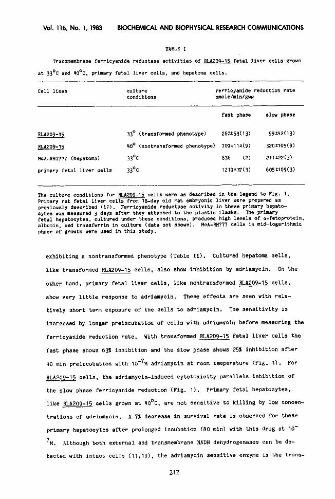
TABLE I
Transmembrane ferricyanide reductase activities of RLA209-15 fetal liver cells grown
at 33’C and 40°C, primary fetal liver cells, and hepatoma cells.
Cell lines culture conditions
-
Ferricyanide reduction rate nmole/min/gww
fast phase slow phase
RLA209-15 33’ (transformed phenotype) 26@53(13) 99t42(13)
RLA209-15 40’ (nontransformed phenotype) 709f114(9) 320*105(g)
McA-RH7777 (hepatoma) 33Oc 836 (2) 211?22(3)
primary fetal liver cells 33Oc 1210*37(3) 605?109(3)
The culture conditions for RLA209-15 cells were as described in the legend to Fig. 1. Primary rat fetal liver. cells from la-day old rat embryonic liver were prepared as previously described (17). Ferricyanide reductase activity in these primary hepato- cytes was measured 3 days after they attached to the plastic flasks. The primary fetal hepatocytes, cultured under these conditions, produced high levels of a-fetoprotein, albumin, and transferrin in culture (data not shown). McA-RH777 cells in mid-logarithmic phase of growth were used in this study.
exhibiting a nontransformed phenotype (Table II). Cultured hepatoma cells,
like transformed ALA209-15 cells, also show inhibition by adriamycin. On the
other hand, primary fetal liver cells, like nontransformed RLA209-15 cells,
show very little response to adriamycin. These effects are seen with rela-
tively short term exposure of the cells to adriamycin. The sensitivity is
increased by longer preincubation of cells with adriamycin before measuring the
ferricyanide reduction rate. With transformed RLA209-15 fetal liver cells the
fast phase shows 63% inhibition and the slow phase shows 25% inhibition after
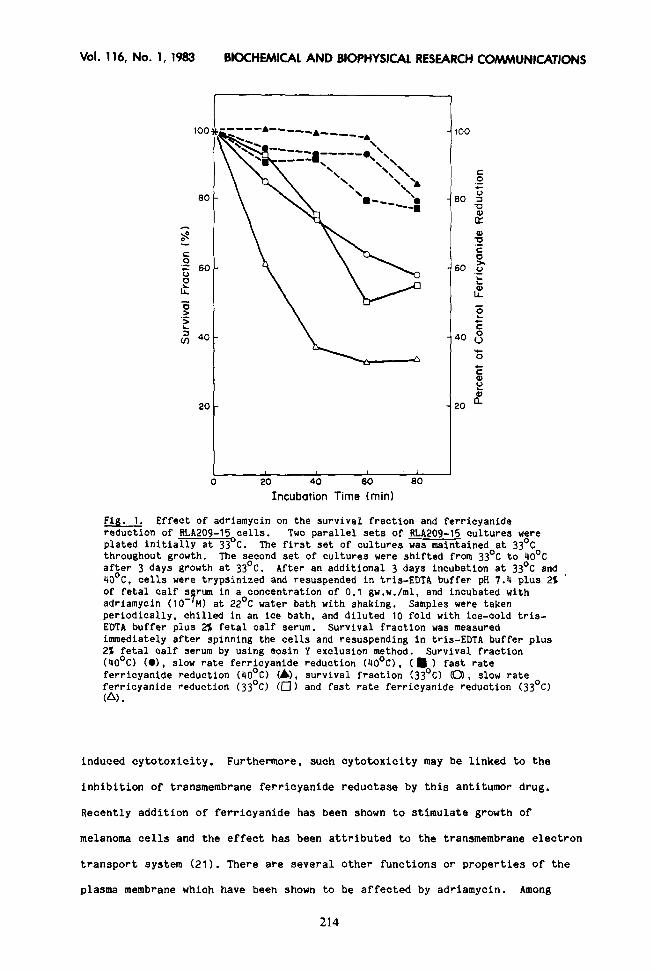
40 min preinoubation with 10e7M adriamycin at room temperature (Fig. 1). For
RLA209-15 cells, the adriamycin-induced cytotoxicity parallels inhibition of
the slow phase ferricyanide reduction (Fig. 1). Primary fetal hepatocytes,
like RLA209-15 cells grown at 40°C, are not sensitive to killing by low concen-
trations of adriamycin. A 7% decrease in survival rate is observed for these
primary hepatocytes after prolonged incubation (80 min) with this drug at lo-
7,. Although both external and transmembrane NADH dehydrogenases can be de-
tected with intact cells (11,191, the adriamycin sensitive enzyme is the trans-
212
Vol. 116, No. 1, 15’83 BIOCHEMICAL AND BIOPHYSICAL RESEARCH COMMUNICATIONS
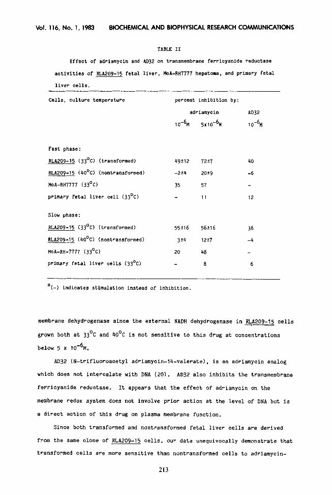
TABLE II
Effect of adriamycin and AD32 on transmembrane ferricyanide reductase
activities of RLA205-15 fetal liver, McA-RH7777 hepatoma, and primary fetal
liver cells.
Cells, culture temperature percent inhibition by:
adriamycin AD32
1 O-6M 5x10⁻⁶M 10%
Fast phase :
RLA209-15 (33’C) (transformed) 49212 72i7
RLA209-15 (4O’C) (nontransformed) -224 2029
McA-RH7777 (33’0 35 57
primary fetal liver cell (33OC) 11
40
-6
12
Slow phase:
RLA209-15 (33’0 (transformed) 55216 56216 36
RLA209-15 (4O’C) (nontransformed) 3t4 1227 -4
McA-RH-7777 (33’0 20 48
primary fetal liver cells (33OC) a 6
a(-) indicates stimulation instead of inhibition.
membrane dehydrogenase since the external NADH dehydrogenase in RLA209-15 cells
grown both at 33’C and 40°C is not sensitive to this drug at concentrations
below 5 x 10m6M.
AD32 (N-trifluoroacetyl adriamycin-14-valerate), is an adriamycin analog
which does not intercalate with DNA (20). AD32 also inhibits the transmembrane
ferricyanide reductase. It appears that the effect of adriamycin on the
membrane redox system does not involve prior action at the level of DNA but is
a direct action of this drug on plasma membrane function.
Since both transformed and nontransformed fetal liver cells are derived
from the same clone of RLA209-15 cells, our data unequivocally demonstrate that
transformed cells are more sensitive than nontransformed cells to adriamycin-
213
Vol. 116, No. 1, 1983
20
BIOCHEMICAL AND BIOPHYSICAL RESEARCH COMMUNICATIONS
I
20 40 60 80 Incubation Time (mid
Fig. 1. Effect of adriamycin on the survival fraction and ferricyanide reduction of RLA205-15 cells. Two parallel sets of RLA209-15 cultures were plated initially at 33 C. The first set of cultures was maintained at 33’C throughout growth. The second set of cultures were shifted from 33’C to 40°C after 3 days growth at 33’C. After an additional 3 days incubation at 33’C and 40°C, cells were trypsinized and resuspended in tris-EDTA buffer pH 7.4 plus 21 ’ of fetal calf s rum in a concentration of 0.1 gw.w./ml, and incubated with adriamycin (10 -9 MI at 22’C water bath with shaking. Samples were taken periodically, chilled in an ice bath, and diluted 10 fold with ice-cold tris- EDTA buffer plus 2% fetal calf serum. Survival fraction was measured immediately after spinning the cells and resuspending in tris-EDTA buffer plus 22 fetal calf serum by using eosin Y exclusion method. Survival fraction (4O’C) (01, slow rate ferricyanide reduction (40°C), (m ) fast rate ferricyanide reduction (4O’C) C&J, survival fraction f33’C) CO), slow rate ferricyanide reduction (33’C) (0) and fast rate ferricyanide reduction (33’C) (A).
induced cytotoxicity. Furthermore, such cytotoxicity may be linked to the
inhibition of transmembrane ferricyanide reductase by this antitumor drug,
Recently addition of ferricyanide has been shown to stimulate growth of
melanoma cells and the effect has been attributed to the transmembrane electron
transport system (21). There are several other functions or properties of the
plasma membrane which have been shown to be affected by adriamycin. Among
214
Vol. 116, No. 1, 1983 BIOCHEMICAL AND BIOPHYSICAL RESEARCH COMMUNICATIONS
these are ion transport (22,23), membrane fluidity (8). membrane fusion and
response to hormones (5). Each of these responses to adriamycin can be based
on adriamycin inhibition of a protonophoric transmembrane dehydrogenase
(15,16,24,25) which can modify membrane potential (26). drive amino acid trans-
port (27') and control the activity of adenylate cyclase (28) or reduce external
iron for uptake (29). The redox system in the plasma membrane, which can be
assayed by external ferricyanide reduction provides a system to measure direct
effects of adriamycin on the plasma membrane. The redox system may also
provide an approach to study the mechanism of adriamycin action at the plasma
membrane,
Acknowledgement
The advice and assistance of L. Jacobsen and the Purdue Cancer Center in culture of cells. Adriamycin was kindly supplied by Dr. M. Ghione Farmitalia, Milano, Italy and AD32 by Dr. J. DoUrOS, National Institute of Health, Division of Cancer Treatment, Developmental Therapeutics Program, Bethesda, MD. Supported in part by a grant from the Indiana Elks, and a career award GMK621839 (FLC) from National Institutes of General Medical Sciences, NIH.
References
1.
::
4. 5. 6.
7.
8.
9.
10.
11.
12. 13. 14.
15. 16.
17. 18.
19.
Blum, R.H. and S.K. Carter (1974) Ann. Internal Med. 80, 249-259. DeMarco, A. (1975) Cancer Chemotherapy Rept. 6. 91-107. Dano, K., S. Frederiksen, and P. Hellung-Larsen (1972) Cancer Res. 32, 1307-1314. Ross, W.E. and M.O. Bradley (1981) Biochim. Biophys. Acta 654, 129-134. Tritton, T.R. and G. Yee (1982) Science 217, 248-250. Tritton, T.R., G. Yee, and L.B. Wingard, Jr. (1983) Fed. Proceed. 42, 204-287. Tokes, Z.A., K. E. Rogers and A. Rembaum (1982) Proc. Natl. Acad. Sci. USA 79, 2026-2030. Siegfried. J.A., K.A. Kennedy, A.C. Satorelli, and T.R. Tritton (1983) J. Biol. Chem. 258, 339-343. Murphree, S.A., L.S. Cunningham, K.M. Hwang and A.C. Sartorelli (1976) Biochem. Pharmacol. 25, 1227-1231. Crane, F.L., W.C. MacKellar, D.J. MorrC, T. Ramasarma, H. Goldenberg, C. Grebing, and H. L(lw (1980) Biochem. Biophys. Res. Communs. 93, 746-754. Cherry, J.M., W. MacKellar, D.J. MorrC, F.L. Crane, L.B. Jacobsen, and V. Schirrmacher (1981) Biochim. Biophys. Acta 634, 11-18. Sun, I.L., W. MacKellar, and F.L. Crane (1981) Fed. Proceed. 40, 1815. Sun, I-L., and F.L. Crane (1982) Fed. Proceed. 41, 737. Clark, M.G., E.J. Partick, G.S. Patten, F.L. Crane, H. Low, and C. Grebing (1981) Biochem. J. 200, 565-572. Craig, T.A. and F.L. Crane (1981) Proceed. Indiana Acad. Sci. 90, 150-155. Crane, F.L., H. Roberts, A.W. Linnane, and H. Low, (1982) J. Bioenerg. Biomemb. 14, 491-205. Chou, J.Y. and S. Schlegle-Haueter (1981) J. Cell Biol. 89, 216-222. Mishell, B.B. and S.M. Shiigi, eds. Selective Methods in Cellular Immunology, W.H. Freeman, San Francisco (1980), pp. 17-18. Crane, F.L., H.E. Crane, I.L. sun, W.C. MacKellar, C. Grebing and H. Low (1982) J. Bioenerg. Biomemb. 14, 425-433.
215
Vol. 116, No. 1, 1983 BIOCHEMICAL AND BIOPHYSICAL RESEARCH COMMUNICATIONS
20. 21.
22.
23.
24. 25.
26.
27.
28. 29.
Israel, M. E.J. Modest and E. Frei (1975) Cancer Res. 38, 365-370. Ellem, K.A.O. and G.F. Kay (1983) Biochem. Biophys. Res. Communs 112, 183- 190. Dasdia, T., A. DiMarco, M. Goffredi, A. Minghetti and A. Necco (1979) Pharmacol. Res. Communs. 11, 19-29. Gosalvez, M., G.D. VanRossum and M.F. Bianco (1979) Cancer Res. 39, 257- 261. Dormandy, T.L. and Z. Zarday (1965) J. Physiol. 180, 684-707. Goldenberg, H., F.L. Crane and D.J. Morre (1979) J. Biol. Chem. 254, 24gl-
2498. LOW, H., C. Grebing and F.L. Crane (1982) Abstracts 12 Internat. Biochem. Congress, Perth p 138. Christensen, H.N. in Advances in Emzymology and Related Areas of Molecular Biology. ed. A. Meister, Wiley, N.Y. Vol 49 (1979) pp 41-101. Low, H. and S. Werner (1976) FEBS Lett. 65, 96-98. Crane, F.L., H. Low and M.G. Clark in Membranes and Transport, Vol. 2, ed. A. Martonosi, Plenum N.Y. (1982) pp. 251-254.
216


![Evaluation in Vitro of Adriamycin …...(CANCER RESEARCH 50. 6600-6607. October 15. 1990] Evaluation in Vitro of Adriamycin Immunoconjugates Synthesized Using an Acid-sensitive Hydrazone](https://img.pdfslide.us/doc/110x75/5e8ee25f90cfc853e1716415/evaluation-in-vitro-of-adriamycin-cancer-research-50-6600-6607-october-15.jpg)