Embed Size (px)
Citation preview
Transformation of Personal Computers and Mobile Phones intoGenetic Diagnostic SystemsFaye M. Walker,†,# Kareem M. Ahmad,‡,# Michael Eisenstein,§,∥ and H. Tom Soh*,‡,§,∥
†Department of Chemistry and Biochemistry, University of California, Santa Barbara, California 93106, United States‡Interdepartmental Program in Biomolecular Science and Engineering, University of California, Santa Barbara, California 93106,United States§Department of Materials, University of California, Santa Barbara, California 93106, United States∥Department of Mechanical Engineering, University of California, Santa Barbara, California 93106, United States
*S Supporting Information
ABSTRACT: Molecular diagnostics based on the polymerase chain reaction (PCR) offer rapid and sensitive means for detectinginfectious disease, but prohibitive costs have impeded their use in resource-limited settings where such diseases are endemic. Inthis work, we report an innovative method for transforming a desktop computer and a mobile camera phonedevices that havebecome readily accessible in developing countriesinto a highly sensitive DNA detection system. This transformation wasachieved by converting a desktop computer into a de facto thermal cycler with software that controls the temperature of thecentral processing unit (CPU), allowing for highly efficient PCR. Next, we reconfigured the mobile phone into a fluorescenceimager by adding a low-cost filter, which enabled us to quantitatively measure the resulting PCR amplicons. Our system is highlysensitive, achieving quantitative detection of as little as 9.6 attograms of target DNA, and we show that its performance iscomparable to advanced laboratory instruments at approximately 1/500th of the cost. Finally, in order to demonstrate clinicalutility, we have used our platform for the successful detection of genomic DNA from the parasite that causes Chagas disease,Trypanosoma cruzi, directly in whole, unprocessed human blood at concentrations 4-fold below the clinical titer of the parasite.
Although advanced molecular diagnostic technologies forthe detection of infectious diseases such as human
immunodeficiency virus (HIV), malaria, and tuberculosis1,2
are widely available in the developed world, prohibitive costs ofequipment and reagents have impeded their adoption in theless developed countries (LDCs) in which these diseases aremost prevalent.3−6 In contrast, access to certain consumerelectronics has surged over the past decade in these sameLDCs. For instance, the number of mobile cellular subscribersin the developing world rose by more than 600 million between2010 and 2011,7 and desktop computer penetration hasdramatically accelerated since the start of the millenium.8
This trend offers an exciting opportunity for leveraging suchtools as a means to affordably improve healthcare. For example,the built-in cameras in mobile phones have been adapted asimaging platforms9,10 for detecting disease biomarkers andinfectious pathogens11−16 in blood and other clinically relevantsamples.17−20 However, as these methods are microscopy-based, they can suffer from poor limits of detection and thechallenge of differentiating among similar species, subspecies,and strains.21 Nucleic acid-based genetic tests offer highersensitivity and exquisite specificity,22,23 and several innovativeapproaches have been explored to develop low-cost assays andinstruments for genetic detection at the point-of-care. For
example, Manage et al. achieved streamlined detection of BKviruses by performing polymerase chain reaction (PCR)directly in whole blood using self-contained gel strips.24 Inaddition, several groups have demonstrated “sample-in-answer-out” systems that integrate multiple process steps into amonolithic device using microfluidics technology.25−28 TheLanders group pioneered the use of microfluidics for geneticanalysis,29 isolating and amplifying nucleic acids directly frombuccal swabs and whole blood for clinical examination.30 Ourgroup has similarly demonstrated direct detection of H1N1influenza viruses in throat swab samples by integratingmagnetic separation with reverse transcriptase PCR (RT-PCR) in a disposable device.31 In practice, however, thedeployment of these systems in low-resource settings ischallenging because they often rely on specialized devices andinstrumentation (e.g., pumps, syringes, and detectors) that havelimited availability and require skilled technicians for operation.We have developed an alternate approach to molecular
diagnostics that largely eliminates the need for suchapparatuses. Instead of relying on custom-built machines, we
Received: June 18, 2014Accepted: August 6, 2014
Article
pubs.acs.org/ac
© XXXX American Chemical Society A dx.doi.org/10.1021/ac5022419 | Anal. Chem. XXXX, XXX, XXX−XXX
Terms of Use
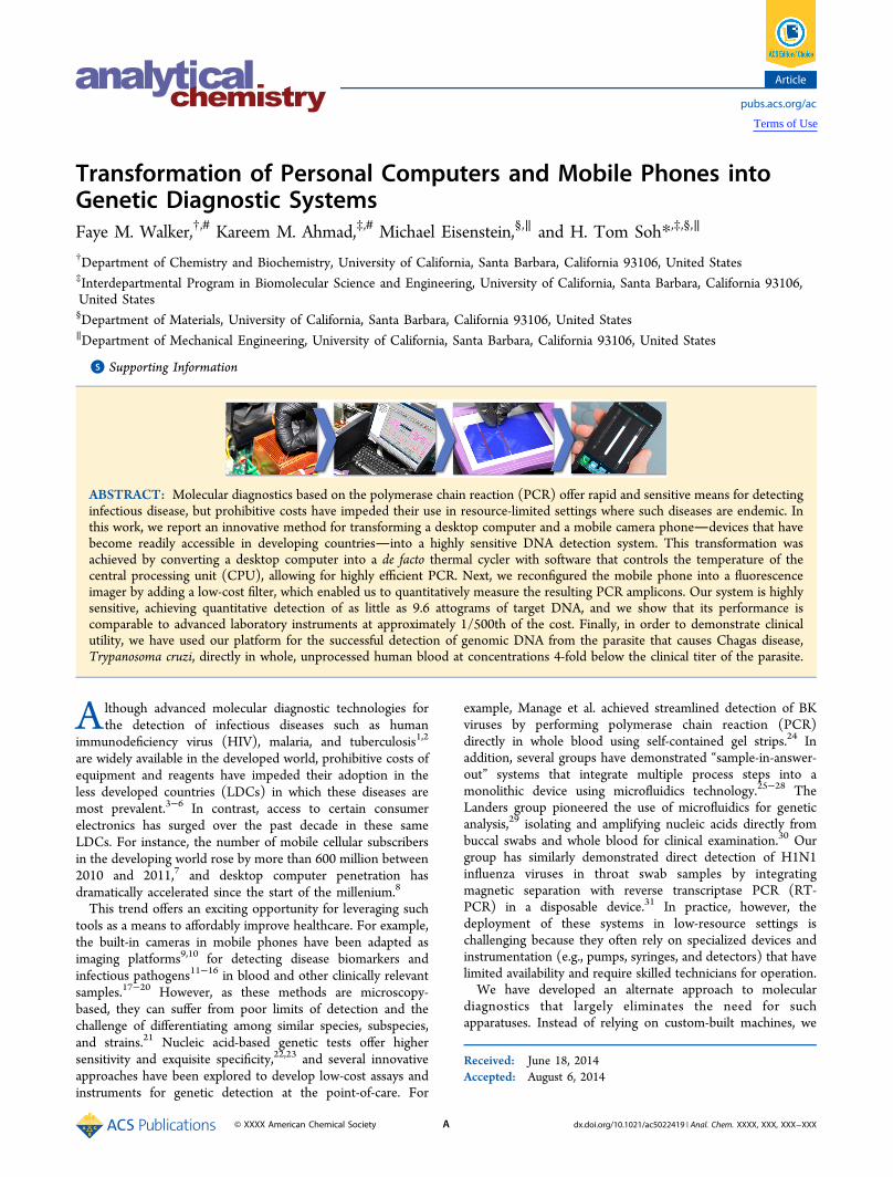
repurpose a desktop PC and a mobile camera phone into ahighly sensitive platform for genetic detection of pathogens.Specifically, we use low-cost software tools to convert the PCinto a de facto thermal cycler for PCR and configure mobilephones as imagers to detect and quantify the resulting PCRamplicons. To our knowledge, this is the first work to performPCR reactions using a PC. In our PC-PCR-Phone (P3) system,a small volume of patient blood is added directly to a length ofdisposable tubing that has been preloaded with PCR reagents(Figure 1, step 1). The tubing is then placed into the heat sinkof the central processing unit (CPU), where PCR is performedby using two software programs to precisely manipulate thePC’s internal temperature (Figure 1, step 2). Subsequently, weuse a mobile phone camera to image the amplified products,which are quantified according to their fluorescence intensity(Figure 1, step 3). As a model, we used our P3 system to detectgenomic DNA (gDNA) from Trypanosoma cruzi (T. cruzi), theparasitic protist responsible for Chagas disease,32,33 whichaffects over 17 million people worldwide.34,35 We demonstratedirect detection from blood with a limit of detection of 0.1 fg/μL, which is well below the average parasitic loads found inclinical samples (0.4 fg/μL).
■ MATERIALS AND METHODSGeneral Information. All synthetic DNA sequences were
purchased from Integrated DNA Technologies (IDT). Hot-start polymerase master mix and nuclease-free water werepurchased from Promega. PCR validation was performed with a100-nt single-stranded DNA sequence reported previously asThr-02,36 using primers AGCAGCACAGAGGTCAGATG andTTCACGGTAGCACGCATAGG. DMSO was obtained fromthe American Type Culture Collection and used at theconcentration indicated. SYBR Green I was obtained fromLife Technologies and used at 0.625× concentration, andEvaGreen was obtained from Biotium and used at 1×concentration. Whole blood preserved in EDTA was obtainedfrom Bioreclamation. Hemo KlenTaq (HKT) polymerase and5× buffer were obtained from New England Biolabs and usedin the manufacturer recommended amount. Melting temper-atures were measured via the iQ5 real-time multicolor detectionsystem (Bio-Rad).PC-Based PCR. For initial validation experiments, we used 2
fmol of 100-nt template, with 1 μM primer in a 50 μL reactionthat included PCR mix (containing DNA polymerase, dNTPs,buffer), EvaGreen, primers, and 13% DMSO. The optimal
DMSO concentration was determined prior to carrying outPCR in a PC by testing the effect of DMSO on dsDNAhybridization in the reaction mixture described above. Meltingtemperatures were determined by performing 40 cycles of two-step PCR on the 100-nt template to generate double-strandedproducts in the iQ5. Post-amplification, we carried out athermal gradient beginning at 65 °C and increasing to 85 °C ata rate of 1 °C per minute and dwell time of 10 s, withfluorescence intensity measured every minute. We thencalculated the negative first derivative of the plot of theresulting melting profile of intensity versus temperature. Themelting temperature is where this differential plot reaches amaximum, as calculated by Bio-Rad iQ5 melt curve analysis(Supplemental Figure 2, Supporting Information). To performPC-based PCR, the reaction mixture was preloaded into shortcapillaries of perfluoroalkoxyl (PFA) tubing. After addingtemplate at a concentration of 40 fg/μL, the capillary tubeswere permanently sealed at both ends by epoxy (Devcon) toyield contaminant-free testers that are amenable to high-throughput manufacturing. The capillary tube was then insertedbetween the CPU heat sink fins of a Dell Pentium 4 desktopcomputer for cycling.For measuring the actual temperature of the sample, we used
a digital thermometer (Fluke) and K type thermocouple(Omega). Thermal measurements were performed by record-ing the solution temperature inside the capillaries with 50 μL ofdistilled water using a thermocouple probe. We used twoprograms to automate the PCR thermocycling process with thecomputer. The heating of the CPU was achieved withBurnInTest software (Passmark), while SpeedFan (Almico)program obtained the CPU temperature from the built-inthermocouples and controlled the fan for cooling.The resulting PCR product was loaded and run on 10% TBE
polyacrylamide gels (Bio-Rad) with a 20 bp DNA ladder (Bio-Rad) in 4 °C running buffer. After 15 min of gel electrophoresisat 300 V, the gels were stained with Gelstar (Lonza) for 10 min.The stained gels were imaged on a Gel Logic System using UVtransillumination and a 535 nm optical filter (Kodak). Thepositive control PCR was performed by taking an aliquot of thesame sample and carrying out amplification in a commercialthermal cycler using 0.2 mL capped tubes (Bio-Rad). In boththe PC and cycler, the template was amplified for no more than20 cycles. This number was calculated to be the appropriatethreshold cycle (Ct) based on real-time quantitative PCR usinga Bio-Rad iQ5.
Figure 1. P3 assay schematic. First, a small drop of blood obtained via finger prick is added to a length of preloaded capillary tubing containing thereagents required for PCR. The tubing is then inserted between the cooling fins on the heat sink in the computer. Commercial software controlsCPU usage, cyclically heating and cooling the computer according to a protocol designed to amplify target DNA. After thermal cycling, the samplesare exposed to UV light and imaged with a camera phone. By comparing a histogram of the pixel intensities for the patient sample relative to controlsamples, the presence or absence of target pathogenic DNA can be determined.
Analytical Chemistry Article
dx.doi.org/10.1021/ac5022419 | Anal. Chem. XXXX, XXX, XXX−XXXB
Genomic DNA from T. cruzi (Tulahuen strain) was obtainedfrom ATCC. The sequence for the Diaz primer set wasCGCAAACAGATATTGACAGA and TGTTCACAC-ACTGGACACCAA,38 which target the 195-bp repetitiveelement in T. cruzi nuclear DNA. Capillaries were preppedwith 20 μL of reaction mixture containing HK Taq polymerase,0.2 mM dNTPs, 1× buffer, SYBR Green I, 0.2 μM primers, and13% DMSO. Spiked human whole blood containing gDNA (orblood itself in the case of the negative control) was added to afinal concentration of 5% (v/v) by pipetting the blood into thetubing and allowing it to settle at the bottom of the buffer layerwithout any vigorous mixing.37 Next, 1 μL of human blood wasloaded into the capillaries, and the capillary tubes were sealedon both ends and heated in a boiling water bath for 2 min tosimulate cell lysis and gDNA denaturation steps prior to PC-PCR amplification. From an initial annealing/extensiontemperature of 64.5 °C, the annealing/extension step wasreduced by a difference of 3 °C every three cycles for the initialstep-down cycling phase. After 15 cycles, we maintained theannealing temperature at 49.5 °C for 40 cycles to complete theamplification phase of SD-PCR (Supplemental Figure S5,Supporting Information).Post-PCR Imaging. Initial characterization of the camera
phone’s (Samsung Galaxy S) fluorescent imaging capabilitieswas performed with EvaGreen dye, and the PCR products weregenerated from the Thr-02 template after 30 cycles of PCR in astandard thermal cycler (Bio-Rad). Real-time quantitative PCRin a Bio-Rad iQ5 was used to determine that 30 cycles was theupper threshold for efficient amplification of a single PCRproduct from this template. Further applications with SYBRGreen I stain and T. cruzi gDNA in whole blood wereperformed according to the SD-PCR protocol described in thesection above. Samples were excited by a UV transilluminator(Kodak). For imaging, a 520 ± 10 nm bandpass filter (EdmundOptics) was placed over the mobile phone camera and held inplace by a silicone case. The phone was situated at fixeddistance above the samples, and the image was captured usingthe “night mode” option on the camera phone. Images weretransferred to a computer and analyzed with ImageJ software(http://www.nih.gov/). Rectangular regions of interest weredrawn around each sample, and the histogram of pixelintensities was obtained. Mean histogram values of all sampleswere then background-subtracted with the mean histogramvalue of an empty tube. Average and standard deviations are theresult of at least four individual trial runs. Data were importedinto MATLAB and plotted. For relative image analysis, theimsubtract function in MATLAB was used to subtract eachelement in a sample image by the same element in the blank(empty) image. From the resulting RGB values, background-subtracted images were graphed using the imshow function.
■ RESULTS AND DISCUSSIONEfficient Amplification of Genomic DNA in Blood
Using a PC. PCR amplification of genomic DNA (gDNA) inblood can be hindered by the presence of enzymatic inhibitorsnaturally found in blood or anticoagulants added after samplecollection.39 To circumvent this problem, we implementedthree key modifications to the standard PCR protocol40 (seeMaterials and Methods). First, we used a step-down (SD)-PCR41 approach that enables specific and high-yieldamplification of long template DNA (i.e., gDNA) with reducedbyproducts.42 Second, we adopted a two-temperature PCRscheme, consisting of a hot start followed by alternating
hybridization/extension and denaturation steps, simplifyingaccurate feedback control of the CPU temperature.To complete the modified PCR protocol associated with our
system, we reduced the denaturation temperature to below themaximum temperature for safe, extended CPU operation (90°C). We achieved this by adding dimethyl sulfoxide (DMSO)to our PCR mixture, which decreased the melting temperature(Tm) of our primer-template duplex by −0.6 °C per 1% DMSO(Supplemental Figure S1, Supporting Information). As anadded benefit, the addition of DMSO also improves yield andfurther reduces undesired byproducts.43,44
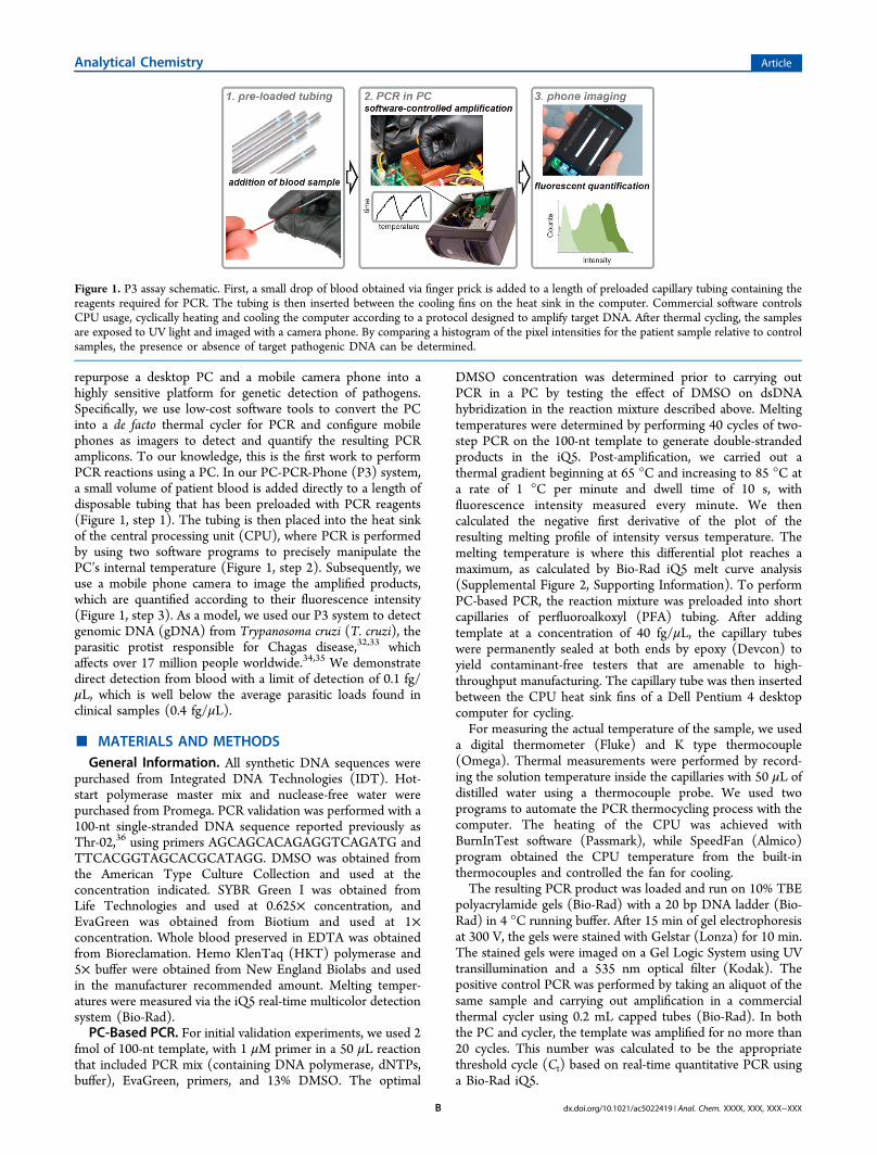
Software-Based Thermal Cycling in a PC. We installedtwo software programs that can effectively convert a desktopPC into a PCR thermal cycler. The first program (BurnIn Test)is used to rapidly increase the CPU temperature throughintensive computational operations. The second program(SpeedFan) measures the temperature of the CPU in real-time using the built-in thermal sensors common to all CPUsand also controls the cooling fan. By running these twoprograms, the surface temperature of the CPU can be preciselyregulated under automated control (Figure 2a, black trace).However, due to thermal resistance between the heat sink andtubing (even in the presence of interfacing compounds such as
Figure 2. DNA amplification achieved via robust thermal cyclingwithin the PC heat sink. (a) Temperature traces of the reported CPUtemperature (black trace) as recorded by SpeedFan software comparedwith the measured sample temperature (green trace) as obtained by athermocouple probe over the course of three cycles. Each two-stepPCR cycle started at a temperature of 55 °C for annealing andextension, which was then raised to 83 °C for melting. The verticalbars indicate when the CPU (heating, red bars) and fans (cooling, bluebars) were active. (b) PAGE image showing amplification of asynthetic 100-nt template from reactions performed within the PCheat sink. Control reactions were performed by carrying out 20 cyclesof PCR on aliquots of the same negative and positive samples in acommercial thermal cycler.
Analytical Chemistry Article
dx.doi.org/10.1021/ac5022419 | Anal. Chem. XXXX, XXX, XXX−XXXC
thermal paste), the temperature of the blood sample is lowerthan that of the CPU. In order to correct for this difference, wemeasured the actual temperature of the sample usingthermocouples (Figure 2a, green trace). These data indicatethat the difference in temperature between the CPU andsample does not vary by more than several degrees and can beaccurately predicted with a calibration curve. We established alinear correlation with excellent fit (R2 = 0.992 for heating and0.995 for cooling, Supplemental Figure S2, SupportingInformation) to account for the effect of thermal resistance.Since this thermal resistance should not vary across models ofPC, we believe our calibration curve can be used generally andthat it is not necessary to calibrate each PC individually.PC-PCR Produces Single-Length Amplicons with High
Yield. Our system is capable of simultaneously performingPCR on up to 29 samples with reproducible yield and nospurious byproducts. To verify that our PCR protocol onlygenerates amplicons of the predicted 100-nt length, weprepared 10 identical samples (20 μL each) and monitoredthe reaction at every other PCR cycle. Visualization withpolyacrylamide gel electrophoresis (PAGE) clearly showed asingle product band that matched positive control ampliconsobtained with a conventional laboratory thermal cycler(Supplemental Figure S3, Supporting Information). Wesubsequently tested the capacity of our system by performingPCR on 29 identical reactions distributed across the CPU heatsink. We observed minimal variability in amplification (Figure2b), both among the various CPU samples and relative to acontrol amplification performed in a laboratory thermal cycler,as measured by image densitometry following PAGE (C.V. <4%).Optical DNA Detection with a Camera Phone. In order
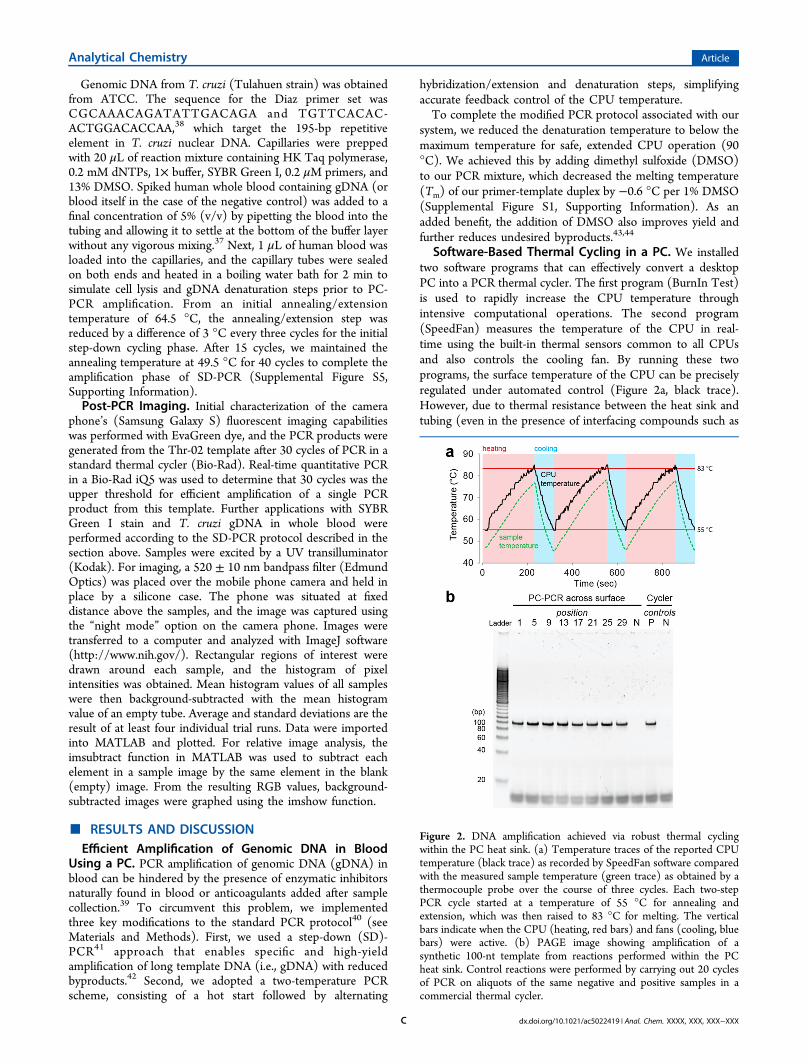
to achieve convenient and quantitative means of readout afteramplification, we repurposed a standard camera phone into aquantitative DNA detection platform capable of measuring aslittle as 9.6 ag of template DNA. Specifically, we outfitted thecamera phone with a monochromatic filter to capturefluorescence from a DNA binding dye (see Materials andMethods). This dye is present in the PCR mix prior to thereaction and emits green light (peak wavelength = 520 nm)under UV excitation when complexed with double-strandedDNA amplicons. Using this setup, we were able to clearlydifferentiate fluorescent signals obtained from PCR reactionsperformed in a conventional thermal cycler with samplescontaining as little as 9.6 ag of template DNA relative to atemplate-free negative control (Figure 3a). Moreover, thedetection performance of our camera phone system iscomparable to that of a laboratory real-time quantitative PCR(qPCR) instrument. Software analysis yielded normalized,mean fluorescence intensity values of our camera phone images(see Materials and Methods), which we plotted as a function oftemplate copy number (Figure 3b, black). We compared theseresults with the normalized end-point fluorescence valuesobtained from a qPCR instrument (Bio-Rad iQ5) (Figure 3b,red). We found that the respective performance of these twoplatforms correlates very closely, with an R2 > 0.99(Supplemental Figure S4, Supporting Information), and fallswithin each other’s error range at low template values (<153ag).Detection of T. cruzi Using the P3 System. The average
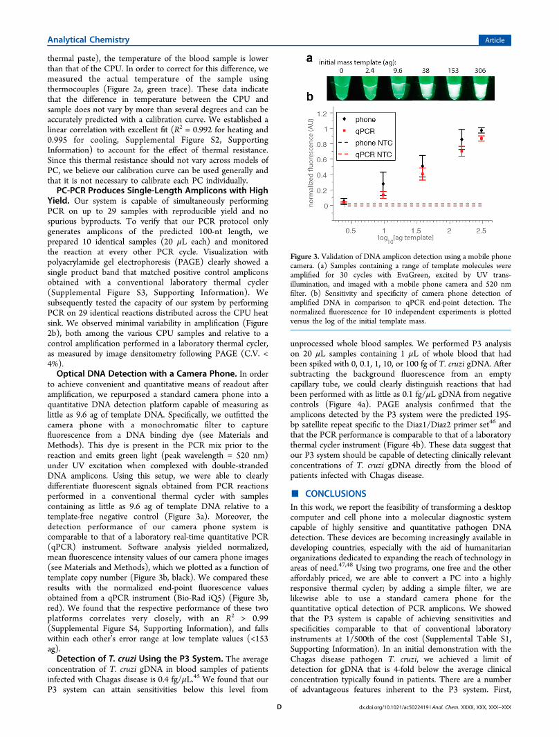
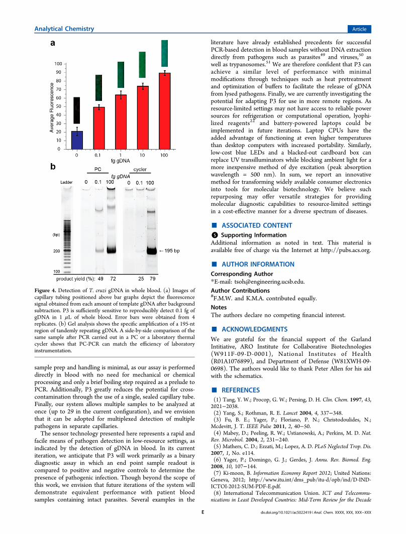
concentration of T. cruzi gDNA in blood samples of patientsinfected with Chagas disease is 0.4 fg/μL.45 We found that ourP3 system can attain sensitivities below this level from
unprocessed whole blood samples. We performed P3 analysison 20 μL samples containing 1 μL of whole blood that hadbeen spiked with 0, 0.1, 1, 10, or 100 fg of T. cruzi gDNA. Aftersubtracting the background fluorescence from an emptycapillary tube, we could clearly distinguish reactions that hadbeen performed with as little as 0.1 fg/μL gDNA from negativecontrols (Figure 4a). PAGE analysis confirmed that theamplicons detected by the P3 system were the predicted 195-bp satellite repeat specific to the Diaz1/Diaz2 primer set46 andthat the PCR performance is comparable to that of a laboratorythermal cycler instrument (Figure 4b). These data suggest thatour P3 system should be capable of detecting clinically relevantconcentrations of T. cruzi gDNA directly from the blood ofpatients infected with Chagas disease.
■ CONCLUSIONSIn this work, we report the feasibility of transforming a desktopcomputer and cell phone into a molecular diagnostic systemcapable of highly sensitive and quantitative pathogen DNAdetection. These devices are becoming increasingly available indeveloping countries, especially with the aid of humanitarianorganizations dedicated to expanding the reach of technology inareas of need.47,48 Using two programs, one free and the otheraffordably priced, we are able to convert a PC into a highlyresponsive thermal cycler; by adding a simple filter, we arelikewise able to use a standard camera phone for thequantitative optical detection of PCR amplicons. We showedthat the P3 system is capable of achieving sensitivities andspecificities comparable to that of conventional laboratoryinstruments at 1/500th of the cost (Supplemental Table S1,Supporting Information). In an initial demonstration with theChagas disease pathogen T. cruzi, we achieved a limit ofdetection for gDNA that is 4-fold below the average clinicalconcentration typically found in patients. There are a numberof advantageous features inherent to the P3 system. First,
Figure 3. Validation of DNA amplicon detection using a mobile phonecamera. (a) Samples containing a range of template molecules wereamplified for 30 cycles with EvaGreen, excited by UV trans-illumination, and imaged with a mobile phone camera and 520 nmfilter. (b) Sensitivity and specificity of camera phone detection ofamplified DNA in comparison to qPCR end-point detection. Thenormalized fluorescence for 10 independent experiments is plottedversus the log of the initial template mass.
Analytical Chemistry Article
dx.doi.org/10.1021/ac5022419 | Anal. Chem. XXXX, XXX, XXX−XXXD
sample prep and handling is minimal, as our assay is performeddirectly in blood with no need for mechanical or chemicalprocessing and only a brief boiling step required as a prelude toPCR. Additionally, P3 greatly reduces the potential for cross-contamination through the use of a single, sealed capillary tube.Finally, our system allows multiple samples to be analyzed atonce (up to 29 in the current configuration), and we envisionthat it can be adopted for multiplexed detection of multiplepathogens in separate capillaries.The sensor technology presented here represents a rapid and
facile means of pathogen detection in low-resource settings, asindicated by the detection of gDNA in blood. In its currentiteration, we anticipate that P3 will work primarily as a binarydiagnostic assay in which an end point sample readout iscompared to positive and negative controls to determine thepresence of pathogenic infection. Though beyond the scope ofthis work, we envision that future iterations of the system willdemonstrate equivalent performance with patient bloodsamples containing intact parasites. Several examples in the
literature have already established precedents for successfulPCR-based detection in blood samples without DNA extractiondirectly from pathogens such as parasites49 and viruses,50 aswell as trypanosomes.51 We are therefore confident that P3 canachieve a similar level of performance with minimalmodifications through techniques such as heat pretreatmentand optimization of buffers to facilitate the release of gDNAfrom lysed pathogens. Finally, we are currently investigating thepotential for adapting P3 for use in more remote regions. Asresource-limited settings may not have access to reliable powersources for refrigeration or computational operation, lyophi-lized reagents22 and battery-powered laptops could beimplemented in future iterations. Laptop CPUs have theadded advantage of functioning at even higher temperaturesthan desktop computers with increased portability. Similarly,low-cost blue LEDs and a blacked-out cardboard box canreplace UV transilluminators while blocking ambient light for amore inexpensive method of dye excitation (peak absorptionwavelength = 500 nm). In sum, we report an innovativemethod for transforming widely available consumer electronicsinto tools for molecular biotechnology. We believe suchrepurposing may offer versatile strategies for providingmolecular diagnostic capabilities to resource-limited settingsin a cost-effective manner for a diverse spectrum of diseases.
■ ASSOCIATED CONTENT*S Supporting InformationAdditional information as noted in text. This material isavailable free of charge via the Internet at http://pubs.acs.org.
■ AUTHOR INFORMATIONCorresponding Author*E-mail: [email protected] Contributions#F.M.W. and K.M.A. contributed equally.NotesThe authors declare no competing financial interest.
■ ACKNOWLEDGMENTSWe are grateful for the financial support of the GarlandIntitiative, ARO Institute for Collaborative Biotechnologies(W911F-09-D-0001), National Institutes of Health(R01A1076899), and Department of Defense (W81XWH-09-0698). The authors would like to thank Peter Allen for his aidwith the schematics.
■ REFERENCES(1) Tang, Y. W.; Procop, G. W.; Persing, D. H. Clin. Chem. 1997, 43,2021−2038.(2) Yang, S.; Rothman, R. E. Lancet 2004, 4, 337−348.(3) Fu, B. E.; Yager, P.; Floriano, P. N.; Christodoulides, N.;Mcdevitt, J. T. IEEE Pulse 2011, 2, 40−50.(4) Mabey, D.; Peeling, R. W.; Ustianowski, A.; Perkins, M. D. Nat.Rev. Microbiol. 2004, 2, 231−240.(5) Mathers, C. D.; Ezzati, M.; Lopez, A. D. PLoS Neglected Trop. Dis.2007, 1, No. e114.(6) Yager, P.; Domingo, G. J.; Gerdes, J. Annu. Rev. Biomed. Eng.2008, 10, 107−144.(7) Ki-moon, B. Information Economy Report 2012; United Nations:Geneva, 2012; http://www.itu.int/dms_pub/itu-d/opb/ind/D-IND-ICTOI-2012-SUM-PDF-E.pdf.(8) International Telecommunication Union. ICT and Telecommu-nications in Least Developed Countries: Mid-Term Review for the Decade
Figure 4. Detection of T. cruzi gDNA in whole blood. (a) Images ofcapillary tubing positioned above bar graphs depict the fluorescencesignal obtained from each amount of template gDNA after backgroundsubtraction. P3 is sufficiently sensitive to reproducibly detect 0.1 fg ofgDNA in 1 μL of whole blood. Error bars were obtained from 4replicates. (b) Gel analysis shows the specific amplification of a 195-ntregion of tandemly repeating gDNA. A side-by-side comparison of thesame sample after PCR carried out in a PC or a laboratory thermalcycler shows that PC-PCR can match the efficiency of laboratoryinstrumentation.
Analytical Chemistry Article
dx.doi.org/10.1021/ac5022419 | Anal. Chem. XXXX, XXX, XXX−XXXE
2001-2010; ITU: Geneva, 2006; http://www.itu.int/ITU-D/ldc/pdf/ictand_telinldc-e.pdf.(9) Stedtfeld, R. D.; Tourlousse, D. M.; Seyrig, G.; Stedtfeld, T. M.;Kronlein, M.; Price, S.; Ahmad, F.; Gulari, E.; Tiedje, J. M.; Hashsham,S. A. Lab Chip 2012, 12, 1454−1462.(10) Lee, D.; Chou, W. P.; Yeh, S. H.; Chen, P. J.; Chen, P. H.Biosens. Bioelectron. 2011, 26, 4349−4354.(11) Preechaburana, P.; Gonzalez, M. C.; Suska, A.; Filippini, D.Angew. Chem. 2012, 124, 11753−11756.(12) Wei, Q.; Qi, H.; Luo, W.; Tseng, D.; Ki, S. J.; Wan, Z.; Gorocs,Z.; Bentolila, L. A.; Wu, T.-T.; Sun, R.; Ozcan, A. ACS Nano 2013, 7,9147−9155.(13) Zhu, H.; Sikora, U.; Ozcan, A. Analyst 2012, 137, 2541−2544.(14) Greenbaum, A.; Luo, W.; Su, T.-W.; Gorocs, Z.; Xue, L.;Isikman, S. O.; Coskun, A. F.; Mudanyali, O.; Ozcan, A. Nat. Methods2012, 9, 889−895.(15) Balsam, J.; Rasooly, R.; Bruck, H. A.; Rasooly, A. Biosens.Bioelectron. 2013, 51C, 1−7.(16) Tseng, D.; Mudanyali, O.; Oztoprak, C.; Isikman, S. O.; Sencan,I.; Yaglidere, O.; Ozcan, A. Lab Chip 2010, 10, 1787−1792.(17) Zhu, H.; Mavandadi, S.; Coskun, A. F.; Yaglidere, O.; Ozcan, A.Anal. Chem. 2011, 83, 6641−6647.(18) Zhu, H.; Yaglidere, O.; Su, T.-W.; Tseng, D.; Ozcan, A. LabChip 2011, 11, 315−322.(19) Breslauer, D. N.; Maamari, R. N.; Switz, N. A.; Lam, W. A.;Fletcher, D. A. PLoS One 2009, 4, No. e6320.(20) Xie, H.; Mire, J.; Kong, Y.; Chang, M.; Hassounah, H. A.;Thornton, C. N.; Sacchettini, J. C.; Cirillo, J. D.; Rao, J. Nat. Chem.2012, 4, 802−809.(21) Gomes, Y. M.; Lorena, V. M. B.; Luquetti, A. O. Mem. Inst.Oswaldo Cruz 2009, 104 (Suppl), 115−121.(22) Niemz, A.; Ferguson, T. M.; Boyle, D. S. Trends Biotechnol.2011, 29, 240−250.(23) Carolina, J. D. Air Water Borne Dis. 2013, 02, 1−10.(24) Manage, D. P.; Lauzon, J.; Atrazhev, A.; Pang, X.; Pilarski, L. M.Lab Chip 2013, 13, 4011−4014.(25) Liu, R. H.; Yang, J.; Lenigk, R.; Bonanno, J.; Grodzinski, P. Anal.Chem. 2004, 76, 1824−1831.(26) Chin, C. D.; Laksanasopin, T.; Cheung, Y. K.; Steinmiller, D.;Linder, V.; Parsa, H.; Wang, J.; Moore, H.; Rouse, R.; Umviligihozo,G.; Karita, E.; Mwambarangwe, L.; Braunstein, S. L.; van de Wijgert, J.;Sahabo, R.; Justman, J. E.; El-Sadr, W.; Sia, S. K. Nat. Med. 2011, 17,1015−1019.(27) Martinez, A. W.; Phillips, S. T.; Carrilho, E.; Thomas, S. W.;Sindi, H.; Whitesides, G. M. Anal. Chem. 2008, 80, 3699−3707.(28) Foudeh, A. M.; Fatanat Didar, T.; Veres, T.; Tabrizian, M. LabChip 2012, 12, 3249−3266.(29) Easley, C. J.; Karlinsey, J. M.; Bienvenue, J. M.; Legendre, L. A.;Roper, M. G.; Feldman, S. H.; Hughes, M. A.; Hewlett, E. L.; Merkel,T. J.; Ferrance, J. P.; Landers, J. P. Proc. Natl. Acad. Sci. U. S. A. 2006,103, 19272−19277.(30) Lounsbury, J. A.; Karlsson, A.; Miranian, D. C.; Cronk, S. M.;Nelson, D. A.; Li, J.; Haverstick, D. M.; Kinnon, P.; Saul, D. J.;Landers, J. P. Lab Chip 2013, 13, 1384−1393.(31) Ferguson, B. S.; Buchsbaum, S. F.; Wu, T.-T.; Hsieh, K.; Xiao,Y.; Sun, R.; Soh, H. T. J. Am. Chem. Soc. 2011, 133, 9129−9135.(32) Rassi, A.; Marin-Neto, J. A. Lancet 2010, 375, 1388−1402.(33) Hotez, P. J.; Dumonteil, E.; Woc-Colburn, L.; Serpa, J. A.;Bezek, S.; Edwards, M. S.; Hallmark, C. J.; Musselwhite, L. W.; Flink,B. J.; Bottazzi, M. E. PLoS Neglected Trop. Dis. 2012, 6, No. e1498.(34) Martins-Melo, F. R.; Alencar, C. H.; Ramos, A. N.; Heukelbach,J. PLoS Neglected Trop. Dis. 2012, 6, No. e1508.(35) Moncayo, A.; Silveira, A. C. Mem. Inst. Oswaldo Cruz 2009, 104(Suppl), 17−30.(36) Ahmad, K. M.; Oh, S. S.; Kim, S.; McClellen, F. M.; Xiao, Y.;Soh, H. T. PLoS One 2011, 6, No. e27051.(37) Kermekchiev, M. B.; Barnes, W. M. Use of whole blood in PCRreactions. U.S. Patent 8470563, 2013.
(38) Diaz, C.; Nussenzweig, V.; Gonzalez, A. Am. J. Trop. Med. Hyg.1992, 46, 616−623.(39) Al-soud, W. A.; Radstrom, P. J. Clin. Microbiol. 2001, 39, 485−493.(40) Mullis, K.; Faloona, F.; Scharf, S.; Saiki, R.; Horn, G.; Erlich, H.Cold Spring Harbor Symp. Quant. Biol. 1986, L1, 263−273.(41) Hecker, K. H.; Roux, K. H. Biotechniques 1996, 20, 478−485.(42) Korbie, D. J.; Mattick, J. S. Nat. Protoc. 2008, 3, 1452−1456.(43) Varadaraj, K.; Skinner, D. M. Gene 1994, 140, 1−5.(44) Von Ahsen, N.; Wittwer, C. T.; Schutz, E. Clin. Chem. 2001, 47,1956−1961.(45) Moreira, O. C.; Ramírez, J. D.; Velazquez, E.; Melo, M. F. A. D.;Lima-Ferreira, C.; Guhl, F.; Sosa-Estani, S.; Marin-Neto, J. A.; Morillo,C. A.; Britto, C. Acta Trop. 2013, 125, 23−31.(46) Virreira, M.; Torrico, F.; Truyens, C.; Alonso-Vega, C.; Solano,M.; Carlier, Y.; Svoboda, M. Am. J. Trop. Med. Hyg. 2003, 68, 574−582.(47) Cristia, J. P.; Ibarraran, P.; Cueto, S.; Santiago, A.; Severín, E.Technology and Child Development: Evidence from the One Laptop PerChild Program; IZA Discussion Paper No. 6401; IZA: Bonn, Germany,2012.(48) Sheriff, R. E., Ed. Electronics and Telecommunications ResearchSeminar Series: 10th Workshop Proceedings; School of Engineering,Design and Technology, University of Bradford: Bradford, UK, 2011.(49) Li, Y.; Kumar, N.; Gopalakrishnan, A.; Ginocchio, C.; Manji, R.;Bythrow, M.; Lemieux, B.; Kong, H. J. Mol. Diagn. 2013, 15, 634−641.(50) Zhang, Z.; Kermekchiev, M. B.; Barnes, W. M. J. Mol. Diagn.2010, 12, 152−161.(51) Ravindran, R.; Rao, J. R.; Mishra, A. K.; Pathak, K. M. L.; Babu,N.; Satheesh, C. C.; Rahul, S. Vet. Arh. 2008, 78, 89−94.
Analytical Chemistry Article
dx.doi.org/10.1021/ac5022419 | Anal. Chem. XXXX, XXX, XXX−XXXF