Embed Size (px)
Citation preview
1
Neurorehabilitation and Neural Repair
Volume XX Number XMonth XXXX xx-xx
© 2009 The American Society of Neurorehabilitation
10.1177/1545968308328723http://nnr.sagepub.com
hosted athttp://online.sagepub.com
Transcutaneously Coupled, High-Frequency Electrical Stimulation of the Pudendal Nerve Blocks External Urethral Sphincter Contractions
Robert A. Gaunt and Arthur Prochazka, PhD
Background. Detrusor-sphincter dyssynergia is a condition in which reflexive contractions of the external urethral sphincter occur during bladder contractions, preventing the expulsion of urine. High-frequency stimulation (kHz range) has been shown to elicit a fast-acting and reversible block of action potential propagation in peripheral nerves, which may be a useful technique in the management of this condition. Objective. The aim of these experiments was to see if a newly developed stimulus delivery system, capable of transmitting current trans-cutaneously to remote peripheral nerves using a passive implanted conductor, was an effective way to transmit high-frequency waveforms to the pudendal nerve to block ongoing sphincter contractions. Methods. High-frequency waveforms were delivered through the skin to the pudendal nerve using a passive implanted conductor in 6 adult cats anesthetized with isoflurane. Five of the experiments were acute, ter-minal procedures, and the remaining cat was implanted with a permanent electrode system allowing evaluation for 6 months. Typical stimulation parameters were in the range of 1 to 10 kHz and 1 to 10 mA. Results. Complete blocking of external urethral sphincter contrac-tions was achieved in 5 of the 6 animals. High-frequency stimulation was also tested in the chronically implanted animal without anesthe-sia, and the stimulation was tolerated with minimal aversive reactions. Conclusions. The transcutaneous passive implanted conductor stimulus delivery system is an effective way to stimulate the pudendal nerve at high frequency, leading to sphincter relaxation. This system may provide a simple means to implement this stimulation paradigm in people with detrusor-sphincter dyssynergia.
Key Words: Detrusor-sphincter dyssyergia; Neuroprosthesis; High-frequency block; Bladder; Spinal cord injury
The act of micturition normally involves the coordinated activation of the detrusor (bladder wall) muscle and relax-
ation of the external urethral sphincter (EUS) muscle. During the maintenance of continence, the detrusor remains relaxed while EUS contractions help prevent leakage; during micturi-tion, the pattern of activity reverses and the EUS relaxes while the detrusor contracts, resulting in the expulsion of urine. Injury and disease involving the nervous system, including spinal cord injury (SCI) and multiple sclerosis, can disrupt this normal control scheme, resulting in an impairment of storage, voiding, or both.
After SCI, detrusor-sphincter dyssynergia (DSD) is a fre-quently observed phenomenon in which the EUS contracts reflexively in response to contractions of the bladder.1-3 DSD therefore prevents normal voiding, and if left untreated it can lead to high intravesical pressures and renal failure. Many tech-niques have been proposed to overcome the problem of DSD,4 but the current best practices involve pharmacological treatment to reduce hyperreflexive bladder contractions combined with clean intermittent or suprapubic catheterization to drain the bladder. Despite these treatments, urinary tract infections and other urinary-related complications remain the leading cause of hospitalization after SCI.5 There remains a strong impetus to develop better solutions to the problem of bladder control after SCI.
One relatively recent approach to overcoming DSD after SCI involves the use of high-frequency stimulation (HFS) of the pudendal nerve to block action potential propagation in the nerve and thus unwanted EUS activity.6-8 The pudendal nerve contains the motor axons to the EUS as well as other efferent and afferent fibers innervating the external anal sphincter, other perineal musculature, and genitalia.9 HFS has been investigated sporadically over the past 100 years and has been shown to block the propagation of action potentials locally in peripheral nerve. Kilgore and Bhadra provide a thorough sum-mary of the history of HFS and the various and inconsistent ways it has been implemented and reported.10 Part of the rea-son for the confusion in this area of research is that the term “high-frequency stimulation” has been used to describe stimu-lation frequencies from less than 100 Hz all the way up to 50 kHz. In addition, a complete theoretical understanding of the mechanism of action of HFS has yet to be achieved. However, recent work has done much to address this confusion and to propose theoretical models to explain the mechanism of high-frequency (HF) blocking.10-16 In addition to reducing unwanted EUS contractions,6-8,17-19 there have been reports of using HFS to control motor axon recruitment order during electrical stimulation20,21 and to suppress tinnitus.22 It should be noted that some of these studies used monophasic rather than biphasic HFS17-21 and that different mechanisms are likely responsible for
From the Department of Biomedical Engineering, University of Alberta, Edmonton, Alberta, Canada. Address correspondence to Arthur Prochazka, PhD, University of Alberta, Center for Neuroscience, 507 HMRC, Edmonton, AB, Canada T6G 2S2. E-mail: [email protected].
Neurorehabil Neural Repair OnlineFirst, published on December 24, 2008 as doi:10.1177/1545968308328723
2 Neurorehabilitation and Neural Repair
the observed reductions in nerve activity in these 2 conditions, with monophasic HFS possibly acting like a direct-current nerve block.10
Recently, a transcutaneous stimulation delivery system, referred to as the stimulus router system (SRS), that passes current directly through the skin rather than relying on induc-tive coupling across the skin was described.23 An external stimulator connected to surface electrodes applied to the skin was used to transmit stimulation pulses to deep-lying nerves using completely passive implanted electrodes. Given the potential usefulness of HFS in blocking unwanted EUS con-tractions, the primary aim of this study was to examine whether this new stimulus delivery system could transmit HF waveforms to the pudendal nerve sufficient to elicit functional blocking of the EUS contractions.
Method
Four adult male (#1, #2, #3, #6) and 2 adult female (#4, #5) cats (age: 1.5-4 years, weight: 2.7-6.5 kg) were used to test the hypothesis that the SRS is an effective means to deliver HF waveforms to the pudendal nerve to block EUS contractions. Acute experiments were performed on all animals except #6. In animal #6 a chronic implant was performed to evaluate the performance of HFS at various time points. All experiments were done with the approval of the University of Alberta Animal Care and Use Committee.
Surgical Procedures
The animals were preoperatively medicated with acepro-mazine (0.1-0.25 mg/kg sc), glycopyrrolate (0.01 mg/kg sc), and buprenorphine (0.01 mg/kg sc) or hydromorphone (0.1 mg/kg sc) and then anesthetized with a mixture of isoflurane (2%-3% in carbogen, flow rate 2 L/min). For the acute experi-ments, the trachea was cannulated and connected to a closed-loop anesthetic system that monitored respiration rate and pressure and ventilated the animal if necessary. For the chronic implant, anesthesia was delivered via a pediatric endotracheal tube. The cephalic vein was catheterized to allow administra-tion of fluids and drugs, and a saline drip was delivered throughout the procedure. Body temperature was maintained using a warm-water heating pad, and the heart rate and SpO2 were monitored throughout.
For the acute experiments, the bladder was exposed through a midline abdominal incision and catheterized at the dome with a 5 Fr. catheter to allow intravesical pressure measurement as well as the addition and withdrawal of fluid. A purse-string suture was placed around the catheter to prevent leakage. The abdomen was closed in layers, with the catheter emerging percutaneously. For the chronic implant, the end of a custom-made silicone catheter was inserted into the dome of the blad-der through a puncture hole created with a 16G hypodermic needle, and a silk purse-string suture was secured around the
catheter. A retaining disk on the catheter was sutured to the bladder wall to ensure that the catheter remained fixed in place. The abdominal musculature was closed with 3-0 Vicryl suture, with the catheter emerging at the rostral end. A trocar was used to draw the catheter subcutaneously to the cat’s head. The abdominal skin was sutured closed with 3-0 Prolene suture. Four stainless steel screws were attached to the skull through small incisions, which were then sutured closed. The screw heads, as well as a Luer fitting attached to the bladder catheter, were embedded in a dental acrylic cap. This provided a secure platform for long-term access to the bladder catheter connector while maintaining skin health. During experimental procedures, bladder pressure was monitored and generally kept below 10 mm Hg by emptying the bladder at regular intervals through the implanted catheter.
The pudendal nerves were exposed bilaterally by incisions lateral to the base of the tail and by blunt dissection through the tissue of the ischio-rectal fossa. For the acute experiments, 2 bipolar platinum hook electrodes or stainless steel electrode nerve cuffs were placed on exposed portions of the pudendal nerve unilaterally. Both the proximally and distally placed electrodes captured the caudal rectal and deep perineal branches of the pudendal nerve.9 In the case of animal #3, a laminectomy was performed, and a bipolar nerve cuff was placed on the S2 spinal nerve root intradurally instead of on the proximal pudendal nerve. For the chronic implant, custom-made implants consisting of 3 SRSs for the left and right sides were implanted (see Figure 1 for a schematic and description of the implant). The nerve cuff used in the chronic implant consisted of a 10-mm length of split silicone tubing containing 3 contacts. The proximal contact was used to deliver low- frequency stimulus waveforms, and the 2 distal electrodes were used in a bipolar configuration to deliver HF waveforms. The stimulating electrode was separated from the blocking electrodes by 4 mm, and the bipolar blocking electrodes had an interelectrode spacing of 2 mm. This particular nerve cuff length and electrode configuration was chosen to fit the avail-able length of pudendal nerve in a chronic implant while still containing the required electrodes.
At the end of the chronic implant, the cat was given keto-profen (1 mg/kg sc) and hydromorphone (0.07 mg/kg sc) suf-ficient to maintain a somnolent state. During postoperative recovery, the cat was kept warm in a heated cage. Analgesia was maintained by giving 2 or 3 additional doses of ketoprofen and/or hydromorphone at 8-hour intervals. Ampicillin was administered for 4 days after surgery, followed by amoxicillin (62.5 mg tablets, 2/day) for 6 additional days. At the termination of all acute experiments, the animals were euthanized with an overdose of pentobarbital sodium.
Stimulus Router System
The SRS used throughout these experiments provided a means to transmit electrical current from surface electrodes through the skin and then to the pudendal nerve via an implanted
Gaunt, Prochazka / Electrical Stimulation of the Pudendal Nerve 3
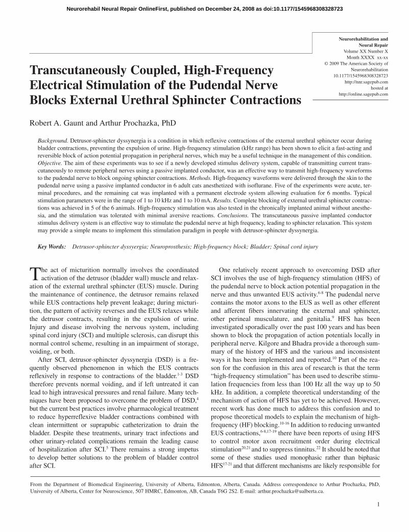
lead wire. The SRS consists of several components: the external stimulator, surface electrodes, and lead wires with pick-up and stimulation terminals. Adhesive gel electrodes placed on the skin were positioned directly over subcutaneously placed pick-up terminals, which were connected via insulated lead wires to stimulation terminals (hook electrodes or nerve cuff electrodes). When current was passed between pairs of surface electrodes, a portion of the current was captured by the implanted lead and routed to the nerve cuff to stimulate the pudendal nerve. Figure 2 shows a schematic representation of this system and the setup for the acute experiments in this study. In this system, the ratio of internal current (current flowing to the nerve) to the external current (total current delivered by the stimulator) has been shown to be in the range of 0.10 to 0.18.23
Stimulation Procedures
Two types of stimulation were used in these experiments: direct stimulation and SRS stimulation. In direct stimulation, the bipolar electrodes of the hook electrode or nerve cuff, placed on the pudendal nerve, were directly connected to a pulse generator. When SRS stimulation was used, the distally placed hook electrodes or nerve cuff electrodes were con-nected as shown in Figure 2. The adhesive gel surface elec-trodes (Kendall Soft-E H69P; Kendall-LTP, Chicopee, MA)
were applied to the skin after it was carefully shaved and cleaned with alcohol. The pick-up electrodes were positioned subcutaneously near the lumbar vertebrae.
Low-frequency (LF) (~15-30 Hz) direct stimulation via proximally placed hook or nerve cuff electrodes on the pudendal nerve was used to elicit contractions of the EUS. In one case (#3), contractions of the EUS were generated by intradural stimulation of the S2 spinal nerve root. LF pulse trains were generated using either a voltage-controlled Grass SD9 stimulator (Grass Technologies, West Warwick, RI) or a current-controlled Neurolog stimulator (Digitimer Ltd, Welwyn Garden City, UK) using modules NL304 (period generator), NL403 (delay-width), NL510 (pulse buf-fer), and NL800 (stimulus isolator). HF square-wave pulse trains were generated using the Neurolog system (animals #1 and #2) or alternatively, a custom-built, constant-current stimulator was used to generate HF sinusoidal waveforms (ani-mals #3-#6). Both the Grass SD9 and Neurolog stimulators
Figure 1 Schematic of the Left-Hand Side Chronic Implant
Note: The nerve cuffs were constructed using Silastic tubing (1.02 mm ID, 2.16 mm OD) and Cooner AS636 stainless steel wire. The Silastic tubes were sliced longitudinally, and a section of deinsulated wire was inserted through one edge of the tube and exited at the opposite edge. At both the entry and exit from the tubing, a bead of silicone was applied to both insu-late the wire and secure it to the tubing. Three such electrodes were placed in each cuff, as shown. The SRS pick-up terminals were made from stainless steel disks 1.5 cm in diameter and were embedded in a custom-made sili-cone base. The disks were separated by 3 cm and a 1 cm diameter window was cut in the silicone base to expose the metal disk. The array of pick-up electrodes was positioned in the subcutaneous space near the lumbar verte-brae. The electrodes in the nerve cuffs were connected to SRS pick-up electrodes via Cooner AS636 wire.
Figure 2 Schematic of the SRS and the Setup for the Acute
Experiments Used to Test HF Blocking of the Pudendal Nerve
Note: Each of the 2 electrodes in the distal “blocking” cuff was connected via insulated wire to a “pick-up terminal” made from a stainless steel metal disk (1-1.5 cm diameter) placed under the skin. Adhesive gel electrodes were applied on the skin directly over the pick-up electrodes and connected to a stimulator. The proximal nerve cuff was connected directly to a stimulator to generate LF pulse trains. An intraurethral catheter was positioned with its side-port in the region of the EUS to measure intraurethral pressure during low-frequency stimulation (LFS) and HFS.
4 Neurorehabilitation and Neural Repair
generated only monophasic pulses, whereas the custom- built stimulator was used to generate biphasic waveforms. Therefore, animals #1 and #2 received monophasic HFS exclusively, whereas the remaining animals received biphasic HFS exclusively.
In the chronic implant (animal #6), both LFS and HFS were delivered via the SRS implants. The response to HFS was normally evaluated with the animal anesthetized with isoflu-rane. Prior to a stimulation session, the skin over each pick-up electrode was shaved and cleaned, and adhesive gel surface electrodes (Kendall H69P) were applied. LF pulse trains were delivered using the Grass SD9 stimulator through the most proximal pudendal nerve electrode (L1; see Figure 1). An additional adhesive gel electrode (Kendall H49P) was posi-tioned over the base of the tail to act as the indifferent (anode) electrode during LF monopolar stimulation. The Grass SD9 stimulator provided voltage-controlled monophasic pulses 200 μs in duration. HF waveforms were current-controlled, charge-balanced sinusoids applied through the L2 and L3 surface electrodes. Six evaluation sessions were performed over the course of 6.5 months.
A typical HFS trial consisted of 10 seconds of LFS deliv-ered through the proximal electrode followed by 5 seconds of LFS + HFS and a final 10 seconds of LFS only. From trial to trial, both the HFS amplitude and frequency were varied. Typically, the HFS parameters ranged from 1 to 10 kHz and 1 to 10 mA (external current). In all experiments, the bipolar HF electrodes were separated by 2 mm and were 0.5 to 1 mm wide.
Pressure Measurement
Bladder and intraurethral pressures were measured using an implanted bladder catheter and a closed-end tomcat catheter (Kendall 3.5 Fr), respectively. Each catheter was connected to a Neurolog NL108D4/10 dome and NL108T4 isolated pres-sure transducer (Digitimer Ltd, Welwyn Garden City, UK). The urethral catheter was modified to block the distal of the 2 side ports at the tip of the catheter. The urethral catheter was also connected to an infusion pump (Pump 22; Harvard Apparatus, Saint Laurent, Quebec, Canada), and during intrau-rethral pressure measurements, sterile saline was infused at a rate of 0.2 or 1.0 mL/min. This method of measuring urethral pressure was first described by Brown and Wickham.24 The side port of the urethral catheter was positioned in the region of the EUS (typically 4-6 cm from the tip of the urethral meatus). Both the bladder and urethral pressure signals were displayed on an oscilloscope, and the data were sampled at a rate of 100 samples per second using a CED 1401 Laboratory Interface and Signal v3 software (Cambridge Electronic Design Ltd, Cambridge, UK).
Data Analysis
An automated analysis of the intraurethral pressure traces was performed using software written in Matlab R2007a (The
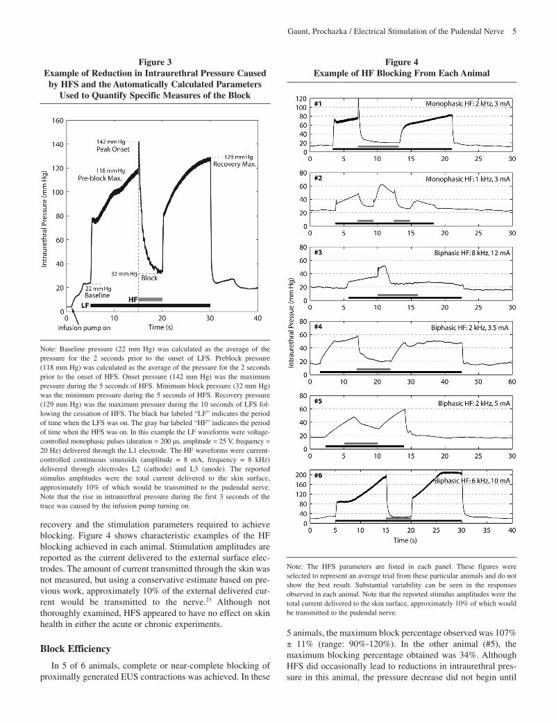
Mathworks, Natick, MA) to extract 3 measures of perfor-mance in the HFS trials. Figure 3 shows an example of a HFS blocking protocol and the resulting intraurethral pressure trace. From this trace, the baseline pressure (22 mm Hg), pre-block pressure (118 mm Hg), peak onset pressure (142 mm Hg), minimum block pressure (32 mm Hg), and peak recovery pressure (129 mm Hg) were found. These values were used to calculate the following 3 measures: block percentage, onset percentage, and recovery percentage. In the example of Figure 3 where the stimulation amplitude was 8 mA and the stimula-tion frequency was 8 kHz, the block percentage was 90%, the onset percentage was 25%, and the recovery percentage was 110%. Block percentage, onset percentage, and recovery percentage were calculated as follows:
Block%= PPreBlockMax −PBlock
PPreBlockMax −PBaseline
× 100
Onset%= PPeakOnset −PPreBlockMax
PPreBlockMax −PBaseline
× 100
Recovery%= PRecoveryMax −PBaseline
PPreBlockMax −PBaseline
× 100
These 3 measures were selected to provide quantitative information regarding the completeness of the block, the size of the onset response, and the effect of the block on subsequent neural and muscular function.
Statistical analysis was performed using Matlab R2007a and SigmaStat v3.5 (Systat Software, Inc, San Jose, CA). Kruskal-Wallis one-way analysis of variance (ANOVA) on ranks was used to test for differences between multiple groups. Student’s t test was used to test for the difference in means of two groups. The text indicates which test was used in specific instances. Simple linear regression was used to examine rela-tionships between variables, and ANOVA was used to test for significance in these cases. In all cases results are reported as significant with P < .05. At various points, the data are divided into two groups: the first group includes all collected data, and the second group includes only data where the block percent-age was > 80%. This second group was selected to provide an indication of those trials in which “near complete” blocking was achieved and where the blocking would likely lead to functionally relevant results. Grouped data are reported in the text as the mean ± standard deviation.
RESULTS
The primary aim of these experiments was to determine whether or not HF waveforms could be transmitted by the SRS to the pudendal nerve to block ongoing EUS contractions. In the first group of 3 animals, the experimental goal was to deter-mine the feasibility of the approach, and in the last 3 animals the experimental goals were to more thoroughly examine the details of the nerve blocking including efficacy, onset, and
Gaunt, Prochazka / Electrical Stimulation of the Pudendal Nerve 5
Figure 3 Example of Reduction in Intraurethral Pressure Caused
by HFS and the Automatically Calculated Parameters Used to Quantify Specific Measures of the Block
Note: Baseline pressure (22 mm Hg) was calculated as the average of the pressure for the 2 seconds prior to the onset of LFS. Preblock pressure (118 mm Hg) was calculated as the average of the pressure for the 2 seconds prior to the onset of HFS. Onset pressure (142 mm Hg) was the maximum pressure during the 5 seconds of HFS. Minimum block pressure (32 mm Hg) was the minimum pressure during the 5 seconds of HFS. Recovery pressure (129 mm Hg) was the maximum pressure during the 10 seconds of LFS fol-lowing the cessation of HFS. The black bar labeled “LF” indicates the period of time when the LFS was on. The gray bar labeled “HF” indicates the period of time when the HFS was on. In this example the LF waveforms were voltage- controlled monophasic pulses (duration = 200 μs, amplitude = 25 V, frequency = 20 Hz) delivered through the L1 electrode. The HF waveforms were current- controlled continuous sinusoids (amplitude = 8 mA, frequency = 8 kHz) delivered through electrodes L2 (cathode) and L3 (anode). The reported stimulus amplitudes were the total current delivered to the skin surface, approximately 10% of which would be transmitted to the pudendal nerve. Note that the rise in intraurethral pressure during the first 3 seconds of the trace was caused by the infusion pump turning on.
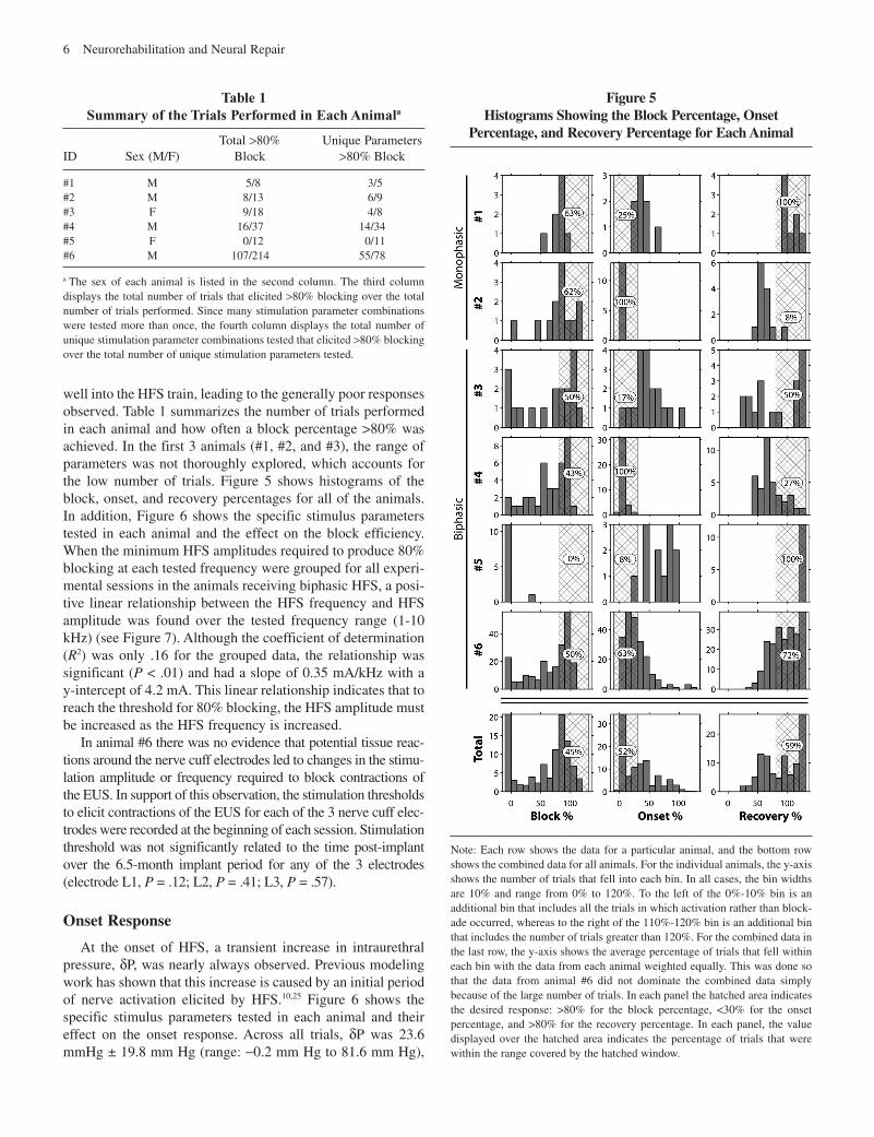
recovery and the stimulation parameters required to achieve blocking. Figure 4 shows characteristic examples of the HF blocking achieved in each animal. Stimulation amplitudes are reported as the current delivered to the external surface elec-trodes. The amount of current transmitted through the skin was not measured, but using a conservative estimate based on pre-vious work, approximately 10% of the external delivered cur-rent would be transmitted to the nerve.23 Although not thoroughly examined, HFS appeared to have no effect on skin health in either the acute or chronic experiments.
Block Efficiency
In 5 of 6 animals, complete or near-complete blocking of proximally generated EUS contractions was achieved. In these
Figure 4 Example of HF Blocking From Each Animal
Note: The HFS parameters are listed in each panel. These figures were selected to represent an average trial from these particular animals and do not show the best result. Substantial variability can be seen in the responses observed in each animal. Note that the reported stimulus amplitudes were the total current delivered to the skin surface, approximately 10% of which would be transmitted to the pudendal nerve.
5 animals, the maximum block percentage observed was 107% ± 11% (range: 90%-120%). In the other animal (#5), the maximum blocking percentage obtained was 34%. Although HFS did occasionally lead to reductions in intraurethral pres-sure in this animal, the pressure decrease did not begin until
6 Neurorehabilitation and Neural Repair
Table 1 Summary of the Trials Performed in Each Animala
Total >80% Unique Parameters ID Sex (M/F) Block >80% Block
#1 M 5/8 3/5#2 M 8/13 6/9#3 F 9/18 4/8#4 M 16/37 14/34#5 F 0/12 0/11#6 M 107/214 55/78
a The sex of each animal is listed in the second column. The third column displays the total number of trials that elicited >80% blocking over the total number of trials performed. Since many stimulation parameter combinations were tested more than once, the fourth column displays the total number of unique stimulation parameter combinations tested that elicited >80% blocking over the total number of unique stimulation parameters tested.
Figure 5 Histograms Showing the Block Percentage, Onset
Percentage, and Recovery Percentage for Each Animal
Note: Each row shows the data for a particular animal, and the bottom row shows the combined data for all animals. For the individual animals, the y-axis shows the number of trials that fell into each bin. In all cases, the bin widths are 10% and range from 0% to 120%. To the left of the 0%-10% bin is an additional bin that includes all the trials in which activation rather than block-ade occurred, whereas to the right of the 110%-120% bin is an additional bin that includes the number of trials greater than 120%. For the combined data in the last row, the y-axis shows the average percentage of trials that fell within each bin with the data from each animal weighted equally. This was done so that the data from animal #6 did not dominate the combined data simply because of the large number of trials. In each panel the hatched area indicates the desired response: >80% for the block percentage, <30% for the onset percentage, and >80% for the recovery percentage. In each panel, the value displayed over the hatched area indicates the percentage of trials that were within the range covered by the hatched window.
well into the HFS train, leading to the generally poor responses observed. Table 1 summarizes the number of trials performed in each animal and how often a block percentage >80% was achieved. In the first 3 animals (#1, #2, and #3), the range of parameters was not thoroughly explored, which accounts for the low number of trials. Figure 5 shows histograms of the block, onset, and recovery percentages for all of the animals. In addition, Figure 6 shows the specific stimulus parameters tested in each animal and the effect on the block efficiency. When the minimum HFS amplitudes required to produce 80% blocking at each tested frequency were grouped for all experi-mental sessions in the animals receiving biphasic HFS, a posi-tive linear relationship between the HFS frequency and HFS amplitude was found over the tested frequency range (1-10 kHz) (see Figure 7). Although the coefficient of determination (R2) was only .16 for the grouped data, the relationship was significant (P < .01) and had a slope of 0.35 mA/kHz with a y-intercept of 4.2 mA. This linear relationship indicates that to reach the threshold for 80% blocking, the HFS amplitude must be increased as the HFS frequency is increased.
In animal #6 there was no evidence that potential tissue reac-tions around the nerve cuff electrodes led to changes in the stimu-lation amplitude or frequency required to block contractions of the EUS. In support of this observation, the stimulation thresholds to elicit contractions of the EUS for each of the 3 nerve cuff elec-trodes were recorded at the beginning of each session. Stimulation threshold was not significantly related to the time post-implant over the 6.5-month implant period for any of the 3 electrodes (electrode L1, P = .12; L2, P = .41; L3, P = .57).
Onset Response
At the onset of HFS, a transient increase in intraurethral pressure, δP, was nearly always observed. Previous modeling work has shown that this increase is caused by an initial period of nerve activation elicited by HFS.10,25 Figure 6 shows the specific stimulus parameters tested in each animal and their effect on the onset response. Across all trials, δP was 23.6 mmHg ± 19.8 mm Hg (range: -0.2 mm Hg to 81.6 mm Hg),
Gaunt, Prochazka / Electrical Stimulation of the Pudendal Nerve 7
Figure 6 Summary of the Effect of Stimulus Frequency and
Amplitude on Block Percentage, Onset Percentage, and Recovery Percentage for Each Animal
Note: Animals #1 and #2 received monophasic HFS, and the remaining animals received biphasic HFS. Each row of subfigures contains the dataset from a sin-gle animal, and the columns show the block percentage, onset percentage, and recovery percentage. Combinations of stimulus frequency and amplitude that elicited a blocking efficiency >80%, an onset percentage <30%, or a recovery percentage >80% are shown as large circles. Stimulation parameters that did not reach these threshold values are shown as small circles. In cases where multiple trials were performed at a given stimulus frequency/ amplitude combinations, the results with the maximum block efficiency are shown. Note that stimulus amplitude refers to the total current delivered to the skin surface, approximately 10% of which would have been transmitted to the pudendal nerve.
Figure 7 Threshold Stimulation Amplitudes at Different Stimulus
Frequencies for Animals Receiving Biphasic HFS
Note: Since no combination of HFS parameters in animal #5 resulted in block efficiencies >80%, no data from this animal are shown in this figure. Data from animal #6 are presented separately for different testing sessions. Increasing the stimulation frequency increased the stimulus amplitude required to achieve a block efficiency of 80%. The regression was significant (P < .01). Note that the reported stimulus amplitudes were the total current delivered to the skin surface, approximately 10% of which would be transmit-ted to the pudendal nerve.
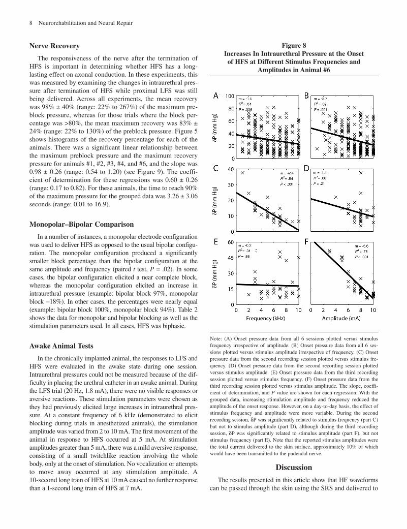
preblock pressure. For example, if the intraurethral pressure was decreasing in the 2 seconds prior to the onset of HFS, it was possible for the average pressure during this window to be higher than the maximum increase in pressure at the onset of HFS, leading to a negative value of δP. In animal #6, in which the widest stimulation parameter range was examined, there was a negative linear relationship between δP and both the HFS frequency (P < .006) and amplitude (P < .001) across all trials (see Figures 8A and 8B). However, the coefficient of determination was 0.036 and 0.09 for the relationships, respec-tively, indicating that HFS frequency and HFS amplitude had only very weak predictive value for δP. When the data from animal #6 were considered on a session-by-session basis, sev-eral high R2 values were found, but they changed substantially between sessions. For example, during the second recording session, R2 for the regression between HFS frequency and δP was .54 (P < .001), whereas it was not significant for the HFS amplitude (see Figures 8C and 8D). During the third recording session, however, the regression between HFS frequency and onset amplitude was not significant, although R2 was .75 for the regression between HFS amplitude and δP (P < .001) (see Figures 8E and 8F). Figure 5 shows histograms of δP expressed as onset percentages for each animal. There was a significant difference in the onset percentages between animals (Kruskal-Wallis, P < .001). In all cases where a significant relationship was present between δP and HFS frequency or amplitude, δP decreased with increasing HFS frequency or amplitude. This was true for the grouped data for each animal, as well as for the individual session data for animal #6.
representing an increase over the preblock pressure of 27.4% ± 23.6% (range: -0.3% to 122.4%). Several negative values of δP were obtained because of the method of calculating the
8 Neurorehabilitation and Neural Repair
Nerve Recovery
The responsiveness of the nerve after the termination of HFS is important in determining whether HFS has a long-lasting effect on axonal conduction. In these experiments, this was measured by examining the changes in intraurethral pres-sure after termination of HFS while proximal LFS was still being delivered. Across all experiments, the mean recovery was 98% ± 40% (range: 22% to 267%) of the maximum pre-block pressure, whereas for those trials where the block per-centage was >80%, the mean maximum recovery was 83% ± 24% (range: 22% to 130%) of the preblock pressure. Figure 5 shows histograms of the recovery percentage for each of the animals. There was a significant linear relationship between the maximum preblock pressure and the maximum recovery pressure for animals #1, #2, #3, #4, and #6, and the slope was 0.98 ± 0.26 (range: 0.54 to 1.20) (see Figure 9). The coeffi-cient of determination for these regressions was 0.60 ± 0.26 (range: 0.17 to 0.82). For these animals, the time to reach 90% of the maximum pressure for the grouped data was 3.26 ± 3.06 seconds (range: 0.01 to 16.9).
Monopolar–Bipolar Comparison
In a number of instances, a monopolar electrode configuration was used to deliver HFS as opposed to the usual bipolar configu-ration. The monopolar configuration produced a significantly smaller block percentage than the bipolar configuration at the same amplitude and frequency (paired t test, P = .02). In some cases, the bipolar configuration elicited a near complete block, whereas the monopolar configuration elicited an increase in intraurethral pressure (example: bipolar block 97%, monopolar block -18%). In other cases, the percentages were nearly equal (example: bipolar block 100%, monopolar block 94%). Table 2 shows the data for monopolar and bipolar blocking as well as the stimulation parameters used. In all cases, HFS was biphasic.
Awake Animal Tests
In the chronically implanted animal, the responses to LFS and HFS were evaluated in the awake state during one session. Intraurethral pressures could not be measured because of the dif-ficulty in placing the urethral catheter in an awake animal. During the LFS trial (20 Hz, 1.8 mA), there were no visible responses or aversive reactions. These stimulation parameters were chosen as they had previously elicited large increases in intraurethral pres-sure. At a constant frequency of 6 kHz (demonstrated to elicit blocking during trials in anesthetized animals), the stimulation amplitude was varied from 2 to 10 mA. The first movement of the animal in response to HFS occurred at 5 mA. At stimulation amplitudes greater than 5 mA, there was a mild aversive response, consisting of a small twitchlike reaction involving the whole body, only at the onset of stimulation. No vocalization or attempts to move away occurred at any stimulation amplitude. A 10-second long train of HFS at 10 mA caused no further response than a 1-second long train of HFS at 7 mA.
Figure 8 Increases In Intraurethral Pressure at the Onset
of HFS at Different Stimulus Frequencies and Amplitudes in Animal #6
Note: (A) Onset pressure data from all 6 sessions plotted versus stimulus frequency irrespective of amplitude. (B) Onset pressure data from all 6 ses-sions plotted versus stimulus amplitude irrespective of frequency. (C) Onset pressure data from the second recording session plotted versus stimulus fre-quency. (D) Onset pressure data from the second recording session plotted versus stimulus amplitude. (E) Onset pressure data from the third recording session plotted versus stimulus frequency. (F) Onset pressure data from the third recording session plotted versus stimulus amplitude. The slope, coeffi-cient of determination, and P value are shown for each regression. With the grouped data, increasing stimulation amplitude and frequency reduced the amplitude of the onset response. However, on a day-to-day basis, the effect of stimulus frequency and amplitude were more variable. During the second recording session, δP was significantly related to stimulus frequency (part C) but not to stimulus amplitude (part D), although during the third recording session, δP was significantly related to stimulus amplitude (part F), but not stimulus frequency (part E). Note that the reported stimulus amplitudes were the total current delivered to the skin surface, approximately 10% of which would have been transmitted to the pudendal nerve.
Discussion
The results presented in this article show that HF waveforms can be passed through the skin using the SRS and delivered to
Gaunt, Prochazka / Electrical Stimulation of the Pudendal Nerve 9
the pudendal nerve to completely block EUS contractions induced by proximal LFS. EUS contraction blocking was evaluated by measuring the change in intraurethral pressure as measured by an intraurethral infusion catheter positioned in the vicinity of the EUS. Complete (ie, >80%) blocking of EUS contractions was achieved in 5 of the 6 animals tested using the SRS, indicating that this is an effective method of deliver-ing HF waveform trains. In addition, HFS using the SRS behaved in a similar manner to that described in previously published studies of HF blocking when direct connections were made from the electrical stimulator to the stimulation electrodes.
Previous studies of HFS have found that higher stimulation frequencies require higher stimulation amplitudes to block peripheral nerve and that these values are linearly related.6,11 The same studies also found that the amplitude of the onset response was minimized at higher stimulation frequencies and amplitudes. Both of these relationships were found in the pres-ent experiments when HF waveforms were delivered to the pudendal nerve using the SRS. Other work on HFS has indi-cated a wide range of minimal frequencies that produce an effective block. One study reported that 600 Hz monophasic stimulation could elicit a block of a sacral root,19 and other studies have suggested minimum blocking frequencies of 1 kHz6 and 4 kHz7,8 to block action potential propagation in the pudendal nerve using biphasic stimulation. We did occasion-ally observe large reductions in intraurethral pressure during HFS at frequencies <1 kHz using monophasic stimulation, however, using biphasic stimulation, the minimum frequency was in the range of 1 to 3 kHz. Modeling studies of HFS sug-gest that the minimum frequency required to block axons is in the range of 3 to 15 kHz,14,15,25,26 although the specific model used can have a large influence on these estimates. The model-ing studies all used models that were developed to predict activation of axons with LF pulse trains and may not necessar-ily be well suited to predict all the details of HF blocking.
In the present experiments it was demonstrated that HFS could be tolerated by an awake animal. Although there were mild aversive responses at the onset of stimulation, suggesting startle or transient discomfort, continued HF stimulation was well tolerated. The reactions were unlikely to be the result of a direct cutaneous sensory response as a result of using the SRS, as this would have resulted in aversive reactions for the duration of the stimulation, not just at the onset, which was observed. The issue of aversive responses is particularly impor-tant for pudendal nerve stimulation, as this nerve contains both motor and sensory fibers and it is possible that stimulation of the pudendal nerve could lead to pain. Although there was evi-dently a sensory volley transmitted through the pudendal nerve coincident with the onset of HFS, there was no evidence of
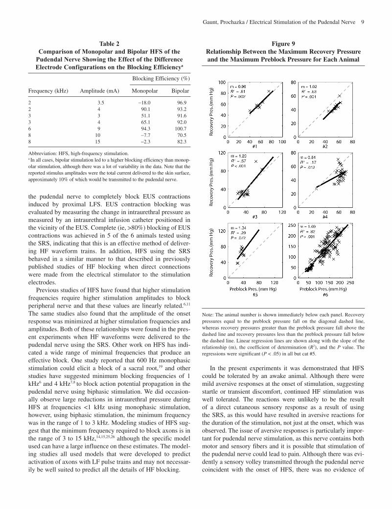
Table 2 Comparison of Monopolar and Bipolar HFS of the
Pudendal Nerve Showing the Effect of the Difference Electrode Configurations on the Blocking Efficiencya
Blocking Efficiency (%)
Frequency (kHz) Amplitude (mA) Monopolar Bipolar
2 3.5 -18.0 96.92 4 90.1 93.23 3 51.1 91.63 4 65.1 92.06 9 94.3 100.78 10 -7.7 70.58 15 -2.3 82.3
Abbreviation: HFS, high-frequency stimulation.a In all cases, bipolar stimulation led to a higher blocking efficiency than monop-olar stimulation, although there was a lot of variability in the data. Note that the reported stimulus amplitudes were the total current delivered to the skin surface, approximately 10% of which would be transmitted to the pudendal nerve.
Figure 9 Relationship Between the Maximum Recovery Pressure and the Maximum Preblock Pressure for Each Animal
Note: The animal number is shown immediately below each panel. Recovery pressures equal to the preblock pressure fall on the diagonal dashed line, whereas recovery pressures greater than the preblock pressure fall above the dashed line and recovery pressures less than the preblock pressure fall below the dashed line. Linear regression lines are shown along with the slope of the relationship (m), the coefficient of determination (R2), and the P value. The regressions were significant (P < .05) in all but cat #5.
10 Neurorehabilitation and Neural Repair
continued unpleasant sensations. This finding suggests that clinical implementation of HFS may be acceptable even when it elicits large onset responses. Although it would be desirable to eliminate any and all unwanted side effects of electrical stimulation of nerves, this is often impossible given the many types of sensory axons in peripheral nerves. Movement of the leg is often an unwanted side effect resulting from activation of efferent fibers in the sacral roots when stimulating them to elicit bladder contractions.27 Yet these are the most successful neuroprostheses for bladder control. One technique that has been proposed to reduce the onset response associated with HFS is to slowly increase the stimulation amplitude as the HFS pulse train is delivered. However, a recent investigation of this proposal concluded that this technique actually increases the duration of the onset response.28
A number of trials comparing the effectiveness of monopo-lar and bipolar HFS were tested. Although monopolar stimula-tion was capable of blocking EUS contractions, it was not as reliable as bipolar stimulation and generally required higher stimulation amplitudes. However, further characterization of the full range of possible parameters using monopolar stimula-tion may show that functionally relevant and consistent HF blocking may be achievable. Given the simplicity of a monop-olar SRS as compared to bipolar or even tripolar systems, there may be practical benefits for pursing HF block using a monop-olar stimulation electrode. One potentially complicating factor in the discussion of monopolar and bipolar electrodes is the existence of virtual anodes and cathodes that form at the ends of the nerve cuffs. In animals #1 through #5 the nerve cuffs were short, but in animal #6 in both monopolar and bipolar configurations, the nerve cuff was asymmetric. This configura-tion can be used to generate unidirectionally propagating action potentials,29,30 but the effect of virtual anodes or cath-odes on HF blocking is unclear.
Although complete blocking of EUS contractions was achieved in 5 of the 6 animals tested, there was a large amount of variability in the quality of the blocking and in the most effective stimulus parameters. During the multiple recording sessions in animal #6, which occurred over 6 months, two very different parameter ranges were observed for eliciting a com-plete block. The higher-frequency range generally elicited effective blocking only when the stimulation frequency was greater than 4 kHz and the amplitude was greater than 7 mA. The lower-frequency range produced effective blocking when the stimulation frequency was 2 to 4 kHz and the amplitude was greater than 2 mA. It is important to note that on those days where the lower-frequency range was effective, no com-bination of stimulation frequency and amplitude could be found above 4 kHz that elicited blocking. Initially, the higher-frequency range was the most effective, but on post-implant days 127 and 176, the lower-frequency range was most effec-tive. During the last test (day 230), the higher-frequency range was again the most effective. In both the lower- and higher- frequency ranges, blocking was complete and occurred rap-idly, but when the lower-frequency range was most effective,
the recovery was incomplete, whereas when the higher-frequency range was most effective, recovery was generally complete. It is unclear why two independent frequency ranges were observed on different testing days. Since the recovery of intraurethral pres-sure after the offset of HFS at the lower-frequency range was poor, it is possible that a neural fatigue process with a slow recov-ery may have contributed to the blocking effect at this lower-fre-quency range. Despite the inconsistencies, complete blocking was achieved on each tested day.
In one of the experimental animals, HFS was generally ineffective in reducing intraurethral pressures. Although the intraurethral pressure usually began to decrease approximately halfway through the HFS train, the magnitude of the pressure decrease was small and the pressure decreased slowly. This particular animal in which HFS elicited poor blocking was a female, making the positioning of the catheter more challeng-ing than in the male. Since the quality of intraurethral pressure measurements depends greatly on the specific position of the catheter in the urethra, it is possible that poor catheter place-ment was a cause of the observed results. It is also possible that the experimental setup, including the nerve cuff contact with the pudendal nerve, was not optimal in this animal, espe-cially given that complete blocking was achieved in another female cat. Given the proposed mechanisms for HF blocking, including steady-state depolarization resulting from sodium channel inactivation12 and hyperpolarization resulting from constant potassium channel activation,16 it is possible that in some animals the effect of isoflurane anesthesia on potassium and sodium channels affects HF blocking.31,32 One other pos-sibility that could account for the poor responses in one animal and some of the variability in the other animals is that both LFS and HFS of the pudendal nerve was done unilaterally. Since the EUS is innervated bilaterally, it is possible that proximal stimulation of the pudendal nerve elicited reflex activity in the contralateral pudendal nerve, which resulted in EUS activation that was recorded by the intraurethral catheter. Though isoflurane suppresses spinal reflexes, a reflex explana-tion cannot be ruled out.
One issue that has yet to be addressed in HFS is the poten-tial for nerve damage caused by long-term stimulation. Because of the potential for nerve damage using monophasic stimulation, the majority of the experiments focused on using biphasic, sinusoidal waveforms. No evidence of nerve damage was found in this study, as assessed by the functional response of the pudendal nerve to LFS. Over the duration of the chronic implant, LFS thresholds remained stable. With current con-trolled sinusoidal waveforms, the charge per phase can be expressed as: Q = 1000 × A/πf, where A is the amplitude in mA, f is the frequency in Hz, and Q is the charge in microcou-lombs (µC). For stainless-steel electrodes, such as the kind used in these experiments, the safe limit of charge per phase has been estimated at 0.4 to 0.8 µC/mm2.33 For constant-cur-rent sinusoidal waveforms, the highest charge per phase values occur at the lowest frequencies and highest amplitudes. For stimulation at 1 kHz and 10 mA (external), the estimated
Gaunt, Prochazka / Electrical Stimulation of the Pudendal Nerve 11
charge per phase in our experiments would have been 0.2 μC/mm2, assuming a 10% capture ratio (1 mA at the pudendal nerve) and an electrode surface area of 1.5 mm2. Using stimu-lation parameters that more commonly resulted in complete blocking (eg, 8 kHz, 8 mA), the charge per phase would have been approximately 0.02 μC/mm2.
Numerous devices and techniques have been proposed to modify bladder and sphincter function after SCI.4 However, very few of these devices have been successful. HFS of the pudendal nerve leading to relaxation of the EUS could be a use-ful way to treat DSD occurring after SCI while reducing the need for catheterization, which can be a significant cause of urinary tract infections. Other methods currently available to reduce EUS tone without using catheters or stents are sphinc-terotomies,34 injections of botulinum toxin,35 and less common procedures such as dorsal rhizotomies.36 Each of these proce-dures has significant disadvantages, including the irreversibility of sphincterotomies and dorsal rhizotomies and the repeated treatments required with botulinum toxin injections. HFS of the pudendal nerve would require only a single implant surgery and could be turned on and off at the desired times.
Several aspects of using the SRS to stimulate the pudendal nerve are attractive from a neuroprosthetics perspective. With the stimulus router, the only implanted components are the leads, with their nerve cuff and pick-up terminals. No implanted electronic components or batteries are required. Since all the stimulation electronics are external, a variety of stimulation paradigms using a wide range of stimulation parameters can be explored. For example, to our knowledge, no implanted stimula-tor capable of generating biphasic pulse trains over 1 kHz exists. Also, some studies indicate that different waveform shapes may be better at exciting smaller diameter axons in a peripheral nerve.37,38 In a normal implanted stimulator, custom electronics would have to be designed to examine these special conditions. With the SRS, an existing implant is capable of transmitting any externally applied waveform. Additionally, maintaining the energy supply to an implanted HF stimulator would be problem-atic, whereas batteries can easily be replaced in an external device. The primary disadvantage of the SRS is that to use the device, external electrodes must be applied to the skin. However, device design could minimize this problem. For example, the use of self-adhesive gel electrodes that can be left in place for a week or more attached to disposable stimulators the size of a matchbox could minimize this disadvantage. Alternatively, since an SRS for HF blocking of EUS contractions might be used as a part of a regularly scheduled bladder voiding routine, electrodes could be temporarily placed and then removed once voiding was complete. Another potential disadvantage of the SRS is that HFS could potentially induce uncomfortable sensa-tions in the skin.
Surgical access to the pudendal nerve in humans is not as simple as in the cat, but it is possible to expose the pudendal nerve for the purpose of nerve cuff implantation in humans.39 Furthermore, a number of other useful responses have been demonstrated with stimulation of the pudendal nerve.
Stimulation of the genital branches of the pudendal nerve in humans has been shown to suppress hyperreflexive bladder contractions,40-42 and recent work has shown that stimulation of urethral branches of the pudendal nerve can elicit reflexive bladder contractions in cats43,44 and humans.45 Taking all of these results together, it may be possible to create a neuropros-thesis where the only implanted component is a lead going to a multipolar nerve cuff on the pudendal nerve that can both inhibit and elicit reflexive bladder contractions as well as inhibit and elicit contractions of the EUS. This range of options represents all of the major actions of the lower urinary tract.
The ultimate goal of HF blocking of EUS contractions is to promote voiding in cases where there are naturally occurring, unwanted EUS contractions, or EUS contractions resulting from proximal stimulation of the pudendal nerve.46,47 We have shown that it is possible to completely block EUS contractions using HFS of the pudendal nerve in anesthetized cats using the SRS. The main objective of future work will be to try to improve the effectiveness of this system in all animals, refine the parameter range, and demonstrate the usefulness of HFS in improving micturition. The simplicity of the SRS and the effectiveness of HFS suggest that continued investigation could lead to the devel-opment of a device that could be useful in managing DSD.
Acknowledgments
We thank Mr Michel Gauthier for his help with the devel-opment of the stimulator used in these experiments, as well as Mr Allen Denington and the staff of HSLAS for their assis-tance with animal care. This work was supported by the National Institute of Neurological Disorders and Stroke con-tract N01-NS-2-2342, the Alberta Heritage Foundation for Medical Research, and the Natural Sciences and Engineering Research Council of Canada. Arthur Prochazka has a commer-cial interest in the development of the SRS.
References
1. Kaplan SA, Chancellor MB, Blaivas JG. Bladder and sphincter behavior in patients with spinal cord lesions. J Urol. 1991;146:113-117.
2. Weld KJ, Dmochowski RR. Association of level of injury and bladder behavior in patients with post-traumatic spinal cord injury. Urology. 2000;55:490-494.
3. Yalla SV, Blunt KJ, Fam BA, Constantinople NL, Gittes RF. Detrusor-urethral sphincter dyssynergia. J Urol. 1977;118:1026-1029.
4. Gaunt RA, Prochazka A. Control of urinary bladder function with devices: successes and failures. Prog Brain Res. 2006;152:163-194.
5. Middleton JW, Lim K, Taylor L, Soden R, Rutkowski S. Patterns of mor-bidity and rehospitalisation following spinal cord injury. Spinal Cord. 2004;42:359-367.
6. Bhadra N, Bhadra N, Kilgore K, Gustafson KJ. High frequency electrical conduction block of the pudendal nerve. J Neural Eng. 2006;3:180-187.
7. Tai C, Roppolo JR, de Groat WC. Block of external urethral sphincter contraction by high frequency electrical stimulation of pudendal nerve. J Urol. 2004;172:2069-2072.
8. Tai C, Roppolo JR, de Groat WC. Response of external urethral sphincter to high frequency biphasic electrical stimulation of pudendal nerve. J Urol. 2005;174:782-786.
12 Neurorehabilitation and Neural Repair
9. Martin WD, Fletcher TF, Bradley WE. Innervation of feline perineal mus-culature. Anat Rec. 1974;180:15-29.
10. Kilgore KL, Bhadra N. Nerve conduction block utilising high-frequency alternating current. Med Biol Eng Comput. 2004;42:394-406.
11. Bhadra N, Kilgore KL. High-frequency electrical conduction block of mammalian peripheral motor nerve. Muscle Nerve. 2005;32:782-790.
12. Bhadra N, Lahowetz EA, Foldes ST, Kilgore KL. Simulation of high- frequency sinusoidal electrical block of mammalian myelinated axons. J Comput Neurosci. 2007;22:313-326.
13. Tai C, de Groat WC, Roppolo JR. Simulation analysis of conduction block in unmyelinated axons induced by high-frequency biphasic electrical cur-rents. IEEE Trans Biomed Eng. 2005;52:1323-1332.
14. Tai C, de Groat WC, Roppolo JR. Simulation of nerve block by high- frequency sinusoidal electrical current based on the Hodgkin-Huxley model. IEEE Trans Neural Syst Rehabil Eng. 2005;13:415-422.
15. Zhang X, Roppolo JR, de Groat WC, Tai C. Simulation analysis of conduction block in myelinated axons induced by high-frequency bipha-sic rectangular pulses. IEEE Trans Biomed Eng. 2006;53:1433-1436.
16. Zhang X, Roppolo JR, de Groat WC, Tai C. Mechanism of nerve conduc-tion block induced by high-frequency biphasic electrical currents. IEEE Trans Biomed Eng. 2006;53:2445-2454.
17. Abdel-Gawad M, Boyer S, Sawan M, Elhilali MM. Reduction of bladder outlet resistance by selective stimulation of the ventral sacral root using high frequency blockade: a chronic study in spinal cord transected dogs. J Urol. 2001;166:728-733.
18. Ishigooka M, Hashimoto T, Sasagawa I, Izumiya K, Nakada T. Modulation of the urethral pressure by high-frequency block stimulus in dogs. Eur Urol. 1994;25:334-337.
19. Shaker HS, Tu LM, Robin S, et al. Reduction of bladder outlet resistance by selective sacral root stimulation using high-frequency blockade in dogs: an acute study. J Urol. 1998;160:901-907.
20. Solomonow M, Eldred E, Lyman J, Foster J. Control of muscle contractile force through indirect high-frequency stimulation. Am J Phys Med. 1983; 62:71-82.
21. Zhou B-H, Baratta R, Solomonow M. Manipulation of Muscle Force with Various Firing Rate and Recruitment Control Strategies. IEEE Trans Biomed Eng. 1987;34:128-139.
22. Rubinstein JT, Tyler RS, Johnson A, Brown CJ. Electrical suppression of tinnitus with high-rate pulse trains. Otol Neurotol. 2003;24:478-485.
23. Gan LS, Prochazka A, Bornes TD, Denington AA, Chan KM. A new means of transcutaneous coupling for neural prostheses. IEEE Trans Biomed Eng. 2007;54:509-517.
24. Brown M, Wickham JE. The urethral pressure profile. Br J Urol. 1969;41: 211-217.
25. Elbasiouny SM, Mushahwar VK. Modulation of motoneuronal firing behavior after spinal cord injury using intraspinal microstimulation current pulses: a modeling study. J Appl Physiol. 2007;103:276-286.
26. Williamson RP, Andrews BJ. Localized electrical nerve blocking. IEEE Trans Biomed Eng. 2005;52:362-370.
27. Brindley GS. The first 500 patients with sacral anterior root stimulator implants: general description. Paraplegia. 1994;32:795-805.
28. Miles JD, Kilgore KL, Bhadra N, Lahowetz EA. Effects of ramped ampli-tude waveforms on the onset response of high-frequency mammalian nerve block. J Neural Eng. 2007;4:390-398.
29. Ungar IJ, Mortimer JT, Sweeney JD. Generation of unidirectionally propagating action potentials using a monopolar electrode cuff. Ann Biomed Eng. 1986;14:437-450.
30. Sweeney JD, Mortimer JT. An asymmetric two electrode cuff for genera-tion of unidirectionally propagated action potentials. IEEE Trans Biomed Eng. 1986;33:541-549.
31. Duch DS, Rehberg B, Vysotskaya TN. Volatile anesthetics significantly suppress central and peripheral mammalian sodium channels. Toxicol Lett. 1998;100-101:255-263.
32. Nau C. Voltage-gated ion channels. Handb Exp Pharmacol. 2008(182): 85-92.
33. Mortimer JT. Motor prostheses. In: Brooks VB, ed. The Nervous System, Vol II: Motor Control. Bethesda, MD: American Physiological Society; 1981:155-187.
34. Reynard JM, Vass J, Sullivan ME, Mamas M. Sphincterotomy and the treatment of detrusor-sphincter dyssynergia: current status, future pros-pects. Spinal Cord. 2003;41:1-11.
35. Smith CP, Nishiguchi J, O’Leary M, Yoshimura N, Chancellor MB. Single-institution experience in 110 patients with botulinum toxin A injec-tion into bladder or urethra. Urology. 2005;65:37-41.
36. Brindley GS, Polkey CE, Rushton DN, Cardozo L. Sacral anterior root stimulators for bladder control in paraplegia: the first 50 cases. J Neurol Neurosurg Psychiatry. 1986;49:1104-1114.
37. Bhadra N, Grunewald V, Creasey G, Mortimer JT. Selective suppression of sphincter activation during sacral anterior nerve root stimulation. Neurourol Urodyn. 2002;21:55-64.
38. Fang ZP, Mortimer JT. Selective activation of small motor axons by quasi-trapezoidal current pulses. IEEE Trans Biomed Eng. 1991;38:168-174.
39. Gustafson KJ, Zelkovic PF, Feng AH, Draper CE, Bodner DR, Grill WM. Fascicular anatomy and surgical access of the human pudendal nerve. World J Urol. 2005;23:411-418.
40. Dalmose AL, Rijkhoff NJ, Kirkeby HJ, Nohr M, Sinkjaer T, Djurhuus JC. Conditional stimulation of the dorsal penile/clitoral nerve may increase cystometric capacity in patients with spinal cord injury. Neurourol Urodyn. 2003;22:130-137.
41. Nakamura M, Sakurai T. Bladder inhibition by penile electrical stimula-tion. Br J Urol. 1984;56:413-415.
42. Wheeler JS Jr, Walter JS, Zaszczurynski PJ. Bladder inhibition by penile nerve stimulation in spinal cord injury patients. J Urol. 1992;147: 100-103.
43. Boggs JW, Wenzel BJ, Gustafson KJ, Grill WM. Spinal micturition reflex mediated by afferents in the deep perineal nerve. J Neurophysiol. 2005;93: 2688-2697.
44. Tai C, Smerin SE, de Groat WC, Roppolo JR. Pudendal-to-bladder reflex in chronic spinal-cord-injured cats. Exp Neurol. 2006;197:225-234.
45. Gustafson KJ, Creasey GH, Grill WM. A urethral afferent mediated excitatory bladder reflex exists in humans. Neurosci Lett. 2004; 360:9-12.
46. Boger A, Bhadra N, Gustafson KJ. Bladder voiding by combined high frequency electrical pudendal nerve block and sacral root stimulation. Neurourol Urodyn. 2008;27:435-439.
47. Tai C, Wang J, Wang X, Roppolo JR, de Groat WC. Voiding reflex in chronic spinal cord injured cats induced by stimulating and blocking pudendal nerves. Neurourol Urodyn. 2007;26:879-886


![Numerical Study on Ship Motion Coupled with LNG tank Sloshing …€¦ · the sea, Kim, B. et al[5] studied the coupled seakeeping and sloshing tanks in frequency domain. A forward-speed](https://img.pdfslide.us/doc/110x75/5f2e02e067c2f941ef5c2b20/numerical-study-on-ship-motion-coupled-with-lng-tank-sloshing-the-sea-kim-b-et.jpg)