Embed Size (px)
Citation preview

Copyright 0 1996 by the Genetics Society of America
Transcription of gypsy Elements in a Y-Chromosome Male Fertility Gene of Drosqbhila hydei
Ron Hochstenbach,*" Harry Harhangi," Karin Schouren," Petra Bindels," Ron Suijkerbuijkt and Wolfgang Hennig"
*Department of Molecular and Developmental Genetics, Faculty of Sciences, Catholic University of Nzj'megen, NL-6525 ED Nzj'megen, The Netherlands and tDepartment of Human Genetics, University Hospital Nijmegen, NL-6525 GA Nijmegen, The Netherlands
Manuscript received November 7, 1994 Accepted for publication October 20, 1995
ABSTRACT We have found that defective gypsy retrotransposons are a major constituent of the lampbrush loop
pair Nooses in the short arm of the Y chromosome of Drosophila hydei. The loop pair is formed by male fertility gene Q during the primary spermatocyte stage of spermatogenesis, each loop being a single transcription unit with an estimated length of 260 kb. Using fluorescent in situ hybridization, we show that throughout the loop transcripts gypsy elements are interspersed with blocks of a tandemly repetitive Y-specific DNA sequence, ayl. Nooses transcripts containing both sequence types show a wide size range on Northern blots, do not migrate to the cytoplasm, and are degraded just before the first meiotic division. Only one strand of ayl and only the coding strand of gypsy can be detected in the loop transcripts. However, as cloned genomic DNA fragments also display opposite orientations of ayl and gypsy, such DNA sections cannot be part of the Nooses. Hence, they are most likely derived from the flanking heterochromatin. The direction of transcription of ayl and gypsy thus appears to be of a functional significance.
A BOUT 40 families of transposable elements reside in the genome of Drosophila melanogaster (BERG
and H o w 1989; FINNEGAN 1990). The most abundant type of transposable elements are called retrotranspo- sons, as they have structural similarity with retroviruses. At least 19 different families of retrotransposons have been identified in this species. They are implicated in the majority of spontaneous mutations (GREEN 1988), and a wealth of data exists on their structure and their genomic and phylogenetic distribution. Also studies on the mechanisms by which they affect normal patterns of gene expression have been carried out.
To cause heritable changes, retrotransposons must transpose within cells of the germ line. This requires an RNA intermediate, as has been shown for the IAP sequence of the mouse (HEIDMANN and HEIDMANN 1991) and also for several retroposons, as for example the L1 element of the mouse (EVANS and PALMITER 1991) and the I factor of D. melanogaster ( JENSEN and HEIDMANN 1991; PELISSON et al. 1991). Therefore, such elements must be transcribed during oogenesis or sper- matogenesis. The Ifactor is transcribed in the female germ line cells (LACHAUME et al. 1992; M c L m et al. 1993), and the gypsy retrotransposon of this species is transcribed in the somatic follicle cells that surround
Corresponding author: Wolfgang Hennig, Department of Molecular and Developmental Genetics, Faculty of Sciences, Catholic University of Nijmegen, Toernooiveld, NL-6525 ED Nijmegen, The Netherlands.
' Present address: Department of Microbiology and Evolutionary Biol- ogy, Faculty of Sciences, Catholic University of Nijmegen, Toer- nooiveld, N U 5 2 5 ED Nijmegen, The Netherlands.
Genetics 142 437-446 (February, 1996)
the oocytes (PELISSON et al. 1994). However, little is known about the expression of retrotransposons in male germ line cells of D. melanogaster, even though the promoters of several retrotransposons have been identified (see for mdg3 ARKHIPOVA et al. 1986, for c@ia SNEDDON and FLAVELL 1990, for mdgl ARKHIPOVA and ILYIN 1991, for gypsy JARRELL and MESELSON 1991). Some retrotransposons display localized expression during embryogenesis (BROOKMAN et al. 1992; FROM- MER et al. 1994; BRONNER et al. 1995). For some families of retrotransposons, the developmental pattern of ex- pression has been determined (PARKHURST and CORCES 1987), but since these studies were based on RNA ex- tracted from entire animals, with males and females mixed, they reveal nothing about retrotransposon tran- scription in either the male or the female germ line.
Previous investigations of our laboratory on the mo- lecular structure of the lampbrush loop-forming male fertility genes on the Y chromosome of D. hydei (re- viewed by HENNIG et al. 1989; HENNIG 1990) have re- vealed that retrotransposons of the micropiafamily (LAN- KENAU 1993) are transcribed in the lampbrush loop pairs Threads and Pseudonucleolus in primary spermato- cytes (HUIJSER et al. 1988). More recently, it has been demonstrated that an antisense transcript of micropia is found in spermatocytes (LANKENAU et al. 1994). This transcript might be involved in the regulation of trans- position frequencies of micropia in the male germ line.
In this paper we show that defective members of the Q~SJ retrotransposon family are abundantly transcribed in the germ line of wild-type D. hydei males. These gypsy
Dow
nloaded from https://academ
ic.oup.com/genetics/article/142/2/437/6016658 by guest on 23 January 2022

438 R. Hochstenbach et al.
elements are located in the lampbrush loop pair Nooses that is associated with male fertility gene Qon the short a rm of the Y chromosome. The gypsy elements are co- transcribed with repeats of the Y-specific ayl family of repetitive DNA sequences that was earlier identified as the major constituent of the Nooses DNA (VOGT et al. 1982; VOW a n d HENNIG 1986a,b; HOCHSTENBACH el al. 1993a,b, 1994a).
MATERIALS AND METHODS
Drosophila stocks: Both the D. hydei Tubingen wild-type strain and the D. eohydei wild-type strain were from our labora- tory collection. D. hydeimales of the genotype X/m(vQl were used as a control, since they lack the short arm of the Y chromosome, and therefore, they lack fertility gene Q. Follow- ing its induction by EMS in 1979, the ms(vQl Ychromosome was cytologically normal, carrying a sterile allele of gene Q on the short arm (HACKSTEIN et al. 1982; HACKSTEIN and HENNIG 1982). During subsequent maintenance of the chro- mosome in males of the genotype T(X;y)59/m(Y)Ql, the short arm became deleted (J. H. P. HACKSTEIN, personal com- munication). T(X; y)59 is a translocation of the short arm of the Y chromosome to the euchromatic arm of the Xchromo- some, complementing the absence of gene Q. It carries the markers yellow, miniature, and c h q (HACKSTEIN et al. 1982). The X/ms(Y)Ql males used for isolation of RNA were ob- tained by crossing T(X;y)59/ms(Y)Ql males to virgin wild- type females. Absence of the short arm was confirmed by inspection of neuroblast metaphases of X/rns(vQl third in- star larvae and by the failure of an ayl repeat probe to hybrid- ize to Southern blots of genomic DNA of X/rns(Y)Ql adults. Repeats of the Y-specific ayl family are located exclusively on the short arm of the Y chromosome (VOGT and HENNIG 1983). Flies were grown at 18" or 24" as described (HOCHSTEN- BACH et al. 1993a).
Isolation of nucleic acids: RNA was isolated from testes of 3- to 5day old adult males by the method of CHIRCWIN et al. (1979) as described by BRAND and HENNIC (1989). Plasmid DNA was isolated according to a boiling procedure recom- mended by Stratagene.
Nucleic acid probes: Two probes were used for the detec- tion of Nooses transcripts. As a probe for detecting transcripts of the Y-specific ayl family of repetitive DNA sequences we used an EcoRI DNA fragment of 393 bp that represents the sequence complexity of this family (VOCT and HENNIG 1986a). This particular repeat is called ayl. As a probe for detecting transcripts of the Y-associated DNA sequences of the Nooses loop pair we used the 5.8-kb BarnHI-EcoRI DNA fragment of the genomic clone DhNo9O (HOCHSTENBACH et al. 1993a). Both DNA fragments were subcloned in pBluescript I1 KS+ plasmid vectors (Stratagene). Integrity of RNA samples was verified using DmK2-30, a 1.2-kb cDNA clone containing parts of exons 16 and 17 of the D. rnelanogastermuscle myosin heavy- chain gene (GEORGE et al. 1989). This probe (kindly provided by Dr. K MIEDEMA) hybridizes to major transcripts of 6.6 and 4.5 kb, and to less abundant transcripts of 6.1 and 4.2 kb in testis RNA of D. hydei (MIEDEMA 1994).
DNA sequence analysis: Restriction fragments for DNA se- quencing were subcloned in M13mp18 or M13mp19 vectors, and sequences were determined using the dideoxy chain-ter- mination method, all following procedures provided by Amel-- sham. DNA sequences were analyzed using the software pack- age of the University of Wisconsin Genetics Computer Group (DEVEREUX et al. 1984). For sequence database searches and DNA sequence alignments we used the programs FASTA and LFASTA, respectively (PEARSON and LIPMAN 1988).
Labeling of probes: Strand-specific RNA probes for in situ hybridization were prepared by in vitro transcription using either T3 or T7 polymerase (Stratagene) from linearized plas- mid DNA, following protocols from Boehringer Mannheim. Such probes were labeled either by incorporation of digoxi- genin-11-UTP or biotin-16UTP (both from Boerhringer Mannheim). Control hybridizations of these probes to plas- mid DNA indicated comparable labeling of both strands (data not shown). RNA probes for hybridization to Northern blots were labeled by incorporation of [a-"PI-UTP. Equal amounts of probe of each strand, labeled to comparable specific activi- ties, were used. In some experiments single-stranded DNA probes were used for this purpose. Such probes were prepared from plasmid DNA using the Klenow fragment of Eschm'chia coli DNA polymerase, and they were labeled by incorporation of [a-"'P]dCTP, following conventional methods (SAMBROOK et uZ. 1989).
Hybridization to Northern blots: Samples of testis RNA were denatured by glyoxal/dimethylsulfoxide, separated on 1-2% denaturing agarose gels, transferred to Hybond mem- branes (New England Nuclear), hybridized, and washed as described by BRAND and HENNIC (1989). Approximately 20 pg total RNA was loaded in each lane.
Transcript in situ hybridization: Transcript in situ hybridiza- tion on squashed testis was performed by a modification of the method of TAUTZ and PFEIFLE (1988), as described in detail by H0CHSTENBAC:H et al. (1993a). If only a single probe was hybridized, we used digoxigenin for probe labeling. In this case probe detection was by an anti-digoxigenin antibody conjugated with alkaline phosphatase (Boehringer Mann- heim), and the probe was visualized by conventional phase contrast microscopy. If two probes were hybridized simulta- neously, one probe was labeled with digoxigenin and the other with biotin. In this case probe detection was by indirect immu- nofluorescence, following essentially the procedure described by HOCHSTENnA(:H et al. (1993b), except that digoxigenin was detected by successive incubations with rhodaminconjugated sheep antidigoxigenin Fabfragments (Boehringer Mann- heim, 1:20 dilution), Texas Red-conjugated rabbit anti-sheep antibodies (Jackson Immunoresearch Laboratories, West Grove, PA, 1:lOO dilution), and Texas Red-conjugated donkey anti-rabbit antibodies (Jackson Immunoresearch, 1:lOO dilu- tion). Probe visualization by fluorescence microscopy, digital image recording, and computer-assisted image processing were as described (HOCHSTENBACH et al. 1993b).
RESULTS
Cotranscription of ayl and Yassociated DNA se- quences in the Noma lampbrush loop pair: The apsy elements were identified in genomic clones that were isolated as potential segments of the lampbrush loop pair Nooses. Our earlier molecular studies revealed that the Y-specific ayl family of repetitive DNA sequences accounts for about two-thirds of the 260 kb of DNA transcribed in this loop pair, but that, in addition, other DNA sequences are transcribed in the loops that are also present on other chromosomes. These sequences were therefore designated as Yassociated (VOGT a n d HENNIC 1983, 1986a,b; HOCHSTENBACH et al. 1993a,b). Using ayl repeats as a probe to screen genomic libraries, we recov- ered 300 kb of genomic DNA in plasmid, lambda and cosmid clones containing both ayl and Y-associated DNA sequences (HOCHSTENBACH et al. 1993a).
Three of the lambda clones are shown in Figure 1.
Dow
nloaded from https://academ
ic.oup.com/genetics/article/142/2/437/6016658 by guest on 23 January 2022

Y Chromosomal wj~sy Transcripts
PR RT RN IN I S'LTR ORFI, ORF2 ORF3 LTR3' .. . .. . .. . .. . .. . . . . . .. . .. . .. . .. . .. . ..
439
.. .. . .. . .. . .. :: .. .. .. .. .. .. .. .. .. : ? b .. :: .. 71 ! ' . . DhNo86 . .
DhNo9O 1-j 04
P H S E 8
DhNol9 b
60 59
FIGURE 1.-Alignment of D. hydei Y-associated Ripsr elements with the g ~ j y element of D. melnnognstpr. Restriction maps of three ayl-containing lambda clones are shown. In each clone gypsy sequences are indicated as open rectangles, and restriction fragments hybridizing to ayl probes are indicated by dark shading. Individual ayl repeats, as identified by sequence comparison with the basic 393bp ayl repeat defined by VOCT and HENNIC (1986a), are depicted as black arrowheads, which also indicate the direction of transcription of ayl in the Nooses lampbrush loop pair. Restriction fragmena that hybridize neither to avl nor gypsy probes are hatched. The sequenced parts of these fragments have no obvious similarities to any sequence in the EMBL database (Release 40, September 1994). For each Y-associated &xysequence, the direction of transcription of the coding strand is indicated by an arrow. The numbers below the gypsy fragments indicate the percentage of sequence similarity to the corresponding sequences from the gypsy elements of D. mplnnoguster (upper numbers) and D. vin'lis (lower numbers). In the D. melnnogmtm gypsy element at the top, the LTR, open reading frames (ORF), as well as the positions of the protease (PR), reverse transcriptase (RT), ribonuclease (RN), and integrase (IN) activities encoded by ORF2 are indicated. The start site of g ~ j q transcription is marked by the small arrow above the 5' LTR. The vertical lines in the .~J~.YJ elements demarcate the limits of the different ORFs. The A in the largest ~ p s y sequence in DhNol9 indicates a poly(A)-tail that is located between ORF2 and ORFS. A more detailed analysis of these and other Y-associated gypsy sequences has been presented elsewhere (HOCHSTENRACI-~ et a/. 1994b). Restriction enzyme abbreviations are as follows: A, AvaI; B, RnmHl; E, EcoRI; H, HindIII; P, PsfI and S, Sail. The complete nucleotide sequence of DhNol9 has been submitted to the EMBL database under accession number X74538, the partial sequence of DhNo86 has been submitted under accession numbers X74539, X74540, X74541 and X74542, and the partial sequence of DhNo9O under accession numbers X74536, X74537 and X74543.
These clones have different restriction maps and hence, they do not overlap. Each of them contains ayl repeats that are organized in one to several clusters of tandem repeats. In addition, they share Y-associated DNA sequences. In clone DhNo86 the shared se- quences are located in a 3.8-kb BamHI-Hind111 frag- ment, in clone DhNo9O in a 5.8-kb BamHI-EmRI frag- ment, and in clone DhNol9 in a 3.7-kb EcoRI-EcoRI fragment. These DNA fragments were designated DhNo86BH3.8, DhNo9OBE5.8 and DhNol9EE3.7, re- spectively. On Southern blots of these clones, the Y- associated fragments cross-hybridize with one another after washing under nonstringent, but not under strin- gent conditions (HOCHSTENRACH et al. 1993a). Most of the copies on the other chromosomes are in the centromere-associated heterochromatin of the Xchro- mosome and the autosomes. Using highly stringent conditions for in situ hybridization, the Y-associated DNA sequences hybridize to Nooses transcripts in pri- mary spermatocytes (HOCHSTENRACH et al. 1993a).
The hybridization pattern of the shared Y-associated DNA sequence on noose.^ transcripts is highly similar to that of ayl. This was shown by fluorescent transcript in, situ hybridization, using a biotin-labeled, strand-specific RNA probe for ayl and a digoxigenin-labeled strand-
specific RNA probe for DhNo9OBE5.8. This fragment was chosen because it is present in at least four different ayl-containing genomic clones (HOCHSTENBACH rt al. 1993a), and it may therefore occur in multiple copies within the transcribed DNA of the loop. As shown in Figure 2, the two signals almost completely overlap and cover the entire Noose.$ loop pair. The slight differences in the patterns are due to the different sensitivities of detection at the different wave lengths. The overlap in signals indicates that both types of DNA sequences are interspersed throughout the Noos~s loop pair, consistent with our analysis of the genomic DNA of the lampbrush loop. Moreover, no major parts of the transcription unit are devoid of either sequence. In this case partially differing patterns would be expected. Also in D. eohydei, a species closely related to D. hydpi (WASSERMAN 1982), both sequences are cotranscribed in a lampbrush loop pair. This loop pair does not correspond to any of the four loop pairs previously described for this species (HENNIG 1978).
The results of the transcript in situ hybridization experiments were confirmed using Northern blots pre- pared from total testis RNA of D. hydpi (Figure 3). Both probes hybridize in a similar pattern to RNA fragments heterogeneous in size, with the largest fragments 10-
Dow
nloaded from https://academ
ic.oup.com/genetics/article/142/2/437/6016658 by guest on 23 January 2022

R. Hochstenbach et al.
FIGURE 2.-Cotranscription of ayl and gypsy in the Noows loop pair of U . hydei and in a loop pair of D. eo/zydei. In A and D a primary spermatocyte nucleus is shown for each species (phase contrast). Unfixed unstained nuclei are shown because cytology is severely distorted by in situ hybridization. The loop pairs of D. hydei (A) are as follows: Th, Threads; Ps, Pseudonucleolus; C1, Clubs; Tr, Tubular ribbons; Ns, Nooses (HESS and MEWR 1968). Those of D. eohydei (D) are as follows: g, granular loop; cl, clublike loop; dl, diffuse loop and pn, proximal loop (HENNIG 1978). Nucleolus organizers are marked. Fixed testis tissue of D. hydei (B and C ) and D. eohyda' (E and F) was hybridized simultaneously with the biotin-labeled ayl probe and a digoxigenin-labeled probe for the DhNo90BE5.8 fragment, which contains a gypsy sequence. The ayl probe was detected by fluorescein isothiocyanate fluorescence (B and E), and the gypsy probe was detected by Texas Red fluorescence (C and F) (see MATERIAIS AND METHODS). Two nuclei, each containing one labeled loop pair, are shown for each species. The opposite strand of both probes both fail to give detectable signals on testis transcripts in situ (see for ayl LIFSCHWZ and HAREVEN 1985; TRAPITZ et al. 1988 and for gyp? HOCHSTENBACH et al. 1993a) or on Northern blots of testis RNA (Figure 3). In all figures bar, 10 pm.
20 kb and the smallest only a few hundred bp in length. Using probes of the ayl family, such patterns were also observed in our earlier studies (VOGT et al. 1982) and by other investigators as well (LIFSCHITZ et al. 1983; TRAPITZ et al. 1988). The size heterogeneity is expected given the fact that the growing nascent loop transcripts of the 260-kb Nooses transcription unit display a large size gradient (GROND et al. 1983). In addition, current biochemical methods of RNA isola- tion are not suited to isolate transcripts of several hun- dred kb in length without substantial degradation. However, hybridization with a D. hydpi myosin-cDNA probe still allows the recognition of testis transcripts with sizes >6 kb (Figure 3). The patterns, therefore, indicate that both ayl and the Y-associated DNA seg- ments are components of much larger primary tran- scripts. In testis RNA from males lacking an active gene Q no hybridization is seen with either probe (Figure 3). Moreover, only one strand of DhNo90BE5.8 could be detected on the Northern blots, consistent with our earlier in situ hybridization experiments (HOCHSTEN- BACH et al. 1993a). Thus, within the Nooses transcrip- tion unit not only the ayl repeats (LIFSCHYI-z and HAR- EVEN 1985; TRAPITZ et al. 1988; PAPENBROCK 1991), but also all copies of the Y-associated DhNo90BE5.8
sequence, seem to have the same orientation. In addi- tion, we have also found that the heterogeneous ayl- containing testis transcripts are not polyadenylated (HOCHSTENBACH 1994).
Y-associated DNA sequences of the Nooses loop pair are defective @sy elements: We sequenced DhNo9O- BE5.8, and the related sequences from DhNol9 and DhNo86. As shown in Figure 1, each of the three lambda clones contains a 4 to 5-kb-long DNA sequence with a high degree of similarity to the g@sy retrotranspo- son, known from D. mlanogaster (MARLOR et al. 1986) and D. vin'lis (MIZROKHI and MAZO 1991). These gypsy elements, as well as all other Y-associated gypsy elements of D. hydei that have been sequenced so far, are defec- tive. In particular, they have lost their protein coding capacity, since all open reading frames are destroyed by deletions or frame shifts, as shown by detailed sequence analysis (HOCHSTENBACH et al. 1994b). In addition, those DNA sequences that in complete gypsy elements control transcription are absent due to truncations at either the 5' end, the 3' end, or at both ends. For example, the 5' long terminal repeat (LTR), which con- tains the gypsy promoter ( JARRELL and MESELSON 1991) as well as the binding sites for the protein encoded by the s u w e s s w of Hairy-wing (su(Hru)) gene (SPANA et al.
Dow
nloaded from https://academ
ic.oup.com/genetics/article/142/2/437/6016658 by guest on 23 January 2022

2.4 -
1.35-
Y Chromosomal gypsy Transcripts
4 M 5 6 7 (kb)
9.5- 7.4-
4.4-
2.4-
1.35-
8
I
0
44 1
M 9 10 (kbl
9.5 - 7.4-
4.4-
2.4-
1.35-
0.24- 0.24-
0.24-
FIGURE 3.-Only one strand of ayl and only one strand of gypsy can be detected in testis transcripts. Twenty micrograms total testis RNA of wild-type D. hyda' males (lanes 1, 2, 4, 5, 6, 8, 9 and 10) or of males of the genotype X/m(Y)QI (lanes 3 and 7) were loaded in each lane. The blots shown in lanes 1-8 were hybridized with ['3'P]-labeled strand-specific RNA probes for ayl (lanes 1-4) or for DhNo90BE5.8 (lanes 5-8). These blots were stringently washed in 0.02 M sodium phosphate buffer at 50" and exposed for 48 hr using two intensifying screens. The blots shown in lanes 1 and 5 are shorter exposures of those in lanes 2 and 6, respectively. The ayl probe hybridizes to testis transcripts of a heterogeneous size [but only if the short arm of the Y chromosome is present (lanes 1-3)] and so does the probe for the coding strand of gypsy (lanes 5-7). At a level of -2 kb comigrating ribosomal RNA causes a distortion of the signals. The blots shown in lanes 2 and 6 were stripped and subsequently hybridized under identical conditions with equal amounts of probes for the opposite strand of ayl and the noncoding strand of gypsy, and also exposed for 48 hr using two intensifjmg screens (lanes 4 and 8, respectively). As a control for the integrity of the RNA we used a probe for the D. melunogmter muscle myosin heavy chain gene. The blot shown in lane 9 was hybridized with a ["PI-labeled strand-specific DNA probe for ayl, stripped, and then hybridized again with the myosin probe (lane 10).
1988), are absent in the gypsy element of DhNo9O and in the large gypsy element of DhNol9.
Y-associated @sy elements outside of the Nooses tran- scription unit have random orientations relative to adja- cent ayl repeats: The orientations of the gypsy elements in DhNo90BE5.8 and DhNo86BH3.8 with respect to the T3 and T7 promoters of the pBluescript vectors used for subcloning implied that the coding strand of gypsy is represented in the Nooses transcripts. To confirm this finding, we determined the orientation of the ayl repeats immediately flanking the gypsy elements in clones DhNo9O and DhNo86 by partial sequence deter- mination of ayl repeat clusters. DhNol9 was completely sequenced as its restriction map revealed the presence of at least three separate clusters of ayl repeats ( HOCH- STENBACH et al. 1993a). Comparisons of the orientations of adjacent gypsy and ayl sequences show that the gypsy fragments in DhNo9O and DhNo86 are indeed tran- scribed from the same strand of DNA as the ayl repeats in these clones (Figure l ) , suggesting that DhNo9O and DhNo86 represent genuine segments of the Nooses.
In contrast, DhNol9 contains six different gypsy frag- ments, with only two in the same orientation as the ayl
repeats, which, on the other hand, all have the same orientation within the clone (Figure 1). Since only one strand of gyp9 is detectable in Nooses transcripts, both by in situ hybridization (HOCHSTENBACH et al. 1993a) and by hybridization to Northern blots (Figure 3), it is unlikely that the genomic clone DhNol9 represents a part of the Nooses transcription unit. This finding em- phasizes that ayl repeats that are interspersed by Y- associated DNA sequences are not necessarily located within the loop. Consistent with this conclusion, we have shown that the Ychromosome contains more DNA with interspersed ayl repeats than predicted by the 260- kb length estimate for the Nooses transcription unit (HOCHSTENBACH et al. 1993a,b). However, clones such as DhNol9 are exceptional, since from nine lambda and three cosmid clones in which both gypsy and ayl have been identified, it is the only clone with gypsy se- quences in the opposite orientation relative to ayl ( HOCHSTENBACH 1994; HOCHSTENBACH et nl. 1994b).
Distribution of Nooses transcripts during male germ cell development: Because retrotransposon transcripts encode proteins, we investigated whether the Nooses transcripts are transported from the nucleus to the cyto-
Dow
nloaded from https://academ
ic.oup.com/genetics/article/142/2/437/6016658 by guest on 23 January 2022
442 R. Hochstenbach P t 01.
5 - : 1 a"
FIGURE 4.-Distribution of N o o m transcripts during spermatogenesis, as followed by in situ hybridization using the digoxigenin- labeled ayl probe. The probe was detected using an antidigoxigenin antibody conjugated with alkaline phosphatase (see MATERIAIS AND w r t m m ) . (A) In the tip of the testis tube, spermatogonia (SG) fail to become labeled, whereas the nuclei of adjacent primary spermatocytes (SPC) contain a labeled NoosP.T loop pair (indicated by arrows). This is more clearly seen in B. Label is only detected in the nuclei of primary spermatocytes, but not in the cytoplasm of these cells. In the center of the figure, a cyst of secondary spermatocytes (containing almost the complete number of 16 cells) during anaphase I1 of meiosis is seen (MEI), and at the left, there is a complete cyst of 32 spermatids of an early postmeiotic stage (F") , with round or slightly oval Nebenkern derivatives. All cells of both cysts are completely free of label. Detailed descriptions of the different stages of spermatogenesis in D. hydh have been given by HESS and MEEK (1968), HENNIC (1985) and HENNIC and KREMER (1990). Phase contrast. Bar, 100 pm.
plasm. We used the ayl probe to follow the distribution of the loop transcripts during spermatogenesis in wild- type males of D. hydei. Identical results were obtained using the DhNo90BE5.8 lrvpsr probe (data not shown).
Spermatogenesis starts in the tip of the testis tube where primordial germ cells differentiate into spermato- gonia, which subsequently proliferate by mitotic divi- sions. In such cells the Ychromosome is not active (HEN- NIG 1967, 1985), and, as expected, we did not detect transcripts containing ayl in such cells (Figure 4A).
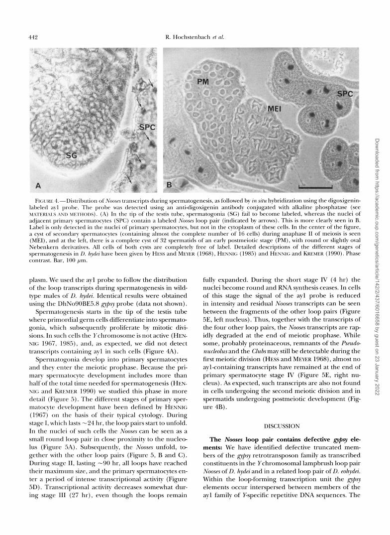
Spermatogonia develop into primary spermatocytes and they enter the meiotic prophase. Because the pri- mary spermatocyte development includes more than half of the total time needed for spermatogenesis (HEN- XI<; and KRICMER 1990) we studied this phase in more detail (Figure 5). The different stages of primary sper- matocyte development have been defined by HENNIG (1967) on the basis of their typical cytology. During stage I, which lasts "24 hr, the loop pairs start to unfold. In the nuclei of such cells the Noose~s can be seen as a small round loop pair in close proximity to the nucleo- lus (Figure 5A). Subsequently, the Nooses unfold, to- gether with the other loop pairs (Figure 5, B and C ) . During stage 11, lasting -90 hr, all loops have reached their maximum size, and the primary spermatocytes en- ter a period of intense transcriptional activity (Figure 5D). Transcriptional activity decreases somewhat dur- ing stage I11 (27 hr), even though the loops remain
fully expanded. During the short stage IV (4 hr) the nuclei become round and RNA synthesis ceases. In cells of this stage the signal of the ayl probe is reduced in intensity and residual Nooses transcripts can be seen between the fragments of the other loop pairs (Figure 5E, left nucleus). Thus, together with the transcripts of the four other loop pairs, the Nooses transcripts are rap- idly degraded at the end of meiotic prophase. While some, probably proteinaceous, remnants of the Pseudo- nucleolus and the Clubs may still be detectable during the first meiotic division (HESS and MEYER 1968), almost no aylcontaining transcripts have remained at the end of primary spermatocyte stage lV (Figure 5E, right nu- cleus). As expected, such transcripts are also not found in cells undergoing the second meiotic division and in spermatids undergoing postmeiotic development (Fig- ure 4B).
DISCUSSION
The Nooses loop pair contains defective glrpsy el6 ments We have identified defective truncated mem- bers of the gypsy retrotransposon family as transcribed constituents in the Ychromosomal lampbrush loop pair Nooses of D. hydiri and in a related loop pair of D. eohydiri. Within the loop-forming transcription unit the @sy elements occur interspersed between members of the ayl family of Y-specific repetitive DNA sequences. The
Dow
nloaded from https://academ
ic.oup.com/genetics/article/142/2/437/6016658 by guest on 23 January 2022
Y Chromosomal g ~ p s y Transcripts
t ' ' $
hybridization patterns of ayl and gypsy to the loop tran- scripts in situ are highly similar in both species, sug- gesting that both sequence types are intermingled throughout the entire transcription unit.
We can detect only the coding strand of gypsy and only one strand of ayl in the loop transcripts. If seg- ments of the other strand are present as well, they are either too short or too highly diverged. Alternatively, they may escape detection because they are located at the very end of the transcription unit. It may also be argued that the inverted ayl or gyp.^ sequences are undetectable because they are spliced out from a giant primary loop transcript. However, from Miller spreads of nascent loop chromatin there is no indication that Nooses transcripts undergo splicing (GROND et al. 1983). In addition, such an explanation would also require that the inverted repeats are preferentially spliced out. Therefore, we conclude that most, if not all, copies of ayl and gypsy are present in only one orientation within the Nooses.
Probes containing gypsy sequences result in strong signals on Nooses transcripts, both in situ (Figure 2, also see HOCHSTENBACH et al. 199%) and on Northern blots (Figure S), suggesting that gypsy sequences represent a major part of the loop. We have no means to accurately assess the copy number of the gypsy elements in the Nooses loop. However, all Y chromosomal gypsy elements are clustered together in a distal region on the short
443
FIGURE !%--Gradual unfolding of the Nooses loop pair during the successive stages of primary sperm- atocyte development. The Nooses loop pair was visualized by tran- script in situ hybridization using the digoxigenin-labeled ayl probe. (A) In early Stage I primary sperm- atocytes, the Nooses loop pair (Ns) starts to unfold from a position close to the round nucleolus (Nu). The other loop pairs cannot be seen in this nucleus. (B and C ) Subsequent, gradual unfolding of the Nooses, together with the other loop pairs, during stage I. Other loop pairs seen are the Threads (Th) and the Psmdonuckolzw (Ps). (D) During stages I1 and 111, the Nooses, as well as the other loop pairs, are seen at their maximum expansion. (E) During stage IV, the nuclei become round, and the loops are degraded. In the nucleus on the left, some residual aylcon- taining transcripts are present (indicated by the white arrow). In the nucleus on the right, which is about to enter metaphase I, such transcript5 (indicated by the black arrow) are barely detectable. Phase contrast. Bar, 10 pm.
arm (HOCHSTENBACH et al. 1993b). From genomic Southern blots we estimated that this region contains "10 copies of DhNo90BE5.8, 10 of the related se- quence in DhNo86BH3.8 and at least two of the related sequence in DhNo19EE3.7 (HOCHSTENBACH et al. 1993a). If all these Y chromosomal copies of gypsy, as recognized by their hybridization to either DhNo9O- BE5.8 or DhNo86BH3.8, are located within the tran- scription unit, gypsy would represent more than half of the estimated 80-90 kb of %associated DNA of the 260- kb-long loop. gyp? does not interfere with fertility gene func-
tion: Male fertility gene rZ, forming the loop pair Nooses, is not the only loopforming fertility gene of D. hydei containing defective retrotransposons. Members of the micrqia family, found in the loop pairs Threads and Pseu- donuckolus, that are formed by fertility genes A and C, respectively, have also lost their proteincoding capacity (HUIJSER et al. 1988). It is remarkable that also in the case of micropia only the coding strand of the retro- transposon can be detected in the loop transcripts (LAN- KENAu et al. 1994). Thus, each loopforming fertility gene appears to contain a few, or even only one family of retrotransposons, with all members in the same orien- tation within the loopforming transcription unit.
An immediate question raised by these observations is why these retrotransposons do not interfere with the function of the respective fertility gene. Insertions of
Dow
nloaded from https://academ
ic.oup.com/genetics/article/142/2/437/6016658 by guest on 23 January 2022

444 R. Hochstenbach et al.
retrotransposons into genes usually result in mutations (GREEN 1988). In gypsy-induced mutations, the binding of the su(Hw) protein, an ubiquitous nuclear zinc-finger protein (PARKHURST et al. 1988; SPANA et al. 1988; I-IAR- REON et al. 1993), to its binding sites in gypsy is sufficient for mediating the mutagenic effects of the element on the expression of adjacent genes (GEYER et al. 1988; PEIFER and BENDER 1988; MAZO et al. 1989; GEYER and CORCES 1992; SMITH and CORCES 1992; ROSEMAN et al. 1993). We have shown that at least the gypsy element in clone DhNo90 has lost the binding sites for the su(Hw) protein (also see HOCHSTENBACH et al. 1994b). The gypsy element of this clone was also identified in three additional aylcontaining clones (HOCHSTENBACH et al. 1993a), and therefore, several copies related to this cloned fragment occur in the Nooses loop. We do not know whether all the gypsy elements in the loop have lost their capacity to bind the su(Hw) protein. However, a probe containing the su(Hw)-binding sites from the D. melanogaster element failed to reveal male-specific DNA fragments in D. hydei (HOCHSTENBACH et al. 1994b), suggesting that there are no such DNA se- quences on the Ychromosome. This would make plausi- ble why the gypsy elements do not interfere with gene function.
In Miller spreads the Nooses loop can be seen as a single transcription unit (GROND et al. 1983). Hence, the gypsy elements within the loop do not serve as sec- ondary initiation sites for loop transcription, nor do they impede the normal progression of the RNA poly- merase along the loop DNA. This suggests that the promoter sequences in the 5' LTR of gypsy and the transcriptional termination signals in the 3' LTR (ARK- HIPOVA et al. 1986; JARRELL and MESELSON 1991) are either deleted, mutated, or nonfunctional in the con- text of lampbrush loop transcription. Consistent with the first possibility, the gypsy element in DhNo90 has a deletion of the 5' LTR, and the element in DhNo86 has almost completely lost its 3' LTR.
Significance of the elements for fertility gene function: Mutations or deletions in fertility gene Q forming the Nooses loop pair, cause a developmental arrest of spermatogenesis at the end of the elongation stage, before spermatid individualization (HESS and MEYER 1968). Since the molecular basis of this effect is unknown, it is difficult to assess the role of the gypsy elements transcribed in the Nooses for the function of the associated fertility gene Q However, mutant alleles of fertility genes that do not form a loop are sterile (LEONCINI 1977; HACKSTEIN et al. 1982, 1991). There- fore, the transcription of the repetitive loop constit- uents, such as ayl and gypsy, seems to be required for gene function.
The detailed sequence analysis of ayl repeats and Y- associated gypsy elements indicates that, in general, point mutations or deletions of these sequences are unlikely to interfere with the function of gene Q The
gypsy elements, such as those shown in Figure 1, are randomly accumulating pdint mutations and deletions, and they have lost their protein coding potential (HOCHSTENBACH et al. 1994b). The ayl repeats are het- erogeneous in size and they do not share an extended conserved DNA region (VOGT and HENNIG 1986a,b; WLASCHEK et al. 1988; PAPENBROCK 1991; HOCHSTEN- BACH 1994).
In this context it is of interest that ayl and gypsy are absent in the lampbrush loops of most other Drosophila species. The ayl repeats are present only in D. hydei and its closest relatives D. neohydei and D. eohydei (HAREVEN et al. 1986; VOGT et al. 1986). Gypsy is also transcribed in a loop pair of D. eohydei (Figure 2) and in a loop pair of D. virilis (data not shown), but we failed to detect transcription of gypsy in the lampbrush loops of other species with gypsy elements in the genome, as for in- stance D. repbta (HOCHSTENBACH et al. 1994b). Thus, it seems that the function of the loop-forming male fertil- ity genes does not depend on the particular type of repetitive DNA sequences that are transcribed in the loops (also see HENNIG 1990 for discussion).
Following earlier suggestions by HARDY et al. (1981) and GOLDSTEIN et al. (1982) that the loop-forming fertil- ity genes k l - 5 and k l - 3 on the Y chromosome of D. melanogaster encode dynein-like proteins, GEPNER and HAYS (1993) have shown that one member of the dyn- ein P-heavy chain gene family is located in the region containing kl-5. AYLES et al. (1973) have isolated EMS- induced temperature-sensitive alleles of several of the loop-forming fertility genes of D. melanogaster, k l - 5 be- ing one of these genes (GOLDSTEIN et al. 1982), and such alleles have been isolated by LEONCINI (1977) for several of the loop-forming genes of D. hydei, including gene Q (HACKSTEIN et al. 1982). At the restrictive tem- perature the temperature-sensitive allele m(Y) Q4"' of gene Q forms a morphologically normal Nooses loop pair, at least at the level of the light microscope, in which both ayl and gypsy are transcribed (HOCHSTEN- BACH et al. 1994~). This would be expected if the mutant lesion is a point mutation or a small deletion in an exon of a protein-coding gene.
From our limited sample of DNA sequences from putative segments of the Nooses loop pair, we have no indication that this loop contains protein-coding se- quences (HOCHSTENBACH 1994). As discussed by HEN- NIG (1993), such exons may be clustered at the very beginning or at the very end of the loop. It is even possible that the exons are distributed throughout the entire loop, separated by much larger introns that con- tain the rapidly evolving repetitive loop constituents, as proposed by HACKSTEIN et al. (1991). Our finding, however, that the transcripts of the Nooses, as detected by ayl or gypsy probes, lack a specific size, are not poly- adenylated, remain within the nucleus, and are absent postmeiotically, when most proteins of the sperm are being made (HENNIG 1967), does not seem to be com-
Dow
nloaded from https://academ
ic.oup.com/genetics/article/142/2/437/6016658 by guest on 23 January 2022

Y Chromosomal gypsr Transcripts 445
patible with protein coding. Also the observation that loop transcription is sensitive to actinomycin-D but not to a-amanitin (HENNIG 1967) argues against protein coding by loop transcripts.
As shown by hybridization to loop transcripts in situ and on Northern blots, the repetitive loop constituents of D. hydei occur in one orientation within the loop forming transcription units (LIFSCHYTZ and HAREVEN 1985; TRAPITZ et al. 1988). We do not know whether this merely reflects the evolutionary history of the loops, which were most likely generated by successive rounds of sequence amplification (see for discussion VOGT and HENNIG 1986b; HOCHSTENBACH et al. 1993a, 1994b). It is also possible that the distinct orientations of the repetitive DNA sequences is of a functional significance, as opposite orientations may lead to the formation of hairpin structures that could impede the progression of the transcriptional apparatus or induce heterochro- matin formation (ZUCKERKANDL and HENNIG 1995). With respect to the Nooses, the orientations of the ayl and gypsy sequences within the transcription unit will greatly assist the reconstruction of the entire loop in an ordered set of overlapping genomic clones.
We express our gratitude to Dr. JOHANNES HACUTEIN for providing the T(X; Y)58/mr(Y)QI males, to Dr. KOOS MIEDEMA for providing the myosin cDNA, to Drs. REIN BRAND and XtAOPING SUN for assistance in preparing Northern blots, and to WIEI.IJANSSEN for excellent tech- nical support. We thank Drs. HANS BUNEMANN, THOMAS PAPENBROCK and THOMAS HAIS for communicating unpublished data, and Drs. ERWIN SCHMIDT and PETER VOGT and our colleagues ANNA d m - NOVA and Koos MIEDEMA for critical comments on the manuscript.
LITERATURE CITED
ARKHIPOVA, I . R., and Y. V. ILYIN, 1991 Properties of promoter re- gions of mdgl Drosophila retrotransposon indicate that it belongs to a specific class of promoters. EMBO J. 10 1169-1177.
ARKHIPOVA, I. R., A. M. m0, V. A. CHEWSSOVA, T. V. GORFLOVA, N. G. SHUPPE et al., 1986 The steps of reverse transcription of Drosophila mobile dispersed genetic elements and U3R-U5 structure of their LTRs. Cell 44: 555-563.
AXES, G. B., T. G. SANDERS, B. I. KtEFERandD. T. SUZUKI, 1973 Temper- ature-sensitive mutations in Dmsophila mlanogmter XI. Male sterile mutations of the Y chromosome. Dev. Biol. 32: 239-257.
BERG, D. E., and M. M. HOW., 1989 MobiZeDNA. American Society for Microbiology, Washington, DC.
BRAND, R. C., and W. HENNIG, 1989 An abundant testis RNA species shows sequence similarity to Y chromosomal and other genomic sites in Drosophila hydei. Mol. Gen. Genet. 215: 469-477.
BRONNER, G., H. TAUBERT and H. JACKLE, 1995 Mesoderm-specific B104 expression in the Drosophila embryo is mediated by internal cisacting elements of the transposon. Chromosoma 103: 669-675.
BROOKMAN, J. J., A. T. TOOSY, L. S. SHASHIDHARA and R. A. H. WHITE, 1992 The 412 retrotransposon and the development ofgonadal mesoderm in Drosophila. Development 116 1185- 1192.
CHRIGWN, J. M., A. E. PRZYBU, R. J. MACDONALD and W. J. RUTTER, 1979 Isolation of biologically active ribonucleic acid from source enriched in ribonuclease. Biochemistry 24: 5294-5299.
DEVEREUX, J., P. HAEBERLI and 0. SMITHIES, 1984 A comprehensive set of sequence analysis programs for the VAX. Nucleic Acids
EVANS, J. P., and R. P. PALMITER, 1991 Retrotransposition of a mouse
FINNEGAN, D. J., 1990 Transposable elements. Drosophila Inform.
FROMMER, G., R. SCHUH and H. JACKLE, 1994 Localized expression
Res. 12 387-395.
L1 element. Proc. Natl. Acad. Sci. USA 88: 8792-8795.
Sew. 68: 371-382.
of a novel micropia-like element in the blastoderm of Drosophila melanogaster is dependent on the anterior morphogen bicoid. Chromosoma 103: 82-89.
GEORGE, E. L., M. B. OBER and C. P. EMERSON,Jr., 1989 Functional domains of the Drosophila melanogastermuscle myosin heavy-chain gene are encoded by alternatively spliced exons. Mol. Cell. Biol.
GEPNER, J., and T. HAIS, 1993 A fertility region on the Y chromo- some of Drosophila melanogaster encodes the structural gene for a dynein microtubule motor. Proc. Natl. Acad. Sci. USA 90: 11 132- 11136.
GEYER, P. IC, and V. G. CORCES, 1992 DNA-position-specific repres- sion of transcription by a Drosophila zinc finger protein. Genes Dev. 6 1865-1873.
GEYER, P. R, M. M. GREEN and V. G. CORCES, 1988 Mutant gene phenotypes mediated by a Drosophila melanogaster retrotranspo- son require sequences homologous to mammalian enhancers. Proc. Natl. Acad. Sci. USA 85: 8593-8597.
GOLDSTEIN, L. S. B., R. W. HARDY and D. L. LINDSLEY, 1982 Struc- tural genes on the Ychromosome of Drosophila melanogaster. Proc. Natl. Acad. Sci. USA 79: 7405-7409.
GREEN, M. M., 1988 Mobile DNA elements and spontaneous gene mutation, pp. 41 -50 in Banbuly Rqburt 30. Eukalyotic Transposable Elements as Mutagenic Agents, edited by M. E. LAMBERT, J. F. MG DONALD and I. B. WEINSTEIN. Cold Spring Harbor Laboratory, Cold Spring Harbor, Ny.
GROND, C. J., I. SIEGMUND and W. HENNIG, 1983 Visualization of a lampbrush loopforming fertility gene in Drosophila hyda'. Chro- mosoma 88: 50-56.
HACKSTEIN, J. H. P., and W. HENNIG, 1982 New mutants. Drosophila hydei. Drosophila Inform. Serv. 58: 195-203.
HACKSTEIN, J. H. P., 0. LEONCINI, H. BECK, G. PEELEN and W. HENNIG, 1982 Genetic fine structure of the Y chromosome of Drosophila hyda'. Genetics 101: 257-277.
HACKSTEIN, J. H. P., K H. GIATZER and T. J. M. HUISEBOS, 1991 Genetic and cytogenetic analysis of the "Th-Ps" region of the Y chromosome of Drosophila hydei: evidence for dual functions of the lampbrush loopforming fertility genes? Mol. Gen. Genet. 227: 293-305.
HARDY, R. W., K. T. TOKLJXA~U and D. L. LINDSLEY, 1981 Analysis of spermatogenesis in Drosophila mlanogaster bearing deletions for Ychromosome fertility genes. Chromosoma 83: 593-617.
HAREVEN, D., M. ZUCKERMAN and E. LIFSCHYE, 1986 Origin and evolution of the transcribed repeated sequences on the Y chro- mosome lampbrush loops of Drosophila hyda'. Proc. Natl. Acad. Sci. USA 83: 125-129.
HARRISON, D. A,, D. A. GDULA, R. S . COYNE and V. G. CORCES, 1993 A leucine zipper domain of the suppressor of Hairy-wing protein mediates i t s repressive effect on enhancer function. Genes Dev.
HEIDMANN, 0.. and T. HEIDMANN, 1991 Retrotransposition of a mouse IAP sequence tagged with an indicator gene. Cell 64: 159-170.
HENNIG, I . , 1978 Vergleichend-zytologische und genetische Unter- suchungen am Genom der Fruchtfliegen-Arten Drosophila hyda', neohydei und eohydei (Diptera: Drosophilidae). Entomol. Germ.
HENNIG, W., 1967 Untersuchungen zur Struktur und Funktion des Lampenbtirsten-Y-Chromosoms in der Spermatogenese von Dro sophila. Chromosoma 22: 294-357.
HENNIG, W., 1985 Y chromosome function and spermatogenesis in Drosophila hydei. Adv. Genet. 23: 179-234.
HENNIG, W., 1990 The Y chromosome of Drosophila, pp. 213-238 in Chromosomes, Eukaryotic, Proka7yotic and Viral, edited by R W. ADOLPH. CRC Press, Boca Raton, FL.
HENNIG, W., 1993 Conventional protein coding genes in the Dro
solved? Proc. Natl. Acad. Sci. USA 90: 10904-10906. sophila Y chromosome: is the puzzle of the fertility gene function
HENNIG, W., and H. KREMER, 1990 Spermatogenesis of Drosophila hydei. Int. Rev. Cytol. 123 129-175.
HENNIG, W., R. C. BRAND, J. HACKSTEIN, R. HOCHSTENBACH, H. KREMER et al., 1989 Ychromosomal fertility genes of Drosophila: a new type of eukaryotic genes. Genome 31: 561-571.
HESS, O., and G. F. MEYER, 1968 Genetic activities of the Ychromo- some in Drosophila during spermatogenesis. Adv. Genet. 14: 171-223.
9: 2957-2974.
7: 1966-1978.
4 211-223.
Dow
nloaded from https://academ
ic.oup.com/genetics/article/142/2/437/6016658 by guest on 23 January 2022

446 R. Hochstenbach et al.
HOCHSTENBACH, R., 1994 Anatomy of a lampbrush loopforming male fertility gene on the Y chromosome of Drosophila hydei. Ph.D. Thesis, Catholic University of Nijmegen, The Netherlands.
HOCHSTENBACH, R., A. POTGENS, H. MEIJER, R. DIJKHOF, M. mops et al., 1993a Partial reconstruction of the lampbrush loop pair Nooses on the Y chromosome of Drosophila hydei. Chromosoma
HOCHSTENBACH, R., M. WILBRINK, R. SUIJKERBUIJK and W. HENNIG, 1993b Localization of the lampbrush loop pair Nooses on the Y chromosome of Drosophila hydei. Chromosoma 102: 546-552.
HOCHSTENBACH, R., M. KNOPS and W. HENNIG, 1994a Discrimina- tion of transcribed and nontranscribed related repetitive DNA sequences from the Y chromosomes of Drosophila hydei and Dro sophila eohydei. Mol. Gen. Genet. 243: 54-62.
HOCHSTENBACH, R., H. HARHANGI, K. SCHOUREN and W. HENNIG, 199413 Degenerating gpsy retrotransposons in a male fertility gene on the Y chromosome of Drosophila hyda. J. Mol. Evol. 39:
HOCHSTENBACH, R., R. BRAND and W. HENNIG, 1994c Transcription of repetitive DNA sequences in the lampbrush loop pair Nooses formed by sterile alleles of fertility gene Qon the Y chromosome of Drosophila hydei. Mol. Gen. Genet. 244: 653-660.
HUIJSER, P., C. KIRCHHOFF, D.-H. LANKENAU and W. HENNIG, 1988 Retrotransposon-like sequences are expressed in Ychromosomal lampbrush loops of Drosophila hyda. J. Mol. Biol. 203: 689-697.
JARRELI., K. A., and MESELSON, 1991 Drosophila retrotransposon pro- moter includes an essential sequence at the initiation site and
Acad. Sci. USA 8 8 102-104. requires a downstream promoter for full activity. Proc. Natl.
JENSEN, S., and T. HEIDMANN, 1991 An indicator gene for detection of germline retrotransposition in transgenic Drosophila demon- strates RNA-mediated transposition of the LINE I element. EMBO J. 10 1927-1937.
LACHAUME, P., K. BOUHIDEL, M. MESURE and H. PINON, 1992 Spatial and temporal expression of the I factor during oogenesis in Drosophila mlanogaster. Development 115: 729-735.
LANKENAU, D.-H., 1993 The retrotransposon family mirropza in Dre sophila species, pp. 232-241, in Transposable Elements and Evolu- tion, edited by J. F. MCDONALD. Kluwer Academic Publishers, Amsterdam.
LANKENAU, S., V. G. CORCES and D.-H. LANKENAU, 1994 The Drosoph- ila micropia retrotransposon encodes a testis-specific antisense RNA complementary to reverse transcriptase. Mol. Cell. Biol. 1 4
LEONCINI, 0.. 1977 Temperatursensitive Mutanten im Y Chro- mosom von Drosophila hydei. Chromosoma 63: 329-357.
LIFSCHWZ, E., and D. H A R E V E N , 1985 Molecular evidence for partial inactivation of Y loops in T(X;Y)56 males from D. hydei. Mol. Gen. Genet. 199: 46-52.
LIFSCHWL, E., D. HAREVEN, A. AZRIEL and H. BRODSLY, 1983 DNA clones and RNA transcripts of four lampbrush loops from the Y chromosome of Drosophila hydei. Cell 3 2 191-199.
MARLOR, R. L., S. M. PARKHURST and V. G. CORCES, 1986 The Dro sophila melanogaster apsy transposable element encodes putative gene products homologous to retroviral proteins. Mol. Cell. Biol.
MAZO, A. M., L. J. MIZROKHI, A. A. KARAVANOV, Y. A. SEDKOV, A. A. KRICHEVSKAJA et al., 1989 Suppression in Drosophila: su(Hw) and sucf) gene products interact with a region of upsy (mdg4) regulat- ing its transcriptional activity. EMBO J. 8: 903-911.
MCLEAN, C., A. BUCHETON and D. J. FINNEGAN, 1993 The 5' untrans- lated region of the Ifactor, a long interspersed nuclear element- like retrotransposon of Drosophila melanogaster, contains an inter- nal promoter and sequences that regulate transcription. Mol. Cell. Biol. 13 1042-1050.
MIEDEMA, IC, 1994 Muscle myosin heavy chain gene expression in the testis of Drosophila hydei and Drosophila melanogaster. Ph.D. Thesis, Catholic University of Nijmegen, The Netherlands.
MIZROKHI, L. J., and A. M. W z o , 1991 Cloning and analysis of the mobile element gypsy from D. vin'lis. Nucleic Acids Res. 19: 913-916.
PAPENBROCK, T., 1991 Molekulare Analyse der Riesentranskripte aus den Y Chromosomalen Lampenbtirstenschleifen von Drosophila
102 526-545.
452-465.
1764-1775.
6: 1129-1134.
hydei. Ph.D. Thesis, Institut far Genetik, Heinrich Heine Universi- tit, Dtisseldorf, Germany.
PARKHURST, S. M., and V. G. CORCES, 1987 Developmental expres sion of Drosophila mlanogaster transposable elements. EMBO J. 6: 419-424.
PARKHURST, S. M., D. A. HARRISON, M. P. REMINGTON, C. SPANA, R. L. KELLEY et al., 1988 The Drosophila su(Hw) gene, which controls the phenotypic effect of the apsy transposable element, encodes a putative DNA-binding protein. Genes Dev. 2: 1205-1215.
PEARSON, W. R., and D. J. LIPMAN, 1988 Improved tools for biologi- cal sequence comparison. Proc. Natl. Acad. Sci. USA 85: 2444- 2448.
PEIFER, M., and W. BENDER, 1988 Sequences of the gypsy transposon of Drosophila necessary for its effects on adjacent genes. Proc. Natl. Acad. Sci. USA 85: 9650-9654.
PELISSON, A,, D. J. FINNEGAN and A. BUCHETON, 1991 Evidence for retrotransposition of the I factor, a LINE element of Drosophila mlanogaster. Proc. Natl. Acad. Sci. USA 88: 4907-4910.
PELISSON, A,, S. U. SONG, N. PRUD'HOMME, P. A. SMITH, A. BUCHETON et al., 1994 Cypsy transposition correlates with the production of a retroviral envelope-like protein under the tissue-specific con- trol of the DrosophilajZamenco gene. EMBO J. 13: 4401-4411.
ROSEMAN, R. R., V. PIRROTTA and P. K. GEYER, 1993 The su(Hw) protein insulates expression of the Drosophila mlanogaster white gene from chromosomal position effects. EMBO J. 12: 435-442.
SAMBROOK, J., E. F. FRITSCH and T. MANIATIS, 1989 Molecular Clon- ing. A Laboratq Manual, Ed. 2. Cold Spring Harbor Laboratory Press, Cold Spring Harbor, NY.
SMITH, P. A,, and V. G. CORCES, 1992 The suppressor of Haily-wing binding region is required for gypsy mutagenesis. Mol. Gen. Genet. 233: 65-70.
SNEDDON, A., and A. J. FI~VELL, 1989 The transcriptional control regions of the copia retrotransposon. Nucleic Acids Res. 17:
SPANA, C., D. A. HARRISON and V. G. CORCES, 1988 The Drosophila mlanogaster suppressm of Haily-wing protein binds to specific se- quences of the gpsy retrotransposon. Genes Dev. 2: 1414-1423.
TAUTZ, D., and C. PEIFI.E, 1989 A nonradioactive in situ hybridiza- tion method for the localization of specific RNAs in Drowphila embryos reveals translational control of the segmentation gene hunchback. Chromosoma 98: 81-85.
TRAPITZ, P., M. WIASCHEK and H. BUNEMANN, 1988 Structure and function of Y chromosomal DNA 11. Analysis of lampbrush loop associated transcripts in nuclei of primary spermatocytes of Dro sophila hydei hy in situ hybridization using asymmetric RNA probes of four different families of repetitive DNA. Chromosoma 96: 159-170.
VOGT, P., and W. HENNIG, 1983 Y chromosomal DNA of Drosophila hydci. J. Mol. Biol. 167: 37-56.
VOGT, P., and W. HENNIG, 1986a Molecular structure of the lamp brush loops nooses of the Y chromosome of Drosophila hyda' I. The Y chromosome-specific repetitive DNA sequence family ayl is dispersed in the loop DNA. Chromosoma 9 4 449-458.
VOGT, P., and W. HENNIG, 1986b Molecular structure of the lamp- brush loops nooses on the Y chromosome of Drosophila hydci 11. DNA sequences with homologies to multiple genomic locations are major constituents of the loop. Chromosoma 9 4 459-467.
VOGT, P., W. HENNIG, I. SIEGMUND, 1982 Identification of cloned Y chromosomal DNA sequences from a lampbrush loop of Drosoph- ila hydei. Proc. Natl. Acad. Sci. USA 79: 5132-5136.
VOGT, P., W. HENNIG, D. TEN HACKEN and P. VERBOST, 1986 Evolu- tion of Y chromosomal lampbrush loop DNA sequences of Drw sophila. Chromosoma 9 4 367-376.
WASSEW, M., 1982 Evolution of the repleta group, pp. 61-139 in The Genetics and Biology of Drosophila, Vol. 3b, edited by M . ASHBURNER, H. I,. CAR~ON and J. N. THOMPSON. Academic Press, New York.
WLASCHEK, M., A. AWGULEWTSCH and H. BUNEMANN, 1988 Structure and function of Y chromosomal DNA I. Sequence organization and localization of four different families of repetitive DNA on the Ychromosome of Drosophila hydei. Chromosoma 96: 145-158.
ZUCKERKANDL, E., and W. HENNIG, 1995 Tracking heterochromatin. Chromosoma 104 75-83.
4025-4035.
Communicating editor: M. T. FUILER
Dow
nloaded from https://academ
ic.oup.com/genetics/article/142/2/437/6016658 by guest on 23 January 2022