Embed Size (px)
Citation preview
T E C H N I C A L B U L L E T I N
Transcend™ Non-RadioactiveTranslation Detection System
Instructions for Use of Products L5061 and L5070
Revised 8/21 TB182
Promega Corporation · 2800 Woods Hollow Road · Madison, WI 53711-5399 USA · Toll Free in USA 800-356-9526 · 608-274-4330 · Fax 608-277-2516 1www.promega.com TB182 · Revised 8/21
All technical literature is available at: www.promega.com/protocols/ Visit the web site to verify that you are using the most current version of this Technical Bulletin.
E-mail Promega Technical Services if you have questions on use of this system: [email protected]
Transcend™ Non-Radioactive Translation Detection System
1. Description .........................................................................................................................................2
2. General Considerations .......................................................................................................................42.A. Effect of Biotinylated Lysine Incorporation on Expression Levels and Enzyme Activity .....................42.B. Estimating Incorporation Levels of Biotinylated Lysine in Transcend™ Reactions ...........................4
3. Product Components and Storage Conditions ........................................................................................6
4. Biotinylated Lysine Incorporation Using Transcend™ tRNA ...................................................................64.A. Translation Protocol ...................................................................................................................64.B. In Vitro Translation Systems .......................................................................................................8
5. Post-Translational Analysis ..................................................................................................................95.A. Denaturing Gel Analysis of Translation Products ...........................................................................95.B. Electroblotting of Proteins to Membrane .................................................................................... 105.C. Colorimetric (BCIP/NBT) Detection .......................................................................................... 10
6. Troubleshooting................................................................................................................................ 11
7. References ........................................................................................................................................ 12
8. Appendix .......................................................................................................................................... 138.A. Composition of Buffers and Solutions ......................................................................................... 138.B. Related Products ...................................................................................................................... 138.C. Summary of Changes................................................................................................................. 14
2 Promega Corporation · 2800 Woods Hollow Road · Madison, WI 53711-5399 USA · Toll Free in USA 800-356-9526 · 608-274-4330 · Fax 608-277-2516TB182 · Revised 8/21 www.promega.com
1. Description
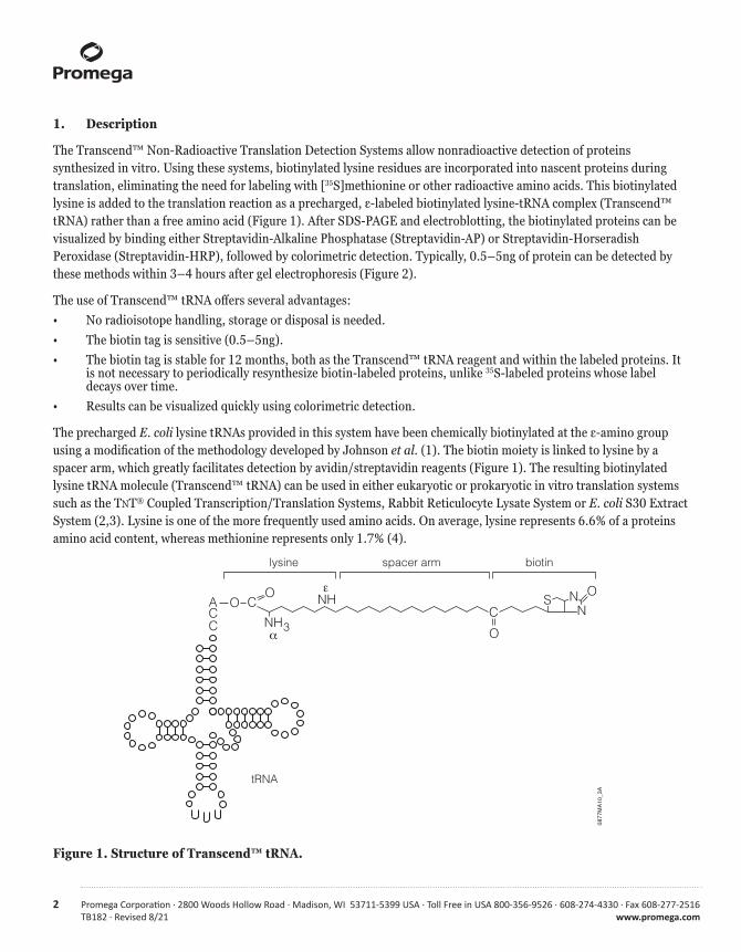
The Transcend™ Non-Radioactive Translation Detection Systems allow nonradioactive detection of proteins synthesized in vitro. Using these systems, biotinylated lysine residues are incorporated into nascent proteins during translation, eliminating the need for labeling with [35S]methionine or other radioactive amino acids. This biotinylated lysine is added to the translation reaction as a precharged, ε-labeled biotinylated lysine-tRNA complex (Transcend™ tRNA) rather than a free amino acid (Figure 1). After SDS-PAGE and electroblotting, the biotinylated proteins can be visualized by binding either Streptavidin-Alkaline Phosphatase (Streptavidin-AP) or Streptavidin-Horseradish Peroxidase (Streptavidin-HRP), followed by colorimetric detection. Typically, 0.5–5ng of protein can be detected by these methods within 3–4 hours after gel electrophoresis (Figure 2).
The use of Transcend™ tRNA offers several advantages:• No radioisotope handling, storage or disposal is needed.• The biotin tag is sensitive (0.5–5ng).• The biotin tag is stable for 12 months, both as the Transcend™ tRNA reagent and within the labeled proteins. It
is not necessary to periodically resynthesize biotin-labeled proteins, unlike 35S-labeled proteins whose label decays over time.
• Results can be visualized quickly using colorimetric detection.
The precharged E. coli lysine tRNAs provided in this system have been chemically biotinylated at the ε-amino group using a modification of the methodology developed by Johnson et al. (1). The biotin moiety is linked to lysine by a spacer arm, which greatly facilitates detection by avidin/streptavidin reagents (Figure 1). The resulting biotinylated lysine tRNA molecule (Transcend™ tRNA) can be used in either eukaryotic or prokaryotic in vitro translation systems such as the TNT® Coupled Transcription/Translation Systems, Rabbit Reticulocyte Lysate System or E. coli S30 Extract System (2,3). Lysine is one of the more frequently used amino acids. On average, lysine represents 6.6% of a proteins amino acid content, whereas methionine represents only 1.7% (4).
Figure 1. Structure of Transcend™ tRNA.
ACC O
CO
O CO
NH3
ε
α
NH SN
N
tRNA
lysine spacer arm biotin
0877MA10_3A
Promega Corporation · 2800 Woods Hollow Road · Madison, WI 53711-5399 USA · Toll Free in USA 800-356-9526 · 608-274-4330 · Fax 608-277-2516 3www.promega.com TB182 · Revised 8/21
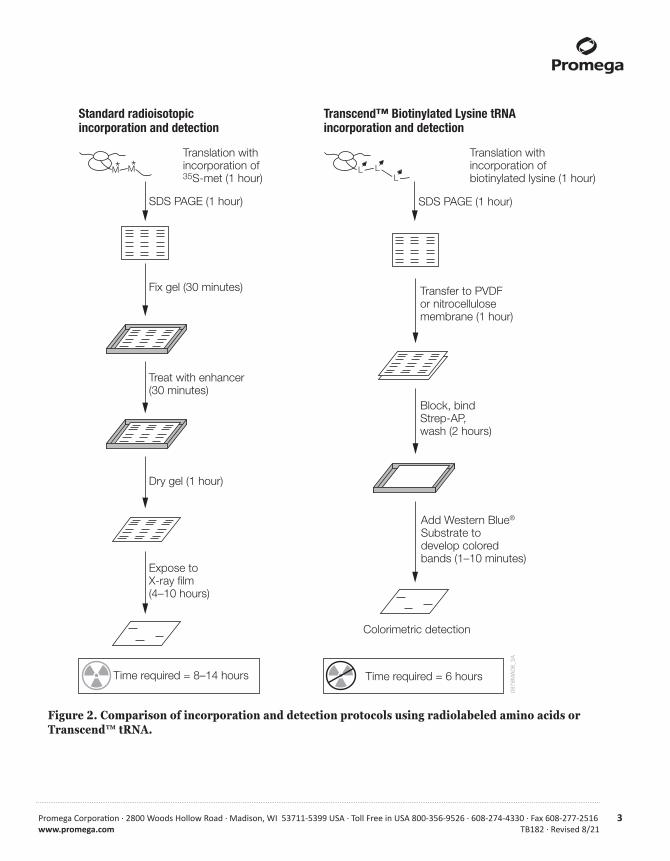
Figure 2. Comparison of incorporation and detection protocols using radiolabeled amino acids or Transcend™ tRNA.
Standard radioisotopicincorporation and detection
Transcend™ Biotinylated Lysine tRNAincorporation and detection
M* M*Translation withincorporation of 35S-met (1 hour)
Translation withincorporation of biotinylated lysine (1 hour)
SDS PAGE (1 hour)
Fix gel (30 minutes)
SDS PAGE (1 hour)
L LL
Colorimetric detection
Transfer to PVDF or nitrocellulose membrane (1 hour)
Block, bind Strep-AP, wash (2 hours)
Add Western Blue® Substrate to develop colored bands (1–10 minutes)
Expose to X-ray film(4–10 hours)
Time required = 8–14 hours Time required = 6 hours
Treat with enhancer (30 minutes)
Dry gel (1 hour)
0878
MA
08_3
A
4 Promega Corporation · 2800 Woods Hollow Road · Madison, WI 53711-5399 USA · Toll Free in USA 800-356-9526 · 608-274-4330 · Fax 608-277-2516TB182 · Revised 8/21 www.promega.com
2. General Considerations
2.A. Effect of Biotinylated Lysine Incorporation on Expression Levels and Enzyme Activity
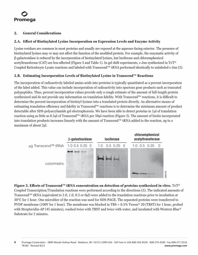
Lysine residues are common in most proteins and usually are exposed at the aqueous-facing exterior. The presence of biotinylated lysines may or may not affect the function of the modified protein. For example, the enzymatic activity of β-galactosidase is reduced by the incorporation of biotinylated lysines, but luciferase and chloramphenicol acetyltransferase (CAT) are less affected (Figure 3 and Table 1). In gel shift experiments, c-Jun synthesized in TNT® Coupled Reticulocyte Lysate reactions and labeled with Transcend™ tRNA performed identically to unlabeled c-Jun (3).
2.B. Estimating Incorporation Levels of Biotinylated Lysine in Transcend™ Reactions
The incorporation of radioactively labeled amino acids into proteins is typically quantitated as a percent incorporation of the label added. This value can include incorporation of radioactivity into spurious gene products such as truncated polypeptides. Thus, percent incorporation values provide only a rough estimate of the amount of full-length protein synthesized and do not provide any information on translation fidelity. With Transcend™ reactions, it is difficult to determine the percent incorporation of biotinyl-lysines into a translated protein directly. An alternative means of estimating translation efficiency and fidelity in Transcend™ reactions is to determine the minimum amount of product detectable after SDS-polyacrylamide gel electrophoresis. We have been able to detect proteins in 1μl of translation reaction using as little as 0.5μl of Transcend™ tRNA per 50μl reaction (Figure 3). The amount of biotin incorporated into translation products increases linearly with the amount of Transcend™ tRNA added to the reaction, up to a maximum of about 2μl.
Figure 3. Effects of Transcend™ tRNA concentration on detection of proteins synthesized in vitro. TNT® Coupled Transcription/Translation reactions were performed according to the directions (5). The indicated amounts of Transcend™ tRNA (equivalent to 2.0, 1.0, 0.5 or 0μl) were added to the translation reactions prior to incubation at 30°C for 1 hour. One microliter of the reaction was used for SDS-PAGE. The separated proteins were transferred to PVDF membrane (100V for 1 hour). The membrane was blocked in TBS + 0.5% Tween® 20 (TBST) for 1 hour, probed with Streptavidin-AP (45 minutes), washed twice with TBST and twice with water, and incubated with Western Blue® Substrate for 2 minutes.
0865
TA
β-galactosidase luciferasechloramphenicolacetyltransferase
µg Transcend™ tRNA
colorimetric
1.0 0.5 0.25 0 1.0 0.5 0.25 0 1.0 0.5 0.25 0
Promega Corporation · 2800 Woods Hollow Road · Madison, WI 53711-5399 USA · Toll Free in USA 800-356-9526 · 608-274-4330 · Fax 608-277-2516 5www.promega.com TB182 · Revised 8/21
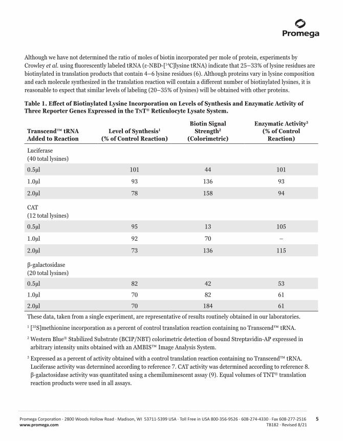
Although we have not determined the ratio of moles of biotin incorporated per mole of protein, experiments by Crowley et al. using fluorescently labeled tRNA (ε-NBD-[14C]lysine tRNA) indicate that 25–33% of lysine residues are biotinylated in translation products that contain 4–6 lysine residues (6). Although proteins vary in lysine composition and each molecule synthesized in the translation reaction will contain a different number of biotinylated lysines, it is reasonable to expect that similar levels of labeling (20–35% of lysines) will be obtained with other proteins.
Table 1. Effect of Biotinylated Lysine Incorporation on Levels of Synthesis and Enzymatic Activity of Three Reporter Genes Expressed in the TNT® Reticulocyte Lysate System.
Transcend™ tRNA Added to Reaction
Level of Synthesis1 (% of Control Reaction)
Biotin Signal Strength2
(Colorimetric)
Enzymatic Activity3 (% of Control
Reaction)
Luciferase (40 total lysines)
0.5μl 101 44 101
1.0μl 93 136 93
2.0μl 78 158 94
CAT (12 total lysines)
0.5μl 95 13 105
1.0μl 92 70 –
2.0μl 73 136 115
β-galactosidase (20 total lysines)
0.5μl 82 42 53
1.0μl 70 82 61
2.0μl 70 184 61
These data, taken from a single experiment, are representative of results routinely obtained in our laboratories.1 [35S]methionine incorporation as a percent of control translation reaction containing no Transcend™ tRNA.2 Western Blue® Stabilized Substrate (BCIP/NBT) colorimetric detection of bound Streptavidin-AP expressed in
arbitrary intensity units obtained with an AMBIS™ Image Analysis System.3 Expressed as a percent of activity obtained with a control translation reaction containing no Transcend™ tRNA.
Luciferase activity was determined according to reference 7. CAT activity was determined according to reference 8. β-galactosidase activity was quantitated using a chemiluminescent assay (9). Equal volumes of TNT® translation reaction products were used in all assays.
6 Promega Corporation · 2800 Woods Hollow Road · Madison, WI 53711-5399 USA · Toll Free in USA 800-356-9526 · 608-274-4330 · Fax 608-277-2516TB182 · Revised 8/21 www.promega.com
3. Product Components and Storage Conditions
P R O D U C T C AT. #
Transcend™ Colorimetric Non-Radioactive Translation Detection System L5070
Each system contains sufficient reagents to label 30 × 50μl translation reactions and perform colorimetric detection of biotinylated proteins on 6 blots (7 × 9cm) using Streptavidin-AP Conjugate and Western Blue® Substrate. Includes:
• 30μl Transcend™ tRNA• 36μl Streptavidin-AP• 35ml Western Blue® Stabilized Substrate for Alkaline Phosphatase
P R O D U C T S I Z E C AT. #
Transcend™ tRNA 30μl L5061
Thirty microliters of Transcend™ tRNA is sufficient for 30 × 50μl translation reactions.
Note: Transcend™ tRNA (Cat.# L5061) is shipped separately from the other components due to differences in storage temperatures.
Storage Conditions: Store Transcend™ tRNA at less than –65°C. Do not subject Transcend™ tRNA to more than 5 freeze-thaw cycles and quickly return it to less than –65°C after use.
Store all other components at +2°C to +10°C. Do not freeze Western Blue® Substrate or Streptavidin-Alkaline Phosphatase.
4. Biotinylated Lysine Incorporation Using Transcend™ tRNA
4.A. Translation Protocol
Materials to Be Supplied by the User• RNasin® Ribonuclease Inhibitor (Cat.# N2111) or Recombinant RNasin® Ribonuclease Inhibitor (Cat.# N2511)• Nuclease-Free Water (Cat.# P1193)• translation extract (e.g., Rabbit Reticulocyte Lysate, E. coli S30 Extract, TNT® Coupled Transcription/Translation
Systems; see Note 1 at the end of this section)• salts, DTT and other components as needed to optimize translation reaction• complete amino acid mix or a combination of two minus amino acid mixes
Use the following protocol as a guideline for setting up a translation reaction using Transcend™ tRNA. In general, Transcend™ tRNA may be used in an in vitro translation protocol at a concentration of 1–2μl Transcend™ tRNA per 50μl reaction. An example of a standard reaction for Rabbit Reticulocyte Lysate is provided. For more information on specific in vitro translation systems, please see Section 4.B.
Promega Corporation · 2800 Woods Hollow Road · Madison, WI 53711-5399 USA · Toll Free in USA 800-356-9526 · 608-274-4330 · Fax 608-277-2516 7www.promega.com TB182 · Revised 8/21
1. Remove the translation and Transcend™ tRNA reagents from storage at less than –65°C. Thaw the Transcend™ tRNA on ice. Thaw the translation lysate by hand warming and immediately place on ice. The other components can be thawed at 37°C and then stored on ice as soon as they thaw.
Note: Do not subject Transcend™ tRNA to more than 5 freeze-thaw cycles. If necessary, store Transcend™ tRNA in multiple aliquots at less than –65°C.
2. On ice, set up 50μl translation reactions as you would for radioactive amino acid incorporation, with the following exception: Add 1μl of a complete amino acid mix (containing 1mM of each amino acid) or a combination of two minus amino acid mixtures (such as 0.5μl of minus methionine and 0.5μl of minus leucine).
Note: In the E. coli S30 Extract Systems, the use of a minus lysine amino acid mixture may significantly reduce the yield of translation product.
Example of a Standard Rabbit Reticulocyte Lysate Reaction Using Transcend™ tRNA.
Rabbit Reticulocyte Lysate 35μl
Nuclease-Free Water 10μl
RNasin® Ribonuclease Inhibitor (40u/μl) 1μl
1mM complete amino acid mixture (or mixture of two minus amino acid mixtures) 1μl
RNA template in Nuclease-Free Water (see Note 3 at the end of this section) 2μl
Transcend™ tRNA (see Note 2 at the end of this section) 1–2μl
final volume 50μl
3. Add all components except the Transcend™ tRNA and gently mix by pipetting the reaction while stirring the reaction with the pipette tip. If necessary, spin briefly in a microcentrifuge to return the sample to the bottom of the tube. Add the Transcend™ tRNA.
Note: We recommend including a control reaction containing Transcend™ tRNA but no added nucleic acid template. This allows measurement of any background incorporation contributed by endogenous mRNA and also reveals any endogenous biotinylated protein(s) in the translation extract (see Note 1 at the end of this section). If using a wheat germ extract, this is especially important.
4. Immediately incubate the translation reaction at 30°C for 60 minutes (see Note 4).
5. Terminate the reaction by placing on ice. If necessary, the translation reaction mix can be stored for several months at –20°C to –70°C.
6. Analyze the results of translation. Procedures for gel analysis of translation products are provided in Section 5.
8 Promega Corporation · 2800 Woods Hollow Road · Madison, WI 53711-5399 USA · Toll Free in USA 800-356-9526 · 608-274-4330 · Fax 608-277-2516TB182 · Revised 8/21 www.promega.com
Notes:
1. Commonly used translation extracts contain endogenous biotinylated proteins, which may be detected when translation products are analyzed by SDS-PAGE, electroblotting and Streptavidin-AP/Streptavidin-HRP detection. Rabbit Reticulocyte Lysate contains one biotinylated protein, which migrates as a faint band at 100kDa and, in some lots, an additional very faint band at 47kDa. E. coli S30 Extract contains one endogenous protein, migrating at 22.5kDa. Wheat Germ Extract contains five major endogenous biotinylated proteins, migrating at 200kDa, 80kDa, 32kDa and a doublet at 17kDa. A comparison to a no-template control will distinguish the endogenous biotinylated protein(s) from the newly synthesized biotinylated translation product.
2. Biotin labeling of poorly expressed proteins or proteins containing few lysines can be increased by doubling the amount of Transcend™ tRNA added per 50μl translation reaction (Table 1 and Figure 3).
3. For maximal expression of your protein, optimize the amount of template added to the reaction and use highly purified RNA or DNA, depending on the translation system used.
Translation: An unfractionated cytoplasmic RNA preparation is 60–70% rRNA and, as a result, translates poorly. Usually such preparations yield no better than 20–30% of the maximum incorporation attainable, and concentrations of 100–200μg/ml (final concentration) are needed to stimulate translation. In contrast, viral RNAs and poly(A)+ mRNAs (including mRNA transcribed in vitro) can be used at much lower concentrations (5–80μg/ml final concentration).
Increased expression levels may be obtained by optimizing the Mg2+ and K+ concentrations in the translation reaction or by using a TNT® Coupled Transcription/Translation System or a TNT® Quick Coupled Transcription/Translation System.
Coupled Transcription/Translation: In E. coli S30 reactions, increased band intensities often can be obtained by using 2–3 times the DNA concentration normally recommended.
4. The appropriate incubation temperature will vary from one translation system to another. Please refer to the appropriate Promega protocol (see Section 4.B) for specific reaction conditions.
4.B. In Vitro Translation Systems
For more information on specific in vitro translation systems, please request the appropriate protocol from Promega.• TNT® Quick Coupled Transcription/Translation System Technical Manual (#TM045)• TNT® Coupled Reticulocyte Lysate Systems Technical Bulletin (#TB126)• TNT® Coupled Wheat Germ Extract Systems Technical Bulletin (#TB165)• Flexi® Rabbit Reticulocyte Lysate System Technical Bulletin (#TB127)• Wheat Germ Extract System Technical Manual (#TM230)• E. coli S30 Extract System for Circular DNA Technical Bulletin (#TB092)• E. coli S30 Extract System for Linear Templates Technical Bulletin (#TB102)• E. coli T7 S30 Extract System for Circular DNA Technical Bulletin (#TB219)• TNT® Quick for PCR DNA Technical Manual (#TM235)
The number of lysines in the translated polypeptide and the efficiency of translation are the two most important factors affecting the SDS-PAGE band intensity of the translation product. To increase the intensity of weak bands, increase the amount of Transcend™ tRNA to up to twice the standard amount (see Figure 3).
Promega Corporation · 2800 Woods Hollow Road · Madison, WI 53711-5399 USA · Toll Free in USA 800-356-9526 · 608-274-4330 · Fax 608-277-2516 9www.promega.com TB182 · Revised 8/21
To reduce the chance of RNase contamination, wear gloves throughout the experiment and use microcentrifuge tubes and pipette tips that have been autoclaved and handled only with gloves. Addition of RNasin® Ribonuclease Inhibitor to the translation reaction is recommended but not required. RNasin® Ribonuclease Inhibitor prevents degradation of sample mRNAs by many contaminating RNases.
If the amount of translation product must be estimated, add radioactive amino acid(s), in addition to the Transcend™ tRNA, to either a control translation reaction or all translation reactions. The percent incorporation of the radioactive amino acid can be used in combination with knowledge of the protein’s amino acid composition to estimate the amount of translation product produced.
5. Post-Translational Analysis
5.A. Denaturing Gel Analysis of Translation Products
Biotinylated protein standards (Bio-Rad Cat.# 161-0319) can be used to determine the apparent molecular weight of the translated biotinylated protein. Alternatively, fluorescently labeled size standards can be observed after transfer and marked with a pencil under UV irradiation. The positions of unlabeled size standards also can be determined by staining the blot after transfer (see Section 5.C).
1. Once the 50μl translation reaction is complete (or at any desired time point), remove a 1μl aliquot and add it to 15μl of SDS sample buffer. The remainder of the reaction may be stored at –20°C.
2. Close the tube and heat at 70°C for 15 minutes to denature the proteins.
3. Load the denatured sample on an SDS-polyacrylamide gel. (Protocols for SDS polyacrylamide gel electrophoresis may be found in the technical literature provided with Promega translation systems, which is listed in Section 4.B.)
4. Perform electrophoresis using standard conditions for your apparatus. Typically, electrophoresis is carried out at a constant current of 20mA. Electrophoresis usually is performed until the bromophenol blue dye has run off the bottom of the gel.
Note: If a gene product is weakly expressed or contains few lysines, up to 2μl of the translation reaction (Reticulocyte Lysate) can be loaded on an SDS gel without the loss of resolution observed with autoradiography. However, loading more of the translation reaction can result in high background on the blot. When loading more than 1μl of an S30 Extract translation reaction, first precipitate the proteins by adding 20μl of acetone per 5μl of extract and incubate on ice for 15 minutes. Centrifuge the acetone-precipitated S30 reaction at 12,000 × g for 5 minutes. Remove the supernatant and dry the pellet. Resuspend the pellet in 5μl of water, add 10μl of SDS sample buffer, heat at 90–100°C for 5 minutes and load 5–15μl of the sample on the gel.
!
10 Promega Corporation · 2800 Woods Hollow Road · Madison, WI 53711-5399 USA · Toll Free in USA 800-356-9526 · 608-274-4330 · Fax 608-277-2516TB182 · Revised 8/21 www.promega.com
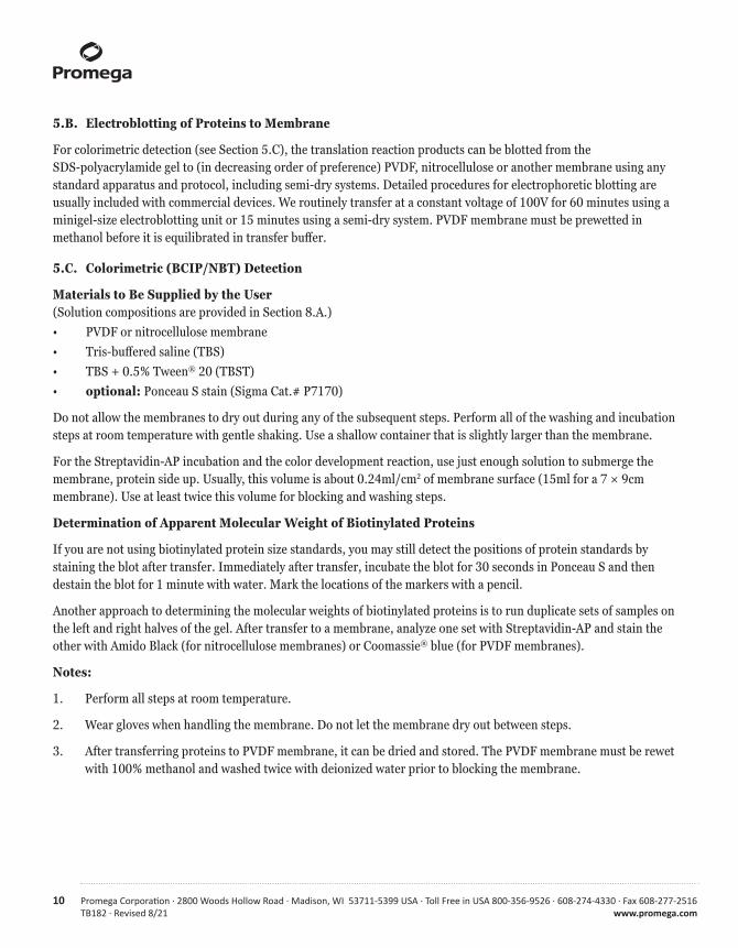
5.B. Electroblotting of Proteins to Membrane
For colorimetric detection (see Section 5.C), the translation reaction products can be blotted from the SDS-polyacrylamide gel to (in decreasing order of preference) PVDF, nitrocellulose or another membrane using any standard apparatus and protocol, including semi-dry systems. Detailed procedures for electrophoretic blotting are usually included with commercial devices. We routinely transfer at a constant voltage of 100V for 60 minutes using a minigel-size electroblotting unit or 15 minutes using a semi-dry system. PVDF membrane must be prewetted in methanol before it is equilibrated in transfer buffer.
5.C. Colorimetric (BCIP/NBT) Detection
Materials to Be Supplied by the User(Solution compositions are provided in Section 8.A.)• PVDF or nitrocellulose membrane• Tris-buffered saline (TBS)• TBS + 0.5% Tween® 20 (TBST)• optional: Ponceau S stain (Sigma Cat.# P7170)
Do not allow the membranes to dry out during any of the subsequent steps. Perform all of the washing and incubation steps at room temperature with gentle shaking. Use a shallow container that is slightly larger than the membrane.
For the Streptavidin-AP incubation and the color development reaction, use just enough solution to submerge the membrane, protein side up. Usually, this volume is about 0.24ml/cm2 of membrane surface (15ml for a 7 × 9cm membrane). Use at least twice this volume for blocking and washing steps.
Determination of Apparent Molecular Weight of Biotinylated Proteins
If you are not using biotinylated protein size standards, you may still detect the positions of protein standards by staining the blot after transfer. Immediately after transfer, incubate the blot for 30 seconds in Ponceau S and then destain the blot for 1 minute with water. Mark the locations of the markers with a pencil.
Another approach to determining the molecular weights of biotinylated proteins is to run duplicate sets of samples on the left and right halves of the gel. After transfer to a membrane, analyze one set with Streptavidin-AP and stain the other with Amido Black (for nitrocellulose membranes) or Coomassie® blue (for PVDF membranes).
Notes:
1. Perform all steps at room temperature.
2. Wear gloves when handling the membrane. Do not let the membrane dry out between steps.
3. After transferring proteins to PVDF membrane, it can be dried and stored. The PVDF membrane must be rewet with 100% methanol and washed twice with deionized water prior to blocking the membrane.
Promega Corporation · 2800 Woods Hollow Road · Madison, WI 53711-5399 USA · Toll Free in USA 800-356-9526 · 608-274-4330 · Fax 608-277-2516 11www.promega.com TB182 · Revised 8/21
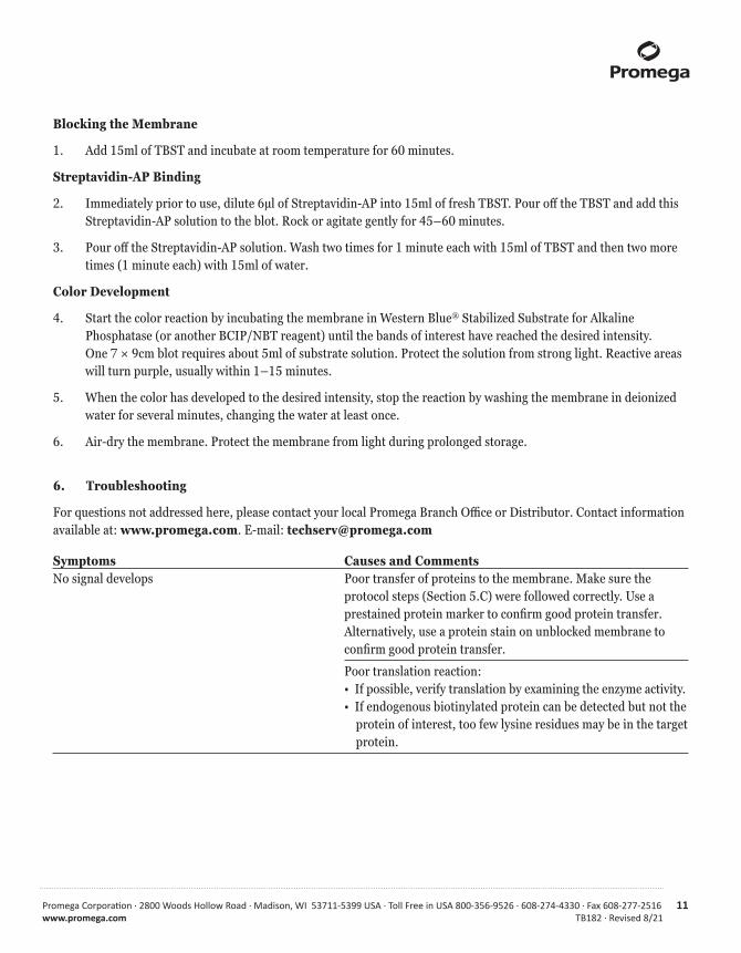
Blocking the Membrane
1. Add 15ml of TBST and incubate at room temperature for 60 minutes.
Streptavidin-AP Binding
2. Immediately prior to use, dilute 6μl of Streptavidin-AP into 15ml of fresh TBST. Pour off the TBST and add this Streptavidin-AP solution to the blot. Rock or agitate gently for 45–60 minutes.
3. Pour off the Streptavidin-AP solution. Wash two times for 1 minute each with 15ml of TBST and then two more times (1 minute each) with 15ml of water.
Color Development
4. Start the color reaction by incubating the membrane in Western Blue® Stabilized Substrate for Alkaline Phosphatase (or another BCIP/NBT reagent) until the bands of interest have reached the desired intensity. One 7 × 9cm blot requires about 5ml of substrate solution. Protect the solution from strong light. Reactive areas will turn purple, usually within 1–15 minutes.
5. When the color has developed to the desired intensity, stop the reaction by washing the membrane in deionized water for several minutes, changing the water at least once.
6. Air-dry the membrane. Protect the membrane from light during prolonged storage.
6. Troubleshooting
For questions not addressed here, please contact your local Promega Branch Office or Distributor. Contact information available at: www.promega.com. E-mail: [email protected]
Symptoms Causes and CommentsNo signal develops Poor transfer of proteins to the membrane. Make sure the protocol steps (Section 5.C) were followed correctly. Use a
prestained protein marker to confirm good protein transfer. Alternatively, use a protein stain on unblocked membrane to confirm good protein transfer.
Poor translation reaction: • If possible, verify translation by examining the enzyme activity. • If endogenous biotinylated protein can be detected but not the protein of interest, too few lysine residues may be in the target protein.
12 Promega Corporation · 2800 Woods Hollow Road · Madison, WI 53711-5399 USA · Toll Free in USA 800-356-9526 · 608-274-4330 · Fax 608-277-2516TB182 · Revised 8/21 www.promega.com
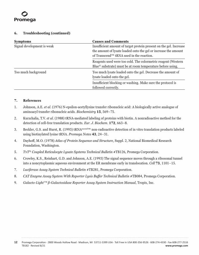
6. Troubleshooting (continued)
Symptoms Causes and CommentsSignal development is weak Insufficient amount of target protein present on the gel. Increase the amount of lysate loaded onto the gel or increase the amount of Transcend™ tRNA used in the reaction.
Reagents used were too cold. The colormetric reagent (Western Blue® substrate) must be at room temperature before using.
Too much background Too much lysate loaded onto the gel. Decrease the amount of lysate loaded onto the gel.
Insufficient blocking or washing. Make sure the protocol is followed correctly.
7. References
1. Johnson, A.E. et al. (1976) N-epsilon-acetyllysine transfer ribonucleic acid: A biologically active analogue of aminoacyl transfer ribonucleic acids. Biochemistry 15, 569–75.
2. Kurzchalia, T.V. et al. (1988) tRNA-mediated labeling of proteins with biotin. A nonradioactive method for the detection of cell-free translation products. Eur. J. Biochem. 172, 663–8.
3. Beckler, G.S. and Hurst, R. (1993) tRNAnscend™ non-radioactive detection of in vitro translation products labeled using biotinylated lysine tRNA. Promega Notes 43, 24–31.
4. Dayhoff, M.O. (1978) Atlas of Protein Sequence and Structure, Suppl. 2, National Biomedical Research Foundation, Washington.
5. TNT® Coupled Reticulocyte Lysate Systems Technical Bulletin #TB126, Promega Corporation.
6. Crowley, K.S., Reinhart, G.D. and Johnson, A.E. (1993) The signal sequence moves through a ribosomal tunnel into a noncytoplasmic aqueous environment at the ER membrane early in translocation. Cell 73, 1101–15.
7. Luciferase Assay System Technical Bulletin #TB281, Promega Corporation.
8. CAT Enzyme Assay System With Reporter Lysis Buffer Technical Bulletin #TB084, Promega Corporation.
9. Galacto-Light™ β-Galactosidase Reporter Assay System Instruction Manual, Tropix, Inc.
Promega Corporation · 2800 Woods Hollow Road · Madison, WI 53711-5399 USA · Toll Free in USA 800-356-9526 · 608-274-4330 · Fax 608-277-2516 13www.promega.com TB182 · Revised 8/21
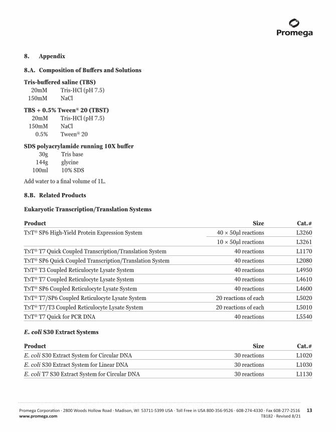
8. Appendix
8.A. Composition of Buffers and Solutions
Tris-buffered saline (TBS) 20mM Tris-HCl (pH 7.5) 150mM NaCl
TBS + 0.5% Tween® 20 (TBST) 20mM Tris-HCl (pH 7.5) 150mM NaCl 0.5% Tween® 20
SDS polyacrylamide running 10X buffer 30g Tris base 144g glycine 100ml 10% SDS
Add water to a final volume of 1L.
8.B. Related Products
Eukaryotic Transcription/Translation Systems
Product Size Cat.#TNT® SP6 High-Yield Protein Expression System 40 × 50μl reactions L3260
10 × 50μl reactions L3261
TNT® T7 Quick Coupled Transcription/Translation System 40 reactions L1170
TNT® SP6 Quick Coupled Transcription/Translation System 40 reactions L2080
TNT® T3 Coupled Reticulocyte Lysate System 40 reactions L4950
TNT® T7 Coupled Reticulocyte Lysate System 40 reactions L4610
TNT® SP6 Coupled Reticulocyte Lysate System 40 reactions L4600
TNT® T7/SP6 Coupled Reticulocyte Lysate System 20 reactions of each L5020
TNT® T7/T3 Coupled Reticulocyte Lysate System 20 reactions of each L5010
TNT® T7 Quick for PCR DNA 40 reactions L5540
E. coli S30 Extract Systems
Product Size Cat.#E. coli S30 Extract System for Circular DNA 30 reactions L1020
E. coli S30 Extract System for Linear DNA 30 reactions L1030
E. coli T7 S30 Extract System for Circular DNA 30 reactions L1130

14 Promega Corporation · 2800 Woods Hollow Road · Madison, WI 53711-5399 USA · Toll Free in USA 800-356-9526 · 608-274-4330 · Fax 608-277-2516TB182 · Revised 8/21 www.promega.com
8.B. Related Products (continued)
Rabbit Reticulocyte Lysate Translation Systems
Product Size Cat.#Rabbit Reticulocyte Lysate, Nuclease Treated 1ml L4960
Flexi® Rabbit Reticulocyte Lysate System 1ml L4540
Rabbit Reticulocyte Lysate/Wheat Germ Extract Combination System
Product Size Cat.#Rabbit Reticulocyte Lysate/Wheat Germ Extract Combination System 12 reactions L4330
Available Separately
Product Size Cat.#Streptavidin Alkaline Phosphatase 0.5ml V5591
Western Blue® Stabilized Substrate for Alkaline Phosphatase 100ml S3841
Amino Acid Mixture, Complete 175μl L4461
8.C. Summary of Changes
The following changes were made to the 8/21 revision of this document:
1. Removed discontinued Cat.# L5080 and associated chemiluminescent detection instructions (Section 5.D).
2. Updated Sections 1 and 6, including Figure 2.
3. Moved to a new template.
© 2011–2021 Promega Corporation. All Rights Reserved.
Flexi, RNasin, TNT and Western Blue are registered trademarks of Promega Corporation. Transcend is a trademark of Promega Corporation.
AMBIS is a trademark of AMBIS, Inc. Coomassie is a registered trademark of Imperial Chemical Industries, Ltd. Corning is a registered trademark of Corning, Inc. Galacto-Light is a trademark of Tropix, Inc. Kodak is a registered trademark of Eastman Kodak Co. Tween is a registered trademark of ICI Americas, Inc.
Products may be covered by pending or issued patents or may have certain limitations. Please visit our Web site for more information.
All prices and specifications are subject to change without prior notice.
Product claims are subject to change. Please contact Promega Technical Services or access the Promega online catalog for the most up-to-date information on Promega products.