Embed Size (px)
Citation preview
FEATUREARTIC
LES
HOW TO DO IT
Transapical Arterial Cannulation for SalvageCardiopulmonary Bypass in TranscatheterAortic Valve ReplacementDerek R. Brinster, MD, Jay A. Patel, Harry L. McCarthy, BS, Ty M. Aron, BS, andZachary M. Gertz, MDDivisions of Cardiothoracic and Vascular Surgery, Virginia Commonwealth University Medical Center, Richmond, Virginia
Hemodynamic instability during transcatheter aorticvalve replacement procedures may require transient car-diopulmonary bypass for support. In patients with severeatherosclerosis, peripheral cannulation may not bepossible. This method of direct left ventricle cannulation
Accepted for publication March 26, 2014.
Address correspondence to Dr Brinster, Virginia Commonwealth Uni-versity, W Hospital Bldg, 7th Flr, S Wing, 1200 E Broad St, PO Box 980068,Richmond VA 23298-0068; e-mail: [email protected].
� 2014 by The Society of Thoracic SurgeonsPublished by Elsevier
during transapical TAVR is a facile means to providearterial inflow.
(Ann Thorac Surg 2014;98:1482–4)� 2014 by The Society of Thoracic Surgeons
ranscatheter aortic valve replacement (TAVR) was
Tapproved in November 2011 by the US Food andDrug Administration for the treatment of inoperable,severe, symptomatic aortic valve stenosis. Since that time,the label has been expanded to include high-risk, butoperable, patients by either transfemoral or transapicalaccess [1].Procedural success rates vary and range between 94%and 100% [2]. Intraprocedural hemodynamic instabilitycan occur at any point of the procedure, but typicallyhappens during rapid ventricular pacing, balloon infla-tion, or sheath access into a small ventricle duringtransapical aortic valve replacement. This report high-lights a facile method of rapid arterial cannulation in apatient undergoing transapical TAVR with hemodynamicinstability and unsuitable for rapid peripheral arterialcannulation access.
Technique
A 90-year-old patient was evaluated by the multidisci-plinary valve team for TAVR for progressive symptoms ofshortness of breath, dyspnea on exertion, and orthopnea.The patient had a past medical history of severe periph-eral arterial disease with femoral to femoral arterybypass, right above-knee amputation (but ambulatorywith prosthesis and walker), hypertension, rheumatoidarthritis, and heart failure. Her echo revealed a meanaortic transvalvular gradient of 42 mm Hg, aortic valvearea of 0.5 cm2, and ejection fraction of 0.75. Of note, theecho also revealed a small, collapsing left ventricle. Herpreoperative coronary catheterization revealed heavily
calcified coronary vessels with luminal irregularities, anda left main coronary with mild distal tapering.The patient had prohibitive arterial access due to
atherosclerosis and occlusion of her iliac vessels, as wellas stenosis of both of her subclavian arteries. Therefore,transapical TAVR was chosen. However, options forpossible salvage cardiopulmonary bypass method as partof the preoperative strategic planning were challengingdue to her small or occluded native vessels. Conse-quently, the cardiopulmonary method planned, in case ofhemodynamic instability, was to place the patient oncardiopulmonary bypass (CPB) after insertion of thevalve, utilizing an arterial cannula through the transapicalTAVR sheath. After successful establishment of bypass, amore appropriate cannulation site could be utilized.The patient was brought to the hybrid operating room
and general anesthesia induced without incidence. Both afemoral transvenous pacing catheter was placed, as wellas an additional femoral venous access on the contralat-eral side for possible venous cannula placement for CPB.A small left thoracotomy incision was performed, and
the left ventricular apex was cannulated with a Bentsonwire (Cook Medical, Bloomington, IN) and a 6 Fr sheath.The Bentson wire was placed through the native aorticvalve and a short extra stiff Amplatz (Boston Scientific,Natick, MA) wire was exchanged through a catheter. Thelarge Ascendra introducer system (Edwards Lifesciences,Irvine, CA) was placed under fluoroscopy into the leftventricle apex to a level of approximately 4 cm within theleft ventricle cavity. The Ascendra sheath then allowedaccess for eventual balloon valvuloplasty as well asTAVR.During transapical balloon valvuloplasty with rapid
ventricular pacing, the patient had moderate hemody-namic instability which responded to vasoactive medi-cations. Upon placement of the 26-mm, Edwards Sapienvalve (Edwards Lifesciences), the blood pressure droppedand did not respond to typical vasoactive medication
0003-4975/$36.00http://dx.doi.org/10.1016/j.athoracsur.2014.03.049

Fig 1. Operative image of transapical cannulation. (a) Arterial inflowcannula. (b) Transcatheter aortic valve replacement transapicalsheath. (c) Left ventricular apex of heart.
1483Ann Thorac Surg HOW TO DO IT BRINSTER ET AL2014;98:1482–4 LV CANNULATION DURING TRANSAPICAL TAVR
FEATUREARTIC
LES
maneuvers. Brief chest compressions of approximately 2minutes were required, and the patient was fully hepa-rinized and prepared for CPB.
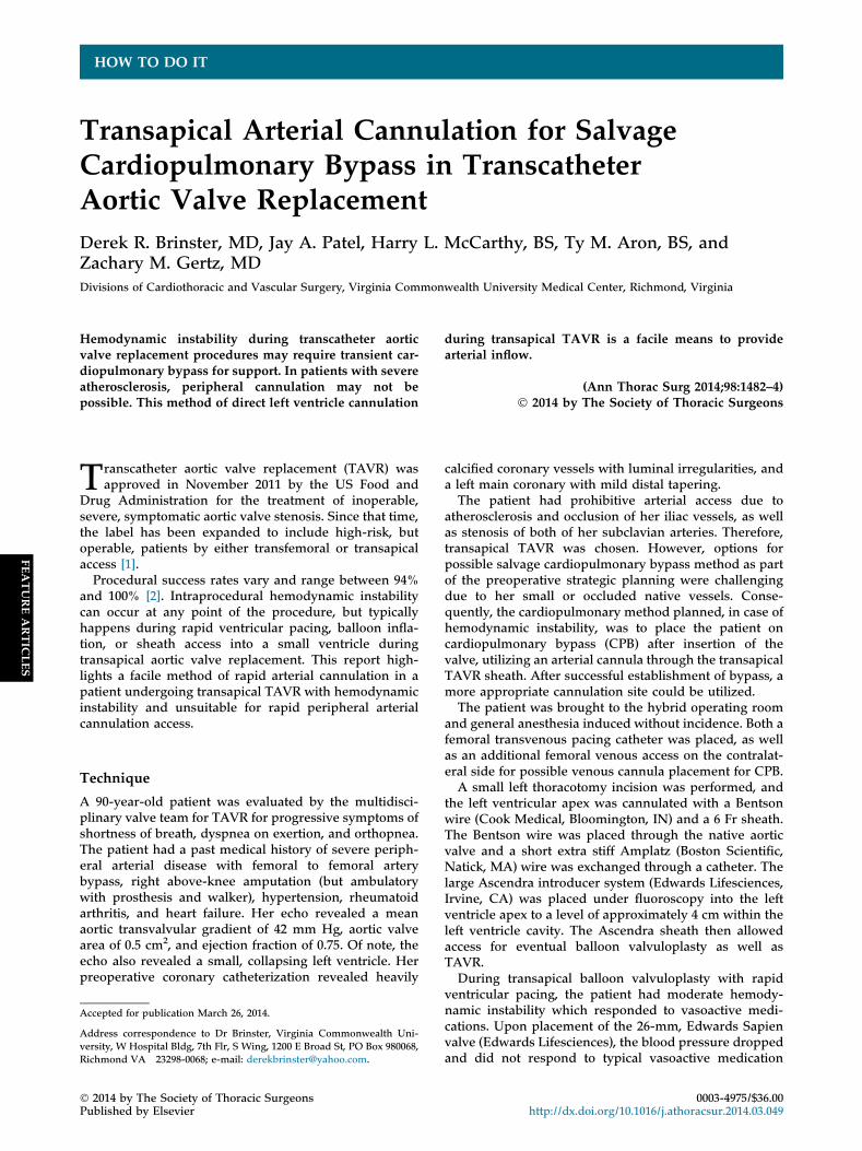
A 22 Fr percutaneous venous cannula (Edwards Life-sciences) was inserted under transesophageal echocardi-ography guidance through the previously accessedcommon femoral vein. At the same time, a long arterialcannula, Bio-Medicus (Medtronic, Inc, Sandy Hook, CT)50-cm 17 Fr percutaneous catheter was inserted throughthe TAVR 25-mm sheath (Fig 1) with fluoroscopic guid-ance (Fig 2A;B). The long 17 Fr arterial cannula wasselected to allow enough length to extend beyond theAscendra sheath and into the ascending aorta. Bypassflows were able to be achieved between 2.5 L/minute and4.0L/minute depending on volume status. The rightaxillary artery was then dissected out and utilized forarterial inflow after performing an endarterectomy, andsewing on a side arm graft. The transapical arterial can-nula was removed once arterial inflow was established inthe left axillary graft. The patient was maintained on CPBuntil hemodynamic recovery, and slowly weaned from
cardiopulmonary support. The valve had trace to mildperivalvular regurgitation with no evidence of movementor injury from passing the arterial cannula through thevalve.The patient was transferred to the intensive care unit
after the procedure and had gradual correction of heracidosis, following commands, and being prepared forextubation by postoperative day 1. Approximately 30hours after the procedure, the patient decompensatedand went into multisystem organ failure with hypoten-sion. The family wished to withdraw care and the patientexpired. The autopsy revealed an occlusion of the leftcoronary artery distal to the orifice of the left main artery,acute tubular necrosis, fibrin thrombi within her lungsconsistent with disseminated intravascular coagulation,and congested liver.
Comment
The uses and indications for TAVR continue to expand.There is approximately a 92% success rate in valve im-plantation. However, despite preoperative planning toprevent complications of hemodynamic instability, therisk of intraprocedural complications requiring emer-gency cardiac surgery is approximately 1.2% [3].There are little data regarding the incidence for using
CPB for transient hemodynamic compromise. Shreenivasand colleagues [4] reported that circulatory support,identified as either the use of cardiopulmonary bypass orintraaortic balloon pump, was required in 203 of 2,538patients (8%) and, compared with noncirculatory supportpatients, support patients were more likely to be male,undergo transapical access, have a higher incidence ofmajor vascular complication (17% vs 5%, p < 0.0001), anda higher 30-day morality (25% vs 5%; p < 0.0001).The optimal therapy for high-risk patients is for pre-
operative preparation of CPB with exposure of an arterialvessel for cannulation and appropriate venous access.Unfortunately, the need for cardiopulmonary supportmay occur abruptly and unexpectedly, or in those whereno optimal site of arterial access is able to be achievedwithout a complex vascular intervention, such as
Fig 2. (A) Fluoroscopic image of transapicalcannulation. (B) Schematic of transapicalcannulation. (a ¼ sheath; b ¼ aortic valve;c ¼ cannula.)

1484 HOW TO DO IT BRINSTER ET AL Ann Thorac SurgLV CANNULATION DURING TRANSAPICAL TAVR 2014;98:1482–4
FEATUREARTIC
LES
endarterectomy or use of prosthetic graft, etc. Thismethod allows rapid institution for CPB in patients withsevere peripheral arterial disease and time to establishappropriate peripheral arterial access for CPB. Once thepatient has cardiopulmonary support transapically, pe-ripheral cannulation may be instituted carefully. How-ever, some concerns of this mode of transapicalcannulation are possible dislodgement of the valve,damage to the valve, creation of aortic valve insufficiencyduring bypass with left ventricular distention, and theneed for eventual peripheral bypass. To our knowledge,this is the first reported method of direct arterial cannu-lation through the TAVR sheath for institution of CPBduring transapical TAVR insertion.
References
1. US Food and Drug Administration. FDA expands approveduse of Sapien artificial heart valve. Available at: http://www.fda.gov/newsevents/newsroom/pressannouncements/ucm323478.htm. Accessed August 27, 2014.
2. Mack MJ, Brennan JM, Brindis R, et al. Outcomes followingtranscatheter aortic valve replacement in the United States.JAMA 2013;310:2069–77.
3. Eggebrecht H, Mehta RH, Kahlert P, et al. Emergent cardiacsurgery during transcatheter aortic valve implantation (TAVI):insights from the Edwards SAPIEN Aortic BioprosthesisEuropean Outcome (SOURCE) registry. EuroIntervention2013. 20130924-01. [Epub ahead of print].
4. Shreenivas SS, Lilly SM, Szeto WY, et al. Circulatory supportis associated with higher mortality during TAVR. JACC2013;62(18_S1):B235.