Embed Size (px)
Citation preview
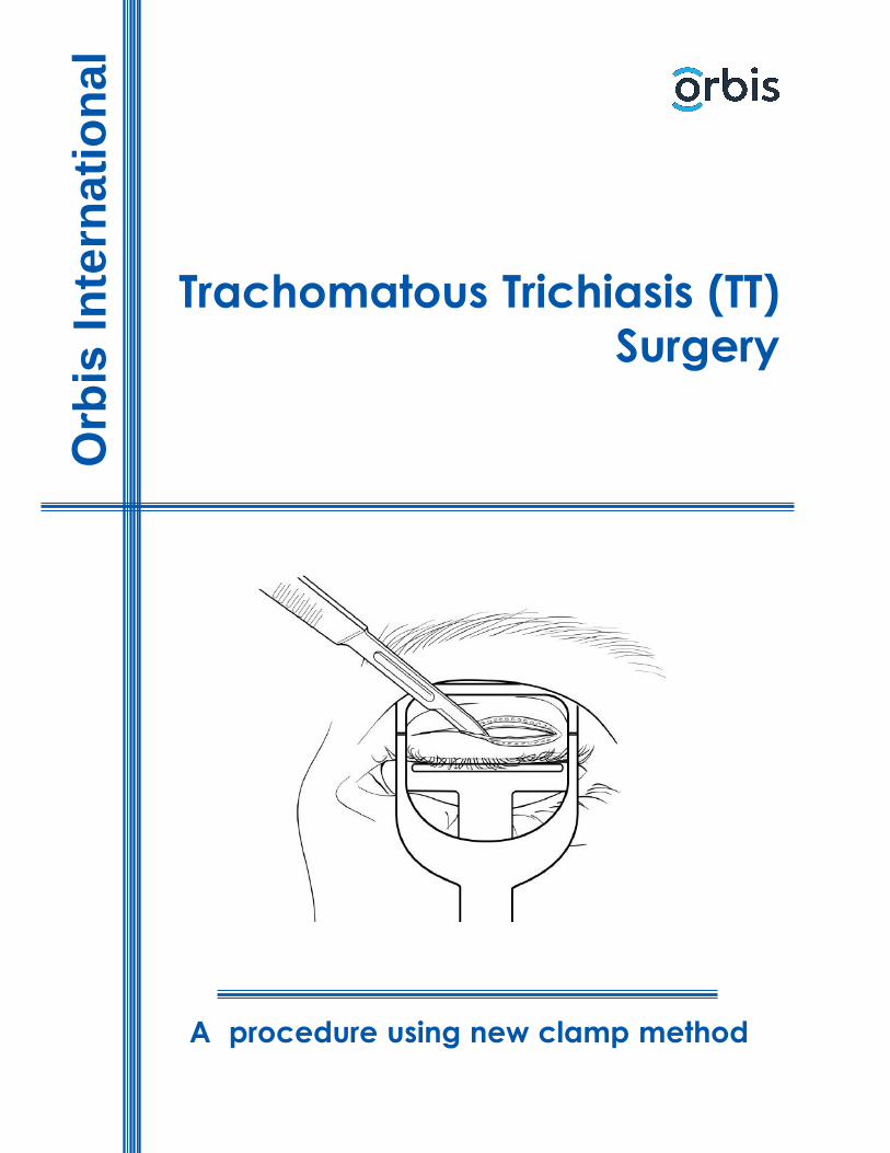
Trachomatous Trichiasis (TT)Surgery
A procedure using new clamp method
Orbis International

Trachomatous Trichiasis (TT) Surgery:
A procedure using new clamp method
Produced by:
Orbis International Ethiopia
Zelalem Eshetu, MD, Ophth. Program Consultant
Alemayehu Sisay, MD, MPH, Ophth. Country Director
Dereje Zewde, MBA, Deputy Country Director
Yilikal Adamu, MD, Ophth.Consultant Oculoplastics Surgeon, Addis Ababa University, Department ofOphthalmology
External
Gabremaskal Habtemariam, PhD
Wondu Alemayehu, MD, MPHGeneral Manager and Consultant ophthalmologist at Berhan Public Health and EyeCare Consultancy
Editors
Eugene Helveston, MDOrbis International, Consultant Ophthalmologist and Cyber-Sight Director
Lynda SmallwoodOrbis International, Senior Manager Cyber-Sight

© February, 2014
Table of Contents
Introduction . . . . . . . . . . . . . . . . . . . . . . . . . . . . . . . . . . . . . . . . . . . . . . . . . . . . . . . . . 1
Part I: Trachoma Background . . . . . . . . . . . . . . . . . . . . . . . . . . . . . . . . . . . . . . . . . . 1Trachoma - Definition . . . . . . . . . . . . . . . . . . . . . . . . . . . . . . . . . . . . . . . . . . . . . 1Stages of Trachoma . . . . . . . . . . . . . . . . . . . . . . . . . . . . . . . . . . . . . . . . . . . . . . 2
Stage 1 - Follicular Conjunctivitis . . . . . . . . . . . . . . . . . . . . . . . . . . . . . . . 2Stage 2 - Conjunctival Scarring . . . . . . . . . . . . . . . . . . . . . . . . . . . . . . . . 2Stage 3 - Trachomatous Trichiasis . . . . . . . . . . . . . . . . . . . . . . . . . . . . . 2Stage 4 - Corneal Scarring . . . . . . . . . . . . . . . . . . . . . . . . . . . . . . . . . . . 2
Trachoma Magnitude . . . . . . . . . . . . . . . . . . . . . . . . . . . . . . . . . . . . . . . . . . . . . 3Global . . . . . . . . . . . . . . . . . . . . . . . . . . . . . . . . . . . . . . . . . . . . . . . . . . . . 3Sub-Saharan Africa . . . . . . . . . . . . . . . . . . . . . . . . . . . . . . . . . . . . . . . . . 3Ethiopia . . . . . . . . . . . . . . . . . . . . . . . . . . . . . . . . . . . . . . . . . . . . . . . . . . 4
Impact of Trachoma . . . . . . . . . . . . . . . . . . . . . . . . . . . . . . . . . . . . . . . . . . . . . . 5Orbis Involvement in Trachoma Control in Ethiopia . . . . . . . . . . . . . . . . . . . . 6Trachoma Control Project Objectives . . . . . . . . . . . . . . . . . . . . . . . . . . . . . . . . . 7Strategies Employed by Orbis to Control Trachoma . . . . . . . . . . . . . . . . . . . . 7
TT Surgery . . . . . . . . . . . . . . . . . . . . . . . . . . . . . . . . . . . . . . . . . . . . . . . . 8Mass Drug Administration (MDA) . . . . . . . . . . . . . . . . . . . . . . . . . . . . . . 8Facial Cleanliness . . . . . . . . . . . . . . . . . . . . . . . . . . . . . . . . . . . . . . . . . . 8Environmental Improvement . . . . . . . . . . . . . . . . . . . . . . . . . . . . . . . . . . 8
Implementation of the SAFE Strategy . . . . . . . . . . . . . . . . . . . . . . . . . . . . . . . . 8Challenges of Trachoma Prevention in Orbis Project Areas . . . . . . . . . . . . . . 9The Way Forward . . . . . . . . . . . . . . . . . . . . . . . . . . . . . . . . . . . . . . . . . . . . . . . 10
Part II: Trachomatous Trichiasis (TT) Surgery . . . . . . . . . . . . . . . . . . . . . . . . . . . . . 11The Eye . . . . . . . . . . . . . . . . . . . . . . . . . . . . . . . . . . . . . . . . . . . . . . . . . . . . . . . 12Step 1: Placement of Anesthetic Drops in Cul-de-sac . . . . . . . . . . . . . . . . . . . 14Step 2: Injection of Anesthetic . . . . . . . . . . . . . . . . . . . . . . . . . . . . . . . . . . . . . 14Step 3: Insertion of Clamp . . . . . . . . . . . . . . . . . . . . . . . . . . . . . . . . . . . . . . . . 16Step 4: Incision of Lid . . . . . . . . . . . . . . . . . . . . . . . . . . . . . . . . . . . . . . . . . . . . 17Step 5: Inspection of Incision . . . . . . . . . . . . . . . . . . . . . . . . . . . . . . . . . . . . . . 18Step 6: Placement of Sutures . . . . . . . . . . . . . . . . . . . . . . . . . . . . . . . . . . . . . . 18Step 7: Tying of Sutures . . . . . . . . . . . . . . . . . . . . . . . . . . . . . . . . . . . . . . . . . . 21Postoperative Course . . . . . . . . . . . . . . . . . . . . . . . . . . . . . . . . . . . . . . . . . . . . 23
Conclusion . . . . . . . . . . . . . . . . . . . . . . . . . . . . . . . . . . . . . . . . . . . . . . . . . . . . . . . . 24

1
Introduction
This manual is designed to show a technique for trachomatous trichiasis (TT) surgery using a clamp that protects the cornea, maintains hemostasis, and provides markers for standardizing the surgical technique. As an introduction to the subject, background information about trachoma is included. This will enable the reader to understand the scientific background of trachoma, the magnitude of the problem, and the Orbis experience with trachoma control in Ethiopia. The first part is based primarily on experience in Ethiopia with Orbis supported programs. However, the activity described can be used as a model for a program in any part of the world where trachoma and trachoma-related blindness is a problem. Moreover, this information is intended for any local organization or international group interested in and dedicated to this work wherever it can be carried out.
This manual is prepared in two parts. The first part deals with background information fortrachoma and the second part describes a surgical technique for TT surgery that employs anewly designed clamp that promises to promote faster, and safer, surgery with morepredictable results.
Part 1: Trachoma BackgroundTrachoma - Definition
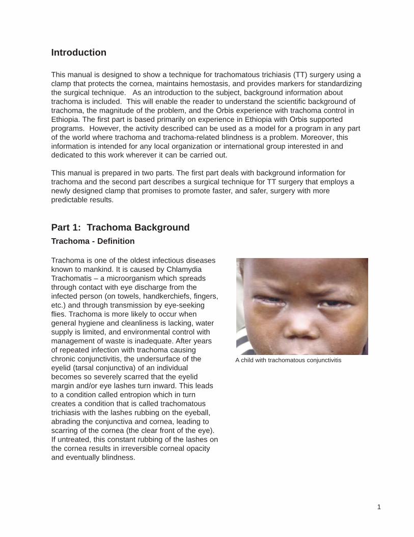
Trachoma is one of the oldest infectious diseases known to mankind. It is caused by Chlamydia�Trachomatis – a microorganism which spreads through contact with eye discharge from theinfected person (on towels, handkerchiefs, fingers,etc.) and through transmission by eye-seekingflies. Trachoma is more likely to occur whengeneral hygiene and cleanliness is lacking, watersupply is limited, and environmental control withmanagement of waste is inadequate. After yearsof repeated infection with trachoma causingchronic conjunctivitis, the undersurface of theeyelid (tarsal conjunctiva) of an individualbecomes so severely scarred that the eyelidmargin and/or eye lashes turn inward. This leadsto a condition called entropion which in turncreates a condition that is called trachomatoustrichiasis with the lashes rubbing on the eyeball,abrading the conjunctiva and cornea, leading toscarring of the cornea (the clear front of the eye).If untreated, this constant rubbing of the lashes onthe cornea results in irreversible corneal opacityand eventually blindness.
A child with trachomatous conjunctivitis
2
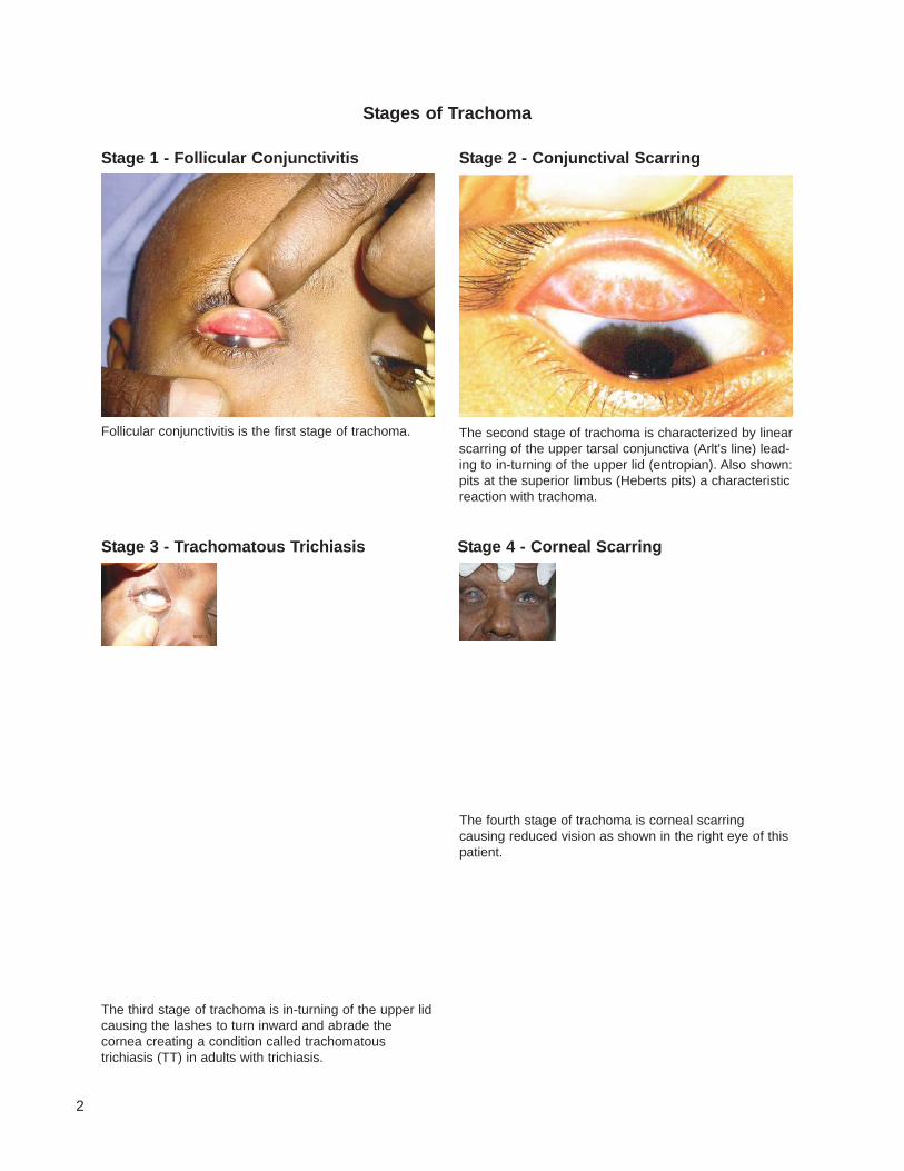
Follicular conjunctivitis is the first stage of trachoma.
The third stage of trachoma is in-turning of the upper lidcausing the lashes to turn inward and abrade thecornea creating a condition called trachomatoustrichiasis (TT) in adults with trichiasis.
The fourth stage of trachoma is corneal scarringcausing reduced vision as shown in the right eye of thispatient.
The second stage of trachoma is characterized by linearscarring of the upper tarsal conjunctiva (Arlt's line) lead-ing to in-turning of the upper lid (entropian). Also shown:pits at the superior limbus (Heberts pits) a characteristicreaction with trachoma.
Stage 1 - Follicular Conjunctivitis
Stage 3 - Trachomatous Trichiasis Stage 4 - Corneal Scarring
Stage 2 - Conjunctival Scarring
Stages of Trachoma
3
Trachoma Magnitude
Global
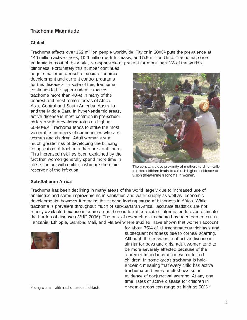
Trachoma affects over 162 million people worldwide. Taylor in 20081 puts the prevalence at146 million active cases, 10.6 million with trichiasis, and 5.9 million blind. Trachoma, onceendemic in most of the world, is responsible at present for more than 3% of the world’sblindness. Fortunately this number continuesto get smaller as a result of socio-economicdevelopment and current control programs for this disease.2 In spite of this, trachomacontinues to be hyper-endemic (activetrachoma more than 40%) in many of thepoorest and most remote areas of Africa,Asia, Central and South America, Australiaand the Middle East. In hyper-endemic areas,active disease is most common in pre-schoolchildren with prevalence rates as high as 60-90%.2 Trachoma tends to strike the mostvulnerable members of communities who arewomen and children. Adult women are atmuch greater risk of developing the blindingcomplication of trachoma than are adult men.This increased risk has been explained by thefact that women generally spend more time inclose contact with children who are the mainreservoir of the infection.
The constant close proximity of mothers to chronicallyinfected children leads to a much higher incidence ofvision threatening trachoma in women.
for about 75% of all trachomatous trichiasis andsubsequent blindness due to corneal scarring.Although the prevalence of active disease issimilar for boys and girls, adult women tend tobe more severely affected because of theaforementioned interaction with infectedchildren. In some areas trachoma is holo-endemic meaning that every child has activetrachoma and every adult shows someevidence of conjunctival scarring. At any onetime, rates of active disease for children inendemic areas can range as high as 50%.3
Sub-Saharan Africa
Trachoma has been declining in many areas of the world largely due to increased use ofantibiotics and some improvements in sanitation and water supply as well as economicdevelopments; however it remains the second leading cause of blindness in Africa. Whiletrachoma is prevalent throughout much of sub-Saharan Africa, accurate statistics are notreadily available because in some areas there is too little reliable information to even estimatethe burden of disease (WHO 2006). The bulk of research on trachoma has been carried out inTanzania, Ethiopia, Gambia, Mali, and Malawi where studies have shown that women account
Young woman with trachomatous trichiasis
4
Prevalence of trachomatous trichiasis (TT), a chronic form of trachoma, is estimated to be3.1% for Ethiopia. This means there are an estimated 1.3 million people age 15 and above whoare at risk of blindness unless treated urgently.
The Southern Nation Nationality Peoples Region (SNNPR), where an Orbis Rural Program is located, is the third most populated (~ 15 million) region in Ethiopia among the 9 regional states and two city administrations. It is located in the southern part of the country and has more than 45 different ethnic groups. The SNNPR has a prevalence of trachomatous trichiasis (TT) of 2%, which is one of the highest in the country.4
Trachoma patients waiting for examination at a clinic
Ethiopia
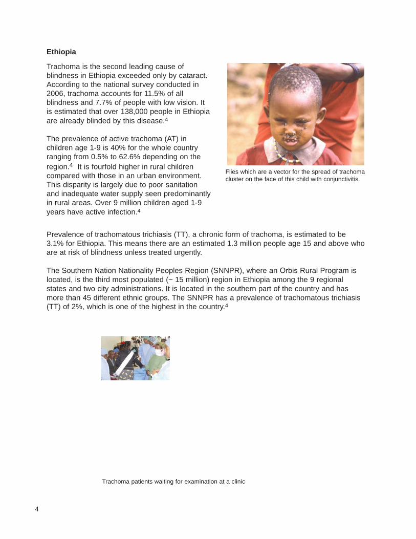
Trachoma is the second leading cause ofblindness in Ethiopia exceeded only by cataract.According to the national survey conducted in2006, trachoma accounts for 11.5% of allblindness and 7.7% of people with low vision. Itis estimated that over 138,000 people in Ethiopiaare already blinded by this disease.4
The prevalence of active trachoma (AT) inchildren age 1-9 is 40% for the whole countryranging from 0.5% to 62.6% depending on theregion.4 It is fourfold higher in rural childrencompared with those in an urban environment.This disparity is largely due to poor sanitationand inadequate water supply seen predominantlyin rural areas. Over 9 million children aged 1-9years have active infection.4
Flies which are a vector for the spread of trachomacluster on the face of this child with conjunctivitis.
5
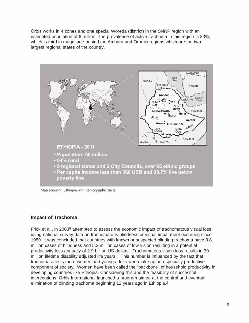
Orbis works in 4 zones and one special Woreda (district) in the SNNP region with an estimated population of 6 million. The prevalence of active trachoma in this region is 33%, which is third in magnitude behind the Amhara and Oromia regions which are the two largest regional states of the country.
Impact of Trachoma
Frick et al., in 20035 attempted to assess the economic impact of trachomatous visual lossusing national survey data on trachomatous blindness or visual impairment occurring since1980. It was concluded that countries with known or suspected blinding trachoma have 3.8million cases of blindness and 5.3 million cases of low vision resulting in a potential productivity loss annually of 2.9 billion US dollars. Trachomatous vision loss results in 39 million lifetime disability adjusted life years. This number is influenced by the fact that trachoma affects more women and young adults who make up an especially productive component of society. Women have been called the “backbone” of household productivity in developing countries like Ethiopia. Considering this and the feasibility of successful interventions, Orbis International launched a program aimed at the control and eventual elimination of blinding trachoma beginning 12 years ago in Ethiopia.2
Map showing Ethiopia with demographic facts
6
Orbis Involvement in Trachoma Control in Ethiopia
The Orbis Ethiopia country office was officially registered in 1999. Between 2000 and 2001, a comprehensive assessment of the extent of blindness and low vision, including the prevalence of trachoma, was conducted in the Gurage zone to learn more about the magnitude of eye health problems from trachoma. Training for nurses enabling them to perform trichiasis surgery was initiated in 2000 and a study of the outcome of surgeries done by these trained nurses known as Integrated Eye Care Workers (IECWs) compared with that done by ophthalmologists was conducted. It was determined that results of surgery done by these two groups were comparable.6
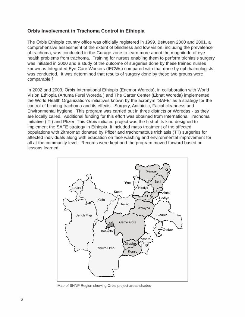
In 2002 and 2003, Orbis International Ethiopia (Enemor Woreda), in collaboration with World Vision Ethiopia (Artuma Fursi Woreda ) and The Carter Center (Ebnat Woreda) implemented the World Health Organization’s initiatives known by the acronym “SAFE” as a strategy for the control of blinding trachoma and its effects: Surgery, Antibiotic, Facial cleanness and Environmental hygiene. This program was carried out in three districts or Woredas - as they are locally called. Additional funding for this effort was obtained from International Trachoma Initiative (ITI) and Pfizer. This Orbis initiated project was the first of its kind designed to implement the SAFE strategy in Ethiopia. It included mass treatment of the affected populations with Zithromax donated by Pfizer and trachomatous trichiasis (TT) surgeries for affected individuals along with education on face washing and environmental improvement for all at the community level. Records were kept and the program moved forward based on lessons learned.
Map of SNNP Region showing Orbis project areas shaded
7
Trachoma Control Project Objectives
The project objectives were aimed at addressing each of the four components of SAFE asfollows:
Surgery
u Train nurses to perform trichiasis surgery and carry out primary eye care.u Equip and supply health centers to deliver trichiasis surgery and primary eye care
service to reduce backlog of trichiasis through both static and outreach services.u Determine the recurrence of TT after surgery with and without Zithromax treatment at
time of surgery. u Develop and standardize training and certification of trichiasis surgeons.
Antibiotics
u Reduce prevalence of active trachoma through antibiotic (Zithromax) treatment aimedat reducing the pool of infection and interrupting transmission, as well as providingtreatment of active infections through mass distribution at health facilities.
u Provide information and education activities to the population to create awareness onhow to prevent trachoma through the use of antibiotics.
u Carry out a variety of different studies on ways to improve the distribution of Zithromaxand to determine the effectiveness of this treatment.
Face Washing
u Increase the level of understanding and awareness about the value of face washingthrough education in schools and in the community by producing and distributingeducational materials.
u Upgrade local water sources to enable a safe and adequate water supply.u Collaborate with other NGOs working on water and sanitation improvement.
Environmental Improvement
u Raise awareness to increase utilization of appropriate sanitary methods for the humanand animal waste and household rubbish.
u Construct model household latrines to be used in the community.u Construct school and communal latrines in selected areas.
Strategies Employed by Orbis to Control Trachoma
Orbis has employed the WHO endorsed SAFE strategy for control of trachoma as described above in the belief that reducing one or more of the factors in the transmission of the bacteria that causes trachoma, Chlamydia�Trachomatis,�will reduce the prevalence of blindness from trachoma.
It would be best to simply prevent trachoma by encouraging better personal hygiene, improvedenvironmental cleanliness, and by controlling the flies that serve as vectors of transmission.However, once trachoma is prevalent and is considered a public health problem, it is necessaryto apply all�four�components of the SAFE strategy for control.
8
Facial Cleanliness
The third component is facial cleanliness. This is an important part of trachoma controlstrategy. It is aimed primarily at eliminating transmission of the infecting agent from uncleanfaces by flies, and as a means of maintaining individuals in a trachoma free state after initialtreatment is received through MDA. This is a key component of trachoma control in that itreduces the rate of re-infection with trachoma.
Environmental Improvement
The fourth component is environmental improvement. This is an important strategy forcontrolling flies which transmit the infecting agent for trachoma and which flourish in an uncleanenvironment. Breeding of flies can be reduced by proper disposal of refuse through burying orburning and of human and animal waste by using latrines and other appropriate methods thateliminate breeding places for flies. Supplying abundant clean water is also a goal.
Implementation of the SAFE Strategy
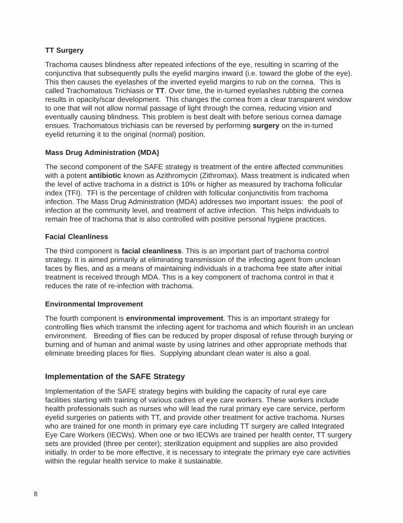
Implementation of the SAFE strategy begins with building the capacity of rural eye carefacilities starting with training of various cadres of eye care workers. These workers includehealth professionals such as nurses who will lead the rural primary eye care service, performeyelid surgeries on patients with TT, and provide other treatment for active trachoma. Nurseswho are trained for one month in primary eye care including TT surgery are called IntegratedEye Care Workers (IECWs). When one or two IECWs are trained per health center, TT surgerysets are provided (three per center); sterilization equipment and supplies are also providedinitially. In order to be more effective, it is necessary to integrate the primary eye care activitieswithin the regular health service to make it sustainable.
TT Surgery
Trachoma causes blindness after repeated infections of the eye, resulting in scarring of theconjunctiva that subsequently pulls the eyelid margins inward (i.e. toward the globe of the eye).This then causes the eyelashes of the inverted eyelid margins to rub on the cornea. This iscalled Trachomatous Trichiasis or TT. Over time, the in-turned eyelashes rubbing the cornearesults in opacity/scar development. This changes the cornea from a clear transparent windowto one that will not allow normal passage of light through the cornea, reducing vision andeventually causing blindness. This problem is best dealt with before serious cornea damageensues. Trachomatous trichiasis can be reversed by performing surgery on the in-turnedeyelid returning it to the original (normal) position.
Mass Drug Administration (MDA)
The second component of the SAFE strategy is treatment of the entire affected communitieswith a potent antibiotic known as Azithromycin (Zithromax). Mass treatment is indicated whenthe level of active trachoma in a district is 10% or higher as measured by trachoma follicularindex (TFI). TFI is the percentage of children with follicular conjunctivitis from trachomainfection. The Mass Drug Administration (MDA) addresses two important issues: the pool ofinfection at the community level, and treatment of active infection. This helps individuals toremain free of trachoma that is also controlled with positive personal hygiene practices.
9
Teachers in primary schools are trained to inform their students about the prevention oftrachoma through personal and environmental hygiene. Community level eye care personnelsuch as Health Extension Workers (HEWs) and volunteers such as Community Health Agents(CHAs) and Women Group Leaders (WGLs) are trained to teach community members abouttrachoma and tell them about preventive measures such as face washing and environmentalcleanliness. These community eye care workers also identify and refer patients to Primary EyeCare Units (PECUs) where they can receive medical and surgical treatment for trachoma asneeded.
All cadres mentioned above should receive close support including annual refresher training tokeep them motivated and to help them stay current with methods to provide quality eye care. Itis also important to produce targeted and effective key messages that these community eyecare workers can use to educate community members.
As part of the overall support to communities to practice the whole SAFE package, it is necessary to address the overall problems of sanitation, something outside of the usual expertise of a blindness prevention organization such as Orbis. This can be addressed by collaboration with other agencies who deal with clean water and sanitation utilizing their expertise and resources. Success in the eradication of blinding trachoma will require a team effort.
Challenges of Trachoma Prevention in Orbis Project Areas
u Lack of awareness of eye problems associated with trachoma on the part of thepopulation and the decision makersA large proportion of the population is unaware that trachoma is a “water washed”disease, and one that can be prevented by personal hygiene including face washingwith soap every day. Many people are not aware that trachomatous trichiasis (TT) canbe treated and blindness prevented in some cases through surgical interventions.There is a need for local decision makers to acknowledge that lack of eye care is asignificant health problem.
u Lack of suitable supplies of clean water and of adequate sanitationIn addition to the lack of overall awareness, clean water and adequate sanitationfacilities are lacking in many areas, especially rural villages. This problem isexacerbated by a lack of coordination between responsible agencies.
u Limited availability of eye care servicesThe type and volume of services that are currently available are inadequate. This ismainly due to scarcity of trained eye care professionals, lack of infrastructure andsupplies, and insufficient allocation of budget for eye care.
u AffordabilityThe majority of the population affected with trachoma is rural and lives in a lowsocioeconomic status. Households for the most part depend on subsistence farming.Even if services are available, surgery or medical treatment is unaffordable not onlydue to direct cost, but also indirect costs associated with transportation,accommodation, and food for most patients. It is especially difficult to obtain serviceswhen travel is required.
10
u Scarcity and high turnover of trained eye care professionals in rural areasThere is a scarcity of eye care professional in rural areas. Part of this is due to attritionof IECWs resulting from the lack of a career pathway for eye care providers that wouldattract them to the job and make them remain in a given location.
u Limited ownership of programs by the governmentThere is poor resource allocation on the part of the government particularly for humanresources. This contributes to inadequate monitoring and evaluation and allocation offunds to cover operational costs for eye care services.
The Way Forward
1. Training of more eye care professionals including volunteers: Ensure availability of atleast two IECWs (or a sufficient number according to population) to provide primary eye careservices in each of the existing health centers in all project areas. In addition, collaborate withall stakeholders while continuing to explore ways in which primary eye care is included in thetraining for health professionals with an emphasis on prevention. Train every new graduatehealth professional to be skilled in providing basic eye care; this could ensure continuity ofservices. Emphasize recruitment of new IECWs for ophthalmic nurse training.
2. Strengthening ownership of the program by partners: This will ensure sustainability ofservices through integration of eye care into the general health services, increasing financialallocation, and cost recovery.
3. Establishing linkage between rural eye care units with higher level eye centers:Establish linkage of Primary Eye Care Units (PECUs) with Secondary Eye Care Units (SECUs)for technical support, referral, and backup for the PECUs. The universities should provide back-up support to Secondary Eye Care Unit activities in all areas of eye health care.
4. Identify and collaborate with organizations working in the area of water andsanitation in the project areas: Strive to create a common understanding among allstakeholders on their roles for ensuring availability of clean water and sanitation for preventingeye disease.
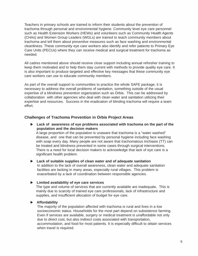
Training eye care workers Performing TT surgery
11
Part II: Trachomatous Trichiasis (TT) Surgery
Trachomatous trichiasis (TT) is the chronic sequel of active trachoma. Epilation (removal oflashes) and surgery to evert the lid thereby redirecting the lashes away from the cornea, aretwo methods for treatment of trachomatous trichiasis (TT). Epilation is often performed at thecommunity level and is usually done by patients themselves. However after simply pulling outthe lashes, the new lashes that replace those that are pulled out are often sharp and moredamaging to the cornea than the original in-turned or the remaining non-epilated lashes.Although entropion can recur in some cases after surgery, this is currently the method of choicefor treating TT. The two most widely practiced surgical procedures are: bilamellar tarsal rotation(BTR) and tarsal plate rotation (TPR/tarsatomy).
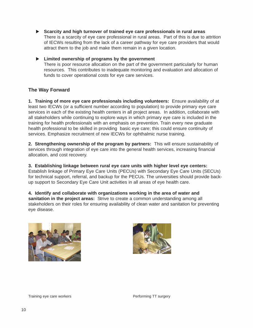
BTR surgery is the surgical procedure recommended by the WHO for trachomatous trichiasis. This surgery is being done by eye care professionals in many parts of the world. Recently a new design for the instrument used to perform BTR surgery has been introduced. It includes a plate to protect the cornea and provide a surface for a clamp that controls bleeding and which can have design features that marks the incision site leading to more consistency. One such instrument was introduced by Dr. Keith Waddell and is manufactured by Collton in the UK. Orbis International is currently using this new clamp at tertiary eye centers and at Orbis rural field facilities. In the last year, ten IECWs and two ophthalmologists used the clamp for surgery done on more than 100 patients. There were no recurrences during the specified follow up period. The feedback from surgeons using this new clamp has been uniform. Their experience compared with a free-hand technique originally recommended by the World Health Organization is summarized as follows:
Advantages of the new clamp:
u Surgery time is reduced.u The through and through lid incision is made in one step.u Less surgical equipment is needed.u Protection for the globe is provided.u Bleeding is reduced.u Surgeons have more confidence when performing the surgery.
Disadvantages:
u Need to have different sizes for different ageand size of palpabral fissure
u Prolonged clamp placement causingreduced blood flow
u Not available on the local marketu Cost
The surgeon inspecting the surgicalinstruments.
12
The Eye
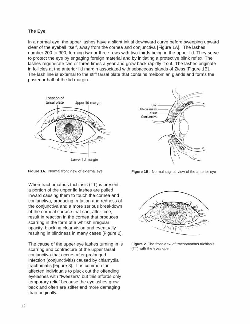
In a normal eye, the upper lashes have a slight initial downward curve before sweeping upwardclear of the eyeball itself, away from the cornea and conjunctiva [Figure 1A]. The lashesnumber 200 to 300, forming two or three rows with two-thirds being in the upper lid. They serveto protect the eye by engaging foreign material and by initiating a protective blink reflex. Thelashes regenerate two or three times a year and grow back rapidly if cut. The lashes originatein follicles at the anterior lid margin associated with sebaceous glands of Ziess [Figure 1B].The lash line is external to the stiff tarsal plate that contains meibomian glands and forms theposterior half of the lid margin.
Figure 1A. Normal front view of external eye Figure 1B. Normal sagittal view of the anterior eye
Figure 2. The front view of trachomatous trichiasis(TT) with the eyes open
When trachomatous trichiasis (TT) is present,a portion of the upper lid lashes are pulledinward causing them to touch the cornea andconjunctiva, producing irritation and redness ofthe conjunctiva and a more serious breakdownof the corneal surface that can, after time,result in reaction in the cornea that producesscarring in the form of a whitish irregularopacity, blocking clear vision and eventuallyresulting in blindness in many cases [Figure 2].
The cause of the upper eye lashes turning in isscarring and contracture of the upper tarsalconjunctiva that occurs after prolongedinfection (conjunctivitis) caused by chlamydiatrachomatis [Figure 3]. It is common foraffected individuals to pluck out the offendingeyelashes with “tweezers” but this affords onlytemporary relief because the eyelashes growback and often are stiffer and more damagingthan originally.
13
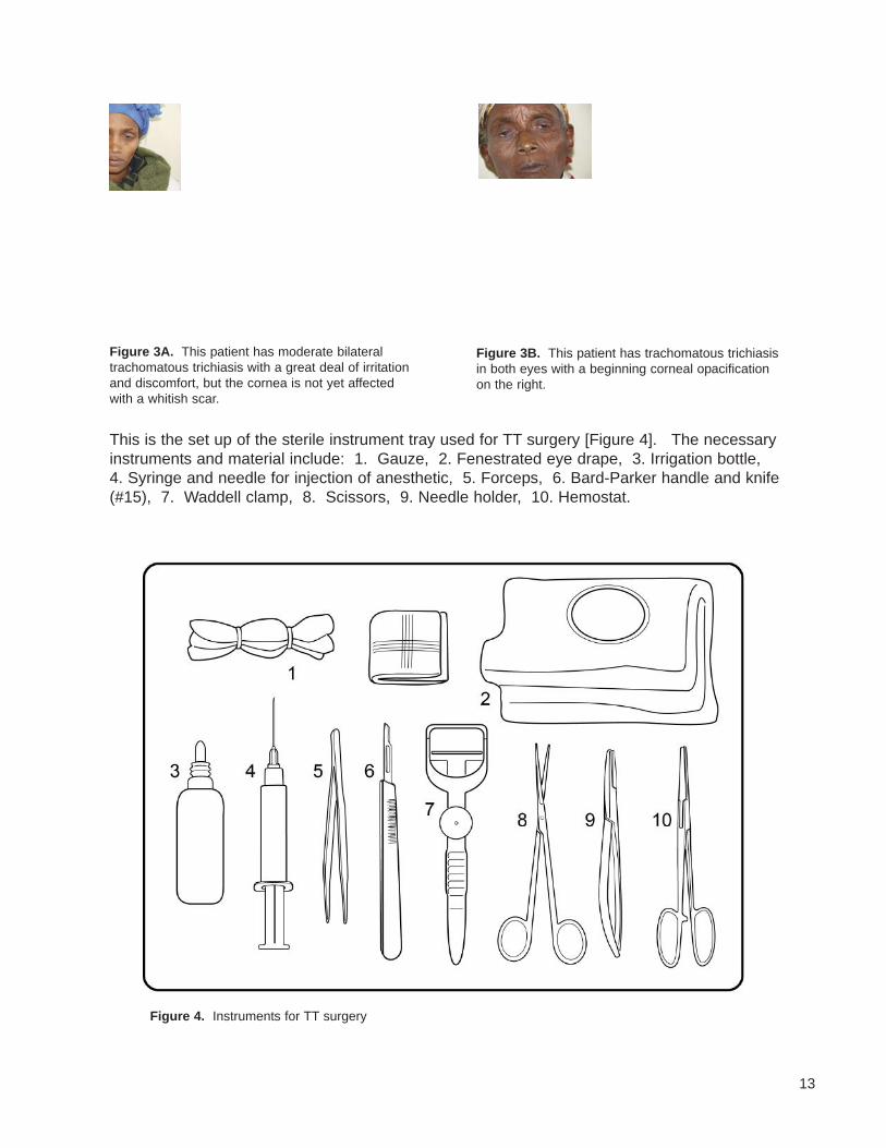
This is the set up of the sterile instrument tray used for TT surgery [Figure 4]. The necessaryinstruments and material include: 1. Gauze, 2. Fenestrated eye drape, 3. Irrigation bottle, 4. Syringe and needle for injection of anesthetic, 5. Forceps, 6. Bard-Parker handle and knife(#15), 7. Waddell clamp, 8. Scissors, 9. Needle holder, 10. Hemostat.
Figure 3B. This patient has trachomatous trichiasisin both eyes with a beginning corneal opacificationon the right.
Figure 3A. This patient has moderate bilateraltrachomatous trichiasis with a great deal of irritationand discomfort, but the cornea is not yet affectedwith a whitish scar.
Figure 4. Instruments for TT surgery
14
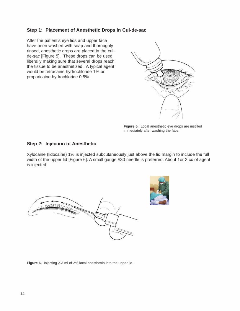
Step 2: Injection of Anesthetic
Xylocaine (lidocaine) 1% is injected subcutaneously just above the lid margin to include the fullwidth of the upper lid [Figure 6]. A small gauge #30 needle is preferred. About 1or 2 cc of agentis injected.
Step 1: Placement of Anesthetic Drops in Cul-de-sac
Figure 5. Local anesthetic eye drops are instilledimmediately after washing the face.
Figure 6. Injecting 2-3 ml of 2% local anesthesia into the upper lid.
After the patient’s eye lids and upper facehave been washed with soap and thoroughlyrinsed, anesthetic drops are placed in the cul-de-sac [Figure 5]. These drops can be usedliberally making sure that several drops reachthe tissue to be anesthetized. A typical agentwould be tetracaine hydrochloride 1% orproparicaine hydrochloride 0.5%.
15
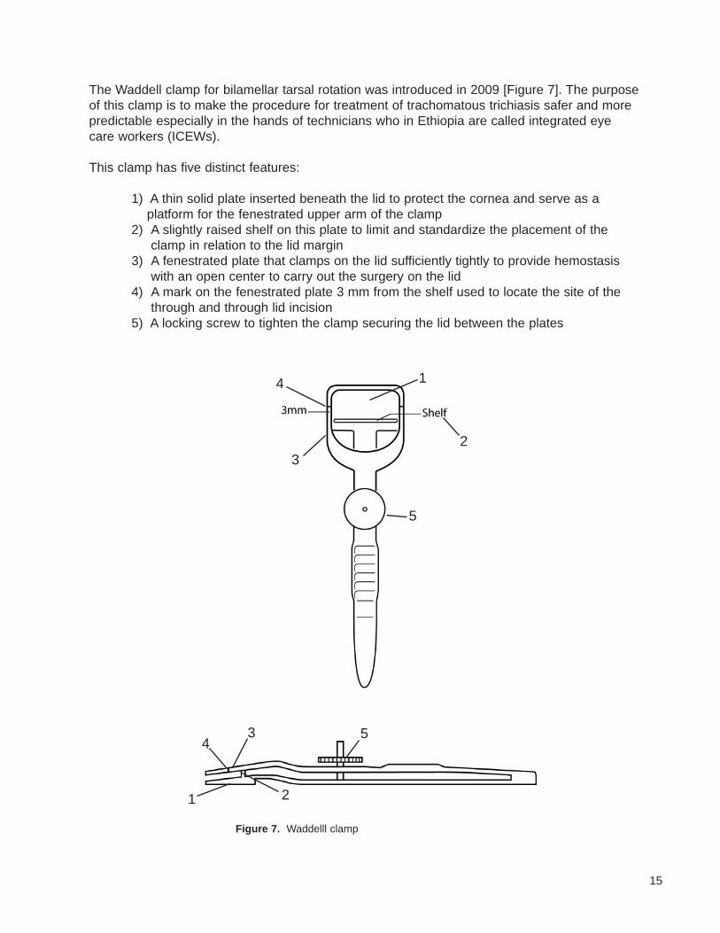
The Waddell clamp for bilamellar tarsal rotation was introduced in 2009 [Figure 7]. The purposeof this clamp is to make the procedure for treatment of trachomatous trichiasis safer and morepredictable especially in the hands of technicians who in Ethiopia are called integrated eyecare workers (ICEWs).
This clamp has five distinct features:
1) A thin solid plate inserted beneath the lid to protect the cornea and serve as aplatform for the fenestrated upper arm of the clamp
2) A slightly raised shelf on this plate to limit and standardize the placement of theclamp in relation to the lid margin
3) A fenestrated plate that clamps on the lid sufficiently tightly to provide hemostasiswith an open center to carry out the surgery on the lid
4) A mark on the fenestrated plate 3 mm from the shelf used to locate the site of thethrough and through lid incision
5) A locking screw to tighten the clamp securing the lid between the plates
Figure 7. Waddelll clamp
1
2
3
3
4
4
5
5
21
16
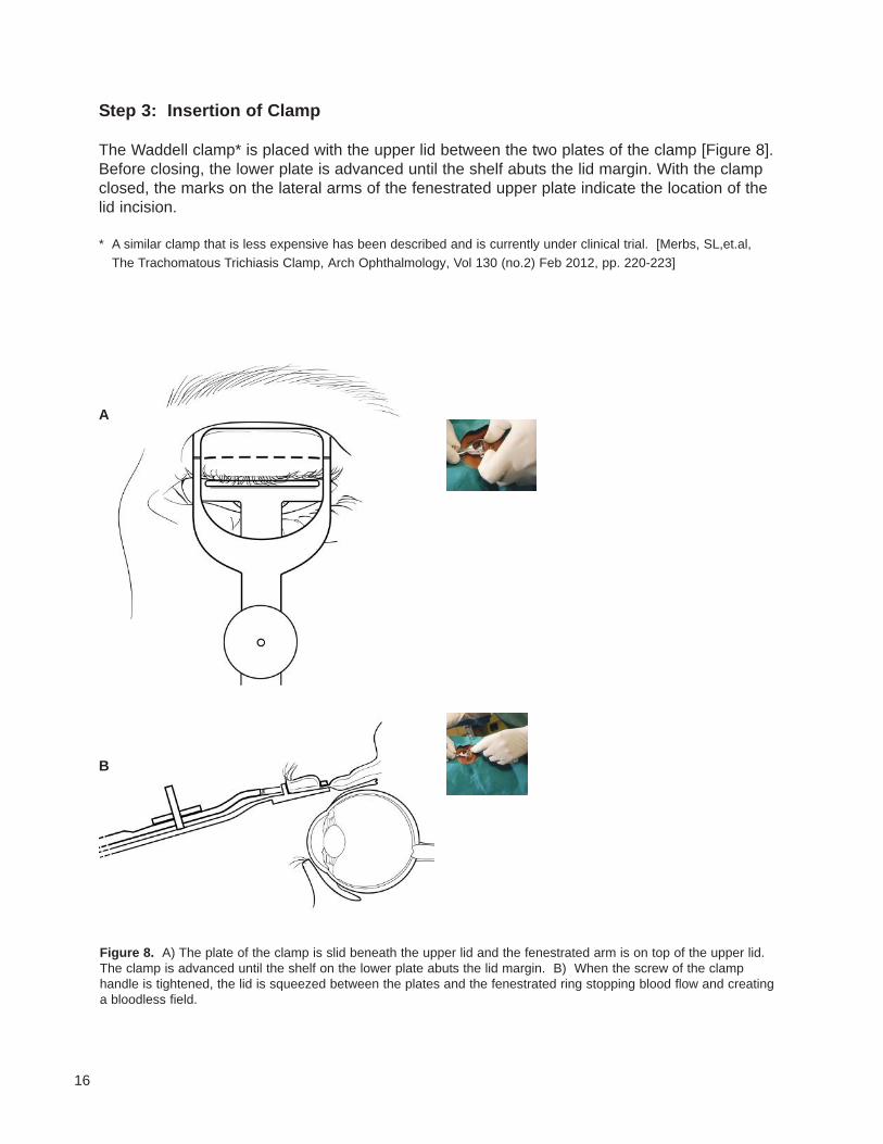
Step 3: Insertion of Clamp
The Waddell clamp* is placed with the upper lid between the two plates of the clamp [Figure 8].Before closing, the lower plate is advanced until the shelf abuts the lid margin. With the clampclosed, the marks on the lateral arms of the fenestrated upper plate indicate the location of thelid incision.
* A similar clamp that is less expensive has been described and is currently under clinical trial. [Merbs, SL,et.al, The Trachomatous Trichiasis Clamp, Arch�Ophthalmology, Vol 130 (no.2) Feb 2012, pp. 220-223]
Figure 8. A) The plate of the clamp is slid beneath the upper lid and the fenestrated arm is on top of the upper lid.The clamp is advanced until the shelf on the lower plate abuts the lid margin. B) When the screw of the clamphandle is tightened, the lid is squeezed between the plates and the fenestrated ring stopping blood flow and creatinga bloodless field.
A
B
17
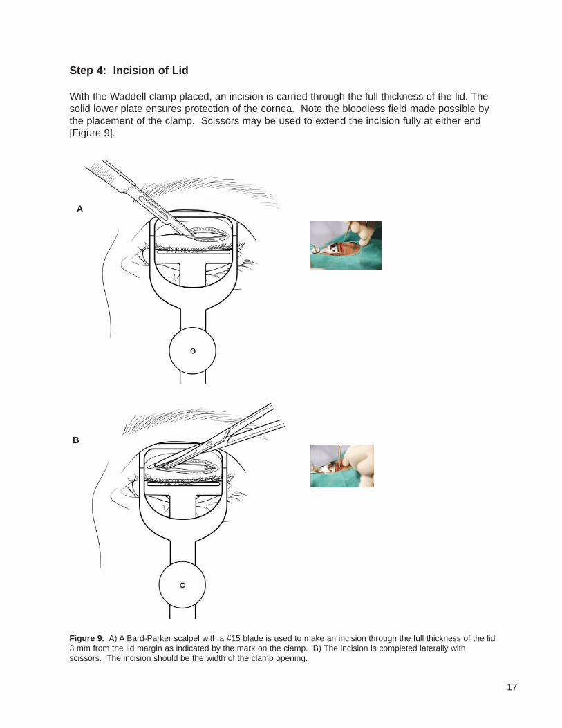
Step 4: Incision of Lid
With the Waddell clamp placed, an incision is carried through the full thickness of the lid. Thesolid lower plate ensures protection of the cornea. Note the bloodless field made possible bythe placement of the clamp. Scissors may be used to extend the incision fully at either end[Figure 9].
Figure 9. A) A Bard-Parker scalpel with a #15 blade is used to make an incision through the full thickness of the lid3 mm from the lid margin as indicated by the mark on the clamp. B) The incision is completed laterally withscissors. The incision should be the width of the clamp opening.
A
B
18
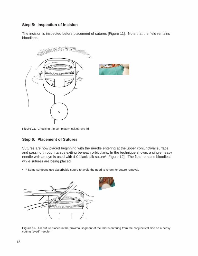
Step 5: Inspection of Incision
The incision is inspected before placement of sutures [Figure 11]. Note that the field remainsbloodless.
Figure 11. Checking the completely incised eye lid
Step 6: Placement of Sutures
Sutures are now placed beginning with the needle entering at the upper conjunctival surfaceand passing through tarsus exiting beneath orbicularis. In the technique shown, a single heavyneedle with an eye is used with 4-0 black silk suture* [Figure 12]. The field remains bloodlesswhile sutures are being placed.
• * Some surgeons use absorbable suture to avoid the need to return for suture removal.
Figure 12. 4-0 suture placed in the proximal segment of the tarsus entering from the conjunctival side on a heavycutting “eyed” needle.
19
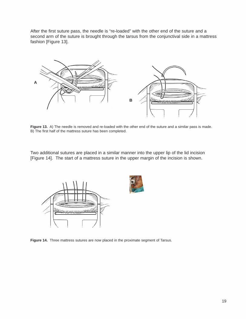
After the first suture pass, the needle is “re-loaded” with the other end of the suture and asecond arm of the suture is brought through the tarsus from the conjunctival side in a mattressfashion [Figure 13].
Figure 13. A) The needle is removed and re-loaded with the other end of the suture and a similar pass is made.B) The first half of the mattress suture has been completed.
A
B
Two additional sutures are placed in a similar manner into the upper lip of the lid incision[Figure 14]. The start of a mattress suture in the upper margin of the incision is shown.
Figure 14. Three mattress sutures are now placed in the proximate segment of Tarsus.
20
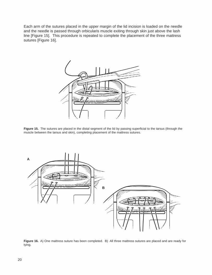
Each arm of the sutures placed in the upper margin of the lid incision is loaded on the needleand the needle is passed through orbicularis muscle exiting through skin just above the lashline [Figure 15]. This procedure is repeated to complete the placement of the three mattresssutures [Figure 16].
Figure 15. The sutures are placed in the distal segment of the lid by passing superficial to the tarsus (through themuscle between the tarsus and skin), completing placement of the mattress sutures.
Figure 16. A) One mattress suture has been completed. B) All three mattress sutures are placed and are ready fortying.
A
B
21
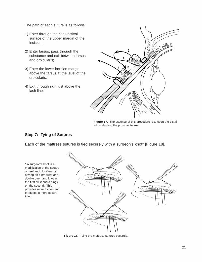
The path of each suture is as follows:
1) Enter through the conjunctivalsurface of the upper margin of theincision;
2) Enter tarsus, pass through thesubstance and exit between tarsusand orbicularis;
3) Enter the lower incision marginabove the tarsus at the level of theorbicularis;
4) Exit through skin just above thelash line.
Figure 17. The essence of this procedure is to evert the distallid by abutting the proximal tarsus.
Step 7: Tying of Sutures
Each of the mattress sutures is tied securely with a surgeon’s knot* [Figure 18].
Figure 18. Tying the mattress sutures securely.
* A surgeon’s knot is amodification of the squareor reef knot. It differs byhaving an extra twist or adouble overhand knot inthe first twist and a singleon the second. Thisprovides more friction andproduces a more secureknot.
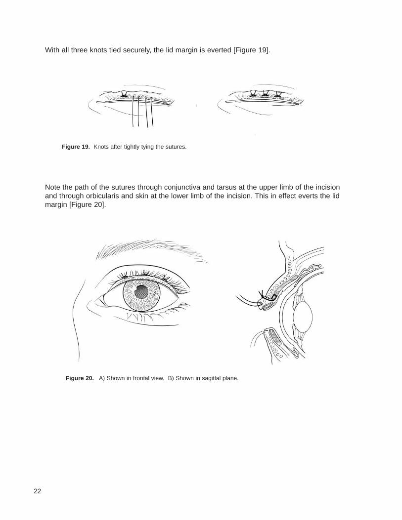
Figure 20. A) Shown in frontal view. B) Shown in sagittal plane.
Figure 19. Knots after tightly tying the sutures.
22
With all three knots tied securely, the lid margin is everted [Figure 19].
Note the path of the sutures through conjunctiva and tarsus at the upper limb of the incisionand through orbicularis and skin at the lower limb of the incision. This in effect everts the lidmargin [Figure 20].
23
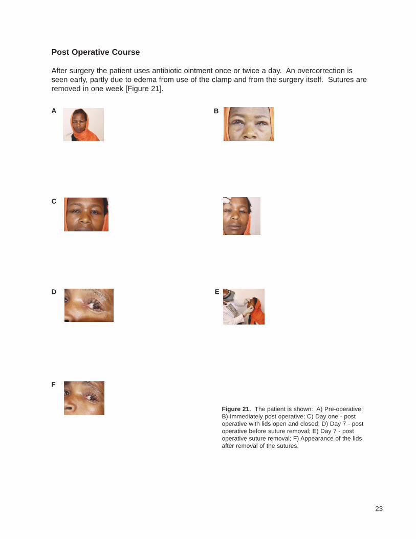
Post Operative Course
After surgery the patient uses antibiotic ointment once or twice a day. An overcorrection isseen early, partly due to edema from use of the clamp and from the surgery itself. Sutures areremoved in one week [Figure 21].
Figure 21. The patient is shown: A) Pre-operative;B) Immediately post operative; C) Day one - postoperative with lids open and closed; D) Day 7 - postoperative before suture removal; E) Day 7 - postoperative suture removal; F) Appearance of the lidsafter removal of the sutures.
A B
C
D E
F
24
References
1. Taylor HR. Trachoma: A Blinding Scourge from the Bronze Age to the Twenty First Century,Victoria, Australia: Haddington Press, 2008.
2. World Health Organization (WHO). Prevention of Blindness and Visual Impairment:Trachoma. Available at: http://www.who.int/blindness/causes/trachoma/en. Accessed July24, 2012.
3. Lewallen S, Courtright P. Blindness in Africa: present situation and future needs. Br.�J.Ophthalmology 2001; 85:897-903
4. Berhane Y, et al. National survey on blindness, low vision and trachoma in Ethiopia:Methods and study clusters profile (2006). Ethiop.�J.�Health�Dev.� 2007; 21(3):185-203.
5. Frick KD, Hanson CL, Lacobson GA. Global burden of trachoma and economics of thedisease, Am�J�Trop�Med�Hyg 2003; 69:(5 suppl):1-10.
6. Alemayehu W, et al. Surgery for trichiasis by ophthalmologists versus integrated eye careworkers: a randomized trial. Ophthalmology�2004; 111:578-84.
Conclusion
Orbis trained integrated eye care workers (IECWs) who had been using the “free hand” technique for trachomatous trichiasis (TT) surgery as recommended by the World Health Organization (WHO) were provided instruction in the use of the new Waddell clamp. After successfully completing the training, these workers and the ophthalmologist who trained them performed TT surgery on more than 100 patients in the course of a year. After this experience the ICEWs reported the following: 1) surgery time was reduced; 2) there was less bleeding; 3) the operators had more confidence; 4) the surgery was conducted in a more controlled andstandardized manner; and 5) the chance of harming the cornea was eliminated because ofprotection provided by the clamp.
This experience in Ethiopia suggests that a technique for TT surgery that provides protectionfor the cornea, creates a bloodless field, and offers a way to standardize the procedure shouldbe considered by anyone doing this type of surgery. The Waddell clamp which utilizes theprinciple that provides all of these advantages is one such clamp. Another clamp designed byMerbs that is lower cost and comes in different sizes is now undergoing evaluation.
This manual is not intended to endorse a specific clamp but instead to alert those who will bedoing this type of surgery to consider using some type of clamp that offers the advantagesdescribed here.


Copyright 2014


![Population-based prevalence survey of follicular trachoma ......inwards and scratch the cornea (trachomatous trichiasis, TT), leading to corneal opacity and blindness [2]. The World](https://img.pdfslide.us/doc/110x75/606ee2729bde5e77a30c4adf/population-based-prevalence-survey-of-follicular-trachoma-inwards-and-scratch.jpg)