Embed Size (px)
Citation preview
C H A P T E R
6
Toxoplasmosis in wild and domesticanimals
David S. Lindsay1 and J.P. Dubey21Department of Biomedical Sciences and Pathobiology, Virginia Maryland College of Veterinary
Medicine, Virginia Tech, Blacksburg, VA, United States 2Animal Parasitic Diseases Laboratory, United
States Department of Agriculture, Agricultural Research Service, Beltsville, MD, United States
6.1 Introduction
Toxoplasma gondii is widely distributed inwild and domestic animals. The present chapterreviews toxoplasmosis in wild and domestic ani-mals. Coverage in wild animal species is limitedto confirmed cases of toxoplasmosis, cases withparasite isolation, cases with parasite detectionby polymerase chain reaction (PCR), and experi-mental infection studies (Figs. 6.1�6.3). Studiesconcerning serological prevalence have not beenincluded for the majority of host species. Thiswas done because many serological tests, e.g.latex agglutination (LAT), indirect fluorescentantibody (IFAT), and indirect hemagglutina-tion), have been demonstrated to underestimatethe prevalence of T. gondii.
6.2 Toxoplasmosis in wildlife
6.2.1 Felids
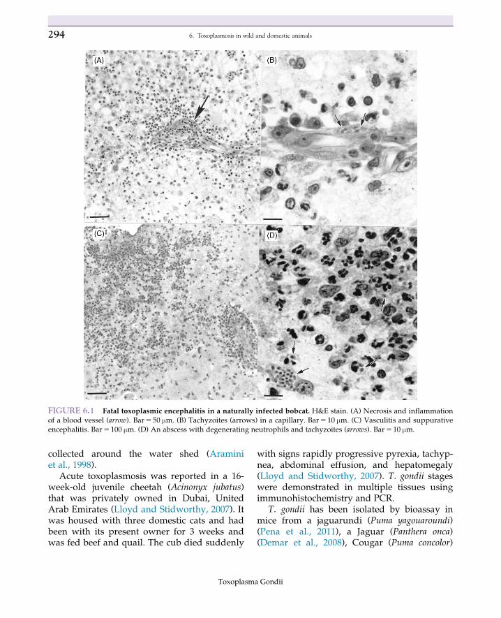
Congenital toxoplasmosis has been reportedin bobcats (Felis rufus) kits (Dubey et al., 1987).Toxoplasmic meningoencephalitis has beenobserved in a 6-month-old bobcat (Smith et al.,1995). T. gondii has been isolated from the tis-sues of adult bobcats (Lindsay et al., 1997b;Dubey et al., 2004b). Bobcats are important inmaintaining T. gondii in wild herbivores inmany areas of the United States (Fig. 6.1).Oocysts excreted by cougars (Felis concolor)were thought to be the source of a large waterborne outbreak of human toxoplasmosis inVictoria, British Columbia, Canada, andoocysts were isolated from the feces of cougars
293Toxoplasma Gondii
DOI: https://doi.org/10.1016/B978-0-12-815041-2.00006-2 © 2020 Elsevier Ltd. All rights reserved.
collected around the water shed (Araminiet al., 1998).
Acute toxoplasmosis was reported in a 16-week-old juvenile cheetah (Acinonyx jubatus)that was privately owned in Dubai, UnitedArab Emirates (Lloyd and Stidworthy, 2007). Itwas housed with three domestic cats and hadbeen with its present owner for 3 weeks andwas fed beef and quail. The cub died suddenly
with signs rapidly progressive pyrexia, tachyp-nea, abdominal effusion, and hepatomegaly(Lloyd and Stidworthy, 2007). T. gondii stageswere demonstrated in multiple tissues usingimmunohistochemistry and PCR.
T. gondii has been isolated by bioassay inmice from a jaguarundi (Puma yagouaroundi)(Pena et al., 2011), a Jaguar (Panthera onca)(Demar et al., 2008), Cougar (Puma concolor)
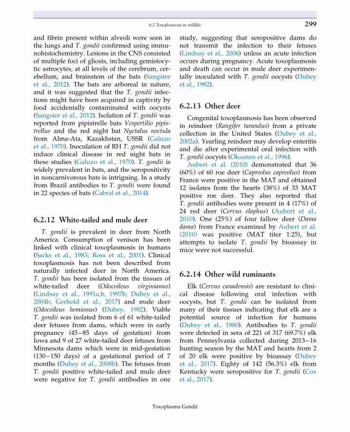
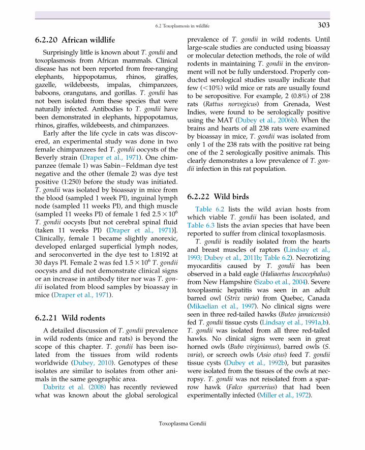
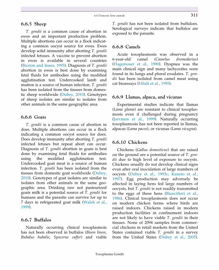
FIGURE 6.1 Fatal toxoplasmic encephalitis in a naturally infected bobcat. H&E stain. (A) Necrosis and inflammationof a blood vessel (arrow). Bar5 50 µm. (B) Tachyzoites (arrows) in a capillary. Bar5 10 µm. (C) Vasculitis and suppurativeencephalitis. Bar5 100 µm. (D) An abscess with degenerating neutrophils and tachyzoites (arrows). Bar5 10 µm.
Toxoplasma Gondii
294 6. Toxoplasmosis in wild and domestic animals
(Dubey et al., 2008a,b), and sand cat (Felis mar-garita) (Dubey et al., 2010). Experimental infec-tions resulting in oocyst excretion have beendemonstrated in jaguarundi (P. yagouaroundi),ocelot (Furcifer pardalis), bobcats (Lynx rufus),and cheetah (A. jubatus) (Jewell et al., 1972;Miller et al., 1972). In general, these felids arenot as efficient at producing oocysts as aredomestic cats. Congenital toxoplasmosis is amajor factor hindering breeding programs forendangered Pallas’s cats (Otocolobus manul)and sand cats (F. margarita) in zoos worldwide(see next).
6.2.2 Canids
Acute toxoplasmosis has been reported inarctic foxes (Alopex lagopus) (Sorensen et al.,2005), Fennec foxes (Fennecus zerda) (Kottwitzet al., 2004), gray foxes (Urocyon cinereoargen-teus) (Davidson et al., 1992; Dubey and Lin,1994; Kelly and Sleeman, 2003), red foxes
(Vulpes vulpes) (Reed and Turek, 1985; Dubeyet al., 1990; Kelly and Sleeman, 2003), and sandfoxes (Vulpes rueppellii) (Pas and Dubey 2008c).Coinfection with canine distemper virus isoften associated with clinical toxoplasmosis ingray (Davidson et al., 1992; Kelly and Sleeman,2003) and red foxes (Reed and Turek, 1985).Clinical toxoplasmosis has not been documen-ted in wolves, coyotes, hyenas, or dingos. T.gondii has been isolated from artic foxes (Dubeyet al., 2011b), red foxes (Smith and Frenkel,1995; Dubey et al., 2004b, 2011b), gray foxes(Dubey et al., 2004b), and coyotes (Lindsayet al., 1997b; Dubey et al., 2004b). Aubert et al.(2010) found modified agglutination test (MAT)antibodies in 14 of 19 (74%) red foxes fromFrance and isolated T. gondii from the hearts of9 (69%) of 13 seropositive red foxes. The iso-lates were all genotype Type II.
Herrmann et al. (2012) used serology (immu-noblot) and PCR to examine the prevalence ofT. gondii in red foxes and rodents from theGerman Federal States of Brandenburg and
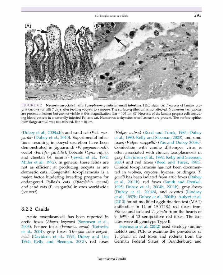
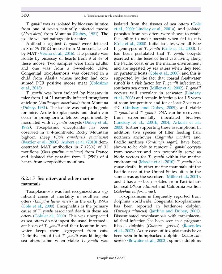
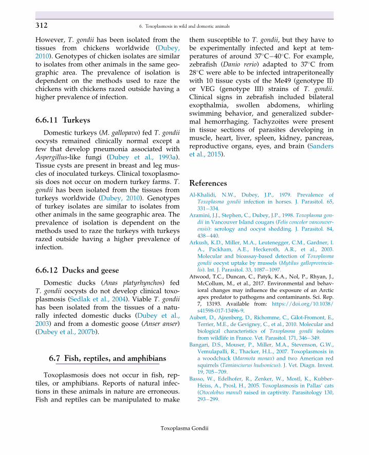
FIGURE 6.2 Necrosis associated with Toxoplasma gondii in small intestine. H&E stain. (A) Necrosis of lamina pro-pria (arrows) of villi 7 days after feeding oocysts to a mouse. The surface epithelium is not affected. Numerous tachyzoitesare present in lesions but are not visible at this magnification. Bar5 100 µm. (B) Necrosis of the lamina propria cells includ-ing blood vessels in a naturally infected Pallas’s cat. Numerous tachyzoites (small arrows) are present. The surface epithe-lium (large arrow) was not affected. Bar5 10 µm.
Toxoplasma Gondii
2956.2 Toxoplasmosis in wildlife
Saxony-Anhalt. They found 152/204 (74.5%) and149/176 (84.7%) of red foxes in Brandenburg andSaxony-Anhalt were immunoblot positive, respec-tively, but none of 72 rodents (69 common volesMicrotus arvalis, 2 Mediterranean water shrewsNeomys anomalus, and 1 striped field mouseApodemus agrarius) had antibodies to T. gondii.PCR was conducted on heart tissue from seropos-itive red fox tissues and 28/152 (18%) and 20/149(13%) of seropositive foxes from Brandenburgand Saxony-Anhalt, respectively, were positive(Herrmann et al., 2012). PCR was done on heartand lung samples from the 72 rodents and nonetested positive (Herrmann et al., 2012).
6.2.3 Bears
Clinical toxoplasmosis has not beenreported from bears. Viable T. gondii has beenisolated from black bears (Ursus americanus)(Dubey et al., 1995a) and brown bears (Ursusarctos horribilis) (Dubey et al., 2011b). The prev-alence of T. gondii in black bears in the UnitedStates is the highest of any hosts for T. gondiiworldwide. In a recent survey, T. gondii antibo-dies were found in 4% of dams and 5% of theirnursing cubs while in dens; the study con-cluded that there is no transplacental transmis-sion of T. gondii in bears and that 50% of bearsacquire infection postnatally by their 10months of age (Dubey et al., 2016). Comparedwith black bears, the prevalence of T. gondii inbrown bears from Alaska is about half (44%) ofblack bears (Ramey et al., 2019). The preva-lence of T. gondii in 527 polar bears (Ursus mari-timus) was 3.6% in cubs still with their damand 21.4% for subadults and adults fromSvalbard and the Barents Sea and EastGreenland (Oksanen et al., 2009) and 6% of 500polar bears from the Beaufort and Chukchi seaareas of the Arctic Ocean (Rah et al, 2005). Theprevalence in polar bears was 23.9% (33) of 105animals from southern Beaufort Sea (Atwoodet al., 2017) and in grizzly bears (U. arctos)(Chomel et al., 1995). Meat from any species ofbear should be considered a potential source ofT. gondii.
6.2.4 Raccoons
Many serosurveys indicate that T. gondii ishighly prevalent in raccoons (Procyon lotor)(reviewed by Hancock et al., 2005). Encysted T.gondii has been isolated from the tissues of nat-urally infected raccoons (Lindsay et al., 1997b;Dubey et al., 2004c, 2011b). Clinical toxoplas-mosis has not been reported from raccoons andthey are resistant to experimental infection(Dubey et al., 1993b).
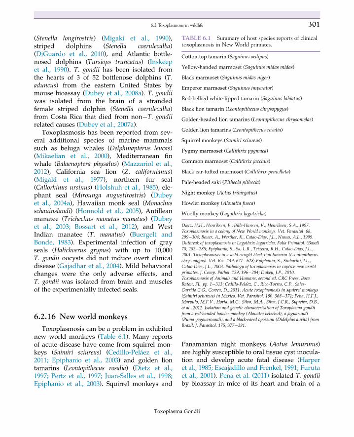
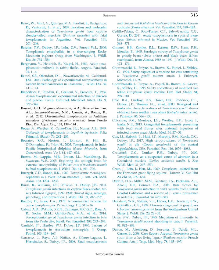
FIGURE 6.3 Section of liver from a gazelle with toxo-plasmosis showing a central area of hepatitis. NoteToxoplasma gondii (arrows) in hepatocytes at the peripheryof the lesion. H&E stain. Bar5 25 µm.
Toxoplasma Gondii
296 6. Toxoplasmosis in wild and domestic animals
6.2.5 Squirrels
Acute toxoplasmosis has been reported ingray squirrels (Sciurus carolinensis) (Dubey et al.,2006a), eastern fox squirrels (Sciurus niger)(Kumar et al., 2018), American red squirrels(Tamiasciurus hudsonicus) (Bangari et al., 2007),13-lined ground squirrels (Citellus tridecemlinea-tus) (van Pelt and Dieterich, 1972), Eurasian redsquirrels (Sciurus vulgaris) (Jokelainen andNylund, 2012), Swinhoe’s striped squirrel(Tamiops swinhoei) (Fayyad et al., 2016), andKorean squirrels (Tanias sibericus) (Carrascoet al., 2006). T. gondii has been isolated from graysquirrels (Smith and Frenkel, 1995) andFormosan giant flying squirrels (Petaurista petaur-ista grandis) (Cross et al., 1969).
6.2.6 Rabbits and hares
Fatal toxoplasmosis has been reported fromthree domestic (Oryctolagus cuniculus) rabbitsfrom two different sources in the United States(Dubey et al., 1992a). The rabbits died after anacute illness characterized by fever, lethargy,and diarrhea in one rabbit and no clinical signsin the other two rabbits. The most strikinglesion in all three rabbits was foci of necrosis ofthe spleen and liver associated with massivepresence of multiplying tachyzoites (Dubeyet al., 1992a). Similar findings were present in2�18-month-old domestic rabbits from 15flocks in Germany. Necropsy examinations of49 rabbits revealed lesions of a generalizedgranulomatous-necrotizing toxoplasmosiswithin the spleen, liver, lungs, and lymphnodes (Bergmann et al., 1980). Both authors ofthe current chapter (DSL and JPD) have inocu-lated domestic rabbits orally and subcutane-ously with T. gondii oocysts (usually 10,000/rabbit) to generate immune serum for immuno-histochemistry. All inoculated rabbits have orwould have developed fatal toxoplasmosis hadthey not been euthanized for humane reasons.Viable T. gondii has been isolated from
domestic rabbits (O. cuniculus) from Brazil(Dubey et al., 2011a).
Brown hares (Lepus europaeus) develop fataltoxoplasmosis after experimental infection withas few as 10 oocysts and all inoculated haresdied within 8�19 days after ingesting oocysts(Sedlak et al., 2000). The typical pathologicalfinding in hares is hemorrhagic enteritis, enlarge-ment and hyperemia of mesenteric lymph nodes,splenomegaly, and multiple necrotic lesions inthe parenchyma of the liver and other organs(Sedlak et al., 2000). Mountain hare (Lepus timi-dus) experimentally inoculated with 50 T. gondiioocysts and examined 7 days later had grosslesions in the mesenteric lymph nodes and liver(Gustafsson et al., 1997). Histologically, the hareshad extensive necrotic areas in the small intes-tine, mesenteric lymph nodes and liver, and lessprominent foci of necrosis in various otherorgans (Gustafsson et al., 1997). Recent retrospec-tive studies in Finland (Jokelainen et al., 2011)have documented natural toxoplasmosis in haressimilar to these experimental reports. Acute gen-eralized toxoplasmosis was demonstrated immu-nohistochemically, and T. gondii was confirmedas the cause of death in 14 (8%) of 173 Europeanbrown hares (L. europaeus) and 4 (3%) of 148mountain hares (L. timidus) from Finland(Jokelainen et al., 2011). Aubert et al. (2010) dem-onstrated that 3 (13%) of 23 European brownhares (L. europaeus) from France were positive inthe MAT but were not able to isolate T. gondiifrom the hearts of two seropositive animals.
6.2.7 Skunks and fisher
T. gondii genotype III was isolated from threeof six asymptomatic striped skunks (Mephitismephitis) from Mississippi (Dubey et al., 2004d).Two of the three isolated were mouse patho-genic even thought they were molecularly con-sistent with the mouse avirulent genotype III.Lesions of toxoplasmosis and T. gondii parasiteswere not observed at necropsy of 37 striped
Toxoplasma Gondii
2976.2 Toxoplasmosis in wildlife
skunks from Illinois (Gehrt et al., 2010). Thispopulation was serologically 60% positive forexposure to T. gondii (Gehrt et al., 2010).
T. gondii was detected by PCR from brainand skeletal muscle of a free-ranging juvenilefisher (Martes pennanti) from Maryland(Gerhold et al., 2005). Clinically this animalhad encephalitis, but it was not associated withthe T. gondii infection. T. gondii antibodies werefound using MAT in 100% of 38 and usingIFAT in 71% of 45 fisher from Pennsylvania(Larkin et al., 2011).
6.2.8 Beavers
T. gondii has been isolated from beaver(Castor canadensis) tissue (Dubey, 1983; Smithand Frenkel, 1995). Fatal systematic toxoplas-mosis was seen in a 5-month-old beaver thatwas in a rehabilitation center in Connecticut(Forzan and Frasca, 2004). Histologic lesionscontained T. gondii positive stages by immuno-histochemistry and consisted of lymphohistio-cytic encephalitis, myocarditis, and interstitialpneumonia with multinucleated cells (Forzanand Frasca, 2004).
6.2.9 Woodchuck and other largerodents
Central nervous system toxoplasmosis hasbeen observed in a woodchuck (Marmota mon-ax) (Bangari et al., 2007) from New York. Thewoodchuck was euthanized because of pro-gressive clinical signs of head tilt, circling, andrapid weight loss. The brain and heart werepositive for T. gondii by immunohistochemistryand PCR (Bangari et al., 2007).
Clinical toxoplasmosis has not beenreported in capybara (Hydrochaeris hydrochaeris)or nutria (Myocastor coypus). However, the par-asite has been isolated from capybara fromBrazil (Yai et al., 2009) and T. gondii DNA has
been detected by PCR in nutria from Italy(Nardoni et al., 2011).
6.2.10 Insectivores
Little is known about toxoplasmosis ininsectivores. The prevalence of T. gondii usingthe Sabin�Feldman dye test was ,1% in 578insectivores from the Czech Republic (Hejliceket al., 1997). Fatal toxoplasmosis was diagnosedin a juvenile male common mole (Talpa euro-paea) from Germany (Geisel et al., 1995). Noneof 70 T. europaea from the Netherlands wereserologically positive using the latex agglutina-tion test (LAT), but T. gondii DNA wasdetected by real-time PCR in the brain of twoof these common moles (Krijger et al., 2014).The brains and/or hearts from 3 of 22 white-toothed shrews (Crocidura russula) from organicpig farms in the Netherlands were positive forT. gondii by PCR (Kijlstra et al., 2008). Inanother study from organic pig farms from theNetherlands, none of the brains from 9 com-mon shrews (Sorex araneus) and 2 (2%) brainsfrom 102 white-toothed shrews (C. russula)were positive by PCR for T. gondii (Meerburget al., 2012). None of two Mediterranean watershrews (N. anomalus) from Germany were posi-tive by serology or PCR (Herrmann et al.,2012). T. gondii DNA was detected in the heartof 1 of 578 striped field mice (A. agrarius) fromNorth Korea (Hong et al., 2014).
6.2.11 Bats
Acute toxoplasmosis has been observed in ajuvenile spectacled flying-fox (Pteropus conspi-cillatus) and a juvenile little red flying fox(Pteropus scapulatus) from Australia (Sangsteret al., 2012). One was a captive born member ofa colony, and the other was undergoing reha-bilitation at a wildlife hospital. Severe, acuteinterstitial pneumonia with varying combina-tions of neutrophils, large foamy macrophages,
Toxoplasma Gondii
298 6. Toxoplasmosis in wild and domestic animals
and fibrin present within alveoli were seen inthe lungs and T. gondii confirmed using immu-nohistochemistry. Lesions in the CNS consistedof multiple foci of gliosis, including gemistocy-tic astrocytes, at all levels of the cerebrum, cer-ebellum, and brainstem of the bats (Sangsteret al., 2012). The bats are arboreal in nature,and it was suggested that the T. gondii infec-tions might have been acquired in captivity byfood accidentally contaminated with oocysts(Sangster et al., 2012). Isolation of T. gondii wasreported from pipistrelle bats Vespertilio pipis-trellus and the red night bat Nyctalus noctulafrom Alma-Ata, Kazakhstan, USSR (Galuzoet al., 1970). Inoculation of RH T. gondii did notinduce clinical disease in red night bats inthese studies (Galuzo et al., 1970). T. gondii iswidely prevalent in bats, and the seropositivityin noncarnivorous bats is intriguing. In a studyfrom Brazil antibodies to T. gondii were foundin 22 species of bats (Cabral et al., 2014).
6.2.12 White-tailed and mule deer
T. gondii is prevalent in deer from NorthAmerica. Consumption of venison has beenlinked with clinical toxoplasmosis in humans(Sacks et al., 1983; Ross et al., 2001). Clinicaltoxoplasmosis has not been described fromnaturally infected deer in North America.T. gondii has been isolated from the tissues ofwhite-tailed deer (Odocoileus virginianus)(Lindsay et al., 1991a,b, 1997b; Dubey et al.,2004b; Gerhold et al., 2017) and mule deer(Odocoileus hemionus) (Dubey, 1982). ViableT. gondii was isolated from 6 of 61 white-taileddeer fetuses from dams, which were in earlypregnancy (45�85 days of gestation) fromIowa and 9 of 27 white-tailed deer fetuses fromMinnesota dams which were in mid-gestation(130�150 days) of a gestational period of 7months (Dubey et al., 2008b). The fetuses fromT. gondii positive white-tailed and mule deerwere negative for T. gondii antibodies in one
study, suggesting that seropositive dams donot transmit the infection to their fetuses(Lindsay et al., 2006) unless an acute infectionoccurs during pregnancy. Acute toxoplasmosisand death can occur in mule deer experimen-tally inoculated with T. gondii oocysts (Dubeyet al., 1982).
6.2.13 Other deer
Congenital toxoplasmosis has been observedin reindeer (Rangifer tarandus) from a privatecollection in the United States (Dubey et al.,2002a). Yearling reindeer may develop enteritisand die after experimental oral infection withT. gondii oocysts (Oksanen et al., 1996).
Aubert et al. (2010) demonstrated that 36(60%) of 60 roe deer (Capreolus capreolus) fromFrance were positive in the MAT and obtained12 isolates from the hearts (38%) of 33 MATpositive roe deer. They also reported thatT. gondii antibodies were present in 4 (17%) of24 red deer (Cervus elaphus) (Aubert et al.,2010). One (25%) of four fallow deer (Damadama) from France examined by Aubert et al.(2010) was positive (MAT titer 1:25), butattempts to isolate T. gondii by bioassay inmice were not successful.
6.2.14 Other wild ruminants
Elk (Cervus canadensis) are resistant to clini-cal disease following oral infection withoocysts, but T. gondii can be isolated frommany of their tissues indicating that elk are apotential source of infection for humans(Dubey et al., 1980). Antibodies to T. gondiiwere detected in sera of 221 of 317 (69.7%) elkfrom Pennsylvania collected during 2013�16hunting season by the MAT and hearts from 2of 20 elk were positive by bioassay (Dubeyet al., 2017). Eighty of 142 (56.3%) elk fromKentucky were seropositive for T. gondii (Coxet al., 2017).
Toxoplasma Gondii
2996.2 Toxoplasmosis in wildlife
T. gondii was as isolated by bioassay in micefrom one of seven naturally infected moose(Alces alces) from Montana (Dubey, 1981). Theisolate was not pathogenic for mice.
Antibodies against T. gondii were detectedin 8 of 79 (10%) moose from Minnesota testedby MAT (Verma et al., 2016). The parasite wasisolate by bioassay of hearts from 3 of 68 ofthese moose. Two samples were from adults,and one was from a 3-week-old calve.Congenital toxoplasmosis was observed in achild from Alaska whose mother had con-sumed PCR positive moose meat (Colosimoet al., 2013).
T. gondii was been isolated by bioassay inmice from 1 of 21 naturally infected pronghornantelope (Antilocapra americana) from Montana(Dubey, 1981). The isolate was not pathogenicfor mice. Acute toxoplasmosis and death canoccur in pronghorn antelopes experimentallyinoculated with T. gondii oocysts (Dubey et al.,1982). Toxoplasmic encephalitis has beenobserved in a 4-month-old Rocky Mountainbighorn sheep (Ovis canadensis canadensis)(Baszler et al., 2000). Aubert et al. (2010) dem-onstrated MAT antibodies in 7 (23%) of 31mouflons (Ovis gmelini musimon) from Franceand isolated the parasite from 1 (25%) of 4hearts from seropositive mouflons.
6.2.15 Sea otters and other marinemammals
Toxoplasmosis was first recognized as a sig-nificant cause of mortality in southern seaotters (Enhydra lutris nereis) in the early 1990s(Cole et al., 2000). Encephalitis is the primarycause of T. gondii associated death in these seaotters (Cole et al., 2000). This was unexpectedas sea otters do not ingest the usual intermedi-ate hosts of T. gondii and their location in sea-water keeps them segregated from cats.Definitive proof that T. gondii was killing thesea otters came when viable T. gondii was
isolated from the tissues of sea otters (Coleet al., 2000; Lindsay et al., 2001a), and isolatedparasites from sea otters were shown to retainthe ability to make oocysts when fed to cats(Cole et al., 2000). Initial isolates were all typeII genotypes of T. gondii (Cole et al., 2000). Ithas been postulated that T. gondii oocystsexcreted in the feces of feral cats living alongthe Pacific coast enter the marine environmentand are ingested by sea otters when they feedon paratenic hosts (Cole et al., 2000), and this issupported by the fact that coastal freshwaterrunoff is a risk factor for T. gondii infection insouthern sea otters (Miller et al., 2002). T. gondiioocysts will sporulate in seawater (Lindsayet al., 2003) and remain infectious for 1.5 yearsat room temperature and for at least 2 years at4�C (Lindsay and Dubey, 2009), and viableT. gondii and T. gondii DNA can be recoveredfrom experimentally inoculated bivalves(Lindsay et al., 2001b, 2004; Arkush et al.,2003), further supporting these assumptions. Inaddition, two species of filter feeding fish,northern anchovies (Engraulis mordax) andPacific sardines (Sardinops sagax), have beenshown to be able to remove T. gondii oocystsfrom seawater and can potentially serve asbiotic vectors for T. gondii within the marineenvironment (Massie et al., 2010). T. gondii alsocause deaths in other marine mammals off thePacific coast of the United States often in thesame areas as the sea otters (Miller et al., 2001),and it has also been isolated from Pacific har-bor seal (Phoca vitulina) and California sea lion(Zalophus californianus).
Toxoplasmosis is frequently reported fromdolphins worldwide. Congenital toxoplasmosishas been reported in bottlenose dolphin(Tursiops aduncus) (Jardine and Dubey, 2002).Disseminated toxoplasmosis with transplacen-tal fetal infection has been seen in a pregnantRisso’s dolphin (Grampus griseus) (Resendeset al., 2002). Acute cases of toxoplasmosis havebeen seen in humpbacked dolphins (Sousa chi-nensis) (Bowater et al., 2003), spinner dolphins
Toxoplasma Gondii
300 6. Toxoplasmosis in wild and domestic animals
(Stenella longirostris) (Migaki et al., 1990),striped dolphins (Stenella coeruleoalba)(DiGuardo et al., 2010), and Atlantic bottle-nosed dolphins (Tursiops truncatus) (Inskeepet al., 1990). T. gondii has been isolated fromthe hearts of 3 of 52 bottlenose dolphins (T.aduncus) from the eastern United States bymouse bioassay (Dubey et al., 2008a). T. gondiiwas isolated from the brain of a strandedfemale striped dolphin (Stenella coeruleoalba)from Costa Rica that died from non�T. gondiirelated causes (Dubey et al., 2007a).
Toxoplasmosis has been reported from sev-eral additional species of marine mammalssuch as beluga whales (Delphinapterus leucas)(Mikaelian et al., 2000), Mediterranean finwhale (Balaenoptera physalus) (Mazzariol et al.,2012), California sea lion (Z. californianus)(Migaki et al., 1977), northern fur seal(Callorhinus ursinus) (Holshuh et al., 1985), ele-phant seal (Mirounga angustirostris) (Dubeyet al., 2004a), Hawaiian monk seal (Monachusschauinslandi) (Honnold et al., 2005), Antilleanmanatee (Trichechus manatus manatus) (Dubeyet al., 2003; Bossart et al., 2012), and WestIndian manatee (T. manatus) (Buergelt andBonde, 1983). Experimental infection of grayseals (Halichoerus grypus) with up to 10,000T. gondii oocysts did not induce overt clinicaldisease (Gajadhar et al., 2004). Mild behavioralchanges were the only adverse effects, andT. gondii was isolated from brain and musclesof the experimentally infected seals.
6.2.16 New world monkeys
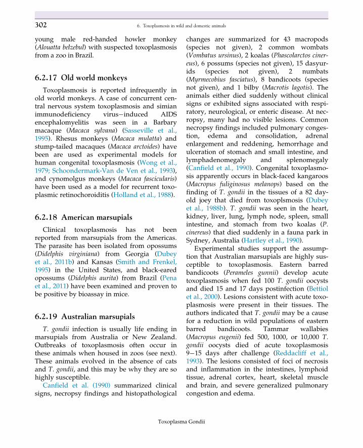
Toxoplasmosis can be a problem in exhibitednew world monkeys (Table 6.1). Many reportsof acute disease have come from squirrel mon-keys (Saimiri sciureus) (Cedillo-Pelaez et al.,2011; Epiphanio et al., 2003) and golden liontamarins (Leontopithecus rosalia) (Dietz et al.,1997; Pertz et al., 1997; Juan-Salles et al., 1998;Epiphanio et al., 2003). Squirrel monkeys and
Panamanian night monkeys (Aotus lemurinus)are highly susceptible to oral tissue cyst inocula-tion and develop acute fatal disease (Harperet al., 1985; Escajadillo and Frenkel, 1991; Furutaet al., 2001). Pena et al. (2011) isolated T. gondiiby bioassay in mice of its heart and brain of a
TABLE 6.1 Summary of host species reports of clinicaltoxoplasmosis in New World primates.
Cotton-top tamarin (Saguinus oedipus)
Yellow-handed marmoset (Saguinus midas midas)
Black marmoset (Saguinus midas niger)
Emperor marmoset (Saguinus imperator)
Red-bellied white-lipped tamarin (Saguinus labiatus)
Black lion tamarin (Leontopithecus chrysopygus)
Golden-headed lion tamarins (Leontopithecus chrysomelas)
Golden lion tamarins (Leontopithecus rosalia)
Squirrel monkeys (Saimiri sciureus)
Pygmy marmoset (Callithrix pygmaea)
Common marmoset (Callithrix jacchus)
Black ear-tufted marmoset (Callithrix penicllata)
Pale-headed saki (Pithecia pithecia)
Night monkey (Aotus trivirgatus)
Howler monkey (Alouatta fusca)
Woolly monkey (Lagothrix lagotricha)
Dietz, H.H., Henriksen, P., Bille-Hansen, V., Henriksen, S.A., 1997.Toxoplasmosis in a colony of New World monkeys. Vet. Parasitol. 68,299�304; Bouer, A., Werther, K., Catao-Dias, J.L., Nunes, A.L., 1999.Outbreak of toxoplasmosis in Lagothrix lagotricha. Folia Primatol. (Basel)70, 282�285; Epiphanio, S., Sa, L.R., Teixeira, R.H., Catao-Dias, J.L.,2001. Toxoplasmosis in a wild-caught black lion tamarin (Leontopithecus
chrysopygus). Vet. Rec. 149, 627�628; Epiphanio, S., Sinhorini, I.L.,Catao-Dias, J.L., 2003. Pathology of toxoplasmosis in captive new worldprimates. J. Comp. Pathol. 129, 196�204; Dubey, J.P., 2010.Toxoplasmosis of Animals and Humans, second ed. CRC Press, BocaRaton, FL, pp. 1�313; Cedillo-Pelaez, C., Rico-Torres, C.P., Sales-Garrido C.G., Correa, D., 2011. Acute toxoplasmosis in squirrel monkeys
(Saimiri sciureus) in Mexico. Vet. Parasitol. 180, 368�371; Pena, H.F.J.,Marvulo, M.F.V., Horta, M.C., Silva, M.A., Silva, J.C.R., Siqueira, D.B.,et al., 2011. Isolation and genetic characterisation of Toxoplasma gondiifrom a red-handed howler monkey (Alouatta belzebul), a jaguarundi(Puma yagouaroundi), and a black-eared opossum (Didelphis aurita) fromBrazil. J. Parasitol. 175, 377�381.
Toxoplasma Gondii
3016.2 Toxoplasmosis in wildlife
young male red-handed howler monkey(Alouatta belzebul) with suspected toxoplasmosisfrom a zoo in Brazil.
6.2.17 Old world monkeys
Toxoplasmosis is reported infrequently inold world monkeys. A case of concurrent cen-tral nervous system toxoplasmosis and simianimmunodeficiency virus�induced AIDSencephalomyelitis was seen in a Barbarymacaque (Macaca sylvana) (Sasseville et al.,1995). Rhesus monkeys (Macaca mulatta) andstump-tailed macaques (Macaca arctoides) havebeen are used as experimental models forhuman congenital toxoplasmosis (Wong et al.,1979; Schoondermark-Van de Ven et al., 1993),and cynomolgus monkeys (Macaca fascicularis)have been used as a model for recurrent toxo-plasmic retinochoroiditis (Holland et al., 1988).
6.2.18 American marsupials
Clinical toxoplasmosis has not beenreported from marsupials from the Americas.The parasite has been isolated from opossums(Didelphis virginiana) from Georgia (Dubeyet al., 2011b) and Kansas (Smith and Frenkel,1995) in the United States, and black-earedopossums (Didelphis aurita) from Brazil (Penaet al., 2011) have been examined and proven tobe positive by bioassay in mice.
6.2.19 Australian marsupials
T. gondii infection is usually life ending inmarsupials from Australia or New Zealand.Outbreaks of toxoplasmosis often occur inthese animals when housed in zoos (see next).These animals evolved in the absence of catsand T. gondii, and this may be why they are sohighly susceptible.
Canfield et al. (1990) summarized clinicalsigns, necropsy findings and histopathological
changes are summarized for 43 macropods(species not given), 2 common wombats(Vombatus ursinus), 2 koalas (Phascolarctos ciner-eus), 6 possums (species not given), 15 dasyur-ids (species not given), 2 numbats(Myrmecobius fasciatus), 8 bandicoots (speciesnot given), and 1 bilby (Macrotis lagotis). Theanimals either died suddenly without clinicalsigns or exhibited signs associated with respi-ratory, neurological, or enteric disease. At nec-ropsy, many had no visible lesions. Commonnecropsy findings included pulmonary conges-tion, edema and consolidation, adrenalenlargement and reddening, hemorrhage andulceration of stomach and small intestine, andlymphadenomegaly and splenomegaly(Canfield et al., 1990). Congenital toxoplasmo-sis apparently occurs in black-faced kangaroos(Macropus fuliginosus melanops) based on thefinding of T. gondii in the tissues of a 82 day-old joey that died from toxoplsmosis (Dubeyet al., 1988b). T. gondii was seen in the heart,kidney, liver, lung, lymph node, spleen, smallintestine, and stomach from two koalas (P.cinereus) that died suddenly in a fauna park inSydney, Australia (Hartley et al., 1990).
Experimental studies support the assump-tion that Australian marsupials are highly sus-ceptible to toxoplasmosis. Eastern barredbandicoots (Perameles gunnii) develop acutetoxoplasmosis when fed 100 T. gondii oocystsand died 15 and 17 days postinfection (Bettiolet al., 2000). Lesions consistent with acute toxo-plasmosis were present in their tissues. Theauthors indicated that T. gondii may be a causefor a reduction in wild populations of easternbarred bandicoots. Tammar wallabies(Macropus eugenii) fed 500, 1000, or 10,000 T.gondii oocysts died of acute toxoplasmosis9�15 days after challenge (Reddacliff et al.,1993). The lesions consisted of foci of necrosisand inflammation in the intestines, lymphoidtissue, adrenal cortex, heart, skeletal muscleand brain, and severe generalized pulmonarycongestion and edema.
Toxoplasma Gondii
302 6. Toxoplasmosis in wild and domestic animals
6.2.20 African wildlife
Surprisingly little is known about T. gondii andtoxoplasmosis from African mammals. Clinicaldisease has not been reported from free-rangingelephants, hippopotamus, rhinos, giraffes,gazelle, wildebeests, impalas, chimpanzees,baboons, orangutans, and gorillas. T. gondii hasnot been isolated from these species that werenaturally infected. Antibodies to T. gondii havebeen demonstrated in elephants, hippopotamus,rhinos, giraffes, wildebeests, and chimpanzees.
Early after the life cycle in cats was discov-ered, an experimental study was done in twofemale chimpanzees fed T. gondii oocysts of theBeverly strain (Draper et al., 1971). One chim-panzee (female 1) was Sabin�Feldman dye testnegative and the other (female 2) was dye testpositive (1:250) before the study was initiated.T. gondii was isolated by bioassay in mice fromthe blood (sampled 1 week PI), inguinal lymphnode (sampled 11 weeks PI), and thigh muscle(sampled 11 weeks PI) of female 1 fed 2.53 106
T. gondii oocysts [but not cerebral spinal fluid(taken 11 weeks PI) (Draper et al., 1971)].Clinically, female 1 became slightly anorexic,developed enlarged superficial lymph nodes,and seroconverted in the dye test to 1:8192 at30 days PI. Female 2 was fed 1.53 106 T. gondiioocysts and did not demonstrate clinical signsor an increase in antibody titer nor was T. gon-dii isolated from blood samples by bioassay inmice (Draper et al., 1971).
6.2.21 Wild rodents
A detailed discussion of T. gondii prevalencein wild rodents (mice and rats) is beyond thescope of this chapter. T. gondii has been iso-lated from the tissues from wild rodentsworldwide (Dubey, 2010). Genotypes of theseisolates are similar to isolates from other ani-mals in the same geographic area.
Dabritz et al. (2008) has recently reviewedwhat was known about the global serological
prevalence of T. gondii in wild rodents. Untillarge-scale studies are conducted using bioassayor molecular detection methods, the role of wildrodents in maintaining T. gondii in the environ-ment will not be fully understood. Properly con-ducted serological studies usually indicate thatfew (,10%) wild mice or rats are usually foundto be seropositive. For example, 2 (0.8%) of 238rats (Rattus norvegicus) from Grenada, WestIndies, were found to be serologically positiveusing the MAT (Dubey et al., 2006b). When thebrains and hearts of all 238 rats were examinedby bioassay in mice, T. gondii was isolated fromonly 1 of the 238 rats with the positive rat beingone of the 2 serologically positive animals. Thisclearly demonstrates a low prevalence of T. gon-dii infection in this rat population.
6.2.22 Wild birds
Table 6.2 lists the wild avian hosts fromwhich viable T. gondii has been isolated, andTable 6.3 lists the avian species that have beenreported to suffer from clinical toxoplasmosis.
T. gondii is readily isolated from the heartsand breast muscles of raptors (Lindsay et al.,1993; Dubey et al., 2011b; Table 6.2). Necrotizingmyocarditis caused by T. gondii has beenobserved in a bald eagle (Haliaeetus leucocephalus)from New Hampshire (Szabo et al., 2004). Severetoxoplasmic hepatitis was seen in an adultbarred owl (Strix varia) from Quebec, Canada(Mikaelian et al., 1997). No clinical signs wereseen in three red-tailed hawks (Buteo jamaicensis)fed T. gondii tissue cysts (Lindsay et al., 1991a,b).T. gondii was isolated from all three red-tailedhawks. No clinical signs were seen in greathorned owls (Bubo virginianus), barred owls (S.varia), or screech owls (Asio otus) feed T. gondiitissue cysts (Dubey et al., 1992b), but parasiteswere isolated from the tissues of the owls at nec-ropsy. T. gondii was not reisolated from a spar-row hawk (Falco sparverius) that had beenexperimentally infected (Miller et al., 1972).
Toxoplasma Gondii
3036.2 Toxoplasmosis in wildlife
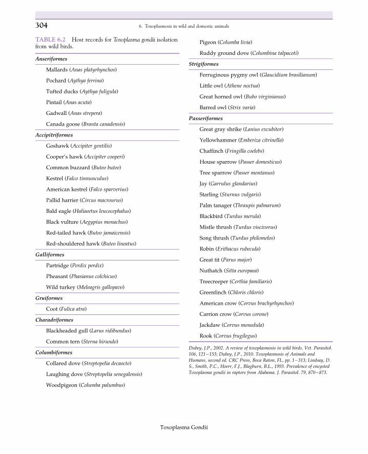
TABLE 6.2 Host records for Toxoplasma gondii isolationfrom wild birds.
Anseriformes
Mallards (Anas platyrhynchos)
Pochard (Aythya ferrina)
Tufted ducks (Aythya fuligula)
Pintail (Anas acuta)
Gadwall (Anas strepera)
Canada goose (Branta canadensis)
Accipitriformes
Goshawk (Accipiter gentilis)
Cooper’s hawk (Accipiter cooperi)
Common buzzard (Buteo buteo)
Kestrel (Falco tinnunculus)
American kestrel (Falco sparverius)
Pallid harrier (Circus macrourus)
Bald eagle (Haliaeetus leucocephalus)
Black vulture (Aegypius monachus)
Red-tailed hawk (Buteo jamaicensis)
Red-shouldered hawk (Buteo lineatus)
Galliformes
Partridge (Perdix perdix)
Pheasant (Phasianus colchicus)
Wild turkey (Meleagris gallopavo)
Gruiformes
Coot (Fulica atra)
Charadriformes
Blackheaded gull (Larus ridibundus)
Common tern (Sterna hirundo)
Columbiformes
Collared dove (Streptopelia decaocto)
Laughing dove (Streptopelia senegalensis)
Woodpigeon (Columba palumbus)
Pigeon (Columba livia)
Ruddy ground dove (Columbina talpacoti)
Strigiformes
Ferruginous pygmy owl (Glaucidium brasilianum)
Little owl (Athene noctua)
Great horned owl (Bubo virginianus)
Barred owl (Strix varia)
Passeriformes
Great gray shrike (Lanius excubitor)
Yellowhammer (Emberiza citrinella)
Chaffinch (Fringilla coelebs)
House sparrow (Passer domesticus)
Tree sparrow (Passer montanus)
Jay (Garrulus glandarius)
Starling (Sturnus vulgaris)
Palm tanager (Thraupis palmarum)
Blackbird (Turdus merula)
Mistle thrush (Turdus viscivorus)
Song thrush (Turdus philomelos)
Robin (Erithacus rubecula)
Great tit (Parus major)
Nuthatch (Sitta europaea)
Treecreeper (Certhia familiaris)
Greenfinch (Chloris chloris)
American crow (Corvus brachyrhynchos)
Carrion crow (Corvus corone)
Jackdaw (Corvus monedula)
Rook (Corvus frugilegus)
Dubey, J.P., 2002. A review of toxoplasmosis in wild birds. Vet. Parasitol.106, 121�153; Dubey, J.P., 2010. Toxoplasmosis of Animals andHumans, second ed. CRC Press, Boca Raton, FL, pp. 1�313; Lindsay, D.S., Smith, P.C., Hoerr, F.J., Blagburn, B.L., 1993. Prevalence of encysted
Toxoplasma gondii in raptors from Alabama. J. Parasitol. 79, 870�873.
Toxoplasma Gondii
304 6. Toxoplasmosis in wild and domestic animals
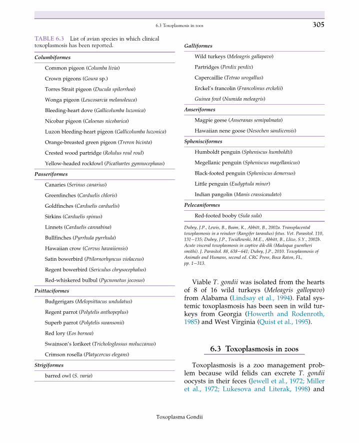
Viable T. gondii was isolated from the heartsof 8 of 16 wild turkeys (Meleagris gallopavo)from Alabama (Lindsay et al., 1994). Fatal sys-temic toxoplasmosis has been seen in wild tur-keys from Georgia (Howerth and Rodenroth,1985) and West Virginia (Quist et al., 1995).
6.3 Toxoplasmosis in zoos
Toxoplasmosis is a zoo management prob-lem because wild felids can excrete T. gondiioocysts in their feces (Jewell et al., 1972; Milleret al., 1972; Lukesova and Literak, 1998) and
TABLE 6.3 List of avian species in which clinicaltoxoplasmosis has been reported.
Columbiformes
Common pigeon (Columba livia)
Crown pigeons (Goura sp.)
Torres Strait pigeon (Ducula spilorrhoa)
Wonga pigeon (Leucosarcia melanoleuca)
Bleeding-heart dove (Gallicolumba luzonica)
Nicobar pigeon (Caloenas nicobarica)
Luzon bleeding-heart pigeon (Gallicolumba luzonica)
Orange-breasted green pigeon (Treron bicinta)
Crested wood partridge (Rolulus roul roul)
Yellow-headed rockfowl (Picathartes gymnocephaus)
Passeriformes
Canaries (Serinus canarius)
Greenfinches (Carduelis chloris)
Goldfinches (Carduelis carduelis)
Sirkins (Carduelis spinus)
Linnets (Carduelis cannabina)
Bullfinches (Pyrrhula pyrrhula)
Hawaiian crow (Corvus hawaiiensis)
Satin bowerbird (Ptilornorhyncus violaceus)
Regent bowerbird (Sericulus chrysocephalus)
Red-whiskered bulbul (Pycnonotus jocosus)
Psittaciformes
Budgerigars (Melopsittacus undulatus)
Regent parrot (Polytelis anthopeplus)
Superb parrot (Polytelis swansonii)
Red lory (Eos bornea)
Swainson’s lorikeet (Trichologlossus moluccanus)
Crimson rosella (Platycercus elegans)
Strigiformes
barred owl (S. varia)
Galliformes
Wild turkeys (Meleagris gallapavo)
Partridges (Perdix perdix)
Capercaillie (Tetrao urogallus)
Erckel’s francolin (Francolinus erckelii)
Guinea fowl (Numida meleagris)
Anseriformes
Magpie geese (Anseranas semipalmata)
Hawaiian nene goose (Nesochen sandicensis)
Sphenisciformes
Humboldt penguin (Spheniscus humboldti)
Megellanic penguin (Spheniscus magellanicus)
Black-footed penguin (Spheniscus demersus)
Little penguin (Eudyptula minor)
Indian pangolin (Manis crassicaudato)
Pelecaniformes
Red-footed booby (Sula sula)
Dubey, J.P., Lewis, B., Beam, K., Abbitt, B., 2002a. Transplacentaltoxoplasmosis in a reindeer (Rangifer tarandus) fetus. Vet. Parasitol. 110,131�135; Dubey, J.P., Tocidlowski, M.E., Abbitt, B., Llizo, S.Y., 2002b.
Acute visceral toxoplasmosis in captive dik-dik (Madoqua guentherismithi). J. Parasitol. 88, 638�641; Dubey, J.P., 2010. Toxoplasmosis ofAnimals and Humans, second ed. CRC Press, Boca Raton, FL,pp. 1�313.
Toxoplasma Gondii
3056.3 Toxoplasmosis in zoos
feral cats occur in zoos (Gorman et al., 1986).Oocysts excreted by these felids can make theirway into highly susceptible species.
Mammalian species that frequently developtoxoplasmosis in zoos include Australian mar-supials (Portas, 2010), New World and arborialmonkeys (Dietz et al., 1997; Pertz et al., 1997;Juan-Salles et al., 1998; Epiphanio et al., 2000),lemurs (Dubey et al., 1985), and Pallas’s cats(O. manul) (Riemann et al., 1974; Dubey et al.,1988a, 2002a,b; Basso et al., 2005) (Fig. 6.2).Lesions in these animals are consistent withacute toxoplasmosis and are usually mostsevere in visceral tissues such as the lungs,liver, and spleen.
Toxoplasmosis is common in lemurs exhib-ited in zoo worldwide (Dubey et al., 1985). Afemale ring-tailed lemur (Lemur catta) died oftoxoplasmosis in a zoo in Spain 1 week after thedelivery of 4 stillborn offspring which all haddisseminated toxoplasmosis (Juan-Salles et al.,2011). T. gondii was isolated from the tissues of a3-year-old secundiparous female ring-tailedlemur from a zoo in Alabama that died of acutetoxoplasmosis (Spencer et al., 2004). The isolatewas not pathogenic for mice and was geneticallya Type II isolate. This case points out the diffi-culty in preventing toxoplasmosis in highly sus-ceptible animals because this lemur was housedin a group on an island in the zoo (Spenceret al., 2004), making it easier to prevent contactwith feral cats. Oocysts on the lemur’s food(fruit etc.) or carried in by black birds were con-sidered likely sources of infection in this case(Spencer et al., 2004).
Sporadic cases of acute toxoplasmosis havebeen reported in exhibited dik-dik (Madoquaguentheri smithi) (Dubey et al., 2002b), slender-tailed meerkats (Suricata suricatta) (Juan-Salleset al., 1997), African crested porcupines (Hystrixcristata) (Harrison et al., 2007), New World por-cupines (Erethizontidae sp.) (Fayyad et al.,2016), and Brazilian prehensile-tailed porcu-pines (Coendou mexicanus) (Morales et al., 1996).Fatal disseminated toxoplasmosis in three cap-tive slender-tailed meerkats (S. suricatta) in a
zoo in La Plata, Argentina, was found to becaused by the normally nonpathogenic geno-type Type III isolate of the parasite suggestingthat meerkats are highly susceptible to infection(Basso et al., 2009). A case of abortion due toT. gondii has been reported in a Greenlandmuskox (Ovibos moshatus wardi) (Crawfordet al., 2000).
Fatal toxoplasmosis was reported in a 7-year-old giant panda (Ailuropoda melanoleuca) in a zooin China (Ma et al., 2015). The animal had acutegastrointestinal and respiratory signs andT. gondiiwas seen in lung lesions. It was anorexicand lethargic and died 2 days after it signs devel-oped despite supportive treatment with intra-muscular cephalosporin and intravenousinfusion of glucose solution. Parasite DNA wasdetected in the liver, spleen, lung, kidney, andintestines using PCR. Antibodies to T. gondiiwere detected in sera from 7 of 19 giant pandasin the breeding program at the ChengduResearch Base of Giant Panda Breeding inSichuan, China (Loeffler et al., 2007).
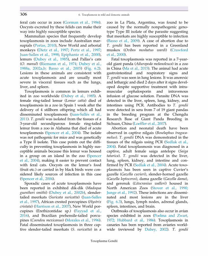
Abortion and neonatal death have beenobserved in captive nilgais (Boselaphus tragoca-melus). T. gondii DNA was demonstrated in thetissues of the nilgais using PCR (Sedlak et al.,2004). Fatal toxoplasmosis was diagnosed in acaptive, adult female saiga antelope (Saigatatarica). T. gondii was detected in the liver,lung, spleen, kidney, and intestine and con-firmed by PCR (Sedlak et al., 2004). Acute toxo-plasmois has been seen in captive Cuvier’sgazelle (Gazella cuvieri), slender-horned gazelle(Gazella leptoceros), dama gazelle (Gazella dama),and gerenuk (Litocranius walleri) housed inNorth American Zoos (Stover et al., 1990;Junge et al., 1992). These infections are dissemi-nated and most lesions are in the liver(Fig. 6.3), lungs, lymph nodes, adrenal glands,spleen, intestines, and brain.
Outbreaks of toxoplasmosis also occur in avianspecies exhibited in zoos (Poelma and Zwart,1972; Hubbard et al., 1986). Toxoplasmosis incanaries has been reported from aviaries world-wide (reviewed by Dubey, 2002). T. gondii
Toxoplasma Gondii
306 6. Toxoplasmosis in wild and domestic animals
genotype III was isolated from five of five black-winged lorys (Eos cyanogenia) from an acute toxo-plasmosis outbreak in an aviary in SouthCarolina (Dubey et al., 2004d). Acute systemictoxoplasmosis was reported to be the cause ofdeath of 3 of 10 Nicobar pigeons (Caloenas nicobar-ica) in an aviary collection in South Africa (Lasand Shivaprasad, 2008). Feral cats were a knownmanagement problem and lesions were consis-tent with oocyst-acquired infection. Three 1�3-month-old black-footed penguin chicks(Spheniscus demersus) died from acute toxoplas-mosis within 24 hours of showing central ner-vous signs (Ploeg et al., 2011). The birds werehoused in a baby penguin creche in a zoo in theNetherlands. A cat with a litter of kittens hadrecently been observed feeding on fish intendedfor the penguins in the zoo, and the cat was sus-pected as the source of infection (Ploeg et al.,2011).
Management and husbandry programs canbe designed to help achieve prevention of toxo-plasmosis in highly susceptible species in zoosand aviaries. Felids should never be fed freshunfrozen meats because of the possibility con-tamination with T. gondii tissue cysts. Meatthat has been frozen solid and then thawed canbe safely fed because freezing kills T. gondii tis-sue cysts (Kotula et al., 1991). Feral cats shouldbe actively controlled in zoos to prevent themfrom shedding oocysts. Highly susceptible spe-cies should not be housed near felids.
Outdoor aviaries are at risk because ofoocysts excreted by domestic cats. Aviariesshould be designed to exclude cat feces andtransport hosts (flies, roaches, etc.) that maybring in T. gondii on or in their bodies.
6.4 Toxoplasma gondii and endangeredspecies
Toxoplasmosis can adversely affect endan-gered avian and mammalian species. The ‘Alala(Hawaiian crow, Corvus hawaiiensis) is an
endangered species, and only about 25 were leftin captivity and the wild in 2000 (Work et al.,2000). Tragically, these birds are highly suscepti-ble to fatal toxoplasmosis and develop diseaseafter being introduced back in to the wild.Toxoplasmosis appears to pose a significantthreat and management challenge to reintroduc-tion programs for ‘Alala in Hawaii (Work et al.,2000).
Captive breeding groups of golden liontamarins (L. rosalia) have developed acute toxo-plasmosis and suffered many fatalities both inNorth American and European zoos (Pertzet al., 1997; Juan-Salles et al., 1998). These arbo-real monkeys are endangered and attempts tobreed them in captivity for eventual release inthe wild are hammered, because it is difficultto keep them from being exposed to T. gondii.
Repeated transplacental transmission ofT. gondii by Pallas’s cats maybe responsible forthe high rate of impact of this disease on thePallas’s cat population in zoos. Efforts byNorth American zoos to establish geneticallyviable captive populations of Pallas’s cats(O. manul) have been compromised by highnewborn kitten mortality due to toxoplasmosis(Brown et al., 2005). In their natural environ-ment, Pallas’s cats generally have little expo-sure to T. gondii, and it is believed that theyacquire T. gondii infection after captivity(Brown et al., 2005). The mortality rate for toxo-plasmosis of Pallas’s cat kittens born in Zoos inthe United States is 35%�60% (Kenny et al.,2002; Brown et al., 2005).
Sand cats (F. margarita) housed at theBreeding Centre for Endangered ArabianWildlife in the United Arab Emirates and AlWabra Wildlife Preservation, Qatar, have beenreported to suffer from congenital (Pas andDubey 2008a) and acquired toxoplasmosis(Dubey et al., 2010). Serological examination ofendangered Gordon’s wildcat (Felis silvestrisgordoni) kept at the same institution (Pas andDubey 2008b) indicated that seropositiveGordon’s wildcats were present but no clinical
Toxoplasma Gondii
3076.4 Toxoplasma gondii and endangered species
history consistent with toxoplasmosis has beenreported in these animals. Unlike domesticcats, Sand cat (F. margarita) queens will repeat-edly infect litters of kittens making it very diffi-cult to keep up numbers of healthy kittens inbreeding programs. Fortunately, Gordon’swildcats appear to behave like domestic cats intheir responses to T. gondii infection.
6.5 Toxoplasmosis in pets
6.5.1 Cats
Most cats are asymptomatic during a pri-mary T. gondii infection. Fever (40.0�C�41.7�C)is present in many cats with clinical toxoplas-mosis. Clinical signs of dyspnea, polypnea,icterus, and signs of abdominal discomfortwere the most frequent findings in 100 catswith histologically confirmed toxoplasmosis(Dubey and Carpenter, 1993). Uveitis and reti-nochoroiditis are also common clinical signs incats with toxoplasmosis. Gross and micro-scopic lesions are found in many organs butare most common in the lungs. Gross lesions inthe lungs consist of edema and congestion, fail-ure to collapse, and multifocal areas of firm,white to yellow, discoloration. Pericardial andabdominal effusions may be present. The liveris the most frequently affected abdominalorgan and diffuse necrotizing hepatitis may bevisible grossly. Gross lesions associated withnecrosis can also be observed in the mesentericlymph nodes and pancreas.
All ages, sexes, and breeds of domestic catsare susceptible to T. gondii infection (Dubeyet al., 1977). Transplacentally or lactogenicallyinfected kittens will excrete oocysts, but theprepatent period is usually 3 weeks or more,because the kittens are infected with tachy-zoites (Dubey et al., 1995b). Domestic catsunder 1 year of age produce the most numbersof T. gondii oocysts. Cats that are born andraised outdoors usually become infected with
T. gondii shortly after they are weaned andbegin to hunt. T. gondii naive adult domesticcats will excrete oocysts if fed tissue cysts, butthey usually will excrete fewer numbers ofoocysts and excrete oocysts for a shorter periodof time than recently weaned kittens.
Intestinal immunity to T. gondii is strong incats that have excreted oocysts (Dubey, 1995).Primary T. gondii infection in cats does notcause immunosuppression (Lappin et al., 1992;Davis and Dubey, 1995). Serum antibody doesnot play a significant role in resistance to intes-tinal infection and intestinal immunity is mostlikely cell mediated. Oocysts begin to beexcreted in the feces before IgM, IgG, or IgAantibodies are present in the serum (Lappinet al., 1989). Partial development of the enteroe-pithelial stages occur in the intestines ofimmune cats, but oocyst production is pre-vented (Davis and Dubey, 1995). Most cats thathave excreted oocysts once do not reexcreteoocysts if challenged within 6 months to 1 year.Intestinal immunity will last up to 6 years inabout 55% of cats (Dubey, 1995).
Vaccination of cats against intestinal T. gondiiinfection has been successfully achieved using amutant strain (T-263) of the parasite (Frenkelet al., 1991; Freyre et al., 1993). Oral administra-tion of strain T-263 bradyzoites results in intesti-nal infection but does not result in oocystproduction in cats. These vaccinated cats do notexcrete oocysts when challenged with oocystproducing strains of T. gondii. The T-263 strain issafe to use in healthy cats. It is not recom-mended for use in pregnant cats or FeLV posi-tive cats or immunocompromised cats(Choromanski et al., 1994, 1995). It has only lim-ited ability to persist in the tissues of cats andcannot survive more than 3 back-passages incats. No reversion to oocyst excretion or increasein virulence has been observed in over 200 inoc-ulated cats. The T-263 strain is rapidly clearedfrom the mouth of inoculated cats.
It is logical to assume that cat owners andveterinarians would be at a greater risk for
Toxoplasma Gondii
308 6. Toxoplasmosis in wild and domestic animals
developing toxoplasmosis; however, serologi-cal studies do not confirm this assumption. Inone study in AIDS patients, it was conclusivelyshown that owning cats did not increase therisk of developing toxoplasmosis (Wallaceet al., 1993). The role of cat ownership andexposure to T. gondii is, however, notcompletely clear at present. Many studies havebeen conducted to determine the associationbetween cat ownership or cat exposure and theprevalence of T. gondii infection in humans.Many studies do not find a positive relation-ship while many find a positive relationship. Itmust be stressed that preventing exposure tocats is not the same as preventing exposure toT. gondii oocysts. Pregnant women or immuno-compromised individuals should not changethe cat’s litter box. If feces are removed daily,this will also help prevent exposure by remov-ing oocysts before they can sporulate. T. gondiioocyst can survive in the soil for years and canbe disseminated from the original site of depo-sition by erosion, other mechanical means, andby phoretic vectors. Inhalation of oocysts stir-red up in the dust by horses has been associ-ated with an outbreak of human toxoplasmosisat a riding stable (Teutsch et al., 1979). Oocystsare not likely to remain in the air for extendedperiods of time. Washing fruits andvegetables and wearing gloves while garden-ing are means of preventing exposure tooocysts.
T. gondii oocysts were not isolated from thefur of oocyst-excreting cats (Dubey, 1995).Therefore it is unlikely that infection can beobtained by petting a cat. Tachyzoites are notlikely to be present in the oral cavity of catswith active T. gondii infection, and none wouldbe in a chronic infection; therefore it is unlikelythat a cat bite would transmit T. gondii infec-tion. Cat scratches are also unlikely to transmitT. gondii infection.
T. gondii has been isolated from the tissuesfrom domestic cats worldwide (Dubey, 2010).Genotypes of feline isolates are similar to
isolates from other animals in the same geo-graphic area.
6.5.2 Dogs
T. gondii was once confused with Neosporacaninum as a cause of disease in dogs, andmany reports of toxoplasmosis in dogs areactually neosporosis (Dubey and Lindsay,1996; Lindsay and Dubey, 2000). True toxoplas-mosis does occur in dogs (Dubey et al., 1989).Clinical toxoplasmosis in dogs is often associ-ated with immunosuppression induced bycanine distemper virus infection. Clinical signsare usually most apparent in the respiratoryand hepatic systems and probably result fromreactivation of latent infections (Dubey et al.,1989). Transplacental infections have not yetbeen confirmed in naturally infected dogs.Dogs are resistant to experimental infectionwith tissue cysts and oocysts (Lindsay et al.,1996, 1997a).
A role for dogs in the transmission ofT. gondii to humans has been postulatedbased on serological surveys and observa-tions that dogs ingest cat feces and often rolein cat feces and other foul smelling sub-stances (Frenkel et al., 2003). It is believedthat dogs can bring oocysts to a home afteringesting them and deposit them in or aroundthe home when they defecate. Experimentallyinfective T. gondii oocysts can be found in dogfeces for up to 2 days after they ingest oocysts(Lindsay et al., 1997a). T. gondii oocysts willnot sporulate when placed on dog fur(Lindsay et al., 1997a). Schares et al. (2005)found viable T. gondii oocysts in 2 of 24,089dogs in Germany. The role of dogs as poten-tial transport hosts for T. gondii needs furtherexamination. T. gondii has been isolated fromthe tissues from domestic dogs worldwide(Dubey, 2010). Genotypes of feline isolates aresimilar to isolates from other animals inthe same geographic area. A related
Toxoplasma Gondii
3096.5 Toxoplasmosis in pets
Apicomplexan parasitic protozoa N. caninumis present in dogs.
6.5.3 Ferrets
Congenital toxoplasmosis has been observedin farmed razed ferrets (Mustela putorius furo)from New Zealand (Thornton and Cook, 1986).Thirty percent of the kits on the farm diedacutely and had lesions of disseminated toxo-plasmosis. An epizootic of toxoplasmosisoccurred among a population of endangeredblack-footed ferrets (Mustela nigripes) at a zooin the United States (Burns et al., 2003).Twenty-two adults and 30 kits died from acutetoxoplasmosis and an additional 13 adults diedfrom chronic toxoplasmosis after the initialoutbreak.
6.6 Domestic farm animals
6.6.1 Mink
Acute toxoplasmosis with abortions hasbeen reported in farmed mink (Mustela vison)from Europe and the United States (Frank,2001; Smielewska-Los and Turniak, 2004). Thepractice of feeding nonfrozen slaughter offalwas blamed for acute toxoplasmosis in onereport (Smielewska-Los and Turniak, 2004).Toxoplasmosis was diagnosed using PCR andimmunohistochemistry in a young free-rangingmink (M. vison) that had signs of left hind limblameness, ataxia, head tremors, and bilateralblindness and was found on a college campusin Michigan (Jones et al., 2006). T. gondii hasbeen isolated from wild mink from the UnitedStates (Smith and Frenkel, 1995).
6.6.2 Horses
Horses are resistant to experimental infec-tion with 13 104 or 13 105 oocysts. T. gondiican persist in edible tissues of horses for up to
476 days (Dubey, 1985). Although T. gondii hasbeen isolated from tissues of horses, there is noconfirmed report of clinical toxoplasmosis inhorses (Al-Khalidi and Dubey, 1979). A relatedApicomplexan parasitic protozoa Neosporahughesi is present in horses.
6.6.3 Swine
Abortion in sows is the most common signof toxoplasmosis in swine. Sows only abortonce. Abortions are rare in most pork produc-ing regions of the world with the exception ofTaiwan. Pigs raised on dirt are more likely tohave T. gondii in their tissues. Diagnosis ofT. gondii abortion in sows is best done byexamining fetal fluids for antibodies using themodified agglutination test. Undercooked porkis a source of human infection, and viable tis-sue cysts can remain in pork for up to 865 days(Dubey, 1988). T. gondii has been isolated fromthe tissues from domestic pigs worldwide(Dubey, 2010). Genotypes of pig isolates aresimilar to isolates from other animals in thesame geographic area.
6.6.4 Cattle
Clinical toxoplasmosis in cattle is rare, andabortions are uncommon. Many reports ofbovine abortion due to T. gondii are actually dueto N. caninum (Dubey and Lindsay, 1996).Attempts to isolate T. gondii from seropositivecattle are often unsuccessful indicating that beefmay not be a significant source of human infec-tion in the United States (Dubey et al., 2005). Forexample, no T. gondii was isolated from 2094samples of beef obtained from retail markets inthe United States (Dubey et al., 2005). However,viable tissue cysts can remain in cattle for up to1191 days (Dubey and Thulliez, 1993).Additional studies are needed to full documentthese experimental findings.
Toxoplasma Gondii
310 6. Toxoplasmosis in wild and domestic animals
6.6.5 Sheep
T. gondii is a common cause of abortion inewes and an important production problem.Multiple abortions can occur in a flock indicat-ing a common oocyst source for ewes. Ewesdevelop solid immunity after aborting T. gondiiinfected fetuses. A vaccine to prevent abortionin ewes is available in several countries(Buxton and Innes, 1995). Diagnosis of T. gondiiabortion in ewes is best done by examiningfetal fluids for antibodies using the modifiedagglutination test. Undercooked lamb andmutton is a source of human infection. T. gondiihas been isolated from the tissues from domes-tic sheep worldwide (Dubey, 2010). Genotypesof sheep isolates are similar to isolates fromother animals in the same geographic area.
6.6.6 Goats
T. gondii is a common cause of abortion indoes. Multiple abortions can occur in a flockindicating a common oocyst source for does.Does develop immunity after aborting T. gondiiinfected fetuses but repeat abort can occur.Diagnosis of T. gondii abortion in goats is bestdone by examining fetal fluids for antibodiesusing the modified agglutination test.Undercooked goat meat is a source of humaninfection. T. gondii has been isolated from thetissues from domestic goat worldwide (Dubey,2010). Genotypes of goat isolates are similar toisolates from other animals in the same geo-graphic area. Drinking raw not pasteurizedgoats milk is a potential source of T. gondii forhumans and the parasite can survive for up to7 days in refrigerated goat milk (Walsh et al.,1999).
6.6.7 Buffalos
Naturally occurring clinical toxoplasmoishas not been observed in buffalos (Bison bison,Bubalus bubalis, Syncerus caffer) and viable
T. gondii has not been isolated from buffaloes.Serological surveys indicate that buffalos areexposed to the parasite.
6.6.8 Camels
Acute toxoplasmosis was observed in a6-year-old camel (Camelus dromedarius)(Hagemoser et al., 1990). Dyspnea was themain clinical sign and many tachyzoites werefound in its lungs and plural exudates. T. gon-dii has been isolated from camel meat usingcat bioassays (Hilali et al., 1995).
6.6.9 Llamas, alpaca, and vicunas
Experimental studies indicate that llamas(Lama glama) are resistant to clinical toxoplas-mosis even if challenged during pregnancy(Jarvinen et al., 1999). Naturally occurringtoxoplasmosis has not been reported in llamas,alpacas (Lama pacos), or vicunas (Lama vicugna).
6.6.10 Chickens
Chickens (Gallus domesticus) that are raisedon the ground are a potential source of T. gon-dii due to high level of exposure to oocysts.Chickens usually do not develop clinical signseven after oral inoculation of large numbers ofoocysts (Dubey et al., 1993c; Kaneto et al.,1997). Egg production may adversely beaffected in laying hens fed large numbers ofoocysts, but T. gondii is not readily transmittedto the eggs of these hens (Biancifiori et al.,1986). Clinical toxoplasmosis does not occuron modern chicken farms where birds areraised indoors. Chickens raised in modernproduction facilities in confinement indoorsare not likely to have viable T. gondii in theirtissues. None of 2094 samples from commer-cial chickens in retail markets from the UnitedStates contained viable T. gondii in a surveyfrom the United States (Dubey et al., 2005).
Toxoplasma Gondii
3116.6 Domestic farm animals
However, T. gondii has been isolated from thetissues from chickens worldwide (Dubey,2010). Genotypes of chicken isolates are similarto isolates from other animals in the same geo-graphic area. The prevalence of isolation isdependent on the methods used to raze thechickens with chickens razed outside having ahigher prevalence of infection.
6.6.11 Turkeys
Domestic turkeys (M. gallopavo) fed T. gondiioocysts remained clinically normal except afew that develop pneumonia associated withAspergillus-like fungi (Dubey et al., 1993a).Tissue cysts are present in breast and leg mus-cles of inoculated turkeys. Clinical toxoplasmo-sis does not occur on modern turkey farms. T.gondii has been isolated from the tissues fromturkeys worldwide (Dubey, 2010). Genotypesof turkey isolates are similar to isolates fromother animals in the same geographic area. Theprevalence of isolation is dependent on themethods used to raze the turkeys with turkeysrazed outside having a higher prevalence ofinfection.
6.6.12 Ducks and geese
Domestic ducks (Anas platyrhynchos) fedT. gondii oocysts do not develop clinical toxo-plasmosis (Sedlak et al., 2004). Viable T. gondiihas been isolated from the tissues of a natu-rally infected domestic ducks (Dubey et al.,2003) and from a domestic goose (Anser anser)(Dubey et al., 2007b).
6.7 Fish, reptiles, and amphibians
Toxoplasmosis does not occur in fish, rep-tiles, or amphibians. Reports of natural infec-tions in these animals in nature are erroneous.Fish and reptiles can be manipulated to make
them susceptible to T. gondii, but they have tobe experimentally infected and kept at tem-peratures of around 37�C�40�C. For example,zebrafish (Danio rerio) adapted to 37�C from28�C were able to be infected intraperitoneallywith 10 tissue cysts of the Me49 (genotype II)or VEG (genotype III) strains of T. gondii.Clinical signs in zebrafish included bilateralexopthalmia, swollen abdomens, whirlingswimming behavior, and generalized subder-mal hemorrhaging. Tachyzoites were presentin tissue sections of parasites developing inmuscle, heart, liver, spleen, kidney, pancreas,reproductive organs, eyes, and brain (Sanderset al., 2015).
References
Al-Khalidi, N.W., Dubey, J.P., 1979. Prevalence ofToxoplasma gondii infection in horses. J. Parasitol. 65,331�334.
Aramini, J.J., Stephen, C., Dubey, J.P., 1998. Toxoplasma gon-dii in Vancouver Island cougars (Felis concolor vancouver-ensis): serology and oocyst shedding. J. Parasitol. 84,438�440.
Arkush, K.D., Miller, M.A., Leutenegger, C.M., Gardner, I.A., Packham, A.E., Heckeroth, A.R., et al., 2003.Molecular and bioassay-based detection of Toxoplasmagondii oocyst uptake by mussels (Mytilus galloprovincia-lis). Int. J. Parasitol. 33, 1087�1097.
Atwood, T.C., Duncan, C., Patyk, K.A., Nol, P., Rhyan, J.,McCollum, M., et al., 2017. Environmental and behav-ioral changes may influence the exposure of an Arcticapex predator to pathogens and contaminants. Sci. Rep.7, 13193. Available from: https://doi.org/10.1038/s41598-017-13496-9.
Aubert, D., Ajzenberg, D., Richomme, C., Gilot-Fromont, E.,Terrier, M.E., de Gevigney, C., et al., 2010. Molecular andbiological characteristics of Toxoplasma gondii isolatesfrom wildlife in France. Vet. Parasitol. 171, 346�349.
Bangari, D.S., Mouser, P., Miller, M.A., Stevenson, G.W.,Vemulapalli, R., Thacker, H.L., 2007. Toxoplasmosis ina woodchuck (Marmota monax) and two American redsquirrels (Tamiasciurus hudsonicus). J. Vet. Diagn. Invest.19, 705�709.
Basso, W., Edelhofer, R., Zenker, W., Mostl, K., Kubber-Heiss, A., Prosl, H., 2005. Toxoplasmosis in Pallas’ cats(Otocolobus manul) raised in captivity. Parasitology 130,293�299.
Toxoplasma Gondii
312 6. Toxoplasmosis in wild and domestic animals
Basso, W., More, G., Quiroga, M.A., Pardini, L., Bacigalupe,D., Venturini, L., et al., 2009. Isolation and molecularcharacterization of Toxoplasma gondii from captiveslender-tailed meerkats (Suricata suricatta) with fataltoxoplasmosis in Argentina. Vet. Parasitol. 161,201�206.
Baszler, T.V., Dubey, J.P., Lohr, C.V., Foreyt, W.J., 2000.Toxoplasmic encephalitis in a free-ranging RockyMountain bighorn sheep from Washington. J. Wildl.Dis. 36, 752�754.
Bergmann, V., Heidrich, R., Kiupel, H., 1980. Acute toxo-plasmosis outbreak in rabbit flocks. Angew. Parasitol.21, 1�6.
Bettiol, S.S., Obendorf, D.L., Nowarkowski, M., Goldsmid,J.M., 2000. Pathology of experimental toxoplasmosis ineastern barred bandicoots in Tasmania. J. Wildl. Dis. 36,141�144.
Biancifiori, F., Rondini, C., Grelloni, V., Frescura, T., 1986.Avian toxoplasmosis: experimental infection of chickenand pigeon. Comp. Immunol. Microbiol. Infect. Dis. 9,337�346.
Bossart, G.D., Mignucci-Giannoni, A.A., Rivera-Guzman,A.L., Jimenez-Marrero, N.M., Camus, A., Bonde, R.,et al., 2012. Disseminated toxoplasmosis in Antilleanmanatees (Trichechus manatus manatus) from PuertoRico. Dis. Aqua. Org. 101, 139�144 .
Bouer, A., Werther, K., Catao-Dias, J.L., Nunes, A.L., 1999.Outbreak of toxoplasmosis in Lagothrix lagotricha. FoliaPrimatol. (Basel). 70, 282�285.
Bowater, R.O., Norton, J., Johnson, S., Hill, B.,O’Donoghue, P., Prior, H., 2003. Toxoplasmosis in Indo-Pacific humpbacked dolphins (Sousa chinensis), fromQueensland. Aust. Vet J. 81, 627�632.
Brown, M., Lappin, M.R., Brown, J.L., Munkhtsog, B.,Swanson, W.F., 2005. Exploring the ecologic basis forextreme susceptibility of Pallas’ cats (Otocolobus manul)to fatal toxoplasmosis. J. Wildl. Dis. 41, 691�700.
Buergelt, C.D., Bonde, R.K., 1983. Toxoplasmic meningoen-cephalitis in a West Indian manatee. J. Am. Vet. Med.Assoc. 183, 1294�1296.
Burns, R., Williams, E.S., O’Toole, D., Dubey, J.P., 2003.Toxoplasma gondii infections in captive black-footed fer-rets (Mustela nigripes), 1992-1998: clinical signs, serology,pathology, and prevention. J. Wildl. Dis. 39, 787�797.
Buxton, D., Innes, E.A., 1995. A commercial vaccine forovine toxoplasmosis. Parasitology 110, S11�16.
Cabral, A.D., D’Auria, S.R.N., Camargo, M.C.G.O., Rosa, A.R., Sodre, M.M., Galvao-Dias, M.A., et al., 2014.Seroepidemiology of Toxoplasma gondii infection in batsfrom Sao Paulo city, Brazil. Vet. Parasitol. 206, 293�296.
Canfield, P.J., Hartley, W.J., Dubey, J.P., 1990. Lesions oftoxoplasmosis in Australian marsupials. J. Comp.Pathol. 103, 159�167.
Carrasco, L., Raya, A.I., Nunez, A., Gomez-Laguna, J.,Hernandez, S., Dubey, J.P., 2006. Fatal toxoplasmosis
and concurrent (Calodium hepaticum) infection in Koreansquirrels (Tanias sibericus). Vet. Parasitol. 137, 180�183.
Cedillo-Pelaez, C., Rico-Torres, C.P., Sales-Garrido, C.G.,Correa, D., 2011. Acute toxoplasmosis in squirrel mon-keys (Saimiri sciureus) in Mexico. Vet. Parasitol. 180,368�371.
Chomel, B.B., Zarnke, R.L., Kasten, R.W., Kass, P.H.,Mendes, E., 1995. Serologic survey of Toxoplasma gondiiin grizzly bears (Ursus arctos) and black bears (Ursusamericanus), from Alaska, 1988 to 1991. J. Wildl. Dis. 31,472�479.
Choromanski, L., Freyre, A., Brown, K., Popiel, I., Shibley,G., 1994. Safety aspects of a vaccine for cats containinga Toxoplasma gondii mutant strain. J. Eukaryot.Microbiol. 41, 8S.
Choromanski, L., Freyre, A., Popiel, R., Brown, K., Grieve,R., Shibley, G., 1995. Safety and efficacy of modified livefeline Toxoplasma gondii vaccine. Dev. Biol. Stand. 84,269�281.
Cole, R.A., Lindsay, D.S., Howe, D.K., Roderick, C.L.,Dubey, J.P., Thomas, N.J., et al., 2000. Biological andmolecular characterizations of Toxoplasma gondii strainsobtained from southern sea otters (Enhydra lutris nereis).J. Parasitol. 86, 526�530.
Colosimo, S.M., Montoya, J.G., Westley, B.P., Jacob, J.,Isada, N.B., 2013. Congenital toxoplasmosis presentingwith fetal atrial flutter after maternal ingestion ofinfected moose meat. Alaska Med. 54, 27�31.
Cox, J.J., Slabach, B., Hast, J.T., Murphy, S.M., Kwok, O.C.,Dubey, J.P., 2017. High seroprevalence of Toxoplasmagondii in elk (Cervus canadensis) of the centralAppalachians, USA. Parasitol. Res. 116, 1079�1083.
Crawford, G.C., Dunker, F.H., Dubey, J.P., 2000.Toxoplasmosis as a suspected cause of abortion in aGreenland muskox (Ovibos moshatus wardi). J. ZooWildl. Med. 31, 247�250.
Cross, J., Lein, J., Hsu, M., 1969. Toxoplasma isolated fromthe Formosan giant flying squirrel. Taiwan Yi Xue HuiZa Zhi 68, 678�683.
Dabritz, H.A., Miller, M.M., Gardner, I.A., Packham, A.E.,Atwill, E.R., Conrad, P.A., 2008. Risk factors forToxoplasma gondii infection in wild rodents from CentralCoastal California and a review of T. gondii prevalencein rodents. J. Parasitol. 94, 675�683.
Davidson, W.R., Nettles, V.F., Hayes, L.E., Howerth, E.W.,Couvillion, C.E., 1992. Diseases diagnosed in gray foxes(Urocyon cinereoargenteus) from the southeastern UnitedStates. J. Wildl. Dis. 28, 28�33.
Davis, S.W., Dubey, J.P., 1995. Mediation of immunity toToxoplasma gondii oocyst shedding in cats. J. Parasitol.81, 882�886.
Demar, M., Ajzenberg, D., Serrurier, B., Darde, M.L.,Carme, B., 2008. Case Report: Atypical Toxoplasma gondiistrain from a free-living Jaguar (Panthera onca) in FrenchGuiana. Am. J. Trop. Med. Hyg. 78, 195�197.
Toxoplasma Gondii
313References
Dietz, H.H., Henriksen, P., Bille-Hansen, V., Henriksen, S.A., 1997. Toxoplasmosis in a colony of New Worldmonkeys. Vet. Parasitol. 68, 299�304.
DiGuardo, G., Proietto, U., Di Francesco, C.E., Marsilio, F.,Zaccaroni, A., Scaravelli, D., et al., 2010. Cerebral toxo-plasmosis in striped dolphins (Stenella coeruleoalba)stranded along the Ligurian Sea Coast of Italy. Vet.Pathol. 47, 245�253.
Draper, C.C., Killick-Kendrick, R., Hutchison, W.M., Siim,J.C., Garnham, P.C.C., 1971. Experimental toxoplasmo-sis in chimpanzees. Br. Med. J. 2, 375�378.
Dubey, J.P., 1981. Isolation of encysted Toxoplasma gondiifrom musculature of moose and pronghorn in Montana.Am. J. Vet. Res. 42, 126�127.
Dubey, J.P., 1982. Isolation of encysted Toxoplasma gondiifrom muscles of mule deer in Montana. J. Am. Vet.Med. Assoc. 181, 1535.
Dubey, J.P., 1983. Toxoplasma gondii infection in rodentsand insectivores from Montana. J. Wildl. Dis. 19,149�150.
Dubey, J.P., 1985. Persistence of encysted Toxoplasma gondiiin tissues of equids fed oocysts. Am. J. Vet. Res. 46,1753�1754.
Dubey, J.P., 1988. Long-term persistence of Toxoplasma gon-dii in tissues of pigs inoculated with T. gondii oocystsand effect of freezing on viability of tissue cysts in pork.Am. J. Vet. Res. 49, 910�913.
Dubey, J.P., 1995. Duration of immunity to shedding ofToxoplasma gondii oocysts by cats. J. Parasitol. 81,410�415.
Dubey, J.P., 2002. A review of toxoplasmosis in wild birds.Vet. Parasitol. 106, 121�153.
Dubey, J.P., 2010. Toxoplasmosis of Animals andHumans, second ed. CRC Press, Boca Raton, FL,pp. 1�313.
Dubey, J.P., Carpenter, J.L., 1993. Histologically confirmedclinical toxoplasmosis in cats: 100 cases (1952�1990).J. Am. Vet. Med. Assoc. 203, 1556�1566.
Dubey, J.P., Lin, T.L., 1994. Acute toxoplasmosis in a grayfox (Urocyon cinereoargenteus). Vet. Parasitol. 51,321�325.
Dubey, J.P., Lindsay, D.S., 1996. A review of Neospora cani-num and neosporosis. Vet Parasitol. 67, 1�59.
Dubey, J.P., Thulliez, P., 1993. Persistence of tissue cysts inedible tissues of cattle fed Toxoplasma gondii oocysts.Am. J. Vet. Res. 54, 270�273.
Dubey, J.P., Hoover, E.A., Walls, K.W., 1977. Effect of ageand sex on the acquisition of immunity to toxoplasmo-sis in cats. J. Protozool. 24, 184�186.
Dubey, J.P., Thorne, E.T., Sharma, S.P., 1980. Experimentaltoxoplasmosis in elk (Cervus canadensis). Am. J. Vet. Res.41, 792�793.
Dubey, J.P., Thorne, E.T., Williams, E.S., 1982. Inducedtoxoplasmosis in pronghorns and mule deer. J. Am.Vet. Med. Assoc. 181, 1263�1267.
Dubey, J.P., Kramer, L.W., Weisbrode, S.E., 1985. Acutedeath associated with Toxoplasma gondii in ring-tailedlemurs. J. Am. Vet. Med. Assoc. 187, 1272�1273.
Dubey, J.P., Quinn, W.J., Weinandy, D., 1987. Fatal neona-tal toxoplasmosis in a bobcat (Lynx rufus). J. Wildl. Dis.23, 324�327.
Dubey, J.P., Gendron-Fitzpatrick, A.P., Lenhard, A.L.,Bowman, D., 1988a. Fatal toxoplasmosis and enteroe-pithelial stages of Toxoplasma gondii in a Pallas cat (Felismanul). J. Protozool. 35, 528�530.
Dubey, J.P., Ott-Joslin, J., Torgerson, R.W., Topper, M.J.,Sundberg, J.P., 1988b. Toxoplasmosis in black-facedkangaroos (Macropus fuliginosus melanops). Vet.Parasitol. 30, 97�105.
Dubey, J.P., Carpenter, J.L., Topper, M.L., Uggla, A., 1989.Fatal toxoplasmosis in dogs. J. Am. Anim. Hosp. Assoc.25, 659�664.
Dubey, J.P., Hamir, A.N., Rupprecht, C.E., 1990. Acute dis-seminated toxoplasmosis in a red fox (Vulpes vulpes). J.Wildl. Dis. 26, 286�290.
Dubey, J.P., Brown, C.A., Carpenter, J.L., Moore III, J.J.,1992a. Fatal toxoplasmosis in domestic rabbits in theUSA. Vet. Parasitol. 44, 305�309.
Dubey, J.P., Porteer, S.I., Tseng, F., Shen, S.K., Thulliez, P.,1992b. Induced toxoplasmosis in owls. J. Zoo Wildl.Med. 23, 98�102.
Dubey, J.P., Camargo, M.E., Ruff, M.D., Wilkins, G.C.,Shen, S.K., Kwok, O.C., et al., 1993a. Experimental toxo-plasmosis in turkeys. J. Parasitol. 79, 949�952.
Dubey, J.P., Hamir, A.N., Shen, S.K., Thulliez, P.,Rupprecht, C.E., 1993b. Experimental Toxoplasma gondiiinfection in raccoons (Procyon lotor). J. Parasitol. 79,548�552.
Dubey, J.P., Ruff, M.D., Camargo, M.E., Shen, S.K., Wilkins,G.L., Kwok, O.C., et al., 1993c. Serologic and parasito-logic responses of domestic chickens after oral inocula-tion with Toxoplasma gondii oocysts. Am. J. Vet. Res. 54,1668�1672.
Dubey, J.P., Humphreys, J.G., Thulliez, P., 1995a.Prevalence of viable Toxoplasma gondii tissue cysts andantibodies to T. gondii by various serologic tests in blackbears (Ursus americanus) from Pennsylvania. J. Parasitol.81, 109�112.
Dubey, J.P., Lappin, M.R., Thulliez, P., 1995b. Diagnosis ofinduced toxoplasmosis in neonatal cats. J. Am. Vet.Med. Assoc. 207, 179�185.
Dubey, J.P., Lewis, B., Beam, K., Abbitt, B., 2002a.Transplacental toxoplasmosis in a reindeer (Rangifer tar-andus) fetus. Vet. Parasitol. 110, 131�135.
Dubey, J.P., Tocidlowski, M.E., Abbitt, B., Llizo, S.Y., 2002b.Acute visceral toxoplasmosis in captive dik-dik(Madoqua guentheri smithi). J. Parasitol. 88, 638�641.
Dubey, J.P., Zarnke, R., Thomas, N.J., Wong, S.K., VanBonn, W., Briggs, M., et al., 2003. Toxoplasma gondii,Neospora caninum, Sarcocystis neurona, and Sarcocystis
Toxoplasma Gondii
314 6. Toxoplasmosis in wild and domestic animals
canis-like infections in marine mammals. Vet. Parasitol.116, 275�296.
Dubey, J.P., Lipscomb, T.P., Mense, M., 2004a.Toxoplasmosis in an elephant seal (Mirounga angustiros-tris). J. Parasitol. 90, 410�411.
Dubey, J.P., Graham, D.H., De Young, R.W., Dahl, E.,Eberhard, M.L., Nace, E.K., et al., 2004b. Molecular andbiologic characteristics of Toxoplasma gondii isolatesfrom wildlife in the United States. J. Parasitol. 90,67�71.
Dubey, J.P., Navarro, I.T., Sreekumar, C., Dahl, E., Freire,R.L., Kawabata, H.H., et al., 2004c. Toxoplasma gondiiinfections in cats from Parana, Brazil: seroprevalence,tissue distribution, and biologic and genetic characteri-zation of isolates. J. Parasitol. 90, 721�726.
Dubey, J.P., Parnell, P.G., Sreekumar, C., Vianna, M.C., DeYoung, R.W., Dahl, E., et al., 2004d. Biologic and molec-ular characteristics of Toxoplasma gondii isolates fromstriped skunk (Mephitis mephitis), Canada goose (Brantacanadensis), black-winged lory (Eos cyanogenia), and cats(Felis catus). J. Parasitol. 90, 1171�1174.
Dubey, J.P., Hill, D.E., Jones, J.L., Hightower, A.W.,Kirkland, E., Roberts, J.M., et al., 2005. Prevalence ofviable Toxoplasma gondii in beef, chicken and pork fromretail meat stores in the United States; risk assessmentto consumers. J. Parasitol. 91, 1082�1093.
Dubey, J.P., Hodgin, E.C., Hamir, A.N., 2006a. Acute fataltoxoplasmosis in squirrels (Sciurus carolensis) with brady-zoites in visceral tissues. J. Parasitol. 92, 658�659.
Dubey, J.P., Bhaiyat, M.I., Macpherson, C.N.L., de Allie, C.,Chikweto, A., Kwok, O.C.H., et al., 2006b. Prevalence ofToxoplasma gondii in rats (Rattus norvegicus) in Grenada,West Indies. J. Parasitol. 92, 1107�1108.
Dubey, J.P., Morales, J.A., Sundar, N., Velmurugan, G.V.,Gonzalez-Barrientos, C.R., Hernandez-Mora, G., et al.,2007a. Isolation and genetic characterization ofToxoplasma gondii from striped dolphin (Stenella coeru-leoalba) from Costa Rica. J. Parasitol. 93, 710�711.
Dubey, J.P., Webb, D.M., Sundar, N., Velmurugan, G.V.,Bandini, L.A., Kwok, O.C.H., et al., 2007b. Endemicavian toxoplasmosis on a farm in Illinois: Clinical dis-ease, diagnosis, biologic and genetic characteristics ofToxoplasma gondii isolates from chickens (Gallus domesti-cus), and a goose (Anser anser). Vet. Parasitol. 148,207�212.
Dubey, J.P., Fair, P.A., Sundar, N., Velmurugan, G., Kwok,O.C.H., McFee, W.E., et al., 2008a. Isolation ofToxoplasma gondii from bottlenose dolphins (Tursiopstruncatus). J. Parasitol. 94, 821�823.
Dubey, J.P., Velmurugan, G.V., Ulrich, V., Gill, J.,Carstensen, M., Sundar, N., et al., 2008b. Transplacentaltoxoplasmosis in naturally-infected white-tailed deer:isolation and genetic characterisation of Toxoplasma gon-dii from foetuses of different gestational ages. Int. J.Parasitol. 38, 1057�1063.
Dubey, J.P., Pas, A., Rajendran, C., Kwok, O.C.H., Ferreira,L.R., Martinc, J., et al., 2010. Toxoplasmosis in Sand cats(Felis margarita) and other animals in the BreedingCentre for Endangered Arabian Wildlife in the UnitedArab Emirates and Al Wabra Wildlife Preservation, theState of Qatar. Vet. Parasitol. 172, 195�203.
Dubey, J.P., Passos, I.M.F., Rajendran, C., Ferreira, I.R.,Gennari, S.M., Su, C., 2011a. Isolation of viableToxoplasma gondii from feral guinea fowl (Numida melea-gris) and domestic rabbits (Oryctolagus cuniculus) fromBrazil. J. Parasitol. 97, 842�845.
Dubey, J.P., Velmurugan, G.V., Rajendran, C., Yabsley, M.J., Thomas, N.J., Beckmen, K.B., et al., 2011b. Geneticcharacterisation of Toxoplasma gondii in wildlife fromNorth America revealed widespread and high preva-lence of the fourth clonal type. Int J Parasitol. 41,1139�1147.
Dubey, J.P., Brown, J., Ternent, M., Verma, S.K., Hill, D.E.,Cerqueira-Cezar, C.K., et al., 2016. Seroepidemiologicstudy on the prevalence of Toxoplasma gondii andTrichinella spp. infections in black bears (Ursus ameri-canus) in Pennsylvania, USA. Vet. Parasitol. 229, 76�80.
Dubey, J.P., Brown, J., Verma, S.K., Cerqueira-Cerar, C.K.,Banfield, J., Kowk, O.C.H., et al., 2017. Isolation of via-ble Toxoplasma gondii, molecular characterization, andseroprevalence in elk (Cervus canadensis) inPennsylvania, USA. Vet. Parasitol. 243, 1�5.
Epiphanio, S., Guimaraes, M.A., Fedullo, D.L., Correa, S.H.,Catao-Dias, J.L., 2000. Toxoplasmosis in golden-headedlion tamarins (Leontopithecus chrysomelas) and emperormarmosets (Saguinus imperator) in captivity. J. ZooWildl. Med. 31, 231�235.
Epiphanio, S., Sa, L.R., Teixeira, R.H., Catao-Dias, J.L.,2001. Toxoplasmosis in a wild-caught black lion tamarin(Leontopithecus chrysopygus). Vet. Rec. 149, 627�628.
Epiphanio, S., Sinhorini, I.L., Catao-Dias, J.L., 2003.Pathology of toxoplasmosis in captive new world pri-mates. J. Comp. Pathol. 129, 196�204.
Escajadillo, A., Frenkel, J.K., 1991. Experimental toxoplas-mosis and vaccine tests in Aotus monkeys. Am. J. Trop.Med. Hyg. 44, 382�389.
Fayyad, A., Kummerfeld, M., Davina, I., Wohlsein, P.,Beineke, A., Baumgartner, W., et al., 2016. ). Fatal sys-temic Toxoplasma gondii infection in a red squirrel(Sciurus vulgaris), a swinhoe’s striped squirrel (Tamiopsswinhoei) and a New World Porcupine (Erethizontidaesp.). J. Comp Pathol. 154, 263�267.
Forzan, M.J., Frasca, S., 2004. Systemic toxoplasmosis in afive-month-old beaver, (Castor canadensis). J. Zoo Wildl.Med. 35, 113�115.
Frank, R.K., 2001. An outbreak of toxoplasmosis in farmedmink (Mustela vison S.). J. Vet. Diagn. Invest. 13,245�249.
Frenkel, J.K., Pfefferkorn, E.R., Smith, D.D., Fishback, J.L.,1991. Prospective vaccine prepared from a new mutant
Toxoplasma Gondii
315References
of Toxoplasma gondii for use in cats. Am. J. Vet. Res. 52,759�763.
Frenkel, J.K., Lindsay, D.S., Parker, B.B., Dobesh, M., 2003.Dogs as possible mechanical carriers of Toxoplasma anddog fur as source of infection for young children. Int. J.Infect. Dis. 7, 292�293.
Freyre, A., Choromanski, L., Fishback, J.L., Popiel, I., 1993.Immunization of cats with tissue cysts, bradyzoites, andtachyzoites of the T-263 strain of Toxoplasma gondii. J.Parasitol 79, 716�719.
Furuta, T., Une, Y., Omura, M., Matsutani, N., Nomura, Y.,Kikuchi, T., et al., 2001. Horizontal transmission ofToxoplasma gondii in squirrel monkeys (Saimiri sciureus).Exp. Anim. 50, 299�306.
Gajadhar, A.A., Measures, L., Forbes, L.B., Kapel, C.,Dubey, J.P., 2004. Experimental Toxoplasma gondii infec-tion in grey seals (Halichoerus grypus). J. Parasitol. 90,255�259.
Galuzo, I.G., Vysokova, L.A., Krivkova, A.M., 1970.Toxoplasmosis in bats. In: Fitzgerald, P.R. (Ed.),Toxoplasmosis in Animals. University of Illinois, Collegeof Veterinary Medicine, Urbana, IL, pp. 190�194.
Gehrt, S.D., Michael, J., Kinsel, M.J., Anchor, C., 2010.Pathogen dynamics and morbidity of striped skunks inthe absence of rabies. J. Wildl. Dis. 46, 335�347.
Geisel, O., Breuer, W., Minkus, G., Hermanns, W., 1995.Toxoplasmosis causing death in a mole (Talpa europaea).Berl. Munch. Tierarztl. Wochenschr. 108, 241�243.
Gerhold, R.W., Howerth, E.W., Lindsay, D.S., 2005.Sarcocystis neurona-associated meningoencephalitis anddescription of intramuscular sarcocysts in a fisher(Martes pennanti). J. Wildl. Dis. 41, 224�230.
Gerhold, R.W., Saraf, P., Chapman, A., Zou, X., Hickling,G., Stiver, W.H., et al., 2017. Toxoplasma gondii seroprev-alence and genotype diversity in select wildlife speciesfrom the southeastern United States. Para. Vec. 10, 508.
Gorman, T.R., Riveros, V., Alcaino, H.A., Salas, D.R.,Thiermann, E.R., 1986. Helminthiasis and toxoplasmosisamong exotic mammals at the Santiago National Zoo. J.Am. Vet. Med. Assoc. 189, 1068�1070.
Gustafsson, K., Uggla, A., Jarplid, B., 1997. Toxoplasma gon-dii infection in the mountain hare (Lepus timidus) anddomestic rabbit (Oryctolagus cuniculus). I. Pathology. J.Comp. Pathol. 117, 351�360.
Hagemoser, W.A., Dubey, J.P., Thompson, J.R., 1990. Acutetoxoplasmosis in a camel. J. Am. Vet. Med. Assoc. 196,347.
Hancock, K., Thiele, L.A., Zajac, A.M., Elvinger, F.,Lindsay, D.S., 2005. Prevalence of antibodies toToxoplasma gondii in raccoons (Procyon lotor) from anurban area of Northern Virginia. J. Parasitol. 91,694�695.
Harper III, J.S., London, W.T., Sever, J.L., 1985. Five drugregimens for treatment of acute toxoplasmosis in squir-rel monkeys. Am. J. Trop. Med. Hyg. 34, 50�57.
Harrison, M., Moorman, J.B., Bolin, S.R., Grosjean, N.L.,Lim, A., Fitzgerald, S.D., 2007. Toxoplasma gondii in anAfrican crested porcupine (Hystrix cristata). J. Vet.Diagn. Invest. 19, 191�194.
Hartley, W.J., Dubey, J.P., Spielman, D.S., 1990. Fatal toxo-plasmosis in koalas (Phascolarctos cinereus). J. Parasitol.76, 271�272.
Hejlicek, K., Literak, I., Nezval, J., 1997. Toxoplasmosis inwild mammals from the Czech Republic. J. Wildl. Dis.33, 480�485.
Herrmann, D.C., Maksimov, P., Maksimov, A., Sutor, A.,Schwarz, S., Jaschke, W., et al., 2012. Toxoplasma gondiiin foxes and rodents from the German Federal States ofBrandenburg and Saxony-Anhalt: seroprevalence andgenotypes. Vet. Parasitol. 185, 78�85.
Hilali, M., Fatani, A., al-Atiya, S., 1995. Isolation of tissuecysts of Toxoplasma, Isospora, Hammondia and Sarcocystisfrom camel (Camelus dromedarius) meat in Saudi Arabia.Vet. Parasitol. 58, 353�356.
Holland, G.N., O’Connor, G.R., Diaz, R.F., Minasi, P.,Wara, W.M., 1988. Ocular toxoplasmosis in immuno-suppressed nonhuman primates. Invest. Ophthalmol.Vis. Sci. 29, 835�842.
Holshuh, H.J., Sherrod, A.E., Taylor, C.R., Andrews, B.F.,Howard, E.B., 1985. Toxoplasmosis in a feral northernfur seal. J. Am. Vet. Med. Assoc. 187, 1229�1230.
Hong, S.H., Lee, S.E., Jeong, Y.I., Kim, H.C., Chong, S.T.,Klein, T.A., et al., 2014. Prevalence and molecular char-acterizations of Toxoplasma gondii and Babesia microtifrom small mammals captured in Gyeonggi andGangwon Provinces, Republic of Korea. Vet Parasitol.205, 512�517.
Honnold, S.P., Braun, R., Scott, D.P., Sreekumar, C., Dubey,J.P., 2005. Toxoplasmosis in a Hawaiian monk seal(Monachus schauinslandi). J. Parasitol. 91, 695�697.
Howerth, E.W., Rodenroth, N., 1985. Fatal systemic toxo-plasmosis in a wild turkey. J. Wildl. Dis. 21, 446�449.
Hubbard, G., Witt, W., Healy, M., Schmidt, R., 1986. Anoutbreak of toxoplasmosis in zoo birds. Vet. Pathol. 23,639�641.
Inskeep II, W., Gardiner, C.H., Harris, R.K., Dubey, J.P.,Goldston, R.T., 1990. Toxoplasmosis in Atlantic bottle-nosed dolphins (Tursiops truncatus). J. Wildl. Dis. 26,377�382.
Jardine, J.E., Dubey, J.P., 2002. Congenital toxoplasmosis ina Indo-Pacific bottlenose dolphin (Tursiops aduncus).J. Parasitol. 88, 197�199.
Jarvinen, J.A., Dubey, J.P., Althouse, G.,C., 1999. Clinicaland serologic evaluation of two llamas (Lama glama)infected with Toxoplasma gondii during gestation.J. Parasitol. 85, 142�144.
Jewell, M.L., Frenkel, J.K., Johnson, K.M., Reed, V.,Ruiz, A., 1972. Development of Toxoplasma oocysts inneotropical felidae. Am. J. Trop. Med. Hyg. 21,512�517.
Toxoplasma Gondii
316 6. Toxoplasmosis in wild and domestic animals
Jokelainen, P., Nylund, M., 2012. Acute fatal toxoplasmosisin three Eurasian Red Squirrels (Sciurus vulgaris) causedby genotype II of Toxoplasma gondii. J. Wildl. Dis 48,454�457.
Jokelainen, P., Isomursu, M., Nareaho, A., Oksanen, A.,2011. Natural Toxoplasma gondii infections in Europeanbrown hares and mountain hares in Finland: propor-tional mortality rate, antibody prevalence, and geneticcharacterization. J. Wildl. Dis. 47, 154�163.
Jones, Y.L., Fitzgerald, S.D., Sikarske, J.G., Murphy, A.,Grosjean, N., Kiupel, M., 2006. Toxoplasmosis in a free-ranging mink. J. Wildl. Dis. 42, 865�869.
Juan-Salles, C., Prats, N., Lopez, S., Domingo, M., Marco,A.J., Moran, J.F., 1997. Epizootic disseminated toxoplas-mosis in captive slender-tailed meerkats (Suricata suri-catta). Vet. Pathol. 34, 1�7.
Juan-Salles, C., Prats, N., Marco, A.J., Ramos-Vara, J.A.,Borras, D., Fernandez, J., 1998. Fatal acute toxoplasmo-sis in three golden lion tamarins (Leontopithecus rosalia).J. Zoo Wildl. Med. 29, 55�60.
Juan-Salles, C., Mainez, M., Marco, A., Sanchıs, A.M.M.,2011. Localized toxoplasmosis in a ring-tailed lemur(Lemur catta) causing placentitis, stillbirths, and dissemi-nated fetal infection. J. Vet. Diag. Invest. 23, 1041�1045.
Junge, R.E., Fischer, J.R., Dubey, J.P., 1992. Fatal dissemi-nated toxoplasmosis in a captive Cuvier’s gazelle(Gazella cuvieri). J. Zoo Wildl. Med. 23, 342�345.
Kaneto, C.N., Costa, A.J., Paulillo, A.C., Moraes, F.R.,Murakami, T.O., Meireles, M.V., 1997. Experimental toxo-plasmosis in broiler chicks. Vet. Parasitol. 69, 203�210.
Kelly, T.R., Sleeman, J.M., 2003. Morbidity and mortality ofred foxes (Vulpes vulpes) and gray foxes (Urocyon ciner-eoargenteus) admitted to the Wildlife Center of Virginia,1993-2001. J. Wildl. Dis. 39, 467�469.
Kenny, D.E., Lappin, M.R., Knightly, F., Baler, J., Brewer,M., Getzy, D.M., 2002. Toxoplasmosis in Pallas’ cats(Otocolobus felis manul) at the Denver ZoologicalGardens. J, Zoo Wildl, Med. 33, 131�138.
Kijlstra, A., Meerburg, B., Cornelissen, J., De Craeye, S.,Vereijken, P., Jongert, E., 2008. The role of rodents andshrews in the transmission of Toxoplasma gondii to pigs.Vet. Parasitol. 156, 183�190.
Kottwitz, J.J., Preziosi, D.E., Miller, M.A., Ramos-Vara, J.A.,Maggs, D.J., Bonagura, J.D., 2004. Heart failure causedby toxoplasmosis in a Fennec fox (Fennecus zerda). J.Am. Anim. Hosp. Assoc. 40, 501�507.
Kotula, A.W., Dubey, J.P., Sharar, A.K., Andrews, C.D.,Shen, S.K., Lindsay, D.S., 1991. Effect of freezing oninfectivity of Toxoplasma gondii tissue cysts in pork. J.Food Protect. 54, 687�690.
Krijger, I.M., Cornelissen, J.B.W.J., Wisselink, H.J., Meerburg,B.G., 2014. Prevalence of Toxoplasma gondii in commonmoles (Talpa europaea). Acta Vet. Scand. 56, 48. Availablefrom: https://doi.org/10.1186/s13028-014-0048-0.
Kumar, A., Melotti, J.R., Cooley, T.M., Fitzgerald, S.D.,2018. Mortality due to toxoplasmosis in suburban east-ern fox squirrels (Sciurus niger) in Michigan, USA. JWildl Dis. 55, 213�217.
Lappin, M.R., Greene, C.E., Prestwood, A.K., Dawe, D.L.,Tarleton, R.L., 1989. Diagnosis of recent Toxoplasma gon-dii infection in cats by use of an enzyme-linked immu-nosorbent assay for immunoglobulin M. Am. J. Vet.Res. 50, 1580�1585.
Lappin, M.R., Dawe, D.L., Lindl, P., Greene, C.E.,Prestwood, A.K., 1992. Mitogen and antigen-specificinduction of lymphoblast transformation in cats withsubclinical toxoplasmosis. Vet. Immunol.Immunopathol. 30, 207�220.
Larkin, J.L., Gabriel, M., Gerhold, R.W., Yabsley, M.J.,Wester, J.C., Humphreys, J.G., et al., 2011. Prevalence toToxoplasma gondii and Sarcocystis spp. in a reintroducedfisher (Martes pennanti) population in Pennsylvania. J.Parasitol. 97, 425�429.
Las, R.D., Shivaprasad, H.L., 2008. An outbreak of toxo-plasmosis in an aviary collection of Nicobar pigeons(Caloenas nicobarica). J. S. Afr. Vet. Assoc. 79, 149�152.
Lindsay, D.S., Dubey, J.P., 2000. Canine neosporsis. J. Vet.Parasitol. 14, 4�14.
Lindsay, D.S., Dubey, J.P., 2009. Long-term survival ofToxoplasma gondii sporulated oocysts in seawater. J.Parasitol. 95, 1019�1020.
Lindsay, D.S., Blagburn, B.L., Dubey, J.P., Mason, W.H.,1991a. Prevalence and isolation of Toxoplasma gondiifrom white-tailed deer in Alabama. J. Parasitol. 77,62�64.
Lindsay, D.S., Dubey, J.P., Blagburn, B.L., 1991b.Toxoplasma gondii infections in red-tailed hawks inocu-lated orally with tissue cysts. J. Parasitol. 77, 322�325.
Lindsay, D.S., Smith, P.C., Hoerr, F.J., Blagburn, B.L., 1993.Prevalence of encysted Toxoplasma gondii in raptorsfrom Alabama. J. Parasitol. 79, 870�873.
Lindsay, D.S., Smith, P.C., Blagburn, B.L., 1994. Prevalenceand isolation of Toxoplasma gondii from wild turkeys inAlabama. J. Helminthol. Soc. Wash. 61, 115�117.
Lindsay, D.S., Dubey, J.P., Butler, J.M., Blagburn, B.L.,1996. Experimental tissue cyst induced Toxoplasma gon-dii infections in dogs. J. Eukaryot. Microbiol. 43, 113S.
Lindsay, D.S., Dubey, J.P., Butler, J.M., Blagburn, B.L.,1997a. Mechanical transmission of Toxoplasma gondiioocysts by dogs. Vet. Parasitol. 73, 27�33.
Lindsay, D.S., Sundermann, C.A., Dubey, J.P., Blagburn, B.L., 1997b. Update on Toxoplasma gondii infections inwildlife and exotic animals from Alabama. J. AlabamaAcad. Sci. 68, 246�254.
Lindsay, D.S., Thomas, N.J., Rosypal, A.C., Dubey, J.P.,2001a. Dual Sarcocystis neurona and Toxoplasma gondiiinfection in a Northern sea otter from Washington state,USA. Vet. Parasitol. 97, 319�327.
Toxoplasma Gondii
317References
Lindsay, D.S., Phelps, K.K., Smith, S.A., Flick, G., Sumner,S.S., Dubey, J.P., 2001b. Removal of Toxoplasma gondiioocysts from sea water by eastern oysters (Crassostreavirginica). J. Eukaryot. Microbiol. (Suppl), 197S�198S.
Lindsay, D.S., Collins, M.V., Mitchell, S.M., Cole, R.A.,Flick, G.J., Wetch, C.N., et al., 2003. Sporulation and sur-vival of Toxoplasma gondii oocysts in seawater. J.Eukaryot. Microbiol. 50 (Suppl), 687�688.
Lindsay, D.S., Collins, M.V., Mitchell, S.M., Wetch, C.N.,Rosypal, A.C., Flick, G.J., et al., 2004. Survival ofToxoplasma gondii oocysts in Eastern oysters (Crassostreavirginica). J. Parasitol. 90, 1054�1057.
Lindsay, D.S., McKown, R.D., DiCristina, J., Jordan, C.N.,Mitchell, S.M., Oates, D.W., et al., 2006. Prevalence ofagglutinating antibodies to Toxoplasma gondii in adultand fetal mule deer (Odocoileus hemionus) fromNebraska. J. Parasitol. 91, 475�478.
Lloyd, C., Stidworthy, M.F., 2007. Acute disseminated toxo-plasmosis in a juvenile cheetah (Acinonyx jubatus). J.Zoo Wildl. Med. 38, 475�478.
Loeffler, I.K., Howard, J., Montali, R.J., Hayek, L.A.,Dubovi, E., Zhang, Z., et al., 2007. Serosurvey of ex situgiant pandas (Ailuropoda melanoleuca) and red pandas(Ailurus fulgens) in China with implications for speciesconservation. J. Zoo Wildl. Med. 38, 559�566.
Lukesova, D., Literak, I., 1998. Shedding of Toxoplasma gon-dii oocysts by Felidae in zoos in the Czech Republic.Vet. Parasitol. 74, 1�7.
Ma, H., Wang, Z., Wang, C., Li, C., Wei, F., Liu, Q., 2015.Fatal Toxoplasma gondii infection in the giant panda.Parasite 22, 30. Available from: https://doi.org/10.1051/parasite/2015030.
Massie, G.N., Ware, M.N., Villegas, E.N., Black, M.W.,2010. Uptake and transmission of Toxoplasma gondiioocysts by migratory, filter-feeding fish. Vet. Parasitol.169, 296�303.
Mazzariol, S., Federica Marcer, F., Mignone, W., 2012.Toxoplasma gondii coinfection in a Mediterranean finwhale (Balaenoptera physalus). BMC Vet. Res. 8, 20.
Meerburg, B.G., De Craeye, S., Dierick, K.A., Kijlstra, A.,2012. Neospora caninum and Toxoplasma gondii in braintissue of feral rodents and insectivores caught on farmsin the Netherlands. Vet. Parasitol. 184, 317�320.
Migaki, G., Allen, J.F., Casey, H.W., 1977. Toxoplasmosis ina California sea lion (Zalophus californianus). Am. J. Vet.Res. 38, 135�136.
Migaki, G., Sawa, T.R., Dubey, J.P., 1990. Fatal dissemi-nated toxoplasmosis in a spinner dolphin (Stenella long-irostris). Vet. Pathol. 27, 463�464.
Mikaelian, I., Dubey, J.P., Martineau, D., 1997. Severe hepati-tis resulting from toxoplasmosis in a barred owl (Strixvaria) from Quebec, Canada. Avian Dis. 41, 738�740.
Mikaelian, I., Boisclair, J., Dubey, J.P., Kennedy, S.,Martineau, D., 2000. Toxoplasmosis in beluga whales(Delphinapterus leucas) from the St Lawrence estuary:two case reports and a serological survey. J. Comp.Pathol. 122, 73�76.
Miller, N.L., Frenkel, J.K., Dubey, J.P., 1972. Oral infectionswith Toxoplasma cysts and oocysts in felines, othermammals, and in birds. J. Parasitol. 58, 928�937.
Miller, M.A., Sverlow, K., Crosbie, P.R., Barr, B.C.,Lowenstine, L.J., Gulland, F.M., et al., 2001. Isolationand characterization of two parasitic protozoa from aPacific harbor seal (Phoca vitulina richardsi) with menin-goencephalomyelitis. J. Parasitol. 87, 816�822.
Miller, M.A., Gardner, I.A., Kreuder, C., Paradies, D.M.,Worcester, K.R., Jessup, D.A., et al., 2002. Coastal fresh-water runoff is a risk factor for Toxoplasma gondii infec-tion of southern sea otters (Enhydra lutris nereis). Int. J.Parasitol. 32, 997�1006.
Morales, J.A., Pena, M.A., Dubey, J.P., 1996. Disseminatedtoxoplasmosis in a captive porcupine (Coendou mexica-nus) from Costa Rica. J. Parasitol. 82, 185�186.
Nardoni, S., Angelici, M.C., Mugnaini, L., Mancianti, F.,2011. Prevalence of Toxoplasma gondii infection inMyocastor coypus in a protected Italian wetland. Parasit.Vect. 4, 240.
Oksanen, A., Gustafsson, K., Lunden, A., Dubey, J.P.,Thulliez, P., Uggla, A., 1996. Experimental Toxoplasmagondii infection leading to fatal enteritis in reindeer(Rangifer tarandus). J. Parasitol. 82, 843�845.
Oksanen, A., Asbakk, K., Prestrud, K.W., Aars, J.,Derocher, A.E., Tryland, M., et al., 2009. Prevalence ofantibodies against Toxoplasma gondii in polar bears(Ursus maritimus) from Svalbard and East Greenland. J.Parasitol. 95, 89�94.
Pas, A., Dubey, J.P., 2008a. Fatal toxoplasmosis in sand cats(Felis margarita). J. Zoo Wildl. Med. 39, 362�369.
Pas, A., Dubey, J.P., 2008b. Seroprevalence of antibodies toToxoplasma gondii in Gordon’s wild cat (Felis silvestrisgordoni) in the Middle East. J. Parasitol. 94, 1169.
Pas, A., Dubey, J.P., 2008c. Toxoplasmosis in Sand fox(Vulpes rueppellii). J. Parasitol. 94, 976�977.
Pena, H.F.J., Marvulo, M.F.V., Horta, M.C., Silva, M.A.,Silva, J.C.R., Siqueira, D.B., et al., 2011. Isolation andgenetic characterisation of Toxoplasma gondii from ared-handed howler monkey (Alouatta belzebul), ajaguarundi (Puma yagouaroundi), and a black-earedopossum (Didelphis aurita) from Brazil. J. Parasitol.175, 377�381.
Pertz, C., Dubielzig, R.R., Lindsay, D.S., 1997. FatalToxoplasma gondii infection in golden lion tamarins(Leontopithecus rosalia rosalia). J. Zoo Wildl. Med. 28,491�493.
Toxoplasma Gondii
318 6. Toxoplasmosis in wild and domestic animals
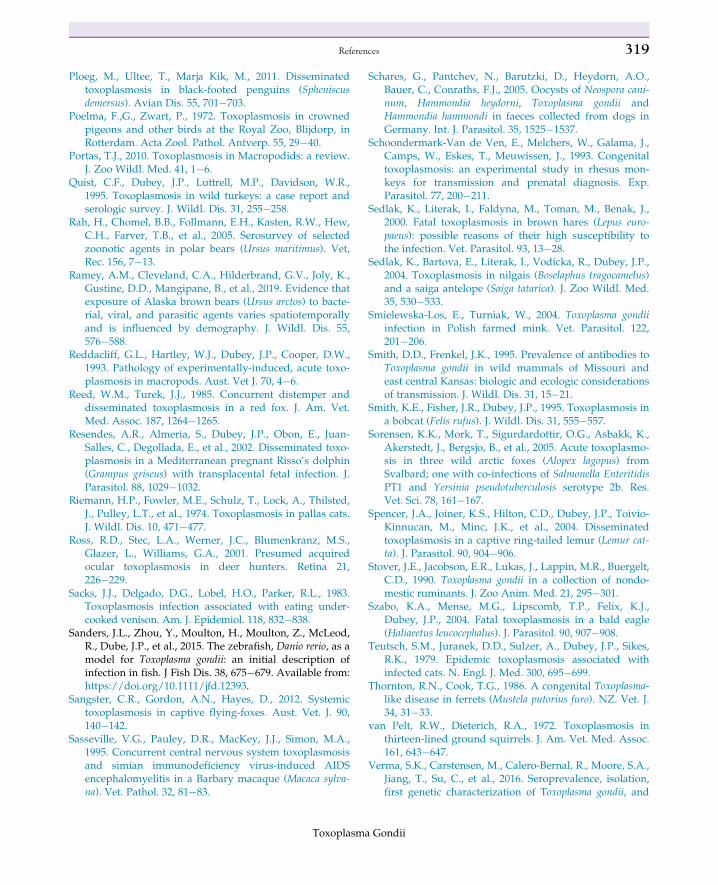
Ploeg, M., Ultee, T., Marja Kik, M., 2011. Disseminatedtoxoplasmosis in black-footed penguins (Spheniscusdemersus). Avian Dis. 55, 701�703.
Poelma, F.,G., Zwart, P., 1972. Toxoplasmosis in crownedpigeons and other birds at the Royal Zoo, Blijdorp, inRotterdam. Acta Zool. Pathol. Antverp. 55, 29�40.
Portas, T.J., 2010. Toxoplasmosis in Macropodids: a review.J. Zoo Wildl. Med. 41, 1�6.
Quist, C.F., Dubey, J.P., Luttrell, M.P., Davidson, W.R.,1995. Toxoplasmosis in wild turkeys: a case report andserologic survey. J. Wildl. Dis. 31, 255�258.
Rah, H., Chomel, B.B., Follmann, E.H., Kasten, R.W., Hew,C.H., Farver, T.B., et al., 2005. Serosurvey of selectedzoonotic agents in polar bears (Ursus maritimus). Vet,Rec. 156, 7�13.
Ramey, A.M., Cleveland, C.A., Hilderbrand, G.V., Joly, K.,Gustine, D.D., Mangipane, B., et al., 2019. Evidence thatexposure of Alaska brown bears (Ursus arctos) to bacte-rial, viral, and parasitic agents varies spatiotemporallyand is influenced by demography. J. Wildl. Dis. 55,576�588.
Reddacliff, G.L., Hartley, W.J., Dubey, J.P., Cooper, D.W.,1993. Pathology of experimentally-induced, acute toxo-plasmosis in macropods. Aust. Vet J. 70, 4�6.
Reed, W.M., Turek, J.J., 1985. Concurrent distemper anddisseminated toxoplasmosis in a red fox. J. Am. Vet.Med. Assoc. 187, 1264�1265.
Resendes, A.R., Almeria, S., Dubey, J.P., Obon, E., Juan-Salles, C., Degollada, E., et al., 2002. Disseminated toxo-plasmosis in a Mediterranean pregnant Risso’s dolphin(Grampus griseus) with transplacental fetal infection. J.Parasitol. 88, 1029�1032.
Riemann, H.P., Fowler, M.E., Schulz, T., Lock, A., Thilsted,J., Pulley, L.T., et al., 1974. Toxoplasmosis in pallas cats.J. Wildl. Dis. 10, 471�477.
Ross, R.D., Stec, L.A., Werner, J.C., Blumenkranz, M.S.,Glazer, L., Williams, G.A., 2001. Presumed acquiredocular toxoplasmosis in deer hunters. Retina 21,226�229.
Sacks, J.J., Delgado, D.G., Lobel, H.O., Parker, R.L., 1983.Toxoplasmosis infection associated with eating under-cooked venison. Am. J. Epidemiol. 118, 832�838.
Sanders, J.L., Zhou, Y., Moulton, H., Moulton, Z., McLeod,R., Dube, J.P., et al., 2015. The zebrafish, Danio rerio, as amodel for Toxoplasma gondii: an initial description ofinfection in fish. J Fish Dis. 38, 675�679. Available from:https://doi.org/10.1111/jfd.12393.
Sangster, C.R., Gordon, A.N., Hayes, D., 2012. Systemictoxoplasmosis in captive flying-foxes. Aust. Vet. J. 90,140�142.
Sasseville, V.G., Pauley, D.R., MacKey, J.J., Simon, M.A.,1995. Concurrent central nervous system toxoplasmosisand simian immunodeficiency virus-induced AIDSencephalomyelitis in a Barbary macaque (Macaca sylva-na). Vet. Pathol. 32, 81�83.
Schares, G., Pantchev, N., Barutzki, D., Heydorn, A.O.,Bauer, C., Conraths, F.J., 2005. Oocysts of Neospora cani-num, Hammondia heydorni, Toxoplasma gondii andHammondia hammondi in faeces collected from dogs inGermany. Int. J. Parasitol. 35, 1525�1537.
Schoondermark-Van de Ven, E., Melchers, W., Galama, J.,Camps, W., Eskes, T., Meuwissen, J., 1993. Congenitaltoxoplasmosis: an experimental study in rhesus mon-keys for transmission and prenatal diagnosis. Exp.Parasitol. 77, 200�211.
Sedlak, K., Literak, I., Faldyna, M., Toman, M., Benak, J.,2000. Fatal toxoplasmosis in brown hares (Lepus euro-paeus): possible reasons of their high susceptibility tothe infection. Vet. Parasitol. 93, 13�28.
Sedlak, K., Bartova, E., Literak, I., Vodicka, R., Dubey, J.P.,2004. Toxoplasmosis in nilgais (Boselaphus tragocamelus)and a saiga antelope (Saiga tatarica). J. Zoo Wildl. Med.35, 530�533.
Smielewska-Los, E., Turniak, W., 2004. Toxoplasma gondiiinfection in Polish farmed mink. Vet. Parasitol. 122,201�206.
Smith, D.D., Frenkel, J.K., 1995. Prevalence of antibodies toToxoplasma gondii in wild mammals of Missouri andeast central Kansas: biologic and ecologic considerationsof transmission. J. Wildl. Dis. 31, 15�21.
Smith, K.E., Fisher, J.R., Dubey, J.P., 1995. Toxoplasmosis ina bobcat (Felis rufus). J. Wildl. Dis. 31, 555�557.
Sorensen, K.K., Mork, T., Sigurdardottir, O.G., Asbakk, K.,Akerstedt, J., Bergsjo, B., et al., 2005. Acute toxoplasmo-sis in three wild arctic foxes (Alopex lagopus) fromSvalbard; one with co-infections of Salmonella EnteritidisPT1 and Yersinia pseudotuberculosis serotype 2b. Res.Vet. Sci. 78, 161�167.
Spencer, J.A., Joiner, K.S., Hilton, C.D., Dubey, J.P., Toivio-Kinnucan, M., Minc, J.K., et al., 2004. Disseminatedtoxoplasmosis in a captive ring-tailed lemur (Lemur cat-ta). J. Parasitol. 90, 904�906.
Stover, J.E., Jacobson, E.R., Lukas, J., Lappin, M.R., Buergelt,C.D., 1990. Toxoplasma gondii in a collection of nondo-mestic ruminants. J. Zoo Anim. Med. 21, 295�301.
Szabo, K.A., Mense, M.G., Lipscomb, T.P., Felix, K.J.,Dubey, J.P., 2004. Fatal toxoplasmosis in a bald eagle(Haliaeetus leucocephalus). J. Parasitol. 90, 907�908.
Teutsch, S.M., Juranek, D.D., Sulzer, A., Dubey, J.P., Sikes,R.K., 1979. Epidemic toxoplasmosis associated withinfected cats. N. Engl. J. Med. 300, 695�699.
Thornton, R.N., Cook, T.G., 1986. A congenital Toxoplasma-like disease in ferrets (Mustela putorius furo). NZ. Vet. J.34, 31�33.
van Pelt, R.W., Dieterich, R.A., 1972. Toxoplasmosis inthirteen-lined ground squirrels. J. Am. Vet. Med. Assoc.161, 643�647.
Verma, S.K., Carstensen, M., Calero-Bernal, R., Moore, S.A.,Jiang, T., Su, C., et al., 2016. Seroprevalence, isolation,first genetic characterization of Toxoplasma gondii, and
Toxoplasma Gondii
319References

possible congenital transmission in wild moose fromMinnesota, USA. Parasitol. Res. 115, 687�690.
Wallace, M.R., Rossetti, R.J., Olson, P.E., 1993. Cats andtoxoplasmosis risk in HIV-infected adults. J. Am. Med.Assoc. 269, 76�77.
Walsh, C.P., Hammond, S.E., Zajac, A.M., Lindsay, D.S.,1999. Survival of Toxoplasma gondii tachyzoites in goatmilk: potential source of human toxoplasmosis. J.Eukart. Microbiol. 73s�74s.
Wong, M.M., Kozek, W.J., Karr Jr., S.L., Brayton, M.A.,Theis, J.H., Hendrickx, A.G., 1979. Experimental con-genital infection of Toxoplasma gondii in Macaca arctoides.Asian J. Infect. Dis. 3, 61�67.
Work, T.M., Massey, J.G., Rideout, B.A., Gardiner, C.H.,Ledig, D.B., Kwok, O.C., et al., 2000. Fatal toxoplasmo-sis in free-ranging endangered ‘Alala from Hawaii. J.Wildl. Dis. 36, 205�212.
Yai, L.E.O., Ragozo, A.M.A., Soares, R.M., Pena, H.F.J.,Su, C., Gennari, S.M., 2009. Genetic diversity amongcapybara (Hydrochaeris hydrochaeris) isolates ofToxoplasma gondii from Brazil. Vet. Parasitol. 162,332�337.
Further reading
DiGuardo, G., Agrimi, U., Morelli, L., Cardeti, G.,Terracciano, G., Kennedy, S., 1995. Post mortem investi-gations on cetaceans found stranded on the coasts ofItaly between 1990 and 1993. Vet. Rec. 136, 439�442.
Sreekumar, C., Graham, D.H., Dahl, E., Lehmann, T.,Raman, M., Bhalerao, D.P., et al., 2003. Genotyping ofToxoplasma gondii isolates from chickens from India.Vet. Parasitol. 118, 187�194.
320 6. Toxoplasmosis in wild and domestic animals
Toxoplasma Gondii