Embed Size (px)
Citation preview

Using a Genetically Encoded Sensor to Identify Inhibitors ofToxoplasma gondii Ca2� Signaling*□S
Received for publication, November 10, 2015, and in revised form, February 23, 2016 Published, JBC Papers in Press, March 1, 2016, DOI 10.1074/jbc.M115.703546
Saima M. Sidik‡, Miryam A. Hortua Triana§, Aditya S. Paul¶, Majida El Bakkouri�, Caroline G. Hackett‡, Fanny Tran**,Nicholas J. Westwood**, Raymond Hui�, William J. Zuercher‡‡, Manoj T. Duraisingh¶, Silvia N. J. Moreno§,and Sebastian Lourido‡1
From the ‡Whitehead Institute for Biomedical Research, Cambridge, Massachusetts 02142, the §Center for Tropical and EmergingGlobal Diseases, Department of Cellular Biology, University of Georgia, Athens, Georgia 30602, the ¶Department of Immunologyand Infectious Diseases, Harvard T. H. Chan School of Public Health, Boston, Massachusetts 02115, the �Structural GenomicsConsortium, University of Toronto, Toronto, Ontario M5G 1L7, Canada, the **School of Chemistry and Biomedical SciencesResearch Complex, University of St. Andrews and EaStCHEM, North Haugh, St. Andrews, Fife KY16 9ST, Scotland, United Kingdom,and the ‡‡Division of Chemical Biology and Medicinal Chemistry, UNC Eshelman School of Pharmacy, University of North Carolina,Chapel Hill, North Carolina 27599
The life cycles of apicomplexan parasites progress in accord-ance with fluxes in cytosolic Ca2�. Such fluxes are necessary forevents like motility and egress from host cells. We used geneti-cally encoded Ca2� indicators (GCaMPs) to develop a cell-basedphenotypic screen for compounds that modulate Ca2� signalingin the model apicomplexan Toxoplasma gondii. In doing so, wetook advantage of the phosphodiesterase inhibitor zaprinast,which we show acts in part through cGMP-dependent proteinkinase (protein kinase G; PKG) to raise levels of cytosolic Ca2�.We define the pool of Ca2� regulated by PKG to be a neutralstore distinct from the endoplasmic reticulum. Screening alibrary of 823 ATP mimetics, we identify both inhibitors andenhancers of Ca2� signaling. Two such compounds constitutenovel PKG inhibitors and prevent zaprinast from increasingcytosolic Ca2�. The enhancers identified are capable of releas-ing intracellular Ca2� stores independently of zaprinast or PKG.One of these enhancers blocks parasite egress and invasion andshows strong antiparasitic activity against T. gondii. The samecompound inhibits invasion of the most lethal malaria parasite,Plasmodium falciparum. Inhibition of Ca2�-related phenotypesin these two apicomplexan parasites suggests that depletion ofintracellular Ca2� stores by the enhancer may be an effectiveantiparasitic strategy. These results establish a powerful newstrategy for identifying compounds that modulate the essentialparasite signaling pathways regulated by Ca2�, underscoring theimportance of these pathways and the therapeutic potential oftheir inhibition.
Apicomplexan parasites, such as Toxoplasma gondii andPlasmodium spp., the causative agents of toxoplasmosis andmalaria, require changes in cytosolic Ca2� concentrations toegress from host cells and move within the infected organism(1– 6). These pathways therefore hold tremendous therapeuticpotential, not only due to their importance in parasite biologybut because of their divergence from similar pathways in hostcells. Compounds targeting Ca2� signaling in parasites havebeen shown to be effective antiparasitics (discussed below).However, few of the molecules involved in regulating Ca2�
homeostasis and signaling have been identified in parasites, andtheir interplay is only evident in live cells. This has created aneed for new methods to study Ca2� signaling pathways inapicomplexan parasites, with the hope of defining the essentialcomponents and identifying novel inhibitors.
The mechanisms for Ca2� entry into the cytoplasm and thephysiologically relevant sources of Ca2� remain poorly definedin apicomplexan parasites. Ca2� can be mobilized from theparasite’s intracellular stores, or it can be drawn from the envi-ronment. Current evidence points toward intracellular storesbeing sufficient for parasites to move between cells (6 – 8),although virulence of T. gondii is enhanced by extracellularCa2� (9). The best studied of these intracellular stores is theendoplasmic reticulum (ER).2 This organelle is a highly net-worked, dynamic structure (10) that has been shown to consti-tute multiple spatially independent Ca2� stores in some celltypes (11). Such compartmentalization has also been hypothe-sized to occur in T. gondii (4). Mammalian cells store Ca2� inendosomes, lysosomes (12), and the Golgi (13), in addition tothe ER (14). Some alveolates, like Paramecium, additionallycontain a network of alveolar sacs that sequester Ca2� in anATP-dependent manner, with physiologically relevant affini-ties (15, 16). Whether the inner membrane complex, an api-complexan structure homologous to alveolar sacs, also storesCa2� remains to be determined. Like many other eukaryotes,
* This work was supported in part by National Institutes of Health GrantsAI-110027 and AI-096836 (to S. N. J. M.) and 1DP5OD017892 (to S. L.). Theauthors declare that they have no conflicts of interest with the contents ofthis article. The content is solely the responsibility of the authors and doesnot necessarily represent the official views of the National Institutes ofHealth.Author’s Choice—Final version free via Creative Commons CC-BY license.
□S This article contains supplemental Table S1, Figs. S1 and S2, and VideosS1–S3.
1 To whom correspondence should be addressed: Whitehead Institute forBiomedical Research, 9 Cambridge Center, Cambridge, MA 02142. Tel.:617-324-4920; Fax: 617-258-5213; E-mail: [email protected].
2 The abbreviations used are: ER, endoplasmic reticulum; GPN, L-phenylala-nine-naphthylamide; IP3, inositol 1,4,5-triphosphate; PKG, protein kinaseG; HFF, human foreskin fibroblast; PP, pyrazolopyridazine; Cpd1 and Cpd2,compound 1 and 2, respectively; Enh1 and Enh2, enhancer 1 and 2, respec-tively; Inh1 and Inh2, inhibitor 1 and 2, respectively.
THE JOURNAL OF BIOLOGICAL CHEMISTRY VOL. 291, NO. 18, pp. 9566 –9580, April 29, 2016Author’s Choice © 2016 by The American Society for Biochemistry and Molecular Biology, Inc. Published in the U.S.A.
crossmark
9566 JOURNAL OF BIOLOGICAL CHEMISTRY VOLUME 291 • NUMBER 18 • APRIL 29, 2016
at Massachusetts Institute of T
echnology on May 17, 2016
http://ww
w.jbc.org/
Dow
nloaded from
at Massachusetts Institute of T
echnology on May 17, 2016
http://ww
w.jbc.org/
Dow
nloaded from
at Massachusetts Institute of T
echnology on May 17, 2016
http://ww
w.jbc.org/
Dow
nloaded from
at Massachusetts Institute of T
echnology on May 17, 2016
http://ww
w.jbc.org/
Dow
nloaded from
apicomplexans also possess acidic vacuoles known as acidocal-cisomes that contain Ca2� in complex with pyrophosphate andpolyphosphates. This Ca2� can be released pharmacologically,but the function of acidocalcisomes remains unclear in apicom-plexans (17). A final acidic Ca2� store described in T. gondii isthe plantlike vacuole or vacuolar compartment. The plantlikevacuole is an acidic organelle that releases Ca2� upon treat-ment with L-phenylalanine-naphthylamide (GPN), which inother systems causes ion leakage from lysosomal compart-ments (18). Although implicated in ionic homeostasis, thesephenotypes have not been linked to the plantlike vacuole’sfunction as a Ca2� store (18). Due to the lack of characterizedregulatable Ca2� channels, it remains an open question whichof these Ca2� sources are involved in parasite motility andinvasion.
Recent evidence suggests that PKG may play a role in regu-lating parasite Ca2�. In Plasmodium berghei, PKG has beenproposed to influence Ca2� homeostasis, thereby regulatingegress from host cells. Activation of PKG leads to change in thelevels of the lipid precursors of inositol 1,4,5-triphosphate (IP3).This has been hypothesized to increase IP3, which causesrelease of Ca2� from the ER through binding to the IP3 recep-tor, although such a channel remains to be identified in apicom-plexan parasites (19). PKG is also known to regulate egress inT. gondii, although its effect on parasite Ca2� has not beendetermined (20). In both parasites, PKG can be pharmacologi-cally activated using the mammalian phosphodiesterase inhib-itor zaprinast (20, 21). In Plasmodium, zaprinast treatmentleads to an increase in cyclic GMP levels that presumably acti-vates PKG (22). Further characterization of zaprinast’s mecha-nism of action could therefore shed light on Ca2� signaling.
Targeting the downstream effectors of Ca2� signaling hasbeen shown to hold therapeutic value against T. gondii. Com-pounds targeting Ca2�-dependent protein kinase 1, a regulatorof egress and invasion, reduce proliferation in cell culture andcyst burden in the brains of T. gondii-infected mice (23, 24).Drugs targeting Ca2�-related processes are appealing not onlybecause of this historical success but also because many pro-teins involved in parasite Ca2� signaling are sufficiently diver-gent from their mammalian counterparts to enable the designof drugs with minimal off-target effects. For example, proteinkinase G (PKG), which regulates egress, invasion, and motilityin T. gondii and Plasmodium spp. (19, 20, 25, 26), is sufficientlydifferent from mammalian PKG to be selectively inhibited (27).Similarly, the Ca2�-dependent protein kinases lack homo-logues in mammalian cells (28), making them attractive drugtargets.
In this study, we use both chemical and genetic Ca2� indica-tors to define the regulatory circuits that mediate Ca2� releasein T. gondii and identify small molecules that modulate thisprocess. We determine the effect of T. gondii PKG on cytosolicCa2� following treatment with zaprinast and characterize thesource of the Ca2� released in this process as a neutral storedistinct from the ER. Using genetically encoded Ca2� indica-tors recently established in T. gondii (29), we develop a cell-based phenotypic screen that allows us to monitor Ca2� signal-ing in live cells without the technical challenges of conventionalchemical Ca2� indicators. Using this platform, we have been
able to identify, in an unbiased manner, compounds that inter-fere with Ca2� signaling. In contrast to enzyme-based assays,this system enables us to probe a broader swath of parasitebiology. Recent analysis indicates that such phenotypic screensare more likely to lead to clinically approved drugs than the farmore prevalent molecular target-based approaches (30). Ourscreen identified two novel PKG inhibitors that abrogate theeffect of zaprinast, as well as two compounds that increase cyto-solic Ca2� through an independent pathway. From the latter,one compound blocks invasion of both T. gondii and Plasmo-dium falciparum. Our results demonstrate the potential of thisstrategy to explore parasite Ca2� signaling and identify newcompounds with antiparasitic potential against multiple api-complexan parasites.
Experimental Procedures
Strain Construction and Maintenance—Recombinant hu-man T. gondii strain RH parasites were maintained in humanforeskin fibroblasts (HFFs) grown in DMEM supplementedwith 10% heat-inactivated fetal bovine serum (FBS) and 10�g/ml gentamicin. PKG-T and PKG-M alleles were con-structed as described previously (31). GCaMP5 was amplifiedfrom pCMV-GCaMP5G (32) with primers containing NsiI andPacI restriction sites (forward primer, 5�-gcg atg cat cct ttt tcgaca aaa tgg gtt ctc atc atc atc atc atc; reverse primer, 5�-gcg ttaatt aat cac ttc gct gtc atc att tg) and cloned directionally, replac-ing the CAT gene in pSAG1/2-CAT (33) to generate pSAG1-GCaMP5. Recombinant human parasites were co-transfectedwith pSAG1-GCaMP5 and pSAG1/2-CAT and selected withchloramphenicol (40 �M), and clones were isolated by limitingdilution. The GCaMP6f strains was similarly derived, asdescribed previously (29). Both GCaMP strains were main-tained under selection to prevent loss of the transgene. TheGFP-expressing strain was kindly provided by Jeroen P. J. Saeij(34).
Store Activation and Cpd1 Inhibition Experiments withGCaMP6f—GCaMP6f-expressing T. gondii were suspended at2 � 107 parasites/ml in basal Ca2� buffer (140 mM NaCl, 10 mM
potassium gluconate, 2.7 mM MgSO4, 2 mM glucose, 250 �M
EGTA, 85 �M CaCl2, 10 mM HEPES, pH 7.3) or extracellularCa2� buffer (140 mM NaCl, 10 mM potassium gluconate, 2.7 mM
MgSO4, 2 mM glucose, 1 mM CaCl2, 10 mM HEPES, pH 7.3),supplemented with 1% FBS when noted. For the Cpd1 inhibi-tion experiments, parasites were suspended in Ringer’s solution(115 mM NaCl, 3 mM KCl, 2 mM CaCl2, 1 mM MgCl2, 3 mM
NaH2PO4, 10 mM HEPES, 10 mM glucose, 1% FBS). In all cases,100 �l of suspended parasites were applied to each well of a CellCarrier 96-well plate (PerkinElmer Life Sciences). For storeactivation experiments, parasites were incubated on ice for 5min before the addition of Enh1 or Enh2 (10 �M final concen-tration), zaprinast (100 �M final), or DMSO (0.3% final) sus-pended in the same buffers as the parasites to which they wereadded. To examine the effect of Cpd1 on zaprinast or Enh1treatment of GCaMP6-expressing parasites, fluorescence wasrecorded for 100 s before adding Cpd1 to a concentration of 1.2�M. Fluorescence was recorded for an additional 300 s beforetreatment with 10 �M Enh1 or 100 �M zaprinast. Parasites wereincubated on ice for 5 min before the addition of zaprinast to
Identifying Modulators of Apicomplexan Ca2� Signaling
APRIL 29, 2016 • VOLUME 291 • NUMBER 18 JOURNAL OF BIOLOGICAL CHEMISTRY 9567
at Massachusetts Institute of T
echnology on May 17, 2016
http://ww
w.jbc.org/
Dow
nloaded from

slow down the response and facilitate observation of peak fluo-rescence. Fluorescence was read with an excitation wavelengthof 485 nm and an emission wavelength of 528 nm every 10 s ina BioTek Cytation 3. The assay plate was shaken for 1 s beforeeach read.
Compound Screen—GCaMP5-expressing parasites were sus-pended in Ringer’s solution at 4 � 107 parasites/ml, and 50 �l ofparasites were applied to each well of a 96-well plate (Costar,catalog no. 3631). Parasites were treated with compounds fromthe GSK libraries PKIS and PKIS2 at 13.3 �M or 1.33% DMSO asa vehicle control and then incubated at 37 °C with 5% CO2 for10 min. Parasites were then treated with 100 �M zaprinast or0.1% DMSO as a vehicle control and incubated at 37 °C with 5%CO2 for an additional 4 min before measuring fluorescencewith excitation and emission wavelengths of 485 and 525 nm,respectively, on a SpectraMax M3 (Molecular Devices). Basalfluorescence, measured from untreated parasites, was sub-tracted from all values, and results were expressed as -foldchange from parasites treated only with zaprinast. Z� factorswere determined from (i) parasites treated with zaprinast versusuntreated parasites and (ii) parasites treated with a final con-centration of 1 �M Cpd2 or a vehicle control followed by zapri-nast, using the formula, Z� � 1 � 3(�p � �n)/(��p � �n�), where�p and �n indicate S.D. values of positive and negative controls,and �p and �n indicate the means of positive and negative con-trols, respectively.
In Vitro PKG Assays—Recombinant T. gondii PKG wasexpressed using a baculovirus system. Synthetic DNA codingfor the protein was amplified and subcloned into the pFBOH-MHL vector, which confers an N-terminal His6 tag with atobacco etch virus cleavage site. The resulting plasmid wastransformed into DH10BacTM Escherichia coli competent cellsto produce recombinant viral DNA. P3 viral stocks were used toinfect Sf9 insect cells grown in HyQ SFX insect serum-freemedium (Thermo Fisher Scientific). The culture was incubatedat 27 °C and shaken at 100 rpm. After 60 –72 h, the cells wereharvested. The His-tagged T. gondii PKG samples were purifiedby affinity chromatography and size exclusion chromatographyusing an ÄKTAxpress system equipped with a SuperdexTM 20010/300 column (GE Healthcare, Mississauga, Canada). In vitrokinase assays were performed using a PKG assay kit (CycLex) asper the manufacturer’s instructions. Inhibitors were testedagainst 6.76 nM recombinant PKG, which was active within thelinear range of the assay.
Structural Analysis—Structures of various protein kinases incomplex with pyrazolopyridazine (PP) and oxindole derivativeswere obtained from the RCSB Protein Data Bank (35) andaligned on the basis of their kinase domains. For the PP analysis,the CDK2 structures 3EID and 3EJ1 (36) and the p38 MAPKstructure 3GCP (37) were aligned to the ERK2 structure 1WZY(38). For the oxindole analysis, the PDK1 structures 2PE0 and2PE2 (39) and the Alk5 structure 2X7O were aligned to theNek2 structure 2JAV (40).
Lactate Dehydrogenase Release Egress Assays—ConfluentHFF monolayers in 96-well plates were infected with 5 � 104
parasites/well. The HFF monolayer was washed once with Rin-ger’s solution �18 h later, and 50 �l of Ringer’s solution wasapplied to each well. Drugs suspended in Ringer’s solution were
applied at 1.33 times the indicated concentrations. 1.33%DMSO in Ringer’s solution was used as a vehicle control. Cellswere incubated at 37 °C with 5% CO2 for 20 min, stimulatedwith 500 �M zaprinast or 0.5% DMSO (vehicle), and incubatedagain for an additional 5 min. The cells were centrifuged at400 � g for 5 min, before collecting 50 �l of each supernatant.The lactate dehydrogenase levels in the supernatant sampleswere quantified using the CytoTox 96 cytotoxicity assay (Pro-mega) as per the manufacturer’s instructions.
Plaque Assays—T. gondii parasites were suspended in growthmedium supplemented with Enh1, zaprinast, or DMSO alone atthe indicated concentrations. The amount of DMSO in alltreatment groups was normalized to 0.1%. Parasites were incu-bated at 37 °C with 5% CO2 for 20 min before infecting HFFmonolayers in 6-well plates using 3 ml carrying 100 parasites/well. Medium containing the highest concentration of eachdrug was also applied to HFF cells in the absence of parasites toassess the effects of the drugs on host cell viability. The parasiteswere allowed to plaque for 8 days before fixing with 70% ethanoland staining with 0.1% crystal violet.
Lytic Assays—Drugs were suspended at twice the indicatedconcentrations in growth medium and mixed with an equalvolume of parasites at an initial concentration of 106 parasites/ml. The maximum concentration of DMSO was added as avehicle control: 1% DMSO in comparisons with zaprinast and0.013% in comparisons with Enh1. Parasites were preincubatedwith the compounds for 20 min at 37 °C with 5% CO2, and then200 �l/well (105 parasites) was added to host cell monolayers in96-well plates. Assay plates were incubated at 37 °C with 5%CO2 for 3 days and then fixed in 70% ethanol and stained with0.1% crystal violet. Absorbance at 590 nm was read as a measureof host cell lysis.
Cell-wounding Assays—Parasites suspended in Ringer’s solu-tion at 5 � 105 parasites/ml were pretreated with varying con-centrations of zaprinast (as indicated) for 20 min at 37 °C with5% CO2. 105 parasites/well were then applied to confluent hostcell monolayers, and plates were centrifuged at 290 � g for 5min. Following 1 h at 37 °C with 5% CO2, plates were centri-fuged again at 500 � g for 5 min. 50 �l of supernatant werecollected from each well, and lactate dehydrogenase was quan-tified using the CytoTox 96 cytotoxicity assay (Promega).
Video Microscopy Egress Assays—Host cell monolayers inCell Carrier 96-well plates (PerkinElmer Life Sciences) wereinfected with 5 � 104 GFP-expressing parasites/well. 18 hpostinfection, the medium was exchanged for Ringer’s solution,and the intracellular parasites were treated with 10 �M Enh1,500 �M zaprinast, or 0.5% DMSO. Images were acquired every10 s for 30 min. To measure the effect of Enh1 on zaprinast andA23187-induced egress, the procedure was repeated, addingeither 12.5 �M Enh1 or 0.13% DMSO and imaging for 10 min,before stimulation with 500 �M zaprinast or 1 �M A23187 (Cal-biochem) and imaging for an additional 10 min. In all cases,images were acquired with a �4 objective on a Cytation3 reader(BioTek) using excitation and emission wavelengths of 485 and528 nm, respectively. Intact vacuoles were defined as objects ofat least 78 �m2 with a circularity of at least 0.5, as determined inFiji after default thresholding (41).
Identifying Modulators of Apicomplexan Ca2� Signaling
9568 JOURNAL OF BIOLOGICAL CHEMISTRY VOLUME 291 • NUMBER 18 • APRIL 29, 2016
at Massachusetts Institute of T
echnology on May 17, 2016
http://ww
w.jbc.org/
Dow
nloaded from

Video Microscopy—Parasites expressing GCaMP5 weretransfected with p30-DsRed and used to infect host cell mono-layers in 3-cm glass bottom dishes (MatTek). Approximately20 h after infection, medium was exchanged for Ringer’s solu-tion, and parasites were imaged on a Nikon Eclipse Ti epifluo-rescence microscope, equipped with an enclosure heated to37 °C. Images were acquired every 5 s for 10 min following theaddition of zaprinast to a final concentration of 100 �M. Toquantify changes in fluorescence, videos were analyzed in Fiji tomeasure the average fluorescence intensity in specific circularregions of interest 10 �m in diameter. To examine the effects ofzaprinast and Enh1 on mammalian cells, 6 � 103 HeLa cellswere seeded in each well of a Cell Carrier 96-well plate. Approx-imately 24 h later, HeLa cells were transfected with 100 ng ofthe pCMV-R-GECO (42) using Fugene (Promega) as per themanufacturer’s instructions. Cells were washed in Ringer’ssolution and then treated with 10 �M Enh1, 500 �M zaprinast,or 2 �M A23187. Images were acquired every 12 s for 30 min ona Cytation3 reader (BioTek) using an excitation wavelength of531 and an emission wavelength of 593. Higher resolution vid-eos were similarly acquired from HeLa cells seeded in 3-cmglass bottom dishes (MatTek) and transfected with 200 ng ofpCMV-R-GECO per dish. Cells were treated with either 500 �M
zaprinast or 10 �M Enh1 in Ringer’s solution and imaged every250 ms using a Nikon Eclipse Ti microscope. Videos were ana-lyzed in Fiji using default thresholding for the red channel todetermine the mean gray value in each slice for individual cells.Kymographs were constructed from videos of intracellularGCaMP6f-expressing parasites, following the same treatmentas for the video microscopy egress assays, except half as manyparasites were used to infect the monolayers, and a �20 objec-tive was used to acquire the images. Regions of interest weredefined by outlining parasites in Fiji and measuring the meanfluorescence of each region over time.
Fura-2 Recordings—Tachyzoite loading with Fura-2/AM wasdone as described previously (17). Briefly, freshly lysed parasiteswere washed twice with buffer A (116 mM NaCl, 5.4 mM KCl, 0.8mM MgSO4, 5.5 mM D-glucose, and 50 mM HEPES, pH 7.4) andresuspended to a final density of 1 � l09 parasites/ml in loadingbuffer (buffer A plus 1.5% sucrose and 5 �M Fura-2/AM). Thesuspension was incubated for 26 min at 26 °C with mild agita-tion. Subsequently, the parasites were washed twice with bufferA to remove extracellular dye, resuspended to a final density of1 � 109 parasites/ml in buffer A, and kept in ice. Parasites areviable for a few hours under these conditions. For fluorescencemeasurements, 2 � 107 parasites/ml were placed in a cuvettewith 2.5 ml of Ringer’s solution. Fluorescence measurementswere done in a thermostatically controlled Hitachi F-7000 spec-trofluorometer using the Fura-2 conditions for excitation (340and 380 nm) and emission (510 nm). The Fura-2 fluorescenceresponse to Ca2� was calibrated from the ratio of 340/380-nmfluorescence values after subtraction of the background fluo-rescence of the cells at 340 and 380 nm, as described previously(43). The Ca2� release rate is the change in Ca2� concentrationduring the initial 20 s after the addition of compound.
Chemical Susceptibility of P. falciparum Egress and Erythro-cyte Invasion—Blood stage P. falciparum parasites of strain3D7 were obtained from the Walter and Eliza Hall Institute
(Melbourne, Australia) and cultured as described previously(44). Parasites were maintained in O� human erythrocytes(Research Blood Components, Boston, MA), at 2% hematocrit,in RPMI 1640 supplemented with HEPES (25 mM), hypoxan-thine (50 mg/liter), sodium bicarbonate (2.42 mM), and Albu-max (4.31 mg/ml). Cultures were incubated at 37 °C inmicroaerophilic atmospheric conditions (1% O2, 5% CO2, 94%N2) within modular incubator chambers.
To test the specific effects of Enh1 on egress of parasites fromschizonts as well as erythrocyte invasion by liberated merozo-ites, mature schizont stage parasites were purified from 4 ml ofblood stage culture (10 –20% hematocrit) by centrifugation(930 � g, 15 min, low acceleration and deceleration) on 4 ml ofa 60% Percoll cushion (45). Schizonts were retrieved from themedium supernatant-Percoll interface. After at least threewashes in excess volumes of RPMI culture medium, schizontswere diluted with uninfected erythrocytes for a parasitemia of3–5% and further supplemented with 2 �M Cpd2 to allow sch-izont maturation up to the point of egress (22). After 1– 4 h atstandard culture conditions, cultures were washed at least threetimes in excess RPMI culture medium to remove Cpd2 andadded to 1 volume of Enh1 in RPMI or RPMI only (no drug) fora sample volume of 100 �l at 1% hematocrit. We similarly pre-pared samples of parasites supplemented with heparin (100units/ml), a specific inhibitor of erythrocyte invasion (46), toassess the background signal for ring stage parasitemia (seebelow). We fixed samples (4% paraformaldehyde and 0.0075%glutaraldehyde in PBS (47)) at the outset of the experiment(untreated only) and after 1–2 h of incubation in standard cul-ture conditions (all samples). After extensive washing in PBSand staining with SYBR Green I (1:1000 dilution in PBS; Invit-rogen), schizont and ring stage parasitemia were measured byflow cytometry in the FITC channel as described previously(48, 49).
T. gondii Invasion Assays—Invasion assays were performedas described previously (20). Briefly, freshly lysed tachyzoiteswere preincubated for 10 min in varying concentrations ofEnh1, keeping the total concentration of DMSO (vehicle) con-stant in all samples. HFF monolayers, seeded 48 h earlier, wereinfected at a multiplicity of infection of �10, and invasion wasallowed to proceed for 10 min at 37 °C. Following fixation,intracellular parasites were enumerated by immunofluores-cence and normalized to the number of host cells in a givenfield.
Results
Zaprinast Increases Cytosolic Ca2� in a PKG-dependentManner—Previous work has shown that zaprinast triggersT. gondii and P. falciparum egress in a PKG-dependent manner(20, 22). Because Ca2� is a necessary second messenger duringegress (8), we hypothesized that zaprinast might stimulateegress by increasing cytosolic Ca2� in the parasite. To test this,we generated a strain that expressed the genetically encodedCa2� indicator GCaMP5 (32) in the wild-type T. gondii recom-binant human background. These parasites were transfectedwith a plasmid encoding a constitutively secreted fluorescentfusion protein, p30-DsRed, which accumulates in the parasito-phorous vacuole before egress (50). This second sensor enabled
Identifying Modulators of Apicomplexan Ca2� Signaling
APRIL 29, 2016 • VOLUME 291 • NUMBER 18 JOURNAL OF BIOLOGICAL CHEMISTRY 9569
at Massachusetts Institute of T
echnology on May 17, 2016
http://ww
w.jbc.org/
Dow
nloaded from

us to visualize permeabilization of the parasitophorous vacuole,which has been demonstrated to occur via the Ca2�-regulatedsecretion of a perforin-like protein (50). Human fibroblastsinfected with recombinant human GCaMP5 were treated withzaprinast and monitored by live video microscopy. Zaprinastelicited a rapid increase in fluorescence compared with thevehicle alone, which was followed by a second, less intense peakof fluorescence (Fig. 1, A and B). This led to permeabilization ofthe vacuole membrane, as indicated by diffusion of DsRed, andsubsequent egress of the parasites from the host cell (supple-mental Video S1).
To determine whether the zaprinast-induced increase inCa2� represented release of intracellular stores or Ca2� entry,
we loaded wild-type parasites with the ratiometric Ca2� indi-cator Fura-2/AM and stimulated them with zaprinast in bufferscontaining either a basal Ca2� concentration (100 nM freeCa2�) or one resembling the extracellular environment (1.8mM). We found that cytosolic Ca2� increased in response tozaprinast under both conditions (Fig. 1C), although theresponse was magnified in the presence of extracellular Ca2�.This indicates that zaprinast-mediated Ca2� mobilizationoccurs through release of intracellular stores, which mayenhance Ca2� entry, as has been reported for other agonistscapable of releasing intracellular stores (9).
Compound 1 (Cpd1), a specific inhibitor of apicomplexanPKG (25), blocks zaprinast-induced egress (20). To determine
FIGURE 1. Zaprinast raises cytosolic Ca2� through the activation of PKG. A, video microscopy of intracellular parasites expressing both GCaMP5 andconstitutively secreted DsRed, following the addition of zaprinast at 0 s. B, GCaMP5 fluorescence in the region of the parasitophorous vacuole (green) or DsRedfluorescence in adjacent areas of the infected host cell (red) following the addition of zaprinast. Results shown are mean � S.E. for four experiments.Kymographs for GCaMP5 fluorescence in all four experiments are shown to indicate times of peak fluorescence (white asterisks) relative to the initiation ofegress from the region (white vertical lines). C, intracellular Ca2� concentrations, monitored over time, for wild-type parasites loaded with Fura-2/AM, sus-pended in buffer containing extracellular (1.8 mM) or basal (100 nM) free Ca2�, and stimulated with 100 �M zaprinast at 400 s. D, similar measurements wereperformed for PKG-M and PKG-T parasites loaded with Fura-2/AM and suspended in basal Ca2�. Cpd1 or vehicle was added at 100 s, and zaprinast was addedat 400 s, as indicated. E–G, intracellular calcium concentrations from parasites loaded with Fura-2/AM and then treated with zaprinast, ionomycin, GPN, orthapsigargin at 100 s (1) and 400 s (2), as indicated. Traces are representative of three independent experiments. Bar graphs report the change in cytosoliccalcium over 20 s following the addition of each drug. Results shown are mean � S.E. (error bars). Z, zaprinast; I, ionomycin; G, GPN; T, thapsigargin; *, p 0.05;one-tailed t test.
Identifying Modulators of Apicomplexan Ca2� Signaling
9570 JOURNAL OF BIOLOGICAL CHEMISTRY VOLUME 291 • NUMBER 18 • APRIL 29, 2016
at Massachusetts Institute of T
echnology on May 17, 2016
http://ww
w.jbc.org/
Dow
nloaded from

whether zaprinast’s effect on parasite Ca2� depends on PKG,we measured the response to zaprinast after Cpd1 treatment.Sensitivity of apicomplexan PKG to Cpd1 relies on the identityof a residue at the base of the ATP-binding pocket known as thegatekeeper (25). We therefore used strains engineered toexpress PKG with either the wild-type threonine or a bulkymethionine gatekeeper residue that renders PKG refractory toCpd1 inhibition (31). These strains were loaded with Fura-2/AM, treated with Cpd1 or vehicle, and then stimulated withzaprinast. Parasites treated with vehicle responded to the addi-tion of zaprinast with a sharp Ca2� spike. The addition of Cpd1did not change the response of the insensitive PKG-M strain.However, the Ca2� spike was severely diminished by Cpd1 inthe sensitive PKG-T strain, indicating the need for PKG in thisprocess (Fig. 1D). We also observed a slight decrease in thecytosolic Ca2� level of the PKG-T strain after Cpd1 treatment,perhaps indicating that PKG regulates the basal Ca2� level inaddition to enhancing it before egress.
Zaprinast Mobilizes a Neutral, SERCA-independent Ca2�
Store—To further characterize the source of zaprinast-mobi-lized Ca2�, we assessed the involvement of various Ca2� stor-age organelles. We loaded parasites with Fura-2/AM and sus-pended them in a buffer containing basal Ca2� and then addedthe Ca2� ionophore ionomycin. Binding of ionomycin to Ca2�
is pH-dependent and falls to negligible levels below pH 7 (51).Ionomycin has therefore been used to specifically mobilize neu-tral Ca2� stores in both mammalian cells (52) and T. gondii(17). We observed a peak in cytosolic Ca2� levels followingionomycin treatment, indicating that Ca2� had been mobilizedfrom neutral stores. Subsequent treatment with zaprinast didnot produce an additional Ca2� peak, indicating that the zapri-nast-mobilized store had already been depleted by ionomycin.In contrast, treating parasites with zaprinast followed by iono-mycin produced a Ca2� spike in response to each compound.These results were corroborated by analyzing the rate of Ca2�
release during the 20 s that followed the addition of each com-pound (Fig. 1E). Taken together, these results indicate that thezaprinast-mobilized store comprises a subset of the ionomycin-mobilized stores and as such is predicted to be neutral.
To further rule out acidic Ca2� stores, we treated parasiteswith GPN before or after the addition of zaprinast. GPN is spe-cifically hydrolyzed in lysosomal compartments, leading totheir leakage, as has been shown for the plantlike vacuole inT. gondii (18). As predicted by mobilization with ionomycin,the zaprinast-mobilized store was independent from this acidiccompartment (Fig. 1F).
The ER is the major neutral Ca2� store in many organisms(53). Thapsigargin is an inhibitor of the Ca2� reuptake pumpSERCA, which partially localizes to the ER in extracellularT. gondii tachyzoites (54). We treated parasites with thapsi-gargin before or after the addition of zaprinast and found thatzaprinast mobilized Ca2� with the same efficiency regardless ofthe treatment order. The independence of the zaprinast- andthapsigargin-mobilized stores was evident in the rate of Ca2�
release following each treatment (Fig. 1F). These results indi-cate either that zaprinast mobilizes a neutral store that is sepa-rate from the ER or that the ER is segmented into SERCA-de-
pendent and SERCA-independent Ca2� stores, as has beenhypothesized (4).
Identifying Small Molecules That Modulate Parasite Ca2�—Despite its central importance during infection, few com-pounds have been demonstrated to specifically interfere withthe Ca2� signaling pathways of apicomplexan parasites. Com-pounds that modulate Ca2� signaling can have clinical value aswell as being useful in research. We predicted that the zaprinastresponse measured in GCaMP5-expressing parasites couldform the basis for a phenotypic screen. Such a screen wouldbenefit from the known importance of Ca2� signaling in para-site biology along with the reported success rate of cell-basedphenotypic assays. We designed a screen wherein extracellularGCaMP5-expressing parasites were pretreated with com-pounds for 10 min before stimulation with zaprinast, and par-asite fluorescence was measured 4 min later. We expected thatcompounds that interfered with the zaprinast response wouldreduce fluorescence. We calculated a Z�-factor for this screento determine its dynamic range and suitability to high through-put screening. To do so, we compared zaprinast-treated para-sites to untreated parasites or those pretreated with the PKGinhibitor Compound 2 (Cpd2) (55). These scenarios yieldedZ�-factors of 0.58 and 0.55, respectively, well above the 0.5Z�-factor considered acceptable for high throughput screening(56). This phenotypic screen is therefore suitable for analyzinglarge libraries of compounds to identify compounds interferingwith parasite Ca2� signaling (Fig. 2A).
We obtained the compound libraries published kinase inhib-itor sets PKIS1 (57) and PKIS2 (unpublished), from Glaxo-SmithKline and applied our phenotypic screen to the 823 ATPmimetics represented in these collections (Fig. 2B and supple-mental Table S1). The compounds were screened at a single 10�M dose with two biological replicates. We identified 37 puta-tive inhibitors and 14 putative enhancers, defined as com-pounds that resulted in fluorescence readings that were morethan two S.D. values below or above the mean of all compounds,respectively. Compounds that interfered with accurate mea-surement of GCaMP5 fluorescence, including two putativeenhancers, were identified and excluded from subsequentexperiments.
We wondered whether we could identify classes of moleculesthat inhibit or enhance Ca2� mobilization. To this end, weexamined analogs of the most potent enhancer and inhibitor,which we here refer to as Enh1 (PKIS: GSK260205A) and Inh1(PKIS: GW827099X) (Fig. 2C). Compounds with the same corestructure as Enh1 generally produced a -fold change in fluores-cence of 1 in our screen, indicating that parasite fluorescencewas greater in the presence of such compounds than with zapri-nast alone (Fig. 2D and supplemental Fig. S1). A Kolmogorov-Smirnov test comparing the values obtained from all com-pounds with those obtained from the Enh1 analogs revealed asignificant difference (p � 0.0042) (Fig. 2D). The combinationof a phenyl group at the R1 position and a 3-aminopropyloxygroup at the R3 position, as found in Enh1, appeared to producea particularly effective enhancer; compounds having eithergroup without the other produced only a mild effect. A similaranalysis of Inh1 analogs revealed that these compounds tendedto inhibit zaprinast-induced Ca2� mobilization (Fig. 2D and
Identifying Modulators of Apicomplexan Ca2� Signaling
APRIL 29, 2016 • VOLUME 291 • NUMBER 18 JOURNAL OF BIOLOGICAL CHEMISTRY 9571
at Massachusetts Institute of T
echnology on May 17, 2016
http://ww
w.jbc.org/
Dow
nloaded from

supplemental Fig. S2). The fluorophenyl group in the R1 posi-tion of Inh1 may be responsible for this compound’s stronginhibitory effect (as discussed below). This 4-fluorophenyl isalso found in Cpd1 and Cpd2. No other R1 side group produceda similarly strong response, although an analogue containing atrifluoromethyl group at the R1 position elicited the secondlargest response in the group. In summary, although Enh1 andInh1 are unique within the set of compounds tested for theirabilities to augment and repress cytosolic Ca2�, their corestructures appear to be generally well suited to these purposes.Indeed, further exploration of these scaffolds could lead to theidentification of improved modulators of parasite Ca2�
signaling.Two Inhibitors Target PKG Using Distinct Chemical Scaf-
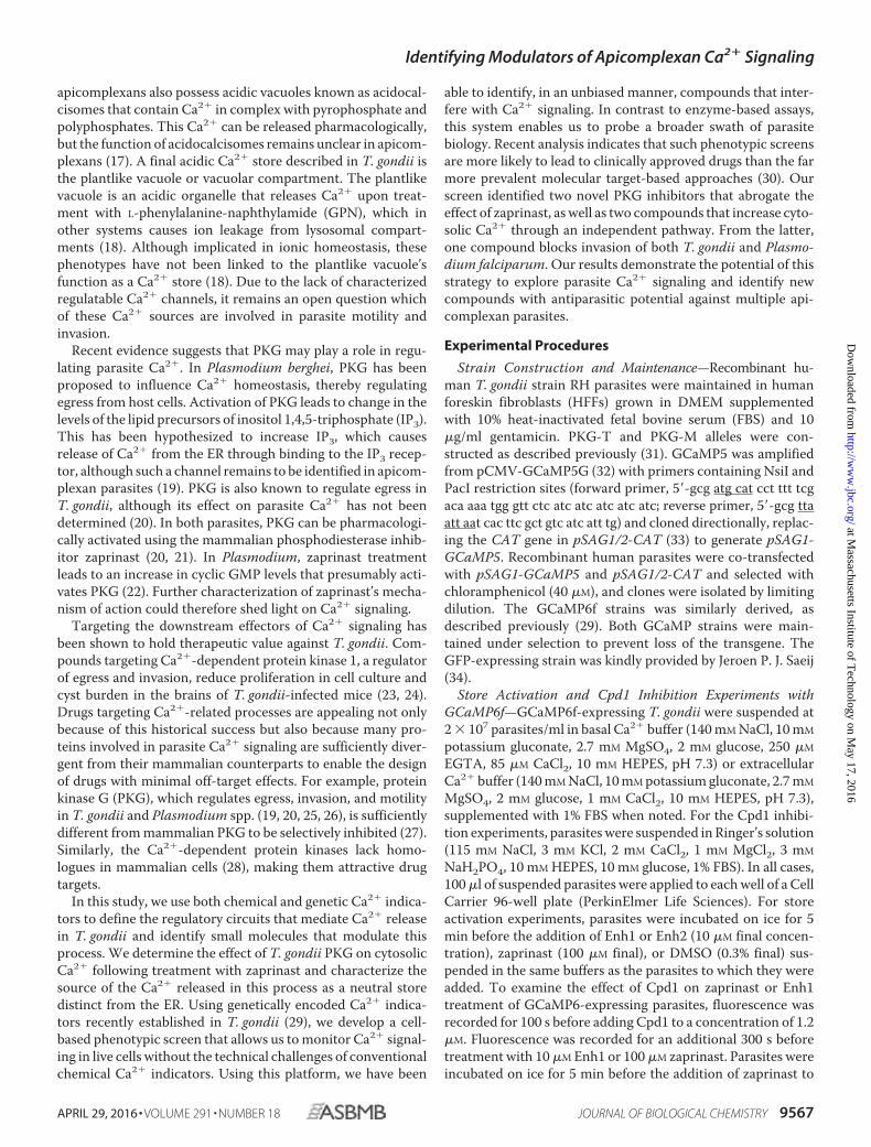
folds—Because known PKG inhibitors could suppress zapri-nast-induced Ca2� release (Fig. 1D), we hypothesized that thenewly identified compounds could act in a similar manner. Wetested the effects of Inh1 and Inh2 (compound GW407323Afrom the screen) on the in vitro activity of recombinant T. gon-dii PKG. Both compounds inhibited PKG activity with IC50values of 580 nM for Inh1 and 670 nM for Inh2 (Fig. 3A), sug-gesting that PKG inhibition is a plausible explanation for theiractivity. Furthermore, neither inhibitor showed appreciableactivity against human PKG or related AGC kinases (57), dem-onstrating selectivity for the parasite enzyme.
The two inhibitors are derivatives of distinct bicyclic heter-ocyclic scaffolds with proven activity against protein kinases.Inh1 is a PP with substitutions in the C2 and C3 positions (Fig.3B). Several available kinase structures display a similar orien-tation of the PP scaffold in the ATP-binding pocket, with the C3position engaging the hinge region and the C2 position orientedtoward the gatekeeper residue. The structures of two such
inhibitors complexed with human CDK2 demonstrate theproximity of the C2 position to the bulky phenylalanine-gate-keeper residue (Fig. 3B, middle). Substitutions at this positiondecreased the activity of these inhibitors presumably due tosteric clash with the gatekeeper (36). The 4-fluorophenyl groupin the C2 position of Inh1 is therefore expected to restrict bind-ing of the inhibitor to kinases with relatively small gatekeepers,like the threonine found in T. gondii PKG. This type of bindingis evident in the structure of p38 MAPK with a ligand similar toInh1 (SB203580, a monocyclic heterocycle rather than a bicy-clic one) that extends the 4-fluorophenyl functional group intothe hydrophobic pocket created by a threonine gatekeeper (37)(Fig. 3B, right). Furthermore, the related human kinase ERK2,normally resistant to SB203580, can be rendered sensitive tothis inhibitor by mutation of its gatekeeper glutamine to eitheran alanine or a threonine (58). This strongly suggests that asmall gatekeeper is required to accommodate heterocycles withsubstitutions similar to those found on Inh1.
Inh2 is an oxindole derivative, with substitutions at the C3and C5 positions (Fig. 3C). Disubstituted and trisubstitutedoxindoles are well studied protein kinase inhibitors. Severalstructures are available of kinases in complex with such inhib-itors. In every instance, the oxindole is positioned such that the2-oxygen and the nitrogen of the bicyclic scaffold engage thehinge residues (Fig. 3C, left). This configuration leaves the C3and C5 positions pointing away from the binding pocket.Although the C6 substituent is inward pointing, it does notapproach the direction of the gatekeeper. The binding config-uration of oxindole derivatives is highly consistent acrossseveral different kinase structures, suggesting that Inh2(a 3,5-disubstituted oxindole) is likely to be gatekeeper-independent.
FIGURE 2. Compound screen identifies modulators of zaprinast-induced Ca2� signaling. A, GCaMP5-expressing parasites pretreated with Cpd2 or avehicle control were stimulated with zaprinast or a vehicle control, and fluorescence was measured to determine a Z� score for a zaprinast-based screen forCa2� modulators. B, GCaMP5-expressing parasites were pretreated with 823 compounds from the PKIS libraries. Fluorescence was measured after zaprinaststimulation. Results are -fold change from zaprinast alone after background subtraction. Compounds that fluoresced independently are indicated (blue) alongwith selected enhancers (Enh; green) and inhibitors (Inh; red). The mean � S.D. (error bars) of two experiments is shown, and dashed lines indicate two S.D. valuesabove and below the mean of all compounds. C, structures of Enh1, Enh2, Inh1, and Inh2 as well as the known PKG inhibitors: Cpd1 and Cpd2. D, cumulativefrequencies of screen values for all compounds (black), Inh1 and its analogs (red), or Enh1 and its analogs (green).
Identifying Modulators of Apicomplexan Ca2� Signaling
9572 JOURNAL OF BIOLOGICAL CHEMISTRY VOLUME 291 • NUMBER 18 • APRIL 29, 2016
at Massachusetts Institute of T
echnology on May 17, 2016
http://ww
w.jbc.org/
Dow
nloaded from
To determine whether these inhibitors could interfere withother PKG-dependent processes, we next assessed their abilityto block zaprinast-induced egress. We directly tested the struc-tural predictions regarding the effect of the gatekeeper on Inh1,but not Inh2, by performing these experiments with thePKG-M and PKG-T strains. We measured zaprinast-inducedegress by lactate dehydrogenase release from host cells infectedwith either strain (Fig. 3D). As expected, PKG-T parasites didnot egress after Cpd2 treatment, whereas the refractoryPKG-M strain egressed similarly to vehicle-treated parasites.Both Inh1 and Inh2 reliably reduced egress relative to vehicle-treated parasites. The similar potencies of these compounds invivo and in vitro are consistent with inhibition of PKG being theprimary mechanism through which Inh1 and Inh2 block egress(Fig. 3D). As predicted from our structural analysis, Inh1worked in a gatekeeper-dependent manner, reducing egressfrom cells infected with PKG-T parasites with an IC50 of 1.3 �M
but leaving PKG-M parasites unaffected. In contrast, Inh2reduced egress from cells infected with either strain, exhibitingan IC50 of 2.5 �M for PKG-T and 3.5 �M for PKG-M. Theseresults were satisfying because we were able to recover inhibi-
tors with distinct scaffolds and modes of inhibiting an enzymeknown to be involved in the zaprinast response. Identificationof two novel PKG inhibitors through a screen of only 823 com-pounds demonstrates the power of our approach to identifycompounds with biological effects in pathways critical to para-site survival.
Two Enhancers Directly Release Intracellular Ca2� Stores—Having shown that a screen for modulators of zaprinast-in-duced Ca2� mobilization reveals novel PKG inhibitors, weturned our attention to Enh1 and another putative enhancer:Enh2, or GSK2188764A (Fig. 2C). During the course of thisstudy, the genetically encoded Ca2� indicator GCaMP6fbecame available. Because GCaMP6f has a broader dynamicrange and faster kinetics than GCaMP5 (59), we used GCaMP6fthroughout much of this study. We tested whether the enhanc-ers could increase parasite Ca2� independently of zaprinast.Indeed both enhancers could mobilize Ca2� on their own,although they did so with significantly slower kinetics thanzaprinast (Fig. 4A).
To determine the source of the Ca2� mobilized by theenhancers, we suspended parasites in buffers containing extra-
FIGURE 3. PKG inhibitors display distinct modes of inhibition. A, activity of recombinant PKG in the presence of increasing concentrations of Inh1, Inh2, orthe PKG inhibitor Cpd2. B, binding of PP derivatives like Inh1 to protein kinases orients the C2 position toward the gatekeeper residue (orange). Left, the PPscaffold’s orientation for a disubstituted PP in complex with CDK2. Middle, the structure of Inh1 is highlighted to indicate the PP scaffold (red) and the C2fluorophenyl group (green). Two other similar compounds from other kinase structures are superimposed over the CDK2 structure in complex with its inhibitor,all showing a similar positioning of the PP scaffold. Right, human p38 MAPK with a trisubstituted monocyclic heterocycle oriented similarly as the PP scaffoldand extending a fluorophenyl group in the direction of the gatekeeper. C, oxindole derivatives bind to protein kinases in a manner that orients their C2 and C5positions away from the gatekeeper. Left, oxindole scaffold of a derivative in complex with NEK2. Right, the oxindole scaffold of Inh2. Two other similarcompounds from other kinase structures have been superimposed on the structure of NEK2 with its inhibitor. D, zaprinast-induced egress of parasites carryingCpd2-sensitive (PKG-T) or resistant (PKG-M) alleles of PKG, following pretreatment with Inh1, Inh2, or Cpd2. Results shown are mean � S.E. (error bars) for n �3 independent experiments.
Identifying Modulators of Apicomplexan Ca2� Signaling
APRIL 29, 2016 • VOLUME 291 • NUMBER 18 JOURNAL OF BIOLOGICAL CHEMISTRY 9573
at Massachusetts Institute of T
echnology on May 17, 2016
http://ww
w.jbc.org/
Dow
nloaded from

cellular (1 mM) or basal (100 nM) levels of free Ca2�, treatedthese parasites with 10 �M Enh1 or Enh2, and monitoredGCaMP6f fluorescence (Fig. 4A). Enh1 increased fluorescenceunder both conditions, indicating that it mobilizes intracellularstores. Surprisingly, we observed only weak mobilization ofCa2� in response to Enh2 under both conditions. Because theoriginal screen was performed in buffer containing 1% FBS, wetested the effect of this component. The addition of 1% FBSincreased Enh2-mediated Ca2� mobilization. We interpretedthis to mean that Enh2 requires a cofactor found in FBS.Because the activity of Enh2 is relatively weak and depends onspecific conditions, we focused subsequent experiments onEnh1.
We wondered whether, like zaprinast, Enh1 functionsthrough PKG to increase parasite Ca2�. Significant overlap inthe emission spectra of Enh1 and Fura-2 prevented us from
using this ratiometric indicator. However, we were able to useGCaMP6f to make semiquantitative measurements. Werecorded fluorescence from GCaMP6f-expressing parasitestreated with Cpd1 or vehicle control, followed by stimulationwith Enh1 or zaprinast. To record the peak fluorescenceinduced by zaprinast, we had to incubate parasites on ice beforestimulation so as to slow down the response. In contrast to thezaprinast response, Cpd1-treated parasites responded to Enh1in the same manner as vehicle-treated parasites, indicating thatEnh1 does not act through PKG. Although Cpd1 acceleratedthe return to basal Ca2� levels following zaprinast treatment, itonly partially decreased the initial Ca2� spike induced by zapri-nast (Fig. 4B). This is reminiscent of results obtained from Fura-2-loaded parasites, where the increase in Ca2� following zapri-nast could never be fully suppressed by PKG inhibition(Fig. 1D).
We wondered whether zaprinast or Enh1 could induce Ca2�
fluxes in mammalian cells. We transfected HeLa cells with theplasmid CMV-R-GECO (42), which encodes a variation of theGCaMP Ca2� indicators under the mammalian CMV pro-moter. We treated these transfected cells with zaprinast, Enh1,or, as a positive control, the Ca2� ionophore A23187. Videomicroscopy revealed that R-GECO-expressing HeLa cellsincreased fluorescence in response to A23187 but that neitherzaprinast nor Enh1 produced a significant change (Fig. 4C).Given that zaprinast is known to inhibit phosphodiesterases inmammalian cells (60), we considered that these drugs could behaving a subtle effect that was below our limit of detection. Wetherefore repeated the assay with greater spatial and temporalresolution. We observed a slight increase in R-GECO fluores-cence in response to zaprinast during the first 10 s of treatment.However, Enh1 had no effect on host cell Ca2� (Fig. 4D), indi-cating that the effects of Enh1 are specific to Ca2� signaling inthe parasite.
Enh1 Inhibits T. gondii Survival—Zaprinast and relatedphosphodiesterase inhibitors were recently shown to blockT. gondii proliferation in human fibroblasts (61). Altering Ca2�
levels using ionophores has also been shown to affect parasiteviability (62). We therefore hypothesized that Enh1 would sim-ilarly inhibit survival of T. gondii. We tested the effect of thesecompounds using plaque assays as an indication of parasite sur-vival following treatment with Enh1 or zaprinast. Plaque for-mation was inhibited by zaprinast and Enh1 at 100 and 0.5 �M,respectively, whereas 10-fold lower concentrations allowednormal plaque formation similar to vehicle alone (Fig. 5A).These results for zaprinast agree with previously publishedwork (61). In order to determine the inhibitory concentrationsof these compounds more precisely, we performed lytic assaysin which host cells were exposed to parasites pretreated withvarying concentrations of zaprinast or Enh1, and the degree ofhost cell lysis was assessed by crystal violet staining of themonolayer, 3 days later. Enh1 showed a steep dose response,with 350 nM completely inhibiting parasite-induced cell death,and an EC50 of 180 nM. In contrast, 500 �M zaprinast wasrequired to cause complete inhibition of parasite-induced celldeath, and the IC50 of zaprinast was 200 �M (Fig. 5B).
We noticed the discrepancy between the amount of zaprinastrequired to kill T. gondii in plaque assays (100 �M) and the
FIGURE 4. Enh1 and Enh2 mobilize intracellular Ca2� stores. A, GCaMP6-expressing parasites suspended in buffer supplemented with either extracel-lular (1 mM) or basal (100 nM) Ca2� concentrations, with or without 1% FBS,and treated with 10 �M Enh1 or Enh2 at time 0. B, GCaMP6-expressing para-sites treated with 1 �M Cpd1 or vehicle at 100 s and then with 10 �M Enh1 or100 �M zaprinast at 400 or 750 s, respectively. A gap indicates incubation onice before the addition of zaprinast, in order to capture the peak of theresponse. Measurements represent fluorescence after the subtraction ofbackground obtained from samples treated with Cpd1 or vehicle, as indi-cated. Results shown are mean � S.E. (error bars) for n � 3 independentexperiments. C and D, intensity of R-GECO-expressing HeLa cells treated withEnh1, zaprinast, or A23187 over 10 min (C) or acquired at a faster rate for 1 min(D).
Identifying Modulators of Apicomplexan Ca2� Signaling
9574 JOURNAL OF BIOLOGICAL CHEMISTRY VOLUME 291 • NUMBER 18 • APRIL 29, 2016
at Massachusetts Institute of T
echnology on May 17, 2016
http://ww
w.jbc.org/
Dow
nloaded from

amount needed in lytic assays (500 �M). Because a rise in cyto-solic Ca2� stimulates parasite motility (2), we wondered if themovement of T. gondii in response to concentrations between100 and 500 �M might mechanically wound and kill host cells,which would be indistinguishable from parasite survival in thelytic assay. We tested this hypothesis by incubating host cellmonolayers with parasites pretreated with various concentra-tions of zaprinast and then measuring lactate dehydrogenaserelease from host cells. We found that treatment with 64 –510�M zaprinast resulted in host cell lysis, with a maximal effect at130 �M. Host cell lysis was not observed with zaprinast in theabsence of T. gondii (Fig. 5C). Due to the lower multiplicity ofinfection used in plaque assays compared with lytic assays, hostcells in plaque assays would be unlikely to experience observ-able cell wounding. We therefore expect the plaque assays toprovide a more accurate measure of zaprinast’s antiparasiticactivity.
Enh1 Induces Asynchronous Ca2� Fluxes—We thought itlikely that the plaquing defect caused by Enh1 was related to theCa2� disregulation induced by this compound. We thereforecharacterized the Ca2� response to Enh1 in more detail. Werecorded videos of intracellular GCaMP6f-expressing parasitestreated with Enh1 and compared the result with zaprinast treat-ment, which we have shown induces egress. We observedGCaMP6f activity in both cases, indicating that Enh1 can act onintracellular parasites. However, the profiles of zaprinast andEnh1-mediated Ca2� mobilization were remarkably different.Whereas zaprinast induced a fast, strong Ca2� peak (Fig. 6Aand supplemental Video S2), Enh1 elicited a slow, asynchro-nous effect in which some parasites appeared to experience
multiple Ca2� fluxes of similar magnitudes over the course ofseveral minutes (Fig. 6A and supplemental Video S3). Theresponse to these compounds was similar in extracellular par-asites (data not shown).
We generated kymographs illustrating the responses of indi-vidual GCaMP6f parasites to Enh1. Wondering whether pre-treatment with Enh1 would affect the ability of parasites torespond to other Ca2� agonists, we treated some of these par-asites with zaprinast as well (Fig. 6B). The sporadic flashes ofCa2� triggered by Enh1 were recapitulated, and all vehicle-treated parasites experienced an increase in Ca2� immediatelyafter the addition of zaprinast. However, Enh1-treated parasitesvaried in their responses to zaprinast, exhibiting asynchronousCa2� fluxes and inconsistent magnitudes in their Ca2�
increases. In rare cases, Ca2� concentrations in Enh1-treatedparasites even dropped upon zaprinast treatment. We quanti-fied the changes in fluorescence of individual parasites over the40 s following zaprinast addition and observed a more variedand overall diminished response to zaprinast in Enh1-treatedparasites (Fig. 6C). Taken together, these results suggest thatEnh1 may partially deplete the Ca2� stores mobilized byzaprinast.
Enh1 Blocks Ca2�-related Phenotypes in T. gondii andP. falciparum—While examining Enh1-treated parasites, wenoticed that this compound did not induce egress and in factsuppressed egress in response to zaprinast (Fig. 6C). To confirmthese effects, we treated intracellular GFP-expressing parasiteswith Enh1 and quantified the number of intact vacuoles at var-ious times after treatment, using fluorescence to monitor theinfection using automated image analysis. This assay is capableof directly measuring egress for hundreds of vacuoles per sam-ple. Surprisingly, despite its effects on extracellular parasites,Enh1 failed to stimulate egress beyond the spontaneous egressobserved in the vehicle control. In contrast, zaprinast treatmentresulted in �90% of vacuoles egressing within 30 min (Fig. 7A),as described previously (20). We then examined the effect of a10-min Enh1 pretreatment and found that it robustly blockedzaprinast-induced egress. Analysis of the dose-dependent inhi-bition of egress by Enh1 revealed an EC50 of 290 nM, within therange of concentrations that inhibit plaque formation (Fig. 7B).These results suggest that the antiparasitic activity of Enh1 ismediated by its inhibition of parasite egress.
We wondered whether we could generalize the Enh1-associ-ated egress defect beyond zaprinast-induced egress. We there-fore tested the ability of Enh1 to block egress induced by theCa2� ionophore A23187. Enh1 also produced a significantblock on A23187-induced egress (Fig. 7C), demonstrating thatits effects on parasite Ca2� signaling extend to a variety ofagonists.
We tested whether Enh1 also affects other apicomplexanparasites that use Ca2�-based signal transduction. Asexual,blood stage Plasmodium parasites undergo Ca2�-dependentegress from infected erythrocytes, followed by Ca2�-dependentinvasion into new erythrocytes (6, 63). To assess the effect ofEnh1 on these processes at the relevant developmental stage,we allowed purified schizonts to complete their intracellularmaturation while blocked with Cpd2 from rupturing andegressing (22). We administered Enh1 to parasites immediately
FIGURE 5. Enhancers of Ca2� mobilization show antiparasitic activity. A,plaque formation in the presence of the indicated concentrations of Enh1 orzaprinast. The drug concentrations indicated did not affect host cell survival.B, dose-dependent effect of Enh1 and zaprinast on parasite viability, assayedby monolayer disruption, 3 days postinfection, at the indicated drug concen-trations. Results shown are mean � S.E. (error bars) for n � 3 independentexperiments. C, host cell lysis following 1 h of infection in the presence ofvarying zaprinast concentrations.
Identifying Modulators of Apicomplexan Ca2� Signaling
APRIL 29, 2016 • VOLUME 291 • NUMBER 18 JOURNAL OF BIOLOGICAL CHEMISTRY 9575
at Massachusetts Institute of T
echnology on May 17, 2016
http://ww
w.jbc.org/
Dow
nloaded from

following washout of Cpd2. After allowing 1–2 h of incubationwith the compound, we measured egress and reinvasion using aflow cytometry-based assay that distinguishes schizonts fromrecently invaded ring stage parasites (49). Whereas Enh1 doesnot reduce parasite egress at concentrations up to at least 10 �M
(Fig. 7D), the compound completely blocks invasion within thetested range (IC50 � 3.2 �M) (Fig. 7E). Wondering whetherEnh1 could also inhibit invasion in T. gondii, we incubatedtachyzoites with varying doses of Enh1 for 20 min and testedtheir ability to infect host cells. As in P. falciparum, Enh1strongly inhibited T. gondii invasion (Fig. 7F). Furthermore, theIC50 of Enh1 in the invasion assay was 180 �M, similar to that inthe egress assay, arguing for a common mechanism for theeffect of Enh1 on both Ca2�-related phenotypes. In summary,Enh1 modulates Ca2�-dependent processes in diverse apicom-plexan parasites.
Discussion
Ca2� signaling plays a central role in apicomplexan biology,yet few of the components that regulate Ca2� uptake and
release have been identified. In this study, we extend our under-standing of these processes by demonstrating that PKG activityis needed for the robust Ca2� response elicited by phosphodi-esterase inhibitors. Furthermore, we determine the source ofCa2� to be distinct from other previously described stores. Weuse this phenomenon as the basis of a phenotypic screen thatallowed us to identify several novel inhibitors and enhancers ofCa2� signaling. Two of the inhibitors could be shown to inter-fere with Ca2� signaling by specifically targeting parasite PKG.In contrast, the enhancers could be shown to increase Ca2�
independently from zaprinast and in fact prevented parasiteegress by apparently depleting intracellular Ca2� stores. Thiscompound displayed antiparasitic properties against bothT. gondii and P. falciparum, establishing a new mechanism forinterfering with apicomplexan parasitism.
The signaling events that trigger parasite egress remainpoorly defined. Ca2� ionophores have long been known tostimulate egress in T. gondii (64). However, more recently,phosphodiesterase inhibitors were shown to have similareffects (20, 61). Our experiments revealed a strong and rapid
FIGURE 6. Enh1 elicits asynchronous cytosolic Ca2� fluxes and blocks zaprinast-induced egress. A, video microscopy of GCaMP6f-expressing parasitestreated with Enh1 or zaprinast. Time after the addition of the compound is indicated. Different times were used to capture the fast and slow responses ofzaprinast and Enh1, respectively. B, kymographs illustrate average fluorescence intensities of individual parasites, per row, during the course of the treatmentindicated. Black indicates that parasites egressed from vacuoles. C, change in fluorescence of the parasites illustrated in B over the 40 s following the additionof zaprinast. Measurements from each biological replicate are colored separately. Mean change for each group is indicated with a horizontal line. ****, p 0.0001, two-tailed t test.
Identifying Modulators of Apicomplexan Ca2� Signaling
9576 JOURNAL OF BIOLOGICAL CHEMISTRY VOLUME 291 • NUMBER 18 • APRIL 29, 2016
at Massachusetts Institute of T
echnology on May 17, 2016
http://ww
w.jbc.org/
Dow
nloaded from

release of Ca2� in response to zaprinast treatment through theuse of both the established Ca2� indicator Fura-2 and the newlyadapted genetically encoded sensors GCaMP5 and GCaMP6f(29). We provide conclusive evidence linking PKG to the zapri-nast-induced increase in cytosolic Ca2�, using a chemical-ge-netic approach to demonstrate specific inhibition of PKG byCpd1 and Cpd2. Sensitivity to both inhibitors depends on therelatively small gatekeeper of apicomplexan PKGs, which wemutated to a methionine that preserves kinase activity but ren-ders PKG refractory to inhibition (31). The changes in the
zaprinast response caused by Cpd1 can therefore be fully attrib-uted to PKG because no such changes were observed upontreatment of the resistant strain (PKG-M). However, compar-ing the response to zaprinast in parasites pretreated with Cpd1with those pretreated with vehicle shows that the initial sharpCa2� peak induced by zaprinast is incompletely suppressed byCpd1, in contrast to its complete inhibition of zaprinast-in-duced egress (20). Suppression of this peak appeared greaterwhen assayed using Fura-2 (Fig. 1D) than when using GCaMP6(Fig. 4B). This may result from the semiquantitative nature ofGCaMP6 and the potentially non-linear relationship betweenfluorescence and Ca2� concentration. Additionally, differencesin the subcellular distribution of the two indicators may influ-ence their responses to different sources of Ca2�. Ratiometricmeasurements with Fura-2 also revealed that inhibition of PKGby Cpd1 decreased basal Ca2� concentrations in extracellularparasites, suggesting that PKG might also be necessary to main-tain resting levels of Ca2� in extracellular parasites. Thesechanges in basal Ca2� concentrations might not be evident withGCaMP6f due to its higher Kd (375 nM) (59) compared with thatof Fura-2 (135 nM) (43).
We characterized the zaprinast-mobilized store as neutralbecause it can be depleted by ionomycin. However, we foundthat this store is independent of the thapsigargin-mobilizablestore. This is surprising, given that the ER is the only neutralCa2� store that has been characterized in T. gondii. SERCA, thetarget of thapsigargin, localizes to the ER in intracellular para-sites but redistributes so as to only partially colocalize with theER in extracellular parasites (54). Because our experimentswere done in extracellular parasites, it is possible that zaprinastmobilizes Ca2� from a section of the ER lacking SERCA underthese conditions, as previously suggested for the ethanol-mobi-lized Ca2� stores (65). In P. falciparum, zaprinast has beenshown to work through P. falciparum PKG to trigger changesin the levels of various precursors of the second messenger IP3.Presumably, this increases IP3, which then interacts with theIP3 receptor to stimulate release of Ca2� from intracellularstores (19). An IP3 receptor has not been identified in apicom-plexans, but treating parasites with ethanol raises levels of IP3and stimulates Ca2� release, providing evidence for the pres-ence of such a channel (66). Our results therefore indicate thatzaprinast functions, at least in part, through T. gondii PKG andprobably mobilizes a neutral, SERCA-independent, IP3 recep-tor-gated store.
Genetically encoded calcium indicators provide excellentreproducibility and circumvent many problems associated withloading and compartmentalization of chemical probes. Here wedemonstrate that such indicators can be used to identify com-pounds that alter apicomplexan Ca2� signaling and its depen-dent processes, such as egress and invasion. The simplicity androbustness of this cell-based phenotypic screen, with a Z�-fac-tor 0.5, makes it compatible with high throughput screeningefforts. As proof of concept, we screened a library of 823 ATPmimetics from GlaxoSmithKline for compounds that couldalter the zaprinast-induced increase in cytosolic Ca2�. Identi-fication of two PKG inhibitors, with distinct chemical scaffoldsand mechanisms of inhibition, validated our screen and dem-onstrates the power of the approach. Unexpectedly, our screen
FIGURE 7. Enh1 blocks egress Ca2�-related phenotypes in T. gondii andP. falciparum. A, egress of intracellular parasites treated with zaprinast, Enh1,or a vehicle control. The number of intact vacuoles was monitored by livemicroscopy over 30 min. Representative images before and after treatmentare shown. B, dose-dependent inhibition of zaprinast-induced egress follow-ing pretreatment with Enh1 or vehicle. C, Enh1 inhibition of egress induced byeither zaprinast or A23187. Results shown are mean � S.E. (error bars) for n �3 independent experiments. ***, p 0.001; **, p 0.01. D–E, schizonts werereleased from Cpd2 arrest immediately preceding the addition of Enh1. D,after 1–2 h, the remaining schizonts (mean � S.D. for three technical repli-cates) were counted and normalized to their initial abundance (3.6 and 5.4%in each experiment, respectively). E, Enh1 blocks invasion of erythrocytes byP. falciparum, measured 1–2 h following release from Cpd2, as assessed fromthe ring stage parasitemia. Mean invasion � S.E. is expressed as a percentageof invasion without drug (9.5 and 8.1% in each experiment, respectively).Background was assessed using heparin as a specific blocker of invasion andwas comparable with the signal observed with saturating concentrations ofEnh1. F, dose-dependent inhibition of T. gondii invasion following 10-minpretreatment of parasites before invasion. Results shown are mean � S.E. forn � 3 independent experiments.
Identifying Modulators of Apicomplexan Ca2� Signaling
APRIL 29, 2016 • VOLUME 291 • NUMBER 18 JOURNAL OF BIOLOGICAL CHEMISTRY 9577
at Massachusetts Institute of T
echnology on May 17, 2016
http://ww
w.jbc.org/
Dow
nloaded from

also revealed two compounds that, when used in combinationwith zaprinast, augmented Ca2� levels. One such compound,Enh1, elicited repeated cycles of Ca2� increase and decreasewith overall cytosolic Ca2� building at a population level. Theserepeated cycles are reminiscent of what others have seen whenFluo-4/AM-loaded parasites were treated with thapsigargin(54), perhaps indicating that Enh1 also inhibits Ca2� uptakepathways.
In contrast to zaprinast and Ca2� ionophores, Enh1 failed tostimulate egress despite raising cytosolic Ca2� levels. In fact,Enh1 blocked the ability of these agonists to stimulate parasiteegress. Furthermore, treatment with Enh1 blocked tachyzoiteinvasion and plaque formation at concentrations similar tothose required to block egress. These results suggest that Enh1depletes essential intracellular Ca2� stores, which have beenpreviously suggested to mediate invasion and egress. Consist-ent with this view, we observed diminished changes in GCaMPfluorescence in response to zaprinast, following Enh1 treat-ment (Fig. 6C). However, without a complete understanding ofEnh1 function, we cannot rule out the possibility that inhibitionof egress may be independent of its ability to modulate Ca2�
because (i) some parasites in which Enh1 did not elicit Ca2�
fluctuations still failed to egress in response to zaprinast, and(ii) A23187, which should equilibrate Ca2� across membranes,failed to overcome Enh1 inhibition. Compounds similar toEnh1 have been shown to inhibit mammalian kinases belongingto the AGC family (67). Having already established that T. gon-dii PKG, a member of the AGC kinase family, mediates Ca2�
release, it is an intriguing possibility that a related kinase mightoppose its activity.
Enh1 robustly inhibited the ability of P. falciparum to invadeerythrocytes, although it did not affect egress. Despite the manyparallels between egress of P. falciparum and T. gondii, sub-stantial differences have also been uncovered. Plasmodiumegress is a fast, highly synchronized process, dependent on acascade of proteolytic activity (68). Genetic evidence supportsthis distinction with mutants in the calcium-responsive proteinDOC2.1 blocking both egress and invasion in T. gondii but onlyinvasion in P. falciparum (69), mirroring the effects of Enh1.The intracellular Ca2� stores of Plasmodium have not beeninvestigated as thoroughly as those of T. gondii, but there issome evidence that it is not solely dependent on its intracellularCa2� stores and in fact utilizes Ca2� found within the parasito-phorous vacuole (70). In light of this, the differential responsesof P. falciparum and T. gondii to Enh1 are perhaps not surpris-ing. That Enh1 blocks invasion of P. falciparum demonstratesthis compound’s ability to perturb Ca2�-dependent processesacross multiple members of the Apicomplexa.
Resistance to front line antimalarials is increasing (71, 72),and new treatment options are needed. Rational design strate-gies can identify drugs with minimal off-target effects but focuson a limited repertoire of signaling pathways. The ToxoplasmaCa2� signaling network shows evidence of conservation withinapicomplexans while being dissimilar to human signaling path-ways. By screening for modulation of this pathway, we haveprioritized compounds that are likely to interfere selectivelywith infection without focusing on a single parasite protein.This screen can be performed with commonly available equip-
ment and should be easily scalable to larger collections of com-pounds. Our success in identifying new modulators of Ca2�
signaling with effects that extend to multiple apicomplexanshighlights the power of such an approach. In particular, Enh1inhibits parasite viability with an EC50 in the nanomolar rangeand provides a good lead for the development of antiparasiticcompounds. Further characterization of these compounds mayidentify novel components of apicomplexan Ca2� signalingpathways and improve our ability to target these pathwaysspecifically.
Author Contributions—S. M. S., M. A. H. T., and A. S. P. designedand conducted the experiments and analyzed the data. S. M. S. wrotethe majority of the manuscript, with specific sections contributed byM. A. H. T. and A. S. P. C. G. H. constructed the T. gondii strainswith different PKG alleles. M. E. B., R. H., and W. J. Z. provided keyreagents and advice. F. T. and N. J. W. synthesized Cpd2. M. T. D.,S. N. J. M., and S. L. supervised the work in their respective labora-tories and contributed to the analysis of experiments and writing ofthe manuscript.
Acknowledgments—We thank David Drewry for helpful adviceregarding the compound libraries, Jeroen P. J. Saeij for providing theGFP-expressing strain, and Emily M. Shortt for technical assistance.
References1. Carruthers, V. B., and Sibley, L. D. (1999) Mobilization of intracellular
calcium stimulates microneme discharge in Toxoplasma gondii. Mol. Mi-crobiol. 31, 421– 428
2. Wetzel, D. M., Chen, L. A., Ruiz, F. A., Moreno, S. N. J., and Sibley, L. D.(2004) Calcium-mediated protein secretion potentiates motility in Toxo-plasma gondii. J. Cell Sci. 117, 5739 –5748
3. Billker, O., Lourido, S., and Sibley, L. D. (2009) Calcium-dependent sig-naling and kinases in apicomplexan parasites. Cell Host Microbe 5,612– 622
4. Arrizabalaga, G., and Boothroyd, J. C. (2004) Role of calcium during Tox-oplasma gondii invasion and egress. Int. J. Parasitol. 34, 361–368
5. McCallum-Deighton, N., and Holder, A. A. (1992) The role of calcium inthe invasion of human erythrocytes by Plasmodium falciparum. Mol.Biochem. Parasitol. 50, 317–323
6. Singh, S., Alam, M. M., Pal-Bhowmick, I., Brzostowski, J. A., and Chitnis,C. E. (2010) Distinct external signals trigger sequential release of apicalorganelles during erythrocyte invasion by malaria parasites. PLoS Pathog.6, e1000746
7. Lovett, J. L., and Sibley, L. D. (2003) Intracellular calcium stores in Toxo-plasma gondii govern invasion of host cells. J. Cell Sci. 116, 3009 –3016
8. Moudy, R., Manning, T. J., and Beckers, C. J. (2001) The loss of cytoplas-mic potassium upon host cell breakdown triggers egress of Toxoplasmagondii. J. Biol. Chem. 276, 41492– 41501
9. Pace, D. A., McKnight, C. A., Liu, J., Jimenez, V., and Moreno, S. N. J.(2014) Calcium entry in Toxoplasma gondii and its enhancing effect ofinvasion-linked traits. J. Biol. Chem. 289, 19637–19647
10. English, A. R., and Voeltz, G. K. (2013) Endoplasmic reticulum structureand interconnections with other organelles. Cold Spring Harb. Perspect.Biol. 5, a013227
11. Golovina, V. A., and Blaustein, M. P. (1997) Spatially and functionallydistinct Ca2� stores in sarcoplasmic and endoplasmic reticulum. Science275, 1643–1648
12. Calcraft, P. J., Ruas, M., Pan, Z., Cheng, X., Arredouani, A., Hao, X., Tang,J., Rietdorf, K., Teboul, L., Chuang, K.-T., Lin, P., Xiao, R., Wang, C., Zhu,Y., Lin, Y., et al. (2009) NAADP mobilizes calcium from acidic organellesthrough two-pore channels. Nature 459, 596 – 600
13. Pinton, P., Pozzan, T., and Rizzuto, R. (1998) The Golgi apparatus is aninositol 1,4,5-trisphosphate-sensitive Ca2� store, with functional proper-
Identifying Modulators of Apicomplexan Ca2� Signaling
9578 JOURNAL OF BIOLOGICAL CHEMISTRY VOLUME 291 • NUMBER 18 • APRIL 29, 2016
at Massachusetts Institute of T
echnology on May 17, 2016
http://ww
w.jbc.org/
Dow
nloaded from
ties distinct from those of the endoplasmic reticulum. EMBO J. 17,5298 –5308
14. Koch, G. L. E. (1990) The endoplasmic reticulum and calcium storage.Bioessays 12, 527–531
15. Stelly, N., Mauger, J. P., Claret, M., and Adoutte, A. (1991) Cortical alveoliof Paramecium: a vast submembranous calcium storage compartment.J. Cell Biol. 113, 103–112
16. Plattner, H., Habermann, A., Kissmehl, R., Klauke, N., Majoul, I., andSöling, H. D. (1997) Differential distribution of calcium stores in Parame-cium cells: occurrence of a subplasmalemmal store with a calsequestrin-like protein. Eur. J. Cell Biol. 72, 297–306
17. Moreno, S. N., and Zhong, L. (1996) Acidocalcisomes in Toxoplasma gon-dii tachyzoites. Biochem. J. 313, 655– 659
18. Miranda, K., Pace, D. A., Cintron, R., Rodrigues, J. C. F., Fang, J., Smith, A.,Rohloff, P., Coelho, E., de Haas, F., de Souza, W., Coppens, I., Sibley, L. D.,and Moreno, S. N. J. (2010) Characterization of a novel organelle in Tox-oplasma gondii with similar composition and function to the plant vacu-ole. Mol. Microbiol. 76, 1358 –1375
19. Brochet, M., Collins, M. O., Smith, T. K., Thompson, E., Sebastian, S.,Volkmann, K., Schwach, F., Chappell, L., Gomes, A. R., Berriman, M.,Rayner, J. C., Baker, D. A., Choudhary, J., and Billker, O. (2014) Phosphoi-nositide metabolism links cGMP-dependent protein kinase G to essentialCa2� signals at key decision points in the life cycle of malaria parasites.PLoS Biol. 12, e1001806
20. Lourido, S., Tang, K., and Sibley, L. D. (2012) Distinct signalling pathwayscontrol Toxoplasma egress and host-cell invasion. EMBO J. 31,4524 – 4534
21. Yuasa, K., Mi-Ichi, F., Kobayashi, T., Yamanouchi, M., Kotera, J., Kita, K.,and Omori, K. (2005) PfPDE1, a novel cGMP-specific phosphodiesterasefrom the human malaria parasite Plasmodium falciparum. Biochem. J.392, 221–229
22. Collins, C. R., Hackett, F., Strath, M., Penzo, M., Withers-Martinez, C.,Baker, D. A., and Blackman, M. J. (2013) Malaria parasite cGMP-depen-dent protein kinase regulates blood stage merozoite secretory organelledischarge and egress. PLoS Pathog. 9, e1003344
23. Lourido, S., Zhang, C., Lopez, M. S., Tang, K., Barks, J., Wang, Q., Wild-man, S. A., Shokat, K. M., and Sibley, L. D. (2013) Optimizing small mol-ecule inhibitors of calcium-dependent protein kinase 1 to prevent infec-tion by Toxoplasma gondii. J. Med. Chem. 56, 3068 –3077
24. Johnson, S. M., Murphy, R. C., Geiger, J. A., DeRocher, A. E., Zhang, Z.,Ojo, K. K., Larson, E. T., Perera, B. G. K., Dale, E. J., He, P., Reid, M. C., Fox,A. M. W., Mueller, N. R., Merritt, E. A., Fan, E., et al. (2012) Developmentof Toxoplasma gondii calcium-dependent protein kinase 1 (TgCDPK1)inhibitors with potent anti-toxoplasma activity. J. Med. Chem. 55,2416 –2426
25. Wiersma, H. I., Galuska, S. E., Tomley, F. M., Sibley, L. D., Liberator, P. A.,and Donald, R. G. K. (2004) A role for coccidian cGMP-dependent proteinkinase in motility and invasion. Int. J. Parasitol. 34, 369 –380
26. Taylor, H. M., McRobert, L., Grainger, M., Sicard, A., Dluzewski, A. R.,Hopp, C. S., Holder, A. A., and Baker, D. A. (2010) The malaria parasitecyclic GMP-dependent protein kinase plays a central role in blood-stageschizogony. Eukaryot. Cell 9, 37– 45
27. Gurnett, A. M., Liberator, P. A., Dulski, P. M., Salowe, S. P., Donald,R. G. K., Anderson, J. W., Wiltsie, J., Diaz, C. A., Harris, G., Chang, B.,Darkin-Rattray, S. J., Nare, B., Crumley, T., Blum, P. S., Misura, A. S., et al.(2002) Purification and molecular characterization of cGMP-dependentprotein kinase from Apicomplexan parasites: a novel chemotherapeutictarget. J. Biol. Chem. 277, 15913–15922
28. Lim, D. C., Cooke, B. M., Doerig, C., and Saeij, J. P. J. (2012) Toxoplasmaand Plasmodium protein kinases: roles in invasion and host cell remodel-ling. Int. J. Parasitol. 42, 21–32
29. Borges-Pereira, L., Budu, A., McKnight, C. A., Moore, C. A., Vella, S. A.,Hortua Triana, M. A., Liu, J., Garcia, C. R. S., Pace, D. A., and Moreno,S. N. J. (2015) Calcium signaling throughout the Toxoplasma gondii lyticcycle. J. Biol. Chem. 290, 26914 –26926
30. Zheng, W., Thorne, N., and McKew, J. C. (2013) Phenotypic screens as arenewed approach for drug discovery. Drug Discov. Today 18, 1067–1073
31. Sidik, S. M., Hackett, C. G., Tran, F., Westwood, N. J., and Lourido, S.
(2014) Efficient genome engineering of Toxoplasma gondii using CRISPR/Cas9. PLoS One 9, e100450
32. Akerboom, J., Chen, T.-W., Wardill, T. J., Tian, L., Marvin, J. S., Mutlu, S.,Calderón, N. C., Esposti, F., Borghuis, B. G., Sun, X. R., Gordus, A., Orger,M. B., Portugues, R., Engert, F., Macklin, J. J., et al. (2012) Optimization ofa GCaMP calcium indicator for neural activity imaging. J. Neurosci. 32,13819 –13840
33. Soldati, D., and Boothroyd, J. C. (1995) A selector of transcription initia-tion in the protozoan parasite Toxoplasma gondii. Mol. Cell. Biol. 15,87–93
34. Boyle, J. P., Saeij, J. P. J., and Boothroyd, J. C. (2007) Toxoplasma gondii:inconsistent dissemination patterns following oral infection in mice. Exp.Parasitol. 116, 302–305
35. Berman, H. M., Westbrook, J., Feng, Z., Gilliland, G., Bhat, T. N., Weissig,H., Shindyalov, I. N., and Bourne, P. E. (2000) The Protein Data Bank.Nucleic Acids Res. 28, 235–242
36. Stevens, K. L., Reno, M. J., Alberti, J. B., Price, D. J., Kane-Carson, L. S.,Knick, V. B., Shewchuk, L. M., Hassell, A. M., Veal, J. M., Davis, S. T.,Griffin, R. J., and Peel, M. R. (2008) Synthesis and evaluation of pyra-zolo[1,5-b]pyridazines as selective cyclin dependent kinase inhibitors.Bioorg. Med. Chem. Lett. 18, 5758 –5762
37. Simard, J. R., Getlik, M., Grütter, C., Pawar, V., Wulfert, S., Rabiller, M.,and Rauh, D. (2009) Development of a fluorescent-tagged kinase assaysystem for the detection and characterization of allosteric kinase inhibi-tors. J. Am. Chem. Soc. 131, 13286 –13296
38. Kinoshita, T., Warizaya, M., Ohori, M., Sato, K., Neya, M., and Fujii, T.(2006) Crystal structure of human ERK2 complexed with a pyrazolo[3,4-c]pyridazine derivative. Bioorg. Med. Chem. Lett. 16, 55–58
39. Islam, I., Bryant, J., Chou, Y.-L., Kochanny, M. J., Lee, W., Phillips, G. B.,Yu, H., Adler, M., Whitlow, M., Ho, E., Lentz, D., Polokoff, M. A., Subra-manyam, B., Wu, J. M., Zhu, D., et al. (2007) Indolinone based phos-phoinositide-dependent kinase-1 (PDK1) inhibitors. Part 1: design, syn-thesis and biological activity. Bioorg. Med. Chem. Lett. 17, 3814 –3818
40. Rellos, P., Ivins, F. J., Baxter, J. E., Pike, A., Nott, T. J., Parkinson, D.-M.,Das, S., Howell, S., Fedorov, O., Shen, Q. Y., Fry, A. M., Knapp, S., andSmerdon, S. J. (2007) Structure and regulation of the human Nek2 centro-somal kinase. J. Biol. Chem. 282, 6833– 6842
41. Schindelin, J., Arganda-Carreras, I., Frise, E., Kaynig, V., Longair, M., Pi-etzsch, T., Preibisch, S., Rueden, C., Saalfeld, S., Schmid, B., Tinevez, J.-Y.,White, D. J., Hartenstein, V., Eliceiri, K., Tomancak, P., et al. (2012) Fiji: anopen-source platform for biological-image analysis. Nat. Methods 9,676 – 682
42. Zhao, Y., Araki, S., Wu, J., Teramoto, T., Chang, Y.-F., Nakano, M., Ab-delfattah, A. S., Fujiwara, M., Ishihara, T., Nagai, T., and Campbell, R. E.(2011) An expanded palette of genetically encoded Ca2� indicators. Sci-ence 333, 1888 –1891
43. Grynkiewicz, G., Poenie, M., and Tsien, R. Y. (1985) A new generation ofCa2� indicators with greatly improved fluorescence properties. J. Biol.Chem. 260, 3440 –3450
44. Trager, W., and Jensen, J. B. (1976) Human malaria parasites in continu-ous culture. Science 193, 673– 675
45. Wahlgren, M., Berzins, K., Perlmann, P., and Björkman, A. (1983) Char-acterization of the humoral immune response in Plasmodium falciparummalaria. I. Estimation of antibodies to P. falciparum or human erythro-cytes by means of microELISA. Clin. Exp. Immunol. 54, 127–134
46. Boyle, M. J., Richards, J. S., Gilson, P. R., Chai, W., and Beeson, J. G. (2010)Interactions with heparin-like molecules during erythrocyte invasion byPlasmodium falciparum merozoites. Blood 115, 4559 – 4568
47. Tonkin, C. J., van Dooren, G. G., Spurck, T. P., Struck, N. S., Good, R. T.,Handman, E., Cowman, A. F., and McFadden, G. I. (2004) Localization oforganellar proteins in Plasmodium falciparum using a novel set of trans-fection vectors and a new immunofluorescence fixation method. Mol.Biochem. Parasitol. 137, 13–21
48. Bei, A. K., Desimone, T. M., Badiane, A. S., Ahouidi, A. D., Dieye, T.,Ndiaye, D., Sarr, O., Ndir, O., Mboup, S., and Duraisingh, M. T. (2010) Aflow cytometry-based assay for measuring invasion of red blood cells byPlasmodium falciparum. Am. J. Hematol. 85, 234 –237
49. Paul, A. S., Saha, S., Engelberg, K., Jiang, R. H. Y., Coleman, B. I., Kosber,
Identifying Modulators of Apicomplexan Ca2� Signaling
APRIL 29, 2016 • VOLUME 291 • NUMBER 18 JOURNAL OF BIOLOGICAL CHEMISTRY 9579
at Massachusetts Institute of T
echnology on May 17, 2016
http://ww
w.jbc.org/
Dow
nloaded from
A. L., Chen, C.-T., Ganter, M., Espy, N., Gilberger, T. W., Gubbels, M.-J.,and Duraisingh, M. T. (2015) Parasite calcineurin regulates host cell rec-ognition and attachment by apicomplexans. Cell Host Microbe 18, 49 – 60
50. Kafsack, B. F. C., Pena, J. D. O., Coppens, I., Ravindran, S., Boothroyd, J. C.,and Carruthers, V. B. (2009) Rapid membrane disruption by a perforin-like protein facilitates parasite exit from host cells. Science 323, 530 –533
51. Liu, C., and Hermann, T. E. (1978) Characterization of ionomycin as acalcium ionophore. J. Biol. Chem. 253, 5892–5894
52. Fasolato, C., Zottini, M., Clementi, E., Zacchetti, D., Meldolesi, J., andPozzan, T. (1991) Intracellular Ca2� pools in PC12 cells: three intracellularpools are distinguished by their turnover and mechanisms of Ca2� accu-mulation, storage, and release. J. Biol. Chem. 266, 20159 –20167
53. Moreno, S. N. J., Ayong, L., and Pace, D. A. (2011) Calcium storage andfunction in apicomplexan parasites. Essays Biochem. 51, 97–110
54. Nagamune, K., Beatty, W. L., and Sibley, L. D. (2007) Artemisinin inducescalcium-dependent protein secretion in the protozoan parasite Toxo-plasma gondii. Eukaryot. Cell 6, 2147–2156
55. Donald, R. G. K., Zhong, T., Wiersma, H., Nare, B., Yao, D., Lee, A., Al-locco, J., and Liberator, P. A. (2006) Anticoccidial kinase inhibitors: iden-tification of protein kinase targets secondary to cGMP-dependent proteinkinase. Mol. Biochem. Parasitol. 149, 86 –98
56. Zhang, J. H., Chung, T. D., and Oldenburg, K. R. (1999) A simple statisticalparameter for use in evaluation and validation of high throughput screen-ing assays. J. Biomol. Screen. 4, 67–73
57. Elkins, J. M., Fedele, V., Szklarz, M., Abdul Azeez, K. R., Salah, E., Mikola-jczyk, J., Romanov, S., Sepetov, N., Huang, X.-P., Roth, B. L., Al Haj Zen, A.,Fourches, D., Muratov, E., Tropsha, A., Morris, J., et al. (2016) Compre-hensive characterization of the published kinase inhibitor set. Nat. Bio-technol. 34, 95–103
58. Fox, T., Coll, J. T., Xie, X., Ford, P. J., Germann, U. A., Porter, M. D.,Pazhanisamy, S., Fleming, M. A., Galullo, V., Su, M. S., and Wilson, K. P.(1998) A single amino acid substitution makes ERK2 susceptible to pyr-idinyl imidazole inhibitors of p38 MAP kinase. Protein Sci. 7, 2249 –2255
59. Chen, T.-W., Wardill, T. J., Sun, Y., Pulver, S. R., Renninger, S. L., Baohan,A., Schreiter, E. R., Kerr, R. A., Orger, M. B., Jayaraman, V., Looger, L. L.,Svoboda, K., and Kim, D. S. (2013) Ultrasensitive fluorescent proteins forimaging neuronal activity. Nature 499, 295–300
60. Prigent, A. F., Fougier, S., Nemoz, G., Anker, G., Pacheco, H., Lugnier, C.,Lebec, A., and Stoclet, J. C. (1988) Comparison of cyclic nucleotide phos-phodiesterase isoforms from rat heart and bovine aorta. Separation andinhibition by selective reference phosphodiesterase inhibitors. Biochem.Pharmacol. 37, 3671–3681
61. Howard, B. L., Harvey, K. L., Stewart, R. J., Azevedo, M. F., Crabb, B. S.,
Jennings, I. G., Sanders, P. R., Manallack, D. T., Thompson, P. E., Tonkin,C. J., and Gilson, P. R. (2015) Identification of potent phosphodiesteraseinhibitors that demonstrate cyclic nucleotide-dependent functions in api-complexan parasites. ACS Chem. Biol. 10, 1145–1154
62. Arrizabalaga, G., Ruiz, F., Moreno, S., and Boothroyd, J. C. (2004) Iono-phore-resistant mutant of Toxoplasma gondii reveals involvement of asodium/hydrogen exchanger in calcium regulation. J. Cell Biol. 165,653– 662
63. Glushakova, S., Lizunov, V., Blank, P. S., Melikov, K., Humphrey, G., andZimmerberg, J. (2013) Cytoplasmic free Ca2� is essential for multiplesteps in malaria parasite egress from infected erythrocytes. Malar. J. 12,41– 41
64. Endo, T., Sethi, K. K., and Piekarski, G. (1982) Toxoplasma gondii: calciumionophore A23187-mediated exit of trophozoites from infected murinemacrophages. Exp. Parasitol. 53, 179 –188
65. Carruthers, V. B., Moreno, S. N., and Sibley, L. D. (1999) Ethanol andacetaldehyde elevate intracellular [Ca2�] and stimulate microneme dis-charge in Toxoplasma gondii. Biochem. J. 342, 379 –386
66. Lovett, J. L., Marchesini, N., Moreno, S. N. J., and Sibley, L. D. (2002)Toxoplasma gondii microneme secretion involves intracellular Ca2� re-lease from inositol 1,4,5-triphosphate (IP3)/ryanodine-sensitive stores.J. Biol. Chem. 277, 25870 –25876
67. Naqvi, S., Macdonald, A., McCoy, C. E., Darragh, J., Reith, A. D., andArthur, J. S. C. (2012) Characterization of the cellular action of the MSKinhibitor SB-747651A. Biochem. J. 441, 347–357
68. Blackman, M. J. (2008) Malarial proteases and host cell egress: an “emerg-ing” cascade. Cell Microbiol. 10, 1925–1934
69. Farrell, A., Thirugnanam, S., Lorestani, A., Dvorin, J. D., Eidell, K. P., Fer-guson, D. J. P., Anderson-White, B. R., Duraisingh, M. T., Marth, G. T.,and Gubbels, M.-J. (2012) A DOC2 protein identified by mutational pro-filing is essential for apicomplexan parasite exocytosis. Science 335,218 –221
70. Gazarini, M. L., Thomas, A. P., Pozzan, T., and Garcia, C. R. S. (2003)Calcium signaling in a low calcium environment how the intracellularmalaria parasite solves the problem. J. Cell Biol. 161, 103–110
71. Dondorp, A. M., Nosten, F., Yi, P., Das, D., Phyo, A. P., Tarning, J., Lwin,K. M., Ariey, F., Hanpithakpong, W., Lee, S. J., Ringwald, P., Silamut, K.,Imwong, M., Chotivanich, K., Lim, P., et al. (2009) Artemisinin resistancein Plasmodium falciparum malaria. N. Engl. J. Med. 361, 455– 467
72. Cheeseman, I. H., Miller, B. A., Nair, S., Nkhoma, S., Tan, A., Tan, J. C., AlSaai, S., Phyo, A. P., Moo, C. L., Lwin, K. M., McGready, R., Ashley, E.,Imwong, M., Stepniewska, K., Yi, P., et al. (2012) A major genome regionunderlying artemisinin resistance in malaria. Science 336, 79 – 82
Identifying Modulators of Apicomplexan Ca2� Signaling
9580 JOURNAL OF BIOLOGICAL CHEMISTRY VOLUME 291 • NUMBER 18 • APRIL 29, 2016
at Massachusetts Institute of T
echnology on May 17, 2016
http://ww
w.jbc.org/
Dow
nloaded from
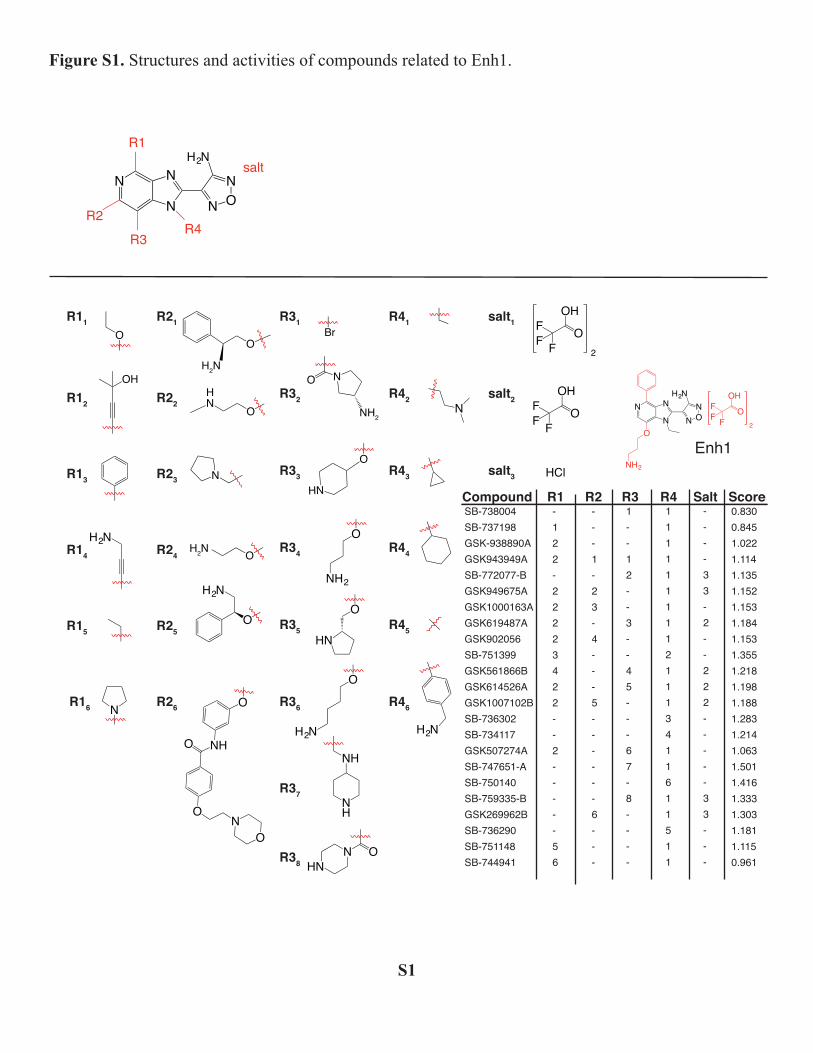
N
N
N
N ON
H2NR1
R2
R3R4
O
OH
H2N
N
O
H2N
OHN
N
OH2N
O
H2N
Br
O N
NH2
HN
O
O
NH2
O
HN
O
H2N
NH
NH
N OHN
N
R11
R12
R13
R14
R15
R16
R21
R22
R23
R24
R25
R26
R31
R32
R33
R34
R35
R36
R37
R38
R41
R42
R43
R44
R45
R46
NN
N
O
NH2
N ON
H2N
F
FF
OH
O2
Enh1
salt1
salt2
salt3
F
FF
OH
O2
F
FF
OH
O
HCl
salt
O
NH
ON
O
O
H2N
SB-738004SB-737198GSK-938890AGSK943949ASB-772077-BGSK949675AGSK1000163AGSK619487AGSK902056SB-751399GSK561866BGSK614526AGSK1007102BSB-736302SB-734117GSK507274ASB-747651-ASB-750140SB-759335-BGSK269962BSB-736290SB-751148SB-744941
Compound R1 R2 R3 R4 Salt-122-22223422--2-----56
---1-23-4---5------6---
1--12--3--45---67-8----
11111111121113411611511
----33-2--222-----33---
Score0.8300.8451.0221.1141.1351.1521.1531.1841.1531.3551.2181.1981.1881.2831.2141.0631.5011.4161.3331.3031.1811.1150.961
Figure S1. Structures and activities of compounds related to Enh1.
S1

F
NNN
N
N
NH O
O
R31
R22
R21
R13
R12
R11
F
FF
O
O
Cl
O
F
F
F
NNN
NN
NH
R1
R2
R3
Compound R1 R2 R3 Score
GW832467XGW827105XGW827106XGW829055X
1223
121-
1---
0.6660.7960.9891.188
Inh1
Figure S2. Structures and activities of compounds related to Inh1.
S2
Zuercher, Manoj T. Duraisingh, Silvia N. J. Moreno and Sebastian LouridoCaroline G. Hackett, Fanny Tran, Nicholas J. Westwood, Raymond Hui, William J.
Saima M. Sidik, Miryam A. Hortua Triana, Aditya S. Paul, Majida El Bakkouri, Signaling2+
CaToxoplasma gondiiUsing a Genetically Encoded Sensor to Identify Inhibitors of
doi: 10.1074/jbc.M115.703546 originally published online March 1, 20162016, 291:9566-9580.J. Biol. Chem.
10.1074/jbc.M115.703546Access the most updated version of this article at doi:
Alerts:
When a correction for this article is posted•
When this article is cited•
to choose from all of JBC's e-mail alertsClick here
Supplemental material:
http://www.jbc.org/content/suppl/2016/03/01/M115.703546.DC1.html
http://www.jbc.org/content/291/18/9566.full.html#ref-list-1
This article cites 72 references, 35 of which can be accessed free at
at Massachusetts Institute of T
echnology on May 17, 2016
http://ww
w.jbc.org/
Dow
nloaded from


![Evidence of Ca2+-Dependent Carbohydrate Association ... · Ca2+I2+ and [2Lex + Ca2+]2+. The CID experiments of the [2Lex-LacCer + Ca2+I2+ dimers resulted in a neutral loss covalently](https://img.pdfslide.us/doc/110x75/5f8af1f17b5f935beb015692/evidence-of-ca2-dependent-carbohydrate-association-ca2i2-and-2lex-ca22.jpg)