Embed Size (px)
Citation preview
Veterinary World, EISSN: 2231-0916 194
Veterinary World, EISSN: 2231-0916Available at www.veterinaryworld.org/Vol.10/February-2017/10.pdf
RESEARCH ARTICLEOpen Access
Toxoplasma gondii infection in meat animals from Africa: Systematic review and meta-analysis of sero-epidemiological studies
Aretas Babatoundé Nounnagnon Tonouhewa1, Yao Akpo2, Philippe Sessou1, Camus Adoligbe1, Eric Yessinou1, Yaovi Gildas Hounmanou1,3, Marc Napoléon Assogba1, Issaka Youssao1 and Souaïbou Farougou1
1. Laboratory of Research in Applied Biology, Polytechnic School of Abomey-Calavi, University of Abomey-Calavi, 01 P.O. Box 2009, Cotonou, Benin; 2. Laboratory of Ecology, Health and animal Production, Faculty of Agronomy, University of Parakou, P.O. Box 123 Parakou, Benin; 3. Department of Veterinary Medicine and Public Health, Sokoine University of
Agriculture, P.O. Box 3121, Chuo Kikoo, Morogoro, Tanzania.Corresponding author: Aretas Babatoundé Nounnagnon Tonouhewa, e-mail: [email protected],
Co-authors: YA: [email protected], PS: [email protected], CA: [email protected], EY: [email protected], YGH: [email protected], MNA: [email protected], IY: [email protected],
SF: [email protected]: 08-09-2016, Accepted: 10-01-2017, Published online: 16-02-2017
doi: 10.14202/vetworld.2017.194-208 How to cite this article: Tonouhewa ABN, Akpo Y, Sessou P, Adoligbe C, Yessinou E, Hounmanou YG, Assogba MN, Youssao I, Farougou S (2017) Toxoplasma gondii infection in meat animals from Africa: Systematic review and meta-analysis of sero-epidemiological studies, Veterinary World, 10(2): 194-208.
AbstractAim: Toxoplasma gondii is an ubiquitous apicomplexan parasite which causes toxoplasmosis in humans and animals. Felids especially cats are definitive hosts and almost all warm-blooded mammals, including livestock and human can serve as intermediate hosts. Food animals can be reservoirs for T. gondii and act as one of the sources for parasite transmission to humans. The objective of this study is to collect serological data on the prevalence of anti-T. gondii antibody, and risk factors for certain food animals from Africa to provide a quantitative estimate of T. gondii infection among these species from different African countries.
Materials and Methods: Four databases were used to search seroepidemiological data on the prevalence of anti-T. gondii antibody in food animals between 1969 and 2016 from African countries. The search focused on data obtained by serologic test in food animals and meta-analyses were performed per species.
Results: A total of 30,742 individual samples from 24 countries, described in 68 articles were studied. The overall estimated prevalence for toxoplasmosis in chicken, camel, cattle, sheep, goat, pig were 37.4% (29.2-46.0%), 36% (18-56%), 12% (8-17%), 26.1% (17.0-37.0%), 22.9% (12.3-36.0%), and 26.0% (20-32.0%), respectively. Moreover, major risk factor of infection was age, farming system, and farm location.
Conclusions: A significant variation in the seroepidemiological data was observed within each species and country. The results can aid in an updated epidemiological analysis but also can be used as an important input in quantitative microbial risk assessment models. Further studies are required for a better and continual evaluation of the occurrence of this zoonotic infection.
Keywords: animal health, meta-analysis, Toxoplasmosis, zoonosis.
Introduction
Toxoplasma gondii is a coccidian parasite that is globally widespread and causes a common infection in animal and human. The parasite was described for the first time in a North African rodent (Ctenodactylus gon-dii) independently by Nicolle, Manceaux, and Splendore in 1908 [1]. Felids especially cats are definitive hosts and represent the key element in the epidemiology of disease caused by this parasite. Almost all warm-blooded mammals, including livestock, and human can serve as intermediate hosts [2]. T. gondii can infect all homeotherms and is responsible for many abortions and fetal malformations in human and animal [3].
According to estimates, approximately 1/3 of the world’s population would be infected [4] and T. gondii infection represent the most prevalent parasitic zoo-notic disease worldwide [5]. This parasite is present on all continents, and the rate of infection vary highly according to areas [2]. However, climate change has led to an increase of T. gondii infections in different regions of the world as a result of changing environ-mental conditions [6].
Humans get infected after ingesting under-cooked or raw meat, by ingesting cat-shed oocysts via contaminated soil, food, water or congenitally by transplacental transmission of tachyzoites [5]. However, the clinical disease is seen only in few cases with serious consequences in immunocompromised people and pregnant women [7]. Toxoplasmosis is a major cause of reproductive failure in sheep, goats, and pigs [8,9] and also recognized as a serious prob-lem in immunocompromised patients particularly AIDS patient [10,11]. Furthermore, recent studies have shown that toxoplasmosis is a risk factor for
Copyright: Tonouhewa, et al. Open Access. This article is distributed under the terms of the Creative Commons Attribution 4.0 International License (http://creativecommons.org/licenses/by/4.0/), which permits unrestricted use, distribution, and reproduction in any medium, provided you give appropriate credit to the original author(s) and the source, provide a link to the Creative Commons license, and indicate if changes were made. The Creative Commons Public Domain Dedication waiver (http://creativecommons.org/publicdomain/zero/1.0/) applies to the data made available in this article, unless otherwise stated.
Veterinary World, EISSN: 2231-0916 195
Available at www.veterinaryworld.org/Vol.10/February-2017/10.pdf
schizophrenia [12], epilepsy [13], and traffic acci-dents [14] and highly virulent atypical strains of T. gondii have been incriminated with pneumonia, even in immunocompetent people [15].
Toxoplasmosis, especially cerebral toxoplasmo-sis has become the most common opportunistic infec-tion of the central nervous system during HIV infec-tion in the world [10,11]. Africa is the most continent affected by HIV/AIDS infection that affects about 30 million people on the continent [16]. Unluckily countries most affected are those least able to meet the cost of prevention and treatment of disease. Thus, toxoplasmosis has become an important public health problem on the continent account to the severity of the infection in AIDS patients more frequent in Africa. The absence of public health schemes to manage the spread of this disease places African populations at risk of ongoing and possibly increasing incidence and prevalence, as well as a corresponding increase in mortality and morbidity due to toxoplasmosis [17].
Food animals are important livestock species, especially in developing countries and their prod-ucts (meat and milk) are used in various parts of the world. Pork and chicken are the most consumed meat in the world with global production estimated at 115.5 and 108.7 million tons in 2014 [18]. In Africa; cat-tle, chicken, sheep, goat, pig, and camel represent the most consumed animal species. According to estimate, the meat production on the continent was estimated at 17352 thousands of tons in 2013 and increasing every year [18]. Food animals can be reservoirs for T. gondii and act as one of the sources for parasite transmission to humans. Many epidemiologic studies have found an association between consumption of undercooked or raw meat and T. gondii infection in human [19,20]. Based on limited population-based data, the Food and Agriculture Organization and World Health Organization estimated that approximately 22% of human T. gondii infections are meatborne [21].
To detect T. gondii in meat animal, three meth-ods have been used. These methods include serolog-ical assays, bioassay, and polymerase chain reaction (PCR) [22]. Among these three methods, serological assays are rapid and have good accuracy for detecting anti-T. gondii antibodies in food animals [23-25] and the modified agglutination test (MAT) and enzyme-linked immunosorbent assay (ELISA), are the most commonly used serological test.
Compared to other continents, few studies have been conducted on toxoplasmosis in Africa. Studies available on the seroprevalence of toxoplasmosis in African countries are still fragmented, except some countries including Ethiopia where the infection is well documented. Therefore, there have been a few studies on seroprevalence rates of T. gondii in animal species on the continent, and the results of the avail-able studies are sometimes contradictory.
Meta-analysis is a method to synthesize the results of various studies for a given question and was
applied to a wide range of food safety questions [26]. The quantitative results obtained from meta-analysis were used as inputs in risk assessment models [27]. According to Gliner al. [28], the advantages of per-forming a meta-analysis include providing summary statistics based on multiple individual studies, increas-ing precision in estimating effects, and taking the size of studies into account.
The aim of this systematic review and meta-anal-ysis study is to collect serological data on the preva-lence of anti-T. gondii antibody, and risk factors for most consumed food animals from Africa to provide a quantitative estimate of T. gondii infection among these species.Materials and MethodsEthical approval
This study did not require an ethical approval as it was based on information/data retrieved from pub-lished studies already available in the public domain.Data sources and searches
We conducted a systematic literature review on the seroprevalence of T. gondii among food animals in African countries as per preferred reporting items for systematic reviews and meta-analyses criteria [29]. Relevant studies were identified by searching four lit-erature databases including PubMed, Web of Science, Scopus, and Google Scholar. No time limitation was imposed. The search criteria were specified in advance and the search was executed on 11/12/2015 and last updated on 01/04/2016. The search string used was the following: “toxoplasma” OR “toxoplasmosis” AND “seroprevalence” OR “seroepidemiology” AND “sheep” OR “goat” OR “pig” OR “cattle” OR “chicken” OR “camel” AND “Africa”.Data collection and eligibility criteria
For this review, only articles written in English and French were considered. Two investigators studied titles and abstract of all the articles and retrieved data. Several criteria were used to select eligible studies (1) study were performed in animals raised in different African countries; (2) the prevalence of T. gondii had to be detected by serologic methods (ELISA, MAT, direct agglutination test [DAT], modified direct agglu-tination test [MDAT], indirect fluorescent antibody test [IFAT], latex agglutination test [LAT], and Sabin and Feldman test [SFT]); (3) samples had to originate from food animals (cattle, chicken, camel, pigs, sheep and goat); (4) samples had to be collected from animals which were naturally infected; (5) sampling strategy had to be directed toward a random population; (6) the sample size was <35. The extracted data included: Year of publication, host, country of the study, sample size, number of cases, diagnostic test, and risk factors. Reference lists of full-text publications and textbooks were also examined to identify studies not retrieved by the original search. All studies were coded accord-ing to the previously chosen parameters, and data were recorded in Microsoft Excel table.
Veterinary World, EISSN: 2231-0916 196
Available at www.veterinaryworld.org/Vol.10/February-2017/10.pdf
Quality and bias assessment of eligible studiesEach eligible study was assessed for quality and
bias using the risk of bias tool, which is a methodolog-ical quality assessment checklist for prevalence stud-ies [30]. 10 questions were contained in this checklist, and each of the 10 questions was scored 1 or 0 based on the quality of each eligible study [30]. This ques-tions were as follows:Q1: Was the study’s target population a close repre-
sentation of the national population in relation to relevant variables?
Q2: Was the sampling frame a true or close represen-tation of the target population?
Q3: Was some form of random selection used to select the samples, or, was a census undertaken?
Q4: Was the likelihood of non-response bias minimal?Q5: Were data collected directly from the subjects (as
opposed to a proxy)?Q6: Was an acceptable case definition used in the
study?Q7: Was the study instrument that measured the
parameter of interest shown to have reliability and validity (if necessary)?
Yes (if using MAT, ELISA, DAT, and MDAT), No (using other serologic detection methods).
Q8: Was the same mode of data collection used for all subjects?
Q9: Was the length of the shortest prevalence period for the parameter of interest appropriate? Q10: Were the numerator(s) and denominator(s) for the parameter of interest appropriate?Eight different detection methods were used in
these eligible studies. For question 7, which was to determine the reliability and validity of the measure-ment, MAT, ELISA, DAT and MDAT were considered as reliable diagnostic methods (score 1) [24,25], and other diagnostic tests such as LAT, indirect immuno-flourescent assay (IFA), indirect hemagglutination assay (IHA), SFT, were determined as unreliable methods (score 0). A quality score was determined by rescaling the sum of scores of each eligible study between 0 and 1 [30]. Quality assessment was com-pleted independently by two assessors, and a table of quality score computation for each eligible study is provided in the Supplementary Table-S1.Data analysis
Data were recorded in Microsoft Excel spread-sheet and analysed by MetaXL version 4.0 software (EpiGear Int Pty Ltd., Wilston) [31] for the meta-anal-yses and graphed as forest plot. For pooled preva-lence analysis, random effects model was adopted over fixed effect model because there is more robust when analyzing heterogeneous studies [32]. Data were transformed by a double arcsine transformation as described by Barendregt et al. [33] to stabilize the variance. Publication bias was assessed by funnel plots representing the double arcsine transformation of the prevalence against the standard error [34]. Heterogeneity among studies was evaluated by
Cochrane Q and I2 statistical methods. A significant value (p<0.05) in the Cochrane Q method suggests a real effect difference in the meta-analysis. A value of I2 was used to measure the inconsistency across stud-ies. Values of 25%, 50%, and 75% were considered as having a low, moderate, and high degree of heteroge-neity, respectively [35].Results
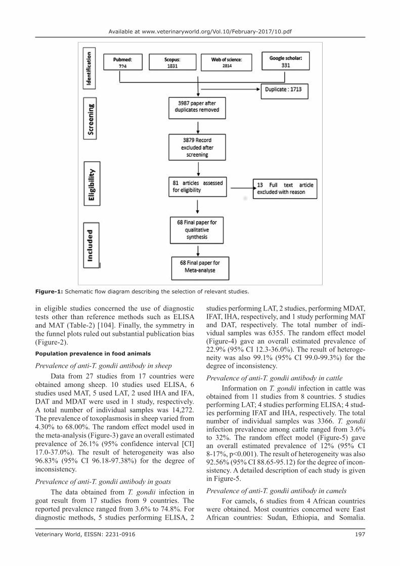
Schematic flow diagram describing the selection of relevant studies Figure-1.Characteristics of eligible studies
Figure-1 shows the flow diagram of the selection of eligible studies. A total of 5700 papers published between 1969 and 2016 were identified by literature search among the four database searched. After dupli-cate removed and irrelevant studies based on titles and abstracts, 81 articles were retrieved for detailed full-text analysis. 13 were excluded due to the following reasons: Two were not available; the sample size was lower than 35 in four study; the diagnosis was estab-lished on the basis of other methods than serologic test in seven studies. Table-1 shows the characteristics of included studies [36-103]. Finally, a total of 68 arti-cles from 24 countries were included in this system-atic review and meta-analysis study. Approximately, 60% (41/68) of the studies were published within the last 10 decade (2007-2016) of the review period. The regional distribution of studies was west Africa (18), East Africa (17), North Africa (21), Southern Africa (8), and Central Africa (4). Our analysis included a totally 30,742 individual samples distributed as fol-lows: 14,272 sheep, 6355 goats, 3366 cattle, 2798 chickens, 2080 pigs, and 1621 camels. Eight different types of diagnostic tests were employed to evaluate T. gondii infection. These diagnostic methods were MAT, ELISA, IHA, DAT, MDAT, IFA, LAT, and SFT. The most used diagnostic tests in 47 year surveys were ELISA and MAT in 24 and 20 studies, which was followed by LAT (14), IHA (13), DAT (6), IFA (6), MDAT (3) and SFT (1). Sensitivity and specificity of diagnostic test are described in Table-2 as reported in literature.Quality and bias assessments
Supplementary Table-S1 (Appendix) represents the quality score of different eligible study. The quality score in 54/84 eligible studies ranged from 6 and 8 (Table-S1) [36-103]. It shows that the risk of bias in these studies was moderate. Besides, many of the eligible studies were conducted in regional and local farms or slaughterhouses, which were not representative of the national population of animals sampled in these countries. Only 5 of the 84 stud-ies were conducted at the national level (Table-S1). Moreover, studies on animal toxoplasmosis were available only in 24 countries out of 54 of African continent. The risk of bias due to quality deficiency in eligible studies was mainly due to external valid-ity criteria, while the flaws internal validity recorded
Veterinary World, EISSN: 2231-0916 197
Available at www.veterinaryworld.org/Vol.10/February-2017/10.pdf
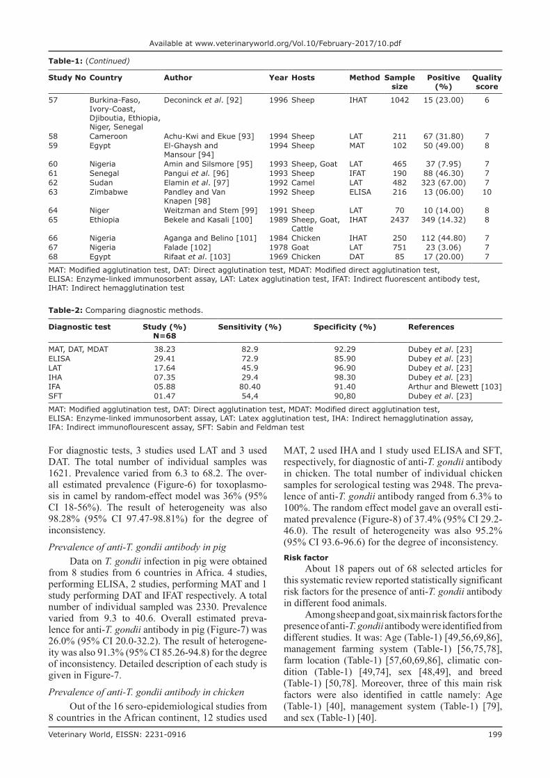
in eligible studies concerned the use of diagnostic tests other than reference methods such as ELISA and MAT (Table-2) [104]. Finally, the symmetry in the funnel plots ruled out substantial publication bias (Figure-2).Population prevalence in food animals
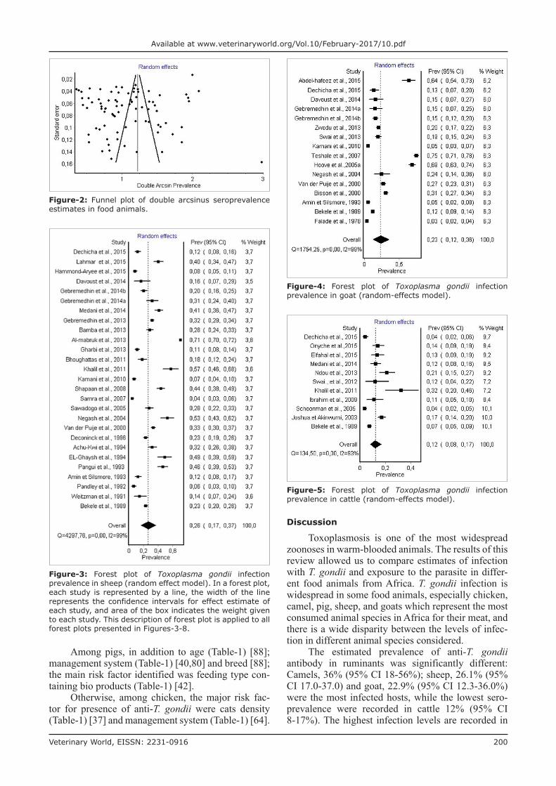
Prevalence of anti-T. gondii antibody in sheepData from 27 studies from 17 countries were
obtained among sheep. 10 studies used ELISA, 6 studies used MAT, 5 used LAT, 2 used IHA and IFA, DAT and MDAT were used in 1 study, respectively. A total number of individual samples was 14,272. The prevalence of toxoplasmosis in sheep varied from 4.30% to 68.00%. The random effect model used in the meta-analysis (Figure-3) gave an overall estimated prevalence of 26.1% (95% confidence interval [CI] 17.0-37.0%). The result of heterogeneity was also 96.83% (95% CI 96.18-97.38%) for the degree of inconsistency.
Prevalence of anti-T. gondii antibody in goatsThe data obtained from T. gondii infection in
goat result from 17 studies from 9 countries. The reported prevalence ranged from 3.6% to 74.8%. For diagnostic methods, 5 studies performing ELISA, 2
studies performing LAT, 2 studies, performing MDAT, IFAT, IHA, respectively, and 1 study performing MAT and DAT, respectively. The total number of indi-vidual samples was 6355. The random effect model (Figure-4) gave an overall estimated prevalence of 22.9% (95% CI 12.3-36.0%). The result of heteroge-neity was also 99.1% (95% CI 99.0-99.3%) for the degree of inconsistency.Prevalence of anti-T. gondii antibody in cattle
Information on T. gondii infection in cattle was obtained from 11 studies from 8 countries. 5 studies performing LAT; 4 studies performing ELISA; 4 stud-ies performing IFAT and IHA, respectively. The total number of individual samples was 3366. T. gondii infection prevalence among cattle ranged from 3.6% to 32%. The random effect model (Figure-5) gave an overall estimated prevalence of 12% (95% CI 8-17%, p<0.001). The result of heterogeneity was also 92.56% (95% CI 88.65-95.12) for the degree of incon-sistency. A detailed description of each study is given in Figure-5.Prevalence of anti-T. gondii antibody in camels
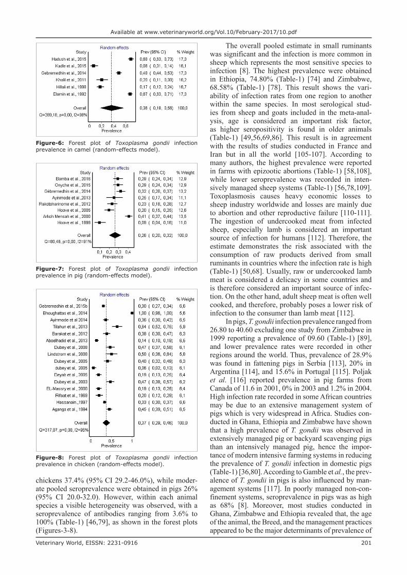
For camels, 6 studies from 4 African countries were obtained. Most countries concerned were East African countries: Sudan, Ethiopia, and Somalia.
Figure-1: Schematic flow diagram describing the selection of relevant studies.
Veterinary World, EISSN: 2231-0916 198
Available at www.veterinaryworld.org/Vol.10/February-2017/10.pdf
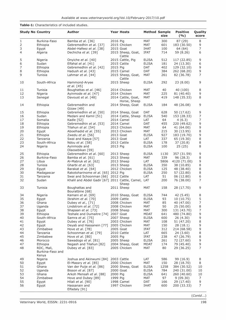
Table-1: Characteristics of included studies.
Study No Country Author Year Hosts Method Sample size
Positive (%)
Quality score
1 Burkina-Faso Bamba et al. [36] 2016 Pig MAT 300 87 (29) 82 Ethiopia Gebremedhin et al. [37] 2015 Chicken MAT 601 183 (30.50) 93 Egypt Abdel-Hafeez et al. [38] 2015 Goat IHAT 100 64 (64) 74 Algeria Dechicha et al. [39] 2015 Sheep, Goat,
CattleIFAT 714 59 (8.26) 6
5 Nigeria Onyiche et al. [40] 2015 Cattle, Pig ELISA 512 117 (22.85) 96 Sudan Elfahal et al. [41] 2015 Cattle ELISA 181 24 (13.30) 67 Ethiopia Gebremedhin et al. [42] 2015 Pig DAT 402 129 (32.10) 98 Ethiopia Hadush et al. [43] 2015 Camel DAT 384 262 (68.20) 99 Tunisia Lahmar et al. [44] 2015 Sheep, Goat,
CattleMAT 261 82 (36.78) 7
10 South-Africa Hammond-Aryee et al. [45]
2015 Sheep ELISA 292 23 (8.00) 9
11 Tunisia Boughattas et al. [46] 2014 Chicken MAT 40 40 (100) 812 Nigeria Ayinmode et al. [47] 2014 Chicken MAT 225 81 (40.40) 913 Senegal Davoust et al. [48] 2014 Cattle, Goat,
Horse, SheepMAT 419 148 (35.33) 8
14 Ethiopia Gebremedhin and Gizaw [49]
2014 Sheep, Goat ELISA 184 48 (26.08) 9
15 Ethiopia Gebremedhin et al. [50] 2014 Sheep, Goat DAT 628 50 (17.62) 916 Sudan Medani and Kamil [51] 2014 Cattle, Sheep ELISA 540 153 (28.33) 717 Somalia Kadle [52] 2014 Camel LAT 64 4 (6.3) 718 Ethiopia Gebremedhin et al. [53] 2014 Camel DAT 455 220 (49.62) 919 Ethiopia Tilahun et al. [54] 2013 Chicken MAT 64 41 (64.00) 920 Egypt Aboelhadid et al. [55] 2013 Chicken MAT 215 30 (13.95) 821 Ethiopia Zwedu et al. [56] 2013 Goat ELISA 927 183 (19.70) 922 Tanzania Swai and Kaaya [57] 2013 Goat LAT 337 65 (19.30) 823 South-Africa Ndou et al. [58] 2013 Cattle ELISA 178 37 (20.8) 824 Nigeria Ayinmode and
Olaosebikan [59]2013 Pig ELISA 100 25 (25) 8
25 Ethiopia Gebremedhin et al. [60] 2013 Sheep ELISA 1130 357 (31.59) 926 Burkina-Faso Bamba et al. [61] 2013 Sheep MAT 339 96 (28.3) 827 Libya Al-Mabruk et al. [62] 2013 Sheep LAT 5806 4120 (71.00) 928 Tunisia Gharbi et al. [63] 2013 Sheep ELISA 350 38 (10.85) 829 Egypt Barakat et al. [64] 2012 Chicken ELISA 125 48 (38.40) 830 Madagascar Rakotoharinome et al. [65] 2012 Pig ELISA 250 57 (22.80) 831 Tanzania Swai and Schoonman [66] 2012 Cattle LAT 51 06 (12.80) 632 Sudan Khalil and Abdel Gadir [67] 2011 Cattle, Camel,
SheepLAT 200 76 (38.00) 7
33 Tunisia Boughattas and Bouratbine [68]
2011 Sheep MAT 158 28 (17.70) 9
34 Nigeria Kamani et al. [69] 2010 Sheep, Goat ELISA 744 42 (5.45) 835 Egypt Ibrahim et al. [70] 2009 Cattle ELISA 93 10 (10.75) 536 Ghana Dubey et al., [71] 2008 Chicken MAT 85 40 (47.00) 737 Uganda Lindstrom et al. [72] 2008 Chicken MAT 50 25 (50.00) 838 Egypt Shapaan et al. [73] 2008 Sheep MAT 300 131 (43.70) 739 Ethiopia Teshale and Dumaitre [74] 2007 Goat MDAT 641 480 (74.80) 940 South-Africa Samra et al. [75] 2007 Sheep ELISA 600 26 (4.30) 941 Egypt Dubey et al. [76] 2003 Chicken MAT 108 51 (47.20) 842 Egypt Deyab and Hassanein [77] 2005 Chicken MAT 150 28 (18.1) 943 Zimbabwe Hove et al. [78] 2005 Goat IFAT 312 214 (68.58) 944 Tanzania Schoonman et al. [79] 2010 Cattle LAT 665 24 (3.60) 845 Zimbabwe Hove et al. [80] 2005 Pig IFAT 238 47 (26.79) 846 Morocco Sawadogo et al. [81] 2005 Sheep ELISA 261 72 (27.60) 947 Ethiopia Negash and Tilahun [82] 2004 Sheep, Goat MDAT 174 79 (45.40) 948 RDC, Mali,
Burkina-Faso and Kenya
Dubey et al. [83] 2005 Chicken MAT 80 29 (36.25) 7
49 Nigeria Joshua and Akinwumi [84] 2003 Cattle LAT 586 99 (16.9) 850 Egypt El-Massry et al. [85] 2000 Chicken MAT 150 28 (18.70) 851 Ghana Van der Puije et al. [86] 2000 Sheep, Goat ELISA 1258 384 (30.52) 1052 Uganda Bisson et al. [87] 2000 Goat ELISA 784 240 (31.00) 1053 Ghana Arkoh Mensah et al. [88] 2000 Pig ELISA 641 260 (40.60) 1054 Zimbabwe Hove and Dubey [89] 1999 Pig MAT 97 9 (09.30) 755 Egypt Hilali et al. [90] 1998 Camel DAT 166 29 (17.40) 956 Egypt Hassanain and
Elfadaly [91]1997 Chicken IHAT 600 200 (33.33) 7
(Contd...)
Veterinary World, EISSN: 2231-0916 199
Available at www.veterinaryworld.org/Vol.10/February-2017/10.pdf
For diagnostic tests, 3 studies used LAT and 3 used DAT. The total number of individual samples was 1621. Prevalence varied from 6.3 to 68.2. The over-all estimated prevalence (Figure-6) for toxoplasmo-sis in camel by random-effect model was 36% (95% CI 18-56%). The result of heterogeneity was also 98.28% (95% CI 97.47-98.81%) for the degree of inconsistency.
Prevalence of anti-T. gondii antibody in pigData on T. gondii infection in pig were obtained
from 8 studies from 6 countries in Africa. 4 studies, performing ELISA, 2 studies, performing MAT and 1 study performing DAT and IFAT respectively. A total number of individual sampled was 2330. Prevalence varied from 9.3 to 40.6. Overall estimated preva-lence for anti-T. gondii antibody in pig (Figure-7) was 26.0% (95% CI 20.0-32.2). The result of heterogene-ity was also 91.3% (95% CI 85.26-94.8) for the degree of inconsistency. Detailed description of each study is given in Figure-7.
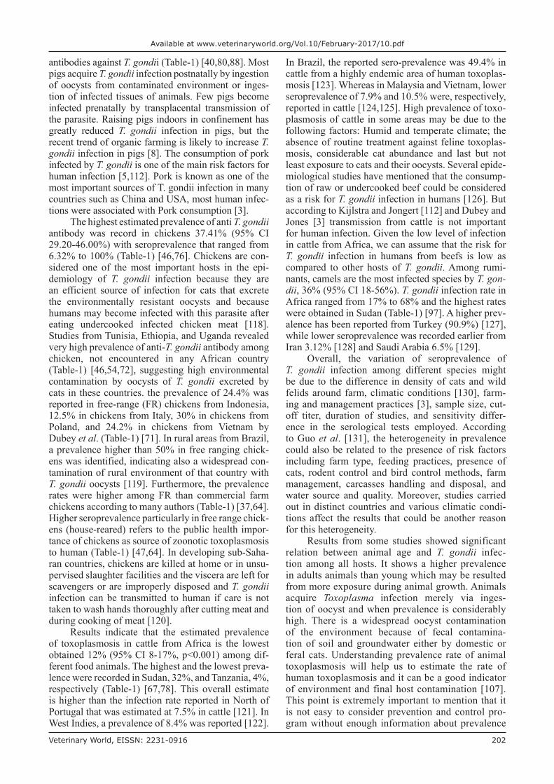
Prevalence of anti-T. gondii antibody in chickenOut of the 16 sero-epidemiological studies from
8 countries in the African continent, 12 studies used
MAT, 2 used IHA and 1 study used ELISA and SFT, respectively, for diagnostic of anti-T. gondii antibody in chicken. The total number of individual chicken samples for serological testing was 2948. The preva-lence of anti-T. gondii antibody ranged from 6.3% to 100%. The random effect model gave an overall esti-mated prevalence (Figure-8) of 37.4% (95% CI 29.2-46.0). The result of heterogeneity was also 95.2% (95% CI 93.6-96.6) for the degree of inconsistency.Risk factor
About 18 papers out of 68 selected articles for this systematic review reported statistically significant risk factors for the presence of anti-T. gondii antibody in different food animals.
Among sheep and goat, six main risk factors for the presence of anti-T. gondii antibody were identified from different studies. It was: Age (Table-1) [49,56,69,86], management farming system (Table-1) [56,75,78], farm location (Table-1) [57,60,69,86], climatic con-dition (Table-1) [49,74], sex [48,49], and breed (Table-1) [50,78]. Moreover, three of this main risk factors were also identified in cattle namely: Age (Table-1) [40], management system (Table-1) [79], and sex (Table-1) [40].
Table-1: (Continued)
Study No Country Author Year Hosts Method Sample size
Positive (%)
Quality score
57 Burkina-Faso, Ivory-Coast, Djiboutia, Ethiopia, Niger, Senegal
Deconinck et al. [92] 1996 Sheep IHAT 1042 15 (23.00) 6
58 Cameroon Achu-Kwi and Ekue [93] 1994 Sheep LAT 211 67 (31.80) 759 Egypt El-Ghaysh and
Mansour [94]1994 Sheep MAT 102 50 (49.00) 8
60 Nigeria Amin and Silsmore [95] 1993 Sheep, Goat LAT 465 37 (7.95) 761 Senegal Pangui et al. [96] 1993 Sheep IFAT 190 88 (46.30) 762 Sudan Elamin et al. [97] 1992 Camel LAT 482 323 (67.00) 763 Zimbabwe Pandley and Van
Knapen [98]1992 Sheep ELISA 216 13 (06.00) 10
64 Niger Weitzman and Stem [99] 1991 Sheep LAT 70 10 (14.00) 865 Ethiopia Bekele and Kasali [100] 1989 Sheep, Goat,
CattleIHAT 2437 349 (14.32) 8
66 Nigeria Aganga and Belino [101] 1984 Chicken IHAT 250 112 (44.80) 767 Nigeria Falade [102] 1978 Goat LAT 751 23 (3.06) 768 Egypt Rifaat et al. [103] 1969 Chicken DAT 85 17 (20.00) 7
MAT: Modified agglutination test, DAT: Direct agglutination test, MDAT: Modified direct agglutination test, ELISA: Enzyme-linked immunosorbent assay, LAT: Latex agglutination test, IFAT: Indirect fluorescent antibody test, IHAT: Indirect hemagglutination test
Table-2: Comparing diagnostic methods.
Diagnostic test Study (%)N=68
Sensitivity (%) Specificity (%) References
MAT, DAT, MDAT 38.23 82.9 92.29 Dubey et al. [23]ELISA 29.41 72.9 85.90 Dubey et al. [23]LAT 17.64 45.9 96.90 Dubey et al. [23]IHA 07.35 29.4 98.30 Dubey et al. [23]IFA 05.88 80.40 91.40 Arthur and Blewett [103]SFT 01.47 54,4 90,80 Dubey et al. [23]
MAT: Modified agglutination test, DAT: Direct agglutination test, MDAT: Modified direct agglutination test, ELISA: Enzyme-linked immunosorbent assay, LAT: Latex agglutination test, IHA: Indirect hemagglutination assay, IFA: Indirect immunoflourescent assay, SFT: Sabin and Feldman test
Veterinary World, EISSN: 2231-0916 200
Available at www.veterinaryworld.org/Vol.10/February-2017/10.pdf
Figure-2: Funnel plot of double arcsinus seroprevalence estimates in food animals.
Figure-3: Forest plot of Toxoplasma gondii infection prevalence in sheep (random effect model). In a forest plot, each study is represented by a line, the width of the line represents the confidence intervals for effect estimate of each study, and area of the box indicates the weight given to each study. This description of forest plot is applied to all forest plots presented in Figures-3-8.
Figure-4: Forest plot of Toxoplasma gondii infection prevalence in goat (random-effects model).
Figure-5: Forest plot of Toxoplasma gondii infection prevalence in cattle (random-effects model).
Among pigs, in addition to age (Table-1) [88]; management system (Table-1) [40,80] and breed [88]; the main risk factor identified was feeding type con-taining bio products (Table-1) [42].
Otherwise, among chicken, the major risk fac-tor for presence of anti-T. gondii were cats density (Table-1) [37] and management system (Table-1) [64].
Discussion
Toxoplasmosis is one of the most widespread zoonoses in warm-blooded animals. The results of this review allowed us to compare estimates of infection with T. gondii and exposure to the parasite in differ-ent food animals from Africa. T. gondii infection is widespread in some food animals, especially chicken, camel, pig, sheep, and goats which represent the most consumed animal species in Africa for their meat, and there is a wide disparity between the levels of infec-tion in different animal species considered.
The estimated prevalence of anti-T. gondii antibody in ruminants was significantly different: Camels, 36% (95% CI 18-56%); sheep, 26.1% (95% CI 17.0-37.0) and goat, 22.9% (95% CI 12.3-36.0%) were the most infected hosts, while the lowest sero-prevalence were recorded in cattle 12% (95% CI 8-17%). The highest infection levels are recorded in
Veterinary World, EISSN: 2231-0916 201
Available at www.veterinaryworld.org/Vol.10/February-2017/10.pdf
The overall pooled estimate in small ruminants was significant and the infection is more common in sheep which represents the most sensitive species to infection [8]. The highest prevalence were obtained in Ethiopia, 74.80% (Table-1) [74] and Zimbabwe, 68.58% (Table-1) [78]. This result shows the vari-ability of infection rates from one region to another within the same species. In most serological stud-ies from sheep and goats included in the meta-anal-ysis, age is considered an important risk factor, as higher seropositivity is found in older animals (Table-1) [49,56,69,86]. This result is in agreement with the results of studies conducted in France and Iran but in all the world [105-107]. According to many authors, the highest prevalence were reported in farms with epizootic abortions (Table-1) [58,108], while lower seroprevalence was recorded in inten-sively managed sheep systems (Table-1) [56,78,109]. Toxoplasmosis causes heavy economic losses to sheep industry worldwide and losses are mainly due to abortion and other reproductive failure [110-111]. The ingestion of undercooked meat from infected sheep, especially lamb is considered an important source of infection for humans [112]. Therefore, the estimate demonstrates the risk associated with the consumption of raw products derived from small ruminants in countries where the infection rate is high (Table-1) [50,68]. Usually, raw or undercooked lamb meat is considered a delicacy in some countries and is therefore considered an important source of infec-tion. On the other hand, adult sheep meat is often well cooked, and therefore, probably poses a lower risk of infection to the consumer than lamb meat [112].
In pigs, T. gondii infection prevalence ranged from 26.80 to 40.60 excluding one study from Zimbabwe in 1999 reporting a prevalence of 09.60 (Table-1) [89], and lower prevalence rates were recorded in other regions around the world. Thus, prevalence of 28.9% was found in fattening pigs in Serbia [113], 20% in Argentina [114], and 15.6% in Portugal [115]. Poljak et al. [116] reported prevalence in pig farms from Canada of 11.6 in 2001, 0% in 2003 and 1.2% in 2004. High infection rate recorded in some African countries may be due to an extensive management system of pigs which is very widespread in Africa. Studies con-ducted in Ghana, Ethiopia and Zimbabwe have shown that a high prevalence of T. gondii was observed in extensively managed pig or backyard scavenging pigs than an intensively managed pig, hence the impor-tance of modern intensive farming systems in reducing the prevalence of T. gondii infection in domestic pigs (Table-1) [36,80]. According to Gamble et al., the prev-alence of T. gondii in pigs is also influenced by man-agement systems [117]. In poorly managed non-con-finement systems, seroprevalence in pigs was as high as 68% [8]. Moreover, most studies conducted in Ghana, Zimbabwe and Ethiopia revealed that, the age of the animal, the Breed, and the management practices appeared to be the major determinants of prevalence of
Figure-6: Forest plot of Toxoplasma gondii infection prevalence in camel (random-effects model).
Figure-7: Forest plot of Toxoplasma gondii infection prevalence in pig (random-effects model).
Figure-8: Forest plot of Toxoplasma gondii infection prevalence in chicken (random-effects model).
chickens 37.4% (95% CI 29.2-46.0%), while moder-ate pooled seroprevalence were obtained in pigs 26% (95% CI 20.0-32.0). However, within each animal species a visible heterogeneity was observed, with a seroprevalence of antibodies ranging from 3.6% to 100% (Table-1) [46,79], as shown in the forest plots (Figures-3-8).
Veterinary World, EISSN: 2231-0916 202
Available at www.veterinaryworld.org/Vol.10/February-2017/10.pdf
antibodies against T. gondii (Table-1) [40,80,88]. Most pigs acquire T. gondii infection postnatally by ingestion of oocysts from contaminated environment or inges-tion of infected tissues of animals. Few pigs become infected prenatally by transplacental transmission of the parasite. Raising pigs indoors in confinement has greatly reduced T. gondii infection in pigs, but the recent trend of organic farming is likely to increase T. gondii infection in pigs [8]. The consumption of pork infected by T. gondii is one of the main risk factors for human infection [5,112]. Pork is known as one of the most important sources of T. gondii infection in many countries such as China and USA, most human infec-tions were associated with Pork consumption [3].
The highest estimated prevalence of anti T. gondii antibody was record in chickens 37.41% (95% CI 29.20-46.00%) with seroprevalence that ranged from 6.32% to 100% (Table-1) [46,76]. Chickens are con-sidered one of the most important hosts in the epi-demiology of T. gondii infection because they are an efficient source of infection for cats that excrete the environmentally resistant oocysts and because humans may become infected with this parasite after eating undercooked infected chicken meat [118]. Studies from Tunisia, Ethiopia, and Uganda revealed very high prevalence of anti-T. gondii antibody among chicken, not encountered in any African country (Table-1) [46,54,72], suggesting high environmental contamination by oocysts of T. gondii excreted by cats in these countries. the prevalence of 24.4% was reported in free-range (FR) chickens from Indonesia, 12.5% in chickens from Italy, 30% in chickens from Poland, and 24.2% in chickens from Vietnam by Dubey et al. (Table-1) [71]. In rural areas from Brazil, a prevalence higher than 50% in free ranging chick-ens was identified, indicating also a widespread con-tamination of rural environment of that country with T. gondii oocysts [119]. Furthermore, the prevalence rates were higher among FR than commercial farm chickens according to many authors (Table-1) [37,64]. Higher seroprevalence particularly in free range chick-ens (house-reared) refers to the public health impor-tance of chickens as source of zoonotic toxoplasmosis to human (Table-1) [47,64]. In developing sub-Saha-ran countries, chickens are killed at home or in unsu-pervised slaughter facilities and the viscera are left for scavengers or are improperly disposed and T. gondii infection can be transmitted to human if care is not taken to wash hands thoroughly after cutting meat and during cooking of meat [120].
Results indicate that the estimated prevalence of toxoplasmosis in cattle from Africa is the lowest obtained 12% (95% CI 8-17%, p<0.001) among dif-ferent food animals. The highest and the lowest preva-lence were recorded in Sudan, 32%, and Tanzania, 4%, respectively (Table-1) [67,78]. This overall estimate is higher than the infection rate reported in North of Portugal that was estimated at 7.5% in cattle [121]. In West Indies, a prevalence of 8.4% was reported [122].
In Brazil, the reported sero-prevalence was 49.4% in cattle from a highly endemic area of human toxoplas-mosis [123]. Whereas in Malaysia and Vietnam, lower seroprevalence of 7.9% and 10.5% were, respectively, reported in cattle [124,125]. High prevalence of toxo-plasmosis of cattle in some areas may be due to the following factors: Humid and temperate climate; the absence of routine treatment against feline toxoplas-mosis, considerable cat abundance and last but not least exposure to cats and their oocysts. Several epide-miological studies have mentioned that the consump-tion of raw or undercooked beef could be considered as a risk for T. gondii infection in humans [126]. But according to Kijlstra and Jongert [112] and Dubey and Jones [3] transmission from cattle is not important for human infection. Given the low level of infection in cattle from Africa, we can assume that the risk for T. gondii infection in humans from beefs is low as compared to other hosts of T. gondii. Among rumi-nants, camels are the most infected species by T. gon-dii, 36% (95% CI 18-56%). T. gondii infection rate in Africa ranged from 17% to 68% and the highest rates were obtained in Sudan (Table-1) [97]. A higher prev-alence has been reported from Turkey (90.9%) [127], while lower seroprevalence was recorded earlier from Iran 3.12% [128] and Saudi Arabia 6.5% [129].
Overall, the variation of seroprevalence of T. gondii infection among different species might be due to the difference in density of cats and wild felids around farm, climatic conditions [130], farm-ing and management practices [3], sample size, cut-off titer, duration of studies, and sensitivity differ-ence in the serological tests employed. According to Guo et al. [131], the heterogeneity in prevalence could also be related to the presence of risk factors including farm type, feeding practices, presence of cats, rodent control and bird control methods, farm management, carcasses handling and disposal, and water source and quality. Moreover, studies carried out in distinct countries and various climatic condi-tions affect the results that could be another reason for this heterogeneity.
Results from some studies showed significant relation between animal age and T. gondii infec-tion among all hosts. It shows a higher prevalence in adults animals than young which may be resulted from more exposure during animal growth. Animals acquire Toxoplasma infection merely via inges-tion of oocyst and when prevalence is considerably high. There is a widespread oocyst contamination of the environment because of fecal contamina-tion of soil and groundwater either by domestic or feral cats. Understanding prevalence rate of animal toxoplasmosis will help us to estimate the rate of human toxoplasmosis and it can be a good indicator of environment and final host contamination [107]. This point is extremely important to mention that it is not easy to consider prevention and control pro-gram without enough information about prevalence
Veterinary World, EISSN: 2231-0916 203
Available at www.veterinaryworld.org/Vol.10/February-2017/10.pdf
of toxoplasmosis in animal since they are a major source of transmission to human.
Given the vital role of animals in the transmission of T. gondii to humans via their products (meat and milk) and the prominent role of cats in disseminating and contamination of the environment by oocysts [1], more emphasis should be placed on the prevention of animal toxoplasmosis in Africa.
Caution is warranted in the interpretation of results of T. gondii prevalence in camel. Regarding such species, the prevalence data used in this study were analyzed based on a limited number of national studies, and nationwide surveys are not available in these meat animals, which resulted in a wide 95% CI of the estimated prevalence.Conclusion
This systematic review was performed to evalu-ate the prevalence of T. gondii infection among sheep, goat, cattle, pig, camel, and chicken which represent the most consumed food animal species in different African countries. The Random-effects meta-anal-ysis approach in this current study provided an esti-mate of T. gondii prevalence in various meat animals with an increased level of precision. The widespread prevalence of T. gondii in sheep, chicken, camel, pig, and goats indicates a food safety concern in differ-ent African countries, especially countries where the infection is more important. Other studies are required for a better and continual evaluation of the occurrence of this zoonotic infection.Authors’ Contributions
The study was conceptualized and proto-cols were carried out by YA. ABNT and PS were involved in the database search, data extraction, statistical analysis, and manuscript written. CA and EY Studied titles and abstract of all the articles and retrieved data. Quality assessment of each study was completed independently by YGH and IY. MNA and SF oversaw data collection and analysis of statisti-cal results. All authors have read and approved the content.Acknowledgments
The authors are grateful to the Ministry of Higher Education and Scientific Research of Benin, Through the Doctoral Support Program No. 125 MESRS/CAB/DC/SGM/DRFM/DRH/REGIE of 24 February 2015.Competing Interests
The authors declare that they have no competing interests.References1. Ferguson, D.J. (2009) Toxoplasma gondii: 1908-2008,
homage to Nicolle, Manceaux and Splendore. Mem. Inst. Oswaldo Cruz., 104(2): 133-148.
2. Dubey, J.P. (2010) Toxoplasmosis of Animals and Humans. 2nd ed. CRC Press, Florida, USA.
3. Dubey, J.P. and Jones, J.L. (2008) Toxoplasma gondii infec-tion in humans and animals in the United States. Int. J. Parasitol., 38(11): 1257-1278.
4. Moncada, P.A. and Montoya, J.G. (2012) Toxoplasmosis in the fetus and newborn: An update on prevalence, diagnosis and treatment. Expert. Rev. Anti Infect. Ther., 10(7): 815-828.
5. Tenter, A.M., Heckerotha, A.R. and Weiss, L.M. (2000) Toxoplasma gondii: From animals to humans. Int. J. Parasitol., 30: 1217-1258.
6. Patz, J.A., Graczyk, T.K., Geller, N. and Vittor, A.Y. (2000) Effects of environmental change on emerging parasitic dis-eases. Int. J. Parasitol., 30(12): 1395-1405.
7. Torgerson, P.R. and Mastroiacovo, P. (2013) The global bur-den of congenital toxoplasmosis: A systematic review. Bull. World Health Organ., 91(7): 501-508.
8. Dubey, J.P. (2009a) Toxoplasmosis in pigs: The last 20 years. Vet. Parasitol., 164: 89-103.
9. Dubey, J.P. (2009b) Toxoplasmosis in sheeps: The last 20 years. Vet. Parasitol., 152: 25-80.
10. Bourée, P., Dumazedier, D., Magdeleine, C. and Sobesky, G. (1997) Toxoplasmose cérébrale et SIDA à la martinique. Méd. Trop., 57(3): 259-261.
11. Weiss, L.M. and Dubey, J.P. (2009) Toxoplasmosis: A history of clinical observations. Int. J. Parasitol., 39(8): 895-901.
12. Torrey, E.F., Bartko, J.J., Lun, Z.R. and Yolken, R.H. (2007) Antibodies to Toxoplasma gondii in patients with schizo-phrenia: A meta-analysis. Schizophr. Bull., 33: 729-736.
13. Palmer, B.S. (2007) Meta-analysis of three case controlled studies and an ecological study into the link between cryp-togenic epilepsy and chronic toxoplasmosis infection. Seizure, 16(8): 657-663.
14. Flegr, J., Klose, J., Novotná, M., Berenreitterová, M. and Havlíček, J. (2009) Increased incidence of traffic accidents in Toxoplasma-infected military drivers and protective effect RhD molecule revealed by a large-scale prospective cohort study. BMC Infect. Dis., 9: 72.
15. Leal, F.E., Cavazzana, C.L., de Andrade, H.F.J., Galisteo, A.Jr., de Mendonca, J.S. and Kallas, E.G. (2007) Toxoplasma gon-dii pneumonia in immunocompetent subjects: Case report and review. Clin. Infect. Dis., 44(6): e62-e66.
16. Pasquali, P. (2007) Infections au VIH et zoonoses. Food Agric. Organ., Vol.163.
17. Hammond-Aryee, K., Esser, M. and Van Helden, P.D. (2015) Toxoplasma gondii seroprevalence studies on humans and animals in Africa. S. Afr. Fam. Pract., 56(2): 119-124.
18. Available from: http://www.faostat.fao.org/site/569/default.aspx#ancor. Accessed on 20-06-2016.
19. Cook, A.J.C., Gilbert, R.E., Buffolano, W., Zufferey, J., Petersen, E., Jenum, P.A., Foulon, W., Semprini, A.E. and Dunn, D.T. (2000) Sources of Toxoplasma infection in preg-nant women: European multicentre case-control study. Br. Med. J., 321: 142-147.
20. Boyer, K.M., Holfels, E., Roizen, N., Swisher, C., Mack, D., Remington, J., Withers, S., Meier, P. and McLeod, R. (2005) Risk factors for Toxoplasma gondii infection in mothers of infants with congenital toxoplasmosis: Implications for pre-natal management and screening. Am. J. Obstet. Gynecol., 192: 564-571.
21. FAO-OMS. (2015) Joint FAO/WHO food standards pro-gram codex committee on food hygiene. Forty-seventh session Boston, Massachusetts, United States of America, 9-13 November 2015.
22. Montoya, J.G. (2002) Laboratory diagnosis of Toxoplasma gondii infection and toxoplasmosis. J. Infect. Dis., 185(1): S73-S82.
23. Dubey, J.P., Thulliez, P., Weigel, R.M., Andrews, C.D., Lind, P. and Powell, E.C. (1995) Sensitivity and specific-ity of various serologic tests for detection of Toxoplasma gondii infection in naturally infected sows. Am. J. Vet. Res., 56(8): 1030-1036.
24. Villena, I., Durand, B., Aubert, D., Blaga, R., Geers, R., Thomas, M., Perret, C., Alliot, A., Escotte-Binet, S.,
Veterinary World, EISSN: 2231-0916 204
Available at www.veterinaryworld.org/Vol.10/February-2017/10.pdf
Thebault, A., Boireau, P. and Halos, L. (2012) New strategy for the survey of Toxoplasma gondii in meat for human con-sumption. Vet. Parasitol., 183(3-4): 203-208.
25. Gamble, H.R., Dubey, J.P. and Lambillotte, D.N. (2005) Comparison of a commercial ELISA with the modified agglutination test for detection of Toxoplasma infection in the domestic pig. Vet. Parasitol., 128: 177-181.
26. Gonzales-Barron, U. and Butler, U. (2011) The use of meta-analytical tools in risk assessment for food safety. Food Microbiol., 28: 823-827.
27. Barron, U.G., Soumpasis, I., Butler, F., Prendergast, D., Duggan, S. and Duffy, G. (2009) Estimation of prevalence of Salmonella on pig carcasses and pork joints, using a quantitative risk assessment model aided by meta-analysis. J. Food Prot., 72: 274-285.
28. Gliner, G.A., Morgan, N.L. and Leech, O. (2009) Research Methods in Applied Settings: An Integrated Approach to Design and Analysis. 2nd ed. Routledge, Abingdon, UK.
29. Hutton, B., Salanti, G., Caldwell, D.M., Chaimani, A., Schmid, C.H., Cameron, C., Ioannidis, J.P., Straus, S., Thorlund, K., Jansen, J.P., Mulrow, C., Catalá-López, F., Gøtzsche, P.C., Dickersin, K., Boutron, I., Altman, D.G. and Moher, D. (2015) The PRISMA extension statement for reporting of systematic reviews incorporating network meta-analyses of health care interventions: Checklist and explanations. Ann. Intern. Med., 162(11): 777-784.
30. Hoy, D., Brooks, P., Woolf, A., Blyth, F., March, L., Bain, C., Baker, P., Smith, E. and Buchbinder, R. (2012) Assessing risk of bias in prevalence studies: Modification of an existing tool and evidence of inter rater agreement. J. Clin. Epidemiol., 65: 934-939.
31. Available from: http://www.epigear.com/index_files/metaxl.html. Accessed on 22 May 2016, 11h 55 min.
32. Doi, S.A., Barendregt, J.J. and Mozurkewich, E.L. (2011) Meta-analysis of heterogeneous clinical trials: An empirical example. Contemp. Clin. Trials, 32: 288-298.
33. Barendregt, J.J., Doi, S.A., Lee, Y.Y., Norman, R.E. and Vos, T. (2013) Meta-analysis of prevalence. J. Epidemiol. Community Health, 67: 974-978.
34. Sterne, J.A. and Egger, M. (2001) Funnel plots for detecting bias in meta-analysis: Guidelines on choice of axis. J. Clin. Epidemiol., 54: 1046-1055.
35. Higgins, J.P., Thompson, S.G., Deeks, J.J. and Altman, D.G. (2003) Measuring inconsistency in meta-analyses. Br. Med. J., 327: 557-560.
36. Bamba, S., Halos, F., Tarnagda, Z., Alanio, A., Macé, E., Moukoury, S., Sangaré, I., Guiguemdé, R., Costa, J.M. and Bretagne, S. (2016) Seroprevalence of Toxoplasma gondii and direct genotyping using mini-sequencing in free-range pigs in Burkina Faso. Int. J. Food Microbiol., 230: 10-15.
37. Gebremedhin, E.Z., Tesfamaryam, G., Yunus, H.A., Duguma, R., Tilahun, G., Di Marco, V. and Vitale, M. (2015b) Seroepidemiology of Toxoplasma gondii infec-tion in free-range chickens (Gallus domesticus) of Central Ethiopia. Epidemiol. Infect., 143(3):608-617.
38. Abdel-Hafeez, E.H., Kamal, A.M., Abdelgelil, N.H. and Abdel-Fatah, M. (2015) Parasites transmitted to human by ingestion of different types of meat, El-Minia City, El-Minia Governorate, Egypt. J. Egypt. Soc. Parasitol., 45(3): 671-680.
39. Dechicha, A.S., Bachi, F., Gharbi, I., Gourbdji, E., Baazize-Ammi, D. and Guetarni, D. (2015) Sero-epidemiological survey on toxoplasmosis in cattle, sheep and goats in Algeria. Afr. J. Agric. Res., 10(20): 2113-2119.
40. Onyiche, T.G.E. and Ademola, I.O. (2015) Seroprevalence of anti-Toxoplasma gondii antibodies in cattle and pigs in Ibadan, Nigeria. J. Parasit. Dis., 39(2): 309-314.
41. Elfahal, A.M., Elhassan, A.M., Hussien, M.O., Enan, K.A., Musa, A.B. and El Hussein, A.M. (2013) Seroprevalence of Toxoplasma gondii in dairy cattle with reproductive prob-lems in Sudan. ISRN Vet. Sci., 89: 51-65.
42. Gebremedhin, E.Z., Kebeta, M.M., Asaye, M., Ashenafi, H., Di Marco, V. and Vitale, M. (2015a) First report on seroepi-demiology of Toxoplasma gondii infection in pigs in central Ethiopia. BMC Vet. Res., 11: 59.
43. Hadush, A., Gebru, M.U., Zeru, F., Hadush, T., Tesfamaryam, G. and Feleke, A. (2015) Sero-epidemiology of camel toxoplasmosis and public awareness on its zoo-notic importance in central afar region, North East Ethiopia. World Appl. Sci. J., 33(12): 1880-1887.
44. Lahmar, I., Lachkhem, A., Slama, D., Sakly, W., Haouas, N., Gorcii, M. and Babba, H. (2015) Prevalence of toxoplas-mosis in sheep, goats and cattle in Southern Tunisia. J. Bacteriol. Parasitol., 6: 245.
45. Hammond-Aryee, K., Van Helden, L.S. and Van Helden, P.D. (2015) The prevalence of antibodies to Toxoplasma gondii in sheep in the Western Cape, South Africa. Onderstepoort J. Vet. Res., 82(1): E1-E5.
46. Boughattas, S., Ayari, K., Sa, T., Aoun, K. and Bouratbine, A. (2014) Survey of the parasite Toxoplasma gondii in human consumed ovine meat in Tunis city. PLoS One, 9(1): e85044.
47. Ayinmode, A.B. and Olaosebikan, R.I. (2014) Seroprevalence of Toxoplasma gondii infection in free ranged chicken from rural and urban settlements in Oyo State, Nigeria. Afr. J. Med. Med. Sci., 43 Suppl: 51-57.
48. Davoust, B., Mediannikov, O., Roqueplo, C., Perret, C., Demoncheaux, J.P., Sambou, M., Guillot, J. and Blaga, R. (2015) Serological survey of animal toxoplasmosis in Senegal. Bull. Soc. Pathol. Exot., 108(1): 73-77.
49. Gebremedhin, E.Z. and Gizaw, D. (2014a) Seroprevalence of Toxoplasma gondii Infection in sheep and goats in three districts of Southern nations, nationalities and peoples region of Ethiopia. World Appl. Sci. J., 31(11): 1891-1896.
50. Gebremedhin, E.Z., Abdurahaman, M., Hadush, T. and Tessema, T.S. (2014b) Seroprevalence and risk factors of Toxoplasma gondii infection in sheep and goats slaughtered for human consumption in Central Ethiopia. BMC Res. Notes, 7: 696.
51. Medani, M.Y.I. and Kamil, I.Y. (2014) Serosurvey of Toxoplasma gondii in sheep and cattle for human consump-tion in Khartoum slaughterhouses, Sudan. Int. J. Infect. Dis., 21: 157.
52. Kadle, A.A.H. (2014) Sero-prevalence of toxoposis in domestic animals in Benadir Region, Somalia. Open. J. Vet. Med., 4(08): 170.
53. Gebremedhin, E.Z., Yunus, H.A., Tesfamaryam, G., Tessema, T.S., Dawo, F., Terefe, G., Di Marco, V. and Vitale, M. (2014) First report of Toxoplasma gondii in camels (Camelus dromedarius) in Ethiopia: Bioassay and seroepidemiological investigation. BMC Vet. Res., 10: 222.
54. Tilahun, G., Tiao, N., Ferreira, L.R., Choudhary, S., Oliveira, S., Verma, S.K., Kwok, O.C., Molla, B., Saville, W.J., Medhin, G., Kassa, T., Aleme, H., Gebreyes, W.A., Su, C. and Dubey, J.P. (2013) Prevalence of Toxoplasma gondii from free-range chickens (Gallus domesticus) from Addis Ababa, Ethiopia. J. Parasitol., 99(4): 740-741.
55. Aboelhadid, S.M., Abdel-Ghany, A.E., Ibrahim, M.A. and Mahran, H.A. (2013) Seroprevalence of Toxoplasma gon-dii infection in chickens and humans in Beni Suef, Egypt. Glob. Vet., 11: 139-144.
56. Zewdu, E., Agonafir, A., Tessema, T.S., Tilahun, G., Medhin, G., Vitale, M., Di Marco, V., Cox, E., Vercruysse, J. and Dorny, P. (2013) Seroepidemiological study of cap-rine toxoplasmosis in East and West Shewa Zones, Oromia Regional State, Central Ethiopia. Res. Vet. Sci., 94(1): 43-48.
57. Swai, E.S. and Kaaya, J.E. (2013) A survey of Toxoplasma gondii antibodies by latex agglutination assay in dairy goats in Northern Tanzania. Trop. Anim. Health Prod., 45(1): 211-217.
58. Ndou, R.V., Pelele, W.P.S., Dzoma, B.M., Nyirenda, M.,
Veterinary World, EISSN: 2231-0916 205
Available at www.veterinaryworld.org/Vol.10/February-2017/10.pdf
Motsei, L.E. and Bakunzi, F.R. (2013) An investigation into the prevalence of Toxoplasma gondii among indigenous, communally reared goats in the Mafikeng Area of the North West Province of South Africa. Life Sci. J., 8 Suppl 1: 38-41.
59. Ayinmode, A.B. and Olaosebikan, R.I. (2014) Antibodies to Toxoplasma gondii in backyard and wandering pigs in Ibadan, Nigeria: Implications for pork consumption. Bull. Anim. Health Prod. Afr., 61(3): 493-497.
60. Gebremedhin, E.Z., Agonafir, A., Tessema, T.S., Tilahun, G., Medhin, G., Vitale, M., Di Marco, V., Cox, E., Vercruysse, J. and Dorny, P. (2013) Seroepidemiological study of ovine toxoplasmosis in East and West Shewa Zones of Oromia Regional State, Central Ethiopia. BMC Vet. Res., 9: 117.
61. Bamba, S., Kamga-Waladjo, A.R., Cissé, M. and Tarnagda, Z. (2012, 2013) Serological survey on bovine toxoplasmosis in Bobo-Dioulasso, Burkina Faso. Rev. Med. Vet. Trop., 65: 63-66.
62. Al-Mabruk, A.A., Alkhunfas, S.R., El-Buni, A.A., Annajar, B.B. and Elsaid, M.M.A. (2013) Seroprevalence of Toxoplasma gondii antibodies in sheep from Libya. Int. J. Adv. Res., 1(9): 148-154.
63. Gharbi, M., Zribi, L., Jedidi, M., Chakkhari, H., Hamdi, S., R’hayem, S. and Darghouth, M.A. (2013) Prevalence of Toxoplasma gondii infection in sheep from Tunisia. Bull. Soc. Pathol. Exot., 106(3): 184-187.
64. Barakat, A.M., Salem, L.M., El-Newishy, A.M., Shaapan, R.M. and El-Mahllawy, E.K. (2012) Zoonotic chicken toxoplasmosis in some Egyptians Governorates. Pak. J. Biol. Sci., 15(17): 821-826.
65. Rakotoharinome, M., Andriamanivo, H., Blaga, R., Perret, C., Lacour, S., Grasset-Chevillot, A., Mace, P., Thomas, M., Villena, I., Aubert, D., Boireau, P. and Porphyre, V. (2012) Toxoplasmosis and trichinellosis: An epidemiological survey of pig population in Madagascar. European Multicolloquium on Parasitology (EMOP) XI, Cluj Napoca, Roumanie.
66. Swai, E.S. and Schoonman, L. (2012) A survey of zoonotic diseases in trade cattle slaughtered at Tanga city abattoir: A cause of public health concern. Asian. Pac. J. Trop. Biomed., 2(1): 55-60.
67. Khalil, K.M. and Abdel Gadir, A.E. (2007) Prevalence of Toxoplasma gondii antibodies in camels and their herders in three ecologically different areas in Sudan. J. Camel Pract. Res., 14(1): 11-13.
68. Boughattas, S. and Bouratbine, A. (2014) Prevalence of food-borne Toxoplasma gondii in free-ranging chickens sold in Tunis, Tunisia. J. Food Qual. Hazards Control, 1(3): 89-92.
69. Kamani, J., Mani, A.U. and Egwu, G.O. (2010) Seroprevalence of Toxoplasma gondii infection in domestic sheep and goats in Borno state, Nigeria. Trop. Anim. Health Prod., 42(4): 793-797.
70. Ibrahim, H.M., Huang, P., Salem, T.A., Talaat, R.M., Nasr, M.I., Xuan, X. and Nishikawa, Y. (2009) Short report: Prevalence of Neospora caninum and Toxoplasma gondii antibodies in northern Egypt. Am. J. Trop. Med. Hyg., 80(2): 263-267.
71. Dubey, J.P., Huong, L.T., Lawson, B.W., Subekti, D.T., Tassi, P., Cabaj, W., Sundar, N., Velmurugan, G.V., Kwok, O.C. and Su, C. (2008) Seroprevalence and isolation of Toxoplasma gondii from free-range chickens in Ghana, Indonesia, Italy, Poland, and Vietnam. J. Parasitol., 94(1): 68-71.
72. Lindstrom, I., Sundar, N., Lindh, J., Kironde, F., Kabasa, J.D., Kwok, O.C., Dubey, J.P. and Smith, J.E. (2008) Isolation and genotyping of Toxoplasma gondii from Ugandan chickens reveals frequentmultiple infections. Parasitology, 135: 39-45.
73. Shaapan, R.M., El-Nawawi, F.A. and Tawfik, M.A. (2008) Sensitivity and specificity of various serological tests for
the detection of Toxoplasma gondii infection in naturally infected sheep. Vet. Parasitol., 153(3-4): 359-362.
74. Teshale, S. and Dumaitre, A. (2007) Serological survey of caprine toxopiasmosis in Ethiopia: Prevalence and risk fac-tors. Parasite, 14(2): 155-159.
75. Samra, N.A., McCrindle, C.M., Penzhorn, B.L. and Cenci-Goga, B. (2007) Seroprevalence of toxoplasmosis in sheep in South Africa. J. S. Afr. Vet. Assoc., 78(3): 116-120.
76. Dubey, J.P., Graham, D.H., Dahl, E., Hilali, M., El-Ghaysh, A., Sreekumar, C., Kwok, O.C., Shen, S.K. and Lehmann, T. (2003) Isolation and molecular characteriza-tion of Toxoplasma gondii from chickens and ducks from Egypt. Vet. Parasitol., 114(2): 89-95.
77. Deyab, A.K. and Hassanein, R. (2005) Zoonotic toxoplas-mosis in chicken. J. Egypt. Soc. Parasitol., 147(5): 341-350.
78. Hove, T., Lind, P. and Mukaratirwa, S. (2005b) Seroprevalence of Toxoplasma gondii infection in goats and sheep in Zimbabwe. Onderstepoort J. Vet. Res., 72(4): 267-272.
79. Schoonman, L.B., Wilsmore, T. and Swai, E.S. (2010) Sero-epidemiological investigation of bovine toxoplasmosis in traditional and smallholder cattle production systems of Tanga Region, Tanzania. Trop. Anim. Health Prod., 42(4): 579-587.
80. Hove, T., Lind, P. and Mukaratirwa, S. (2005a) Seroprevalence of Toxoplasma gondii infection in domes-tic pigs reared under different management systems in Zimbabwe. Onderstepoort J. Vet. Res., 72(3): 231-237.
81. Sawadogo, P., Hafid, J., Bellete, B., Sung, R.T., Chakdi, M., Flori, P., Raberin, H., Hamouni, I.B., Chait, A. and Dalal, A. (2005) Seroprevalence of T. gondii in sheep from Marrakech, Morocco. Vet. Parasitol., 130(1-2): 89-92.
82. Negash, T. and Tilahun, G. (2004) Serological survey on toxoplasmosis in sheep and goats in Nazareth, Ethiopia. Re. Med. Vet., 155(10): 486-487.
83. Dubey, J.P., Karhemere, S., Dahl, E., Sreekumar, C., Diabate, A., Dabire, K.R., Vianna, M.C., Kwok, O.C. and Lehmann, T. (2005) First biologic and genetic characteri-zation of Toxoplasma gondii isolates from chickens from Africa (Democratic Republic of Congo, Mali, Burkina Faso, and Kenya). J. Parasitol., 91(1): 69-72.
84. Joshua, R.A. and Akinwumi, K.A. (2004) Prevalence of antibodies to Toxoplasma gondii in four breeds of cattle at Ibadan, Nigeria. Trop. Vet., 21(3): 134-137.
85. El-Massry, A., Mahdy, O.A., El-Ghaysh, A. and Dubey, J.P. (2000) Prevalence of Toxoplasma gondii antibodies in sera of turkeys, chickens, and ducks from Egypt. J. Parasitol., 86(3): 627-628.
86. Van der Puije, W.N., Bosompem, K.M., Canacoo, E.A., Wastling, J.M. and Akanmori, B.D. (2000) The prevalence of anti-Toxoplasma gondii antibodies in Ghanaian sheep and goats. Acta Trop., 76(1): 21-26.
87. Bisson, A., Maley, S., Rubaire-Akiiki, C.M. and Wastling, J.M. (2000) The seroprevalence of antibodies to Toxoplasma gondii in domestic goats in Uganda. Acta Trop., 76(1): 33-38.
88. Arko-Mensah, J., Bosompem, K.M., Canacoo, E.A., Wastling, J.M. and Akanmori, B.D. (2000) The seroprev-alence of toxoplasmosis in pigs in Ghana. Acta Trop., 76(1): 27-31.
89. Hove, T. and Dubey, J.P. (1999) Prevalence of Toxoplasma gondii antibodies in sera of domestic pigs and some wild game species from Zimbabwe. J. Parasitol., 85(2): 372-337.
90. Hilali, M., Romand, S., Thulliez, P., Kwok, O.C. and Dubey, J.P. (1998) Prevalence of Neospora caninum and Toxoplasma gondii antibodies in sera from camels from Egypt. Vet. Parasitol., 75(42-43): 269-271.
91. Hassanain, M.A. and Elfadaly, H.A. (1997) Biological assay of Toxoplasma gondii Egyptian mutton isolates. Int. J. Zool. Res., 7(4): 330-337.
92. Deconinck, P., Pangui, L.J., Akakpo, J., Garrouste, A.,
Veterinary World, EISSN: 2231-0916 206
Available at www.veterinaryworld.org/Vol.10/February-2017/10.pdf
Ouattara, L., Roger, F., Tibayrenc, R. and Dorchies, P. (1996) Prevalence of toxoplasmosis in small ruminants in tropical Africa: Results of a sero-epidemiological survey of 1042 animals. Rev. Méd. Vét., 19: 30-36.
93. Achu-Kwi, M.D. and Ekue, N.F. (1994) Prevalence of Toxoplasma gondii antibodies in Djallonke sheep flocks in the Vina Division, Cameroon. Bull. Anim. Health Prod. Afr., 42(2): 89-92.
94. El-Ghaysh, A.A. and Mansour, M.M. (1994) Detection of antibodies to Toxoplasma gondii in an Egyptian sheep-herd using modern serological techniques. J. Egypt. Assoc. Immunol., 1: 117-121.
95. Amin, J.D. and Silsmore, A.J. (1993) A serological survey of some abortifacient diseases of sheep and goats in the Maiduguri area of Nigeria. Bull. Anim. Health Prod. Afr., 41(2): 123-128.
96. Pangui, L.J., Lahamdi, A. and Samb, F. (1993) Use of IFI and ELISA in a serological survey of toxoplasmosis in sheep in Dakar-Senegal. Rev. Méd. Vét., 16: 25-29.
97. Elamin, E.A., Elias, S., Daugschies, A. and Rommel, M. (1992) Prevalence of Toxoplasma gondii antibodies in pastoral camels (Camelus dromedarius) in the Butana plains, mid-Eastern Sudan. Vet. Parasitol., 43(3-4): 171-175.
98. Pandey, V.S. and Van Knapen, F. (1992) The seroprevalence of toxoplasmosis in sheep, goats and pigs in Zimbabwe. Ann. Trop. Med. Parasitol., 86(3): 313-315.
99. Weitzman, G.L. and Stem, E.C. (1991) Preliminary sero-logical survey for bluetongue and toxoplasmosis in sheep in Niger. Trop. Anim. Health Prod., 23(4): 258.
100. Bekele, T. and Kasali, O.B. (1989) Toxoplasmosis in sheep, goats and cattle in central Ethiopia. Vet. Res. Commun., 13(5): 371-375.
101. Aganga, A.O. and Belino, E.D. (1984) Toxoplasmosis in local breed of chicken in Zaria, Nigeria. Int. J. Zoonoses., 11(2): 170-172.
102. Falade, S. (1978) Toxoplasma gondii antibodies in Nigerian goats. Trop. Anim. Health Prod., 10(1): 175-177.
103. Rifaat, M.A., Morsy, T.A. and Sadek, M.S.M. (1969) Toxoplasmosis in chickens and pigeons in U.A.R. (Preliminary Report). J. Trop. Med. Hyg., 72: 193-194.
104. Arthur, M.J. and Blewett, D.A. (1988) IFAT detection of IgG specific to Toxoplasma in thoracic fluids from aborted lambs: Evaluation on routine diagnostic submissions. Vet. Rec., 122: 29-31.
105. Dumetre, A., Daniel, A., Luc, R., Aurelien, M. and Marie-Laure, D. (2006) Toxoplasma gondii infection in sheep from Haute-Vienne, France: Seroprevalence and iso-late genotyping by microsatellite analysis. Vet. Parasitol., 142(3-4): 376-379.
106. Sharif, M., Sarvi, S., Shokri, A., Teshnizi, S.H., Rahimi, M.T., Mizani, A. and Daryani, A. (2015) Toxoplasma gondii infec-tion among sheep and goats in Iran: A systematic review and meta-analysis. Parasitol. Res., 114(1): 1-16.
107. Guo, M., Dubey, J.P., Hill, D., Buchanan, R.L., Gamble, H.R., Jones, J.L. and Pradhan, A.K. (2015) Prevalence and risk factors for Toxoplasma gondii infection in meat animals and meat products destined for human con-sumption. J. Food Prot., 78: 457-476.
108. Dubey, J.P. and Kirkbride, C.A. (1989) Enzootic toxoplas-mosis in sheep in North-Central United-States. J. Parasitol., 75: 673-676.
109. Ragozo, A.M.A., Yai, L.E.O., Oliveira, L.N., Dias, R.A., Dubey, J.P. and Gennari, S.M. (2008) Seroprevalence and isolation of Toxoplasma gondii from sheep from São Paulo state, Brazil. J. Parasitol., 94(6): 1259-1263.
110. Buxton, D., Maley, S.W., Wright, S.E., Rodger, S., Bartley, P. and Innes, E.A. (2007) Toxoplasma gondii and ovine toxoplasmosis: New aspects of an old story. Vet. Parasitol., 149(1): 25-28.
111. da Silva, A.F., Brandão, F.Z., Oliveira, F.C.R. and Ferreira, A.M.R. (2013) Toxoplasma gondii in the sheep
industry: A global overview and the situation in Brazil. Rev. Bras. Ciên. Vet., 20(4): 179-188.
112. Kijlstra, A. and Jongert, E. (2008) Control of the risk of human toxoplasmosis transmitted by meat. Int. J. Parasitol., 38: 1359-1370.
113. Klun, I., Djurković-Djaković, O., Katić-Radivojević, S. and Nikolić, A. (2006) Cross-sectional survey on Toxoplasma gondii infection in cattle, sheep and pigs in Serbia: Seroprevalence and risk factors. Vet. Parasitol., 135(2): 121-131.
114. Venturini, M.C., Bacigalupe, D., Venturini, L., Rambeaud, M., Basso, W., Unzaga, J.M. and Perfumo, C.J. (2004) Seroprevalence of Toxoplasma gondii in sows from slaughterhouses and in pigs from an indoor and an outdoor farm in Argentina. Vet. Parasitol., 124(3): 161-165.
115. de Sousa, S., Ajzenberg, D., Canada, N., Freire, L., da Costa, J.C., Dardé, M.L. and Dubey, J.P. (2006) Biologic and molecular characterization of Toxoplasma gondii iso-lates from pigs from Portugal. Vet. Parasitol., 135(2): 133-136.
116. Poljak, Z., Dewey, C.E., Friendship, R.M., Martin, S.W., Christensen, J., Ojkic, D. and Chow, E. (2008) Pig and herd level prevalence of Toxoplasma gondii in Ontario finisher pigs in 2001, 2003, and 2004. Can. J. Vet. Res., 72(4):303-310.
117. Gamble, H.R., Brady, R.C. and Dubey, J.P. (1999) Prevalence of Toxoplasma gondii infection in domestic pigs in the New England states. Vet. Parasitol., 82(2): 129-136.
118. Jones, J.L. and Dubey, J.P. (2012) Foodborne toxoplasmo-sis. Clin. Infect. Dis., 55(6): 845-851.
119. de Oliveira, L.N., Costa Junior, L.M., de Melo, C.F., Ramos Silva, J.C., Bevilaqua, C.M., Azevedo, S.S. and Gennari, S.M. (2009) Toxoplasma gondii isolates from free-range chickens from the northeast region of Brazil. J. Parasitol., 95(1): 235-237.
120. Dubey, J.P. (2010) Toxoplasma gondii infections in chick-ens (Gallus domesticus): Prevalence, clinical disease, diagnosis and public health significance. Zoonoses Public Health, 57(1): 60-73.
121. Lopes, A.P., Dubey, J.P., Neto, F., Rodrigues, A., Martins, T., Rodrigues, M. and Cardoso, L. (2013) Seroprevalence of Toxoplasma gondii infection in cattle, sheep, goats and pigs from the North of Portugal for human consumption. Vet. Parasitol., 193(1): 266-269.
122. Chikweto, A., Kumthekar, S., Tiwari, K., Nyack, B., Deokar, M.S., Stratton, G. and Dubey, J.P. (2011) Seroprevalence of Toxoplasma gondii in pigs, sheep, goats, and cattle from Grenada and Carriacou, West Indies. J. Parasitol., 97(5): 950-951.
123. Costa, G.H.N., Cabral, D.D., Varandas, N.P., de Almeida Sobral, E., de Almeida Borges, F. and Castagnolli, K.C. (2001) Frequency of anti-Neospora caninum and anti-Toxoplasma gondii antibodies in bovine sera from the states of São Paulo and Minas Gerais. Sem. Ciên. Agrárias, 22(1): 61-66.
124. Chandrawathani, P., Nurulaini, R., Zanin, C., Premaalatha, B., Adnan, M., Jamnah, O. and Seah, T.C. (2008) Research note seroprevalence of Toxoplasma gondii antibodies in pigs, goats, cattle, dogs and cats in Peninsular Malaysia. Trop. Biomed., 25: 257-258.
125. Huong, L.T.T., Ljungström, B.L., Uggla, A. and Björkman, C. (1998) Prevalence of antibodies to Neospora caninum and Toxoplasma gondii in cattle and water buffa-loes in Southern Vietnam. Vet. Parasitol., 75(1): 53-57.
126. Baril, L., Ancelle, T., Goulet, V., Thulliez, P., Tirard-Fleury, V. and Carme, B. (1999) Risk factors for Toxoplasma infection in pregnancy: A case-control study in France. Scand. J. Infect. Dis., 31(3): 305-309.
127. Utuk, A.E., Kirbas, A., Babur, C. and Balkaya, I. (2012) Detection of Toxoplasma gondii antibodies and some hel-minthic parasites in camels from Nevsehir Province of
Veterinary World, EISSN: 2231-0916 207
Available at www.veterinaryworld.org/Vol.10/February-2017/10.pdf
Turkey. Isr. J. Vet. Med., 67(2): 106-108.128. Dehkordi, F.S., Borujeni, M.R.H., Rahimi, E. and
Abdizadeh, R. (2013) Detection of Toxoplasma gondii in raw caprine, ovine, buffalo, bovine, and camel milk using cell cultivation, cat bioassay, capture ELISA, and PCR methods in Iran. Foodborne Pathog. Dis., 10(2): 120-125.
129. Al-Anazi, A.D. (2011) Prevalence of Neospora caninum and Toxoplasma gondii antibodies in sera from camels
(Camelus dromedarius) in Riyadh Province, Saudi Arabia. J. Egypt. Soc. Parasitol., 41: 245-250.
130. Montoya, J.G. and Liesenfeld, O. (2004) Toxoplasmosis. Lancet, 363: 1965-1976.
131. Guo, M., Mishra, A., Buchanan, R.L., Dubey, J.P., Hill, D.E. and Gamble, H.R. (2016) A systematic meta-analysis of Toxoplasma gondii prevalence in food animals in the United States. Foodborne Pathog. Dis., 13(3): 109-118.
********
Appendix
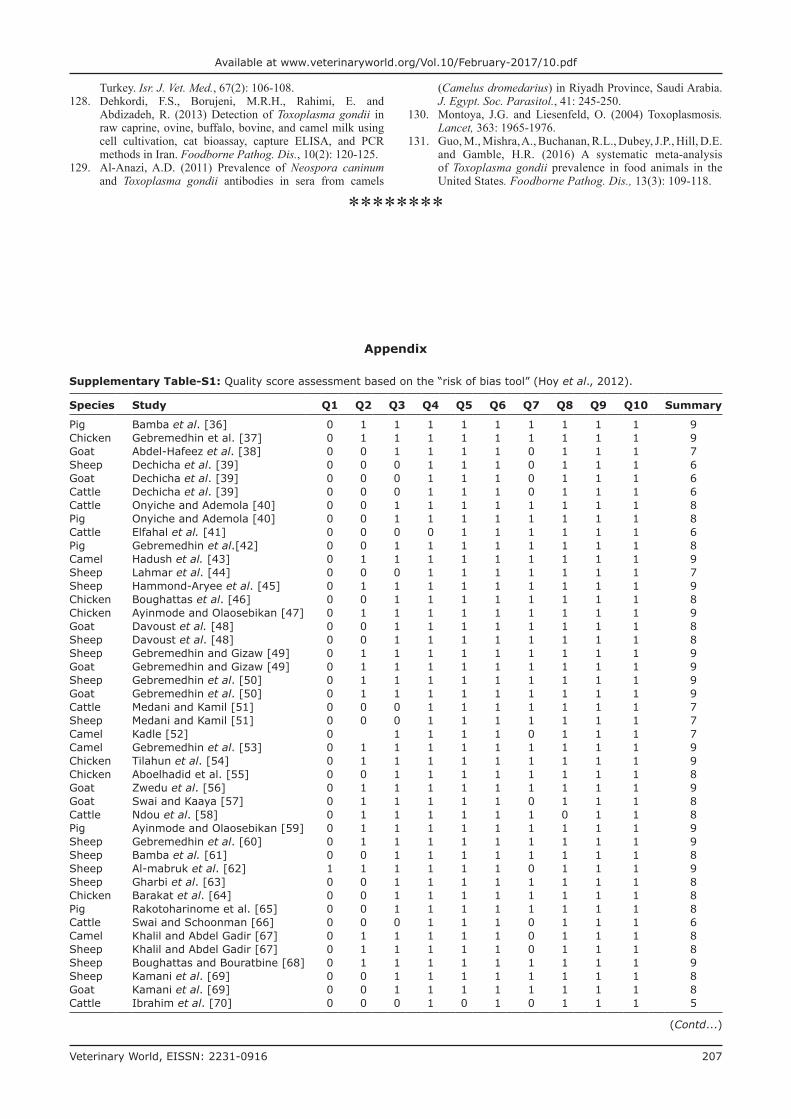
Supplementary Table-S1: Quality score assessment based on the “risk of bias tool” (Hoy et al., 2012).
Species Study Q1 Q2 Q3 Q4 Q5 Q6 Q7 Q8 Q9 Q10 Summary
Pig Bamba et al. [36] 0 1 1 1 1 1 1 1 1 1 9Chicken Gebremedhin et al. [37] 0 1 1 1 1 1 1 1 1 1 9Goat Abdel-Hafeez et al. [38] 0 0 1 1 1 1 0 1 1 1 7Sheep Dechicha et al. [39] 0 0 0 1 1 1 0 1 1 1 6Goat Dechicha et al. [39] 0 0 0 1 1 1 0 1 1 1 6Cattle Dechicha et al. [39] 0 0 0 1 1 1 0 1 1 1 6Cattle Onyiche and Ademola [40] 0 0 1 1 1 1 1 1 1 1 8Pig Onyiche and Ademola [40] 0 0 1 1 1 1 1 1 1 1 8Cattle Elfahal et al. [41] 0 0 0 0 1 1 1 1 1 1 6Pig Gebremedhin et al.[42] 0 0 1 1 1 1 1 1 1 1 8Camel Hadush et al. [43] 0 1 1 1 1 1 1 1 1 1 9Sheep Lahmar et al. [44] 0 0 0 1 1 1 1 1 1 1 7Sheep Hammond-Aryee et al. [45] 0 1 1 1 1 1 1 1 1 1 9Chicken Boughattas et al. [46] 0 0 1 1 1 1 1 1 1 1 8Chicken Ayinmode and Olaosebikan [47] 0 1 1 1 1 1 1 1 1 1 9Goat Davoust et al. [48] 0 0 1 1 1 1 1 1 1 1 8Sheep Davoust et al. [48] 0 0 1 1 1 1 1 1 1 1 8Sheep Gebremedhin and Gizaw [49] 0 1 1 1 1 1 1 1 1 1 9Goat Gebremedhin and Gizaw [49] 0 1 1 1 1 1 1 1 1 1 9Sheep Gebremedhin et al. [50] 0 1 1 1 1 1 1 1 1 1 9Goat Gebremedhin et al. [50] 0 1 1 1 1 1 1 1 1 1 9Cattle Medani and Kamil [51] 0 0 0 1 1 1 1 1 1 1 7Sheep Medani and Kamil [51] 0 0 0 1 1 1 1 1 1 1 7Camel Kadle [52] 0 1 1 1 1 0 1 1 1 7Camel Gebremedhin et al. [53] 0 1 1 1 1 1 1 1 1 1 9Chicken Tilahun et al. [54] 0 1 1 1 1 1 1 1 1 1 9Chicken Aboelhadid et al. [55] 0 0 1 1 1 1 1 1 1 1 8Goat Zwedu et al. [56] 0 1 1 1 1 1 1 1 1 1 9Goat Swai and Kaaya [57] 0 1 1 1 1 1 0 1 1 1 8Cattle Ndou et al. [58] 0 1 1 1 1 1 1 0 1 1 8Pig Ayinmode and Olaosebikan [59] 0 1 1 1 1 1 1 1 1 1 9Sheep Gebremedhin et al. [60] 0 1 1 1 1 1 1 1 1 1 9Sheep Bamba et al. [61] 0 0 1 1 1 1 1 1 1 1 8Sheep Al-mabruk et al. [62] 1 1 1 1 1 1 0 1 1 1 9Sheep Gharbi et al. [63] 0 0 1 1 1 1 1 1 1 1 8Chicken Barakat et al. [64] 0 0 1 1 1 1 1 1 1 1 8Pig Rakotoharinome et al. [65] 0 0 1 1 1 1 1 1 1 1 8Cattle Swai and Schoonman [66] 0 0 0 1 1 1 0 1 1 1 6Camel Khalil and Abdel Gadir [67] 0 1 1 1 1 1 0 1 1 1 8Sheep Khalil and Abdel Gadir [67] 0 1 1 1 1 1 0 1 1 1 8Sheep Boughattas and Bouratbine [68] 0 1 1 1 1 1 1 1 1 1 9Sheep Kamani et al. [69] 0 0 1 1 1 1 1 1 1 1 8Goat Kamani et al. [69] 0 0 1 1 1 1 1 1 1 1 8Cattle Ibrahim et al. [70] 0 0 0 1 0 1 0 1 1 1 5
(Contd...)
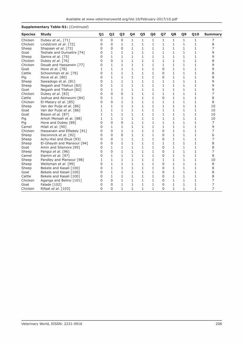
Veterinary World, EISSN: 2231-0916 208
Available at www.veterinaryworld.org/Vol.10/February-2017/10.pdf
Supplementary Table-S1: (Continued)
Species Study Q1 Q2 Q3 Q4 Q5 Q6 Q7 Q8 Q9 Q10 Summary
Chicken Dubey et al., [71] 0 0 0 1 1 1 1 1 1 1 7Chicken Lindstrom et al. [72] 0 0 1 1 1 1 1 1 1 1 8Sheep Shapaan et al. [73] 0 0 0 1 1 1 1 1 1 1 7Goat Teshale and Dumaitre [74] 0 1 1 1 1 1 1 1 1 1 9Sheep Samra et al. [75] 0 1 1 1 1 1 1 1 1 1 9Chicken Dubey et al. [76] 0 0 1 1 1 1 1 1 1 1 8Chicken Deyab and Hassanein [77] 0 1 1 1 1 1 1 1 1 1 9Goat Hove et al. [78] 1 1 1 1 1 1 0 1 1 1 9Cattle Schoonman et al. [79] 0 1 1 1 1 1 0 1 1 1 8Pig Hove et al. [80] 0 1 1 1 1 1 0 1 1 1 8Sheep Sawadogo et al. [81] 0 1 1 1 1 1 1 1 1 1 9Sheep Negash and Tilahun [82] 0 1 1 1 1 1 1 1 1 1 9Goat Negash and Tilahun [82] 0 1 1 1 1 1 1 1 1 1 9Chicken Dubey et al. [83] 0 0 0 1 1 1 1 1 1 1 7Cattle Joshua and Akinwumi [84] 0 1 1 1 1 1 0 1 1 1 8Chicken El-Massry et al. [85] 0 0 1 1 1 1 1 1 1 1 8Sheep Van der Puije et al. [86] 1 1 1 1 1 1 1 1 1 1 10Goat Van der Puije et al. [86] 1 1 1 1 1 1 1 1 1 1 10Goat Bisson et al. [87] 1 1 1 1 1 1 1 1 1 1 10Pig Arkoh Mensah et al. [88] 1 1 1 1 1 1 1 1 1 1 10Pig Hove and Dubey [89] 0 0 0 1 1 1 1 1 1 1 7Camel Hilali et al. [90] 0 1 1 1 1 1 1 1 1 1 9Chicken Hassanain and Elfadaly [91] 0 0 1 1 1 1 0 1 1 1 7Sheep Deconinck et al. [92] 0 0 0 1 1 1 0 1 1 1 6Sheep Achu-Kwi and Ekue [93] 0 0 1 1 1 1 0 1 1 1 7Sheep El-Ghaysh and Mansour [94] 0 0 1 1 1 1 1 1 1 1 8Goat Amin and Silsmore [95] 0 1 1 1 1 1 0 1 1 1 8Sheep Pangui et al. [96] 0 0 1 1 1 1 0 1 1 1 7Camel Elamin et al. [97] 0 1 1 1 1 1 0 1 1 1 8Sheep Pandley and Mansour [98] 1 1 1 1 1 1 1 1 1 1 10Sheep Weitzman et al. [99] 0 1 1 1 1 1 0 1 1 1 8Sheep Bekele and Kasali [100] 0 1 1 1 1 1 0 1 1 1 8Goat Bekele and Kasali [100] 0 1 1 1 1 1 0 1 1 1 8Cattle Bekele and Kasali [100] 0 1 1 1 1 1 0 1 1 1 8Chicken Aganga and Belino [101] 0 0 1 1 1 1 0 1 1 1 7Goat Falade [102] 0 0 1 1 1 1 0 1 1 1 7Chicken Rifaat et al. [103] 0 0 1 1 1 1 0 1 1 1 7

![Research Article Toxoplasma gondii Pave the Road for Dementia?downloads.hindawi.com/journals/jpr/2020/8859857.pdf · including Toxoplasma gondii (T. gondii) [6], Herpes simplex virus-1](https://img.pdfslide.us/doc/110x75/5f9376ed91220772b35c9b7d/research-article-toxoplasma-gondii-pave-the-road-for-dementia-including-toxoplasma.jpg)


![Toxoplasma gondii - PCRmax · Toxoplasma gondii is a species of parasitic protozoa in the genus Toxoplasma.[1] The definitive host of T. gondii is the cat, but the parasite can be](https://img.pdfslide.us/doc/110x75/5cc21bb288c993ed078d60e8/toxoplasma-gondii-toxoplasma-gondii-is-a-species-of-parasitic-protozoa-in.jpg)