Embed Size (px)
Citation preview
1 3
Arch Toxicol (2014) 88:1987–2012DOI 10.1007/s00204-014-1361-0
REVIEW ARTICLE
Toxicology of chemically modified graphene‑based materials for medical application
Toktam Nezakati · Brian G. Cousins · Alexander M. Seifalian
Received: 26 August 2014 / Accepted: 3 September 2014 / Published online: 19 September 2014 © The Author(s) 2014. This article is published with open access at Springerlink.com
biotechnology, and nanomedicine to aid in the diagnosis and treatment of a variety of debilitating diseases.
Keywords Biocompatibility · Graphene · Graphene oxide · In vitro · In vivo · Toxicity
Abbreviations°C Centigrade0D Zero dimensional1D One-dimensional2D Two-dimensional3D Three-dimensional4T1 Murine breast tumor miceA549 Human lung adenocarcinoma epithelial cell
lineAu Aurum, goldCD105 EndoglinCe6 Chlorin e6CGN Thermo-sensitive nanogelCNT Carbon nanotubeCrGO Chemically reduced GOCu CopperCVD Chemical vapor depositionDa DaltonDNA Deoxyribonucleic acidDOX DoxorubicinFA Folic acidFACS Fluorescence-activated cell sortingFDA Food and drug administrationFe3O4 Ferric oxideFGO Fibrin-coated GOFMA Fluorescein O-methacrylateg GramG GrapheneGa Gallium
Abstract This review article aims to provide an over-view of chemically modified graphene, and graphene oxide (GO), and their impact on toxicology when present in bio-logical systems. Graphene is one of the most promising nanomaterials due to unique physicochemical properties including enhanced optical, thermal, and electrically con-ductive behavior in addition to mechanical strength and high surface-to-volume ratio. Graphene-based nanomate-rials have received much attention over the last 5 years in the biomedical field ranging from their use as polymeric conduits for nerve regeneration, carriers for targeted drug delivery and in the treatment of cancer via photo-thermal therapy. Both in vitro and in vivo biological studies of gra-phene-based nanomaterials help understand their relative toxicity and biocompatibility when used for biomedical applications. Several studies investigating important mate-rial properties such as surface charge, concentration, shape, size, structural defects, and chemical functional groups relate to their safety profile and influence cyto- and geno-toxicology. In this review, we highlight the most recent studies of graphene-based nanomaterials and outline their unique properties, which determine their interactions under a range of environmental conditions. The advent of gra-phene technology has led to many promising new oppor-tunities for future applications in the field of electronics,
T. Nezakati (*) · B. G. Cousins · A. M. Seifalian UCL Centre for Nanotechnology and Regeneration Medicine, Division of Surgery and Interventional Science, University College London, London, UKe-mail: [email protected]
A. M. Seifalian e-mail: [email protected]
A. M. Seifalian Royal Free London NHS Foundation Trust, London, UK
1988 Arch Toxicol (2014) 88:1987–2012
1 3
GAP-43 Growth-associated protein 43Gel GelatinGelatin-GNS Gelatin graphene nanosheetsGO Graphene oxideGOT Graphene oxide/TiO2
HB Hypocrellin BHeLa Henrietta Lacks cellHepG2 Human hepatomaHLF Human lung fibroblasthMSC Human mesenchymal stem cellsHPPH 2-(1-Hexyloxyethyl)-2-devinyl
pyropheophorbide-alphaIONP Iron oxide nanoparticlesiPSC Induced pluripotent stem celliTRAQ Isobaric tags used for relative and absolute
quantificationK KelvinKClO3 Potassium chlorateKg KilogramLA Lactobionic acidLC3 Light chain 3LC–MS/MS Liquid chromatography–tandem mass
spectrometryLP Linear polyethyleniminem MeterMCF7 Michigan cancer foundation-7 breast can-
cer cellMFG Multi-functional graphenemg MilligramMG-63 Osteoblast-like cell lineMHC Major histocompatibility complexMHRA Medicines and healthcare products regula-
tory agencymL MilliliterMR Magnetic resonanceM-rGO Microbially reduced graphene oxideMTT Methyl thiazolyl tetrazoliumMTX MethotrexateMWNT Multi-wall carbon nanotubeMyoD MyogeninNGR Nitrogen ion-implanted grapheneNi NickelNIH-3T3 National institute of health 3T3 mouse
fibroblast cellNIR Near-infraredNOTA 1,4,7-Triazacyclononane-1,4,7-triacetic
acidNSC Neural stem cellO-GNR Oxidized graphene nanoribbonsPDT Photodynamic thermal therapyPEG Polyethylene glycolPEI PolyethyleniminePET Positron emission tomography
PGE-DSPE 1,2-Distearoyl-sn-glycero- 3-phosphoethanolamine-N-[amino(polyethylene glycol)]
MCF7 Michigan cancer foundation-7 breast cancer cell
pH Power of hydrogenPLA Polylactic acidPMEF Primary mouse embryonic fibroblastPTT Photo-thermal therapyPVP PolyvinylpyrrolidoneRBC Red blood cellrGO Reduced graphene oxiderGONP Reduced graphene oxide nanoplateletRNA Ribonucleic acidSiC Silicon carbideSKBR3 Sloan–Kettering breast cancer cellSWNT Single-wall carbon nanotubeT TroponinTCP Tissue culture polystyreneTi TitaniumTiO2 Titanium dioxideTPa Tera pascalTRC105 Human chimeric monoclonal antibody to
CD105U251 Human glioma cellUCNP Up-conversion nanoparticlesUK United KingdomUS United StatesW Wattμg Microgram
Introduction
There is only a relatively small contribution regarding the safety profile and toxicology data in the literature on gra-phene-based materials outlining their interactions in bio-logical systems with cells and tissues. Over the last 5 years alone, over 424 publications and cited articles relate to gra-phene toxicology, which has increased to 1,015 publication by 2009 to approximately 3,753 in 2013, whereby the vast majority focus on the physical and material properties of graphene and is a subject of intensive research (Liao et al. 2011; Hu et al. 2011). The physicochemical interaction of graphene, and their use in biological systems, is perhaps one of the newest and fastest growth areas of carbon-based nanomaterials research. Much study in this area is inspired by the myriad of possibilities of many useful biomedi-cal applications relating to their unique properties and to address healthcare concerns relating to nanotoxicology (Liu et al. 2008; Chang et al. 2011a). There has been an intensive focus over the last 10 years in the application of carbon-based nanomaterials such as charcoal, graphite,

1989Arch Toxicol (2014) 88:1987–2012
1 3
fullerene, single-wall carbon nanotubes (SWCNTs), multi-wall carbon nanotubes (MWCNTs), and graphene. This is due to the exploitation of their unique properties, such as enhanced electrical, thermal, mechanical, and optical properties, which provides a range of different application areas from advanced electronics and imaging to biomateri-als and biological sensors for diagnostic use. However, a major concern, involving graphene-based materials, is that there is a limited knowledge relating to their environmental toxicity and biological safety profile. The UK government body, the Medicines and Healthcare Products Regulatory Agency (MHRA), and the US Food and Drug Adminis-tration (FDA) are now reviewing all forms of graphene and functionalized graphene oxide (GO) due to their poor solubility, high agglomeration, long-term retention, and relatively long circulation time in the blood (Begum et al. 2011). Extensive testing is now deemed essential for gra-phene-based materials both for now and in the near future to assess their biological safety profile, which is depend-ent upon different physicochemical factors relating to their surface chemistry, charge, size, shape, and relative concen-tration. Yet still there are many unresolved issues, which remain and need to be clarified before their eventual use for healthcare applications can be fully realized. The bio-compatibility and toxicity behavior of graphene-based material in biological systems gives rise to many important
fundamental issues that require significant attention, and numerous studies are now needed to fill the knowledge gap before being considered as truly ‘safe’ for human use.
Graphene structure and related properties
Graphene is composed of single-carbon atoms forming a sheet of close-packed hexagonal array of SP2 hybridized bonds and can be considered as large aromatic molecule. As such, they have attracted a significant amount of atten-tion in recent times, especially in various areas of biophys-ics and biotechnological applications (Mao et al. 2013b). The two-dimensional, graphene flat sheets can be formed into different geometries, which can be wrapped into spher-ical structures (0D fullerenes, C20, C40, C60), rolled into 1D structures as a single-sheet CNTs, or stacked into 3D-lay-ered structures such as graphite (Fig. 1) (Geim and Novo-selov 2007). This is due to their exceptional material prop-erties giving rise to unique chemical, electrical and thermal conductivity (~5,000 Wm−1 K−1), mechanical, optical transmittance (~97.7 %), structural, and thermal behavior, and has shown great promise for many application areas relating to electronics, semiconductor fabrication, and the biomedical industry (Zhu et al. 2010; Compton and Nguyen 2010; Rao et al. 2009). Graphene has a number
Fig. 1 Graphene is a 2D build-ing material for allotropes of carbon nanomaterials. It can be wrapped up into 0D buckyball, rolled into a 1D nanotube, or stacked into 3D graphite (Geim and Novoselov 2007)
1990 Arch Toxicol (2014) 88:1987–2012
1 3
of fascinating physical characteristics such as the highest surface area (~2,600 m2/g) (Li et al. 2008) and a relatively high Young’s modulus (<1 TPa) among all known materials (Lee et al. 2008), and capable of mass production through a number of chemical manufacturing and material process-ing such as non-covalent and covalent surface modification using surfactants, and biofunctionalization to exploit their unique properties (Shao et al. 2010). Moreover, graphene consists of a layer of π-conjugated systems usually involv-ing six-atom rings. This planar structure offers an excellent capability to interact with a variety of aromatic compounds through π–π stacking interactions in the manufacture of nanocomposite materials and in the immobilisation of bio-molecules such as peptides, antibodies, and other therapeu-tic agents (Boehm 1986; Wintterlin and Bocquet 2009; Van Bommel et al. 1975; Lu et al. 1999a, b; Novoselov et al. 2004). Therefore, graphene has generated great interest in the field of nanomedicine and has been successfully applied in biosensing applications via targeted and selective deliv-ery (Shao et al. 2010; Akhavan et al. 2012b), bioimaging, cell culture, cancer detection, gene delivery (Boehm 1986; Wintterlin and Bocquet 2009; Van Bommel et al. 1975; Lu et al. 1999a, b; Novoselov et al. 2004; Berger et al. 2004; Li et al. 2009; Stankovich et al. 2006), disease diagno-sis (Mohanty and Berry 2008), anti-bacterial compounds (Akhavan and Ghaderi 2009, 2010, 2012; Hu et al. 2010; Ma et al. 2011; Akhavan et al. 2011), anti-viral materials (Akhavan et al. 2012c), photo-thermal therapy (Yang et al. 2012b; Zhang et al. 2011a; Akhavan et al. 2012a), drug delivery (Sun et al. 2008; Liu et al. 2008, Li et al. 2011; Zhang et al. 2010a), and tissue engineering applications (Park et al. 2010; Agarwal et al. 2010; Heo et al. 2011). Therefore, all of their interesting material properties propel graphene from the research laboratory to real-life biologi-cal and clinical applications and show great potential for further exploitation and use within the biomedical industry ready for clinical use.
Graphene preparation and manufacture
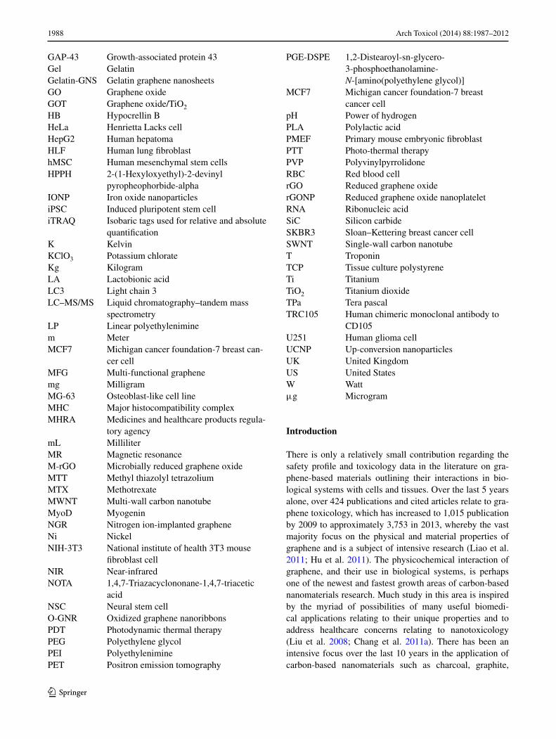
The preparation of graphene can be divided in two main categories: (1) bottom-up and (2) top-down fabrication techniques. Bottom-up fabrication is achieved using several methods to prepare high-quality graphene such as chemical vapor deposition (CVD). These methods produce highly crystalline graphene, but are not suitable for mass produc-tion (Graphene et al. 2010; Kim et al. 2011). For example, CVD is a method which opens up scalable and transparent high-quality graphene in ultra-high vacuum (UHV) condi-tions (10−4–10−6 pa) at high temperature (1,000 °C) using gasses such as methane –CH4 (g) as a carbon source as high-lighted in Fig. 2. The CVD process revolves around a piece of copper (Cu) foil on silicon substrate, which is used as catalyst, which graphene is able to grow as a fibrous ‘mat’-like material. At very high temperatures in an extreme clean, UHV chamber (or environment), carbon from CH4 forms graphene on top of the Cu or nickel (Ni) foil (Fig. 2). Current methods are derived from chemical modification, and functionalized GO and reduced graphene oxide (rGO) within the top-down category are achieved through chemi-cal exfoliation (Novoselov et al. 2012; Dreyer et al. 2010). Chemical exfoliation, described by Schafhaeutl, in the 1940s is a method, which uses a wide range of chemicals such as acid or alkali metals (e.g., potassium), fluoride salts of various types, and transition metals (e.g., iron, nickel), to obtain GO (Dreyer et al. 2010). Nineteen years after Schaf-haeutl described this method, British chemist, Broid, used a chemical exfoliation process to manufacture GO. This method can characterize the molecular weight of graphite by using acids (e.g., sulfuric and nitric), as well as oxi-dants, such as potassium chlorate (KClO3). Further exfo-liation with ultrasonication, thermal or energetic conditions help to oxidize stacked layers of hexagonally arranged carbon atoms that are bonded together with an inter-planar force to obtain graphene layers. The use of this method led
Methane
CVD Graphene grown on substrate+
Hydrogen
Ni/Cu
Fig. 2 Bottom-up fabrication, by chemical vapor deposition (CVD)
1991Arch Toxicol (2014) 88:1987–2012
1 3
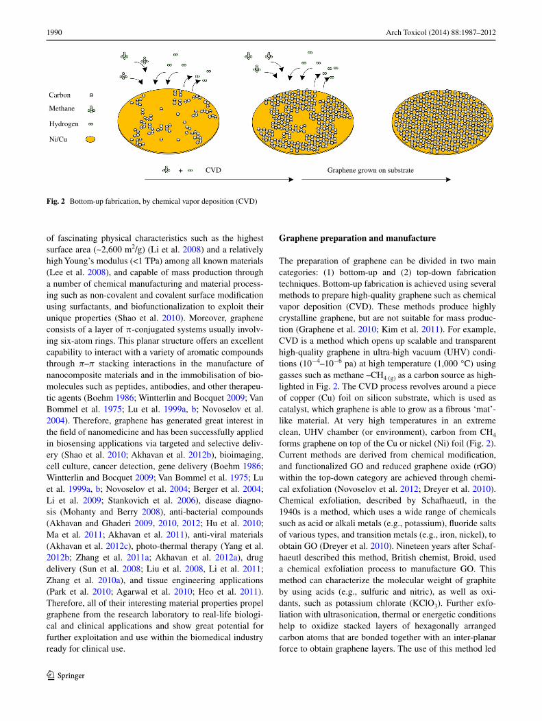
to the formation and production of single-layer-reduced GO (Dreyer et al. 2010). Top-down fabrication involves a chemical reduction based on Hummer’s method (Hummers and Offeman 1958), and chemical oxidation of graphite followed by ultrasonication is highlighted in Fig. 3.
Use of graphene in biomedical applications
Owing to graphene’s low level of toxicity, the lethal dose (LD50) of graphite has been reported as 2 g/per kilo (Sebastian 2012); the following sections outline some of the most promising application areas, use of graphene for enhanced imaging, diagnostics and therapeutic applica-tions in nanomedicine, and their use as novel materials for improved medical devices via improvements in their mechanical properties, and photosensitivity has received considerable attention, along with their health and safety and regulatory concerns (Yang et al. 2010, 2011a, b).
Drug delivery applications
The treatment of cancer represents a global challenge to public health care and is a leading cause of over 7 million deaths worldwide annually (Wood 2013; Boyle and Levin 2008). One significant and area of great importance in the treatment of cancer is the application of chemotherapy. This approach has proved successful in the treatment of various cancers, such as acute promyelocytic leukemia (Jing 2001; Chen et al. 1997), lung (Carney et al. 1983; Umezawa et al. 1966; Kouranos et al. 2011), head and neck cancers. How-ever, the lack of therapeutic efficacy confines such clini-cal applications due to drug resistivity, low efficiency of cellular uptake, and high proportion of side effects, such as liver and kidney damage (Calvert et al. 1989; Kintzel and Dorrt 1995), hair loss (Jaracz et al. 2005; Narang and Varia 2011), nausea and cardiac toxicity (Chithrani et al. 2009; Geiger et al. 2010; Voortman and Giaccone 2006).
Therefore, novel materials with minimal side effects, low toxicity, and high efficiency of targeted drug delivery enhance the bioavailability for chemotherapy, which is an area of increasing research interest (Abou-jawde et al. 2003; Manuscript 2009). Lung cancer is the primary cause of death for all known cancers worldwide (Deaths 2011; Jemal et al. 2011), and due to the size and distribution, cyto-reductive surgery is rarely a viable treatment option. Chemotherapy based on cytotoxic drugs kills cancer cells, which is the main popular approach for treatment of lung cancer. However, the lack of targeting specificity leads to severe side effects such as hemorrhage (Manuscript and Factors 2008). More effective localized delivery can lead to substantial improvements in curative and therapeutic modes of action not only for chemical-based treatments, but for MRI gene delivery including contrast enhancers and radiation sensitizers. In addition, the precise diagnosis and therapy are difficult in most cases for the limited options available (Shi et al. 2013a). Therefore, enormous endeavor in biomedical research has been dedicated to developing new approaches for early-stage detection, diagnosis, and therapy of cancer, which is now commonly referred to as ‘theranostics’ (Mura and Couvreur 2012). Driven by an unmet clinical need, highly integrated drug delivery nano-carriers rely for simultaneous imaging and therapy are cur-rently being evaluated (Huang et al. 2012; Melancon et al. 2011; Liang 2011; Jokerst and Gambhir 2011). Graphene and its derivatives, such as GO, reduced GO, and GO nano-composites, are some of the more well-known examples (Feng and Liu 2011). Externally controlled non-invasive drugs with reliable remote sensing and repeatable ‘on’ and ‘off’ molecular switches to control drug release have recently been receiving attention (Thomas et al. 2010). This method consists of drug-releasing technology via an external stimulus to induce carrier responsive and material properties. The external stimulus is usually derived from polarized or infrared (IR) light (Yavuz et al. 2009; Sherlock et al. 2012; Lu et al. 2008), magnetic field strength (Hoare
Fig. 3 Top-down fabrication, solution based on Hummer’s method using ultrasonication
1992 Arch Toxicol (2014) 88:1987–2012
1 3
et al. 2009; Thomas et al. 2010), ultrasound (Hu and Zhou 2014), and radio frequency-induced drug delivery (Santini et al. 1999; Grayson et al. 2003).
Photo-thermal therapy (PTT)
Photo-thermal therapy (PTT) converts light or opti-cal energy to heat by absorption of a range of nanomate-rial (e.g., silica-coated gold nanoparticles), leading to the thermal ablation resulting in the death of cancer cells. In recent years, PTT as a minimally invasive, controllable, and highly efficient treatment method has drawn wide-spread attention in the treatment of cancer. A large number of research groups have developed various light-absorbing nanomaterials as PTT agents (Huang et al. 2006; Chen et al. 2007; Yavuz et al. 2009; Wu et al. 2010; Dong et al. 2011; Tian et al. 2011; Cheng et al. 2011, 2012; Yang et al. 2010, 2012b, c; Moon et al. 2009; Liu et al. 2011; Wang et al. 2011, 2012), all with absorbance values in the near-infrared (NIR) region (560–760 nm), which is the region ideal for controlling interactions with biological tissues. Despite the great promise of PTT in cancer treatment using nanomaterials, the development of a new generation of PTT agents with enhanced NIR absorption and multiple functions to realize imaging-guided highly effective cancer therapy still merits further effort. Carbon-based nanomate-rials, such as CNTs, carbon nanohorns, and graphene, are being extensively studied as potential PTT agents (Moon et al. 2009; Liu et al. 2011; Wang et al. 2011, 2012; Yang et al. 2010, 2012b). Besides inorganic materials, organic nanoparticles, such as polypyrrole and other light-absorb-ing conductive polymers, have also shown potential in
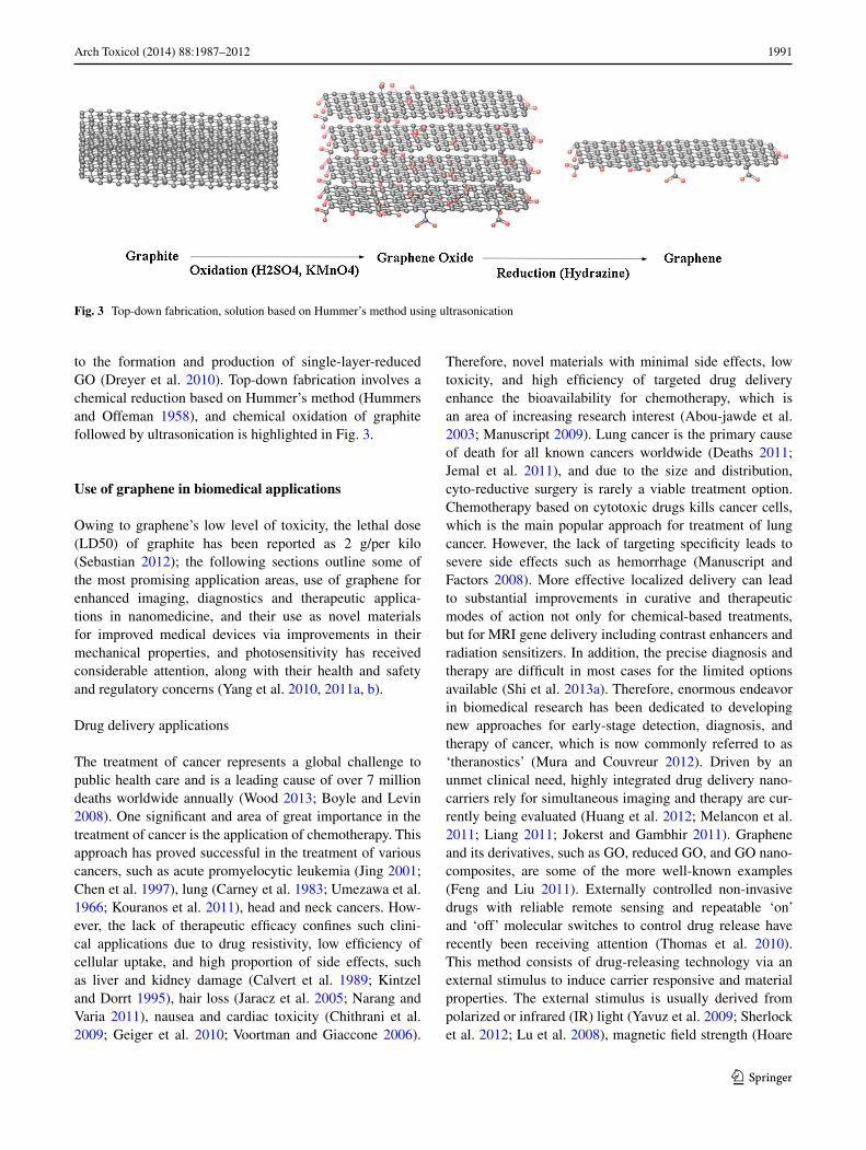
PTT cancer ablation in a few recent studies (Cheng et al. 2012; Yang et al. 2012c; Chen et al. 2012c; Zha et al. 2013). Nanoparticle-based NIR-PTT provides an encourag-ing remedy and strategy for efficient tumor ablation with minimum injury to the surrounding tissues. Up-conversion of nanoparticles (UCNPs) is a further approach to PTT. As an example, UNCP, water-dispersible nanocrystals, which is fluorophores and magnetic nanoparticles, whereby fer-ric oxide (Fe3O4) is reacted with polyethylenimine-modi-fied GO (PEI-GO) acting as a nanocarrier attached to the nanocrystals to yield PEI-GO–nanocrystal (Yan et al. 2013). PEI-GO–UCNP is able to load water-insoluble anti-cancer drugs, such as doxorubicin (DOX), with a superior loading capacity of 100 wt%, through hydrophobic, π–π stacking interaction between PEI-GO–UCNP, and an aro-matic drug highlighted in Fig. 4. Chemotherapy and PTT when used in combination have been proven to reduce drug resistance, and to be an effective strategy to improve the cancer therapy efficacy (Tang et al. 2010; Tang and Mcgoron 2009; Hauck et al. 2008; Lee et al. 2010). In con-trast, undesired damage to normal tissues may be caused by non-specific, untargeted drug delivery and heat sup-plied to the tumor area. Moreover, recent studies suggest that graphene possesses a higher photo-thermal sensitivity than CNTs, and is more effective in PTT in the treatment of cancer (Markovic et al. 2011; Yang et al. 2010, 2012a; Tian et al. 2011).
Nerve repair and regeneration
There is currently an unmet clinical need for biocom-patible and conductive materials used for neurological
Rare Earth (RE): Er, Tm, Y, Yb
NaYF4: Er3+, Tm3+,Yb3+
NaYF4 (UCNP)
Er3+, Tm3+,Yb3+
NH2
NaYF4 - NH2
GO GO-PEI
CO
OH
CO
OH
CO
OH
CO
OH
CO
OH
CO
OH
CO
OH
COOH
COOH PEI
UCNP-NH2
C
GO-PEI-UCNP
Fig. 4 Schematic diagram of the procedure for GO–PEI–UNCP: Numbers of core-shell structured UCNPs covalently grafted with GO through polyethylenimine for advanced imaging, drug delivery, and photo-thermal therapy
1993Arch Toxicol (2014) 88:1987–2012
1 3
applications, which are crucial in the development of next generation of chronic (long-term) implants used in the peripheral and central nervous system (CNS). Nanoparti-cles incorporated into polymeric conduits, acting as fillers, such as, graphene, CNTs, and fullerene, can become one possible solution in the production of conducting materials, which are necessary for stimulating cell growth, and deliv-ery of therapeutic agents. Identification of neural stem cell differentiation is an essential stage for the practical appli-cation of stem cell technology in regenerative medicine. Cell differentiation and monitoring is incredibly important for the application of neural stem cells (NSCs) in the treat-ment of neurodegenerative disease such as Alzheimer’s (Steindler and Okun 2012), Parkinson’s disease (Steindler and Okun 2012; Daadi et al. 2012; Xie et al. 2012), and also, traumatic spinal cord injury (Li et al. 2012; Donnelly et al. 2012). Many conventional tools have been used to detect the differentiation potential of NSCs, as well as to distinguish the undifferentiated NSCs from differentiated neuronal and glial cells (Danova-alt et al. 2012; Ganat et al. 2012; Piao et al. 2012; Buján et al. 2005; Xu et al. 2012).
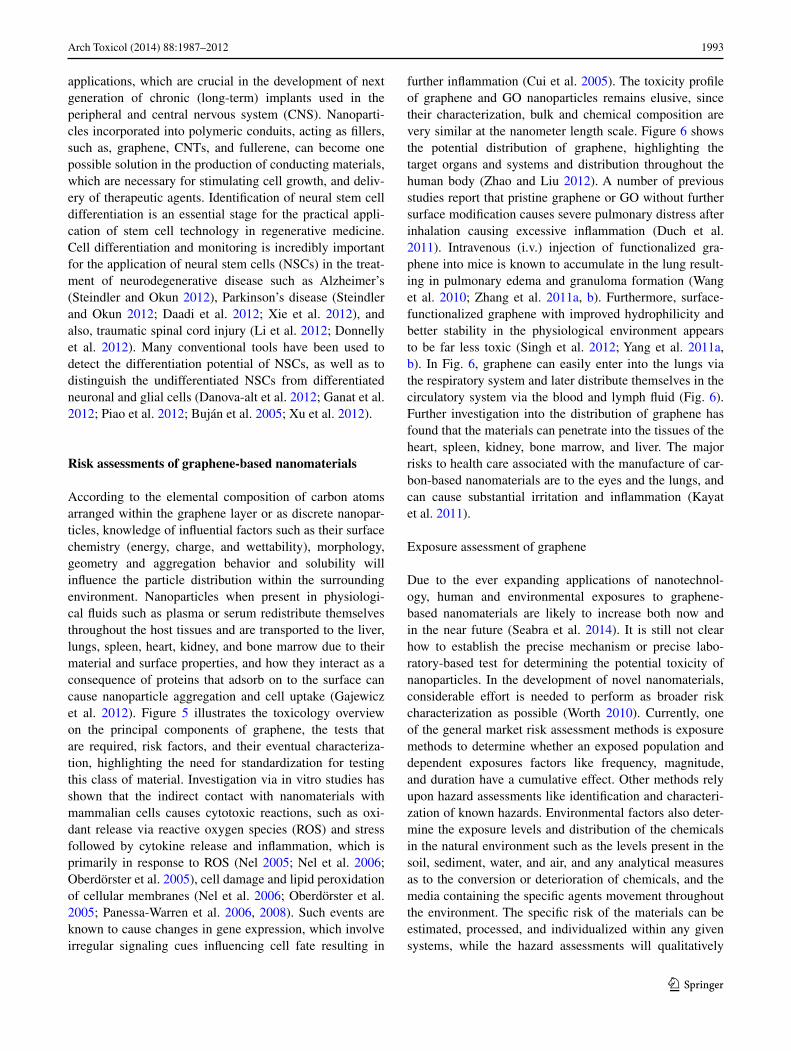
Risk assessments of graphene‑based nanomaterials
According to the elemental composition of carbon atoms arranged within the graphene layer or as discrete nanopar-ticles, knowledge of influential factors such as their surface chemistry (energy, charge, and wettability), morphology, geometry and aggregation behavior and solubility will influence the particle distribution within the surrounding environment. Nanoparticles when present in physiologi-cal fluids such as plasma or serum redistribute themselves throughout the host tissues and are transported to the liver, lungs, spleen, heart, kidney, and bone marrow due to their material and surface properties, and how they interact as a consequence of proteins that adsorb on to the surface can cause nanoparticle aggregation and cell uptake (Gajewicz et al. 2012). Figure 5 illustrates the toxicology overview on the principal components of graphene, the tests that are required, risk factors, and their eventual characteriza-tion, highlighting the need for standardization for testing this class of material. Investigation via in vitro studies has shown that the indirect contact with nanomaterials with mammalian cells causes cytotoxic reactions, such as oxi-dant release via reactive oxygen species (ROS) and stress followed by cytokine release and inflammation, which is primarily in response to ROS (Nel 2005; Nel et al. 2006; Oberdörster et al. 2005), cell damage and lipid peroxidation of cellular membranes (Nel et al. 2006; Oberdörster et al. 2005; Panessa-Warren et al. 2006, 2008). Such events are known to cause changes in gene expression, which involve irregular signaling cues influencing cell fate resulting in
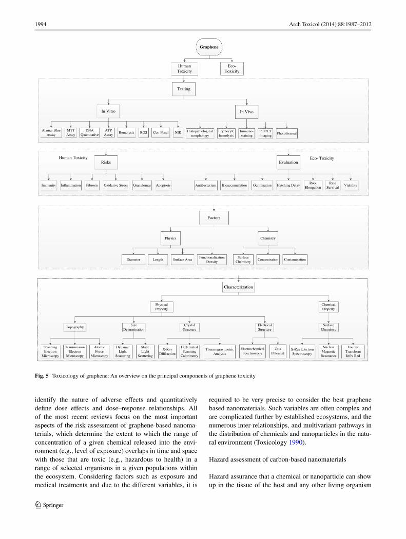
further inflammation (Cui et al. 2005). The toxicity profile of graphene and GO nanoparticles remains elusive, since their characterization, bulk and chemical composition are very similar at the nanometer length scale. Figure 6 shows the potential distribution of graphene, highlighting the target organs and systems and distribution throughout the human body (Zhao and Liu 2012). A number of previous studies report that pristine graphene or GO without further surface modification causes severe pulmonary distress after inhalation causing excessive inflammation (Duch et al. 2011). Intravenous (i.v.) injection of functionalized gra-phene into mice is known to accumulate in the lung result-ing in pulmonary edema and granuloma formation (Wang et al. 2010; Zhang et al. 2011a, b). Furthermore, surface-functionalized graphene with improved hydrophilicity and better stability in the physiological environment appears to be far less toxic (Singh et al. 2012; Yang et al. 2011a, b). In Fig. 6, graphene can easily enter into the lungs via the respiratory system and later distribute themselves in the circulatory system via the blood and lymph fluid (Fig. 6). Further investigation into the distribution of graphene has found that the materials can penetrate into the tissues of the heart, spleen, kidney, bone marrow, and liver. The major risks to health care associated with the manufacture of car-bon-based nanomaterials are to the eyes and the lungs, and can cause substantial irritation and inflammation (Kayat et al. 2011).
Exposure assessment of graphene
Due to the ever expanding applications of nanotechnol-ogy, human and environmental exposures to graphene-based nanomaterials are likely to increase both now and in the near future (Seabra et al. 2014). It is still not clear how to establish the precise mechanism or precise labo-ratory-based test for determining the potential toxicity of nanoparticles. In the development of novel nanomaterials, considerable effort is needed to perform as broader risk characterization as possible (Worth 2010). Currently, one of the general market risk assessment methods is exposure methods to determine whether an exposed population and dependent exposures factors like frequency, magnitude, and duration have a cumulative effect. Other methods rely upon hazard assessments like identification and characteri-zation of known hazards. Environmental factors also deter-mine the exposure levels and distribution of the chemicals in the natural environment such as the levels present in the soil, sediment, water, and air, and any analytical measures as to the conversion or deterioration of chemicals, and the media containing the specific agents movement throughout the environment. The specific risk of the materials can be estimated, processed, and individualized within any given systems, while the hazard assessments will qualitatively
1994 Arch Toxicol (2014) 88:1987–2012
1 3
identify the nature of adverse effects and quantitatively define dose effects and dose–response relationships. All of the most recent reviews focus on the most important aspects of the risk assessment of graphene-based nanoma-terials, which determine the extent to which the range of concentration of a given chemical released into the envi-ronment (e.g., level of exposure) overlaps in time and space with those that are toxic (e.g., hazardous to health) in a range of selected organisms in a given populations within the ecosystem. Considering factors such as exposure and medical treatments and due to the different variables, it is
required to be very precise to consider the best graphene based nanomaterials. Such variables are often complex and are complicated further by established ecosystems, and the numerous inter-relationships, and multivariant pathways in the distribution of chemicals and nanoparticles in the natu-ral environment (Toxicology 1990).
Hazard assessment of carbon-based nanomaterials
Hazard assurance that a chemical or nanoparticle can show up in the tissue of the host and any other living organism
Graphene
HumanToxicity
Eco-Toxicity
Factors
ScanningElectron
Microscopy
TransmissionElectron
Microscopy
SurfaceChemistry
Characterization
Topography
X-Ray ElectronSpectroscopy
NuclearMagnetic
Resonance
FourierTransformInfra Red
AtomicForce
Microscopy
X-RayDiffraction
StaticLight
Scattering
CrystalStructure
DifferentialScanning
Calorimetry
ThermogravimetricAnalysis
ElectrochemicalSpectroscopy
ZetaPotential
ElectricalStructure
Physics
Diameter Surface AreaSurface
ChemistryConcentrationLength
Immunity Inflammation Fibrosis Oxidative Stress Granulomas
DynamicLight
Scattering
SizeDetermination
Chemistry
ContaminationFunctionalization
Density
PhysicalProperty
ChemicalProperty
Risks
Apoptosis BioaccumulationAntibacterium Germination Hatching DelayRoot
ElongationRate
SurvivalViability
Evaluation
Testing
In VivoIn Vitro
Alamar BlueAssay
MTTAssay
DNAQuantitative
HemolysisATP
AssayROS Photothermal
Histopathologicalmorphology
Erythocytehemolysis
Immuno-staining
PET/CTimaging
Con-Focal NIR
Human Toxicity Eco- Toxicity
Fig. 5 Toxicology of graphene: An overview on the principal components of graphene toxicity
1995Arch Toxicol (2014) 88:1987–2012
1 3
is usually dependent upon the frequency, concentration, and duration of exposure of the materials due to factors including the magnitude, extent, and duration to cause toxic side effects. Individual assessments and combinato-rial hazard assessments are now required, and are in great demand to identify and characterize the causative agents. This is primary due to exposure (as described previously), as a result of environment toxicity or in vitro or in vivo medical treatments using graphene either as an implantable device or as drug delivery carrier. Animal experiments are performed according to policy guidelines standardized by the Organization for Economic Co-operation and Develop-ment (OECD) in the UK, and are known to determine com-mon toxic characteristics of a broad range of materials and chemicals. This includes eco-toxicity or animal and human toxicity.
Animal and human toxicity
Toxicology studies are now becoming more advanced in small- and large-scale animal studies in vivo and human cell lines in vitro. Direct hazard assessments and current method-ology for studying nanomaterials help to reveal gaps in the knowledge and deficiency of current assessments. For exam-ple, the lethal dose (LD50) of graphite, CNTs, and fullerenes reported as 2 g/Kg (Sebastian 2012), 2 mg/Kg (Ragot et al. 2010), and 1.2 g/Kg (Da Ros et al. 2001), respectively, in
animals. It is essential to employ traditional risk assessments and control of substances hazardous to health (COSHH) procedures when dealing with engineered nanoparticles, as information from toxicology studies is often deficient for broad risk assessments to be made with regard to nanoma-terials based on carbon due to their vast heterogeneity. The most common cytotoxicity assays to evaluate toxicity of graphene-related materials are apoptosis assay’s such as caspase-3,7 assays to measure cell death, cell adhesion and morphology, cytokine detection, hemocompatibility, hemol-ysis; lactate dehydrogenase (LDH) assay to assess mem-brane integrity; methyl thiazolyl tetrazolium (MTT) assay as a measure of metabolic activity, platelet activation, ROS generation, and genotoxicity (Bitounis et al. 2013; Valla-bani et al. 2011; Zhang et al. 2010b; Schinwald et al. 2012; Chang et al. 2011b; Akhavan et al. 2012d; Liao et al. 2011; Sasidharan et al. 2012; Seabra et al. 2014). Since there is a very close relationship between mutation and damage to DNA, genotoxicity assays are considered as an early and important indicator of toxicity, which may lead to cancer and tumor development (Agemy et al. 2010). The MTT is perhaps the most popular assay among the cell-based assays, and used for cytotoxicity and cell viability when character-izing nanomaterials. In addition, this method using CNTs as the test substrate has been reported to be problematic as a consequence of using graphene-based materials (Bitounis et al. 2013; Liao et al. 2011; Seabra et al. 2014). Therefore,
PrimaryDistribution
SecondaryDistribution
TertiaryDistribution
Central and PeripheralNervous System
Bone Marrow
Spleen
Kidney Liver
Heart
Lymph
Respiratory System
TertiaryDistribution
SecondaryDistribution
PrimaryDistribution
ExposureInjection
Fig. 6 Distribution of graphene in the body
1996 Arch Toxicol (2014) 88:1987–2012
1 3
the cell proliferation assay (WST-8) is a preferred method instead of MTT (Liao et al. 2011). Thus, the most appropri-ate cytotoxic assays must be used to evaluate the toxicity of graphene-based materials to avoid false data. Graphene-based nanomaterials’ in vivo studies are mostly based on the evaluation of tissue distribution (bioaccumulation) and excretion from the body. The most common animal model used to evaluate in vivo toxicity of graphene-based materi-als is the Zebrafish model (Fako and Furgeson 2009). The route of administration should be considered as an important parameter that impacts the toxicity of nanomaterials (Yang et al. 2013a). Based on the recent literature, it is clear that due to the increase in the importance of graphene-based materials, meticulous and accurate in vitro and in vivo stud-ies and accurate testing models of toxicity of the growing graphene family are required and are now in great demand.
Eco‑toxicity of graphene‑based nanomaterials
Above all other living creatures on the earth, continual evo-lution brought intelligence to mankind. Therefore, it is nec-essary that the study of eco-toxicological hazards and their assessments are of considerable importance. In toxicology, the focus is on human as a species, but in ecotoxicology the focus is broadened significantly, regarding the safety and well-being of millions of other common and rare species. So environment factors could put human needs at risk due to indirectly addressing the safety of harmful chemicals, which can degrade or even destroy ecosystem (Toxicol-ogy 1990). The terrestrial environment is similar ecologi-cally to aquatic environments, because living organisms are often intertwined and share a common food chain associ-ated with their natural environment. Two of the well-known organized biological communities are the plant and animal kingdom. The plants are almost affected directly by the level of exposure and the presence of pollutants in the air and in rain fall. However, animals and sometimes humans can become contaminated with heavy metals by uptake toxic compounds through the food chain, e.g., mercury compounds. In the aquatic ecosystem, there is direct con-tact between the pelagic fauna and flora with the chemicals suspended or dissolved in water. In contrast, the food chain is considered as indirect contamination or deemed much slower than direct contamination. Both ecosystems can be contaminated by direct exposure or ingestion of the parti-cles. The quantities of chemical substances, which are car-ried by different media, such as in the air, soil, or water, are often variable in nature. Furthermore, in different media the bioavailability and dynamics are very different. Eventually, such variability and different approaches applied for ter-restrial and aquatic organisms raise substantial concerns in terms of accurately determining the levels of toxicity when comparing similar ecosystems.
Toxicity of functionalized graphene oxide and functionalized graphene
There are very few reports available on the toxicity of gra-phene in vitro (Liu et al. 2009; Singh et al. 2011; Sasid-haran et al. 2011) and in vivo (Chang et al. 2011a; Yang et al. 2011a, b) compared to carbon nanostructures, fuller-enes (Service 2003), and CNTs (Nakamura and Isobe 2003; Lacerda et al. 2006). The main parameters affecting cyto-toxicity of this class of nanomaterial including graphene (Wang et al. 2010), GO (Hu et al. 2010), CNTs (Chang et al. 2011a; Lam et al. 2006), gold and silver nanoparti-cles (Lee et al. 2011) in vitro and in vivo are concentra-tion, shape, size, surface charge, energy, and wettability. Also, in vitro studies could be divided in two discrete sec-tions: (1) cytotoxicity and (2) genotoxicity. Extensive stud-ies have been performed relating to in vitro cytotoxicity of GO over the last 5 years. However, the investigation of new areas of concern relating to genotoxicity of nanomaterials is an important research theme, as there is a close correla-tion between DNA damage, mutation, and the formation of cancers (Agemy et al. 2010). There is insufficient research carried out on the genotoxicity of graphene-based materials at present and warrants much further investigation.
Toxicity of functionalized GO
GO is water-soluble nanomaterial and has been investi-gated extensively as a material for industrial applications for electronics and use in biomedical engineering. This is due to the large vacancy of planar surface area for efficient filling of aromatic drug molecules through π–π stacking interactions, and carboxyl (–COOH), epoxy (–C–O–C–), and hydroxyl (–OH) functional groups. Moreover, limita-tion of GO use in a variety of biomedical applications is due to the absence of stable dispersions. In the following sections, we review current nanotoxicity studies carried out with GO over the last 5 years. Moreover, Tables 1 and 2 provide a thorough summary of all of the current studies, which address functionalized GO cytotoxicity from in vitro and in vivo studies.
Functionalized graphene oxide toxicity in vitro
GO cytotoxicity Initially, the influence of GO on the via-bility of A549 (human lung adenocarcinoma epithelial cell line) cells based on current data has shown that at low con-centrations, GO does not enter into the cells and shows no signs of cytotoxicity. However, GO is known to be cyto-toxic and is dose-dependent and known to cause oxidative stress in A549 cells, and induce a loss in cell viability at high concentrations (Chang et al. 2011a). Cell viabil-ity tests depict significant cell destruction by 1.0 μg/mL
1997Arch Toxicol (2014) 88:1987–2012
1 3
Tabl
e 1
Sum
mar
y of
in v
itro
stud
y of
fun
ctio
naliz
ed g
raph
ene
oxid
e to
xici
ty r
evie
wed
Func
tiona
lizat
ion
Cel
l lin
e/an
imal
mod
elC
once
ntra
tion
and
dura
tion
Sum
mar
y re
sults
Ref
eren
ces
GO
A54
90,
10,
25,
50,
100
, 200
μg/
mL
, 24
hG
O h
ardl
y en
ters
cel
ls a
nd s
how
s go
od b
io-
com
patib
ility
, dos
e an
d si
ze r
elat
edC
hang
et a
l. (2
011a
)
Chi
tosa
n-G
OM
C3T
3-E
1 m
ouse
pre
-ost
eobl
ast c
ell l
ine
CS-
1 w
t% G
O, C
S-3
wt%
GO
, 14
days
GO
into
a C
S ne
twor
k fa
vora
bly
mod
ulat
ed
the
biol
ogic
al r
espo
nse
of o
steo
blas
ts, s
uch
that
cel
l atta
chm
ent,
prol
ifer
atio
n, a
nd
grow
th w
ere
sign
ifica
ntly
enh
ance
d
Dep
an e
t al.
(201
1)
GO
/TiO
2H
eLa
25, 5
0, 7
5, 1
00 μ
g/m
L, 2
0 m
inG
OT
cau
sed
antio
xida
nt e
nzym
e ac
tiviti
es
redu
ctio
n an
d va
riou
s ap
opto
tic e
vent
s in
H
eLa
cell
line,
and
indu
ced
apop
totic
dea
th
Hu
et a
l. (2
012)
GO
/DO
X g
elhu
man
nas
opha
ryng
eal c
arci
nom
a C
NE
1 ce
lls6
mg/
mL
GO
4, 2
, 1 m
g/m
L D
OX
, 14
days
The
gel
exh
ibite
d go
od in
ject
abili
ty, p
artic
u-la
rly
in th
e ca
se o
f hi
gher
am
ount
s of
GO
or
DO
X. T
he in
situ
enc
apsu
late
d D
OX
sh
owed
a s
usta
ined
rel
ease
beh
avio
r an
d an
titum
or e
ffica
cy
Ma
et a
l. (2
012)
HB
-GO
HeL
a, S
MM
C-7
721,
SG
C-7
901,
A54
9H
B–G
O (
2:1)
, HB
–GO
(1:
1)T
he a
ctiv
e up
take
of
HB
–GO
into
tum
or c
ells
an
d si
gnifi
cant
dam
age
to s
uch
impr
eg-
nate
d ce
lls w
as o
bser
ved
upon
irra
diat
ion
Zho
u et
al.
(201
2b)
RG
O/G
elra
bbit’
s fib
robl
ast c
ells
0.1,
0.3
, 0.5
, and
0.7
wt
RG
O h
ad n
o ne
gativ
e ef
fect
on
cell
grow
th,
so th
e R
GO
/gel
com
posi
te m
ay b
e a
prom
-is
ing
biom
ater
ial,
with
goo
d ce
ll co
mpa
t-ib
ility
Wan
g et
al.
(201
2)
MB
/GO
DN
AT
he tr
eate
d D
NA
incr
ease
d th
e qu
ench
ing
effic
ienc
y of
GO
on
MB
com
pare
d to
inta
ct
targ
et D
NA
, ind
icat
ing
that
all
of th
em
exer
t dam
age
effe
ct o
n D
NA
Zho
u et
al.
(201
2a)
GO
Mic
e fib
robl
ast c
ells
line
L92
910
0 μ
g/m
L, 4
8 h
Mat
eria
ls s
how
rel
ativ
ely
good
cyt
o-co
mpa
ti-bi
lity,
the
degr
ee d
epen
ds o
n th
e co
ncen
tra-
tion
and
type
of
disp
ersa
nt
Woj
toni
szak
et a
l. (2
012)
LP-
GO
HE
K29
3 an
d H
eLa
cells
0.1
mg/
mL
Effi
cien
tly c
onde
nsed
pD
NA
and
del
iver
ed it
to
the
insi
des
of th
e ce
lls. L
P-G
O-2
sho
wed
th
e ca
pabi
lity
to d
eliv
er s
iRN
A e
ffici
ently
in
to th
e ce
lls
Tri
path
i et a
l. (2
013)
GO
MG
-63
cells
25, 5
0, 1
00, 2
00 μ
g/m
L, 1
4 da
ysG
O s
how
s no
n-un
ifor
mity
in s
ize
and
shap
e of
its
part
icle
s an
d si
ze v
aria
tion
ham
per
the
tran
sfec
tion
of n
anoc
ompo
site
into
the
cells
Dee
pach
itra
et a
l. (2
013)
PEI-
GO
/PE
I-G
O–U
CN
PM
CF-
7/K
un M
ing
Mou
se10
, 20,
40,
60,
80
μg/
mL
, 48
hE
ffici
ent,
vers
atile
PE
I-G
O–U
CN
P w
ith
up-c
onve
rsio
n lu
min
esce
nce
exhi
bite
d hi
gh d
rug
load
ing
effic
ienc
y an
d co
ntro
lled
rele
ase
of D
OX
to k
ill c
ance
r ce
lls
Yan
et a
l. (2
013)

1998 Arch Toxicol (2014) 88:1987–2012
1 3
Tabl
e 1
con
tinue
d
Func
tiona
lizat
ion
Cel
l lin
e/an
imal
mod
elC
once
ntra
tion
and
dura
tion
Sum
mar
y re
sults
Ref
eren
ces
GO
Neu
robl
asto
ma
SH-S
Y5Y
cel
ls0,
0.4
, 4, 4
0, 4
00 μ
g/m
L, 2
4 h
With
Vpr
13-3
3, g
ivin
g ri
se to
the
tran
sitio
n in
con
form
atio
n, m
orph
olog
y an
d di
men
-si
on c
hang
es o
f ag
greg
ates
, and
red
uced
cy
toto
xici
ty o
f Vpr
13-3
3
Zha
ng e
t al.
(201
3)
GO
Mou
se s
kele
tal m
yobl
asts
C2C
121.
5 m
g/m
L, 2
4 h
The
enh
ance
d ce
llula
r be
havi
or o
n gr
aphe
ne
deri
vativ
es w
as a
ttrib
uted
to s
urfa
ce r
ough
-ne
ss a
nd s
urfa
ce o
xyge
n co
nten
t tha
t infl
u-en
ces
the
adso
rptio
n of
ser
um p
rote
ins
Ku
and
Park
(20
13)
GN
Ps o
n IT
ON
E-4
C n
euro
ecto
derm
al s
tem
cel
ls27
5 m
g/l
Ver
y ef
fect
ive
for
in s
itu m
onito
ring
of
the
undi
ffer
entia
ted
and
diff
eren
tiate
d st
ate
of
stem
cel
ls
Kim
et a
l. (2
013)
GO
Mou
se p
erito
neal
mac
roph
ages
0, 1
, 5, 1
0, 5
0 μ
g/m
L, 2
4 h
Pote
ntia
l tox
ic m
echa
nism
of
carb
on
nano
mat
eria
ls a
nd s
ugge
st c
autio
n on
thei
r ut
iliza
tion
Wan
et a
l. (2
013)
GO
PME
Fs0,
20,
40,
60,
80,
100
μg/
mL
, 24
hM
-rG
O s
how
s si
gnifi
cant
bio
com
patib
ility
th
an G
O a
t hig
her
conc
entr
atio
nsG
urun
atha
n et
al.
(201
3b)
FA–N
GO
–PV
PH
ela,
A54
90,
30,
60,
100
μg/
mL
, 72
hC
ellu
lar
upta
ke d
emon
stra
ted
inte
rnal
iza-
tion
of F
A–N
GO
–PV
P in
to tu
mor
cel
ls
via
rece
ptor
-med
iate
d en
docy
tosi
s an
d ex
hibi
ted
the
cyto
toxi
city
to H
ela
Qin
et a
l. (2
013)
GO
, LA
-PE
G-G
OH
LF
cells
1, 5
0, 1
00 μ
g/m
L, 2
4 h
DN
A d
amag
e in
duce
d by
LA
-PE
G m
odifi
ed
GO
was
mild
com
pare
d w
ith th
at in
duce
d by
oth
er G
O d
eriv
ativ
es
Wan
g et
al.
(201
3a)
GO
E. c
oli
0, 2
5, 5
0, 7
5, 1
00, 1
25, 1
50 m
g/m
L, 4
hA
ntib
acte
rial
act
iviti
es a
re ti
me
and
conc
en-
trat
ion
depe
nden
t; th
e ba
cter
ial c
ell d
eath
m
ay b
e du
e to
oxi
dativ
e st
ress
and
lead
s to
D
NA
fra
gmen
tatio
n
Gur
unat
han
et a
l. (2
013a
)

1999Arch Toxicol (2014) 88:1987–2012
1 3
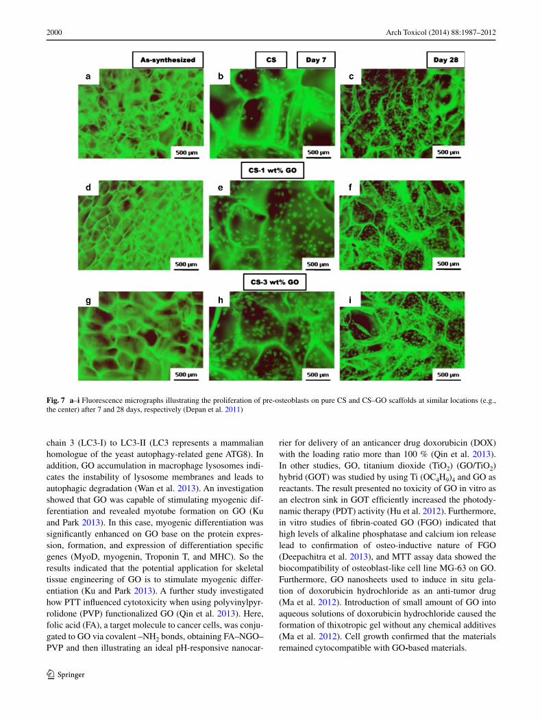
of reduced graphene oxide nanoplatelets (rGONPs) with average dimensions (ALDs) of 11 ± 4 nm, while the rGO sheets with ALDs of 3.8 ± 0.4 μm could show a signifi-cant cytotoxic effect only at high concentration of 100 μg/mL after 1-h exposure. Although oxidative stress and cell wall membrane damage were determined as the main mechanism involved in the cytotoxicity of the rGO sheets, neither of them could completely describe the cell destruc-tions induced by the rGONPs, especially at the concentra-tions ≤1.0 μg/mL (Akhavan et al. 2012e). In other studies, microbially reduced graphene oxide (M-rGO) indicated the significant biocompatibility on primary mouse embryonic fibroblast (PMEF) at concentrations of 100 μg/ml (Guruna-than et al. 2013b). Furthermore, graphene chitosan synthe-sized by covalent linkage of carboxyl groups of GO with amine functionalized groups of chitosan was investigated. The negatively charged GO in chitosan scaffolds was an important physical and chemical factor, which enhanced cell scaffold interactions, as shown in Fig. 7 (Depan et al. 2011). Polyethylenimines (PEIs) are polymeric transfec-tion agents, which are highly branched like and contains primary, secondary, and tertiary amino (–NH2) groups, whereas linear PEIs contain all secondary amines. The pro-duction of linear PEI-grafted GO (LP-GO) conjugates, and their efficacy to transfer nucleic acids into the mammalian cells is investigated (Tripathi et al. 2013). The LP-GOs interact with negatively charged nucleic acids and trans-port them efficiently into cells, therefore, indicating that the conjugates have high transfection efficiency and have bet-ter cell viability compared to LP (Tripathi et al. 2013). In other studies, cytotoxicity and genotoxicity data of GO to human lung fibroblast (HLF) cells have been assessed with the MTT assay, sub-G1 measurements, and comet assays (Wang et al. 2013a), and the results present concentration dependency. This study considered four different concentra-tions, 0, 1, 50, and 100 μg, and indicated better response to the higher concentration range. The cell response has been studied to synthesize lactobionic acid–polyethylene glycol (LA–PEG)-functionalized graphene oxide (LA–PEG–GO), PEG-functionalized graphene oxide (PEG–GO), PEI-func-tionalized graphene oxide (PEI–GO), and GO. The resulting cell was response revealed more positive to GO, PEI–GO, PEG–GO, and LA–PEG–GO, respectively. The genotoxic-ity induced by GO was more severe than the cytotoxicity to HLF cells. The toxic effect can be explained by the oxida-tive stress mediated by GO. In addition, the electric charge on the surface of GO is crucial having shown to decrease the toxicity of GO (Wang et al. 2013a). No toxicity was observed on endothelial cells (ECs) grown on PEI–GO–UNCP, and high potential of dead cancer cells on PEI–GO–UNCP was observed (Yan et al. 2013). In other studies, toxicity evalu-ation of acid-functionalized (Wan et al. 2013) GO induced autophagosome accumulation and the conversion of light Ta
ble
2 S
umm
ary
of in
viv
o st
udy
of f
unct
iona
lized
gra
phen
e ox
ide
toxi
city
rev
iew
ed
Func
tiona
lizat
ion
Cel
l lin
e/an
imal
mod
elC
once
ntra
tion
and
dura
tion
Sum
mar
y re
sults
Ref
eren
ces
188 R
e-G
OM
ale
Kun
Min
g M
ice
20 ±
2 g
, 6–8
wee
ks1
and
10 m
g/m
LH
igh
valu
es o
f %
ID/g
in u
rine
with
in 1
2 h
Zha
ng e
t al.
(201
1b)
NG
O-P
EG
-DO
XE
MT
6 ce
ll/20
Bal
b/c
fem
ale
mic
e 6–
8 w
eeks
24 μ
g/m
L, 2
4 h/
200
μL
, 10
mg/
kg, 7
day
sC
ompl
ete
dest
ruct
ion
of th
e tu
mor
s w
ithou
t w
eigh
t los
s or
rec
urre
nce
of tu
mor
sZ
hang
et a
l. (2
011a
)
66G
a-N
OTA
-GO
-TR
C10
54T
1, M
CF-
7, e
ndot
helia
l cel
ls50
μg/
mL
, 24
hT
umor
targ
etin
g of
NO
TA-G
O-T
RC
105
was
va
scul
atur
e sp
ecifi
c w
ith li
ttle
extr
avas
atio
nH
ong
et a
l. (2
012)
GO
/pG
O5
wee
ks f
emal
e B
alb/
c m
ice,
7 w
eeks
fem
ale
C3H
/HeN
mic
e10
,20,
40 μ
g/m
L, 2
4 h
pGO
acc
umul
ated
to th
e tu
mor
tiss
ues,
and
sys
-te
mic
pG
O n
anop
artic
le-b
ased
co-
deliv
ery
of
Ce6
with
DO
X im
prov
ed th
e ef
ficac
y of
PD
T
Mia
o et
al.
(201
3)
GO
-IO
NP-
Au-
PEG
4T1,
hum
an c
arci
nom
a K
B c
ells
0. 0
.625
, 1.2
5, 2
, 5, 5
, 10
μg/
mL
, 18
days
Cou
ld s
erve
as
a ph
oto-
ther
mal
age
nt f
or P
TT
ca
ncer
cel
l kill
ing
unde
r m
olec
ular
targ
etin
g or
mag
netic
targ
etin
g sh
ows
exce
llent
tum
or
abla
tion
ther
apeu
tic e
ffica
cy
Shi e
t al.
(201
3b)
GO
Mal
e at
hym
ic n
ude
mic
e (C
AnN
.CgF
oxn1
nu/C
rljO
ri, 6
wee
ks o
ld)
0,10
,25,
50 μ
g/m
L, 8
0 h
Plur
onic
-coa
ted
nano
GO
effi
cien
tly s
how
ed
an e
nhan
ced
antic
ance
r ef
fect
by
com
bine
d PD
T–P
TT
eff
ect a
nd e
xhib
ited
high
acc
umu-
latio
n in
tum
or ti
ssue
Sahu
et a
l. (2
013)
2000 Arch Toxicol (2014) 88:1987–2012
1 3
chain 3 (LC3-I) to LC3-II (LC3 represents a mammalian homologue of the yeast autophagy-related gene ATG8). In addition, GO accumulation in macrophage lysosomes indi-cates the instability of lysosome membranes and leads to autophagic degradation (Wan et al. 2013). An investigation showed that GO was capable of stimulating myogenic dif-ferentiation and revealed myotube formation on GO (Ku and Park 2013). In this case, myogenic differentiation was significantly enhanced on GO base on the protein expres-sion, formation, and expression of differentiation specific genes (MyoD, myogenin, Troponin T, and MHC). So the results indicated that the potential application for skeletal tissue engineering of GO is to stimulate myogenic differ-entiation (Ku and Park 2013). A further study investigated how PTT influenced cytotoxicity when using polyvinylpyr-rolidone (PVP) functionalized GO (Qin et al. 2013). Here, folic acid (FA), a target molecule to cancer cells, was conju-gated to GO via covalent –NH2 bonds, obtaining FA–NGO–PVP and then illustrating an ideal pH-responsive nanocar-
rier for delivery of an anticancer drug doxorubicin (DOX) with the loading ratio more than 100 % (Qin et al. 2013). In other studies, GO, titanium dioxide (TiO2) (GO/TiO2) hybrid (GOT) was studied by using Ti (OC4H9)4 and GO as reactants. The result presented no toxicity of GO in vitro as an electron sink in GOT efficiently increased the photody-namic therapy (PDT) activity (Hu et al. 2012). Furthermore, in vitro studies of fibrin-coated GO (FGO) indicated that high levels of alkaline phosphatase and calcium ion release lead to confirmation of osteo-inductive nature of FGO (Deepachitra et al. 2013), and MTT assay data showed the biocompatibility of osteoblast-like cell line MG-63 on GO. Furthermore, GO nanosheets used to induce in situ gela-tion of doxorubicin hydrochloride as an anti-tumor drug (Ma et al. 2012). Introduction of small amount of GO into aqueous solutions of doxorubicin hydrochloride caused the formation of thixotropic gel without any chemical additives (Ma et al. 2012). Cell growth confirmed that the materials remained cytocompatible with GO‑based materials.
Fig. 7 a–i Fluorescence micrographs illustrating the proliferation of pre-osteoblasts on pure CS and CS–GO scaffolds at similar locations (e.g., the center) after 7 and 28 days, respectively (Depan et al. 2011)
2001Arch Toxicol (2014) 88:1987–2012
1 3
Genotoxicity of graphene‑based nanomaterials Investiga-tions using nanoparticles less than 50 nm in each dimension, and GO with a lateral dimension of 2 μm and 1.5 nm in thickness at different concentrations were dependent factors in inducing genotoxicity, and graphene was found to cause the most damage to DNA (Qiao et al. 2013). A further study depicted DNA damage using nanoparticles of silicon diox-ide (SiO2), ZnO, TiO2, tin (Sn), and CNTs at concentration higher than (100 μg/mL). Graphene concentrations higher than 1 μg/mL induced DNA damage at a significantly lower concentrations (Seabra et al. 2014; Qiao et al. 2013). Also the size-dependent genotoxic effects of rGO nanoplatelets (rGONPs) on mesenchymal stem cells (hMSCs) are inves-tigated (Akhavan et al. 2012e). The rGONPs showed geno-toxic effects on hMSCs through DNA fragmentation and chromosomal aberrations even at very low concentration of 0.1 mg/mL, highlighting concerns when using stem cells for applications for use in regenerative medicine.
Functionalized GO toxicity in vivo Functionalized GO tox-icity and their distribution have been studied in mice using radiolabeling techniques (Zhang et al. 2011b). Results indi-cate that GO has sufficient biocompatibility when studied in parallel with red blood cells (RBCs). In addition, GO mainly deposited in the lungs and surrounding tissue, and no pathological variation was illustrated when exposed to mice at 1 mg/kg body weight of GO for 14 days. But at a higher dosage, 10 mg/kg body weight, pulmonary edema, longtime retention, high accumulation, granuloma formation, inflammation, and cell infiltration was observed (Zhang et al. 2011b). Amino group termination covalently attached to GO via a six-arm branched glycol (PEG; 10 kDa) chains were conjugated to NOTA (1,4,7-triazacyclononane-1,4,7-triacetic acid, for 66Ga-labeling) and TRC105 (an antibody that binds to CD105) (Hong et al. 2012), and study of histology vali-dated the characterization of the GO conjugates. The in vivo characterizations were performed in murine breast tumor mice (4T1), and great stability in mouse serum was exhib-ited in 66Ga-NOTA-GO and 66Ga-NOTA-GO-TRC105 con-jugates. Quick accumulation of 66Ga-NOTA-GO-TRC105 in tumor uptake remained stable (Hong et al. 2012). In another study, GO functionalization by iron oxide nanoparticles (IONPs) and gold, forming a multi-functional magnetic and plasmonic GO-IONP-Au nanocomposite with strong super-paramagnetism, significantly enhanced optical absorbance in the NIR region (Shi et al. 2013b). Enhanced photo-thermal cancer ablation effect using GO-IONP-Au-PEG is realized in comparison with PEGylated GO used in earlier studies, as demonstrated in in vivo animal experiments. Moreover, the IONP and Au compartments in the GO-IONP-Au-PEG nanocomposite could prove to be advantageous for mag-netic resonance (MR) and X-ray dual-modal imaging (Shi et al. 2013b). Non-covalently functionalized nanographene
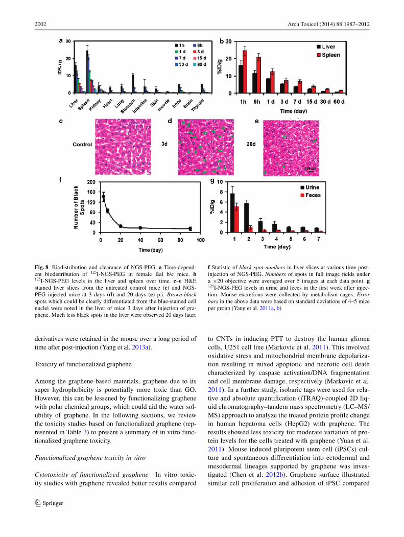
oxide sheet (nano-GO) with pluronic block copolymer and positively charged photosensitizers via electrostatic interac-tions have been previously reported (Sahu et al. 2013). These applications were combined with photodynamic thermal ther-apy (PDT) and PTT for cancer. Cancer cells show increased uptake when compared to normal cells by the use of the nano-GO, and it showed no toxicity to cells in the absence of NIR. High tumor accumulation was observed as a complex was injected intravenously into the tumor. Then, total ablation of tissue caused by NIR explosion via PDT and PTT (Sahu et al. 2013). In further studies, it was shown that doxorubicin loaded on to polyethylene glycol (PEG)ylated graphene oxide (GO–PEG–DOX) facilitates combined chemotherapy and PTT (Zhang et al. 2011a). The GO–PEG–DOX nanopar-ticle ability to combine local, site-specific chemotherapy with external near-infrared-photo-thermal therapy (NIR-PTT) significantly improved the therapeutic efficacy of cancer treatment. In addition, the pathologic examination of main organs improved, as their toxicity study showed less toxicity results with GO–PEG–DOX compared to DOX (Zhang et al. 2011a). Furthermore, injection of 80 mg/kg polyethylene glycol–grafted graphene oxide (PEG–GO) into mice intrave-nously was investigated (Miao et al. 2013) and demonstrated the enhancement of cellular delivery compared to chlorin e6 (Ce6), as a natural molecule, and a promising photosen-sitizer. Accumulation of Ce6/Dox/PEG–GO in tumor tis-sues is shown in molecular imaging of mice, and substantial disruption of tumor nuclei was observed (Miao et al. 2013). Furthermore, photosensitizer molecule, 2-(1-hexyloxyethyl)-2-devinyl pyropheophorbide-alpha (HPPH or Photochlor®) loaded onto PEG-functionalized graphene oxide (GO) via supramolecular π–π stacking investigated and obtained GO–PEG–HPPH complex, shows high HPPH loading efficiency. The in vivo distribution and delivery were tracked by fluo-rescence imaging as well as positron emission tomography (PET) after radiolabeling of HPPH with 64Cu. Compared with free HPPH, GO–PEG–HPPH offers dramatically improved photodynamic cancer cell killing efficacy due to the increased tumor delivery of HPPH (Rong et al. 2014). In vivo biodis-tribution, and potential toxicity of as-made GO and a number of polyethylene glycol (PEG)-functionalized GO derivatives with different sizes and surface coatings, after oral and intra-peritoneal administration at high doses are investigated (Yang et al. 2013a). Insignificant tissue uptake via oral administra-tion on 125I-labeled PEGylated GO derivatives is observed, indicating the rather limited intestinal adsorption of those nanomaterials. In contrast, PEGyalted GO derivatives highly accumulated, but not as-made GO, in the reticuloendothe-lial (RES) system including liver and spleen were observed post-injection (i.p.) and are highlighted in Fig. 8. Moreover, studies based on histological examination of organ slices and hematological analysis discovered that insignificant toxic-ity to the treated animals, although GO and PEGylated GO
2002 Arch Toxicol (2014) 88:1987–2012
1 3
derivatives were retained in the mouse over a long period of time after post-injection (Yang et al. 2013a).
Toxicity of functionalized graphene
Among the graphene-based materials, graphene due to its super hydrophobicity is potentially more toxic than GO. However, this can be lessened by functionalizing graphene with polar chemical groups, which could aid the water sol-ubility of graphene. In the following sections, we review the toxicity studies based on functionalized graphene (rep-resented in Table 3) to present a summary of in vitro func-tionalized graphene toxicity.
Functionalized graphene toxicity in vitro
Cytotoxicity of functionalized graphene In vitro toxic-ity studies with graphene revealed better results compared
to CNTs in inducing PTT to destroy the human glioma cells, U251 cell line (Markovic et al. 2011). This involved oxidative stress and mitochondrial membrane depolariza-tion resulting in mixed apoptotic and necrotic cell death characterized by caspase activation/DNA fragmentation and cell membrane damage, respectively (Markovic et al. 2011). In a further study, isobaric tags were used for rela-tive and absolute quantification (iTRAQ)-coupled 2D liq-uid chromatography–tandem mass spectrometry (LC–MS/MS) approach to analyze the treated protein profile change in human hepatoma cells (HepG2) with graphene. The results showed less toxicity for moderate variation of pro-tein levels for the cells treated with graphene (Yuan et al. 2011). Mouse induced pluripotent stem cell (iPSCs) cul-ture and spontaneous differentiation into ectodermal and mesodermal lineages supported by graphene was inves-tigated (Chen et al. 2012b). Graphene surface illustrated similar cell proliferation and adhesion of iPSC compared
Fig. 8 Biodistribution and clearance of NGS-PEG. a Time-depend-ent biodistribution of 125I-NGS-PEG in female Bal b/c mice. b 125I-NGS-PEG levels in the liver and spleen over time. c–e H&E stained liver slices from the untreated control mice (c) and NGS-PEG injected mice at 3 days (d) and 20 days (e) p.i. Brown-black spots which could be clearly differentiated from the blue-stained cell nuclei were noted in the liver of mice 3 days after injection of gra-phene. Much less black spots in the liver were observed 20 days later.
f Statistic of black spot numbers in liver slices at various time post-injection of NGS-PEG. Numbers of spots in full image fields under a ×20 objective were averaged over 5 images at each data point. g 125I-NGS-PEG levels in urine and feces in the first week after injec-tion. Mouse excretions were collected by metabolism cages. Error bars in the above data were based on standard deviations of 4–5 mice per group (Yang et al. 2011a, b)
2003Arch Toxicol (2014) 88:1987–2012
1 3
Tabl
e 3
Sum
mar
y of
in v
itro
stud
y of
fun
ctio
naliz
ed g
raph
ene
toxi
city
rev
iew
ed
Func
tiona
lizat
ion
Cel
l lin
e/an
imal
mod
elC
once
ntra
tion
and
dura
tion
Sum
mar
y re
sults
Ref
eren
ces
Ghu
man
glio
ma
cell
line
U25
12.
5–10
μg/
mL
, 24
hB
ette
r ph
oto-
ther
mal
effi
cien
cy o
f gr
aphe
ne, d
ue to
dis
pers
ibili
ty/
smal
ler
size
of
grap
hene
, is
supe
rior
to th
at o
f its
str
uctu
ral s
iblin
gM
arko
vic
et a
l. (2
011)
PTC
A/C
CG
HeL
a, M
DA
-MB
-231
, K56
2 ce
lls, N
IH3T
310
0 μ
g/m
L, 7
2 h
Apt
a-se
nsor
has
the
abili
ty to
dif
fere
ntia
te c
ance
r ce
lls a
nd n
orm
al
ones
and
can
be
rege
nera
ted
usin
g A
S141
1 cD
NA
and
reu
sabl
e fo
r ca
ncer
cel
l det
ectio
n
Feng
et a
l. (2
011)
GH
ippo
cam
pus
100
μg/
mL
, 7 d
ays
Bio
com
patib
le a
nd c
apab
le o
f pr
omot
ing
neur
ite s
prou
ting
and
out-
grow
th, d
urin
g th
e ea
rly
deve
lopm
enta
l pha
seL
i et a
l. (2
011)
PGE
/Gra
phen
eH
RP/
DN
A1
μg/
mL
, 24
hG
lyci
dam
ide
coul
d in
duce
mor
e se
riou
s D
NA
dam
age
than
acr
yla-
mid
eQ
iu e
t al.
(201
1)
GH
uman
hep
atom
a H
epG
21
μg/
mL
, 48
hiT
RA
Q-c
oupl
ed 2
D L
C–M
S/M
S pr
oteo
me
anal
ysis
is e
ffec
tive
to th
e ce
llula
r fu
nctio
ns in
res
pons
e to
nan
omat
eria
ls.
Yua
n et
al.
(201
1)
G/G
OM
ouse
iPSC
s ce
ll lin
e 20
D17
1.5
mg/
mL
, 9 d
ays
Allo
w f
or a
ttach
men
t, pr
olif
erat
e on
and
dif
fere
ntia
l dif
fere
ntia
tion
of
iPSC
s an
d pr
omis
e fo
r iP
SCs
Che
n et
al.
(201
2b)
GR
AW
264
.75,
10,
20,
40,
80,
100
μg/
mL
, 48
hG
raph
ene
indu
ce c
ytot
oxic
ity a
nd in
crea
se in
trac
ellu
lar
reac
tive
oxyg
en s
peci
es, a
nd th
en tr
igge
r ap
opto
sis
by a
ctiv
atio
n of
the
mito
chon
dria
l pat
hway
Li e
t al.
(201
2)
rGO
NPs
hMSC
s0.
01–1
00 μ
g/m
L, 2
4 h
The
siz
e- a
nd c
once
ntra
tion-
depe
nden
t cyt
otox
icity
of
the
grap
hene
ox
ide
shee
ts a
nd n
anop
late
lets
in th
e hM
SCs
wer
e st
udie
dA
khav
an e
t al.
(201
2d, e
, 20
13)
GhE
SC li
nes
H9
from
WiC
ell
0, 1
0, 2
5, 5
0 g/
l, 7
days
Neu
rona
l dif
fere
ntia
tion
circ
umve
nts
cyto
toxi
city
and
may
pot
entia
lly
be d
evel
oped
into
3D
AC
-col
lage
n st
ruct
ures
, fur
ther
enh
anci
ng c
el-
lula
r fu
nctio
naliz
atio
n
Che
n et
al.
(201
2a)
rGO
/GO
A54
9, R
AW
264
.720
0 μ
g/m
L, 5
day
sA
n im
port
ant p
aram
eter
det
erm
inin
g th
e bi
olog
ical
eff
ects
of
rGO
/GO
Hor
váth
et a
l. (2
013)
N/g
raph
ene
L92
9 ce
ll lin
e, E
AH
Y92
6 ce
ll lin
e10
0 μ
g/m
L, 7
day
sT
he b
lood
ass
ays
indi
cate
that
N/g
raph
ene
has
slig
htly
low
er p
late
let
adhe
sion
and
pro
long
ed k
inet
ic b
lood
-clo
tting
tim
e th
an p
rist
ine
grap
hene
Guo
et a
l. (2
013)
O-G
NR
coa
ted
with
PE
G-D
SPE
HeL
a, m
ouse
fibr
obla
st c
ells
, SK
BR
3, M
CF7
10, 5
0, 1
00, 2
00, 3
00,
400
μg/
mL
, 48
hT
he h
ighe
r up
take
indi
cate
s th
at O
-GN
R-P
EG
-DSP
Es
have
a d
ose,
an
d tim
e-de
pend
ent,
and
diff
eren
tial c
ytot
oxic
eff
ects
on
the
four
ce
ll lin
es
Mul
lick
Cho
wdh
ury
et a
l. (2
013)
GT
870,
40,
60
mg/
l, 72
hG
raph
ene
indu
ced
necr
osis
in T
87 c
ells
by
inte
rfer
ing
with
the
mor
phol
ogy,
pla
sma
mem
bran
e di
stur
banc
es, a
nd m
itoch
ondr
ial
dysf
unct
ion
Beg
um a
nd F
uget
su (
2013
)
rGO
/QC
-PE
GK
B c
ance
r ce
ll lin
e0.
5, 1
, 10
μg/
mL
, 72
hD
ue to
intr
oduc
tion
of P
lu-S
H, t
he c
reat
ed s
pace
bet
wee
n rG
O/Q
C-
PEG
pla
te a
nd P
lu-S
H p
olym
er a
ids
to e
ntra
p m
ore
DO
X o
r Q
Ds
enab
ling
to s
how
mor
e dr
ug lo
adin
g ef
ficie
ncy
and
fluor
esce
nce
Al-
Nah
ain
et a
l. (2
013)
G/N
afion
HeL
a10
0 μ
g/m
L, 2
4 h
Exc
elle
nt e
lect
roch
emic
al s
ensi
ng c
apab
ility
with
goo
d se
nsiti
vity
, lin
eari
ty o
f re
spon
se, a
nd b
ioaf
finity
Yoo
n et
al.
(201
3a, b
)
PLA
/GN
PM
ouse
em
bryo
fibr
obla
sts
3T3
(AT
CC
CC
L-1
64)
1, 5
, 10
μg/
mL
, 72
hN
o co
nsid
erab
le v
aria
tion
in c
ell p
rolif
erat
ion
at th
e su
rfac
e of
the
film
s w
as o
bser
ved,
exc
ept t
hose
con
tain
ing
GO
aft
er 2
4 h
Pint
o et
al.
(201
3)
GPA
NC
-1G
Film
, 24
hH
ard
coro
na o
n th
e su
rfac
e of
gra
phen
e su
bstr
ates
can
evo
lve
sign
ifi-
cant
ly a
s on
e pa
sses
fro
m p
rote
in c
once
ntra
tions
Mao
et a
l. (2
013a
)
2004 Arch Toxicol (2014) 88:1987–2012
1 3
to glass substrates. Cytotoxicity effect-reduced graphene oxide nanoplatelets (rGONPs) on the human mesenchy-mal stem cells (hMSCs) were investigated (Akhavan et al. 2012e). The cell viability measurement indicated cell destructions at 1.0 μg/mL rGONPs; in contrast, the rGO sheets at concentration of 100 μg/mL illustrated a sig-nificant cytotoxic effect (Akhavan et al. 2012e). A further investigation using a single-layer-reduced GO nanorib-bons (rGONRs) produced via an oxidative unzipping of MWCNTs (Akhavan et al. 2013). The cell viability assay on hMSCs with concentration of 10 μg/mL rGONRs indi-cated significant cytotoxicity effects, while the rGO sheets showed similar cytotoxicity at concentration of 100 μg/mL. The results illustrate the penetration of rGONRs into the cells and DNA fragmentation, as well as, chromosomal aberrations at concentrations of 1.0 μg/mL (Akhavan et al. 2013). The toxicity of graphene on macrophages and epi-thelial cells was also investigated. The results indicate that the initial exposure to these materials is most prominent in respiratory system (Horváth et al. 2013). The interac-tion of nitrogen ion-implanted graphene (NGR) has been evaluated with mouse fibroblast cells and human endothe-lial cells, as well as in blood compatibility studies using rabbit blood (Guo et al. 2013). The results indicated the cell viability and proliferation improvement of cells cul-tured on NGR compared with cells cultured on pristine graphene. Lower platelet adhesion, prolonged kinetic blood-clotting time, and hemolytic rate (below 5 %) pre-sented for NGR showed thrombo-resistance than pristine graphene (Guo et al. 2013). The cytotoxicity of oxidized graphene nanoribbons (O-GNRs) water soluble with PEG–DSPE (1,2-distearoyl-sn-glycero-3-phosphoethanol-amine-N-[amino(polyethylene glycol)]) was investigated (Mullick Chowdhury et al. 2013). The assays were con-ducted on Sloan–Kettering breast cancer cells (SKBR3), Henrietta Lacks cells (HeLa) derived from cervical cancer tissue, Michigan cancer foundation-7 breast cancer cells (MCF7), and National Institute of Health 3T3 mouse fibro-blast cells (NIH-3T3) (Mullick Chowdhury et al. 2013). All of the cells decrease in cell viability, as they repre-sented a time-dependent (12–48 h) and dose-dependent (10–400 μg/mL) response. It was found that SKBR3 and MCF7 show a significantly lowered cytotoxicity compared to HeLa cells. The cells incubated at 10 μg/mL concentra-tion were 100 % viable. As the concentration increased to 400 μg/mL, the cell viability decreased to ~78 % of cells. On the other hand, significant dead cells were observed for HeLa cells even at concentration 10 μg/mL. The results indicated the heterogeneous cytotoxicity of O-GNR–PEG–DSPEs, which possessed different cytotoxicity compared with chemically reduced graphene (Mullick Chowdhury et al. 2013). In a related study, composite poly (lactic acid) (PLA) and PLA film filled with graphene-based materi-Ta
ble
3 c
ontin
ued
Func
tiona
lizat
ion
Cel
l lin
e/an
imal
mod
elC
once
ntra
tion
and
dura
tion
Sum
mar
y re
sults
Ref
eren
ces
GH
T29
, SW
48 tu
mor
cel
l lin
es3
mg/
l, 20
min
Poly
phen
ol g
roup
s at
tach
ed to
rG
O d
urin
g re
duct
ion
proc
ess
are
wel
l se
lect
ive
bind
ing
agen
ts to
can
cer
cell
surf
aces
Abd
olah
ad e
t al.
(201
3)
Gel
atin
-GN
SA
549
5, 1
0, 5
0, 1
00, 1
50,
200,
300
μg/
mL
, 48
hG
elat
in-G
NS
show
ed e
xcel
lent
dis
pers
ibili
ty a
nd s
tabi
lity
in d
istil
led
wat
er a
nd v
ario
us p
hysi
olog
ical
sol
utio
ns, a
lso
exhi
bite
d a
high
dru
g lo
adin
g ca
paci
ty o
f M
TX
An
et a
l. (2
013)
CR
GO
-CO
OH
Mou
se p
rost
ate
canc
er c
ell
line,
TR
AM
P-C
125
μg/
mL
, 180
hU
nifo
rmLy
dis
pers
ed th
erm
o-se
nsiti
ve C
GN
, whi
ch d
ispl
ayed
hig
h ph
oto-
ther
mal
pro
pert
ies
and
reve
rsib
le d
ram
atic
siz
e re
duct
ion
with
te
mpe
ratu
re in
crea
se
Wan
g et
al.
(201
3b)
G-P
EG
Phag
ocyt
ic c
ells
10 μ
g/m
LG
NM
s to
impr
ove
thei
r di
sper
sion
in a
queo
us s
olut
ions
for
bio
med
ical
ap
plic
atio
nsY
ang
et a
l. (2
013b
)
GH
eLa
cells
3 m
g/M
lT
hese
res
ults
dem
onst
rate
the
impo
rtan
ce o
f si
ze-d
epen
dent
gra
phen
e na
nofla
ke to
xici
tyY
oon
et a
l. (2
013a
)

2005Arch Toxicol (2014) 88:1987–2012
1 3
als investigated the biocompatibility. Graphene concentra-tion of 10 μg/mL was used for mouse embryo fibroblasts incubated with both fillers. The results illustrate concen-trations of graphene, and GO may perform decreased in toxicity biomedical applications (Pinto et al. 2013). Study of methotrexate (MTX) attached to the gelatin graphene nanosheets (gelatin-GNS) through strong π–π stacking interactions was conducted (An et al. 2013), and depicted great ability as a drug release, and high drug loading capacity of MTX. Based on the cytotoxicity results on A549 cell, the gelatin-GNS showed non-toxic at specific concentration while the MTX-gelatin-GNS depicted bio-compatibility (An et al. 2013). Another study showed that protein-based facile method for fabrication of nanosized, reduced graphene oxide (nano-rGO) with high stability and low cytotoxicity was also investigated (Sheng et al. 2013). Highly integrated constructed photo-acoustic/ultra-sonic dual modality imaging and photo-thermal therapy platforms further demonstrated that the prepared nano-rGO can be used as ready-to-use theranostic agents for both photo-acoustic imaging and photo-thermal therapy without further surface modification. Intravenous admin-istration of nano-rGO in tumor bearing mice showed rapid and significant photo-acoustic signal enhancement in the tumor region, indicating its excellence for passive tar-geting and photo-acoustic imaging. Meanwhile, using a continuous-wave near-infrared laser, cancer cells in vivo were efficiently ablated, due to the photo-thermal effect of nano-rGO (Sheng et al. 2013). Number of neurite on gra-phene after cell seeding were enhanced during 7 days com-pared with tissue culture polystyrene (TCP) substrates (Li et al. 2011). In addition, the growth-associated protein-43 (GAP-43) was examined. Based on the results, GAP-43 expression was significantly enhanced in graphene when compared to TCP. This could result in the boost of neur-ite sprouting and outgrowth (Li et al. 2011). In brief, the presence of graphene indicated in vitro biocompatibility with different cell lines based on previous investigations. In contrast, there are not many sufficient studies on in vivo biocompatibility, which needs more investigations in this emerging field. Moreover, by functionalizing gra-phene, as polymer filler, the surface topography changes, which causes an increase in roughness and surface energy to modify the wettability. The polar component of surface free energy increased with GO and decreased with gra-phene added to the polymeric matrix.
Genotoxicity studies Hemin-graphene nanosheets (H-GNs) that are able to distinguish intact and damaged DNA and catalytic activity of hemin through graphene π–π interactions have successfully synthesized. Based on this, for detection of single-stranded DNA (ss-DNA) and dam-age DNA induced by chemicals such as styrene oxide (SO),
NaAsO2, and physical radiation, such as UV radiation, a free-label colorimetric method was developed. This method could be used to evaluate the new compounds’ genotoxicity, the maximum limit of pesticide residue, and food additives, due to its simplicity, sensitivity, speed, and cost-effective-ness (Wei et al. 2013). Still, the molecular basis for in vivo graphene oxide (GO) toxicity is unclear. Caenorhabditis elegans has been used to investigate the microRNAs (miR-NAs) control of GO toxicity. A total of 23 up-regulated and 8 down-regulated miRNAs in GO-exposed nematodes were identified with the aid of SOLiD sequencing. The miRNA mutants were confirmed by the functions of identified miR-NAs in regulating the GO toxicity on lifespan. Furthermore, the evidence to raise a hypothesis that GO may reduce lifes-pan through influencing the functions of insulin/IGF signal-ing, TOR signaling, and germline signaling pathways con-trolled by miRNAs provided (Wu et al. 2014).
Functionalized graphene toxicity in vivo
There are no sufficient in vivo studies addressing the tox-icity of graphene based on PubMed database. One of the current ongoing research themes is regarding the mag-netization procedure and in situ reduction, which used to convert GO on to magnetic graphene. This was modified covalently to construct poly-acrylic acid (PAA) for link-ing the fluorescein o-methacrylate (FMA) to yield multi-functional graphene (MFG) with water dispersibility based on 2.5 mg/mL concentration (Gollavelli and Ling 2012). In vivo studies with zebra fish indicated no effect on the survival rate after MFG microinjection, nor any significant abnormalities. Meanwhile, in vivo studies on HeLa cells presented that MFG is a biocompatible imaging probe with concentrations in the range of 100 μg/mL (Gollavelli and Ling 2012). Graphene nanoparticle dispersions indicated the multifunctional agents for in vivo biomedical applica-tions. Therefore, regulatory guidelines for pharmaceuti-cals is followed, which recommend safety pharmacology assessment at least 10–100 times higher than the projected therapeutic dose and present comprehensive single dose response, expanded acute toxicology, toxico-kinetics, and cardiovascular pharmacology results for intravenously administered dextran-coated GO nanoplatelets (GNP-Dex) formulations to rats at doses between 1 and 500 mg/kg (Kanakia et al. 2014). The results presented that the maximum tolerable dose (MTD) of GNP-Dex is between 50 mg/kg ≤ MTD < 125 mg/kg blood half-life <30 min, and majority of nanoparticles excreted within 24 h through feces. Histopathology changes were noted at ≥250 mg/kg in the heart, liver, lung, spleen, and kidney; no changes in the brain and no GNP-Dex-related effects in the cardio-vascular parameters or hematological factors were found (blood, lipid, and metabolic panels) at doses <125 mg/kg
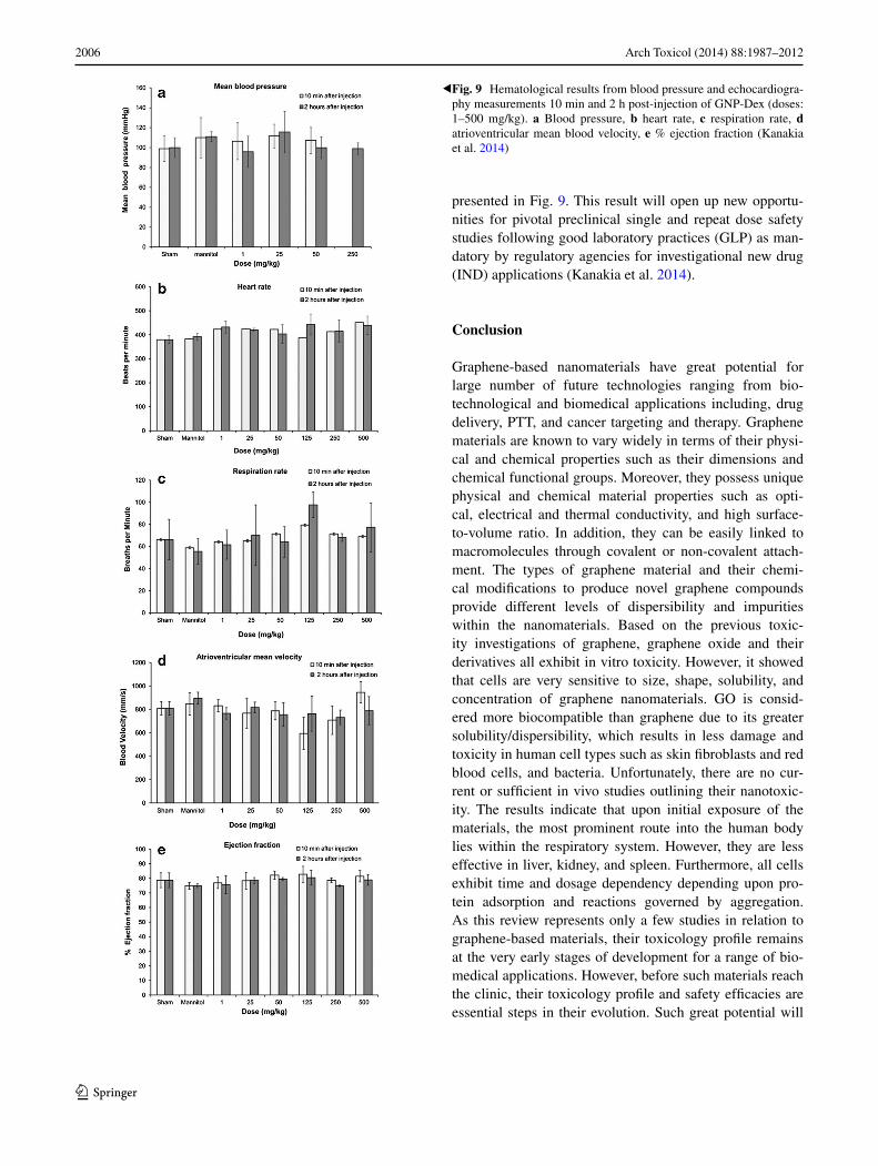
2006 Arch Toxicol (2014) 88:1987–2012
1 3
presented in Fig. 9. This result will open up new opportu-nities for pivotal preclinical single and repeat dose safety studies following good laboratory practices (GLP) as man-datory by regulatory agencies for investigational new drug (IND) applications (Kanakia et al. 2014).
Conclusion
Graphene-based nanomaterials have great potential for large number of future technologies ranging from bio-technological and biomedical applications including, drug delivery, PTT, and cancer targeting and therapy. Graphene materials are known to vary widely in terms of their physi-cal and chemical properties such as their dimensions and chemical functional groups. Moreover, they possess unique physical and chemical material properties such as opti-cal, electrical and thermal conductivity, and high surface-to-volume ratio. In addition, they can be easily linked to macromolecules through covalent or non-covalent attach-ment. The types of graphene material and their chemi-cal modifications to produce novel graphene compounds provide different levels of dispersibility and impurities within the nanomaterials. Based on the previous toxic-ity investigations of graphene, graphene oxide and their derivatives all exhibit in vitro toxicity. However, it showed that cells are very sensitive to size, shape, solubility, and concentration of graphene nanomaterials. GO is consid-ered more biocompatible than graphene due to its greater solubility/dispersibility, which results in less damage and toxicity in human cell types such as skin fibroblasts and red blood cells, and bacteria. Unfortunately, there are no cur-rent or sufficient in vivo studies outlining their nanotoxic-ity. The results indicate that upon initial exposure of the materials, the most prominent route into the human body lies within the respiratory system. However, they are less effective in liver, kidney, and spleen. Furthermore, all cells exhibit time and dosage dependency depending upon pro-tein adsorption and reactions governed by aggregation. As this review represents only a few studies in relation to graphene-based materials, their toxicology profile remains at the very early stages of development for a range of bio-medical applications. However, before such materials reach the clinic, their toxicology profile and safety efficacies are essential steps in their evolution. Such great potential will
Fig. 9 Hematological results from blood pressure and echocardiogra-phy measurements 10 min and 2 h post-injection of GNP-Dex (doses: 1–500 mg/kg). a Blood pressure, b heart rate, c respiration rate, d atrioventricular mean blood velocity, e % ejection fraction (Kanakia et al. 2014)
◂
2007Arch Toxicol (2014) 88:1987–2012
1 3
offer a variety of new and powerful tools based on gra-phene materials for use in the areas of advanced imaging, disease diagnosis, and targeted therapies for the treatment of a range of severely debilitating diseases.
Open Access This article is distributed under the terms of the Crea-tive Commons Attribution License which permits any use, distribu-tion, and reproduction in any medium, provided the original author(s) and the source are credited.
References
Abou-jawde R et al (2003) An overview of targeted treatments in can-cer. Clin Ther 25:2121–2137
Agarwal S et al (2010) Interfacing live cells with nanocarbon sub-strates. Langmuir ACS J Surf Colloids 26(4):2244–2247. http://www.ncbi.nlm.nih.gov/pubmed/20099791. Accessed 14 Oct 2013
Abdolahad M et al (2013) Polyphenols attached graphene nanosheets for high efficiency NIR mediated photodestruction of cancer cells. Mater Sci Eng C Mater Biol Appl 33(3):1498–1505. http://www.ncbi.nlm.nih.gov/pubmed/23827601. Accessed 20 Sept 2013.
Agemy L et al (2010) Nanoparticle-induced vascular block-ade in human prostate cancer. Blood 116(15):2847–2856. http://www.pubmedcentral.nih.gov/articlerender.fcgi?artid=2974592&tool=pmcentrez&rendertype=abstract. Accessed 16 July 2014
Akhavan O et al (2013) Genotoxicity of graphene nanor-ibbons in human mesenchymal stem cells. Carbon 54:419–431. http://linkinghub.elsevier.com/retrieve/pii/S0008622312009499. Accessed 20 Sept 2013
Akhavan O, Ghaderi E, Aghayee S et al (2012a) The use of a glucose-reduced graphene oxide suspension for photothermal cancer therapy. J Mater Chem 22(27):13773. http://xlink.rsc.org/?DOI=c2jm31396k. Accessed 14 Oct 2013
Akhavan O, Ghaderi E, Rahighi R et al (2012b) Toward single-DNA electrochemical biosensing by graphene nanowalls. ACS Nano 6(4):2904–2916
Akhavan O, Choobtashani M, Ghaderi E (2012c) Protein degrada-tion and RNA efflux of viruses photocatalyzed by graphene—tungsten oxide composite under visible light irradiation. J Phys Chem 116:9653–9659
Akhavan O, Ghaderi E, Akhavan A (2012d) Size-dependent genotox-icity of graphene nanoplatelets in human stem cells. Biomateri-als 33(32):8017–8025. http://www.sciencedirect.com/science/article/pii/S0142961212008356. Accessed 28 May 2014
Akhavan O, Ghaderi E, Akhavan A (2012e) Size-dependent geno-toxicity of graphene nanoplatelets in human stem cells. Biomaterials 33(32):8017–8025. http://www.ncbi.nlm.nih.gov/pubmed/22863381. Accessed 20 Sept 2013
Akhavan O, Ghaderi E (2009) Photocatalytic reduction of graphene oxide nanosheets on TiO2 thin film for photoinactivation of bac-teria in solar light irradiation. J Phys Chem 113:20214–20220
Akhavan O, Ghaderi E (2010) Toxicity of graphene and graphene oxide nanowalls against bacteria. ACS Nano 4(10):5731–5736. http://www.ncbi.nlm.nih.gov/pubmed/20925398
Akhavan O, Ghaderi E (2012) Escherichia coli bacteria reduce gra-phene oxide to bactericidal graphene in a self-limiting man-ner. Carbon 50(5):1853–1860. http://linkinghub.elsevier.com/retrieve/pii/S000862231100981X. Accessed 14 Oct 2013
Akhavan O, Ghaderi E, Esfandiar A (2011) Wrapping bacteria by gra-phene nanosheets for isolation from environment, reactivation
by sonication, and inactivation by near-infrared irradiation. J Phys Chem B 115(19):6279–6288. http://www.ncbi.nlm.nih.gov/pubmed/21513335
Al-Nahain A et al (2013) Triggered pH/redox responsive release of doxorubicin from prepared highly stable graphene with thiol grafted pluronic. Int J Pharm 450(1–2):208–217. http://www.ncbi.nlm.nih.gov/pubmed/23624082. Accessed 20 Sept 2013
An J et al (2013) Synthesis of a biocompatible gelatin functionalized graphene nanosheets and its application for drug delivery. Mater Sci Eng C Mater Biol Appl 33(5):2827–2837. http://www.ncbi.nlm.nih.gov/pubmed/23623103. Accessed 20 Sept 2013
Bitounis D et al (2013) Prospects and challenges of graphene in bio-medical applications. Adv Mater 25:2258–2268
Begum P, Fugetsu B (2013) Induction of cell death by graphene in Arabidopsis thaliana (Columbia ecotype) T87 cell suspensions. J Hazard Mater 260:1032–1041. http://www.ncbi.nlm.nih.gov/pubmed/23892171. Accessed 20 Sept 2013
Begum P, Ikhtiari R, Fugetsu B (2011) Graphene phytotoxicity in the seedling stage of cabbage, tomato, red spinach, and let-tuce. Carbon 49(12):3907–3919. http://linkinghub.elsevier.com/retrieve/pii/S0008622311003915. Accessed 20 Sept 2013
Berger C et al (2004) Ultrathin epitaxial graphite: 2D electron gas properties and a route toward graphene-based nanoelectronics. J Phys Chem 108:19912–19916
Boehm HP (1986) Editorial section nomenclature and terminology of graphite. Carbon 24(2):241–245
Boyle P, Levin B (2008) World cancer report 2008. International Agency for Reserach on Cancer
Buján J et al (2005) Muscle-derived stem cells in tissue engineering: defining cell properties suitable for construct design. Histol His-topathol 20:891–899
Calvert AH et al (1989) Carboplatin dosage: prospective evalua-tion of a simple formula based on renal function. J Clin Oncol 7(11):1748–1756
Carney DN et al (1983) In vitro radiation and chemotherapy sensi-tivity of established cell lines of human small cell lung cancer and its large cell morphological variants in vitro radiation and chemotherapy sensitivity of established cell morphological vari-ants. Cancer Res 43:2806–2811
Chang Y et al (2011a) In vitro toxicity evaluation of graphene oxide on A549 cells. Toxicol Lett 200(3): 201–210. http://www.ncbi.nlm.nih.gov/pubmed/21130147. Accessed 17 Sept 2013
Chang Y et al (2011b) In vitro toxicity evaluation of gra-phene oxide on A549 cells. Toxicol Lett 200(3):201–210. http://www.sciencedirect.com/science/article/pii/S0378427410017765. Accessed 28 May 2014
Chen GQ et al (1997) Use of arsenic trioxide (As2O3) in the treat-ment of acute promyelocytic leukemia (APL): I. As2O3 exerts dose-dependent dual effects on APL cells. Blood 89(9):3345–3353. http://www.ncbi.nlm.nih.gov/pubmed/9129041
Chen J et al (2007) Immuno gold nanocages with tailored optical properties for targeted photothermal destruction of cancer cells. Nano Lett 7:1318–1322
Chen EYT et al (2012a) Activated charcoal composite biomaterial promotes human embryonic stem cell differentiation toward neuronal lineage. J Biomed Mater Res Part A 100(8):2006–2017. http://www.ncbi.nlm.nih.gov/pubmed/22623371. Accessed 20 Sept 2013
Chen G-Y et al (2012b) A graphene-based platform for induced pluripotent stem cells culture and differentia-tion. Biomater 33(2):418–427. http://www.ncbi.nlm.nih.gov/pubmed/22014460. Accessed 20 Sept 2013
Chen M et al (2012c) Polypyrrole nanoparticles for high-performance in vivo near-infrared photothermal cancer therapy. In: Elec-tronic supplementary materials (ESI) for chemical communica-tions, pp 1–8

2008 Arch Toxicol (2014) 88:1987–2012
1 3
Cheng L et al (2011) Facile preparation of multifunctional upconver-sion nanoprobes for multimodal imaging and dual-targeted pho-tothermal therapy**. Multifunct Nanoparticles 50:7385–7390
Cheng L et al (2012) Organic stealth nanoparticles for highly effective in vivo near-infrared photothermal therapy of cancer. ACS Nano 6:5605–5613
Chithrani BD et al (2009) Intracellular uptake, transport, and process-ing of nanostructures in cancer cells. Nanomed Nanotechnol Biol Med 5(2):118–127. doi:10.1016/j.nano.2009.01.008
Compton OC, Nguyen ST (2010) Graphene oxide, highly reduced graphene oxide, and graphene: versatile building blocks for carbon-based materials. Small (Weinheim an der Berg-strasse, Germany) 6(6):711–723. http://www.ncbi.nlm.nih.gov/pubmed/20225186. Accessed 19 Sept 2013
Cui D et al (2005) Effect of single wall carbon nanotubes on human HEK293 cells. Toxicol Lett 155(1):73–85. http://www.ncbi.nlm.nih.gov/pubmed/15585362. Accessed 4 June 2014
Daadi MM et al (2012) Dopaminergic neurons from midbrain-spec-ified human embryonic stem cell-derived neural stem cells engrafted in a monkey model of Parkinson’ s disease. PLoS ONE 7:1–11
Danova-alt R et al (2012) Very small embryonic-like stem cells puri-fied from umbilical cord blood lack stem cell characteristics. PLoS ONE 7:1–11
Da Ros T, Spalluto G, Prato M (2001) Biological applications of fullerene derivatives: a brief overview. Croatica Chemica Acta 74(4):743–755
Deaths PC (2011) Cancer statistics, 2011 the impact of eliminat-ing socioeconomic and racial disparities on premature cancer deaths. Cancer J Clin 61:212–236
Deepachitra R et al (2013) Osteo mineralization of fibrin-deco-rated graphene oxide. Carbon 56:64–76. http://linkinghub.elsevier.com/retrieve/pii/S0008622312010378. Accessed 20 Sept 2013
Depan D et al (2011) Structure-process-property relationship of the polar graphene oxide-mediated cellular response and stimu-lated growth of osteoblasts on hybrid chitosan network structure nanocomposite scaffolds. Acta Biomater 7(9):3432–3445. http://www.ncbi.nlm.nih.gov/pubmed/21664303. Accessed 20 Sep-tember 2013
Dong W et al (2011) Facile synthesis of monodisperse superpara-magnetic Fe3 O4 core @ hybrid @ Au shell nanocomposite for bimodal imaging and photothermal therapy. Adv Mater 23:5392–5397
Donnelly EM, Lamanna J, Boulis NM (2012) Stem cell therapy for the spinal cord. Stem Cell Res Therapy 3:1–9
Dreyer DR, Ruoff RS, Bielawski CW (2010) From conception to realization: an historial account of graphene and some per-spectives for its future. Angew Chem (International ed. in English) 49(49):9336–9344. http://www.ncbi.nlm.nih.gov/pubmed/21110353. Accessed 26 Feb 2014
Duch MC et al (2011) Minimizing oxidation and stable nanoscale dis-persion improves the biocompatibility of graphene in the lung. Nano Lett 11:5201–5207
Fako VE, Furgeson DY (2009) Zebrafish as a correlative and pre-dictive model for assessing biomaterial nanotoxicity. Adv Drug Deliv Rev 61(6):478–486. http://www.ncbi.nlm.nih.gov/pubmed/19389433. Accessed 16 July 2014
Feng L et al (2011) A graphene functionalized electrochemical aptasensor for selective label-free detection of cancer cells. Biomaterials 32(11):2930–2937. http://www.ncbi.nlm.nih.gov/pubmed/21256585. Accessed 20 Sept 2013
Feng L, Liu Z (2011) Graphene in biomedicine: opportunities and challenges special report. Future Med 6:317–324
Gajewicz A et al (2012) Advancing risk assessment of engineered nanomaterials: application of computational approaches. Adv
Drug Deliv Rev 64(15):1663–1693. http://www.ncbi.nlm.nih.gov/pubmed/22664229. Accessed 23 May 2014
Ganat YM et al (2012) Identification of embryonic stem cell—derived midbrain dopaminergic neurons for engraftment. J Clin Investig 122:2928–2939
Geiger S et al (2010) Anticancer therapy induced cardiotoxicity: review of the literature. Anticancer Therapy Induced Cardiotox-icity 21:578–590
Geim AK, Novoselov KS (2007) The rise of graphene. Nat Mater 6(3):183–191. http://www.ncbi.nlm.nih.gov/pubmed/17330084
Gollavelli G, Ling Y-C (2012) Multi-functional graphene as an in vitro and in vivo imaging probe. Biomaterials 33(8):2532–2545. http://www.ncbi.nlm.nih.gov/pubmed/22206596. Accessed 20 Sept 2013
Graphene S et al (2010) Organic light-emitting diodes on solution-pro-cessed graphene transparent electrodes. ACS Nano 4(1):43–48
Grayson AMYCR et al (2003) Multi-pulse drug delivery from a resorbable polymeric microchip device. Nat Mater 2:767–772
Guo M et al (2013) Nitrogen ion implanted graphene as thrombo-pro-tective safer and cytoprotective alternative for biomedical appli-cations. Carbon 61:321–328. http://linkinghub.elsevier.com/retrieve/pii/S0008622313004247. Accessed 20 Sept 2013
Gurunathan S, Han JW, Dayem AA et al (2013a) Antibacterial activ-ity of dithiothreitol reduced graphene oxide. J Ind Eng Chem 19(4):1280–1288. http://linkinghub.elsevier.com/retrieve/pii/S1226086X12004492. Accessed 20 Sept 2013
Gurunathan S, Han JW, Eppakayala V et al (2013b). Biocompatibil-ity of microbially reduced graphene oxide in primary mouse embryonic fibroblast cells. Colloids Surf B Biointerfaces 105:58–66. http://www.ncbi.nlm.nih.gov/pubmed/23352948. Accessed 20 Sept 2013
Hauck BTS et al (2008) Enhancing the toxicity of cancer chemo-therapeutics with gold nanorod hyperthermia **. Adv Mater 20:3832–3838
Heo C et al (2011) The control of neural cell-to-cell interactions through non-contact electrical field stimulation using graphene electrodes. Biomaterials 32(1):19–27. http://www.ncbi.nlm.nih.gov/pubmed/20880583. Accessed 24 Sept 2013
Hoare T et al (2009) A magnetically triggered composite membrane for on-demand drug delivery. Nano Lett 9:13–18
Hong H et al (2012) In vivo targeting and positron emission tomography imaging of tumor vasculature with (66)Ga-labeled nano-graphene. Biomaterials 33(16):4147–4156. http://www.pubmedcentral.nih.gov/articlerender.fcgi?artid=3313015&tool=pmcentrez&rendertype=abstract. Accessed 20 Sept 2013
Horváth L et al (2013) Evaluation of the toxicity of graphene derivatives on cells of the lung luminal surface. Car-bon 64:45–60. http://linkinghub.elsevier.com/retrieve/pii/S000862231300626X. Accessed 20 Sept 2013
Hu W et al (2010) Graphene-based antibacterial paper. ACS Nano 4(7):4317–23. http://www.ncbi.nlm.nih.gov/pubmed/20593851
Hu W et al (2011) Protein corona-mediated mitigation of cytotoxicity of graphene oxide. ACS Nano 5(5):3693–3700. http://www.ncbi.nlm.nih.gov/pubmed/21500856
Hu Z et al (2012) Visible light driven photodynamic anticancer activ-ity of graphene oxide/TiO2 hybrid. Carbon 50(3):994–1004. http://linkinghub.elsevier.com/retrieve/pii/S0008622311008219. Accessed 20 Sept 2013
Hu X, Zhou Q (2014) Novel hydrated graphene ribbon unexpectedly promotes aged seed germination and root differentiation. Sci Rep 4:3782. http://www.pubmedcentral.nih.gov/articlerender.fcgi?artid=3896910&tool=pmcentrez&rendertype=abstract. Accessed 18 June 2014
Huang X et al (2006) Cancer cell imaging and photothermal therapy in the near-infrared region by using gold nanorods. J Am Chem Soc 128:2115–2120

2009Arch Toxicol (2014) 88:1987–2012
1 3
Huang P et al (2012) Light-triggered theranostics based on photo-sensitizer- conjugated carbon dots for simultaneous enhanced- fluorescence imaging and photodynamic therapy. Adv Mater 24:5104–5110
Hummers WS, Offeman RE (1958) Preparation of graphitic oxide. J Am Chem Soc 80:1339
Jaracz S et al (2005) Recent advances in tumor-targeting anticancer drug conjugates. Bioorganic Med Chem 13:5043–5054
Jemal A, Bray F, Ferlay J (2011) Global cancer statistics. CA Cancer J Clin 61:69–90
Jing Y (2001) Combined effect of all-trans retinoic acid and arsenic trioxide in acute promyelocytic leukemia cells in vitro and in vivo. Blood 97(1): 264–269. http://www.bloodjournal.org/cgi/doi/10.1182/blood.V97.1.264. Accessed 15 Oct 2013
Jokerst JV, Gambhir SS (2011) Molecular imaging with theranostic nanoparticles. Acc Chem Res 44:1050–1060
Kanakia S et al (2014) Dose ranging, expanded acute toxicity and safety pharmacology studies for intravenously admin-istered functionalized graphene nanoparticle formulations. Biomaterials 35(25):7022–7031. http://www.ncbi.nlm.nih.gov/pubmed/24854092. Accessed 7 July 2014
Kayat J et al (2011) Pulmonary toxicity of carbon nanotubes: a sys-tematic report. Nanomed Nanotechnol Biol Med 7(1):40–49. http://www.ncbi.nlm.nih.gov/pubmed/20620235. Accessed 31 May 2014
Kim Y-K, Kim M-H, Min D-H (2011) Biocompatible reduced graphene oxide prepared by using dextran as a multi-functional reducing agent. Chem Commun (Cambridge, England) 47(11):3195–3197. http://www.ncbi.nlm.nih.gov/pubmed/21286628. Accessed 14 Oct 2013
Kim T-H, Lee K-B, Choi J-W (2013) 3D graphene oxide-encapsu-lated gold nanoparticles to detect neural stem cell differentia-tion. Biomaterials 34(34):8660–8670. http://www.ncbi.nlm.nih.gov/pubmed/23937915. Accessed 20 Sept 2013
Kintzel PE, Dorrt RT (1995) Anticancer drug renal toxicity and elimi-nation: dosing guidelines fro altered renal function. Cancer Treat Rev 21:33–64
Kouranos V et al (2011) Chemotherapy-induced neutropenia in lung cancer patients: the role of antibiotic prophylaxis. Cancer Lett 313(1):9–14. http://www.ncbi.nlm.nih.gov/pubmed/21955615. Accessed 15 Oct 2013
Ku SH, Park CB (2013) Myoblast differentiation on graphene oxide. Biomaterials 34(8):2017–2023. http://www.ncbi.nlm.nih.gov/pubmed/23261212. Accessed 20 Sept 2013
Lacerda L et al (2006) Carbon nanotubes as nanomedicines: from toxicology to pharmacology. Adv Drug Deliv Rev 58(14):1460–1470. http://www.ncbi.nlm.nih.gov/pubmed/17113677. Accessed 22 Sept 2013
Lam C et al (2006) A review of carbon nanotube toxic-ity and assessment of potential occupational and envi-ronmental health risks. Crit Rev Toxicol 36(3):189–217. doi:10.1080/10408440600570233. Accessed 17 Sept 2013
Lee BS, Park H, Yoo K (2010) Synergistic cancer therapeutic effects of locally delivered drug and heat using multifunctional nano-particles. Adv Mater 22:4049–4053
Lee C et al (2008) Measurement of the elastic properties and intrinsic strength of monolayer graphene. Science (New York, NY) 321(5887):385–388. http://www.ncbi.nlm.nih.gov/pubmed/18635798. Accessed 17 Sept 2013
Lee HJ et al (2011) Amine-modified single-walled carbon nano-tubes protect neurons from injury in a rat stroke model. Nat Nanotechnol 6(2):121–5. http://www.ncbi.nlm.nih.gov/pubmed/21278749. Accessed 28 Sept 2013
Li D et al (2008) Processable aqueous dispersions of graphene nanosheets. Nat Nanotechnol 3(2):101–105. http://www.ncbi.nlm.nih.gov/pubmed/18654470. Accessed 24 Sept 2013
Li N et al (2011) The promotion of neurite sprouting and outgrowth of mouse hippocampal cells in culture by graphene substrates. Biomaterials 32(35):9374–9382. http://www.ncbi.nlm.nih.gov/pubmed/21903256. Accessed 20 Sept 2013
Li X et al (2009) Evolution of graphene growth on Ni and Cu by carbon isotope labeling. Nano Lett 9(12):4268–4272. http://pubs.acs.org/doi/abs/10.1021/nl902515k. Accessed 17 Sept 2013
Li X et al (2012a) Engineering neural stem cell fates with hydrogel design for central nervous system regeneration. Prog Polym Sci 37(8):1105–1129. http://linkinghub.elsevier.com/retrieve/pii/S0079670012000202. Accessed 21 Oct 2013
Li Y et al (2012b) The triggering of apoptosis in macrophages by pris-tine graphene through the MAPK and TGF-beta signaling path-ways. Biomaterials 33(2):402–411. http://www.ncbi.nlm.nih.gov/pubmed/22019121. Accessed 20 Sept 2013
Liang X (2011) Theranostic nanoparticles engineered for clinic and pharmaceutics. Acc Chem Res 44:1114–1122
Liao K-H et al (2011) Cytotoxicity of graphene oxide and gra-phene in human erythrocytes and skin fibroblasts. ACS Appl Mater Interfaces 3(7):2607–2615. http://www.ncbi.nlm.nih.gov/pubmed/21650218
Liu X et al (2011) Biomaterials Optimization of surface chemistry on single-walled carbon nanotubes for in vivo photothermal abla-tion of tumors OH O OH O OH O OH O OH O. Biomaterials 32(1):144–151. doi:10.1016/j.biomaterials.2010.08.096
Liu Z et al (2008) PEGylated nanographene oxide for delivery of water-insoluble cancer drugs. J Am Chem Soc 130(33):10876–10877. http://www.pubmedcentral.nih.gov/articlerender.fcgi?artid=2597374&tool=pmcentrez&rendertype=abstract
Liu Z et al (2009) Carbon nanotubes in biology and medicine: in vitro and in vivo detection, imaging and drug delivery. Nano Res 2(2):85–120. www.pubmedcentral.nih.gov/articlerender.fcgi?artid=2824900&tool=pmcentrez&rendertype=abstract. Accessed 23 Sept 2013
Lu J et al (2008) Light-activated nanoimpeller-controlled drug release in cancer cells. Small (Weinheim an der Bergstrasse, Germany) 4(4):421–426. http://www.pubmedcentral.nih.gov/articlerender.fcgi?artid=2712492&tool=pmcentrez&rendertype=abstract. Accessed 19 Oct 2013
Lu X, Huang H et al (1999a) Patterning of highly oriented pyrolytic graphite by oxygen plasma etching. Appl Phys Lett 75(2):193. doi: 10.1063/1.124316. http://link.aip.org/link/APPLAB/v75/i2/p193/s1&Agg=doi
Lu X, Yu M, et al (1999b) Tailoring graphite with the goal of achiev-ing single sheets. Nanotechnology 10(3):269–272. http://stacks.iop.org/0957-4484/10/i=3/a=308?key=crossref.22752dba6bf4b8ec945307fba882d872
Ma J et al (2011) Preparation, characterization and antibacterial proper-ties of silver-modified graphene oxide. J Mater Chem 21(10):3350. http://xlink.rsc.org/?DOI=c0jm02806a. Accessed 14 Oct 2013
Ma D et al (2012) In situ gelation and sustained release of an antitumor drug by graphene oxide nanosheets. Carbon 50(8):3001–3007. http://linkinghub.elsevier.com/retrieve/pii/S0008622312002205. Accessed 20 Sept 2013
Manuscript A (2009) Co-delivery of siRNA and an anticancer drug for treatment of multidrug-resistant cancer. Nanomedicine 3:761–776
Manuscript A, Factors R (2008) Non-small cell lung cancer: epidemi-ology and survivorship. Mayo Clin Proc 83:584–594
Mao H et al (2013a) Hard corona composition and cellular toxicities of the graphene sheets. Colloids Surf B Biointerfaces 109:212–218. http://www.ncbi.nlm.nih.gov/pubmed/23643918. Accessed 20 Sept 2013
Mao HY et al (2013b) Graphene: promises, facts, opportunities, and challenges in nanomedicine. Chem Rev 113(5):3407–3424. http://www.ncbi.nlm.nih.gov/pubmed/23452512

2010 Arch Toxicol (2014) 88:1987–2012
1 3
Markovic ZM et al (2011) In vitro comparison of the photothermal anticancer activity of graphene nanoparticles and carbon nano-tubes. Biomaterials 32(4):1121–1129. http://www.ncbi.nlm.nih.gov/pubmed/21071083. Accessed 20 Sept 2013
Melancon MP, Zhou MIN, Li C (2011) Cancer theranostics with near-infrared light-activatable multimodal nanoparticles. Acc Chem Res 44:947–956
Miao W et al (2013) Safety and tumor tissue accumulation of pegylated graphene oxide nanosheets for co-delivery of antican-cer drug and photosensitizer. Biomaterials 34(13):3402–3410. http://www.ncbi.nlm.nih.gov/pubmed/23380350. Accessed 20 Sept 2013
Mohanty N, Berry V (2008) Graphene-based single-bacterium resolu-tion biodevice and DNA transistor: interfacing graphene deriva-tives with nanoscale and microscale biocomponents. Nano Lett 8(12):4469–4476
Moon HK, Lee SH, Choi HC (2009) In vivo near-infrared mediated tumor destruction by photothermal effect of carbon nanotubes. ACS Nano 3:3707–3713
Mullick Chowdhury S et al (2013) Cell specific cytotoxicity and uptake of graphene nanoribbons. Biomaterials 34(1):283–293. http://www.ncbi.nlm.nih.gov/pubmed/23072942. Accessed 20 Sept 2013
Mura S, Couvreur P (2012) Nanotheranostics for personal-ized medicine ⋆. Adv Drug Deliv Rev 64(13):1394–1416. doi:10.1016/j.addr.2012.06.006
Nakamura E, Isobe H (2003) Functionalized fullerenes in water. The first 10 years of their chemistry, biology, and nanoscience. Accounts Chem Res 36(11):807–815. http://www.ncbi.nlm.nih.gov/pubmed/14622027
Narang AS, Varia S (2011) Role of tumor vascular architec-ture in drug delivery. Adv Drug Deliv Rev 63(8):640–658. doi:10.1016/j.addr.2011.04.002
Nel A (2005) Air pollution-related illness: effects of particles. Science 308:804–806
Nel A et al (2006) Toxic potential of materials at the nanolevel. Sci-ence 311:622–627
Novoselov KS et al (2012) A roadmap for graphene. Nature 490(7419):192–200. http://www.ncbi.nlm.nih.gov/pubmed/23060189. Accessed 20 Jan 2014
Novoselov KS et al (2004) Electric field effect in atomically thin car-bon films. Science (New York, NY) 306(5696):666–669. http://www.ncbi.nlm.nih.gov/pubmed/15499015
Oberdörster G, Oberdörster E, Oberdörster J (2005) Nanotoxicol-ogy : an emerging discipline evolving from studies of ultrafine particles supplemental web sections. Environ Health Perspect 113(7):823–839
Panessa-Warren BJ, Wong SS, Misewich JA (2006) Biological cellular response to carbon nanoparticle toxicity. J Phys 18:2185–2201
Panessa-Warren BJ et al (2008) Human epithelial cell processing of carbon and gold nanoparticles. Int J Nanotechnol 5:55–91
Park S et al (2010) Biocompatible, robust free-standing paper com-posed of a TWEEN/graphene composite. Adv Mat (Deerfield Beach, Fla.), 22(15):1736–1740. http://www.ncbi.nlm.nih.gov/pubmed/20496406. Accessed 8 Oct 2013
Piao S et al (2012) Therapeutic effect of adipose-derived stem cells and BDNF-immobilized PLGA membrane in a rat model of. Int Soc Sex Med 9:1968–1979
Pinto AM et al (2013) Biocompatibility of poly(lactic acid) with incorporated graphene-based materials. Colloids Surf B Biointerfaces 104:229–238. http://www.ncbi.nlm.nih.gov/pubmed/23333912. Accessed 20 Sept 2013
Qiao Y, An J, Ma L (2013) Single cell array based assay for in vitro genotoxicity study of nanomaterials. Anal Chem 85(8):4107–4112. http://www.ncbi.nlm.nih.gov/pubmed/23521522
Qin XC et al (2013) Folic acid-conjugated graphene oxide for can-cer targeted chemo-photothermal therapy. J Photochem Photobiol B Biol 120:156–162. http://www.ncbi.nlm.nih.gov/pubmed/23357205. Accessed 20 Sept 2013
Qiu Y et al (2011) Electrochemical detection of DNA damage induced by acrylamide and its metabolite at the graphene-ionic liquid-Nafion modified pyrolytic graphite electrode. J Hazard Mater 190(1–3):480–485. http://www.ncbi.nlm.nih.gov/pubmed/21497017. Accessed 20 Sept 2013
Ragot J, Ag BM, Stewardship GP (2010) Toxicity of carbon nano-tubes. In: Joint CASG-Nano and ENPRAV workshop on early harvest of research results on nanosafety. Ispra, Italy, 14–15 April 2010
Rao CNR et al (2009) Graphene: the new two-dimensional nanomate-rial. Angew Chem (International ed. in English) 48(42):7752–77. http://www.ncbi.nlm.nih.gov/pubmed/19784976. Accessed 21 Sept 2013
Rong P et al (2014) Photosensitizer loaded nano-graphene for multi-modality imaging guided tumor photodynamic therapy. Thera-nostics 4(3):229–239. http://www.pubmedcentral.nih.gov/articlerender.fcgi?artid=3915087&tool=pmcentrez&rendertype=abstract. Accessed 23 Feb 2014
Sahu A et al (2013) Graphene oxide mediated delivery of methylene blue for combined photodynamic and photothermal therapy. Biomaterials 34(26):6239–6248. http://www.ncbi.nlm.nih.gov/pubmed/23706688. Accessed 20 Sept 2013
Santini JT Jr, Cima MJ, Langer R (1999) A controlled-release micro-chip. Nature 397:335–338
Sasidharan A et al (2011) Differential nano-bio interactions and toxicity effects of pristine versus functionalized graphene. Nanoscale 3(6):2461–2464. http://www.ncbi.nlm.nih.gov/pubmed/21562671. Accessed 4 Oct 2013
Sasidharan A et al (2012) Hemocompatibility and macrophage response of pristine and functionalized graphene. Small (Wein-heim an der Bergstrasse, Germany) 8(8):1251–1263. http://www.ncbi.nlm.nih.gov/pubmed/22334378. Accessed 16 July 2014
Schinwald, A. et al (2012). Graphene-based nanoplatelets: a new risk to the respiratory system as a consequence of their unusual aer-odynamic properties. ACS nano, 6(1), pp 736–46. http://www.ncbi.nlm.nih.gov/pubmed/22195731
Seabra AB et al (2014) Nanotoxicity of graphene and graphene oxide. Chem Res Toxicol 27(2):159–168. http://www.ncbi.nlm.nih.gov/pubmed/24422439
Sebastian D (2012) Material safety data sheet trade name: graphenea-reduced graphene oxide
Service RF (2003) Nanomaterials show signs of toxicity. Science 300:243
Shao Y et al (2010) Graphene based electrochemical sensors and biosensors: a review. Electroanalysis 22(10):1027–1036. doi:10.1002/elan.200900571. Accessed 18 Sept 2013
Sheng Z et al (2013) Protein-assisted fabrication of nano-reduced gra-phene oxide for combined in vivo photoacoustic imaging and photothermal therapy. Biomaterials 34(21):5236–5243. http://www.ncbi.nlm.nih.gov/pubmed/23602365. Accessed 20 Sept 2013
Sherlock SP et al (2012) Photothermally enhanced drug delivery by ultra-small multifunctional FeCo/graphene-shell nanocrystals. ACS Nano 5:1505–1512
Shi S et al (2013a) Biomaterials tumor vasculature targeting and imaging in living mice with reduced graphene oxide. Biomateri-als 34(12):3002–3009. doi:10.1016/j.biomaterials.2013.01.047
Shi X et al (2013b) Graphene-based magnetic plasmonic nano-composite for dual bioimaging and photothermal therapy. Biomaterials 34(20):4786–4793. http://www.ncbi.nlm.nih.gov/pubmed/23557860. Accessed 20 Sept 2013
2011Arch Toxicol (2014) 88:1987–2012
1 3
Singh SK et al (2011) Thrombus inducing property of atomically thin graphene oxide sheets. ACS Nano 5(6):4987–4996. http://www.ncbi.nlm.nih.gov/pubmed/21574593
Singh SK et al (2012) Amine-modified graphene: thrombo-protective safer alternative to graphene oxide for biomedical applica-tions. ACS Nano 6(3):2731–2740. http://www.ncbi.nlm.nih.gov/pubmed/24660818
Stankovich S et al (2006) Graphene-based composite materi-als. Nature 442(7100):282–286. http://www.ncbi.nlm.nih.gov/pubmed/16855586. Accessed 6 Aug 2013
Steindler DA, Okun MS (2012) Stem cell pathologies and neurologi-cal disease. Mod Pathol 25:157–162
Sun X et al (2008) Nano-graphene oxide for cellular imag-ing and drug delivery. Nano Res 1(3):203–212. http://www.pubmedcentral.nih.gov/articlerender.fcgi?artid=2834318&tool=pmcentrez&rendertype=abstract. Accessed 24 Sept 2013
Tang Y, Mcgoron AJ (2009) Combined effects of laser-ICG pho-tothermotherapy and doxorubicin chemotherapy on ovarian cancer cells. J Photochem Photobiol B Biol 97(3):138–144. doi:10.1016/j.jphotobiol.2009.09.001
Tang Y et al (2010) Simultaneous delivery of chemotherapeutic and thermal-optical agents to cancer cells by a polymeric (PLGA) nanocarrier: an in vitro study. Pharm Res 27:2242–2253
Thomas CR et al (2010) Noninvasive remote-controlled release of drug molecules in vitro using magnetic actuation of mechanized nanoparticles. Am Chem Soc 123:10623–10625
Tian B et al (2011) Photothermally enhanced photodynamic therapy delivered by nano-graphene oxide. ACS Nano 9:7000–7009
Toxicology DB (1990) Toxicological versus ecotoxicological testing. In: Bourdeau P (ed) Short-term toxicity tests for non-genotoxic effects. Wiley, New York
Tripathi SK et al (2013) Functionalized graphene oxide mediated nucleic acid delivery. Carbon 51:224–235. http://linkinghub.elsevier.com/retrieve/pii/S0008622312006999. Accessed 20 Sept 2013
Umezawa H et al (1966) New antibiotics, bleomycin A and B. J Anti-biot 19(5):200–209
Van Bommel AJ, Crombeen JE, Van Tooren A (1975) Leed and Auger electron observations of the SiC (0001) surface. Surf Sci 48:463–472
Vallabani NVS et al (2011) Toxicity of graphene in normal human lung cells (BEAS-2B). J Biomed Nanotechnol 7(1):106–107. http://www.ncbi.nlm.nih.gov/pubmed/21485826. Accessed 16 July 2014
Voortman J, Giaccone G (2006) Severe reversible cardiac failure after bortezomib treatment combined with chemotherapy in a non-small cell lung cancer patient: a case report. BMC Cancer 6:4–7
Wan B et al (2013) Single-walled carbon nanotubes and graphene oxides induce autophagosome accumulation and lysosome impairment in primarily cultured murine peritoneal mac-rophages. Toxicol Lett 221(2):118–127. http://www.ncbi.nlm.nih.gov/pubmed/23769962. Accessed 20 Sept 2013
Wang K et al (2010) Biocompatibility of graphene oxide. Nanoscale Res Lett pp 1–8. http://www.nanoscalereslett.com/con-tent/6/1/8. Accessed 27 Sept 2013
Wang C et al (2011) Gold nanoclusters and graphene nanocomposites for drug delivery and imaging of cancer cells **. Cell Interact 50:11644–11648
Wang W et al (2012) Preparation of reduced graphene oxide/gelatin composite films with reinforced mechanical strength. Mater Res Bull 47(9):2245–2251. http://linkinghub.elsevier.com/retrieve/pii/S0025540812004370. Accessed 20 Sept 2013
Wang X et al (2012) Noble metal coated single-walled carbon nano-tubes for applications in surface enhanced raman scattering imaging and photothermal therapy. J Am Soc 134:7414–7422
Wang A et al (2013a) Role of surface charge and oxidative stress in cytotoxicity and genotoxicity of graphene oxide towards human lung fibroblast cells. J Appl Toxicol JAT 33(10):1156–1164. http://www.ncbi.nlm.nih.gov/pubmed/23775274. Accessed 20 Sept 2013
Wang C et al (2013b) A chitosan-modified graphene nanogel for noninvasive controlled drug release. Nanomed Nanotech-nol Biology Medicine. pp 1–9. http://www.ncbi.nlm.nih.gov/pubmed/23352802. Accessed 17 Sept 2013
Wei W et al (2013) Colorimetric detection of DNA damage by using hemin-graphene nanocomposites. Spectrochim Acta Part A Mol Biomol Spectrosc 106:163–169. http://www.ncbi.nlm.nih.gov/pubmed/23376271. Accessed 6 July 2014
Wintterlin J, Bocquet M-L (2009) Graphene on metal surfaces. Surf Sci 603(10–12):1841–1852. http://linkinghub.elsevier.com/retrieve/pii/S003960280900079X. Accessed 25 Sept 2013
Wojtoniszak M et al (2012) Synthesis, dispersion, and cytocompat-ibility of graphene oxide and reduced graphene oxide. Col-loids Surf B Biointerfaces 89:79–85. http://www.ncbi.nlm.nih.gov/pubmed/21962852. Accessed 20 September 2013
Wood B (2013) The to and fro of tumor spread. New Views Res 493:8–9
Worth AP (2010) The role of QSAR methodology in the regulatory assessment of chemicals. Recent Adv QSAR Stud Chall Adv Comput Chem Phys 8:367–382
Wu X et al (2010) High-photoluminescence-yield gold nanocubes: for cell imaging and photothermal therapy. ACS Nano 4:113–120
Wu Q et al (2014) microRNAs control of in vivo toxicity from gra-phene oxide in Caenorhabditis elegans. Nanomed Nano-technol Biol Med. pp 1–10. http://www.ncbi.nlm.nih.gov/pubmed/24780312. Accessed 7 July 2014
Xie Z et al (2012) Floor plate-delivered dopamine neurons from hESCS efficiently engraft in animal models of PD. Nature 480:547–551
Xu Y et al (2012) Promising new potential for mesenchymal stem cells derived from human umbilical cord Wharton’ s jelly : sweat gland cell-like differentiative capacity. J Tissue Eng Regen Med 6:645–654
Yan L et al (2013) The use of polyethylenimine-modified graphene oxide as a nanocarrier for transferring hydrophobic nanocrystals into water to produce water-dispersible hybrids for use in drug delivery. Carbon 57:120–129. http://linkinghub.elsevier.com/retrieve/pii/S0008622313000638. Accessed 20 Sept 2013
Yang K et al (2010) Graphene in mice: ultrahigh in vivo tumor uptake and efficient photothermal therapy. Nano Lett 10(9):3318–3323. http://www.ncbi.nlm.nih.gov/pubmed/20684528. Accessed 26 Sept 2013
Yang K et al (2011a) In vivo pharmacokinetics, long-term biodistribu-tion, and toxicology of pegylated graphene in mice. ACS Nano 5(1):516–522
Yang X et al (2011b) Multi-functionalized graphene oxide based anti-cancer drug-carrier with dual-targeting function and pH-sensi-tivity. J Mater Chem 21:3448–3454
Yang K, Hu L et al (2012a) Multimodal imaging guided photother-mal therapy using functionalized graphene nanosheets anchored with magnetic nanoparticles. Adv Mater 24:1868–1872
Yang K, Wan J, et al (2012b) The influence of surface chemistry and size of nanoscale graphene oxide on photothermal therapy of cancer using ultra-low laser power. Biomaterials 33(7):2206–2214. http://www.ncbi.nlm.nih.gov/pubmed/22169821. Accessed 24 Sept 2013
Yang K, Xu H et al (2012c) In vitro and in vivo near-infrared photo-thermal therapy of cancer using polypyrrole organic nanoparti-cles. Adv Mater 24:5586–5592
Yang K et al (2013a) In vivo biodistribution and toxicology of functionalized nano-graphene oxide in mice after oral and

2012 Arch Toxicol (2014) 88:1987–2012
1 3
intraperitoneal administration. Biomaterials 34(11):2787–2795. http://www.ncbi.nlm.nih.gov/pubmed/23340196. Accessed 16 July 2014
Yang M et al (2013b) A high poly(ethylene glycol) den-sity on graphene nanomaterials reduces the detachment of lipid-poly(ethylene glycol) and macrophage uptake. Acta Biomater 9(1):4744–4753. http://www.ncbi.nlm.nih.gov/pubmed/22995407. Accessed 20 Sept 2013
Yavuz MS et al (2009) Gold nanocages covered by smart polymers for controlled release with near-infrared light. Nat Mater 8(12):935–939. doi:10.1038/nmat2564
Yoon OJ, Kim CH et al (2013a) Toxicity analysis of graphene nano-flakes by cell-based electrochemical sensing using an electrode modified with nanocomposite of graphene and Nafion. Sensors Actuators B Chem 188:454–461. http://linkinghub.elsevier.com/retrieve/pii/S0925400513008368. Accessed 20 Sept 2013
Yoon OJ, Kim I et al (2013b) Toxicity of graphene nanoflakes evalu-ated by cell-based electrochemical impedance biosensing. J Biomed Mater Res Part A. pp 1–7. http://www.ncbi.nlm.nih.gov/pubmed/23894129. Accessed 20 Sept 2013
Yuan J, Gao H, Ching CB (2011) Comparative protein profile of human hepatoma HepG2 cells treated with graphene and single-walled carbon nanotubes: an iTRAQ-coupled 2D LC-MS/MS proteome analysis. Toxicol Lett 207(3):213–221. http://www.ncbi.nlm.nih.gov/pubmed/21963432. Accessed 20 Sept 2013
Zha Z et al (2013) Uniform polypyrrole nanoparticles with high pho-tothermal conversion efficiency for photothermal ablation of cancer cells. Adv Mater 25:777–782
Zhang L et al (2010a) Functional graphene oxide as a nanocarrier for controlled loading and targeted delivery of mixed antican-cer drugs. Drug Deliv 6(4):537–544. http://www.ncbi.nlm.nih.gov/pubmed/20033930. Accessed 3 Oct 2013
Zhang Y et al (2010b) Cytotoxicity effects of graphene and single-wall carbon nanotubes in neural phaeochromocytoma-derived PC12 cells. ACS Nano 4(6):3181–3186
Zhang W et al (2011a) Synergistic effect of chemo-photo-thermal therapy using PEGylated graphene oxide. Bio-materials 32(33):8555–8561. http://www.ncbi.nlm.nih.gov/pubmed/21839507. Accessed 20 Sept 2013
Zhang X et al (2011b) Distribution and biocompatibility studies of graphene oxide in mice after intravenous administration. Car-bon 49(3):986–995. http://linkinghub.elsevier.com/retrieve/pii/S0008622310007967. Accessed 20 Sept 2013
Zhang M et al (2013) The effect of graphene oxide on conformation change, aggregation and cytotoxicity of HIV-1 regulatory pro-tein (Vpr). Biomaterials 34(4):1383–1390. http://www.ncbi.nlm.nih.gov/pubmed/23153418. Accessed 20 Sept 2013
Zhao X, Liu R (2012) Recent progress and perspectives on the toxic-ity of carbon nanotubes at organism, organ, cell, and biomacro-molecule levels. Environ Int 40:244–255
Zhou J et al (2012a) Detection of DNA damage by using hairpin molecular beacon probes and graphene oxide. Talanta 99:625–630. http://www.ncbi.nlm.nih.gov/pubmed/22967603. Accessed 20 Sept 2013
Zhou L et al (2012b) High-efficiency loading of hypocrellin B on graphene oxide for photodynamic therapy. Carbon 50(15):5594–5604. http://linkinghub.elsevier.com/retrieve/pii/S0008622312006653. Accessed 20 Sept 2013
Zhu Y et al (2010) Graphene and graphene oxide: synthe-sis, properties, and applications. Adv Mater (Deerfield Beach, Fla.) 22(35):3906–3924. http://www.ncbi.nlm.nih.gov/pubmed/20706983. Accessed 7 Aug 2013

![Disease of Aquatic Organisms 88:235 - DiVA portaloru.diva-portal.org/smash/get/diva2:771497/FULLTEXT01.pdf · 2.5% polyvinylpyrrolidone [PVP] in 0.1 M sodium phosphate buffer, pH](https://img.pdfslide.us/doc/110x75/5fa39c668b6c0d0da4593cf0/disease-of-aquatic-organisms-88235-diva-771497fulltext01pdf-25-polyvinylpyrrolidone.jpg)