Embed Size (px)
Citation preview
www.wjpr.net Vol 4, Issue 07, 2015.
513
Jadhav et al. World Journal of Pharmaceutical Research
TOXICITIES ASSOCIATED WITH ENGINEERED NANOPARTICLES
Jadhav A.S.1*, Phalke P.L
2, Gadhave M.V.
1, Thorat R.M.
3 and Gaikwad D.D.
1
1Vishal Institute of Pharmaceutical Education & Research, Ale.
2Institute of Pharmacy for Women Ale.
3Institute of Pharmacy, Ale.
ABSTRACT
This Review shows that humans have always been exposed to tiny
particles via dust storms, volcanic ash, and other natural processes to
cause hazardous effects on body. By the development of nanoscience
and nanotechnology, knowledge of interactions between engineered
nanomaterials and cells, tissues and organisms has become very
important, especially in relation to possible hazards to human health.
This review give an overview of current research on nano-
biointeractions, with a focus on the effects of ENP and their
interactions with live cells. We summarize the Types of ENPs And
Also in vivo and in vitro studies of ENPs. Cytotoxic effects are also
discussed. Animal and human studies show that inhaled nanoparticles are less efficiently
removed than larger particles by the macrophage clearance mechanisms in the lung, causing
lung damage, and that nanoparticles can translocate through the circulatory, lymphatic, and
nervous systems to many tissues and organs, including the brain, causing abnormal function
or cell death
KEYWORDS: Engineered Nanoparticle, Endocytosis, Red blood cell, Silanols, Poration,
Cytotoxicity.
INTRODUCTION
Nanomaterials
Nanomaterialsare defined as materials with at least one external dimension in the size range
from approximately 1-100 nanometers. Nanoparticles are objects with all three external
dimensions at the nanoscale.[1]
Nanoparticles that are naturally occurring (e.g., volcanic ash,
soot from forest fires) or are the incidental byproducts of combustion processes (e.g.,
World Journal of Pharmaceutical Research
SJIF Impact Factor 5.990
Volume 4, Issue 7, 513-529. Review Article ISSN 2277– 7105
*Correspondence for
Author
Jadhav A.S.
Vishal Institute of
Pharmaceutical Education
& Research, Ale.
Article Received on
23 April 2015,
Revised on 17 May 2015,
Accepted on 08 June 2015
www.wjpr.net Vol 4, Issue 07, 2015.
514
Jadhav et al. World Journal of Pharmaceutical Research
welding, diesel engines) are usually physically and chemically heterogeneous and often
termed ultrafine particles.
Engineered nanoparticles[2]
ENPs are intentionally produced and designed with very specific properties related to shape,
size, surface properties and chemistry. These properties are reflected in aerosols, colloids, or
powders. Often, the behavior of nanomaterials may depend more on surface area than particle
composition itself. Relative-surface area is one of the principal factors that enhance its
reactivity, strength and electrical properties.
Engineered nanoparticles may be bought from commercial vendors or generated via
experimental procedures by researchers in the laboratory (e.g., CNTs can be produced by
laser ablation, HiPCO (high-pressure carbon monoxide, arc discharge, and chemical vapor
deposition (CVD).
Hazard
United States Environmental Protection Agency (EPA) which defines hazard‘Inherent
toxicity of a compound.[2,3]
According to this definition, if a chemical substance has the
property of being toxic, it is therefore hazardous. Any exposure to a hazardous substance may
lead to adverse health effects in individuals or even death.
Risk
EPA defines Risk_ a measure of the probability that damage to life, health, property, and/or
the environment will occur as a result of a given hazard3. According to this definition, if the
probability of an exposure to a hazardous material is high and the consequences for the health
or environment are significant, then the risk is considered to be high. It is important to
consider both the frequency of the event and the degree of the hazard to estimate risk.[4]
Hazard Identification
Hazard identification (HI) is defined as the Identification of the adverse effects, which a
substance has an inherent capacity to cause. Until recently, much of the discussion about the
environmental and health risks of ENPs was considered to be rather speculative than realistic.
In the last few years, however, a number of experimental studies found that exposure to
certain ENPs can lead to adverse health effects in living organisms.
www.wjpr.net Vol 4, Issue 07, 2015.
515
Jadhav et al. World Journal of Pharmaceutical Research
Nanotoxicity-it is the degree to which nanomaterials can damage an organism, Because of
quantum size effects and large surface area to volume ratio, nanomaterials have unique
properties compared with their larger counterparts.[5]
Two types of nanoparticles (NPs) can be distinguished
(1) Naturally occurring NPs
(e.g., produced naturally in volcanoes, forest fires or as combustion by-products)
(2) Engineered nanoparticles (ENPs)
deliberately developed to be used in application (e.g., carbon black, fumed silica, titanium
dioxide (TiO2), iron oxide (FOx), quantum dots (QDs), fullerenes, carbon nanotubes (CNTs),
Naturally occurring NPs do NOT fall in the scope of this article.[6]
The studies are divided into two categories—in vivo and in vitro studies.
In vivo studies
Carbon nanotubes (CNTs)
A study, performed by Lam et al.[7]
, demonstrated that single-walled carbon nanotubes
(SWCNTs) are able to cause dose-dependent effects of interstitial inflammation and lesions
in mice and rats (0–0.5 mg·kg–1 for 7 to 90 days). Warheit et al. observed pulmonary
granulomas in rats after exposure to SWCNT soot (1 and 5 mg·kg–1 for 24 hours to 3
months). In contrast to Lam et al.[7]
, however, the effects, observed by Warheit et al.[8]
were
not dependent on dose. Smith et al.[9]
tested the ecotoxicity of SWCNTs, dissolved in sodium
dodecyl sulphate (SDS) and sonication on juvenile rainbow trout (0.1, 0.25 and 0.5 mg·L–1
for 24 hours to 10 days) and they observed a dose-dependent rise in ventilation rate, gill
pathologies (oedema, altered mucocytes, hyperplasia), and mucus secretion with SWCNT
precipitation on the gill mucus. They also observed a significant dose-dependent decrease in
thiobarbituric acid reactive substances (TBARS), especially in the gill, brain and liver, which
is an indication of oxidative stress.
Multi-walled carbon nanotubes (MWCNTs) were shown by Carrero-Sanchez et al.[10]
, to
exhibit acute toxicity in rats with LD90 of 5 mg·kg–1. Long MWCNTs were shown by
Poland et al.[11]
to cause significant inflammation and tissue damage in mice, while shorter
MWCNTs caused less inflammation, which suggests that CNT toxicity is influenced by the
www.wjpr.net Vol 4, Issue 07, 2015.
516
Jadhav et al. World Journal of Pharmaceutical Research
particle morphology. In addition, they concluded that water-soluble components of MWCNT
do not produce strong inflammatory effects in mice.
C60 fullerenes
Most studies on the toxicological effects of C60 fullerenes suggest that these materials tend to
induce oxidative stress in living organisms.[12-15]
Lai et al.[12]
observed a significant increase
in lipid peroxidation (LP) products (a sign of oxidative stress) after intravenous
administration of 1 mg kg–1 C60 (OH)18 in male mongrel dogs. Oberdörster[13,14]
studied the
effects of C60 fullerenes in the brain of juvenile largemouth bass and observed high LP
levels (0.5 and 1 ppm for 48 h). Elevated LP was also observed by Zhu et al.[15]
in the brain
and gills of daphnia magna after exposure to hydroxylated C60 fullerenes (C60 (OH)24) and
tetrahydrofuran (THF)-dissolved C60, as it was shown that THF did not contribute to the
effect. Sayes et al.[16]
detected an increase in the numbers of bronchoalveolar lavage (BAL)-
recovered neutrophils (i.e., white blood cells) after intratracheal instillation of C60 and C60
(OH)24 in rats, 1 day after the exposure. They also observed a significant increase in LP
values 1 week after the exposure. Accute effects of functionalized C60 were also reported.
Zhu et al.[15]
estimated LC100 in fathead minnow after exposure to 0.5 ppm of THF-dissolved
C60 for 6–18 hours. Chen et al.[17]
observed a LD50 of 600 mg·kg–1 polyalkylsulfonated
C60 in female rats after intraperitoneal administration (0–2,500 mg·kg–1 for up to two
weeks). Oberdörster[18]
Metal and metal oxide ENPs
Li et al.[19]
found that metal ENPs induce more severe lung toxicity in mice than bulk
particles from the same materials. Gordon et al. [20]
tested the effects on humans of exposure
to zinc (Zn) ENPs. After 2 hours of exposure to 5 mg·m–3 of Zn ENPs, the exposed
individuals started feeling sore throat, chest tightness, headache, fever and chills. Beckett et
al.[21]
repeated that test in three trials, 2 hours each, but at lower concentration (i.e., 500
μg·m–3), and found no indication of adverse effects. The latter two studies suggest that Zn
ENPs toxicity is concentration-dependent and the most probable uptake path is through the
respiratory system. A study of Sayes et al.[17]
concluded that environmental exposure to Zn
ENPs causes pulmonary (lung) inflammatory response in mice. Wang et al.[22]
found that Zn
ENPs can cause severe symptoms of lethargy, anorexia, vomiting, diarrhea, loss of body
weight and even death in mice when gastrointestinally administered, whereas they observed
limited effect for micro-scale Zn at equal concentrations. Yang and Watts[23]
tested the effect
www.wjpr.net Vol 4, Issue 07, 2015.
517
Jadhav et al. World Journal of Pharmaceutical Research
of Aluminium (Al) ENPs on the relative root growth (RRG) in Zea mays (corn), Glycine max
(soybean), Brassica oleracea (cabbage), and Daucus carota (carrot). The study found that the
ENPs significantly inhibited the growth of the plants after administration of 2 mg·mL–1 for
24 h.
Oberdörster[24]
and Oberdörster et al.[25]
observed that smaller TiO2 ENPs tend to cause more
severe pulmonary damage in mice than larger particles. In addition, Warheit et al.[26]
found
that smaller silicon dioxide (SiO2) particles cause stronger lung inflammation in rats than
larger ones. Wang et al.,[27]
noticed that the smaller the TiO2 particle size is, the greater the
concentration in the liver of mice is. Bourrinet et al.[28]
reported hypoactivity, ataxia, emesis,
exophthalmos, salivation, lacrimation, discolored and mucoid feces, injected sclera, and
yellow eyes in dogs after single-dose intravenous bolus administration of 20 and 200 mg·kg–
1 FeO ENPs and a significant increase in fetal skeletal malformations in rats and rabbits.
In vitro studies
Carbon nanotubes (CNTs)
A number of cytotoxicity studies with SWCNTs were reported in the literature. Shvedova et
al.[29]
observed oxidative stress and cellular toxicity in human epidermal keratinocytes, after 2
to 18 hours exposure to unrefined (iron containing) SWCNTs in concentrations, ranging from
0.6 to 0.24 mg·mL–1. Cui et al.[30]
observed dose-and time-dependent inhibition of cell
proliferation and a decrease in cell adhesive ability in human embryo kidney cells after
exposure to SWCNTs in concentrations between 0.8 and 200 μg·mL–1. Sayes et al.[31]
found
that the surface functionalization of SWCNTs plays an important role in their cytotoxicity
towards human dermal fibroblasts. Bottini et al.[32]
noticed that MWCNTs were more
cytotoxic when oxized towards Jurkat T leukemia cells, whereas Monteriro-Riviere et al.[33]
observed a decrease of the viability of human osteoblastic lines and human epidermal
keratinocytes after exposures to 0.1, 0.2, and 0.4 mg·mL–1 of MWCNTs for 1 to 48 hours.
Kang et al.[34]
compared the cytotoxicity of commercially obtained MWCNTs in bacterial
systems before and after physicochemical modification and they observed highest toxicity
when the nanotubes were uncapped, debundled, short, and dispersed in solution. Kang et
al.[34]
concluded that there is need for careful documentation of the physical and chemical
characteristics of CNTs, when reporting their toxicity.
www.wjpr.net Vol 4, Issue 07, 2015.
518
Jadhav et al. World Journal of Pharmaceutical Research
C60 fullerenes
Adelman et al[35]
observed a reduction of the viability of bovine alveolar macrophages after
exposure to sonicated C60 and increased levels of cytokine mediators of inflammation (i.e.,
IL-6, IL-8 and TNF), while Porter et al.[36]
found that C60 and raw soot were not toxic
towards bovine-and human alveolar macrophages. The reason behind the discrepancy
between the results of Adelman et al. and Porter et al. can be attributed to the fact that they
used very different methods. Porter et al. used transmission electron microscopy (TEM) to
image the distributions of the fullerenes within the macrophages, while Adelman et al. used a
viability assay, based on metabolic activity as primary parameter.
Studies on the effects of ENPs on alveolar macrophages are very important because the
alveolar macrophages are the first line of cellular defense against respiratory pathogens[37,38]
Yamawaki and Iwai [39]
observed dose-dependent cytotoxicity of C60 (OH)24 (1–100
μg·mL–1 for 24 hours), resulting in decreased cell density and lactate dehydrogenase (LDH)
release in human umbilical vein endothelial cells cavity (a sign of increase in non-viable cell
numbers). Rouse et al.[40]
observed a dose-dependent decrease in the viability of human
epidermeal keratinocytes after exposure to C60-phenylalanine, as no contribution to the effect
was attributed to the phenylalanine groups.
Quantum dots (QDs)
The toxicity of QDs was found to be influenced by several factors: (1) composition, (2) size,
(3) surface charge and (4) coating of the QDs[41]
,. Jaiswal et al.[42]
found that CdSe/ZnS QDs
(i.e., CdSe QDs in a zinc sulfide (ZnS) matrix), coated with dihydrolipoic acid (DHLA) had
no effect on mammalian cells, while Hoshino et al.[43]
reported adverse effects on mouse
lymphocytes after exposure to CdSe/ZnS QDs, coated with albumin. In addition, Lovríc et
al.[44]
observed that smaller (2.2 ± 0.1 nm), positively charged QDs exhibit stronger
cytotoxicity than larger (5.2 ± 0.1 nm), equally charged QDs under the same conditions. It
was also found that the cytotoxicity of QDs is influenced by the exposure to light and by
temperature.[45,46]
Green and Howman[45]
observed 56% damaged DNA after exposure to
CdSe/ZnS together with UV light versus only 29% after exposure to CdSe/Zn in the absence
of UV light. Chang et al.[46]
found that CdSe/CdS (i.e., CdSe QDs in a cadmium sulfide
(CdS) matrix) were toxic to cancer cells at 37 ºC, but at 4 ºC they were not toxic at all.
www.wjpr.net Vol 4, Issue 07, 2015.
519
Jadhav et al. World Journal of Pharmaceutical Research
Metal and metal oxide ENPs
Sayes et al.[47]
found that anatase TiO2 ENPs are able to kill human dermal fibroblast (HDF)
cells at LC50 of 3.6 μg·mL–1, while Wang et al.[48]
observed decrease in the viability of
human lymphoblastoid cells due to exposure to TiO2 ENPs (0–130 μg·mL–1 for 6–48 h).
Chen & Mikecz[49]
found that SiO2 ENPs do significantly inhibit replication and
transcription in human epithelial HEp-2 cells (25 μg·mL–1 for 24 h). Muller et al.[50]
observed that Fe3O4 ENPs, coated with dextran, decrease the viability of human monocyte
macrophages. Alt et al.[51]
found that nano-particulate silver (Ag) is an effective bactericide
against S. epidermidis, while Baker et al.[52]
noticed that it effectively kills E. coli bacteria
too. Sayes et al.[53]
observed an increase in the production of LDH levels (an indicator of
inflammation) in immortalized rat lung epithelial cells after 1 hour exposure to Zn ENPs at
520 μg·cm–2.
Limitations to hazard identification of ENPs
A lot of studies, relevant for HI, have been carried out with different ENPs, but most of them
were obviously not meant to facilitate risk assessment; they use non-standardized tests,
differing greatly from each other in regard to endpoints, tested species, methods of
administration, dose ranges and exposure periods.[41]
The lack of standardized testing results
in non-reproducible results and makes the univocal HI of ENPs impossible.
Another significant drawback for the HI of ENPs is the serious lack of characterization data,
which makes it difficult to identify which physical and/or chemical characteristics (or
combinations of characteristics) determine the hazards, documented in the (eco)toxicological
studies.[54,55,56]
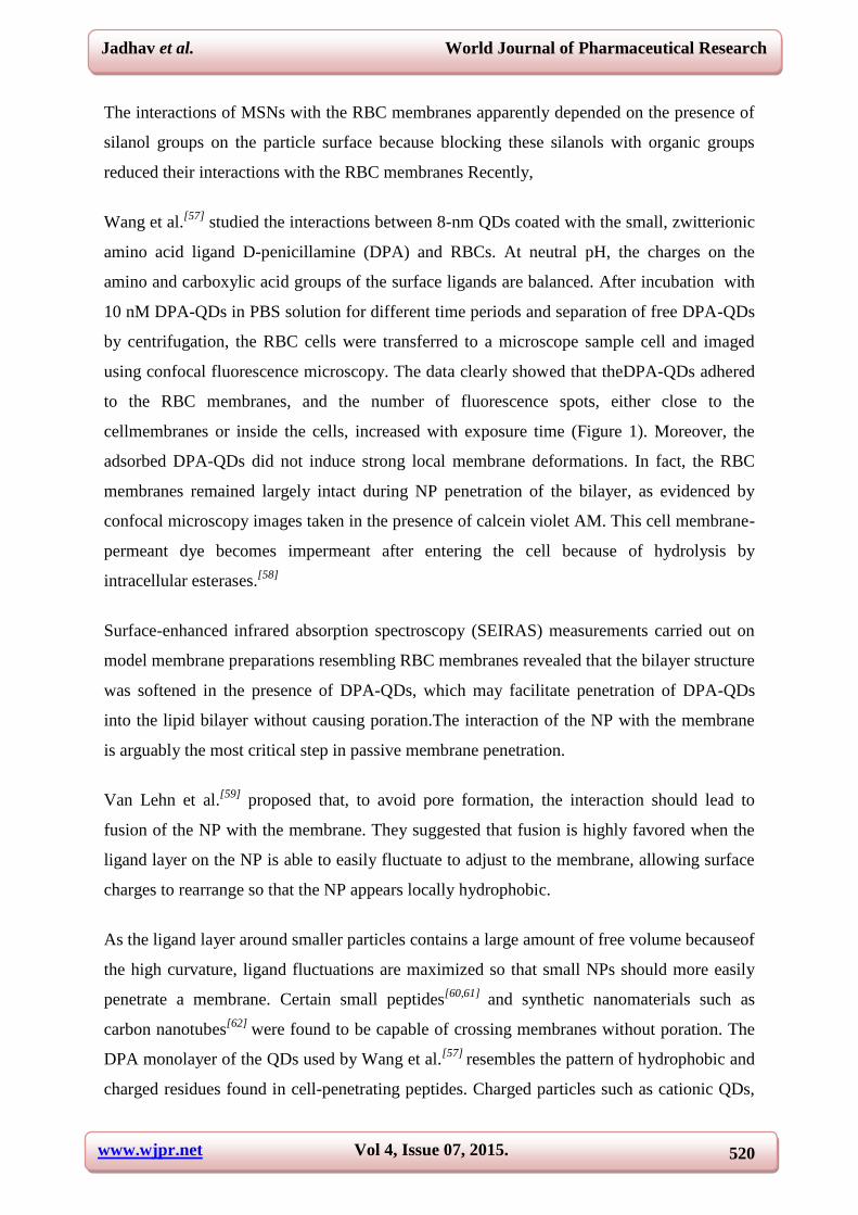
Red blood cells (RBCs)
Using confocal laser scanningmicroscopy (CLSM) in combination with digital data
restoration,conventional TEM, and energy filtering TEM to investigate passive NP uptake ,A
quantitative analysis revealed that only the size determined the uptake efficiency. They
confirmed that particles<200 nm enter RBCs. Small (∼100 nm)adsorbed to the surface of
RBCs without disturbing the membrane or the cell morphology (Figure 1). In
contrast,adsorption of large MSNs (∼600 nm) induced strong local membrane deformations,
followed by internalization of the particles and, eventually, hemolysis.
www.wjpr.net Vol 4, Issue 07, 2015.
520
Jadhav et al. World Journal of Pharmaceutical Research
The interactions of MSNs with the RBC membranes apparently depended on the presence of
silanol groups on the particle surface because blocking these silanols with organic groups
reduced their interactions with the RBC membranes Recently,
Wang et al.[57]
studied the interactions between 8-nm QDs coated with the small, zwitterionic
amino acid ligand D-penicillamine (DPA) and RBCs. At neutral pH, the charges on the
amino and carboxylic acid groups of the surface ligands are balanced. After incubation with
10 nM DPA-QDs in PBS solution for different time periods and separation of free DPA-QDs
by centrifugation, the RBC cells were transferred to a microscope sample cell and imaged
using confocal fluorescence microscopy. The data clearly showed that theDPA-QDs adhered
to the RBC membranes, and the number of fluorescence spots, either close to the
cellmembranes or inside the cells, increased with exposure time (Figure 1). Moreover, the
adsorbed DPA-QDs did not induce strong local membrane deformations. In fact, the RBC
membranes remained largely intact during NP penetration of the bilayer, as evidenced by
confocal microscopy images taken in the presence of calcein violet AM. This cell membrane-
permeant dye becomes impermeant after entering the cell because of hydrolysis by
intracellular esterases.[58]
Surface-enhanced infrared absorption spectroscopy (SEIRAS) measurements carried out on
model membrane preparations resembling RBC membranes revealed that the bilayer structure
was softened in the presence of DPA-QDs, which may facilitate penetration of DPA-QDs
into the lipid bilayer without causing poration.The interaction of the NP with the membrane
is arguably the most critical step in passive membrane penetration.
Van Lehn et al.[59]
proposed that, to avoid pore formation, the interaction should lead to
fusion of the NP with the membrane. They suggested that fusion is highly favored when the
ligand layer on the NP is able to easily fluctuate to adjust to the membrane, allowing surface
charges to rearrange so that the NP appears locally hydrophobic.
As the ligand layer around smaller particles contains a large amount of free volume becauseof
the high curvature, ligand fluctuations are maximized so that small NPs should more easily
penetrate a membrane. Certain small peptides[60,61]
and synthetic nanomaterials such as
carbon nanotubes[62]
were found to be capable of crossing membranes without poration. The
DPA monolayer of the QDs used by Wang et al.[57]
resembles the pattern of hydrophobic and
charged residues found in cell-penetrating peptides. Charged particles such as cationic QDs,
www.wjpr.net Vol 4, Issue 07, 2015.
521
Jadhav et al. World Journal of Pharmaceutical Research
however, typically induce transient poration of the cell membranes, which may result in
cytotoxic effects.[58]
Figure 1: Passive NP uptake by red blood cells. (a – d) Internalization of DPA-QDs (8
nm).[57]
(e – l) Scanning electron micrographs (SEM) of RBCs (5% hematocrit)
incubated with 100 μg mL–1 of (e – h) small (~100 nm) and (i – l) large (~600 nm)
mesoporous silica particles (MSN).[58]
Reproduced with permission from the American
Chemical Society.
Cytotoxic effects of NPs
A protein adsorption layer on the surface confers a new biological identity to the NP, which
may completely modify the subsequent cellular and tissue responses, e.g., the distribution to
various organs, tissues, and cells. Once inside a cell or tissue, the surface layer, including the
adsorbed biomolecules, and also the NP corematerial will likely be metabolized.
Subsequently, the (remnantsof the) NPs may be excreted by the organism. All these
interactions with the biological environment are again dependent on the physicochemical
properties of NPs including their size[64]
(Figure 2). To evaluate the toxicity profile of NPs,
two main approaches have been established: (i) functional assays assess the effects of NPs on
cellular processes, (ii) viability assays probe whether the NPs cause death in a cell or a

www.wjpr.net Vol 4, Issue 07, 2015.
522
Jadhav et al. World Journal of Pharmaceutical Research
system of cells[65]
Although some aspects of size dependent NP toxicity may be reasonably
well predicted by in vitro techniques, it remains difficult to judge whether the observed
cytotoxicity is clinically relevant.As can be inferred from the studies , smaller NPs appear to
be more toxic than larger ones. Small NPs possess a high surface area relative to their total
mass, which increases the chance to interact with surrounding biomolecules and, as a
consequence, to trigger adverse responses. Pan et al.[66]
observed that small AuNPs (1.4 nm)
were highly toxic and caused predominately rapid cell death by necrosis within 12 h, while
larger, 15-nm AuNPs displayed low toxicity, irrespective of cell type and surface ligandsThe
toxic effects in mice were less pronounced after coating the NP surface with peptides that
induced an enhanced immune response. cationic NPs are considered more toxic than neutral
or anionic ones, possibly due to their high affinity towards the negatively charged plasma
membrane. Therefore, NP toxicity must be evaluated by changing NP properties
systematically, one at a time.
Figure 2: Cytotoxic effects of NPs. In the biological environment, NPs may trigger the
production of reactive oxygen species (ROS). Elevated ROS levels may lead to (i)
activation of cellular stress-dependent signaling pathways, (ii) direct damage of
subcellular organelles such as mitochondria and (iii) DNA fragmentation in the nucleus,
resulting in cell cycle arrest, apoptosis, and inflammatory response. NPs may interact
www.wjpr.net Vol 4, Issue 07, 2015.
523
Jadhav et al. World Journal of Pharmaceutical Research
with membrane-bound cellular receptors, e.g., growth factor (GF) receptors and
integrins, inducing cellular phenotypes such as proliferation, apoptosis, differentiation,
and migration. After internalization via endocytic pathways, NPs are trafficked along
the endolysosomal network within vesicles with the help of motor proteins and
cytoskeletal structures. To access cytoplasmic or nuclear targets, NPs must escape from
the endolysosomal network and traverse through the crowded cytoplasm.
CONCLUSION
The smaller partical size of ENPs offers more surface area And show more harmful effects on
body. Interactions between engineered nanomaterials and cells, tissues and organisms has
become very important, especially in relation to possible hazards to human health. current
research on nano-biointeractions, with a focus on the effects of ENP and their interactions
with live cells. Also in vivo and in vitro studies of ENPs showed Toxicities Induced by ENPs
.Hazard identification to facilitate risk assessment due to lack of Standardised Testing And
Characterisation data results in Nonreproducible Results And makes the Univocal HI of
ENPs impossible.
ACKNOWLEDGMENT
The authors gratefully acknowledge the contributions of Prof. Gadhave M.V.HOD M.
Pharm,. Mrs. Thorat R.M.,Principal, Instt.of Pharmacy , Dr.Gaikwad D.D. Principal, Vishal
Institute of Pharmaceutical Education & Research, Ale. for constant motivation and
encouragement.
REFERENCES
1. National Institute for Occupational Safety and Health (NIOSH) Approaches to Safe
Nanotechnology: Managing the Health and Safety Concerns Associated with Engineered
Nanomaterials (March 2009). http://www.cdc.gov/niosh/docs/2009-125/
2. Danail Hristozov * and Ineke Malsch Hazards and Risks of Engineered Nanoparticles for
the Environment and Human Health Malsch TechnoValuation, Vondellaan 90, 3521 GH
Utrecht, The Netherlands; ISSN 2071-1050 www.mdpi.com/journal/sustainability,
Sustainability., 2009; 1: 1161-1194; doi:10.3390/su1041161
3. U.S. Environmental Protection Agency Nanotechnology White Paper; U.S. EPA:
Washington, DC, USA, 2007; Available online:
http://www.epa.gov/osa/pdfs/nanotech/epa-nanotechnology-whitepaper-0207.pdf
(accessed 10 September 2009).

www.wjpr.net Vol 4, Issue 07, 2015.
524
Jadhav et al. World Journal of Pharmaceutical Research
4. Helland, A. Nanoparticles: A Closer Look at the Risks to Human Health and the
Environment. Perceptions and Precautionary Measures of Industry and Regulatory
Bodies in Europe; International Institute for Industrial Environmental Economics (IIIEE):
Lund, Sweden, 2004; Available online:
http://www.iiiee.lu.se/Publication.nsf/$webAll/D9CA9F1E83E4FA12C1256
F9D00539C39/$FILE/Asgeir%20Helland.pdf (accessed 13 October 2009).
5. http://www.merriam-webster.com/dictionary/toxicity
6. Holister, P.; Weener, J.; Vas, C.; Harper, T. Nanoparticles: Technology White Papers nr.
3. Cientifica: London, UK, 2003. Available online: http://images.iop.org/dl/nano/wp/
nanoparticles_WP.pdf (accessed 10 September 2009).
7. Lam, C.; James, J.; McCluskey, R.; Hunter, R. Pulmonary toxicity of single-wall carbon
nanotubes in mice 7 and 90 days after intratracheal instillation. Toxicol. Sci., 2004; 77:
126-134.
8. Warheit, D.; Laurence, B.; Reed, K.; Roach, D.; Reynolds, G.; Webb, T. Comparative
pulmonary toxicity assessment of single-wall carbon nanotubes in rats. Toxicol. Sci.,
2004; 77: 117-125.
9. Smith, C.; Shaw, B.; Handy, R.; Toxicity of single walled carbon nanotubes on rainbow
trout, (Oncorhynchus mykiss): respiratory toxicity, organ pathologies, and other
physiological effects. Aquat. Toxicol., 2007; 82: 94-109.
10. Carrero-Sanchez, J.; Elias, A.; Mancilla, R.; Arrellin, G.; Terrones, H.; Laclette, J.;
Terrones, M. Biocompatibility and toxicological studies of carbon nanotubes doped with
nitrogen. Nano Lett., 2007; 6: 1609-1616.
11. Poland, C.; Duffin, R.; Kinloch, I.; Maynard, A.; Wallace, W.; Seaton, A.; Stone, V.;
Brown, S.; Macnee, W.; Donaldson, K. Carbon nanotubes introduced into the abdominal
cavity of mice show asbestos-like pathogenicity in a pilot study. Nat. Nanotechnol., 2008;
3: 423-428.
12. Lai, H.; Chen, W.; Chiang, L. Free radical scavenging activity of fullerenol on the
ischemia-reperfusion intestine in dogs. World J. Surg., 2000; 24: 450-454.
13. Oberdörster, E. Manufactured nanomaterials (fullerenes, C60) induce oxidative stress in
juvenile largemouth bass. Environ. Health Perspect., 2004; 112: 1058-1062.
14. Oberdörster, E. Toxicity of C60 Fullerenes to Two Aquatic Species: Daphnia and
Largemouth Bass; American Chemical Society: Anaheim, CA, USA, 2004.
www.wjpr.net Vol 4, Issue 07, 2015.
525
Jadhav et al. World Journal of Pharmaceutical Research
15. Zhu, S.; Oberdorster, E.; Haasch, M. Toxicity of an engineered nanoparticle (Fullerene,
C60) in two aquatic species, daphnia and fathead minnow. Mar. Environ. Res., 2006; 62:
S5-S9. Sustainability., 2009; 1: 1188.
16. Sayes, C.; Marchione, A.; Reed, K.; Warheit, D. Comparative pulmonary toxicity
assessments of C60 water suspensions in rats: few differences in fullerene toxicity in vivo
in contrast to in vitro profiles. Nano Lett., 2007; 7: 2399- 2406.
17. Chen, H.; Yu, C.; Ueng, T.; Chen, S.; Chen, B.; Huang, K.; Chiang, L. Acute and
subacute toxicity study of water-soluble polyalkylsulfonated C60 in rats. Toxicol. Pathol.
1998; 26: 143-151.
18. Oberdorster, E.; Zhu, S.; Blickley, T.; Clellan-Green, P.; Haasch, M. Ecotoxicology of
carbon-based engineered nanoparticles: effects of fullerene (C60) on aquatic organisms.
Carbon., 2006; 44: 1112-1120.
19. Li, X.; Brown, D.; Smith S.; MacNee, W.; Donaldson, K. Short term inflammatory
responses following intratracheal instillation of fine and ultrafine carbon black in rats.
Inhal. Toxicol., 1999; 11: 709-731.
20. Gordon, T.; Chen, L.; Fine, J; Schlesinger, R.; Su, W.; Kimmel, T.; Amdur, M.
Pulmonary effects of inhaled zinc-oxide in human-subjects, guinea-pigs, rats, and rabbits.
Am. Ind. Hyg. Assoc. J., 1992; 53: 503-509.
21. Beckett, W.; Chalupa, D.; Pauly-Brown, A.; Speers, D.; Stewart, J.; Frampton, M.; Utell,
M.; Huang, L.; Cox, C.; Zareba, W.; Oberdorster, G. Comparing inhaled ultrafine versus
fine zinc oxide particles in healthy adults: a human inhalation study. Am. J. Respir. Crit.
Care Med., 2005; 171: 1129-1135.
22. Wang, B.; Feng, W.; Wang, T.; Jia, G.; Wang, M.; Shi, J.; Zhang, F.; Zhao, Y.; Chai, Z.
Acute toxicity of nano- and micro-scale zinc powder in healthy adult mice. Toxicol. Lett.,
2006; 161: 115-123.
23. Yang, L.; Watts, D. Particle surface characteristics may play an important role in
phytotoxicity of alumina nanoparticles. Toxicol. Lett. 2005, 158, 122-132.
24. Oberdörster, G. Toxicology of ultrafine particles: in vivo studies. Phil. Trans. R. Soc.
Lond., 2000; A 358: 2719-2740.
25. Oberdörster G.; Ferin, J.; Lehnert, B. Correlation between particle size, in vivo particle
persistence and lung injury. Environ. Health Perspect., 1994; 102: 173-179.
26. Warheit, D.; Webb, T.; Sayes, C.; Colvin, V.; Reed, K. Pulmonary instillation studies
with nanoscale TiO2 rods and dots in rats: toxicity is not dependent upon particle size and
surface area. Toxicol. Sci., 2006; 91: 227-236.
www.wjpr.net Vol 4, Issue 07, 2015.
526
Jadhav et al. World Journal of Pharmaceutical Research
27. Wang, J.; Zhou, G.; Chen, C.; Yu, H.; Wang, T.; Ma, Y.; Jia, G.; Gao, Y.; Li, B.; Sun, J.;
Li, Y.; Jiao, F.; Zhao, Y.; Chai, Z. Acute toxicity and biodistribution of different sized
titanium dioxide particles in mice after oral administration. Toxicol. Lett., 2007; 168:
176-185
28. Bourrinet, P.; Bengele, H.; Bonnemain, B.; Dencausse, A.; Idee, J. M.; Jacobs, P. M.;
Lewis, J. M. Preclinical safety and pharcokinitic profile of ferumoxtran-10, an ultrasmall
superparamagnetic iron oxide magnetic resonance contrast agent. Invest. Radiol., 2006;
41: 313-324.
29. Shvedova, A.; Castranova, V.; Kisin, E.; Schwegler-Berry, D.; Murray, A.R.;
Gandelsman, V.Z.; Maynard, A.; Baron, P. Exposure to carbon nanotube material:
assessment of nanotube cytotoxicity using human keratinocyte cells. J. Toxicol. Environ.
Heal., 2006; 66: 1909-1926.
30. Cui, D.; Tian, F.; Ozkan, C.; Wang, M.; Gao, H. Effect of single wall carbon nanotubes
on human HEK293 cells. Toxicol. Lett., 2005; 155: 73-85.
31. Sayes, C.; Liang, F.; Hudson, J.; Mendez, J.; Guo, W.; Beach, J.; Moore, V.; Doyle, C.;
West, J.; Billups, W.; Ausman, K.; Colvin, V. Functionalization density dependence of
single-walled carbon nanotubes cytotoxicity in vitro. Toxicol. Lett., 2006; 161: 135-142.
32. Bottini, M.; Bruckner, S.; Nika, K.; Bottini, N.; Bellucci, S.; Magrini, A.; Bergamaschi,
A.; Mustelin, T. Multi-walled carbon nanotubes induce t-lymphocyte apoptosis. Toxicol.
Lett, 2006; 160: 121-126.
33. Monteiro-Riviere, N.; Nemanich, R.; Inman, A.; Wang, Y.; Riviere, J. Multi-walled
Carbon nanotube interactions with human epidermal keratinocytes. Toxicol. Lett., 2005;
155: 377-384.
34. Kang, S.; Mauter, M.; Elimelech, M. Physicochemical determinants of multiwalled
carbon nanotube bacterial cytotoxicity. Environ. Sci. Technol., 2008; 42: 5843-5859.
35. Adelman, P.; Baierl, T.; Drosselmeyer, E.; Politis, C.; Polzer, G.; Seidel, A.; Steinleitner
C. Effect of Fullerenes on Alveolar Macrophages in Vitro. In Toxic and Carcinogenic
Effect of Solid Particles in the Respiratory Tract, Mohr, U., Dungworth, D., Mauderly, J.,
Oberdoester, G., Eds. ILSI Press: Washington, DC, USA, 1994; pp. 405-407.
36. Porter, A.; Muller, K.; Skepper, J.; Midgley, P.; Welland, M. Uptake of C60 by human
monocyte macrophages, its localization and implications for toxicity: studied by high
resolution electron microscopy and electron tomography. Acta Biomater., 2006; 2:
409-419.
www.wjpr.net Vol 4, Issue 07, 2015.
527
Jadhav et al. World Journal of Pharmaceutical Research
37. European Commission Technical Guidance Document (TGD) on Risk Assessment;
European Commission Joint Research Center (ECJRC): Location, Country, 2003;
Available online: http://ecb.jrc.ec.europa.eu/tgd/ (accessed 10 September 2009).
38. Rubins, J. Alveolar macrophages: wielding the double-edged sword of inflammation. Am.
J. Respir. Crit. Care Med., 2003; 167: 103-104.
39. Yamawaki, H.; Iwai, N. Cytotoxicity of water-soluble fullerene in vascular endothelial
cells. Am. J. Cell Physiol., 2006; 290: C1495-C1502.
40. Rouse, J.; Yang, J.; Barron, A.; Monteiro-Riviere, N. Fullerene-based amino acid
nanoparticle interactions with human epidermal keratinocytes. Toxicology in Vitro., 2006;
20: 1313-1320.
41. Hansen, S. Regulation and Risk Assessment of Nanomaterials—Too Little, Too Late?
Technical University of Denmark (DTU): Lyngby, Denmark, 2009; Available online:
http://www2.er.dtu.dk/publications/fulltext/2009/ENV2009-069.pdf (accessed 10
September 2009).
42. Jaiswal, J.; Mattoussi, H.; Mauro, J.; Simon, S. Long-term multiple color imaging of live
cells using quantum dot bioconjugates. Nat. Biotechnol., 2003; 21: 47-51.
43. Hoshino, A.; Hanaki, K.; Suzuki, K.; Yamamoto, K. Applications of t-lymphoma labeled
with fluorescent quantum dots to cell tracing markers in mouse body. Biochem. Bioph.
Res. Co., 2004; 314: 46-53.
44. Lovric, J.; Bazzi, H.; Cuie, Y.; Fortin, G.; Winnik, F.; Maysinger, D. Differences in
subcellular distribution and toxicity of green and red emitting CdTe quantum dots. J. Mol.
Med., 2005; 83: 377-385.
45. Green, M.; Howman, E. Semiconductor quantum dots and free radical induced DNA
nicking. Chem. Commun., 2005; 1: 121-123.
46. Chang, E.; Thekkek, N.; Yu, W.; Colvin, V.; Drezek, R. Evaluation of quantum dot
cytotoxicity based on intracellular uptake. Toxicol. Lett., 2006; 2: 1412-1417.
47. Sayes, C.; Wahi, R.; Kurian, P.; Liu, Y.; West, J.; Ausman, K.; Warheit, D.; Colvin, V.
Correlating nanoscale titania structure with toxicity: a cytotoxicity and inflammatory
response study with human dermal fibroblasts and human lung epithelial cells. Toxicol.
Sci., 2006; 92: 174-185.
48. Wang, J.; Sanderson, B.; Wang, H. Cyto-and genotoxicity of ultrafine TiO2 particles in
cultured human lymphoblastoid cells. Mutat. Res-Gen. Tox. En., 2007; 628: 99-106.
49. Chen, M.; von Mikecz, A. Formation of nucleoplasmic protein aggregates impairs nuclear
function in response to SiO2 nanoparticles. Exp. Cell Res., 2005; 305: 51-62.
www.wjpr.net Vol 4, Issue 07, 2015.
528
Jadhav et al. World Journal of Pharmaceutical Research
50. Muller, K.; Skepper, J.; Posfai, M.; Trivedi, R.; Howarth, S.; Corot, C.; Lancelot, E.;
Thompson, P.; Brown, A.; Gillard, J. Effect of ultrasmall superparamagnetic iron oxide
nanoparticles (ferumoxtran-10) on human monocytemacrophages in vitro. Biomaterials.,
2007; 28: 1629-1642.
51. Alt, V.; Bechert, T.; Steinrucke, P.; Wagener, M.; Seidel, P.; Dingeldein, E.; Domann, E.;
Schnettler, R. An in vitro assessment of the antibacterial properties and cytotoxicity of
nanoparticulate silver bone cement. Biomaterials., 2004; 25: 4383-4391.
52. Baker, C.; Pradhan, A.; Pakstis, L.; Pochan, D.; Shah S. Synthesis and antibacterial
properties of silver nanoparticles. J. Nanosci. Nanotechnol., 2005; 5: 244-249.
53. Sayes, C.; Reed, K.; Warheit, D. Assessing toxicity of fine and nanoparticles: comparing
in vitro measurements to in vivo pulmonary toxicity profiles. Toxicol. Sci., 2007; 97:
163-180.
54. Hansen, S.; Larsen, B.; Olsen, S.; Baun, A. Categorization framework to aid hazard
identification of nanomaterials. Nanotoxicology., 2007; 11: 243-250.
55. Warheit, D. How meaningful are the results of nanotoxicity studies in the absence of
adequate material characterization? Toxicol Sci., 2008; 101: 183-185.
56. Plata, D.L.; Gschwend, P.M.; Reddy, C. Industrially synthesized single-walled carbon
nanotubes: compositional data for users, environmental risk assessments, and source
apportionment. Nanotechnology., 2008; 19: 185706.
57. Wang T, Bai J, Jiang X, Nienhaus GU: Cellular uptake of nanoparticles by membrane
penetration: a study combining confocal microscopy with FTIR spectroelectrochemistry.
ACS Nano., 2012; 6: 1251–1259.
58. Zhao Y, Sun X, Zhang G, Trewyn BG, Slowing II, Lin VS: Interaction of mesoporous
silica nanoparticles with human red blood cell membranes: size and surface effects. ACS
Nano., 2011; 5: 1366–1375.
59. Verma A, Uzun O, Hu Y, Han HS, Watson N, Chen S, Irvine DJ, Stellacci F: Surface-
structure-regulated cell-membrane penetration by monolayerprotected nanoparticles. Nat
Mater., 2008; 7: 588–595.
60. Van Lehn RC, Atukorale PU, Carney RP, Yang YS, Stellacci F, Irvine DJ, Alexander-
Katz A: Effect of particle diameter and surface composition on the spontaneous fusion of
monolayer-protected gold nanoparticles with lipid bilayers. Nano Lett., 2013; 13:
4060–4067.
61. Koren E, Torchilin VP: Cell-penetrating peptides: breaking through to the other side.
Trends Mol Med., 2012; 18: 385–393.
www.wjpr.net Vol 4, Issue 07, 2015.
529
Jadhav et al. World Journal of Pharmaceutical Research
62. Heitz F, Morris MC, Divita G: Twenty years of cell-penetrating peptides: from molecular
mechanisms to therapeutics. Br J Pharmacol., 2009; 157: 195–206.
63. Kostarelos K, Lacerda L, Pastorin G, Wu W, Wieckowski S, Luangsivilay J, Godefroy S,
Pantarotto D, Briand JP, Muller S, et al: Cellular uptake of functionalized carbon
nanotubes is independent of functional group and cell type. Nat Nanotechnol., 2007; 2:
108–113.
64. Nel A, Xia T, Madler L, Li N: Toxic potential of materials at the nanolevel. Science.,
2006; 311: 622–627.
65. Love SA, Maurer-Jones MA, Thompson JW, Lin Y-S, Haynes CL: Assessing
nanoparticle toxicity. Annu Rev Anal Chem., 2012; 5: 181–205.
66. Pan Y, Neuss S, Leifert A, Fischler M, Wen F, Simon U, Schmid G, Brandau W, Jahnen-
Dechent W: Size-dependent cytotoxicity of gold nanoparticles. Small., 2007; 3:
1941–1949.


![Engineered Multifunctional Nanocarriers for Cancer Diagnosis ...homepages.uc.edu/~shid/publications/PDFfiles/Engineered...[55–59 ] Magnetic nanoparticles (MNPs) or magnetic nanospheres](https://img.pdfslide.us/doc/110x75/603a819c2af00a55936733e1/engineered-multifunctional-nanocarriers-for-cancer-diagnosis-shidpublicationspdffilesengineered.jpg)