Embed Size (px)
Citation preview

J Clin Exp Tox 2019 Volume 3 Issue 1 1
http://www.alliedacademies.org/clinical-experimental-toxicology/Research Article
IntroductionTuberculosis is a major global health problem. It is the ninth leading cause of death worldwide and the leading cause from a single infectious agent, ranking above HIV/AIDS. In 2016, there were an estimated 1.3 million TB deaths among HIV-negative people (down from 1.7 million in 2000) and an additional 374 000 deaths among HIV-positive people. An estimated 10.4 million people fell ill with TB in 2016: 90% were adults, 65% were male, 10% were people living with HIV [1].
Rifampicin and isoniazid are widely used antimicrobial agents in standard anti-tuberculosis treatment. Their combination along with ethambutol and pyrazinamide has been adjudged best for efficacy and tolerability amongst the available TB drugs and is, therefore, the mainstay “first line” therapy [2,3]. There are historical and clinical proofs that these first-line anti-tuberculosis agents are the most potent oral anti-tuberculous medications [4-6]. In addition, in-vitro and in-vivo clinical data support the use of such individual agents [7,8]. This may be because the combination has been found to be a beneficial and cost-effective treatment for TB [9], but, this is not without some systemic toxicity from the results from human data [10,11].
Since TB affects people in their productive and reproductive age, there have been recent suggestions that anti-tuberculosis agents could produce adverse effects on reproductive health system
[10-13]. Available data on adverse reactions of antituberculous drugs in human and animal experiments particularly with respect to reproduction are limited [14]. Most of the few available studies on effects of anti-tuberculosis agents of female reproduction and/or fertility had focused on maternal and fetal health [15,16]. Unfortunately, there is paucity of data on how the drugs affect materno-foetal link-the placenta.
Rat placental models have generally been useful for evaluating the potential of drugs or chemicals that affect human reproductive development, since there are several similarities between rats and humans in early placental development [17]. The placenta grows rapidly for a short period with high blood flow during pregnancy and has multifaceted functions, such as its barrier function, nutritional transport, drug metabolizing activity and endocrine action. Consequently, the placenta is a highly susceptible target organ for drug- or chemical-induced adverse effects, and many placenta-toxic agents have been reported [18]. On the other hand, the dam and fetus have a close relationship with each other via the placenta and form the maternal-fetal-placental unit in mammalian embryonic development. Drug- or chemical-induced placental functional depression and injury subsequently result in abnormal fetal growth or development leading to fetal resorption or teratogenicity. Thus, the placenta is an important organ for evaluating embryonic developmental toxicity and understanding its mechanism [19]. Unfortunately,
Toxic effects of antituberculosis drugs (isoniazid and rifampicin) on feto-placental unit of wistar rats: a morphological, histological and biochemical study.
Vitalis Chukwuma Ezeuko*, Jacob Ehiagwina Ataman, Dorothea Baxter-Grillo
University of Benin, Nigeria
AbstractThe aim of this study was to investigate the effects of chronic administration of therapeutic doses of isoniazid and rifampicin individually and in combination to the foetal weight, placental weight, foetal/placental weight ratio, placental oxidative stress and placental histomorphology of Wistar rats. A total of twenty-eight adult female Wistar rats weighing between 160 g and 170 g, aged between 90 days and 110 days were used for this study. They were divided into four groups (A, B, C and D) of seven rats each. Group A served as the control group administered with only sterile water. Group B animals were administered 5 mg/kg body weight of isoniazid only. Group C animals were administered 10 mg/kg body weight of rifampicin only. Group D animals were administered 5 mg/kg body weight of isoniazid and 10 mg/kg body weight of rifampicin. Day 20 foetal body weight significantly reduced in the isoniazid-treated and rifampicin-treated groups. Day 20 placental weight was significantly increased while day 20 foetal:placental weight ratio was significantly reduced in all the experimental groups. Placental superoxide dismutase, catalase and glutathione peroxidase activities were significantly reduced in all the experimental groups. Histological studies of the placenta showed varying degrees of alterations including degeneration of trophoblastic giant cells, haemorrhage in the junctional zone, dilation and congestion of maternal sinusoids and mild dilation and congestion of foetal capillaries. In conclusion, administration of isoniazid and rifampicin singly and in combination at therapeutic doses induced intrauterine growth restriction with compensatory placental hypertrophy and associated oxidative stress and alterations in the histomorphology of the placental tissues.
Accepted on January 05, 2019
Keywords: Antituberculosis, wistar rats, pyrazinamide, histomorphology, oxidative.

2
Citation: Ezeuk VC, Ataman JE, Grillo DB. Toxic effects of antituberculosis drugs (isoniazid and rifampicin) on feto-placental unit of wistar rats: a morphological, histological and biochemical study. J Clin Exp Tox. 2019;3(1):1-6. DOI: 10.4066/2630-4570.018.
J Clin Exp Tox 2019 Volume 3 Issue 1
histological examination of the placental tissue is not routinely performed, leaving assessment of placental weight change as the main index for assessment of placental toxicity in rat reproductive toxicity studies [18].
Having considered these gaps in literature, it becomes imperative to investigate the effects of chronic administration of therapeutic doses of isoniazid and rifampicin individually and in combination to the foetal weight, placental weight, foetal/placental weight ratio, placental oxidative stress and placental histomorphology of Wistar rats.
MethodologyAnimalsWistar rats used for the study were bred at the Animal House, Department of Anatomy, School of Basic Medical Sciences, College of Medical Sciences, University of Benin, Benin City, Edo State, Nigeria. They were kept in polypropylene cages under room temperature, with 12 hour light and 12 hour dark cycle photoperiodicity. Protocols for these experiments was in accordance with the guide for the care and use of laboratory animals [20]. The research proposal was approved by the Research and Ethics Committee, College of Medical Sciences, University of Benin, Benin City Edo State Nigeria, with REC approval number CMS/REC/2018/034.
Research designA total of twenty-eight adult female Wistar rats weighing between 160 g and 170 g, aged between 90 days and 110 days were used for this study. They were divided into four groups (A, B, C and D) of seven rats each. Group A served as the control group administered with only sterile water. Group B animals were administered 5 mg/kg body weight of isoniazid (manufactured by Mancare Pharmaceutical Pvt Limited, India) only, dissolved in sterile water. Group C animals were administered 10 mg/kg body weight of rifampicin (manufactured by Bond Chemicals Limited, Nigeria) only, dissolved in sterile water. Group D animals were administered 5 mg/kg body weight of isoniazid and 10 mg/kg body weight of rifampicin dissolved in sterile water. The dosages of rifampicin and isoniazid used in this study were the recommended daily dosages for humans [16,21]. The drugs were administered orally and daily for ninety days. From the seventieth day of drug administration, the animals with four days regular oestrous cycle were paired overnight with sexually active males in the ratio of 2:1. Successful mating was confirmed by the presence of vaginal plug and or sperm in the vaginal smear the following morning between 9.00 and 10.00 hours. The day sperm cells were found in the vaginal smear was considered as day 1 of pregnancy. The rats were weighed daily and physically observed. On day 20 of gestation, each rat
was laparotomized under chloroform anaesthesia. The uterine horns were exteriorized and incised at the greater curvature of the horns. Number and weight of live fetuses were determined. The placentae were also harvested, blotted free of blood and weighed immediately. The day 20 foetal:placental ratio was calculated by dividing day 20 foetal weight (in grams) by day 20 placental weight (in grams).
The placenta tissue for oxidative stress studies (seven per group) was, immediately after harvesting, washed twice in cold phosphate buffered saline (PBS). It was then homogenized using acid-washed sand and PBS in porcelain mortar and pestle. The tissues homogenate was centrifuged at 10000 rpm for 10 minutes at 4oC. The supernatant was immediately processed for analysis of endogenous antioxidants. Superoxide dismutase activity was determined according the method of Misra and Fridovich [22]. Catalase activity was determined according the method of Cohen et al. [23]. Glutathione peroxidase activity was determined according the method of Nyman [24].
The placenta for histopathological studies (seven tissues per group) was preserved in 10% phosphate buffered formalin for twenty-four hours. The tissues were processed via paraffin wax embedding method of Drury and Wallington [25].
ResultsPlacental and foetal weightThe data in Table 1 revealed that day 20 foetal body weight significantly (P<0.05) decreased in the isoniazid-treated and rifampicin-treated groups when compared with the control. However, there was no statistically significant difference (P>0.05) in day 20 foetal body weight between the isoniazid and rifampicin co-treated group and the control group. Day 20 placental weight was significantly increased (P<0.05) in all the experimental groups when compared with the control group. Day 20 foetal:placental weight ratio was significantly decreased (P<0.05) in all the experimental groups when compared with the control group.
Oxidative stress observationsThe data in Table 2 revealed that placental superoxide dismutase, catalase and glutathione peroxidase activities were significantly decreased (P<0.05) in all the experimental groups when compared with the control group.
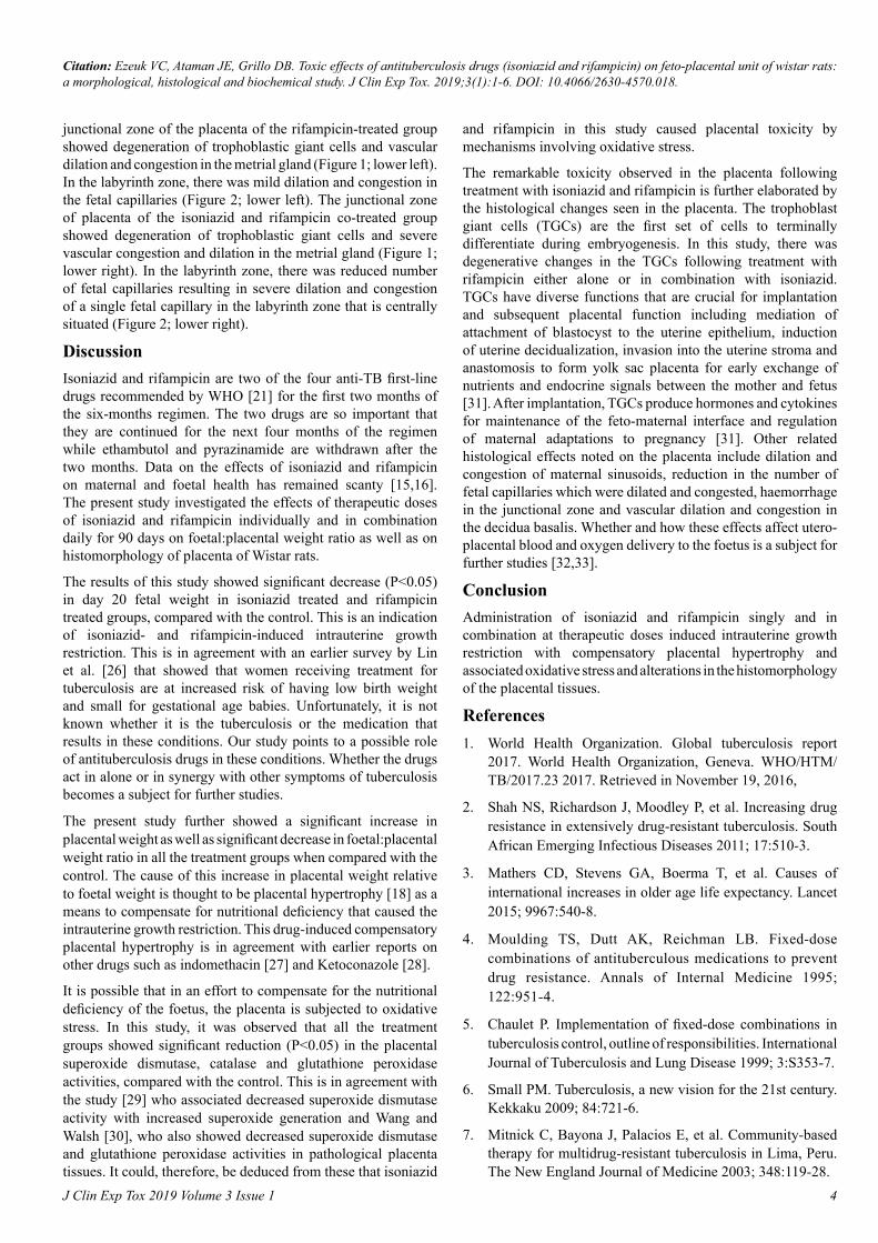
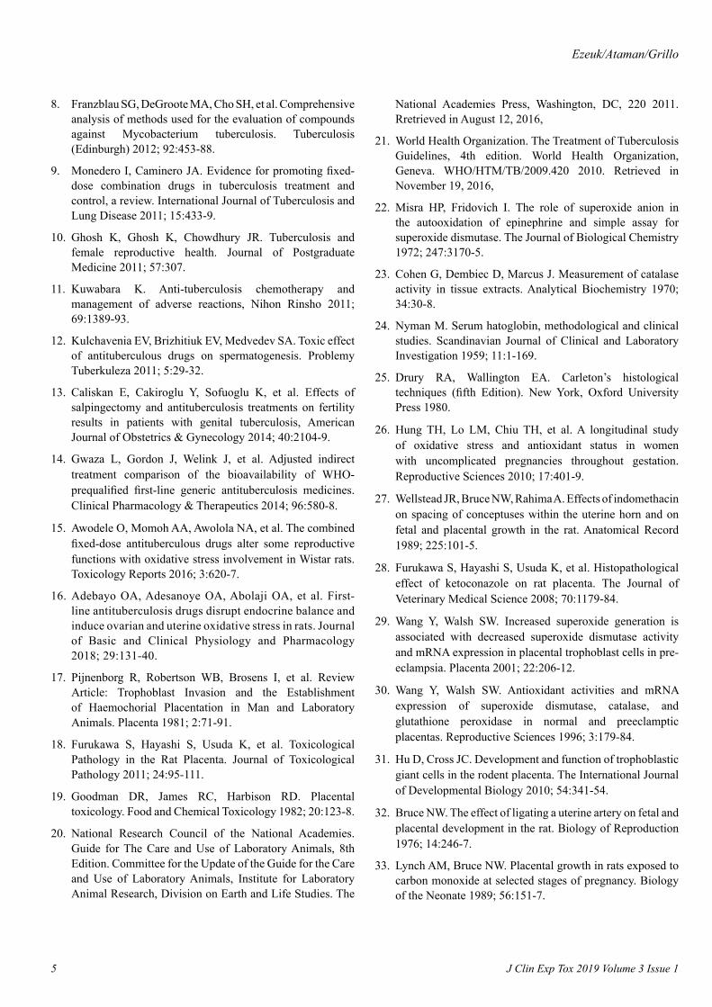
Histological observationHistological studies showed that there were multilayered trophoblastic giant cells and moderate haemorrhage in the junctional zone of the placenta of the isoniazid-treated group (Figure 1; upper right). In the labyrinth zone, there were dilation and congestion of maternal sinusoids and mild dilation and congestion of fetal capillaries (Figure 2: upper left). The
Control Isoniazid only Rifampicin only Isoniazid ± rifampicinDay 20 foetal body weight 4.42 ± 0.13a 3.70 ± 0.06b 3.36 ± 0.07b 4.50 ± 0.09a
Day 20 placental weight 0.46 ± 0.01a 0.53 ± 0.01b 0.70 ± 0.04c 0.60 ± 0.03b
Day 20 foetal/placental weight ratio 9.71 ± 0.31a 6.99 ± 0.18b 4.94 ± 0.37c 7.60 ± 0.38b
• Post hoc comparisons were done using Least Square Differences and shown in superscripts.• Means with unlike superscripts in a row are significantly different (P<0.05) while means with like superscripts in a row are not significantly different (P>0.05).
Table 1. Pregnancy outcome.
Ezeuk/Ataman/Grillo
3 J Clin Exp Tox 2019 Volume 3 Issue 1
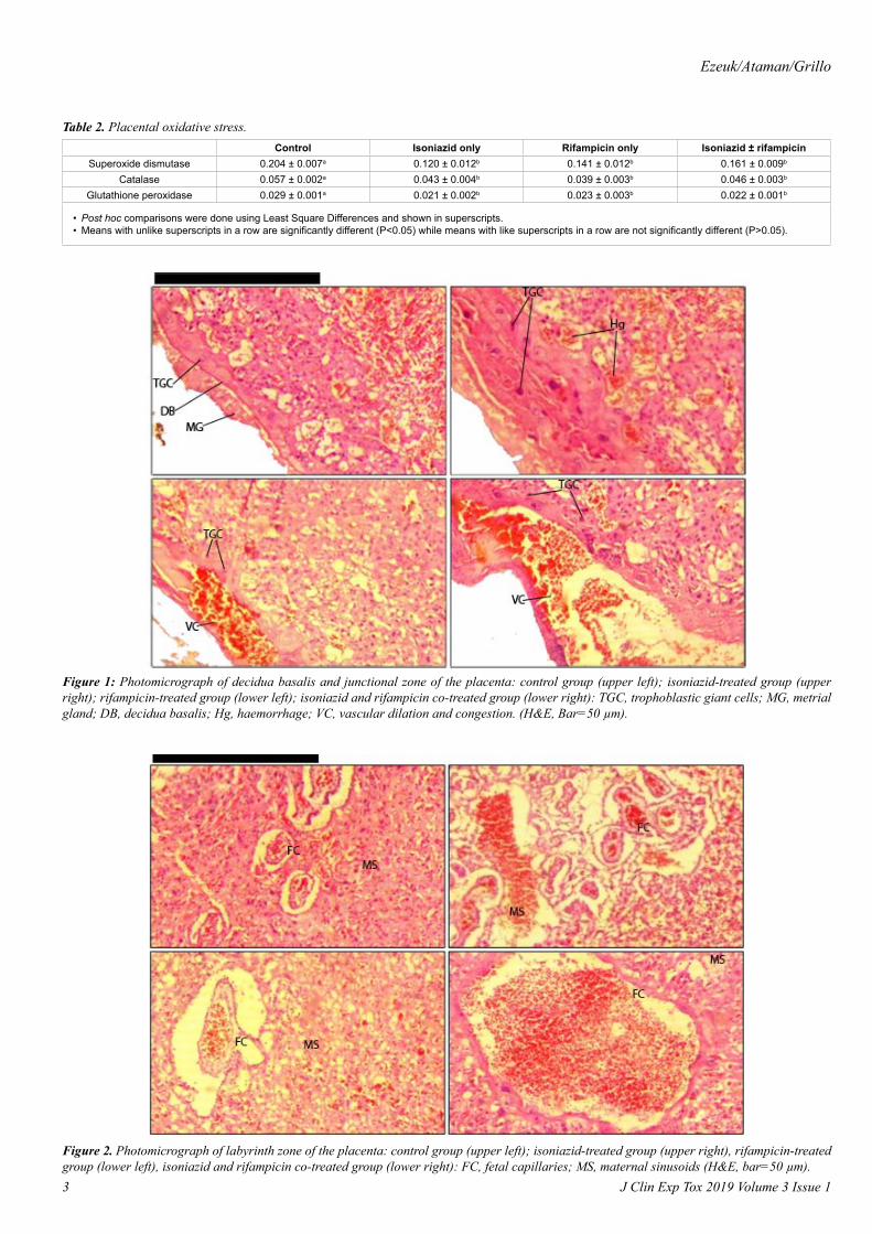
Control Isoniazid only Rifampicin only Isoniazid ± rifampicinSuperoxide dismutase 0.204 ± 0.007a 0.120 ± 0.012b 0.141 ± 0.012b 0.161 ± 0.009b
Catalase 0.057 ± 0.002a 0.043 ± 0.004b 0.039 ± 0.003b 0.046 ± 0.003b
Glutathione peroxidase 0.029 ± 0.001a 0.021 ± 0.002b 0.023 ± 0.003b 0.022 ± 0.001b
• Post hoc comparisons were done using Least Square Differences and shown in superscripts.• Means with unlike superscripts in a row are significantly different (P<0.05) while means with like superscripts in a row are not significantly different (P>0.05).
Table 2. Placental oxidative stress.
Figure 1: Photomicrograph of decidua basalis and junctional zone of the placenta: control group (upper left); isoniazid-treated group (upper right); rifampicin-treated group (lower left); isoniazid and rifampicin co-treated group (lower right): TGC, trophoblastic giant cells; MG, metrial gland; DB, decidua basalis; Hg, haemorrhage; VC, vascular dilation and congestion. (H&E, Bar=50 µm).
Figure 2. Photomicrograph of labyrinth zone of the placenta: control group (upper left); isoniazid-treated group (upper right), rifampicin-treated group (lower left), isoniazid and rifampicin co-treated group (lower right): FC, fetal capillaries; MS, maternal sinusoids (H&E, bar=50 µm).
4
Citation: Ezeuk VC, Ataman JE, Grillo DB. Toxic effects of antituberculosis drugs (isoniazid and rifampicin) on feto-placental unit of wistar rats: a morphological, histological and biochemical study. J Clin Exp Tox. 2019;3(1):1-6. DOI: 10.4066/2630-4570.018.
J Clin Exp Tox 2019 Volume 3 Issue 1
junctional zone of the placenta of the rifampicin-treated group showed degeneration of trophoblastic giant cells and vascular dilation and congestion in the metrial gland (Figure 1; lower left). In the labyrinth zone, there was mild dilation and congestion in the fetal capillaries (Figure 2; lower left). The junctional zone of placenta of the isoniazid and rifampicin co-treated group showed degeneration of trophoblastic giant cells and severe vascular congestion and dilation in the metrial gland (Figure 1; lower right). In the labyrinth zone, there was reduced number of fetal capillaries resulting in severe dilation and congestion of a single fetal capillary in the labyrinth zone that is centrally situated (Figure 2; lower right).
DiscussionIsoniazid and rifampicin are two of the four anti-TB first-line drugs recommended by WHO [21] for the first two months of the six-months regimen. The two drugs are so important that they are continued for the next four months of the regimen while ethambutol and pyrazinamide are withdrawn after the two months. Data on the effects of isoniazid and rifampicin on maternal and foetal health has remained scanty [15,16]. The present study investigated the effects of therapeutic doses of isoniazid and rifampicin individually and in combination daily for 90 days on foetal:placental weight ratio as well as on histomorphology of placenta of Wistar rats.
The results of this study showed significant decrease (P<0.05) in day 20 fetal weight in isoniazid treated and rifampicin treated groups, compared with the control. This is an indication of isoniazid- and rifampicin-induced intrauterine growth restriction. This is in agreement with an earlier survey by Lin et al. [26] that showed that women receiving treatment for tuberculosis are at increased risk of having low birth weight and small for gestational age babies. Unfortunately, it is not known whether it is the tuberculosis or the medication that results in these conditions. Our study points to a possible role of antituberculosis drugs in these conditions. Whether the drugs act in alone or in synergy with other symptoms of tuberculosis becomes a subject for further studies.
The present study further showed a significant increase in placental weight as well as significant decrease in foetal:placental weight ratio in all the treatment groups when compared with the control. The cause of this increase in placental weight relative to foetal weight is thought to be placental hypertrophy [18] as a means to compensate for nutritional deficiency that caused the intrauterine growth restriction. This drug-induced compensatory placental hypertrophy is in agreement with earlier reports on other drugs such as indomethacin [27] and Ketoconazole [28].
It is possible that in an effort to compensate for the nutritional deficiency of the foetus, the placenta is subjected to oxidative stress. In this study, it was observed that all the treatment groups showed significant reduction (P<0.05) in the placental superoxide dismutase, catalase and glutathione peroxidase activities, compared with the control. This is in agreement with the study [29] who associated decreased superoxide dismutase activity with increased superoxide generation and Wang and Walsh [30], who also showed decreased superoxide dismutase and glutathione peroxidase activities in pathological placenta tissues. It could, therefore, be deduced from these that isoniazid
and rifampicin in this study caused placental toxicity by mechanisms involving oxidative stress.
The remarkable toxicity observed in the placenta following treatment with isoniazid and rifampicin is further elaborated by the histological changes seen in the placenta. The trophoblast giant cells (TGCs) are the first set of cells to terminally differentiate during embryogenesis. In this study, there was degenerative changes in the TGCs following treatment with rifampicin either alone or in combination with isoniazid. TGCs have diverse functions that are crucial for implantation and subsequent placental function including mediation of attachment of blastocyst to the uterine epithelium, induction of uterine decidualization, invasion into the uterine stroma and anastomosis to form yolk sac placenta for early exchange of nutrients and endocrine signals between the mother and fetus [31]. After implantation, TGCs produce hormones and cytokines for maintenance of the feto-maternal interface and regulation of maternal adaptations to pregnancy [31]. Other related histological effects noted on the placenta include dilation and congestion of maternal sinusoids, reduction in the number of fetal capillaries which were dilated and congested, haemorrhage in the junctional zone and vascular dilation and congestion in the decidua basalis. Whether and how these effects affect utero-placental blood and oxygen delivery to the foetus is a subject for further studies [32,33].
ConclusionAdministration of isoniazid and rifampicin singly and in combination at therapeutic doses induced intrauterine growth restriction with compensatory placental hypertrophy and associated oxidative stress and alterations in the histomorphology of the placental tissues.
References1. World Health Organization. Global tuberculosis report
2017. World Health Organization, Geneva. WHO/HTM/TB/2017.23 2017. Retrieved in November 19, 2016,
2. Shah NS, Richardson J, Moodley P, et al. Increasing drug resistance in extensively drug-resistant tuberculosis. South African Emerging Infectious Diseases 2011; 17:510-3.
3. Mathers CD, Stevens GA, Boerma T, et al. Causes of international increases in older age life expectancy. Lancet 2015; 9967:540-8.
4. Moulding TS, Dutt AK, Reichman LB. Fixed-dose combinations of antituberculous medications to prevent drug resistance. Annals of Internal Medicine 1995; 122:951-4.
5. Chaulet P. Implementation of fixed-dose combinations in tuberculosis control, outline of responsibilities. International Journal of Tuberculosis and Lung Disease 1999; 3:S353-7.
6. Small PM. Tuberculosis, a new vision for the 21st century. Kekkaku 2009; 84:721-6.
7. Mitnick C, Bayona J, Palacios E, et al. Community-based therapy for multidrug-resistant tuberculosis in Lima, Peru. The New England Journal of Medicine 2003; 348:119-28.
Ezeuk/Ataman/Grillo
5 J Clin Exp Tox 2019 Volume 3 Issue 1
8. Franzblau SG, DeGroote MA, Cho SH, et al. Comprehensive analysis of methods used for the evaluation of compounds against Mycobacterium tuberculosis. Tuberculosis (Edinburgh) 2012; 92:453-88.
9. Monedero I, Caminero JA. Evidence for promoting fixed-dose combination drugs in tuberculosis treatment and control, a review. International Journal of Tuberculosis and Lung Disease 2011; 15:433-9.
10. Ghosh K, Ghosh K, Chowdhury JR. Tuberculosis and female reproductive health. Journal of Postgraduate Medicine 2011; 57:307.
11. Kuwabara K. Anti-tuberculosis chemotherapy and management of adverse reactions, Nihon Rinsho 2011; 69:1389-93.
12. Kulchavenia EV, Brizhitiuk EV, Medvedev SA. Toxic effect of antituberculous drugs on spermatogenesis. Problemy Tuberkuleza 2011; 5:29-32.
13. Caliskan E, Cakiroglu Y, Sofuoglu K, et al. Effects of salpingectomy and antituberculosis treatments on fertility results in patients with genital tuberculosis, American Journal of Obstetrics & Gynecology 2014; 40:2104-9.
14. Gwaza L, Gordon J, Welink J, et al. Adjusted indirect treatment comparison of the bioavailability of WHO-prequalified first-line generic antituberculosis medicines. Clinical Pharmacology & Therapeutics 2014; 96:580-8.
15. Awodele O, Momoh AA, Awolola NA, et al. The combined fixed-dose antituberculous drugs alter some reproductive functions with oxidative stress involvement in Wistar rats. Toxicology Reports 2016; 3:620-7.
16. Adebayo OA, Adesanoye OA, Abolaji OA, et al. First-line antituberculosis drugs disrupt endocrine balance and induce ovarian and uterine oxidative stress in rats. Journal of Basic and Clinical Physiology and Pharmacology 2018; 29:131-40.
17. Pijnenborg R, Robertson WB, Brosens I, et al. Review Article: Trophoblast Invasion and the Establishment of Haemochorial Placentation in Man and Laboratory Animals. Placenta 1981; 2:71-91.
18. Furukawa S, Hayashi S, Usuda K, et al. Toxicological Pathology in the Rat Placenta. Journal of Toxicological Pathology 2011; 24:95-111.
19. Goodman DR, James RC, Harbison RD. Placental toxicology. Food and Chemical Toxicology 1982; 20:123-8.
20. National Research Council of the National Academies. Guide for The Care and Use of Laboratory Animals, 8th Edition. Committee for the Update of the Guide for the Care and Use of Laboratory Animals, Institute for Laboratory Animal Research, Division on Earth and Life Studies. The
National Academies Press, Washington, DC, 220 2011. Rretrieved in August 12, 2016,
21. World Health Organization. The Treatment of Tuberculosis Guidelines, 4th edition. World Health Organization, Geneva. WHO/HTM/TB/2009.420 2010. Retrieved in November 19, 2016,
22. Misra HP, Fridovich I. The role of superoxide anion in the autooxidation of epinephrine and simple assay for superoxide dismutase. The Journal of Biological Chemistry 1972; 247:3170-5.
23. Cohen G, Dembiec D, Marcus J. Measurement of catalase activity in tissue extracts. Analytical Biochemistry 1970; 34:30-8.
24. Nyman M. Serum hatoglobin, methodological and clinical studies. Scandinavian Journal of Clinical and Laboratory Investigation 1959; 11:1-169.
25. Drury RA, Wallington EA. Carleton’s histological techniques (fifth Edition). New York, Oxford University Press 1980.
26. Hung TH, Lo LM, Chiu TH, et al. A longitudinal study of oxidative stress and antioxidant status in women with uncomplicated pregnancies throughout gestation. Reproductive Sciences 2010; 17:401-9.
27. Wellstead JR, Bruce NW, Rahima A. Effects of indomethacin on spacing of conceptuses within the uterine horn and on fetal and placental growth in the rat. Anatomical Record 1989; 225:101-5.
28. Furukawa S, Hayashi S, Usuda K, et al. Histopathological effect of ketoconazole on rat placenta. The Journal of Veterinary Medical Science 2008; 70:1179-84.
29. Wang Y, Walsh SW. Increased superoxide generation is associated with decreased superoxide dismutase activity and mRNA expression in placental trophoblast cells in pre-eclampsia. Placenta 2001; 22:206-12.
30. Wang Y, Walsh SW. Antioxidant activities and mRNA expression of superoxide dismutase, catalase, and glutathione peroxidase in normal and preeclamptic placentas. Reproductive Sciences 1996; 3:179-84.
31. Hu D, Cross JC. Development and function of trophoblastic giant cells in the rodent placenta. The International Journal of Developmental Biology 2010; 54:341-54.
32. Bruce NW. The effect of ligating a uterine artery on fetal and placental development in the rat. Biology of Reproduction 1976; 14:246-7.
33. Lynch AM, Bruce NW. Placental growth in rats exposed to carbon monoxide at selected stages of pregnancy. Biology of the Neonate 1989; 56:151-7.

6
Citation: Ezeuk VC, Ataman JE, Grillo DB. Toxic effects of antituberculosis drugs (isoniazid and rifampicin) on feto-placental unit of wistar rats: a morphological, histological and biochemical study. J Clin Exp Tox. 2019;3(1):1-6. DOI: 10.4066/2630-4570.018.
J Clin Exp Tox 2019 Volume 3 Issue 1
*Correspondence to:Vitalis Chukwuma Ezeuko Anatomy Department University of Benin Nigeria Tel: + 2348061595111 E-mail: [email protected]