Embed Size (px)
Citation preview

Article
Toward Higher Sensitivity in Quantitative MALDI ImagingMass Spectrometry of CNS Drugs Using a Nonpolar Matrix
Ignacy Rzagalinski, Borislav Kovacevic, Nadine Hainz,
Carola Meier, Thomas Tschernig, and Dietrich A. Volmer

1
Analytical Chemistry 26 September 2018
Toward Higher Sensitivity in Quantitative MALDI Imaging Mass
Spectrometry of CNS Drugs Using a Nonpolar Matrix
Ignacy Rzagalinski1, Borislav Kovačević
2, Nadine Hainz
3, Carola Meier
3, Thomas
Tschernig3, Dietrich A. Volmer
4*
1Institute of Bioanalytical Chemistry, Saarland University, 66123 Saarbrücken, Germany
2Group for Computational Life Sciences, Ruđer Bošković Institute, 10000 Zagreb, Croatia
3Institute of Anatomy and Cell Biology, Saarland University, 66421 Homburg, Germany
4Department of Chemistry, Humboldt University of Berlin, 12489 Berlin, Germany
*To whom correspondence should be addressed:
Prof. Dr. Dietrich A. Volmer
Department of Chemistry
Humboldt University of Berlin
Brook-Taylor-Str. 2
12489 Berlin, Germany
Tel: +49 30 2093 7588
Email: [email protected]
Page 1 of 31
ACS Paragon Plus Environment
Analytical Chemistry
123456789101112131415161718192021222324252627282930313233343536373839404142434445464748495051525354555657585960
2
ABSTRACT
Tissue-specific ion suppression is an unavoidable matrix effect in MALDI mass
spectrometry imaging (MSI), the negative impact of which on precision and accuracy in
quantitative MALDI-MSI can be reduced to some extent by applying isotope internal
standards for normalization and matrix-matched calibration routines. The detection sensitivity
still suffers, however, often resulting in significant loss of signal for the investigated analytes.
An MSI application considerably affected by this phenomenon is the quantitative spatial
analysis of central nervous system (CNS) drugs. Most of these drugs are low molecular
weight, lipophilic compounds, which exhibit inefficient desorption and ionization during
MALDI using conventional polar acidic matrices (CHCA, DHB). Here, we present the
application of the (2-[(2E)-3-(4-tert-butylphenyl)-2-methylprop-2-enylidene]malononitrile)
matrix for high sensitivity imaging of CNS drugs in mouse brain sections. Since DCTB is
usually described as electron-transfer matrix, we provide a rationale (i.e. computational
calculations of gas-phase proton affinity and ionization energy) for an additional proton-
transfer ionization mechanism with this matrix. Furthermore, we compare the extent of signal
suppression for five different CNS drugs when employing DCTB versus CHCA matrices. The
results showed that the signal suppression was not only several times lower with DCTB than
with CHCA, but also depended on the specific tissue investigated. Finally, we present the
application of DCTB and ultra-high resolution Fourier-transform ion cyclotron resonance
mass spectrometry to quantitative MALDI imaging of the anesthetic drug xylazine in mouse
brain sections based on a linear matrix-matched calibration curve. DCTB afforded up to 100-
fold signal intensity improvement over CHCA when comparing representative single MSI
pixels, and > 440-fold for the averaged mass spectrum of the adjacent tissue sections.
Keywords: DCTB; 2-[(2E)-3-(4-tert-butylphenyl)-2-methylprop-2-enylidene]malononitrile;
mass spectrometry imaging; MALDI; FTICR; CNS drugs; xylazine
Page 2 of 31
ACS Paragon Plus Environment
Analytical Chemistry
123456789101112131415161718192021222324252627282930313233343536373839404142434445464748495051525354555657585960
3
INTRODUCTION
Disorders affecting the central nervous system (CNS) are growing faster than any
other disease, while progress in drug development in this area continues to be slow.1,2
This
has many reasons including the high complexity of the brain, the frequent occurrence of side
effects with CNS drugs, and above all, the prerequisite for drugs to cross the blood-brain
barrier (BBB).3,4
The latter requirement has limited the current CNS drug portfolio
predominantly to a small group of low molecular weight lipophilic compounds that can cross
the BBB via transcellular passive diffusion (according to the Lipinski’s rule of 5),5–7
even
though new developments in carrier-mediated transport using liposomes8 or nanoparticles
9,10
are of great interest and will potentially play a significant role in the longer perspective.
One of the most essential information at the early stages of drug discovery is the
drug’s and its metabolites’ distribution in the whole body, which provides insight into the
drug’s toxicity as well as its ability to reach the therapeutically desired organ/tissue or target
receptors.11,12
Measuring the drug’s blood/plasma concentrations does not necessarily deliver
adequate information about its organ/tissue distribution13
. This is even relevant for the brain,
as it is tightly isolated by the BBB; the drug distribution into the cerebrospinal fluid (CSF) is
not a measure of BBB permeability into the brain parenchyma, but only through the blood-
CSF barrier (BSCF).14
Consequently, analytical tools are urgently needed to quantitatively
trace the drugs’ spatial distributions in tissue sections, particularly brain sections for CNS
drugs.
Over the last decade, mass spectrometry imaging (MSI) has gained wide acceptance as
a label-free molecular imaging technique in drug distribution studies, providing considerable
advantages over radiolabeled methods such as (quantitative) whole-body autoradiography
((Q)WBA).15,16
In addition, MSI allows for obtaining the information simultaneously from the
parent drugs and its metabolites, as well as combining it with spatiotemporal changes in the
metabolomic/lipidomic profiles.17
Another important advantage of MSI is that it can provide
reliable quantitative information on the amount of drug in the examined sections.18–20
Of all
MSI ionization techniques, matrix-assisted laser desorption/ionization (MALDI) is the most
popular,21–25
particularly for imaging of pharmaceuticals.26,27
A number of recent review
articles have summarized the state-of-the-art of the topic.28–32
One of the most promising applications for MSI is the measurement of the spatial
distribution of xenobiotics in CNS tissue.33
A number of studies have described CNS spatial
distribution of different drugs,34,35,44–46,36–43
narcotics,47–49
neurotoxin50
and positron emission
Page 3 of 31
ACS Paragon Plus Environment
Analytical Chemistry
123456789101112131415161718192021222324252627282930313233343536373839404142434445464748495051525354555657585960

4
tomography (PET) ligands.51
Two recent studies reported more sophisticated uses of MALDI-
MSI, including studying drug-drug interactions and their impact on the BBB permeability,52
and amyloid-binding molecules in an experimental model of Alzheimer's disease.53
Both
examples provide an outlook to the future of MSI in the CNS pharmacology field.54
Although (MALDI-)MSI has proved its usefulness in pharmacology, several factors
still limit its wider applicability to CNS drugs, which are predominantly linked to sensitivity
issues. Firstly, MSI is a “sample volume limited” technique, in which the effective limits of
detection and quantification will strongly depend on the employed pixel size (irradiated single
spot area).55
This is further amplified by the aforementioned low permeability through the
BBB that limits the amount of drug in the CNS parenchyma. Secondly, (MALDI-)MSI suffers
significantly from the presence of tissue-specific ion suppression that cannot be entirely
avoided since no separation step is implemented prior to ionization (in contrast to GC- or LC-
MS).56–58
While its negative impact on precision and accuracy in quantitative MALDI-MSI
can be limited to some extent by applying isotope internal standards for normalization and
matrix-matched calibration routines,19,20
the detection capabilities still suffer from this
phenomenon, often resulting in a significant loss of signal for the investigated analytes.39,59,60
As a result, MSI of tissue in its native state (in situ) has to rely on alternative strategies.
Among the simple strategies are changing the polarity of ionization as well as surface
chemical treatments, such as washing the tissue with different solvents, for improving signal
intensities for proteins,61
peptides,62
lipids,63
small metabolites64
and drugs.46,65
Another
approach relied on using different salt additives that minimize negative ion suppression
effects between different classes of lipids, as shown by Popkova and Schiller66
or Griffiths
and Bunch67
for MALDI-MS, as well as by Sugiyama et al.68
for MALDI-MSI. Other
methods used improved matrix deposition techniques such as matrix-coating assisted by an
electric field (MACEF)69
or matrix spraying by utilizing ultrasonic atomizing tablet and a
simple mini-humidifier.70
Furthermore, improved selectivity toward selected compound
classes by increasing ionization efficiency has been demonstrated for MALDI, by using
alternative ionizing agents (e.g. ionizing cholesterol in a form of silver adducts)71
, applying
on-tissue derivatization reactions72
or even by simply utilizing different MALDI matrices
such as, for example, nanoparticle-based inorganic matrices in surface-assisted laser
desorption/ionization (SALDI).73
Surprisingly, despite the intensive research on new matrices,
the most common method for matrix selection is still the empirical testing of the most popular
(or readily available) compounds. Consequently, despite the aforementioned physicochemical
properties of CNS drugs (small lipophilic molecules with low polar surface area and limited
Page 4 of 31
ACS Paragon Plus Environment
Analytical Chemistry
123456789101112131415161718192021222324252627282930313233343536373839404142434445464748495051525354555657585960
5
hydrogen bonding5–7
), virtually all MALDI-MSI studies of these pharmaceuticals use the two
most common polar and acidic matrices, α-cyano-4-hydroxycinnamic acid
(CHCA)36,39,44,48,50,51,59,74
and 2,5-dihydroxybenzoic acid (DHB).35,38,49,52,53,39–43,45–47
In this study, we successfully applied the nonpolar DCTB (2-[(2E)-3-(4-tert-
butylphenyl)-2-methylprop-2-enylidene]malononitrile) matrix for high sensitivity imaging of
CNS drugs in mouse brain sections. DCTB has been frequently used as an electron-transfer
matrix. We performed computational calculations of gas-phase proton affinity and ionization
energy to explain the efficient, additional proton-transfer ionization mechanism with this
matrix for five selected CNS drugs. Furthermore, using tissue extinction coefficients (TEC),
we compared the extent of signal suppression for CNS drugs when employing DCTB versus
CHCA matrices as well as tissue-specific ionization suppression. Finally, we applied DCTB
for highly sensitive, quantitative MALDI imaging of the anesthetic drug xylazine within
mouse brain sections using normalization to an isotope-labeled internal standard and a tissue-
matched calibration curve.
Page 5 of 31
ACS Paragon Plus Environment
Analytical Chemistry
123456789101112131415161718192021222324252627282930313233343536373839404142434445464748495051525354555657585960

6
EXPERIMENTAL SECTION
Chemicals and reagents. Xylazine, imipramine and clozapine were purchased
from Santa Cruz Biotechnology (Heidelberg, Germany). Ketamine, clonidine, xylazine-d6
(internal standard), α-cyano-4-hydroxycinnamic acid (CHCA), 2,5-dihydroxybenzoic acid
(DHB), trans-2-[3-(4-tert-butylphenyl)-2-methyl-2-propenylidene]malononitrile (DCTB),
methanol, acetonitrile, dichloromethane and standard microscopy glass slides were from
Sigma-Aldrich (Steinheim, Germany). Rabbit brain was obtained from a local butcher store.
Purified water was generated by a Millipore (Bedford, MA, USA) purification system.
Animals, tissue preparation and histological staining. C57BL/6 mice
(12-week old) were purchased from Charles River (Sulzfeld, Germany). Xylazine/ketamine
anesthesia was performed by injecting the following mixture: Rompun (0.5 mL), Ketavet (1
mL) and 0.9% NaCl (8.5 mL); the dose was calculated per body weight (0.1 mL per 10 g
body weight). Deep isoflurane anesthesia (negative control) was carried out by inhalation of
5% isoflurane in oxygen. Animals were perfused with saline to remove the blood from the
organism. Permission for the mouse perfusion was obtained from the local research ethics
committee. The organs were dissected immediately after the sacrifice, snap-frozen in liquid
nitrogen and stored at -80 ºC until sample preparation. The coronal brain tissue sections were
prepared at 14 µm thickness using a Reichert Jung 2800 Frigocut cryostat microtome (Leica
Microsystems, Wetzlar, Germany), thaw-mounted onto plain microscope glass slides and
dried for 30 min in a vacuum desiccator. The tissues were stored at -80 ºC for no longer than 2
d prior to MSI. After MALDI-MSI, the glass slides were washed with 70% ethanol to remove
the matrix. The tissue sections were stained with regressive hematoxylin and eosin (H&E) and
scanned using an Olympus slide optical microscope with an UPLANSAPO 40x/0.90 objective
(Olympus, Tokyo, Japan). Rabbit brain (for ‘unified brain’ preparation) was homogenized
using a Potter-Elvehjem tissue grinder (Sartorius, Göttingen, Germany), transferred into 15
mL conical centrifuge tubes and snap-frozen in liquid nitrogen. The resulting tissue blocks
were sectioned, mounted and dried at the same conditions as the native mouse brain tissues.
MALDI matrix and internal standard deposition. MALDI matrices and
internal standard were homogenously sprayed onto the tissue sections using an automated
home-built sprayer as previously described.75
Solutions of CHCA (5 mg/mL in ACN/H2O,
70/30 [v/v]), DCTB (10 mg/mL in DCM/MeOH, 50/50 [v/v]) and xylazine-d6 (1 µM in
MeOH) were freshly prepared prior to deposition. For TEC experiments, the mixture of 5
Page 6 of 31
ACS Paragon Plus Environment
Analytical Chemistry
123456789101112131415161718192021222324252627282930313233343536373839404142434445464748495051525354555657585960
7
CNS drugs in MeOH (at relatively high concentrations of 100 µM, to obtain a signal from all
compounds with both matrices) was sprayed prior to matrix deposition with increasing flow
rate in the following patterns: 2 layers at 20 µL/min, 2 layers at 40 µL/min and 4 layers at 60
µL/min. For the final imaging experiments, internal standard (xylazine-d6) was sprayed prior
to the matrix deposition in 6 layers at 20 µL/min. For both CHCA and DCTB matrices, 6
layers were sprayed with increasing flow rate in the following patterns: 2 layers at 20 µL/min
and 4 layers at 40 µL/min. The estimated amount of matrix added to the tissue (matrix
density) was calculated at 0.0167 and 0.0334 mg/mm2 for CHCA and DCTB, respectively. In
addition, in-depth visual inspection of the obtained MALDI matrix layers was performed
using scanning electron microscopy (SEM) images, which revealed excellent homogeneity
and reproducibility as well as crystal sizes down to 1 µm for both CHCA and DCTB matrices
(Figure S-1, SI).
Limits of detection and limits of quantification: Limits of detection
(LOD) and quantification (LOQ) were adapted from generally-accepted guidelines for LC-
MS method development, to suit the quantitative MALDI-MSI data. Since a mass spectrum
collected from a single MSI pixel corresponds to a single injection LC/MS run, the sampling
of the entire single calibration spot provided sufficient averaging of multiple analyte
measurements. Here, each calibration area provided 100-150 laser ablation spots, which far
exceeded the number of technical replicates (=injections) of one calibration solution in
LC/MS. LOD was obtained from replicate analysis of multiple blank spots (n=5, of which
every spot contained at least 100 laser shots = single mass spectra) and calculation of the
standard deviation (SD). LOD was defined as the analyte concentration giving a signal equal
to the blank signal plus 3× SD of the blank. LOQ was defined as the lowest concentration of a
sample that can be quantified with acceptable precision (coefficient of variation, CV≤20%)
and bias (±20%). CV was measured within the single calibration spot (showing dispersion of
drug signal intensity values of >100 single MSI pixels) and bias was calculated for the single
spot from the calibration curve.
Mass spectrometry and data analysis. MALDI experiments were performed
in positive ion mode on a Bruker (Bremen, Germany) 7 Tesla Solarix FTICR mass
spectrometer, equipped with a dual ESI/MALDI ion source and Smartbeam II Nd:YAG (355
nm) laser. MALDI imaging data were collected either in a full scan mode from m/z 50 to
1,000 (for TEC experiments) or in CASI (continuous accumulation of selected ions) mode
with a 100 u wide isolation window, set in the quadrupole, centered on m/z 225 with transient
Page 7 of 31
ACS Paragon Plus Environment
Analytical Chemistry
123456789101112131415161718192021222324252627282930313233343536373839404142434445464748495051525354555657585960
8
length of 1.0486 s and resolving power (FWHM) of ~317,000 at m/z 220. Internal mass
calibration was performed using either a series of peaks originating from the MALDI matrix
(TEC experiments) or a lock mass from the internal standard signal (xylazine imaging
experiments). MSI pixel size settings were: 100 µm for the final imaging of xylazine from
dosed animals, which assured dense pixel deposition and avoided overlap between
neighboring laser spots, and 150 µm for TEC experiments (to reduce the time of the entire
experiment). For all MALDI-MS and MALDI-MSI experiments, the laser was set to the
“small” spot size and the repetition rate to 1 kHz. For experiments performed with CHCA and
DHB, the laser power was set to 20 % and the number of laser shots/pixel to 200, while for
the experiments with DCTB, the laser power was set to 15 % and the number of laser
shots/pixel to 50. MALDI experiments with standards were carried out by using dried-droplet
sample preparation onto steel MALDI target plates (Bruker), and collecting and co-adding 16
individual transients for each mass spectrum from three MALDI spots, assuring correction for
intra- and inter-spot variabilities. All MS/MS experiments were performed by isolation of
precursor ions in the external quadrupole (isolation window: 5-10 u) and accumulation in the
hexapole for collision-induced dissociation (CID) at varying collision energies (15-25 V).
Data were processed and analyzed using the Bruker Data Analysis and FlexImaging software
programs for single mass spectra and imaging data sets, respectively. For quantitative MALDI
imaging of xylazine, a series of seven different calibration standard solutions was spotted onto
the blank “unified brain” slices, followed by internal standard and DCTB matrix spraying.
MALDI-MSI experiments were conducted using the same experimental conditions as for the
native brain tissues. Finally, calibration curves were performed by extracting the imzML76
files from FlexImaging and processing with the open-source MSiReader software.77
Computational Experiments. Physicochemical properties (pKa and clogD) of
five selected CNS drugs were predicted with ACD/PhysChem Suite (version 14.0, Advanced
Chemistry Development, Toronto, ON, Canada, 2016). Calculations of gas phase
deprotonation enthalpies (DPE) and gas phase proton affinities (PA) were performed utilizing
B3LYP/6-311+G(2df,p)//B3LYP/6-31G(d) level of theory. This computational model has
been confirmed as a reliable method for calculation of PA and DPE, giving values that were
in good agreement with the experiment.78
However, since the accuracy of the B3LYP method
is not satisfactory for calculation of ionization energies (IE),79
the MP2/6-
31+G(2df,p)//MP2/6-31G(d) approach was applied for this purpose. All calculations were
performed with the Gaussian 09 program package.80
Page 8 of 31
ACS Paragon Plus Environment
Analytical Chemistry
123456789101112131415161718192021222324252627282930313233343536373839404142434445464748495051525354555657585960
9
RESULTS AND DISCUSSION
Rationale for Proton-transfer MALDI with DCTB Matrix. DCTB is
well-known for its efficient desorption/ionization capabilities at low laser fluences, which has
been linked to its high molar absorption coefficient.81
Its application range is somewhat
limited, predominantly for analysis of polymers82
, fullerens83
, organometallics84
and more
recently nanoparticles and nanoclusters85
. DCTB’s mode of action in these applications is that
of an aprotic, electron-transfer (ET) secondary reaction matrix, delivering primarily radical
ions in positive ion mode, or, when purposely forced, cationized molecules of the investigated
compounds. This mechanism of action was previously investigated in detail by Wyatt et al.
for different classes of compounds.86
Interestingly, experiments conducted in our laboratory revealed that DCTB appears
equally suited as proton-transfer (PT) matrix, which prompted us to investigate a mechanistic
rationale for this additional transfer mechanism. Since both current MALDI models (gas
phase protonation and “Lucky Survivor” model) require an excess of protonated matrix ions
for efficient MALDI analyte protonation,87
it was important to study the MALDI mass
spectrum of pure DCTB in positive ion mode (Figure 1A). The spectrum exhibited two major
ions, which were identified as the protonated molecule [M + H]+ and a radical cation after
methyl loss [M – CH3]+·
. In addition, low intensity signals from the radical cation M+·
and a
somewhat unexpected [M – H]+ ion were also observed. The relative intensities of radical
versus protonated DCTB signals depended on the laser fluence, with the latter more
prominent at lower energies. Furthermore, as the secondary reactions occurring in the
expanding MALDI plume are believed to occur under thermodynamic rather than kinetic
control,88
we also performed computational calculations of gas-phase proton affinities (PA).
PA has been shown to determine the resulting MALDI mass spectra in positive ion mode as
well as the ionization efficiencies observed for the chemical matrices.89
The low proton
affinity of DCTB (see Figure 1), which is only slightly higher than that of CHCA and
significantly lower than the two drugs studied here (xylazine and ketamine), suggests that
secondary proton-transfer reactions between protonated matrix and neutral analyte are
thermodynamically possible. These findings were confirmed by analyzing an equimolar
mixture of the two drugs with DCTB, where protonated ketamine and xylazine signals were
readily observed, the latter of which exhibited higher intensity due to the higher PA.
Interestingly, the same experiments performed with two different MALDI matrices (CHCA
Page 9 of 31
ACS Paragon Plus Environment
Analytical Chemistry
123456789101112131415161718192021222324252627282930313233343536373839404142434445464748495051525354555657585960

10
versus DCTB) also revealed ion species of these drugs other than protonated molecules such
as radical ions as well as sodium and/or potassium adducts. Their intensities, however, were
always significantly lower than those of the protonated molecules (relative abundances,
≤1%). Moreover, the relative intensity ratio of radical-to-protonated drug species was higher
for DCTB than for CHCA. This suggests a mixed mechanism of DCTB-assisted laser
desorption/ionization, with the proton-transfer clearly being the dominant pathway. Of note,
the additionally calculated IE confirmed the less favorable nature of potential ET reactions
between radical cation of matrix and neutral analyte. Moreover, calculation of DCTB gas-
phase acidities (deprotonation enthalpy, DPE = 1469 kJ/mol versus 1382 kJ/mol for CHCA
and 1448 kJ/mol for acetic acid) revealed the acidic character of the methyl group attached to
the aliphatic chain of DCTB. This finding is in agreement with NMR data presented by
Gabriel et al.,90
who reported the highly acidic character of this methyl group, in contrast to
the widely accepted aprotic description of DCTB. These data together with the
abovementioned findings support an additional proton-transfer ionization mechanism of
DCTB as MALDI matrix.
MALDI Matrix-dependent Tissue-specific Ion Suppression. To assess
the usefulness of DCTB as nonpolar MALDI matrix for low molecular weight lipophilic CNS
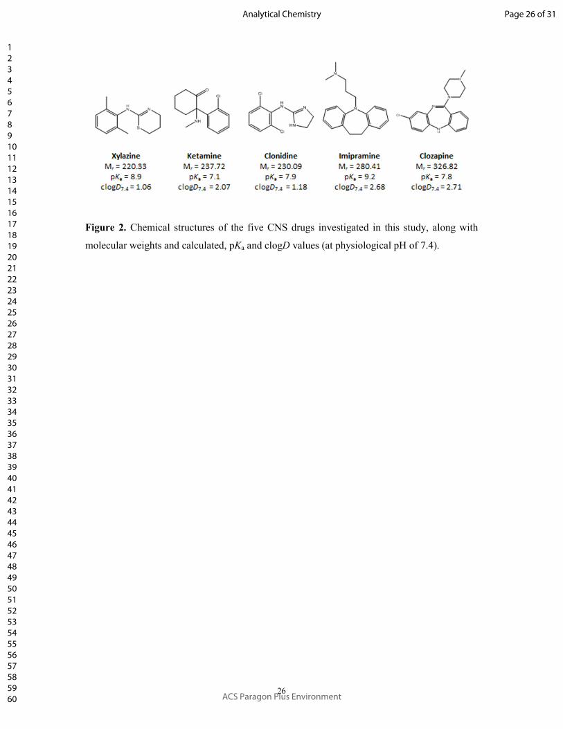
drugs, we selected five model compounds (Figure 2), representing different fields of
neuropharmacological application: xylazine (veterinary tranquilizer/anesthetic), ketamine
(drugs of abuse, but also promising as rapid and potent antidepressant), clonidine (anxiety
disorder and withdrawal syndrome therapeutic), imipramine (tricyclic antidepressant), and
clozapine (atypical antipsychotic treatment for schizophrenia). All selected compounds have
molecular weights in the range of 200-400 g/mol and are predominantly in their neutral forms
at physiological pH of blood and brain; consequently, the calculated distribution coefficients
clogD at pH 7.4 are between 1 and 3 (Figure 2), thus confirming the liphophilic character at
these conditions.
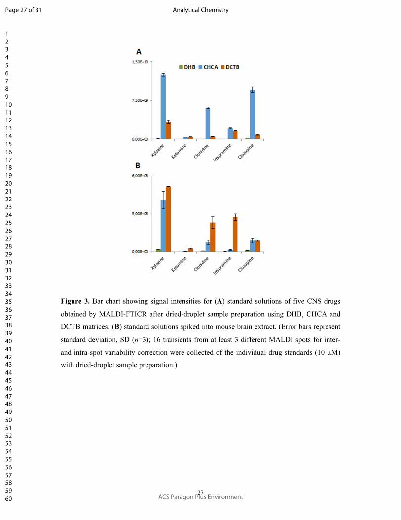
In the first set of experiments, we conducted MALDI-FTICR analyses. As can be seen
in Figure 3A, CHCA matrix provided the highest signal intensities. The same drugs mixed
with brain extract (obtained from isoflurane anesthetized mouse according to a modified
protocol91
) to indicate the presence of potentially suppressing endogenous compounds
exhibited the highest signal intensities when DCTB matrix was used (Figure 3B). This
provided strong evidence for considerably lower signal suppression from endogenous brain
Page 10 of 31
ACS Paragon Plus Environment
Analytical Chemistry
123456789101112131415161718192021222324252627282930313233343536373839404142434445464748495051525354555657585960

11
tissue-related compounds with nonpolar DCTB matrix as compared to CHCA and DHB. DHB
gave significantly lower signal intensities and also suffered from limitations of spatial
resolving power due to the large crystals formed during spraying (see SEM images in Figure
S-1, SI). In all further experiments, we therefore limited all further comparisons to CHCA
versus DCTB.
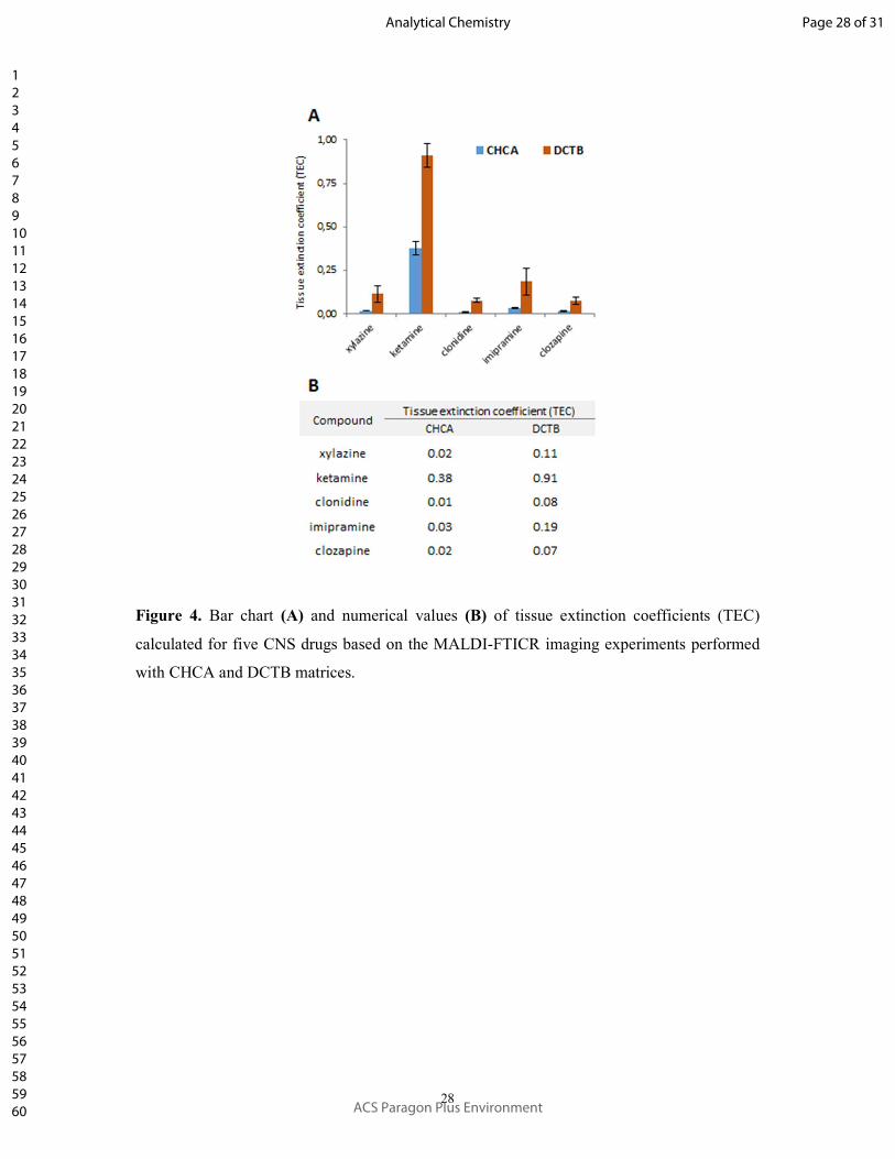
To further validate these findings for MALDI-MSI, we employed the tissue extinction
coefficient (TEC) technique developed by Stoeckli et al.,59
which was later expanded by
Hamm et al.39
Recently, Taylor et al. implemented TECs for systematic comparison of DESI
and MALDI (with CHCA matrix)-related ion suppression of uniformly deposited olanzapine
from heterogeneous transverse brain sections.60
For the TEC experiments, an equimolar
mixture of five CNS drugs was sprayed onto the glass slide containing the three thaw-
mounted negative control (isoflurane anesthetized) brain coronal sections, followed by
MALDI-FTICR-MSI (see Experimental Section). As reference, the off-tissue glass region of
interest was selected and imaged with the same experimental conditions. The final TEC
values were calculated based on the relationship TEC=IA,tissue/IA,ref, where IA are the averaged
analyte ion currents on tissue and reference areas, TEC=1 represents no suppression and
TEC=0 corresponds to total drug signal extinction. The results in Figure 4 show as much as 8-
fold less suppression with DCTB in comparison to CHCA and thus provide strong evidence
for matrix-dependent, tissue-specific ionization suppression. Furthermore, closer investigation
of the ion images of the five CNS drugs (Figure S-2, SI) revealed a heterogenous pattern of
drug signals suppression (white versus grey matter), which was further underpinned by
comparisons of mean mass spectra of the whole brain sections for the two matrices (see
Figure S-3, SI).
In conclusion, while the current results may not provide a complete explanation of the
improved performance of DCTB for MALDI imaging of the investigated CNS drugs from
brain sections in comparison to CHCA, some initial positive conclusions can be drawn.
DCTB clearly showed increased selectivity toward lipophilic CNS drugs as well as higher
“resistance” to brain-specific ionization suppression phenomena from endogenous compounds
such as salts, metabolites, lipids etc. as compared to CHCA. While the organic solvent used
for dissolving DTCB may provide more efficient extraction of neutral analytes in comparison
to CHCA, this does not explain the observed differences, because the lower signal
suppression levels of DCTB were observed not only during MALDI-MSI of tissue sections
but also from MALDI-MS analysis of bulk solutions containing the same interfering
substances from the brain tissue extract. Unfortunately, it was not possible to fully clarify
Page 11 of 31
ACS Paragon Plus Environment
Analytical Chemistry
123456789101112131415161718192021222324252627282930313233343536373839404142434445464748495051525354555657585960
12
whether the observed effects were related to favorable desorption and gas-phase protonation
of the investigated analytes or whether they result from physicochemical processes such as
more efficient incorporation of non-ionized lipophilic drugs and/or less efficient incorporation
of endogenous interfering metabolites/lipids into the non-polar DCTB matrix crystals. Further
work on this topic is ongoing in the present authors’ laboratory.
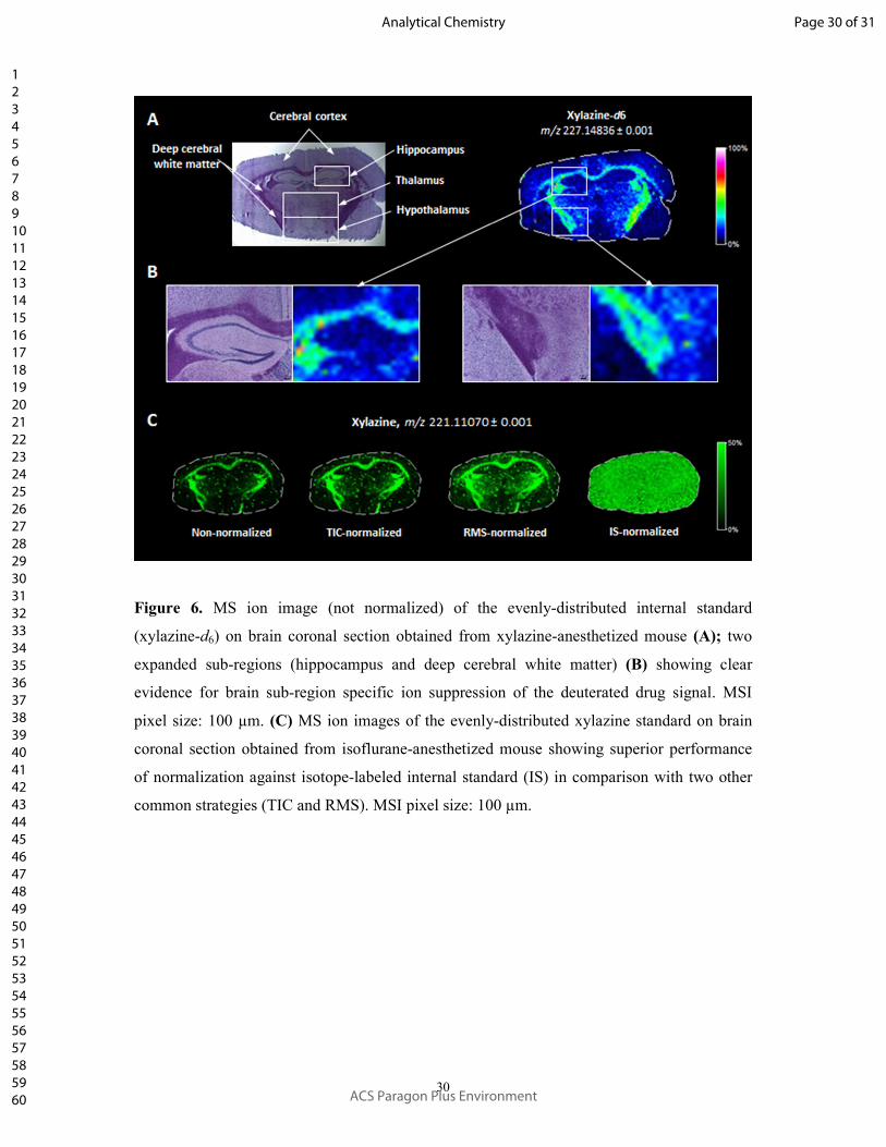
Xylazine Spatial Distribution in Mouse Brain Sections using Ultra-
high Resolution FTICR-MSI. The utility of DCTB for MALDI-MSI of lipophilic CNS
drugs was demonstrated using native brain tissues of drug-dosed animals (single dose
anesthesia with a mixture of xylazine/ketamine, see Experimental Section). The intense
signals detected from xylazine were assigned based on accurate mass measurements and on-
tissue collision-induced dissociation (CID) experiments by comparison to the drug standard
and previously reported fragmentation patterns (Figure S-4, SI).92
We then conducted two
MALDI-MSI experiments on consecutive coronal brain sections at spatial resolution of 100
µm. As illustrated in Figure 5, tissue examined with CHCA matrix showed virtually no
signals from xylazine, whereas tissue sprayed with DCTB matrix clearly revealed the spatial
distribution of the drug (normalized to isotope-labeled internal standard). Since the examined
tissue section clearly shows anatomical regions in the H&E stain of the brain, the MS ion
image of evenly sprayed xylazine-d6 provided additional evidence for regional ion
suppression, with the protonated xylazine molecule clearly less suppressed in the region of
deep cerebral white matter as compared to the cerebral cortex, hippocampus, thalamus and
hypothalamus (Figure 6).
Furthermore, to quantify the differences observed for the two matrices, single MSI
pixel mass spectra from representative regions of the highest xylazine abundance were
compared, showing more than 100-fold higher signal intensity obtained with DCTB over
CHCA. Moreover, when the average mass spectra from the whole examined tissue sections
were compared, >440-fold signal improvement was achieved with DCTB versus CHCA.
Importantly, the ultra-high resolving power of the Fourier-transform ion cyclotron resonance
mass spectrometer used here played a significant role in this comparison, since a DCTB-
derived interfering ion signal was visible only 0.003 u adjacent to protonated xylazine. This
interference had practical implications on the direct on-tissue quantification of xylazine (see
next section). In addition to xylazine, we also interrogated the MSI data sets for the second
administered anesthetic, ketamine as well as for potential metabolites of both drugs. As shown
Page 12 of 31
ACS Paragon Plus Environment
Analytical Chemistry
123456789101112131415161718192021222324252627282930313233343536373839404142434445464748495051525354555657585960
13
in the previous section, ketamine exhibited much lower ionization efficiency in comparison to
xylazine and was therefore not detected in the imaging experiments. Equally, we did not
observe any of the reported biotransformation products of xylazine92
or ketamine.93
On-tissue Quantification of Xylazine. Since most pharmaceutical MALDI-
MSI studies require absolute quantification of the investigated compounds directly from the
tissue surfaces, we also implemented the DCTB matrix for this purpose. Obtaining reliable
quantitative results with MALDI-MSI requires normalization to isotopically-labeled internal
standard (xylazine-d6) and matrix-matched calibration routines. The best results for the latter
are usually obtained by using either the sophisticated “mimetic tissue” model or a simpler
“on-tissue” approach.20
Here, we combined both strategies (the workflow is presented in
Figure S-5, SI). Briefly, rabbit brain was homogenized and snap-frozen in liquid nitrogen for
the best possible emulation of the original intact tissue morphology, as previously reported by
Jadoul et al.94
The prepared blocks of “unified brain” tissue (containing a representation of the
interfering endogenous compounds from different anatomical brain regions) were then cut
with the cryomicrotome and served as large and convenient blank sections for the fast “on-
tissue” spiking approach.
This approach generated linear relationships (coefficients of determination, R2 =
0.996) over a wide concentration range of the calibration curve (almost three orders of
magnitude, see Figure S-5, SI). The limits of detection (LOD, estimated from multiple blank
measurements) and limits of quantification LOQ, (based on a precision of the measurements
of 20% or better, and calculated bias values for the lowest concentrated calibration solution
within 80-120%) were 0.629 µg/g and 3.14 µg/g, respectively (assuming average brain
density of 1.027 g/cm3).
95 Finally, the average absolute concentration of xylazine across the
mouse brain coronal section illustrated in Figure 5-B was calculated to be 21.75 ± 0.41
µg/gtissue. The relatively large confidence interval of the mean was calculated based on the
standard deviation of all scans (scan number, n = 4255), averaged across the whole tissue
section; therefore, it reflects the heterogeneity of xylazine distribution in the brain section.
Page 13 of 31
ACS Paragon Plus Environment
Analytical Chemistry
123456789101112131415161718192021222324252627282930313233343536373839404142434445464748495051525354555657585960
14
CONCLUSIONS
The primary aim of this study was the application of the non-polar DCTB compound
as MALDI matrix for high sensitivity imaging of CNS drugs in mouse brain sections. Based
on five selected neuropharmaceuticals, which all fulfilled the requirements for crossing the
blood-brain barrier via transcellular passive diffusion (i.e., Lipinski’s rule of 5), we
demonstrated that DCTB can be successfully used for high sensitive MALDI mass spectral
imaging of these compounds from murine brain sections using an ultra-high resolution
Fourier-transform ion cyclotron resonance (FTICR) platform. We provided a theoretical and
experimental rationale for an additional proton-transfer ionization mechanism of the DCTB
matrix, even though DCTB is usually considered an electron-transfer matrix. We also
investigated tissue specific ion suppression for DCTB versus the conventional polar and
acidic CHCA matrix, based on tissue extinction coefficients (TEC). These experiments
showed multi-fold lower signal suppression for the drugs using DCTB than CHCA.
Furthermore, we applied DCTB for quantitative imaging of a commonly used veterinary
anesthetic drug, xylazine from mouse coronal brain sections. Compared to CHCA, the data
showed significant signal intensity improvements by as much as 100-fold for representative
single MSI pixels of two serial sections and more than 440-fold for an average mass spectrum
from the whole adjacent tissue sections. Finally, the application of tissue-matched
standardization allowed for excellent linearity of the calibration curve over a wide
concentration range with satisfactory precision.
The use of the DCTB matrix was not without challenges, however, including
difficulties linked to the low melting point (~130 °C versus ~249 °C for CHCA), i.e. limited
stability under high vacuum conditions (which was overcome by normalization to
homogenously sprayed internal standard) and the “volcano” type ablation profiles, which can
constitute a challenge for higher spatial resolutions than used here. We did not observe any
formation of adducts between DCTB and drugs as previously described by Lou et al.96
Our future work will focus on improving the DCTB matrix solubility and stability
under high vacuum conditions. Other work currently in progress includes a detailed
investigation of the tissue-specific ionization suppression of different drugs when using
different MALDI matrices, which will further improve the detection capabilities of MALDI-
MSI for other pharmaceutical drugs.
Page 14 of 31
ACS Paragon Plus Environment
Analytical Chemistry
123456789101112131415161718192021222324252627282930313233343536373839404142434445464748495051525354555657585960
15
ASSOCIATED CONTENT
Supporting Information is available free of charge on the ACS Publications website.
SEM of sprayed MALDI matrices; MS ion images and mean spectra from TEC
experiments; Mean mass spectra obtained from TEC experiments of mouse brain
coronal sections; on-tissue identification of xylazine; on-tissue quantification
workflow and calibration curves.
AUTHOR INFORMATION
Corresponding author:
*E-mail: [email protected]
Notes
The authors declare no competing financial interest.
ACKNOWLEDGMENTS
D.A.V. acknowledges research support by the German Research Foundation (FTICR-MS
Facility, INST 256/356-1). The authors thank Tim Salbert (ACD/Labs) for the use of the
Percepta software, Pascal Schorr (Humboldt University Berlin) for the rabbit brain
preparations, Sylvia Kuhn (Saarland University) for SEM measurements and Alexander
Grißmer (Saarland University Medical Center) for staining and light microscopy experiments.
Page 15 of 31
ACS Paragon Plus Environment
Analytical Chemistry
123456789101112131415161718192021222324252627282930313233343536373839404142434445464748495051525354555657585960
16
REFERENCES
(1) Pangalos, M. N.; Schechter, L. E.; Hurko, O. Drug Development for CNS Disorders:
Strategies for Balancing Risk and Reducing Attrition. Nat. Rev. Drug Discov. 2007, 6
(7), 521–532.
(2) Pardridge, W. M. Blood-Brain Barrier Delivery. Drug Discov. Today 2007, 12 (1–2),
54–61.
(3) Pardridge, W. M. Drug Targeting to the Brain. Pharm. Res. 2007, 24 (9), 1733–1744.
(4) Palmer, A. M.; Alavijeh, M. S. Foundation Review: Translational CNS Medicines
Research. Drug Discov. Today 2012, 17 (19–20), 1068–1078.
(5) Lipinski, C. A. Drug-like Properties and the Causes of Poor Solubility and Poor
Permeability. J. Pharmacol. Toxicol. Methods 2000, 44 (1), 235–249.
(6) Mikitsh, J. L.; Chacko, A. M. Pathways for Small Molecule Delivery to the Central
Nervous System across the Blood-Brain Barrier. Perspect. Medicin. Chem. 2014, No.
6, 11–24.
(7) Pardridge, W. M. Drug Transport across the Blood–Brain Barrier. J. Cereb. Blood
Flow Metab. 2012, 32 (11), 1959–1972.
(8) Lai, F.; Fadda, A. M.; Sinico, C. Liposomes for Brain Delivery. Expert Opin. Drug
Deliv. 2013, 10 (7), 1003–1022.
(9) Wohlfart, S.; Gelperina, S.; Kreuter, J. Transport of Drugs across the Blood-Brain
Barrier by Nanoparticles. J. Control. Release 2012, 161 (2), 264–273.
(10) Saraiva, C.; Praça, C.; Ferreira, R.; Santos, T.; Ferreira, L.; Bernardino, L.
Nanoparticle-Mediated Brain Drug Delivery: Overcoming Blood-Brain Barrier to Treat
Neurodegenerative Diseases. J. Control. Release 2016, 235, 34–47.
(11) Rudin, M.; Weissleder, R. Molecular Imaging in Drug Discovery and Development.
Nat. Rev. Drug Discov. 2003, 2 (2), 123–131.
(12) Willmann, J. K.; van Bruggen, N.; Dinkelborg, L. M.; Gambhir, S. S. Molecular
Imaging in Drug Development. Nat. Rev. Drug Discov. 2008, 7 (7), 591–607.
(13) Pellegatti, M.; Pagliarusco, S. Drug and Metabolite Concentrations in Tissues in
Relationship to Tissue Adverse Findings: A Review. Expert Opin. Drug Metab.
Toxicol. 2011, 7 (2), 137–146.
(14) Pardridge, W. M. CSF, Blood-Brain Barrier, and Brain Drug Delivery. Expert Opin.
Drug Deliv. 2016, 13 (7), 963–975.
Page 16 of 31
ACS Paragon Plus Environment
Analytical Chemistry
123456789101112131415161718192021222324252627282930313233343536373839404142434445464748495051525354555657585960
17
(15) Solon, E. G.; Schweitzer, A.; Stoeckli, M.; Prideaux, B. Autoradiography, MALDI-
MS, and SIMS-MS Imaging in Pharmaceutical Discovery and Development. AAPS J.
2010, 12 (1), 11–26.
(16) Cobice, D. F.; Goodwin, R. J. A.; Andren, P. E.; Nilsson, A.; Mackay, C. L.; Andrew,
R. Future Technology Insight: Mass Spectrometry Imaging as a Tool in Drug Research
and Development. Br. J. Pharmacol. 2015, 172 (13), 3266–3283.
(17) Sugiura, Y.; Setou, M. Imaging Mass Spectrometry for Visualization of Drug and
Endogenous Metabolite Distribution: Toward in Situ Pharmacometabolomes. J.
Neuroimmune Pharmacol. 2010, 5 (1), 31–43.
(18) Sun, N.; Walch, A. Qualitative and Quantitative Mass Spectrometry Imaging of Drugs
and Metabolites in Tissue at Therapeutic Levels. Histochem. Cell Biol. 2013, 140 (2),
93–104.
(19) Ellis, S. R.; Bruinen, A. L.; Heeren, R. M. A. A Critical Evaluation of the Current
State-of-the-Art in Quantitative Imaging Mass Spectrometry. Anal. Bioanal. Chem.
2014, 406 (5), 1275–1289.
(20) Rzagalinski, I.; Volmer, D. A. Quantification of Low Molecular Weight Compounds
by MALDI Imaging Mass Spectrometry – A Tutorial Review. Biochim. Biophys. Acta -
Proteins Proteomics 2017, 1865 (7), 726–739.
(21) Chughtai, K.; Heeren, R. M. A. Mass Spectrometric Imaging for Biomedical Tissue
Analysis. Chem. Rev. 2010, 110 (5), 3237–3277.
(22) Norris, J. L.; Caprioli, R. M. Analysis of Tissue Specimens by Matrix-Assisted Laser
Desorption/Ionization Imaging Mass Spectrometry in Biological and Clinical Research.
Chem. Rev. 2013, 113 (4), 2309–2342.
(23) Spengler, B. Mass Spectrometry Imaging of Biomolecular Information. Anal. Chem.
2015, 87 (1), 64–82.
(24) Bodzon‐Kulakowska, A.; Suder, P. Imaging Mass Spectrometry: Instrumentation,
Applications, and Combination with Other Visualization Techniques. Mass Spectrom.
Rev. 2016, 35 (1), 147–169.
(25) Palmer, A.; Trede, D.; Alexandrov, T. Where Imaging Mass Spectrometry Stands: Here
Are the Numbers. Metabolomics 2016, 12 (6), 107.
(26) Karlsson, O.; Hanrieder, J. Imaging Mass Spectrometry in Drug Development and
Toxicology. Arch. Toxicol. 2017, 91 (6), 2283–2294.
(27) Swales, J. G.; Hamm, G.; Clench, M. R.; Goodwin, R. J. A. Mass Spectrometry
Imaging and Its Application in Pharmaceutical Research and Development: A Concise
Page 17 of 31
ACS Paragon Plus Environment
Analytical Chemistry
123456789101112131415161718192021222324252627282930313233343536373839404142434445464748495051525354555657585960
18
Review. Int. J. Mass Spectrom. 2018, DOI: 10.1016/j.ijms.2018.02.007.
(28) Goodwin, R. J.; Pitt, A. R. Mass Spectrometry Imaging of Pharmacological
Compounds in Tissue Sections. Bioanalysis 2010, 2 (2), 279–293.
(29) Greer, T.; Sturm, R.; Li, L. Mass Spectrometry Imaging for Drugs and Metabolites. J.
Proteomics 2011, 74 (12), 2617–2631.
(30) Prideaux, B.; Stoeckli, M. Mass Spectrometry Imaging for Drug Distribution Studies.
J. Proteomics 2012, 75 (16), 4999–5013.
(31) Hochart, G.; Hamm, G.; Stauber, J. Label-Free MS Imaging from Drug Discovery to
Preclinical Development. Bioanalysis 2014, 6 (20), 2775–2788.
(32) Nilsson, A.; Goodwin, R. J. A.; Shariatgorji, M.; Vallianatou, T.; Webborn, P. J. H.;
Andren, P. E. Mass Spectrometry Imaging in Drug Development. Anal. Chem. 2015,
87 (3), 1437–1455.
(33) Shariatgorji, M.; Svenningsson, P.; Andrén, P. E. Mass Spectrometry Imaging, an
Emerging Technology in Neuropsychopharmacology. Neuropsychopharmacology
2014, 39 (1), 34–39.
(34) Hsieh, Y.; Casale, R.; Fukuda, E.; Chen, J.; Knemeyer, I.; Wingate, J.; Morrison, R.;
Korfmacher, W. Matrix-Assisted Laser Desorption/Ionization Imaging Mass
Spectrometry for Direct Measurement of Clozapine in Rat Brain Tissue. Rapid
Commun. Mass Spectrom. 2006, 20 (6), 965–972.
(35) Li, F.; Hsieh, Y.; Kang, L.; Sondey, C.; Lachowicz, J.; Korfmacher, W. A. MALDI-
Tandem Mass Spectrometry Imaging of Astemizole and Its Primary Metabolite in Rat
Brain Sections. Bioanalysis 2009, 1 (2), 299–307.
(36) Shin, Y. G.; Dong, T.; Chou, B.; Menghrajani, K. Determination of Loperamide in
Mdr1a/1b Knock-out Mouse Brain Tissue Using Matrix-Assisted Laser
Desorption/Ionization Mass Spectrometry and Comparison with Quantitative
Electrospray-Triple Quadrupole Mass Spectrometry Analysis. Arch. Pharm. Res. 2011,
34 (11), 1983–1988.
(37) Shanta, S. R.; Kim, T. Y.; Hong, J. H.; Lee, J. H.; Shin, C. Y.; Kim, K.-H.; Kim, Y. H.;
Kim, S. K.; Kim, K. P. A New Combination MALDI Matrix for Small Molecule
Analysis: Application to Imaging Mass Spectrometry for Drugs and Metabolites.
Analyst 2012, 137 (24), 5757.
(38) Källback, P.; Shariatgorji, M.; Nilsson, A.; Andrén, P. E. Novel Mass Spectrometry
Imaging Software Assisting Labeled Normalization and Quantitation of Drugs and
Neuropeptides Directly in Tissue Sections. J. Proteomics 2012, 75 (16), 4941–4951.
Page 18 of 31
ACS Paragon Plus Environment
Analytical Chemistry
123456789101112131415161718192021222324252627282930313233343536373839404142434445464748495051525354555657585960
19
(39) Hamm, G.; Bonnel, D.; Legouffe, R.; Pamelard, F.; Delbos, J. M.; Bouzom, F.;
Stauber, J. Quantitative Mass Spectrometry Imaging of Propranolol and Olanzapine
Using Tissue Extinction Calculation as Normalization Factor. J. Proteomics 2012, 75
(16), 4952–4961.
(40) Castellino, S.; Groseclose, M. R.; Sigafoos, J.; Wagner, D.; De Serres, M.; Polli, J. W.;
Romach, E.; Myer, J.; Hamilton, B. Central Nervous System Disposition and
Metabolism of Fosdevirine (GSK2248761), a Non-Nucleoside Reverse Transcriptase
Inhibitor: An LC-MS and Matrix-Assisted Laser Desorption/Ionization Imaging MS
Investigation into Central Nervous System Toxicity. Chem. Res. Toxicol. 2013, 26 (2),
241–251.
(41) Salphati, L.; Shahidi-Latham, S.; Quiason, C.; Barck, K.; Nishimura, M.; Alicke, B.;
Pang, J.; Carano, R. A.; Olivero, A. G.; Phillips, H. S. Distribution of the
Phosphatidylinositol 3-Kinase Inhibitors Pictilisib (GDC-0941) and GNE-317 in U87
and GS2 Intracranial Glioblastoma Models - Assessment by Matrix-Assisted Laser
Desorption Ionization Imaging. Drug Metab. Dispos. 2014, 42 (7), 1110–1116.
(42) Swales, J. G.; Tucker, J. W.; Strittmatter, N.; Nilsson, A.; Cobice, D.; Clench, M. R.;
Mackay, C. L.; Andren, P. E.; Takáts, Z.; Webborn, P. J. H.; et al. Mass Spectrometry
Imaging of Cassette-Dosed Drugs for Higher Throughput Pharmacokinetic and
Biodistribution Analysis. Anal. Chem. 2014, 86 (16), 8473–8480.
(43) Liu, X.; Ide, J. L.; Norton, I.; Marchionni, M. A.; Ebling, M. C.; Wang, L. Y.; Davis,
E.; Sauvageot, C. M.; Kesari, S.; Kellersberger, K. A.; et al. Molecular Imaging of
Drug Transit through the Blood-Brain Barrier with MALDI Mass Spectrometry
Imaging. Sci. Rep. 2013, 3, 1–7.
(44) Aikawa, H.; Hayashi, M.; Ryu, S.; Yamashita, M.; Ohtsuka, N.; Nishidate, M.;
Fujiwara, Y.; Hamada, A. Visualizing Spatial Distribution of Alectinib in Murine Brain
Using Quantitative Mass Spectrometry Imaging. Sci. Rep. 2016, 6 (October 2015),
23749.
(45) Tanaka, Y.; Hirata, M.; Shinonome, S.; Torii, M.; Nezasa, K. I.; Tanaka, H.
Distribution Analysis of Epertinib in Brain Metastasis of HER2-Positive Breast Cancer
by Imaging Mass Spectrometry and Prospect for Antitumor Activity. Sci. Rep. 2018, 8
(1), 1–12.
(46) Shariatgorji, M.; Källback, P.; Gustavsson, L.; Schintu, N.; Svenningsson, P.;
Goodwin, R. J. A.; Andren, P. E. Controlled-PH Tissue Cleanup Protocol for Signal
Enhancement of Small Molecule Drugs Analyzed by MALDI-MS Imaging. Anal.
Page 19 of 31
ACS Paragon Plus Environment
Analytical Chemistry
123456789101112131415161718192021222324252627282930313233343536373839404142434445464748495051525354555657585960
20
Chem. 2012, 84 (10), 4603–4607.
(47) Reich, R. F.; Cudzilo, K.; Levisky, J. A.; Yost, R. A. Quantitative MALDI-MSn
Analysis of Cocaine in the Autopsied Brain of a Human Cocaine User Employing a
Wide Isolation Window and Internal Standards. J. Am. Soc. Mass Spectrom. 2010, 21
(4), 564–571.
(48) Kuwayama, K.; Tsujikawa, K.; Miyaguchi, H.; Kanamori, T.; Iwata, Y. T.; Inoue, H.
Distribution Measurements of 3,4-Methylenedioxymethamphetamine and Its
Metabolites in Organs by Matrix-Assisted Laser Desorption/Ionization Imaging Mass
Spectrometry Using an Automatic Matrix Spraying System with an Air Brush and a
Turntable. Anal. Bioanal. Chem. 2012, 404 (6–7), 1823–1830.
(49) Pirman, D. A.; Reich, R. F.; Kiss, A.; Heeren, R. M. A.; Yost, R. A. Quantitative
MALDI Tandem Mass Spectrometric Imaging of Cocaine from Brain Tissue with a
Deuterated Internal Standard. Anal. Chem. 2013, 85 (2), 1081–1089.
(50) Kadar, H.; Le Douaron, G.; Amar, M.; Ferrié, L.; Figadère, B.; Touboul, D.; Brunelle,
A.; Raisman-Vozari, R. MALDI Mass Spectrometry Imaging of 1-Methyl-4-
Phenylpyridinium (MPP+) in Mouse Brain. Neurotox. Res. 2014, 25 (1), 135–145.
(51) Goodwin, R. J. A.; MacKay, C. L.; Nilsson, A.; Harrison, D. J.; Farde, L.; Andren, P.
E.; Iverson, S. L. Qualitative and Quantitative MALDI Imaging of the Positron
Emission Tomography Ligands Raclopride (a D2 Dopamine Antagonist) and SCH
23390 (a D1 Dopamine Antagonist) in Rat Brain Tissue Sections Using a Solvent-Free
Dry Matrix Application Method. Anal. Chem. 2011, 83 (24), 9694–9701.
(52) Vallianatou, T.; Strittmatter, N.; Nilsson, A.; Shariatgorji, M.; Hamm, G.; Pereira, M.;
Källback, P.; Svenningsson, P.; Karlgren, M.; Goodwin, R. J. A.; et al. A Mass
Spectrometry Imaging Approach for Investigating How Drug-Drug Interactions
Influence Drug Blood-Brain Barrier Permeability. Neuroimage 2018, 172 (September
2017), 808–816.
(53) McClure, R. A.; Chumbley, C. W.; Reyzer, M. L.; Wilson, K.; Caprioli, R. M.; Gore, J.
C.; Pham, W. Identification of Promethazine as an Amyloid-Binding Molecule Using a
Fluorescence High-Throughput Assay and MALDI Imaging Mass Spectrometry.
NeuroImage Clin. 2013, 2 (1), 620–629.
(54) Goodwin, R. J.; Webborn, P. J. Future Directions of Imaging MS in Pharmaceutical
R&D. Bioanalysis 2015, 7 (20), 2667–2673.
(55) Castellino, S.; Groseclose, M. R.; Wagner, D. MALDI Imaging Mass Spectrometry:
Bridging Biology and Chemistry in Drug Development. Bioanalysis 2011, 3 (21),
Page 20 of 31
ACS Paragon Plus Environment
Analytical Chemistry
123456789101112131415161718192021222324252627282930313233343536373839404142434445464748495051525354555657585960

21
2427–2441.
(56) Annesley, T. M. Ion Suppression in Mass Spectrometry. Clin. Chem. 2003, 49 (7),
1041–1044.
(57) Jessome, L. L.; Volmer, D. A. Ion Suppression: A Major Concern in Mass
Spectrometry. LCGC North Am. 2006, 24 (5), 498–510.
(58) Furey, A.; Moriarty, M.; Bane, V.; Kinsella, B.; Lehane, M. Ion Suppression; A
Critical Review on Causes, Evaluation, Prevention and Applications. Talanta 2013,
115, 104–122.
(59) Stoeckli, M.; Staab, D.; Schweitzer, A. Compound and Metabolite Distribution
Measured by MALDI Mass Spectrometric Imaging in Whole-Body Tissue Sections.
Int. J. Mass Spectrom. 2007, 260 (2–3), 195–202.
(60) Taylor, A.; Dexter, A.; Bunch, J. Exploring Ion Suppression in Mass Spectrometry
Imaging of a Heterogeneous Tissue. Anal. Chem. 2018, 90, 5637–5645.
(61) Seeley, E. H.; Oppenheimer, S. R.; Mi, D.; Chaurand, P.; Caprioli, R. M. Enhancement
of Protein Sensitivity for MALDI Imaging Mass Spectrometry After Chemical
Treatment of Tissue Sections. J. Am. Soc. Mass Spectrom. 2008, 19 (8), 1069–1077.
(62) Andersson, M.; Groseclose, M. R.; Deutch, A. Y.; Caprioli, R. M. Imaging Mass
Spectrometry of Proteins and Peptides: 3D Volume Reconstruction. Nat. Methods
2008, 5 (1), 101–108.
(63) Wang, H. Y. J.; Wu, H. W.; Tsai, P. J.; Liu, C. Bin. MALDI-Mass Spectrometry
Imaging of Desalted Rat Brain Sections Reveals Ischemia-Mediated Changes of Lipids.
Anal. Bioanal. Chem. 2012, 404 (1), 113–124.
(64) Yang, H.; Ji, W.; Guan, M.; Li, S.; Zhang, Y.; Zhao, Z.; Mao, L. Organic Washes of
Tissue Sections for Comprehensive Analysis of Small Molecule Metabolites by
MALDI MS Imaging of Rat Brain Following Status Epilepticus. Metabolomics 2018,
14 (4), 1–12.
(65) Song, X.; Luo, Z.; Li, X.; Li, T.; Wang, Z.; Sun, C.; Huang, L.; Xie, P.; Liu, X.; He, J.;
et al. In Situ Hydrogel Conditioning of Tissue Samples to Enhance the Drug’s
Sensitivity in Ambient Mass Spectrometry Imaging. Anal. Chem. 2017, 89 (12), 6318–
6323.
(66) Popkova, Y.; Schiller, J. Addition of CsCl Reduces Ion Suppression Effects in the
Matrix-Assisted Laser Desorption/Ionization Mass Spectra of
Triacylglycerol/Phosphatidylcholine Mixtures and Adipose Tissue Extracts. Rapid
Commun. Mass Spectrom. 2017, 31 (5), 411–418.
Page 21 of 31
ACS Paragon Plus Environment
Analytical Chemistry
123456789101112131415161718192021222324252627282930313233343536373839404142434445464748495051525354555657585960

22
(67) Griffiths, R. L.; Bunch, J. A Survey of Useful Salt Additives in Matrix-Assisted Laser
Desorption/Ionization Mass Spectrometry and Tandem Mass Spectrometry of Lipids:
Introducing Nitrates for Improved Analysis. Rapid Commun. Mass Spectrom. 2012, 26
(13), 1557–1566.
(68) Sugiyama, E.; Masaki, N.; Matsushita, S.; Setou, M. Ammonium Sulfate Improves
Detection of Hydrophilic Quaternary Ammonium Compounds through Decreased Ion
Suppression in Matrix-Assisted Laser Desorption/Ionization Imaging Mass
Spectrometry. Anal. Chem. 2015, 87 (22), 11176–11181.
(69) Wang, X.; Han, J.; Yang, J.; Pan, J.; Borchers, C. H. Matrix Coating Assisted by an
Electric Field (MCAEF) for Enhanced Tissue Imaging by MALDI-MS. Chem. Sci.
2015, 6 (1), 729–738.
(70) Huang, X.; Zhan, L.; Sun, J.; Xue, J.; Liu, H.; Xiong, C.; Nie, Z. Utilizing a Mini-
Humidifier to Deposit Matrix for MALDI Imaging. Anal. Chem. 2018, 90 (14), 8309–
8313.
(71) Dufresne, M.; Thomas, A.; Breault-Turcot, J.; Masson, J.-F.; Chaurand, P. Silver-
Assisted Laser Desorption Ionization For High Spatial Resolution Imaging Mass
Spectrometry of Olefins from Thin Tissue Sections. Anal. Chem. 2013, 85 (6), 3318–
3324.
(72) Esteve, C.; Tolner, E. A.; Shyti, R.; van den Maagdenberg, A. M. J. M.; McDonnell, L.
A. Mass Spectrometry Imaging of Amino Neurotransmitters: A Comparison of
Derivatization Methods and Application in Mouse Brain Tissue. Metabolomics 2016,
12 (2), 1–9.
(73) Lin, Z.; Cai, Z. Negative Ion Laser Desorption/Ionization Time-of-Flight Mass
Spectrometric Analysis of Small Molecules by Using Nanostructured Substrate as
Matrices. Mass Spectrom. Rev. 2018, No. February 2017, 1–16.
(74) Wang, H. Y. J.; Jackson, S. N.; McEuen, J.; Woods, A. S. Localization and Analyses of
Small Drug Molecules in Rat Brain Tissue Sections. Anal. Chem. 2005, 77 (20), 6682–
6686.
(75) Rzagalinski, I.; Hainz, N.; Meier, C.; Tschernig, T.; Volmer, D. A. MALDI Mass
Spectral Imaging of Bile Acids Observed as Deprotonated Molecules and Proton-
Bound Dimers from Mouse Liver Sections. J. Am. Soc. Mass Spectrom 2018, 29 (4),
711–722.
(76) Schramm, T.; Hester, A.; Klinkert, I.; Both, J. P.; Heeren, R. M. A.; Brunelle, A.;
Laprévote, O.; Desbenoit, N.; Robbe, M. F.; Stoeckli, M.; et al. ImzML - A Common
Page 22 of 31
ACS Paragon Plus Environment
Analytical Chemistry
123456789101112131415161718192021222324252627282930313233343536373839404142434445464748495051525354555657585960
23
Data Format for the Flexible Exchange and Processing of Mass Spectrometry Imaging
Data. J. Proteomics 2012, 75 (16), 5106–5110.
(77) Bokhart, M. T.; Nazari, M.; Garrard, K. P.; Muddiman, D. C. MSiReader v1.0:
Evolving Open-Source Mass Spectrometry Imaging Software for Targeted and
Untargeted Analyses. J. Am. Soc. Mass Spectrom. 2018, 29 (1), 8–16.
(78) Toomsalu, E.; Koppel, I. A.; Burk, P. Critical Test of Some Computational Chemistry
Methods for Prediction of Gas-Phase Acidities and Basicities. J. Chem. Theory
Comput. 2013, 9 (9), 3947–3958.
(79) Rayne, S.; Forest, K. Benchmarking Semiempirical, Hartree–Fock, DFT, and MP2
Methods against the Ionization Energies and Electron Affinities of Short-through
Long-Chain [n] Acenes and [n] Phenacenes. Can. J. Chem. 2016, 94 (3), 251–258.
(80) Frisch, M.; Trucks, G. W.; Schlegel, H. B.; Scuseria, G. E.; Robb, M. A.; Cheeseman,
J. R.; Scalmani, G.; Barone, V.; Mennucci, B.; Petersson, Ga. Gaussian 09, Revision
A.02,. Gaussian Inc., Wallingford CT 2016.
(81) Ulmer, L.; Mattay, J.; Torres-Garcia, H.; Luftmann, H. The Use of 2-[(2E)-3-(4-Tert-
Butylphenyl)-2- Methylprop-2-Enylidene]Malononitrile as a Matrix for Matrix-
Assisted Laser Desorption/Ionization Mass Spectrometry. Eur. J. Mass Spectrom.
2000, 6 (1), 49.
(82) Mizukado, J.; Sato, H.; Chen, L.; Suzuki, Y.; Yamane, S.; Aoyama, Y.; Suda, H. High-
Resolution MALDI-TOF MS Study on Analysis of Low-Molecular-Weight Products
from Photo-Oxidation of Poly(3-Hexylthiophene). J. Mass Spectrom. 2015, 50 (8),
1006–1012.
(83) Markov, V. Y.; Borschevsky, A. Y.; Sidorov, L. N. MALDI Mass Spectrometry of
Fullerene Derivatives. Int. J. Mass Spectrom. 2012, 325–327, 100–112.
(84) Bergman, N.; Thapper, A.; Styring, S.; Bergquist, J.; Shevchenko, D. Quantitative
Determination of the Ru(Bpy)3 2+ Cation in Photochemical Reactions by Matrix-
Assisted Laser Desorption/Ionization Time-of-Flight Mass Spectrometry. Anal.
Methods 2014, 6 (21), 8513–8518.
(85) Kumara, C.; Dass, A. Au329(SR)84 Nanomolecules: Compositional Assignment of the
76.3 KDa Plasmonic Faradaurates. Anal. Chem. 2014, 86 (9), 4227–4232.
(86) Wyatt, M. F.; Stein, B. K.; Brenton, A. G. Characterization of Various Analytes Using
Matrix Assisted Laser Desorption/Ionization Time of Flight Mass Spectrometry and 2-
[(2E)-3-(4-Tert-Butylphenyl)-2-Methylprop-2-Enylidene]Malononitrile Matrix. Anal.
Chem. 2006, 78 (1), 199–206.
Page 23 of 31
ACS Paragon Plus Environment
Analytical Chemistry
123456789101112131415161718192021222324252627282930313233343536373839404142434445464748495051525354555657585960

24
(87) Jaskolla, T. W.; Karas, M. Compelling Evidence for Lucky Survivor and Gas Phase
Protonation: The Unified MALDI Analyte Protonation Mechanism. J. Am. Soc. Mass
Spectrom. 2011, 22 (6), 976–988.
(88) Knochenmuss, R. Ion Formation Mechanisms in UV-MALDI. Analyst 2006, 131 (9),
966.
(89) Soltwisch, J.; Jaskolla, T. W.; Hillenkamp, F.; Karas, M.; Dreisewerd, K. Ion Yields in
UV-MALDI Mass Spectrometry as a Function of Excitation Laser Wavelength and
Optical and Physico-Chemical Properties of Classical and Halogen-Substituted
MALDI Matrixes. Anal. Chem. 2012, 84 (15), 6567–6576.
(90) Gabriel, S. J.; Pfeifer, D.; Schwarzinger, C.; Panne, U.; Weidner, S. M. Matrix-
Assisted Laser Desorption/Ionization Time-of-Flight Mass Spectrometric Imaging of
Synthetic Polymer Sample Spots Prepared Using Ionic Liquid Matrices. Rapid
Commun. Mass Spectrom. 2014, 28 (5), 489–498.
(91) Want, E. J.; Masson, P.; Michopoulos, F.; Wilson, I. D.; Theodoridis, G.; Plumb, R. S.;
Shockcor, J.; Loftus, N.; Holmes, E.; Nicholson, J. K. Global Metabolic Profiling of
Animal and Human Tissues via UPLC-MS. Nat. Protoc. 2013, 8 (1), 17–32.
(92) Meyer, G. M. J.; Maurer, H. H. Qualitative Metabolism Assessment and Toxicological
Detection of Xylazine, a Veterinary Tranquilizer and Drug of Abuse, in Rat and
Human Urine Using GC-MS, LC-MS n, and LC-HR-MS N. Anal. Bioanal. Chem.
2013, 405 (30), 9779–9789.
(93) Porpiglia, N.; Musile, G.; Bortolotti, F.; De Palo, E. F.; Tagliaro, F. Chiral Separation
and Determination of Ketamine and Norketamine in Hair by Capillary Electrophoresis.
Forensic Sci. Int. 2016, 266, 304–310.
(94) Jadoul, L.; Longuespée, R.; Noël, A.; De Pauw, E. A Spiked Tissue-Based Approach
for Quantification of Phosphatidylcholines in Brain Section by MALDI Mass
Spectrometry Imaging. Anal. Bioanal. Chem. 2015, 407 (8), 2095–2106.
(95) Barber, T. W.; Brockway, J. A.; Higgins, L. S. The Density of Tissues in and about the
Head. Acta Neurol. Scand. 1970, 46 (1), 85–92.
(96) Lou, X.; De Waal, B. F. M.; Van Dongen, J. L. J.; Vekemans, J. A. J. M.; Meijer, E.
W. A Pitfall of Using 2-[(2E)-3-(4-Tert-Butylphenyl)- 2-Methylprop-2-
Enylidene]Malononitrile as a Matrix in MALDI TOF MS: Chemical Adduction of
Matrix to Analyte Amino Groups. J. Mass Spectrom. 2010, 45 (10), 1195–1202.
Page 24 of 31
ACS Paragon Plus Environment
Analytical Chemistry
123456789101112131415161718192021222324252627282930313233343536373839404142434445464748495051525354555657585960

25
Figure 1. (A) Representative MALDI-FTICR mass spectrum of pure DCTB matrix after
dried-droplet sample preparation. (B) Computationally-derived proton affinities (PA) and
ionization energies (IE) for CHCA and DCTB matrices, as well the two CNS drugs (xylazine
and ketamine).
Page 25 of 31
ACS Paragon Plus Environment
Analytical Chemistry
123456789101112131415161718192021222324252627282930313233343536373839404142434445464748495051525354555657585960
26
Figure 2. Chemical structures of the five CNS drugs investigated in this study, along with
molecular weights and calculated, pKa and clogD values (at physiological pH of 7.4).
Page 26 of 31
ACS Paragon Plus Environment
Analytical Chemistry
123456789101112131415161718192021222324252627282930313233343536373839404142434445464748495051525354555657585960
27
Figure 3. Bar chart showing signal intensities for (A) standard solutions of five CNS drugs
obtained by MALDI-FTICR after dried-droplet sample preparation using DHB, CHCA and
DCTB matrices; (B) standard solutions spiked into mouse brain extract. (Error bars represent
standard deviation, SD (n=3); 16 transients from at least 3 different MALDI spots for inter-
and intra-spot variability correction were collected of the individual drug standards (10 µM)
with dried-droplet sample preparation.)
Page 27 of 31
ACS Paragon Plus Environment
Analytical Chemistry
123456789101112131415161718192021222324252627282930313233343536373839404142434445464748495051525354555657585960
28
Figure 4. Bar chart (A) and numerical values (B) of tissue extinction coefficients (TEC)
calculated for five CNS drugs based on the MALDI-FTICR imaging experiments performed
with CHCA and DCTB matrices.
Page 28 of 31
ACS Paragon Plus Environment
Analytical Chemistry
123456789101112131415161718192021222324252627282930313233343536373839404142434445464748495051525354555657585960

29
Figure 5. Spatial distribution of xylazine in mouse brain coronal sections obtained from
MALDI-FTICR imaging experiments performed on two consecutive sections with two
different MALDI matrices, CHCA (A) and DCTB (B), along with single MSI pixel mass
spectra extracted from representative regions of the highest abundance of xylazine detected
with CHCA (C) and DCTB (D). MSI pixel size: 100 µm. Scale bars: 2 mm.
Page 29 of 31
ACS Paragon Plus Environment
Analytical Chemistry
123456789101112131415161718192021222324252627282930313233343536373839404142434445464748495051525354555657585960
30
Figure 6. MS ion image (not normalized) of the evenly-distributed internal standard
(xylazine-d6) on brain coronal section obtained from xylazine-anesthetized mouse (A); two
expanded sub-regions (hippocampus and deep cerebral white matter) (B) showing clear
evidence for brain sub-region specific ion suppression of the deuterated drug signal. MSI
pixel size: 100 µm. (C) MS ion images of the evenly-distributed xylazine standard on brain
coronal section obtained from isoflurane-anesthetized mouse showing superior performance
of normalization against isotope-labeled internal standard (IS) in comparison with two other
common strategies (TIC and RMS). MSI pixel size: 100 µm.
Page 30 of 31
ACS Paragon Plus Environment
Analytical Chemistry
123456789101112131415161718192021222324252627282930313233343536373839404142434445464748495051525354555657585960

31
For Table of Content (TOC) only
Page 31 of 31
ACS Paragon Plus Environment
Analytical Chemistry
123456789101112131415161718192021222324252627282930313233343536373839404142434445464748495051525354555657585960


![PRACTICE OF POLYMER MALDI 11maldi.ch.pw.edu.pl/pomiary/Artykuly/liang-li/ocr/021-040_unity-reader.pdf · 2.2 PRACTICE OF POLYMER MALDI 11 MALDI sources [10]; these have also become](https://img.pdfslide.us/doc/110x75/5e20a425f916e801c369e9f7/practice-of-polymer-maldi-22-practice-of-polymer-maldi-11-maldi-sources-10-these.jpg)