Embed Size (px)
Citation preview

Torticollis

Do obstetric risk factors truly influence the etiopathogenesisof congenital muscular torticollis?
Abstract
Background Congenital muscular torticollis (CMT) is seen
in childhood and presents within months after birth. The
etiology remains unknown; however, medical textbooks
suggest trauma at birth as a main reason. The aim of this
study was to systematically describe obstetric and perinatal
outcomes in a population of children with a confirmed
congenital muscular torticollis diagnosis.
Materials and methods Children with a validated diagnosis
of congenital muscular torticollis born at Aarhus Univer-
sity Hospital from 2000 to 2014 were included in the study.
Information on perinatal, intrapartum and neonatal char-
acteristics were obtained from databases and from medical
records, and systematically described.
Results In this study, there were no differences in birth
characteristics in children with left- and right-sided torti-
collis, between boys and girls or between the conservatively
treated and the children who needed surgery. Most of the
children with congenital muscular torticollis in this study
were delivered at term without signs of birth complications
or trauma. None experienced moderate or severe asphyxia.
Conclusions The results of the present study suggests that
complicated birth or birth trauma may not be the main
cause of congenital muscular torticollis and point towards
intrauterine and prenatal reasons for its development.
Level of evidence according to OCEBM levels of evidence
working group 3
Keywords Child � Congenital muscular torticollis �Obstetric � Perinatal � Risk factors
Introduction
Torticollis is a clinical diagnosis where the sternocleido-
mastoid muscle (SCM) is shortened on the involved side,
leading to a lateral tilt towards the affected muscle and
contralateral rotation of the face and chin [1–3]. Several
obstetric and newborn risk factors have been proposed for
the development of CMT, including prolonged labor,
macrosomia, breech or other irregular fetal presentations
[4–6]. The theory of birth trauma proposes disruption of the
SCM muscle during the birth process [7], and medical text
books state that trauma at birth is associated with CMT
[8–10], although the true etiology remains unknown [7].
Recent research has proposed intrauterine risk factors
[7, 11, 12], but only a few larger studies [6, 13, 14] have
systematically collected information in an effort to describe
the etiology, and none have used systematically collected
obstetric outcomes.
The aim of this study was to describe obstetric outcomes
in a population of children with a confirmed diagnosis of
CMT
Materials and methods
This study was designed as an observational case study of
children referred to the Department of Children’s Ortho-
pedics (DCO) at Aarhus University Hospital (AUH)
Results
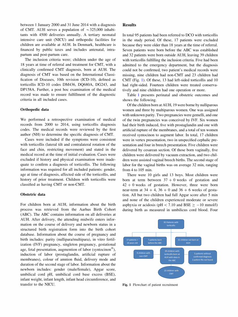
In total 95 patients had been referred to DCO with torticollis
in the study period. Of these, 17 patients were excluded
because they were older than 18 years at the time of referral.
Seven patients were born before the ABC was established
and 32 patients were born outside AUH, leaving 39 children
with torticollis fulfilling the inclusion criteria. Five had been
admitted to the emergency department, but the diagnosis
could not be confirmed, two patient’s medical records were
missing, nine children had non-CMT and 23 children had
CMT (Fig. 1). Of these, 13 had left-sided torticollis and 10
had right-sided. Fourteen children were treated conserva-
tively and nine children had one operation or more.
Table 1 presents perinatal and obstetric outcomes and
shows the following.
Of the children born at AUH, 19were borne by nulliparous
women and three by multiparous women. One was assigned
with unknown parity. Two pregnancies were gemelli, and one
of the twin pregnancies was conceived by IVF. Six women
had their birth induced, five with prostaglandin and one with
artificial rupture of the membranes, and a total of ten women
received syntocinon to augment labor. In total, 17 children
were in vertex presentation, two in unspecified cephalic pre-
sentation and four in breech presentation. Five children were
delivered by cesarean section. Of those born vaginally, five
children were delivered by vacuum extraction, and two chil-
dren were assisted vaginal breech births. The second stage of
labor for the vaginal births was on average 32 min, ranging
from 4 to 105 min.
There were 10 girls and 13 boys. Most children were
born at term between 37 ? 0 weeks of gestation and
42 ? 0 weeks of gestation. However, three were born
near-term at 34 ? 4, 36 ? 0 and 36 ? 6 weeks of gesta-
tion. All but two children had full Apgar score after 5 min
and none of the children experienced moderate or severe
asphyxia or acidosis (pH\ 7.10 and BSE C -10 mmol/l)
during birth as measured in umbilicus cord blood. Four
95 Pa�ents with tor�collis
17 pa�ents > 18 years old
7 pa�ents born before the ABC
32 children not born at AUH
39 children with tor�collis born at AUH with data in
the ABC
9 children with non-CMT
5 pa�ents without confirmed diagnosis
2 pa�ent file not found
23 children with CMT
Fig. 1 Flowchart of patient recruitment
between 1 January 2000 and 31 June 2014 with a diagnosisof CMT. AUH serves a population of *325,000 inhabi-tants with 4500 deliveries annually. A tertiary neonatal intensive care unit (NICU) and orthopedic facilities for children are available at AUH. In Denmark, healthcare is financed by public taxes and includes antenatal, intra-partum and post-partum care.
The inclusion criteria were; children under the age of 18 years at time of referral and treatment for CMT, with a clinically confirmed CMT diagnosis, born at AUH. The diagnosis of CMT was based on the International Classi-fication of Diseases, 10th revision (ICD-10), defined as torticollis ICD-10 codes DM436, DQ680A, DG243, and DP158A. Further, a post hoc examination of the medical record was made to ensure fulfillment of the diagnostic criteria in all included cases.
Orthopedic data
We performed a retrospective examination of medical records from 2000 to 2014, using torticollis diagnosis codes. The medical records were reviewed by the first author (NH) to determine the specific diagnosis of CMT.
Cases were included if the symptoms were consistent with torticollis (lateral tilt and contralateral rotation of the face and chin, restricting movement) and stated in the medical record at the time of initial evaluation. Cases were excluded if history and physical examination were inade-quate to confirm a diagnosis of torticollis. The following information was required for all included patients: gender, age at time of diagnosis, affected side of the torticollis, and history of prior treatment. Children with torticollis were classified as having CMT or non-CMT.
Obstetric data
For children born at AUH, information about the birth process was retrieved from the Aarhus Birth Cohort (ABC). The ABC contains information on all deliveries at AUH. After delivery, the attending midwife enters infor-mation on the course of delivery and newborn status in a structured birth registration form into the birth cohort database. Information about the course of pregnancy and birth includes: parity (nullipara/multipara), in vitro fertil-ization (IVF) pregnancy, singleton pregnancy, gestational age, fetal presentation, augmention of labor (syntocinon�), induction of labor (prostaglandin, artificial rupture of membranes), colour of amnion fluid, delivery mode and duration of the second stage of labor. Information about the newborn includes: gender (male/female), Apgar score, umbilical cord pH, umbilical cord base excess (BSE), infant weight, infant length, infant head circumference, and transfer to the NICU.

Table
1Perinatal
andobstetricoutcomein
23childrenwithadiagnosisoftorticollisborn
atAarhusUniversity
Hospital
from
2000to
2014
Pregnancy
Gender
IVF
pregnancy
Singleton
Parity
Gestational
age
Fetal
presentation
Augmention
(Syntocinon�)
Induction
Amnion
fluid
Delivery
mode
Durationofstage2
(min)
#1
Boy
No
Yes
Nulli
41?
2Vertex
Yes
No
Clear
Vaginal
38
#2
Girl
No
Yes
Nulli
39?
5Vertex
Yes
No
Thick
green
Vaginal
62
#3
Boy
No
Yes
Nulli
40?
1Vertex
No
Prostaglandin
Clear
Vaginal
40
#4
Boy
No
Yes
Nulli
40?
6Vertex
No
No
Light
green
Metal
cup
4
#5
Girl
No
Yes
Nulli
40?
0Cephalica
Yes
No
Clear
CSacute
–
#6
Boy
No
Yes
Nulli
40?
2Vertex
No
No
Green
Vaginal
54
#7
Girl
No
Yes
Nulli
40?
2Vertex
No
No
Clear
Vaginal
31
#8
Girl
No
Yes
Nulli
41?
3Vertex
Yes
No
Clear
Vaginal
19
#9
Girl
No
Yes
Nulli
40?
0Breech
No
Prostaglandin
Clear
Vaginal/
assisted
16
#10
Boy
No
Yes
Multi
39?
1Breech
No
No
Clear
CSplanned
–
#11
Boy
No
Yes
Nulli
41?
0Vertex
No
No
Clear
Vaginal
16
#12
Boy
No
Yes
Nulli
39?
3Vertex
No
No
Clear
Softrubber
cup
30
#13
Boy
No
Yes
Nulli
36?
6Vertex
No
No
Clear
Softrubber
cup
42
#14
Girl
No
No
Multi
37?
3Cephalica
No
No
Clear
CSplanned
–
#15
Boy
No
Yes
Nulli
40?
0Breech
Yes
Prostaglandin
Thick
green
Vaginal/
assisted
10
#16
Girl
No
Yes
Nulli
39?
6Vertex
Yes
Prostaglandin
Clear
Vaginal
19
#17
Girl
No
Yes
Nulli
42?
0Vertex
Yes
Prostaglandin
Light
green
Metal
cup
47
#18
Boy
Yes
No
Nulli
37?
0Vertex
Yes
No
Unknown
CSacute
–
#19
Boy
No
Yes
Nulli
34?
4Vertex
No
No
Clear
Vaginal
22
#20
Girl
No
Yes
Nulli
40?
0Vertex
No
No
Light
green
Vaginal
10
#21
Boy
No
Yes
Multi
38?
4Vertex
No
No
Clear
Vaginal
13
#22
Boy
No
Yes
Missing
36?
0Breech
Yes
No
Clear
CSacute
–
#23
Girl
No
Yes
Nulli
39?
6Vertex
Yes
Rupturesof
mem
branes
Clear
Metal
cup
105
CScesarean
section
aNotspecified

differences in birth characteristics in children with left- and
right-sided CMT, between boys and girls or between the
conservatively treated and the children who needed sur-
gery. The children were primarily born by nulliparous
women. Most were delivered without any trauma, seven
experienced an assisted delivery, either by vacuum
extraction or assisted breech birth, and with a mean second
stage of labor of 32 min. Complicated birth, as measured
by Apgar score, umbilical cord pH, and umbilical cord base
excess, indicated that none of the children suffered mod-
erate or severe asphyxia. Three initially had low Apgar
score but all children had normal scores after 10 min. Most
of the children with CMT in this study were delivered at
term without signs of birth complications or trauma and
none of the children could be classified as macrosomia.
Comparing our data with the existing literature we found
two studies suggesting that the side of the torticollis is
related to CMT either by intrauterine positioning [15]
(head positioning in utero can selectively injure the SCM
muscle) or due to delivering of the first shoulder [16].
Table 2 Neonatal and treatment outcomes in 23 children with a diagnosis of torticollis born at Aarhus University Hospital from 2000 to 2014
Number Apgar Scores
1 min/5 min
Umbilical
cord pH
Umbilical
cord BSE
Fetal
weight (g)
Fetal
length
(cm)
Fetal head
circumference (cm)
NICU Side of
torticollis
Treatment
#1 9/10 a 7.29 -5.7 3990 55 37 No Left Conservative
#2 6/8 v 7.19 -7.9 3280 52 33 Yes Right Operated 9 3
#3 10/10 a 7.22 -3 3640 52 38 No Right Conservative
#4 8/9 a 7.12 -5 3680 54 37 No Left Conservative
#5 10/10 Missing Missing 3910 53 35 No Left Operated 9 3
#6 10/10 v 7.31 -2 3400 52 33 No Left Conservative
#7 10/10 a 7.28 -8 3270 53 35 No Right Conservative
#8 9/10 a 7.27 -5 3160 51 34 No Left Conservative
#9 6/10 a 7.40 -4.6 3350 55 36 No Right Operated 9 1
#10 10/10 v 7.38 -1 3590 51 36 Yes Right Operated 9 2
#11 10/10 v 7.30 Missing 3850 52 32 No Left Conservative
#12 10/10 v 7.43 -5 2620 48 34 No Left Conservative
#13 10/10 a7.27 -8 3240 50 35 No Right Operated 9 1
#14 10/10 a 7.31 -1 2610 47 32 No Left Conservative
#15 9/10 v 7.36 Missing 2680 48 Missing No Left Conservative
#16 10/10 a 7.35 -2 2700 49 32 No Right Operated 9 2
#17 7/10 a 7.18 -6 3630 54 34 No Right Conservative
#18 10/10 Missing Missing 3515 51 36 No Right Conservative
#19 10/10 v 7.21 -3 2260 54 34 No Left Conservative
#20 9/10 v 7.19 -5.5 3560 50 34 No Left Operated 9 1
#21 10/10 7.41 -1 2850 41 35 No Left Conservative
#22 8/10 7.11 Missing 2330 49 33 Yes Left Operated 9 3
#23 5/10 v 7.22 -10 3830 51 36 Yes Right Operated 9 1
a arterial, v venous, NICU neonatal intensive care unit
children were admitted to the neonatal ward; one due to thick meconium-stained amnion fluid and respiratory dis-tress together with an affected Apgar score (6/1, 8/5), and two children after emergency cesarean section, due to asymmetric head shape and low birth weight, and the last child was referred for antibiotic treatment because of pro-longed rupture of membranes.
The median birth weight was 3259 g, ranging from 2260 to 3990 g. The median length was 49.2 cm, ranging from 41 to 55 cm and the median head circumference was 34 cm for the 23 children (one child’s head circumference was not mea-
sured). Table 2 presents neonatal and treatment outcomes in the 23 children with a diagnosis of CMT born at AUH.
Discussion
In this observational case study we systematically reviewed the obstetric outcomes in a population of children with a confirmed diagnosis of CMT and found that there were no

Information about first delivered shoulder, the final fetal
position being either left occipital or right occipital, was
not available in the ABC cohort. Moreover, ultrasound is
only done as routine in Denmark at around gestational
weeks 12 and 19 and could therefore not provide further
information about the specific fetal position during
pregnancy.
No previous studies with information about parity,
augmentation, or induction of labor are available. A case–
control study [7] examined gestational age and birth weight
for CMT patients, but not in relation to complicated birth
or developing CMT. Several have studied fetal presentation
and delivery mode related to CMT [7, 11, 12, 15–19]. To
our knowledge no former studies have examined the
duration of second stage of labor, Apgar score, umbilical
cord pH and base excess, infant head circumference and
transfer to the neonatal ward as indicators of complicated
birth.
In our data, we found a lower prevalence of breech
presentation in children with CMT, compared to earlier
studies [17]. Half of the children in breech presentation
were delivered vaginally and the other half by cesarean
section. In a former case control study with 178 patients,
Lee et al. [7] compared vaginal births with cesarean sec-
tions and found no difference in the clinical severity of
CMT according to the mode of delivery, suggesting that
prenatal factors most likely cause CMT due to the reduced
risk of birth trauma in cesarean sections. This is in accor-
dance with two case reports [12, 15], questioning the
traumatic vaginal breech delivery theory as being the
dominant pathophysiology behind CMT.
Other studies questioned trauma and difficult birth, and
instead pointed towards sequelae from intrauterine and
prenatal factors as the main cause of CMT. Stellwagen
et al. [11] found an association between torticollis and the
fetus being in the same intrauterine position for more than
6 weeks before delivery and Davids et al. [16] used mag-
netic resonance imaging (MRI) to observe the SCM muscle
in infants and found signals similar to those in compart-
ment syndrome.
In contrast, Hollier et al. [19] found a high frequency of
complications during pregnancy and delivery in their small
retrospective study of 11 patients, and Ho et al. [18] found
higher rates of assisted breech births, instrumental deliv-
eries and cesarean sections, which led them to conclude
that birth trauma appears to be the main etiological factor
in CMT. Suzuki et al. [17] suggested that stretching of the
SCM muscle during delivery may be a direct cause of
CMT. In our population only a few cases experienced
moderate birth trauma: mainly those delivered with vac-
uum extraction. In general, most of the studies [17–19] had
only examined fetal presentation and delivery mode, lacking more specific information from obstetric and neonatal medical records.
There seems to be a tendency towards intrauterine and prenatal cause, but the possibility of a perinatal trauma to the SCM muscles cannot be excluded. In our study, most of the children with CMT were born after uncomplicated deliveries, contradictory to the most common theories described in medical textbooks.
However, our study has some limitations. Primarily it only included 23 cases of CMT. One reason for this is that 90–95% of CMT resolves within a year by manual stretching and therefore the majority of these children are never referred to an orthopedic facility. It is therefore expected that children included in this study represent the more severe cases. Our study size was further limited by including only children born at AUH, as this was the only hospital where we were able to retrieve validated obstetric data. However, we believe the study sample to be representative.
We were unable to retrieve family history of CMT in the patient records. A potential genetic association may accu-mulate cases of CMT within families.
Finally, this study was a retrospective observational case study with prospective collected obstetric information. Retrospective studies are useful for studying diseases with low incidence. A large prospective cohort study with evaluation of fetal positioning during pregnancy with sys-tematic examination of the SCM in both the perinatal and the neonatal period using ultrasound or MRI, together with collection of obstetric information may provide further information of CMT etiology.
The results of the present study contribute to existing knowledge by pointing mainly towards intrauterine and prenatal reasons for developing CMT, and indicate that complicated birth and trauma may not be the main cause of CMT, even though this is stated in pediatric orthopedic textbooks.
Conflict of interest The authors declare that they have no conflict of interest.

References
1. Celayir AC (2000) Congenital muscular torticollis: early and
intensive treatment is critical. A prospective study. Pediatr Int
42:504–507
2. Do TT (2006) Congenital muscular torticollis: current concepts
and review of treatment. Curr Opin Pediatr 18:26–29
3. Cheng JC, Tang SP, Chen TM (1999) Sternocleidomastoid
pseudotumor and congenital muscular torticollis in infants: a
prospective study of 510 cases. J Pediatr 134:712–716
4. Cheng JC, Wong MW, Tang SP, Chen TM, Shum SL, Wong EM
(2001) Clinical determinants of the outcome of manual stretching
in the treatment of congenital muscular torticollis in infants. A
prospective study of eight hundred and twenty-one cases. J Bone
Joint Surg Am 83:679–687
5. Shim JS, Jang HP (2008) Operative treatment of congenital tor-
ticollis. J Bone Joint Surg Br 90:934–939
6. Lee YT, Cho SK, Yoon K, Shin HK, Kim E, Kim YB et al (2011)
Risk factors for intrauterine constraint are associated with ultra-
sonographically detected severe fibrosis in early congenital
muscular torticollis. J Pediatr Surg 46:514–519
7. Lee SJ, Han JD, Lee HB, Hwang JH, Kim SY, Park MC et al
(2011) Comparison of clinical severity of congenital muscular
torticollis based on the method of child birth. Ann Rehabil Med
35:641–647
8. Flynn JM, Wiesel SW (2011) Operative techniques in pediatric
orthopedics. Lippincott Williams & Wilkins, Philadelphia,
pp 367–374
9. Stadil F, Lund B, Nordling J (2003) Surgical compendium, vol 3.
New Nordic Publisher, Copenhagen, pp. 1590–1591
10. Lissauer T, Clayden G (eds) (2010) Illustrated textbook of pae-
diatrics, 1st edn. Elsevier Health Sciences, Amsterdam, p 368
11. Stellwagen L, Hubbard E, Chambers C, Jones KL (2008) Torti-
collis, facial asymmetry and plagiocephaly in normal newborns.
Arch Dis Child 93:827–831
12. Khalid S, Zaheer S, Wahab S, Siddiqui MA, Redhu N, Yusuf F
(2012) Fibromatosis colli: a case report. Oman Med J. 27(6)
13. Tatli B, Aydinli N, Caliskan M, Ozmen M, Bilir F, Acar G (2006)
Congenital muscular torticollis: evaluation and classification.
Pediatr Neurol 34:41–44
14. Cheng JC, Au AW (1994) Infantile torticollis: a review of 624
cases. J Pediatr Orthop 14:802–808
15. Sherer DM (1996) Spontaneous torticollis in a breech-presenting
fetus delivered by an atraumatic elective cesarean section: a case
and review of the literature. Am J Perinatol 13:305–307
16. Davids JR, Wenger DR, Mubarak SJ (1993) Congenital muscular
torticollis: sequela of intrauterine or perinatal compartment syn-
drome. J Pediatr Orthop 13:141–147
17. Suzuki S, Yamamuro T, Fujita A (1984) The aetiological rela-
tionship between congenital torticollis and obstetrical paralysis.
Int Orthop 8:175–181
18. Ho BC, Lee EH, Singh K (1999) Epidemiology, presentation and
management of congenital muscular torticollis. Singapore Med J
40:675–679
19. Hollier L, Kim J, Grayson BH, McCarthy JG (2000) Congenital
muscular torticollis and the associated craniofacial changes. Plast
Reconstr Surg 105:827–835

Spinal manual therapy in infants, children and
adolescents: A systematic review and meta-
analysis on treatment indication, technique
and outcomes
Abstract
Background
Studies on effectiveness and safety of specific spinal manual therapy (SMT) techniques in children, which distinguish between age groups, are lacking.
Objective
To conduct a systematic review of the evidence for effectiveness and harms of specific SMT techniques for infants, children and adolescents.
Methods
PubMed, Index to Chiropractic Literature, Embase, CINAHL and Cochrane Library were searched up to December 2017. Controlled studies, describing primary SMT treatment ininfants (<1 year) and children/adolescents (1–18 years), were included to determine effec-
tiveness. Controlled and observational studies and case reports were included to examine harms. One author screened titles and abstracts and two authors independently screened the full text of potentially eligible studies for inclusion. Two authors assessed risk of bias of included studies and quality of the body of evidence using the GRADE methodology. Data were described according to PRISMA guidelines and CONSORT and TIDieR checklists. If appropriate, random-effects meta-analysis was performed.
Results
Of the 1,236 identified studies, 26 studies were eligible. Infants and children/adolescents were treated for various (non-)musculoskeletal indications, hypothesized to be related to spinal joint dysfunction. Studies examining the same population, indication and treatment comparison were scarce. Due to very low quality evidence, it is uncertain whether gentle,
low-velocity mobilizations reduce complaints in infants with colic or torticollis, and whether
high-velocity, low-amplitude manipulations reduce complaints in children/adolescents with
autism, asthma, nocturnal enuresis, headache or idiopathic scoliosis. Five case reports
described severe harms after HVLA manipulations in four infants and one child. Mild, tran-
sient harms were reported after gentle spinal mobilizations in infants and children, and could
be interpreted as side effect of treatment.
Conclusions
Based on GRADE methodology, we found the evidence was of very low quality; this pre-
vented us from drawing conclusions about the effectiveness of specific SMT techniques in
infants, children and adolescents. Outcomes in the included studies were mostly parent or
patient-reported; studies did not report on intermediate outcomes to assess the effective-
ness of SMT techniques in relation to the hypothesized spinal dysfunction. Severe harms
were relatively scarce, poorly described and likely to be associated with underlying missed
pathology. Gentle, low-velocity spinal mobilizations seem to be a safe treatment technique
in infants, children and adolescents. We encourage future research to describe effective-
ness and safety of specific SMT techniques instead of SMT as a general treatment
approach.
Introduction
Is manual therapy effective in reducing or resolving complaints or symptoms in infants, chil-
dren or adolescents? Is it a safe therapeutic approach? Which specific manipulative techniques
are performed? In the field of pediatric care, these questions raise interest of healthcare profes-
sionals, parents and other stakeholders. Worldwide, manual therapy is performed in infants
(<1 year), children (1–11 years) and adolescents (12–18 years), by various healthcare profes-
sionals with different therapeutic backgrounds.[1, 2] They use different conceptual frame-
works regarding the relationship between symptoms and underlying spinal dysfunction.
Manipulative therapeutic techniques differ between professionals and health conditions, and
between infants and children/adolescents.[3–7] Distinctions in techniques are made between
high-velocity, low-amplitude (HVLA) manipulations[8] and low-velocity mobilizations which
can be performed to the full spine or to specific spinal segments. Moreover, treatment indica-
tions vary extensively. Infants and children are frequently treated for musculoskeletal condi-
tions, such as movement related complaints,[9] or non-musculoskeletal conditions, including
colic, otitis media and asthma.[1, 4, 10] Adolescents are mainly treated for musculoskeletal
conditions, such as scoliosis and headache.[1, 2, 4, 10] Non-musculoskeletal conditions as
treatment indication in children differs from manipulative treatment approaches in adults,
which are mainly focused on musculoskeletal conditions, such as headache, neck pain and low
back pain.[11–16]
Pediatric manual therapy and its safety has provoked debates and ethical challenges.[17–19]
Although several literature reviews summarize the evidence of manual therapy in children
with various indications,[2, 4, 5] systematic reviews describing effectiveness of specific manual
therapeutic treatment techniques, specified by treatment indication and age group, are lacking,
especially in the field of spinal manual therapy (SMT).[14] Hypotheses regarding underlying
spinal dysfunction that could be related to complaints in children differ between professionals,
Competing interests: The authors have declared
that no competing interests exist.
and the therapeutic approaches used within SMT overlap. This overlap impedes the interpreta-
tion of effects and harms of SMT. In addition, research concludes on SMT as a general treat-
ment approach instead of on the used techniques. A clear overview of the current state of the
evidence is therefore needed to assess the value of specific SMT techniques in different age
groups.[20, 21] This systematic review and meta-analysis of the literature provides a broad
overview of the evidence regarding the effectiveness and harms of specific SMT techniques in
infants, children and adolescents, related to specified treatment indication.
Methods
We report the results of our systematic review in accordance with the PRISMA guidelines.[22]
Prior to the study, the review protocol was registered at PROSPERO (CRD42017056031).
Literature search strategy
The following electronic databases were searched up to 20 December 2017: PubMed, Index to
Chiropractic Literature, Embase, CINAHL and Cochrane Library. The scientific literature was
systematically searched, combining key words related to “manual therapy” and key words
related to “children”. The search strategy for PubMed is shown in Fig 1. The searches in other
databases were consistent with this strategy. Reference checking of included articles was used
to identify potential studies that were missed with the initial search strategy (n = 1).
Definitions
To date, there is no international consensus on the specific definition of manual therapy in
pediatrics. Overall, three different therapeutic approaches can be recognized. First, chiroprac-
tic manual therapy, which uses high-velocity spinal manipulation or instrumented adjust-
ments using minimal forces (e.g. using an Activator).[1, 23, 24] It aims to influence the
nervous system, visceral functions and/or soft tissue tensions to correct segmental joint dys-
function.[18, 25, 26] Besides spinal manipulative therapy, chiropractic manual therapy incor-
porates additional therapies, such as soft tissue massage, nutritional counseling and exercise.
[27] Second, osteopathic manual therapy, which follows a similar line of reasoning, but also
intends to maintain or restore the flow of body fluids and to support homeostasis of the body.
[26, 28] Third, spinal manual therapy (SMT), which relies on segmental, single spinal joint
low-force oscillating mobilizations and HVLA manipulations,[8] focuses on the biomechanical
aspect of spinal dysfunction by eliciting neurological, physiological and/or muscular changes.
[29]
SMT techniques are integrated in all these treatment approaches, but conclusions on effec-
tiveness and safety are mainly given on treatment approach instead of treatment technique.
Hence, in this systematic review we focused on specific treatment techniques instead of SMT
as a general treatment approach.
In our systematic review, manual therapeutic interventions in which treatment techniques
were primarily performed on the full spine or on specific spinal segments, by any healthcare
professional, were indicated as SMT. We made a distinction between two main SMT tech-
niques: manipulation and mobilization. Manipulation was described as a HVLA or low-veloc-
ity thrust, resulting in a mechanical response of articular surface separation and a cracking
sound, which is also defined as cavitation in the affected joint.[8] Mobilization was described
as low-velocity, low-amplitude oscillating spinal joint play, without a thrust and without cavi-
tation. Infants were defined as those aged between 0 to 12 months; children were defined
being between 1 and 11 years; adolescents as being between 12 and 18 years. Treatment indica-
tions were categorized as musculoskeletal or non-musculoskeletal conditions. Hypothesized

dysfunction could be postulated to have had a primarily biomechanical, neuroreflectory or
physiological origin in the spine or could be described as dysfunction of the whole body, such
as disturbed flow of body fluids, myofascial, visceral or parietal bone problems. Treatment out-
comes were defined as patient- or parent-reported outcomes, such as symptoms (e.g. asymme-
try), behavior (e.g. crying), perceived effect, and quality of life and/or as intermediate
outcomes, which were related to therapist-reported impairment or function, such as asymme-
try, spinal mobility, spinal dysfunction, or performance. Harms were also interpreted as a
treatment outcome and were classified as; mild (transient side effect, lasting <24 hours), mod-
erate (requiring medical and/or general practitioner treatment) and severe (requiring hospital
treatment or adverse event; life threatening situation or death).[30]
Fig 1. Flowchart search strategy.
https://doi.org/10.1371/journal.pone.0218940.g001

Selection procedure and criteria for eligibility
The initial search was performed by the primary author (FD). All studies were collected using
EndNote, an online library system, which enabled us to remove duplicates. Screening of titles
and abstracts was performed by one author (FD) using predefined eligibility criteria (S1
Table). Controlled studies were included to investigate effectiveness and harms. Observational
studies and case reports were included to investigate harms.[31, 32] Subsequently, two authors
(FD, TH) independently reviewed the full text of potentially relevant articles for eligibility. Dis-
crepancies were discussed with all authors until consensus was reached, and eligible studies
were included for an in-depth review.
Assessment of risk of bias of individual studies
The assessment of risk of bias was done independently by two authors. Risk of bias of con-
trolled studies was assessed using the Cochrane Risk of Bias tool, focusing on selection-, per-
formance-, detection-, attrition- and reporting bias[33] by FD and JBS. Observational studies
were assessed with the Item Bank for Assessing Risk of Bias and Confounding for Observa-
tional Studies of Interventions or Exposures (RTI Item Bank)[34] by FD and TH, focusing on
selection-, performance-, detection-, attrition- and reporting bias, and confounding. Risk of
bias of case reports was assessed using the JBI Critical Appraisal Checklist for Case Reports
[35] by FD and JBS.
Data extraction and analysis
Data extraction was performed by FD using a Summary of Findings table, and thereafter
checked by TH in a random sample of 8 studies. Outcomes of effectiveness and harms were
described separately. The CONSORT checklist[36] in conjunction with the TIDieR checklist
[37] were used to describe the extracted data from controlled studies focusing on study popula-
tion, treatment indication, hypothesized dysfunction, specific SMT treatment technique and
outcomes. If appropriate, study outcomes were pooled. For random effects meta-analysis,
outcomes of controlled studies were transformed to standardized mean differences between
baseline and follow-up according to Cochrane recommendations.[33] Meta-analysis was per-
formed when two or more studies described a similar intervention and comparable control
treatment, and used a similar study population regarding condition and age. If appropriate,
intervention groups (�2 groups) were combined into a single group according to the
Cochrane Handbook. Statistical heterogeneity of the intervention effect was assessed using the
I2 statistic (>50% indicates high heterogeneity).[33] All analyses were conducted using Stata
Software, version 12.0 (Stata Inc., College Station, Texas). If studies were not similar, meta-
analysis was not considered appropriate, and findings were narratively reported. Data extrac-
tion to describe harms detailed treatment indication, specific SMT treatment technique and
the reported harm.
Assessment of quality of body of evidence
Quality of the body of evidence related to effectiveness was assessed using the Grading Recom-
mendations Assessment, Development and Evaluation (GRADE) criteria.[38, 39] Each out-
come was assessed in the previously specified age group and treatment indication using five
criteria: 1) risk of bias,[40] 2) inconsistency,[41] 3) indirectness,[42] 4) imprecision[43] and 5)
publication bias.[44] The assessment using GRADE was based on data from the assessment of
risk of bias and the data extraction process. The completion of the GRADE tables was done by
FD. The quality of the body of evidence was assigned as high, moderate, low or very low

(Box 1) and described according to Cochrane recommendations.[45] Randomized controlled
studies were considered high quality evidence and were downgraded by one level for serious
concerns and by two levels for very serious concerns.[31, 46] Non-randomized controlled
studies were automatically downgraded for limitations in the study design. They were further
downgraded for any concerns in the five grading criteria. If the number of studies per specific
age group, intervention and outcome was limited, inconsistency could not be graded and was
interpreted as ‘unknown’.[47] For each comparison and outcome measure, a GRADE table
was completed. Because of the varying designs of studies that solely described harms of SMT,
GRADE was not used; instead, results were reported narratively.
Results
Electronic database searching identified 1,236 articles. After removing duplicates, 1,165 rec-
ords were screened on title and abstract. A total of 1,102 records were excluded because of inel-
igible intervention, study design or study population. For the remaining 63 articles, eligibility
was assessed based on full-text; 38 were excluded because of study population (n = 5), study
design (n = 17), outcomes (n = 8) or the intervention could not be described as SMT (n = 8)
(S2 Table); reference checking added one study (Fig 1). In total 26 studies were included; 12
controlled trials, of which 10 were randomized controlled trials,[48–59] 9 observational stud-
ies[60–68], and 5 case reports.[69–73]
Methodological limitations of controlled studies were related to unclear allocation conceal-
ment, partial or no blinding of participants and personnel, and incomplete outcome reporting.
Limitations of observational studies were related to performance, detection and attrition bias,
and selective outcome reporting. Limitations of case reports were lack of detail or unclear
description of the intervention or treatment procedure. Outcomes of the quality assessments
are presented in S3 and S4 Tables.
Effectiveness
Study characteristics on treatment indication, hypothesized dysfunction, treatment technique
and outcomes of the included 12 controlled studies are shown in Table 1. In the studies involv-
ing infants (n = 5), interventions consisted of low-force, gentle, light fingertip spinal mobiliza-
tions. In studies involving children/adolescents (n = 7), HVLA thrust spinal manipulations
Box 1. GRADE levels describing the quality of the body of evidence(39)
GRADE levels
High: Research provides a very good indication of the likely effect. The likelihood that
the effect will be substantially different is low.
Moderate: Research provides a good indication of the likely effect. The likelihood that
the effect will be substantially different is moderate.
Low: Research provides some indication of the likely effect. The likelihood that the effect
will be substantially different is high.
Very low: Research does not provide a reliable indication of the likely effect. The likeli-
hood that the effect will be substantially different is very high.

Table 1. Treatment indication, hypothesized dysfunction, treatment technique, outcome measures and outcomes of controlled studies (n = 12) on effectiveness of
SMT in infants, children and adolescents.
Studies involving infants
Treatment
indication
Authors Study
population
(age)
Hypothesized
dysfunction
Intervention
(IV)
Outcome
measures
Comparator
(C)
Outcomes Risk of
bias�GRADE��
Colic
(N-MSK)
Olafsdottir
et al., 2001
[49]
86 infants
(3–9 weeks)
Spinal joint
dysfunction
Spinal
mobilizations using
light fingertip
pressure,
performed by a
chiropractor
Crying hours/
day after 8
days
No treatment
(infants were just
held)
Both groups
decreased crying
hours/day (IV: -2
(SD:2.1), C: -2.3 (SD:
2.2)). No significant
difference between
groups (p:0.37).
Moderate
Very low
quality of
evidence
Colic
(N-MSK)
Miller et al.,
2012 [50]
104 infants
(<8 weeks)
Not described Spinal low-force
mobilizations (1
blinded group (IV),
1 not-blinded
group (IV-nb)),
performed by a
chiropractor
Crying hours/
day after 10
days
No treatment
(infants were not
touched)
Both groups
decreased crying
hours/day (IV: -2.4
(SD:2.5), IV-nb: -2.8
(SD:2.2), C: -1.0
(SD:1.6)). Significant
(p<0.05) decrease
(-1.4) in IV group
compared to no
treatment.
Moderate
Colic
(N-MSK)
Browning &
Miller, 2008
[48]
43 infants
(<8 weeks)
Not described Spinal low-force
mobilizations,
performed by a
chiropractor
Crying hours/
day after 14
days
Occipito-sacral
decompression
Both groups
decreased crying
hours/day (IV: -2.1
(SD:2.2), C: -2.0
(SD:1.4)). No
significant difference
between groups
(p:0.85).
Moderate
Very low
quality of
evidence
Colic
(N-MSK)
Wiberg
et al., 1999
[51]
50 infants
(2–10
weeks)
Spinal joint
dysfunction
Spinal
mobilizations using
light fingertip
pressure,
performed by a
chiropractor
Crying hours/
day after 14
days
Dimethicone
medication
Both groups
decreased crying
hours/day (IV: -2.4
(SD:0.4), C: -1.0
(SD:0.6)). Significant
decrease of crying
hours (-1.7 hours/
day) in IV group
compared to
medication
(p = 0.04).
High
Torticollis
(MSK)
Haugen
et al., 2010
[52]
32 infants
(3–6
months)
Upper cervical
dysfunction
Spinal low-force
mobilizations by a
manual therapist
and pediatric
physical therapy
Change in
torticollis after
8 weeks
Pediatric physical
therapy
In both groups
torticollis positively
changed (IV: 80%
improvement, C:
81.3%). No
significant difference
between groups
(p:0.85).
Moderate Very low
quality of
evidence
Studies involving children and/or adolescents
Treatment
indication
Authors Study
population
(age)
Hypothesized
dysfunction
Intervention
(IV)
Outcome
measures
Comparator
(C)
Outcomes Risk of
bias�GRADE��
(Continued)

Table 1. (Continued)
Asthma
(N-MSK)
Balon et al.,
1998 [53]
91 children
(7–16 years)
Spinal joint
dysfunction
Spinal HVLA
manipulations,
performed by a
chiropractor
Peakflow
(FEV1),
symptoms,
medication
use and
quality of life
after 16 weeks
Low-velocity,
low-amplitude
push in gluteal
and scapulae
region
Both groups showed
small increases in
peakflow (IV: 103.6%
(SD:13.7), C: 104.3%
(SD:13.3)),
improvement in
symptoms and
quality of life and
decrease in
medication use.
No significant
differences between
groups (p:0.82).
High
Very low
quality of
evidence
Asthma
(N-MSK)
Bronfort
et al., 2001
[54]
36 children
(6–17 years)
Spinal joint
dysfunction
Spinal HVLA
manipulations,
performed by a
chiropractor, and
standard medical
treatment
Peakflow
(FEV1),
medication
use and
quality of life
after 12 weeks
Light gentle
spinal pressure,
without a thrust,
standard medical
treatment
Little insignificant
increase in peakflow
and quality of life and
decrease in
medication use in
intervention group.
Control group
outcomes not
reported. Groups
could not be
compared.
NA
Autism
(N-MSK)
Khorsid
et al., 2006
[56]
14 children
(age not
specified)
Not described Upper cervical
manipulations,
using the Atlas
Orthogonal,
performed by a
chiropractor
Autism related
symptoms
after 3 months
Diversified
technique SMT
on the full spine
Both groups
decreased in
symptoms (IV: -32%,
C:-19%). No
significant difference
between groups (p-
value not reported).
High Very low
quality of
evidence
Headache
(MSK)
Borusiak
et al., 2009
[55]
56 children
(7–15 years)
Cervical joint
dysfunction
Cervical HVLA
manipulation,
performed by a
manual therapist
Headache
duration
(hours) and
intensity (VAS
scale) after 2
months
Light touch of
spinal segments
Both groups
decreased in
symptoms (duration
IV:-7.5, C:-6.6;
intensity IV:-0.3,
C:0.1). No significant
differences between
groups (p>0.05).
Moderate Very low
quality of
evidence
Nocturnal
enuresis
(N-MSK)
Reed et al.,
1994 [57]
46 children
(5–13 years)
Spinal joint
dysfunction
HVLA
manipulations,
performed by a
chiropractor
Frequency of
bed wetting
after 12 weeks
Instrumented
adjustment using
an Activator on
the thoracic area
Intervention group
decreased in
frequency (IV:-1.2%
(SD:2.2), C:+17.9%
(SD:46.1%). No
significant difference
between groups
(p:0.07).
Moderate Very low
quality of
evidence
Idiopathic
scoliosis
(MSK)
Swierkosz &
Nowak,
2015 [58]
35
adolescents
(15–18
years)
Spinal joint
dysfunction
Lower lumbar
segmental
mobilizations and
traction, performed
by a physical
therapist
Back pain and
quality of life
after 3 weeks
No treatment Pain decreased and
physical health
related quality of life
increased (p<0.001)
within IV group. No
between group
comparisons were
reported.
NA Very low
quality of
evidence
(Continued)

were most frequently used (n = 6). Control interventions consisted of no treatment (n = 3),
sham treatment (n = 4) or other treatments (n = 5), such as physical therapy, medication and
manual therapy using the drop mechanism (Table 1).
Effectiveness of SMT techniques in infants. The review included five studies evaluating
SMT techniques in infants. Four studies included infants with colic [48–51] and one study
infants with torticollis.[52] Outcomes are presented in Table 1.
Infants with colic
Two studies compared SMT to no treatment.[49, 50] Miller et al. compared a blinded treat-
ment group (n = 35), non-blinded treatment group (n = 33) and a non-treatment group
(n = 34) and found that crying hours significantly decreased (p<0.05) with 1.5 hours/day after
10 days between blinded treatment and non-treatment.[50] Olafsdottir et al. showed no signif-
icant differences between the SMT (n = 46) and control group (n = 34) in decrease of crying
hours/day (-2 and -2.3, respectively) after 8 days.[49] Before meta-analysis, the two interven-
tion groups of Miller et al. were combined into one single intervention group. Analysis of the
overall pooled effect of SMT versus no treatment on crying hours/day was -0.33 (95% CI: -0.12
to 0.59; I2: 89.1%, p:0.484). Two studies compared SMT to other treatments.[48, 51] Browning
& Miller found a decrease in crying hours/day of 2.1 hours after SMT (n = 22) and 2.0 hours
after occipitosacral decompression (n = 21) 14 days post-treatment. Groups differed not signif-
icantly.[48] Wiberg et al. compared SMT (n = 25) to daily dimethicone medication (n = 25)
and found a significant decrease in crying hours/day in favor of the SMT group (-2.4 vs. -1.0,
p = 0.04).[51] No meta-analysis could be performed, due to incomparability of the control
treatments. Because of very low quality evidence (serious risk of bias, very serious inconsis-
tency, serious indirectness, serious imprecision) we are uncertain whether SMT consisting of
spinal mobilizations reduces crying hours/day in infants with colic.
Infants with torticollis
Haugen et al. compared pediatric physical therapy combined with SMT (n = 16) to pediat-
ric physical therapy alone (n = 16) on change in torticollis and cervical mobility, and found no
significant differences (SMT improved 80%, pediatric physical therapy alone improved
81.3%).[52] Because of very low quality evidence (unknown inconsistency, very serious impre-
cision) we are uncertain about the effect of SMT consisting of spinal mobilizations on change
of torticollis and increased cervical mobility in infants.
Effectiveness of SMT techniques in children/adolescents. Seven studies investigated the
effectiveness of SMT in children and/or adolescents (Table 1).[53–59]
Children/adolescents with asthma
Two studies compared SMT to sham treatment on lung function and asthma related symp-
toms in children.[53, 54] Balon et al. compared spinal HVLA manipulation (n = 38) to sham
Table 1. (Continued)
Grip
strength-
ening
(MSK)
Botelho &
Andrade,
2012 [59]
18 judo
athletes
Stimulate
nerve
innervations
Cervical HVLA
manipulations,
performed by a
chiropractor
Grip strength SMT using the
head piece drop
mechanism
Significantly better
grip strength
(p<0.05) in IV
(+13.7% mean left/
right hand)
compared to C (+5%)
(p:0.0025).
Moderate Very low
quality of
evidence
IV: Intervention group, C: Control group, MSK: musculoskeletal, N-MSK: non-musculoskeletal, SMT: spinal manual therapy, HVLA: high-velocity, low-amplitude,
FEV1: forced expiratory volume at the end of the first second of expiration
� Risk of bias table is shown in Table A in S3 Table
�� Detailed information about the GRADE assessment (GRADE tables) are presented in S4 Table
https://doi.org/10.1371/journal.pone.0218940.t001

treatment with low-velocity, low-amplitude push in the gluteal and scapulae region (n = 42).
After 16 weeks, lung function (+103.6% after SMT vs. +104.3% after sham treatment), quality
of life and reduction in medication were not significantly different between groups.[53] Bron-
fort et al. compared HVLA spinal manipulations (n = 24) to light gentle manual pressure
(sham treatment) to the spine (n = 12), and found no significant difference between groups in
lung function, quality of life and medication use.[54] No meta-analysis could be performed,
because Bronfort et al. only reported data of outcomes of the intervention group. We contacted
the author, but did not get a response. Because of very low quality evidence (serious risk of
bias, serious inconsistency, very serious imprecision) we are uncertain whether SMT consist-
ing of HVLA manipulations improves lung function in children/adolescents with asthma.
Children/adolescents with autism
Khorshid et al. compared upper cervical SMT (n = 7) to full spine diversified care (n = 7)
on autism related symptoms. No significant differences between groups were found (32%
improvement after SMT, 19% after diversified care).[56] Because of very low quality evidence
(serious risk of bias, unknown inconsistency, very serious imprecision) there is uncertainty
about the effect of SMT consisting of upper cervical manipulations on reducing autism related
symptoms in children/adolescents with autism.
Children/adolescents with headache
Borusiak et al. compared cervical HVLA manipulation (n = 28) to light touch of spinal seg-
ments as sham treatment (n = 28) on headache related symptoms (e.g. days with headache,
duration, intensity) and showed no significant differences after 2 months.[55] Outcomes of
HVLA manipulation versus sham treatment were; days with headache -9.7% vs. -9.4%, dura-
tion (hours) -7.5% vs. -6.6%, intensity (VAS scale) -0.3 vs. 0.1. Because of very low quality evi-
dence (unknown inconsistency, very serious imprecision) we are uncertain about the effect of
cervical SMT with HVLA manipulations on reducing headache related symptoms in children/
adolescents with headache.
Children/adolescents with nocturnal enuresis
Reed et al. compared HVLA adjustments (n = 31) to sham treatment using an Activator at a
non-tension area in the thoracic spine (n = 15). There were no significant differences between
groups after 12 weeks in the frequency of bed-wetting (-1.2% after HVLA adjustments, +17.9%
after Activator).[57] Because of very low quality evidence (serious risk of bias, unknown incon-
sistency, very serious imprecision) we are uncertain whether SMT consisting of HVLA manip-
ulations reduces the frequency of bed-wetting in children with nocturnal enuresis.
Children/adolescents with idiopathic scoliosis
Swierkosz & Nowak compared segmental spinal mobilizations and traction at level L5-S1
(n = 21) to no treatment (n = 11) on back pain and quality of life. Post-treatment outcomes
were only reported for the SMT group. Hence, between group comparison were not described.
[58] Because of very low quality evidence (serious risk of bias, unknown inconsistency, very
serious imprecision) there is uncertainty about the effect of segmental spinal mobilizations on
reducing back pain and increasing quality of life in adolescents with idiopathic scoliosis.
Healthy adolescent judo athletes
Botelho & Andrade compared cervical HVLA manipulations (n = 9) to adjustments using
the head piece drop mechanism (n = 9) on grip strength immediately after treatment. After
cervical HVLA manipulations adolescents showed significantly (p:0.0025) better grip strength
in both hands (mean increase 13.7%) compared to the control group (+5%).[59] Because of
very low quality evidence (unknown inconsistency, serious risk of bias, very serious impreci-
sion) we are uncertain whether cervical HVLA SMT increases transient grip strength in
healthy adolescents.

Harms
Nine observational studies[60–64, 66–68, 74], five case reports[69–73], and four controlled
studies[50, 53, 55, 59] reported on harms. Patient characteristics, treatment indication, treat-
ment technique and related harms are shown in Table 2.
All observational studies and case reports showed methodological shortcomings and mod-
erate-to-high risk of bias, suggesting a negative impact on the quality of evidence (see S3
Table). Studies lacked details about the performed treatment and information on the back-
ground, education/training and experience of professionals were often not provided.
Infants. Three case reports described adverse events in infants after cervical HVLA
manipulations including death[71, 72] and temporary paralysis.[70] In all case reports, these
adverse events could not be demonstrated to be a direct effect of cervical HVLA manipula-
tions, rather, they were suspected to be related to missed underlying pathology. No studies
reporting on harms after full spine HVLA manipulations were found.
One case report described a severe harm of rib fractures after mobilizations of the full spine
using an Activator device in an infant. Physical abuse was suspected but could not be proved.
[73] Two observational studies, including a total of 894 infants showed mild harms in terms of
transient physiological responses and side effects, such as bradycardia and flush (n = 384),
after short, gentle thrust cervical mobilizations.[66, 74] Three studies (n = 412) reported no
harms occurred after spinal mobilizations; a retrospective case series (n = 114) reported no
harms occurred after cervical mobilizations[68] and an observational study (n = 104) and a
controlled study (n = 194) reported that no harms occurred after full spine mobilizations in
infants.[50, 62]
Children/adolescents. Three studies described harms after cervical HVLA manipulation
in children/adolescents. One case report described a severe harm of muscle weakness.[69]
Table 2. Studies on harms of spinal manual therapy: Patients, treatment indication and treatment technique.
Study
population
Treatment
indication
Clinical history Reported harm Treatment technique Study
design
Author Risk of
bias
Cervical spinal manipulation in infants
4 month old boy Congenital torticollis A few hours after manipulation,
the infant was difficult to
arouse, was limp, pale and
moaning. Infant’s mother went
back to the chiropractor, who
manipulated the neck again.
Thereafter the infant moaned
and grunted continuously.
Three hours after the second
cervical manipulation, the
infant was hospitalized, had a
seizure and was comatose. He
suffered from paralysis of both
legs and the right arm. MRI
showed a spinal cord tumor,
which was immediately
removed. After surgery, motor
and sensory function regained
to T4-level. 18 months
postoperatively, the child had
full use of his arms, sensory
function at T9-level and some
spontaneous but nonfunctional
motion of the right leg.
Temporary
quadriplegia
Cervical spinal
manipulation towards
flexion- extension and axial
(un)loading, performed by a
chiropractor
Case
report
Shafrir &
Kaufman,
1996 [70]
Moderate
(Continued)

Table 2. (Continued)
Study
population
Treatment
indication
Clinical history Reported harm Treatment technique Study
design
Author Risk of
bias
3 month old girl Minimal motor
restlessness
After manipulations, the infant
cried heavily and developed
fecal incontinence and breathed
loudly. After 10 minutes infant’s
lips turned blue, muscles were
weak and there was no response
on touching. Infant’s father
started CPR until ambulance
took over. After 1 hour, infant
had her own heart rhythm
again. After hospital exam no
abnormalities were found on x-
ray and CT. MRI showed
abnormalities in the pons and
mesencephalon confirming
vertebrobasilar ischemia,
specifically in the spinal cord.
12 hours after manipulation
treatment, infant had no
spontaneous breathing,
brainstem reflexes and tendon
reflexes. Hospital treatment was
stopped and infant died within
minutes. Autopsy showed
infarcts in spleen and heart due
to oxygen deficiency and multi
organ failure.
Death Manipulations of the
(cervical) spine towards
forced full spine flexion,
performed by a cranio-
sacral therapist
Case
report
Holla et al.,
2009 [71]
High
3 month old girl Torticollis and
muscular hypotonic
Ten minutes after treatment,
the infant looked pale and had
blue lips, cold legs, blue/black
skin and breathing difficulties.
Infant was hospitalized because
of asystole. CPR was started and
the heart was defibrillated for 25
minutes. The infant suffered
from bleeding into the vertebral
arteries at C1 resulting in caudal
brainstem ischemia and
subarachnoid hemorrhage.
Authors state that underlying
cardiovascular and neurological
issues before starting the
treatment could not be ruled
out.
Death Cervical spinal
manipulation towards
forced rotation according to
the Vojta method,
performed by a physical
therapist
Case
report
Jacobi et al.,
2001 [72]
Low
Cervical spinal manipulation in children/adolescents
6 year old boy Sinus infection The day after manipulation,
child experienced complaints of
tingling and numbness in the
left arm and developed gradual
weakness of the left arm during
the week. Two weeks after
manipulation MRI showed a
bilateral lesion in the ventral
horns of the spinal cord from
C3 –C7. A vascular compromise
of vertebral arteries resulting in
anterior cordischemia was
proposed.
Muscle weakness
in the arm
Cervical spinal
manipulation, performed by
a chiropractor
Case
report
Deputy,
2004 [69]
Moderate
(Continued)
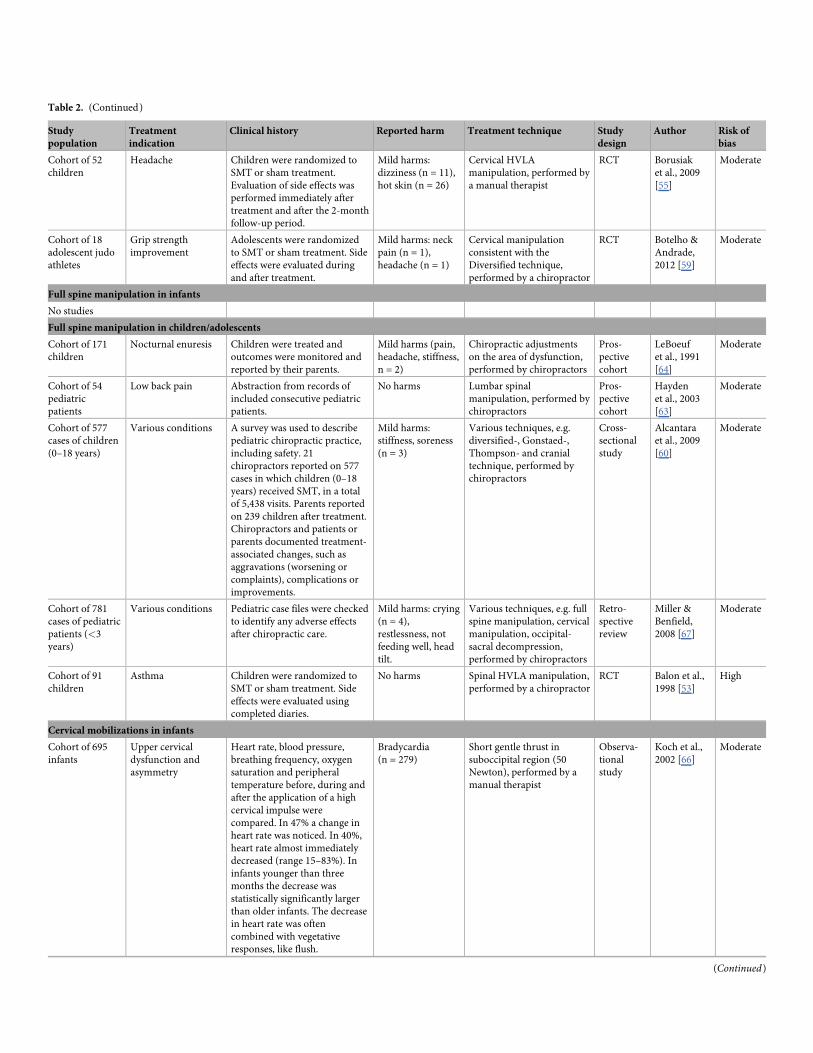
Table 2. (Continued)
Study
population
Treatment
indication
Clinical history Reported harm Treatment technique Study
design
Author Risk of
bias
Cohort of 52
children
Headache Children were randomized to
SMT or sham treatment.
Evaluation of side effects was
performed immediately after
treatment and after the 2-month
follow-up period.
Mild harms:
dizziness (n = 11),
hot skin (n = 26)
Cervical HVLA
manipulation, performed by
a manual therapist
RCT Borusiak
et al., 2009
[55]
Moderate
Cohort of 18
adolescent judo
athletes
Grip strength
improvement
Adolescents were randomized
to SMT or sham treatment. Side
effects were evaluated during
and after treatment.
Mild harms: neck
pain (n = 1),
headache (n = 1)
Cervical manipulation
consistent with the
Diversified technique,
performed by a chiropractor
RCT Botelho &
Andrade,
2012 [59]
Moderate
Full spine manipulation in infants
No studies
Full spine manipulation in children/adolescents
Cohort of 171
children
Nocturnal enuresis Children were treated and
outcomes were monitored and
reported by their parents.
Mild harms (pain,
headache, stiffness,
n = 2)
Chiropractic adjustments
on the area of dysfunction,
performed by chiropractors
Pros-
pective
cohort
LeBoeuf
et al., 1991
[64]
Moderate
Cohort of 54
pediatric
patients
Low back pain Abstraction from records of
included consecutive pediatric
patients.
No harms Lumbar spinal
manipulation, performed by
chiropractors
Pros-
pective
cohort
Hayden
et al., 2003
[63]
Moderate
Cohort of 577
cases of children
(0–18 years)
Various conditions A survey was used to describe
pediatric chiropractic practice,
including safety. 21
chiropractors reported on 577
cases in which children (0–18
years) received SMT, in a total
of 5,438 visits. Parents reported
on 239 children after treatment.
Chiropractors and patients or
parents documented treatment-
associated changes, such as
aggravations (worsening or
complaints), complications or
improvements.
Mild harms:
stiffness, soreness
(n = 3)
Various techniques, e.g.
diversified-, Gonstaed-,
Thompson- and cranial
technique, performed by
chiropractors
Cross-
sectional
study
Alcantara
et al., 2009
[60]
Moderate
Cohort of 781
cases of pediatric
patients (<3
years)
Various conditions Pediatric case files were checked
to identify any adverse effects
after chiropractic care.
Mild harms: crying
(n = 4),
restlessness, not
feeding well, head
tilt.
Various techniques, e.g. full
spine manipulation, cervical
manipulation, occipital-
sacral decompression,
performed by chiropractors
Retro-
spective
review
Miller &
Benfield,
2008 [67]
Moderate
Cohort of 91
children
Asthma Children were randomized to
SMT or sham treatment. Side
effects were evaluated using
completed diaries.
No harms Spinal HVLA manipulation,
performed by a chiropractor
RCT Balon et al.,
1998 [53]
High
Cervical mobilizations in infants
Cohort of 695
infants
Upper cervical
dysfunction and
asymmetry
Heart rate, blood pressure,
breathing frequency, oxygen
saturation and peripheral
temperature before, during and
after the application of a high
cervical impulse were
compared. In 47% a change in
heart rate was noticed. In 40%,
heart rate almost immediately
decreased (range 15–83%). In
infants younger than three
months the decrease was
statistically significantly larger
than older infants. The decrease
in heart rate was often
combined with vegetative
responses, like flush.
Bradycardia
(n = 279)
Short gentle thrust in
suboccipital region (50
Newton), performed by a
manual therapist
Observa-
tional
study
Koch et al.,
2002 [66]
Moderate
(Continued)

Table 2. (Continued)
Study
population
Treatment
indication
Clinical history Reported harm Treatment technique Study
design
Author Risk of
bias
Cohort of 199
infants
Muscle tension
disorders of mouth
or pharynx or
asymmetry of skull,
neck, trunk or hip
Responses after an upper
cervical impulse were
investigated. Physiological
responses were shown in 53%;
flush (49%), short spells of
apnea (22%), hyperextension of
the back and/or neck (13%) and
sweating (8%). The short spells
of apnea lasted less than 10
seconds and breathing pattern
was immediately restored by
blowing into the child’s face.
The authors stated that these
responses were normal
physiological responses and
cannot be interpreted as adverse
reaction or harm.
Physiological
responses
(n = 105)
Short gentle thrust (50
Newton) in suboccipital
region, performed by a
manual therapist
Observa-
tional
study
Koch et al.,
1998 [74]
Moderate
Cohort of 114
cases of infants
(<12 weeks)
Sub-optimal breast-
feeding
Data abstraction out of case
series to describe circumstances,
clinical features, role and
treatment outcomes.
No harms Low force spinal
mobilization, performed by
chiropractors
Retro-
spective
case series
Miller et al.,
2009 [68]
Moderate
Cervical mobilizations in children/adolescents
No studies
Full spine mobilizations in infants
21-day-old girl Colic and fussiness After manipulation infant
immediately cried and fell
asleep. Infant remained fussy
and the mother felt a crackling
sensation of the back. X-ray
showed acute fractures of the 7th
and 8th posterior left ribs. No
additional fractures were found.
Infant went for follow-up to the
child abuse center. Results of
bone laboratory tests were
normal. The center concluded
that child abuse could not be
definitively ruled out, but
chiropractic manipulation was
seen as a plausible explanation
for the fractures.
Rib fractures Spinal fingertip pressure
and adjustments using a
‘spring-activated device’,
performed by a chiropractor
Case
report
Wilson et al.,
2012 [73]
High
Cohort of 194
infants
Various conditions Data were extracted from
mother’s completed
questionnaires about infant
characteristics, symptoms and
perceived effect.
No harms Low-force mobilizations of
spinal joints in the area of
dysfunction, performed by
chiropractors
Cross-
sectional
survey
Nicolas-
Schmid
et al., 2016
[62]
High
Cohort of 104
infants (<4
weeks)
Colic Infants were randomized to
SMT and no treatment. Parents
reported on adverse events
during the treatment period.
No harms Low-force spinal
mobilization (2 Newton),
performed by a chiropractor
RCT Miller et al.,
2012 [50]
Moderate
Full spine mobilizations in children/adolescents
No studies
Unspecified treatment techniques
(Continued)

Two controlled studies reported mild, transient harms in terms of side effects: one study
(n = 52) reported dizziness (n = 11) and hot skin (n = 26),[55] one study (n = 18) reported
neck pain (n = 1) and headache (n = 1).[59] Five studies reported harms after HVLA manipu-
lations performed on the full spine. In three of these studies (n = 1529) a small number of mild
harms (n = 9) was reported;[60, 64, 67] the other two studies (n = 145) reported no harms.[53,
63] No studies were found reporting on harms after cervical or full spine mobilizations. One
study (n = 956) reported side effects or reactions in children after chiropractic treatment
(n = 557), but both side effects or reactions and treatment techniques were not specified.[61]
Hence, conclusions on treatment technique cannot be given.
Discussion
This review provides a unique overview of the evidence investigating the effectiveness and
safety of specific SMT techniques specified per treatment indication and age group, instead of
concluding on SMT as a general treatment approach. We found limited evidence for all age
groups and treatment indications; overall the body of evidence is of very low quality due to
moderate-to-high risk of bias, imprecise estimates, and lack of demonstrated consistency
across studies. The effectiveness of gentle, low-velocity spinal mobilizations in infants with
colic or torticollis remains uncertain. The effectiveness of HVLA spinal manipulations to man-
age asthma, nocturnal enuresis, headache, idiopathic scoliosis, and to improve grip strength in
children and/or adolescents, also remains uncertain. We found that the number of reports of
severe harms as direct side effects of SMT techniques were scarce and may be underreported.
Where reported, harms differed between treatment techniques and between age groups. Gen-
tle, low-velocity mobilization techniques appear to be a safe treatment technique in infants
and children and/or adolescents. Cervical and full spine HVLA manipulations, however,
might be associated with severe harms, although underlying pathology was suspected in the
cases reported on.
Effectiveness of SMT techniques
The very low quality of the body of evidence prevented us from drawing clinically meaningful
conclusions on effectiveness of specific SMT techniques for specified treatment indications.
These findings are consistent with previous reviews investigating the effectiveness of pediatric
manual therapy as a general treatment approach.[1, 2, 4, 13] Specifically, the systematic review
of Bronfort et al. (2010) also concluded that effectiveness of SMT in children is uncertain.[13]
However, Bronfort et al. summarized the evidence regarding general manual treatment per-
formed in both adults and children, and included various interventions, such as spinal and
extremity joint manipulation or mobilization, craniosacral and osteopathic therapies and mas-
sage. In contrast to our systematic review, Bronfort et al. did not distinguish between SMT
techniques in their analysis. Even though in our systematic review five additional (random-
ized) controlled studies were included, available literature was re-examined using the state-of-
Table 2. (Continued)
Study
population
Treatment
indication
Clinical history Reported harm Treatment technique Study
design
Author Risk of
bias
956 chiro-
practors
reported on
treatment of
children (0–18
years)
Various conditions A survey was used to investigate
characteristics of pediatric
chiropractic practice, including
side effects.
Unspecified mild
and moderate
harms (n = 557)
Treatment techniques were
not specified. Treatments
were performed by
chiropractors
Cross-
sectional
survey
Marchand
et al., 2012
[61]
Moderate
https://doi.org/10.1371/journal.pone.0218940.t002

the-art GRADE methodology, and harms were examined in relation to specific treatment tech-
niques, our conclusion about the lack of evidence remains largely the same as previous
research. Our review sets itself apart from previously performed research by focusing on spe-
cific SMT treatment techniques, instead of making conclusions about SMT as a general thera-
peutic approach.
A large number of the included studies in our review showed shortcomings. We highlight
these shortcomings here in an attempt to emphasize the need of high quality future research
and reporting. First, authors reported a hypothesized relation between the child’s (non-)mus-
culoskeletal condition and a particular spinal dysfunction.[49, 51–55, 57, 58] However, inter-
mediate outcomes to assess or indicate this potential dysfunction, such as range of motion,
were often neglected and only scarcely described. All studies assessed parent- or patient-
reported outcomes to indicate perceived treatment effect, while only four out of twelve con-
trolled studies additionally assessed functional outcomes to evaluate spinal dysfunction, such
as change in torticollis,[52] lung function[53, 54] and grip strength.[59] Therefore, currently,
no conclusions on the effect of specific SMT techniques on spinal dysfunction in these patients
can be drawn. In future research it is important to include these intermediate outcomes in
addition to patient-reported outcomes. Second, we would like to highlight that for adequate
interpretation it is of great importance that studies provide a detailed description of the SMT
technique performed. Important information regarding the specific treatment technique was
often omitted from publications. As a consequence, it is challenging (or even impossible) for
researchers and, maybe more importantly, healthcare professionals to interpret study findings
and draw scientifically substantiated conclusions about effective treatment techniques. As
such, this will hamper translation of study findings to clinical practice. Third, in the majority
of the included controlled studies, decrease in complaints/symptoms and improvement in
function over time was seen in both the intervention and control group. This may suggest a
potentially favorable natural course for the indications under study. However, the majority of
studies did not describe or consider this phenomenon. They focused on changes due to the
intervention and only emphasized differences over time within the intervention group, instead
of between group differences. Apart from a potential favorable natural course, the observed
decrease in complaints/symptoms or improvement in function in the sham or control group
may have occurred by other treatment effect, including placebo effect. To manage this, and to
gain a better understanding of the course of complaints/symptoms over the longer time, effec-
tiveness of SMT treatment techniques and potential harms of treatment, we recommend a
change in study designs and a shift in the focus of research. We underline the importance of
RCT designs using three-group-comparisons where a non-treatment group should be
included. Moreover, we recommend research to focus on examining outcomes of specific
SMT techniques and describing effectiveness in relation to these techniques, instead of making
conclusions on SMT as a general treatment approach.
Harms of SMT techniques
Worldwide, manual therapy is regularly performed in children of all ages. Previous reports
indicate that 5 to 40% of patients receiving manual therapy are younger than 18 years old.[3, 9,
10, 27, 75–78] In view of this, severe harms such as death, paralysis and rib fractures after
HVLA manipulations[69–72] or spinal instrumented-adjustments[73] are rare. Authors often
concluded that underlying preexisting pathology was found and potentially related to the
occurrence of these severe harms, and HVLA manipulations were not the direct cause of
harm.[70, 72] Mild, transient harms, such as stiffness, soreness or headache, were reported in
two controlled trials[55, 59], and five larger observational studies,[60, 61, 64, 67, 74] but may
be underreported. Due to the lack of reported information on the specific treatment technique,
specific symptoms and indications, and professional background of the health care profes-
sional, and because of the unknown total prevalence of pediatric SMT performed worldwide,
conclusions about the prevalence of harms cannot be made and harms may be underreported.
Taking these limitations into account, conclusions about the risk of harm and safety of SMT
techniques are hard to draw. As such, we would encourage researchers to include detailed
descriptions of specific performed techniques and details about the education and clinical
experience of performing therapists. Moreover, to improve transparency and quantification of
harms, we acknowledge the importance of continuous review of harms, as previously indicated
by Vohra et al. and Humphreys et al.[79, 80] Observational cohorts with a longer follow up
period could provide a more realistic estimation on risk of harm of a specific intervention in
comparison to non-placebo controlled trials, in which strict inclusion criteria could limit the
representation of a realistic study population.[32] Furthermore, databases and registries of per-
formed treatments in infants and children could facilitate the reporting and review of harms.
Such resources provide a mechanism to continuously monitor treatment outcomes and
harms, and could be more reliable for reporting on harms as they do not aim to collect data for
research in only a specific period and population.[32]
Strengths and limitations
Our systematic review has a number of strengths. Our review sets itself apart from previous
research by focusing on the effectiveness and harms of specific SMT treatment techniques,
instead of concluding about SMT as a general therapeutic approach. A further strength is that
we examined the evidence for infants separately from children and/or adolescents, providing a
more nuanced overview of the effectiveness and safety of SMT techniques in children of differ-
ent ages. In addition, we assessed the quality of the body of evidence using GRADE.
A limitation is that meta-analysis could only be performed for one comparison and on one
outcome due to low consistency across studies. Sparse data meant that the quality of evidence
for any given comparison of treatments and treatment outcome was very low. Finally, many
studies were excluded from the review because they did not report on harms. Importantly, this
does not necessarily indicate absence of harms and may underestimate the occurrence of
harms.
Conclusion
Due to very low quality of the evidence, the effectiveness of gentle, low-velocity mobilizations
in infants and HVLA manipulations in children and/or adolescents is uncertain. Assessments
of intermediate outcomes are lacking in current pediatric SMT research. Therefore, the rela-
tionship between specific treatment and its effect on the hypothesized spinal dysfunction
remains unclear. Gentle, low-velocity spinal mobilizations seem to be a safe treatment tech-
nique. Although scarcely reported, HVLA manipulations in infants and young children could
lead to severe harms. Severe harms were likely to be associated with unexamined or missed
underlying medical pathology. Nevertheless, there is a need for high quality research to
increase certainty about effectiveness and safety of specific SMT techniques in infants, children
and adolescents. We encourage conduction of controlled studies that focus on the effectiveness
of specific SMT techniques on spinal dysfunction, instead of concluding about SMT as a
general treatment approach. Large observational studies could be conducted to monitor the
course of complaints/symptoms in children and to gain a greater understanding of potential
harms.

4. Gotlib A, Rupert R. Chiropractic manipulation in pediatric health conditions—an updated systematic review. Chiropractic & osteopathy. 2008; 16:11.
5. Gleberzon BJ, Arts J, Mei A, McManus EL. The use of spinal manipulative therapy for pediatric health conditions: A systematic review of the literature [review]. The Journal of the Canadian Chiropractic Association. 2012; 56(2):Online access only p 128–41. PMID: 22675226
6. Hestbaek L, Stochkendahl MJ. The evidence base for chiropractic treatment of musculoskeletal condi-tions in children and adolescents: The emperor’s new suit? Chiropractic & osteopathy. 2010; 18:15.
7. Ferrance RJ, Miller J. Chiropractic diagnosis and management of non-musculoskeletal conditions in children and adolescents. Chiropractic & osteopathy. 2010; 18:14.
8. Dobson D, Lucassen PL, Miller JJ, Vlieger AM, Prescott P, Lewith G. Manipulative therapies for infantile colic. The Cochrane database of systematic reviews. 2012; 12:Cd004796. https://doi.org/10.1002/ 14651858.CD004796.pub2 PMID: 23235617
9. Ernst E. Chiropractic spinal manipulation for infant colic: a systematic review of randomised clinical tri-als. International journal of clinical practice. 2009; 63(9):1351–3. https://doi.org/10.1111/j.1742-1241. 2009.02133.x PMID: 19691620
10. Alcantara J. The presenting complaints of pediatric patients for chiropractic care: Results from a prac-
tice-based research network. Clinical Chiropractic. 2008; 11(4):193–8.
11. Evans DW, Lucas N. What is ’manipulation’? A reappraisal. Manual therapy. 2010; 15(3):286–91. https://doi.org/10.1016/j.math.2009.12.009 PMID: 20080431
12. Allen-Unhammer A, Wilson FJ, Hestbaek L. Children and adolescents presenting to chiropractors in Norway: National Health Insurance data and a detailed survey. Chiropractic & manual therapies. 2016; 24:29.
13. Hestbaek L, Jorgensen A, Hartvigsen J. A description of children and adolescents in Danish chiropractic practice: results from a nationwide survey. Journal of manipulative and physiological therapeutics. 2009; 32(8):607–15. https://doi.org/10.1016/j.jmpt.2009.08.024 PMID: 19836596
14. Bronfort G, Assendelft WJ, Evans R, Haas M, Bouter L. Efficacy of spinal manipulation for chronic head-
ache: a systematic review. Journal of manipulative and physiological therapeutics. 2001; 24(7):457–66. PMID: 11562654
15. Bronfort G, Evans R, Anderson AV, Svendsen KH, Bracha Y, Grimm RH. Spinal manipulation, medica-
tion, or home exercise with advice for acute and subacute neck pain: a randomized trial. Annals of inter-
nal medicine [Internet]. 2012; 156(1 Pt 1):[1–10 pp.]. Available from: http://onlinelibrary.wiley.com/o/
cochrane/clcentral/articles/184/CN-00804184/frame.html http://annals.org/pdfaccess.ashx?url=/data/
journals/aim/20370/0000605-201201030-00002.pdf.
16. Bronfort G, Haas M, Evans R, Leininger B, Triano J. Effectiveness of manual therapies: the UK evi-
dence report. Chiropractic & osteopathy. 2010; 18:3.
17. Paige NM, Miake-Lye IM, Booth MS, Beroes JM, Mardian AS, Dougherty P, et al. Association of Spinal Manipulative Therapy With Clinical Benefit and Harm for Acute Low Back Pain: Systematic Review and Meta-analysis. Jama. 2017; 317(14):1451–60. https://doi.org/10.1001/jama.2017.3086 PMID: 28399251
18. Rubinstein SM, Terwee CB, Assendelft WJ, de Boer MR, van Tulder MW. Spinal manipulative therapy for acute low-back pain. The Cochrane database of systematic reviews. 2012(9):CD008880. https://doi. org/10.1002/14651858.CD008880.pub2 PMID: 22972127
19. Rubinstein SM, van Middelkoop M, Assendelft WJ, de Boer MR, van Tulder MW. Spinal manipulative therapy for chronic low-back pain. The Cochrane database of systematic reviews. 2011(2):CD008112. https://doi.org/10.1002/14651858.CD008112.pub2 PMID: 21328304
20. Huijbregts PA. Manual therapy in children: role of the evidencebased clinician. The Journal of manual & manipulative therapy. 2006; 14:7–9.
21. Kemper KJ, Vohra S, Walls R, Task Force on Complementary and Alternative Medicine PSoC, Holistic and Integrative Medicine. American Academy of Pediatrics. The use of complementary and alternative medicine in pediatrics. Pediatrics. 2008; 122:1374–86. https://doi.org/10.1542/peds.2008-2173 PMID: 19047261
22. Canadian Paediatric Society. Chiropractic care for children:controversies and issues. Journal of paedi-
atrics and child health. 2002; 7:85–9.
23. French SD, Walker BF, Perle SM. Chiropractic care for children: too much, too little or not enough? Chi-
ropractic & osteopathy. 2010; 18:17.
24. Leboeuf-Yde C, Hestbaek L. Chiropractic and children: Is more research enough? Chiropractic & oste-
opathy. 2010; 18:11.
25. Liberati A, Altman DG, Tetzlaff J, Mulrow C, Gotzsche PC, Ioannidis JP, et al. The PRISMA statement for reporting systematic reviews and meta-analyses of studies that evaluate healthcare interventions: explanation and elaboration. BMJ. 2009; 339:b2700. https://doi.org/10.1136/bmj.b2700 PMID: 19622552
26. World Health Organization. WHO guidelines on basic training and safety in chiropractic. Geneva: WHO; 2005.
27. Todd AJ, Carroll MT, Mitchell EK. Forces of Commonly Used Chiropractic Techniques for Children: A Review of the Literature. Journal of manipulative and physiological therapeutics. 2016; 39(6):401–10. https://doi.org/10.1016/j.jmpt.2016.05.006 PMID: 27346861

25. Peterson D, Bergmann TF. Chiropractic Technique: Principles and Procedures. Philadelphia: PA:
Mosby; 2002.
26. Meeker WC, Haldeman S. Chiropractic: A Profession at the Crossroads of Mainstream and Alternative
Medicine. Ann Intern Med. 2002; 136(2):216–27.
27. Alcantara J, Ohm J, Kunz D. The chiropractic care of children. Journal of alternative and complementary
medicine. 2010; 16(6):621–6. https://doi.org/10.1089/acm.2009.0369 PMID: 20569028
28. Seffinger MA, King HH, Ward RC, Jones JM, Rogers FJ, Patterson MM. Osteopathic philosophy. Foun-
dations for osteopathic medicine. 2003; 2:3–18.
29. Bialosky JE, Bishop MD, Price DD, Robinson ME, George SZ. The mechanisms of manual therapy in
the treatment of musculoskeletal pain: a comprehensive model. Manual therapy. 2009; 14(5):531–8.
https://doi.org/10.1016/j.math.2008.09.001 PMID: 19027342
30. Marchand A. A Literature Review of Pediatric Spinal Manipulation and Chiropractic Manipulative Ther-
apy: Evaluation of Consistent Use of Safety Terminology. Journal of manipulative and physiological
therapeutics. 2012.
31. Cochrane Effective Practice and Organisation of Care (EPOC). Taking account of adverse effects in
EPOC reviews. EPOC Resources for review authors; 2017.
32. Chou R, Aronson N, Atkins D, Ismaila AS, Santaguida P, Smith DH, et al. AHRQ series paper 4: assess-
ing harms when comparing medical interventions: AHRQ and the effective health-care program. J Clin
Epidemiol. 2010; 63(5):502–12. https://doi.org/10.1016/j.jclinepi.2008.06.007 PMID: 18823754
33. Higgins JPT, Altman DG. Cochrane Handbook for Systematic Reviews of Interventions Version 5.0.1
(updated September 2008): The Cochrane Collaboration; 2008.
34. Viswanathan M, Berkman ND. Development of the RTI item bank on risk of bias and precision of obser-
vational studies. J Clin Epidemiol. 2012; 65(2):163–78. https://doi.org/10.1016/j.jclinepi.2011.05.008
PMID: 21959223
35. The Joanna Briggs Institute. Joanna Briggs Institute Reviewers’ Manual: 2016 edition. Australia: The
Joanna Briggs Institute; 2016.
36. Schulz KF, Altman DG, Moher D, Group C. CONSORT 2010 statement: updated guidelines for report-
ing parallel group randomised trials. BMJ. 2010; 340:c332. https://doi.org/10.1136/bmj.c332 PMID:
20332509
37. Hoffmann TC, Glasziou PP, Boutron I, Milne R, Perera R, Moher D, et al. Better reporting of interven-
tions: template for intervention description and replication (TIDieR) checklist and guide. BMJ. 2014;
348:g1687. https://doi.org/10.1136/bmj.g1687 PMID: 24609605
38. Atkins D, Best D, Briss PA, Eccles M, Falck-Ytter Y, Flottorp S, et al. Grading quality of evidence and
strength of recommendations. BMJ. 2004; 328(7454):1490. https://doi.org/10.1136/bmj.328.7454.1490
PMID: 15205295
39. Guyatt GH, Oxman AD, Vist GE, Kunz R, Falck-Ytter Y, Alonso-Coello P, et al. GRADE: an emerging
consensus on rating quality of evidence and strength of recommendations. BMJ. 2008; 336(7650):924–
6. https://doi.org/10.1136/bmj.39489.470347.AD PMID: 18436948
40. Guyatt GH, Oxman AD, Vist G, Kunz R, Brozek J, Alonso-Coello P, et al. GRADE guidelines: 4. Rating
the quality of evidence—study limitations (risk of bias). J Clin Epidemiol. 2011; 64(4):407–15. https://
doi.org/10.1016/j.jclinepi.2010.07.017 PMID: 21247734
41. Guyatt GH, Oxman AD, Kunz R, Woodcock J, Brozek J, Helfand M, et al. GRADE guidelines: 7. Rating
the quality of evidence—inconsistency. J Clin Epidemiol. 2011; 64(12):1294–302. https://doi.org/10.
1016/j.jclinepi.2011.03.017 PMID: 21803546
42. Guyatt GH, Oxman AD, Kunz R, Woodcock J, Brozek J, Helfand M, et al. GRADE guidelines: 8. Rating
the quality of evidence—indirectness. J Clin Epidemiol. 2011; 64(12):1303–10. https://doi.org/10.1016/
j.jclinepi.2011.04.014 PMID: 21802903
43. Guyatt GH, Oxman AD, Kunz R, Brozek J, Alonso-Coello P, Rind D, et al. GRADE guidelines 6. Rating
the quality of evidence—imprecision. J Clin Epidemiol. 2011; 64(12):1283–93. https://doi.org/10.1016/j.
jclinepi.2011.01.012 PMID: 21839614
44. Guyatt GH, Oxman AD, Montori V, Vist G, Kunz R, Brozek J, et al. GRADE guidelines: 5. Rating the
quality of evidence—publication bias. J Clin Epidemiol. 2011; 64(12):1277–82. https://doi.org/10.1016/j.
jclinepi.2011.01.011 PMID: 21802904
45. Ryan R, Synnot A, Hill S. Describing results. Cochrane Consumers and Communication Group; 2016.
46. Ryan R, Hill S. How to GRADE the quality of evidence. 2016 December 2016.
47. Owens DK, Lohr KN, Atkins D, Treadwell JR, Reston JT, Bass EB, et al. Grading the Strength of a Body
of Evidence When Comparing Medical Interventions. Methods Guide for Effectiveness and Compara-
tive Effectiveness Reviews. AHRQ Methods for Effective Health Care. Rockville (MD)2008.
48. Browning M, Miller J. Comparison of the short-term effects of chiropractic spinal manipulation and occi-
pito-sacral decompression in the treatment of infant colic: A single-blinded, randomised, comparison
trial. Clin Chiropractic. 2008; 11:122–9.
49. Olafsdottir E, Forshei S, Fluge G, Markestad T. Randomised controlled trial of infantile colic treated with
chiropractic spinal manipulation. Archives of disease in childhood. 2001; 84(2):138–41. https://doi.org/
10.1136/adc.84.2.138 PMID: 11159288
50. Miller JE, Newell D, Bolton JE. Efficacy of chiropractic manual therapy on infant colic: a pragmatic sin-
gle-blind, randomized controlled trial. Journal of manipulative and physiological therapeutics. 2012; 35
(8):600–7. https://doi.org/10.1016/j.jmpt.2012.09.010 PMID: 23158465
51. Wiberg JM, Nordsteen J, Nilsson N. The short-term effect of spinal manipulation in the treatment of
infantile colic: a randomized controlled clinical trial with a blinded observer. Journal of manipulative and
physiological therapeutics. 1999; 22(8):517–22. PMID: 10543581
52. Haugen EB, Benth J, Nakstad B. Manual therapy in infantile torticollis: a randomized, controlled pilot
study. Acta paediatrica. 2011; 100(5):687–90. https://doi.org/10.1111/j.1651-2227.2011.02145.x
PMID: 21226758
53. Balon J, Aker PD, Crowther ER, Danielson C, Cox PG, O’Shaughnessy D, et al. A comparison of active
and simulated chiropractic manipulation as adjunctive treatment for childhood asthma. The New
England journal of medicine. 1998; 339(15):1013–20. https://doi.org/10.1056/NEJM199810083391501
PMID: 9761802
54. Bronfort G, Evans RL, Kubic P, Filkin P. Chronic pediatric asthma and chiropractic spinal manipulation:
a prospective clinical series and randomized clinical pilot study. Journal of manipulative and physiologi-
cal therapeutics. 2001; 24(6):369–77. https://doi.org/10.1067/mmt.2001.116417 PMID: 11514813
55. Borusiak P, Biedermann H, Bosserhoff S, Opp J. Lack of efficacy of manual therapy in children and ado-
lescents with suspected cervicogenic headache: results of a prospective, randomized, placebo-con-
trolled, and blinded trial. Headache. 2010; 50(2):224–30. https://doi.org/10.1111/j.1526-4610.2009.
01550.x PMID: 19845788
56. Khorshid KA, Sweat RW, Zemba DA, Zemba BN. Clinical efficacy of upper cervical versus full spine chi-
ropractic care on children with autism: a randomized clinical trial [randomized controlled trial]. Journal of
Vertebral Subluxation Research (JVSR). 2006;Mar(9):Online access only 7 p.
57. Reed WR, Beavers S, Reddy SK, Kern G. Chiropractic management of primary nocturnal enuresis.
Journal of manipulative and physiological therapeutics. 1994; 17(9):596–600. PMID: 7884329
58. Swierkosz S, Nowak Z. Low back pain in adolescents. An assessment of the quality of life in terms of
qualitative and quantitative pain variables. Journal of back and musculoskeletal rehabilitation. 2015; 28
(1):25–34. https://doi.org/10.3233/BMR-140484 PMID: 24867907
59. Botelho MB, Andrade BB. Effect of cervical spine manipulative therapy on judo athletes’ grip strength.
Journal of manipulative and physiological therapeutics. 2012; 35(1):38–44. https://doi.org/10.1016/j.
jmpt.2011.09.005 PMID: 22079053
60. Alcantara J, Ohm J, Kunz D. The safety and effectiveness of pediatric chiropractic: a survey of chiro-
practors and parents in a practice-based research network. Explore. 2009; 5(5):290–5. https://doi.org/
10.1016/j.explore.2009.06.002 PMID: 19733815
61. Marchand AM. Chiropractic care of children from birth to adolescence and classification of reported con-
ditions: an internet cross-sectional survey of 956 European chiropractors. Journal of manipulative and
physiological therapeutics. 2012; 35(5):372–80. https://doi.org/10.1016/j.jmpt.2012.04.008 PMID:
22627100
62. Nicolas-Schmid P, Hetlevik MA, Miller J. Infant presentations and outcomes at a chiropractic clinic in
the UK: Parent report of treatment outcomes using the United Kingdom Infant Questionnaire (UKIQ).
Journal of Clinical Chiropractic Pediatrics (JCCP). 2016; 15(2):Online access only p 1236–41.
63. Hayden JA, Mior SA, Verhoef MJ. Evaluation of chiropractic management of pediatric patients with low
back pain: a prospective cohort study. Journal of manipulative and physiological therapeutics. 2003; 26
(1):1–8. https://doi.org/10.1067/mmt.2003.11 PMID: 12532132
64. Leboeuf C, Brown P, Herman A, Leembruggen K, Walton D, Crisp TC. Chiropractic care of children with
nocturnal enuresis: a prospective outcome study. Journal of manipulative and physiological therapeu-
tics. 1991; 14(2):110–5. PMID: 2019820
65. Koch L, Girnus U. Kraftmessung bei Anwendung der Impulstechnik in der Chirotherapie Physikalische
Darstellung der einwirkenden Kraft in der Manuellen Medizin. Manuel Med. 1998; 36(1):21–6.
66. Koch LE, Koch H, Graumann-Brunt S, Stolle D, Ramirez JM, Saternus KS. Heart rate changes in
response to mild mechanical irritation of the high cervical spinal cord region in infants. Forensic science
international. 2002; 128(3):168–76. PMID: 12175961

67. Miller JE, Benfield K. Adverse effects of spinal manipulative therapy in children younger than 3 years: a
retrospective study in a chiropractic teaching clinic. Journal of manipulative and physiological therapeu-
tics. 2008; 31(6):419–23. https://doi.org/10.1016/j.jmpt.2008.06.002 PMID: 18722196
68. Miller JE, Miller L, Sulesund AK, Yevtushenko A. Contribution of chiropractic therapy to resolving sub-
optimal breastfeeding: a case series of 114 infants. Journal of manipulative and physiological therapeu-
tics. 2009; 32(8):670–4. https://doi.org/10.1016/j.jmpt.2009.08.023 PMID: 19836604
69. Deputy SR. Arm weakness in a child following chiropractor manipulation of the neck. Seminars in pedi-
atric neurology. 2014; 21(2):124–6. https://doi.org/10.1016/j.spen.2014.04.016 PMID: 25149945
70. Shafrir Y, Kaufman BA. Quadriplegia after chiropractic manipulation in an infant with congenital torticol-
lis caused by a spinal cord astrocytoma. The Journal of pediatrics. 1992; 120(2 Pt 1):266–9.
71. Holla M, Ijland MM, van der Vliet AM, Edwards M, Verlaat CW. [Death of an infant following ’craniosa-
cral’ manipulation of the neck and spine]. Ned Tijdschr Geneeskd. 2009; 153(17):828–31. PMID:
19469218
72. Jacobi G, Riepert T, Kieslich M, Bohl J. [Fatal outcome during physiotherapy (Vojta’s method) in a 3-
month old infant. Case report and comments on manual therapy in children]. Klinische Padiatrie. 2001;
213(2):76–85. https://doi.org/10.1055/s-2001-12881 PMID: 11305197
73. Wilson PM, Greiner MV, Duma EM. Posterior rib fractures in a young infant who received chiropractic
care. Pediatrics. 2012; 130(5):e1359–62. https://doi.org/10.1542/peds.2012-0372 PMID: 23027167
74. Koch LE, Biedermann H, Saternus KS. High cervical stress and apnoea. Forensic science international.
1998; 97(1):1–9. PMID: 9854836
75. Miller J. Demographic survey of pediatric patients presenting to a chiropractic teaching clinic. Chiroprac-
tic & Osteopathy. 2010; 18(33):Online access only 13 p.
76. Rubinstein S, Pfeifle CE, van Tulder MW, Assendelft WJ. Chiropractic patients in the Netherlands: a
descriptive study. Journal of manipulative and physiological therapeutics. 2000; 23(8):557–63. https://
doi.org/10.1067/mmt.2000.109675 PMID: 11050613
77. Smith C, Eckert K. Prevalence of complementary and alternative medicine and use among children in
South Australia. Journal of paediatrics and child health. 2006; 42(9):538–43. https://doi.org/10.1111/j.
1440-1754.2006.00918.x PMID: 16925541
78. Ndetan H, Evans MW Jr., Hawk C, Walker C. Chiropractic or osteopathic manipulation for children in
the United States: an analysis of data from the 2007 National Health Interview Survey. Journal of alter-
native and complementary medicine. 2012; 18(4):347–53. https://doi.org/10.1089/acm.2011.0268
PMID: 22384933
79. Vohra S, Johnston BC, Cramer K, Humphreys K. Adverse events associated with pediatric spinal
manipulation: a systematic review. Pediatrics. 2007; 119(1):e275–83. https://doi.org/10.1542/peds.
2006-1392 PMID: 17178922
80. Humphreys BK. Possible adverse events in children treated by manual therapy: a review. Chiropractic
& osteopathy. 2010; 18:12.
Longitudinal follow-up of muscle echotexturein infants with congenital muscular torticollis
Abstract
Unilateral fibrous contracture of the sternocleidomastoid (SCM) muscle is the major pathophysiology in infants with congenital muscular torticollis (CMT). Physical examination is not always sufficient to detect minimal muscle fibrosis in involved SCM muscles.A prospective study for SCM muscle fibrosis in CMT infants by quantifying echotexture and muscle thickness during the course of
treatment is highlighted in the study.Convenience samples of 21 female and 29 male infants with CMT, who were 1 to 12 months old, underwent physiotherapy for at
least 3 months and were followed for 4.7 ± 0.4 months. All infants had at least 2 clinical assessments and ultrasonographic examinations for bilateral SCM muscles during follow-up. The K value, derived from the difference in echo intensities between the involved and uninvolved SCM muscles on longitudinal sonograms, was used to represent the severity of muscle fibrosis. Bilateral SCM muscle thickness and ratio of involved to uninvolved muscle thickness (Ratio I/U) were obtained simultaneously. Clinical outcome was also recorded.No subjects underwent surgical intervention during follow-up. The K value decreased from 6.85 ± 0.58 to 1.30 ± 0.36 at the end of
follow-up (P< 0.001), which reflected the decrease of muscle fibrosis. The Ratio I/U decreased from 1.11 ± 0.04 to 0.97 ± 0.02 during treatment, which was possibly related to the increased uninvolved SCM muscle thickness.In conclusion, echotexture is an efficient indicator for reflecting a wide degree of muscle fibrosis in infants with CMT and is
informative during the treatment course.
Abbreviations: BL = body length, BW = body weight, CMT = congenital muscular torticollis, dB = decibel, DMD = Duchenne muscular dystrophy, EI = echo intensity, MEI = mean echo intensity, Ratio I/U = ratio of involved to uninvolved muscle thickness, ROI = region of interest, SCM = sternocleidomastoid, US = ultrasonography.
Keywords: fibrosis, infant, torticollis, ultrasonography
1. Introduction
Infants with congenital muscular torticollis (CMT) featureunilateral fibrous contracture of the sternocleidomastoid(SCM) muscle with a characteristic head tilt, limited neckrotation, or a palpable mass.[1] Physical examination is notalways sufficient in diagnosing CMT in infants with minimalclinical presentations, and image workup is sometimes requiredfor determining the treatment strategy.[1,2] Because of lack ofrelated cohort studies, randomized controlled trials, or meta-analyses, imaging has been suggested as level II evidence forinfants with CMT, but clinical practice guidelines still recom-mend that physical therapists obtain all image interpretationsbefore informing prognosis.[2] Compression sonoelastogra-phy[3,4] and color Duplex images[5] have been used to helpdescribe the degree of SCM muscle fibrosis. However, lackingconstant loading force and quantification of blood flow in targetmuscles, these new technologies represent less objective analysesfor SCM muscle fibrosis.Ultrasonography (US) can be used for quantifying the degree of
muscle fibrosis,[6] defining the size and location of musclemasses,[1] and guiding the clinical approach and treatmentduration in CMT.[1,7] This imaging type has been widelyadvocated because of the ability to identify different patternsof SCM muscle fibrosis.[1,2,7,8] Most infants showed a pseudo-tumor (type I fibrosis) or diffuse fibrosis mixed with normalmuscle and thickening of involved muscles (type II fibrosis).[1]
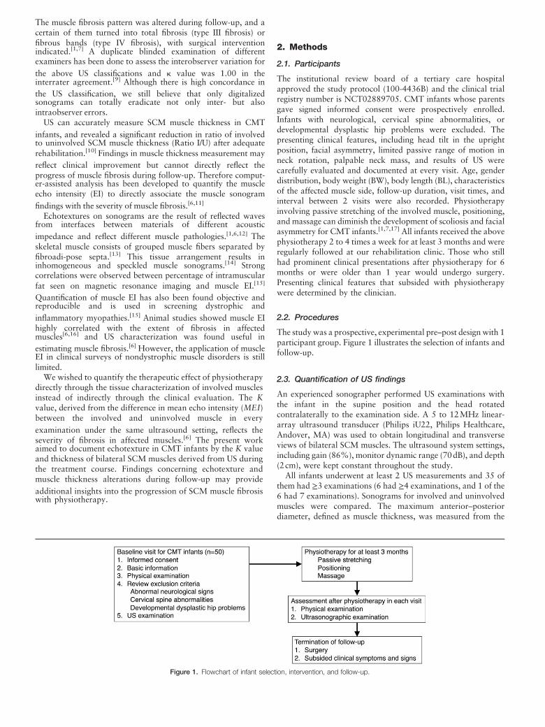
2. Methods
2.1. Participants
The institutional review board of a tertiary care hospitalapproved the study protocol (100-4436B) and the clinical trialregistry number is NCT02889705. CMT infants whose parentsgave signed informed consent were prospectively enrolled.Infants with neurological, cervical spine abnormalities, ordevelopmental dysplastic hip problems were excluded. Thepresenting clinical features, including head tilt in the uprightposition, facial asymmetry, limited passive range of motion inneck rotation, palpable neck mass, and results of US werecarefully evaluated and documented at every visit. Age, genderdistribution, body weight (BW), body length (BL), characteristicsof the affected muscle side, follow-up duration, visit times, andinterval between 2 visits were also recorded. Physiotherapyinvolving passive stretching of the involved muscle, positioning,and massage can diminish the development of scoliosis and facialasymmetry for CMT infants.[1,7,17] All infants received the abovephysiotherapy 2 to 4 times a week for at least 3 months and wereregularly followed at our rehabilitation clinic. Those who stillhad prominent clinical presentations after physiotherapy for 6months or were older than 1 year would undergo surgery.Presenting clinical features that subsided with physiotherapywere determined by the clinician.
2.2. Procedures
The study was a prospective, experimental pre–post design with 1participant group. Figure 1 illustrates the selection of infants andfollow-up.
2.3. Quantification of US findings
An experienced sonographer performed US examinations withthe infant in the supine position and the head rotatedcontralaterally to the examination side. A 5 to 12MHz linear-array ultrasound transducer (Philips iU22, Philips Healthcare,Andover, MA) was used to obtain longitudinal and transverseviews of bilateral SCM muscles. The ultrasound system settings,including gain (86%), monitor dynamic range (70dB), and depth(2cm), were kept constant throughout the study.All infants underwent at least 2 US measurements and 35 of
them had ≥3 examinations (6 had ≥4 examinations, and 1 of the6 had 7 examinations). Sonograms for involved and uninvolvedmuscles were compared. The maximum anterior–posteriordiameter, defined as muscle thickness, was measured from the
Figure 1. Flowchart of infant selection, intervention, and follow-up.
The muscle fibrosis pattern was altered during follow-up, and a certain of them turned into total fibrosis (type III fibrosis) or fibrous bands (type IV fibrosis), with surgical intervention indicated.[1,7] A duplicate blinded examination of different examiners has been done to assess the interobserver variation for the above US classifications and k value was 1.00 in the interrater agreement.[9] Although there is high concordance in the US classification, we still believe that only digitalized sonograms can totally eradicate not only inter- but also intraobserver errors.US can accurately measure SCM muscle thickness in CMT
infants, and revealed a significant reduction in ratio of involved to uninvolved SCM muscle thickness (Ratio I/U) after adequate rehabilitation.[10] Findings in muscle thickness measurement may reflect clinical improvement but cannot directly reflect the progress of muscle fibrosis during follow-up. Therefore comput-er-assisted analysis has been developed to quantify the muscle echo intensity (EI) to directly associate the muscle sonogram findings with the severity of muscle fibrosis.[6,11]
Echotextures on sonograms are the result of reflected waves from interfaces between materials of different acoustic impedance and reflect different muscle pathologies.[1,6,12] The skeletal muscle consists of grouped muscle fibers separated by fibroadi-pose septa.[13] This tissue arrangement results in inhomogeneous and speckled muscle sonograms.[14] Strong correlations were observed between percentage of intramuscular fat seen on magnetic resonance imaging and muscle EI.[15]
Quantification of muscle EI has also been found objective and reproducible and is used in screening dystrophic and inflammatory myopathies.[15] Animal studies showed muscle EI highly correlated with the extent of fibrosis in affected muscles[6,16] and US characterization was found useful in estimating muscle fibrosis.[6] However, the application of muscle EI in clinical surveys of nondystrophic muscle disorders is still limited.We wished to quantify the therapeutic effect of physiotherapy
directly through the tissue characterization of involved muscles instead of indirectly through the clinical evaluation. The K value, derived from the difference in mean echo intensity (MEI) between the involved and uninvolved muscle in every examination under the same ultrasound setting, reflects the severity of fibrosis in affected muscles.[6] The present work aimed to document echotexture in CMT infants by the K value and thickness of bilateral SCM muscles derived from US during the treatment course. Findings concerning echotexture and muscle thickness alterations during follow-up may provide additional insights into the progression of SCM muscle fibrosis with physiotherapy.

longitudinal view, then the Ratio I/U was derived (Fig. 2A). Themuscle EI was determined by computer-assisted gray-scaleanalysis and calculated using MATLAB 2006b (The Math-Works, Natick, MA). The MEI of every pixel in the region ofinterest (ROI) and the K value used to compare sonogramsbetween infants or between times for the same infant wereestimated by the following equations (Fig. 2B)[6]:
MEI ¼∑rx¼1∑
cy¼1Iðx; yÞr � c
ð1Þ
K ¼ MEIinvolved �MEIuninvolved ð2Þ
where r and c represent the pixel counts for the height andwidth of the ROI on the sonogram, respectively. A decrease in Kvalue indicates reduced muscle fibrosis.[6]
2.4. Statistical analysis
All data are presented as mean± standard error of mean. Becauseonly 1 infant underwent US more than 4 times, the 5th to 7thmeasurements for this infant were incorporated into the 4thmeasurement for statistical analysis. Initial US images in 10different subjects were analyzed as the above procedure in thesame program on the same International Business MachinesCorporation-compatible personal computer by 2 differentoperators. Each obtained image was analyzed 10 times andthe coefficient of variance ranged from 3% to 5% in eachexamination. The analyzed values of each image betweendifferent operators were similar. A generalized estimatingequation was used to compare different US measurements ofmuscle thickness (involved and uninvolved SCMmuscles) and the
K value during follow-up. Student t test was used to compare thethickness between bilateral SCM muscles. Pearson correlationwas used to analyze the relationship between US measurements,especially the initial examination, and clinical information.P<0.05 was considered statistically significant.
3. Results
3.1. Clinical information
We prospectively recruited 50 infants for the study (21 femalesand 29 males) with the mean age of 4.3±0.3 months (range1–12). Among them, 23 infants had left and 27 had right SCMmuscle involvement. Their initial mean BW and BL of all subjectswas 7.0±0.2kg and 63.4±0.9cm, respectively, and was about10kg and about 80cm at the end of follow-up. The mean follow-up period and interval were 4.7±0.4 months (range 1.4–14.7)and 2.5±0.2 months (range 0.7–12.6), respectively. All includedinfants had facial asymmetry and limited neck rotation to thelesion side. Four had a palpable neck mass (right/left: 3/1). Theclinical symptoms and signs subsided after physiotherapy ofdifferent durations. Therefore, no CMT infants had surgicalintervention at the end of follow-up.
3.2. Ultrasonographic measurement of echotexture
Each CMT infant underwent US examination for a mean of 2.9±1.2 times (range: 2–7) during follow-up. The initial mean K valuefor all CMT infants was 6.85±0.58 (range 2.22–26.23) anddecreased to 1.30±0.36 (range 0–7.72) during follow-up (P<0.001) (Fig. 3A). The high 4th measurement for 6 CMT infants
Figure 2. US of bilateral SCM muscles in infants with CMT. (A) Longitudinalsonograms of maximum anterior–posterior diameters of the involved (MTi) anduninvolved (MTu) SCM muscles. Ratio I/U was calculated as MTi/MTu. (B)Longitudinal sonograms of the MEI of every pixel in the ROI in the uninvolved(dotted line) and involved (solid line) muscles. The K value was calculated byMEIuninvolved�MEIinvolved.
Figure 3. Measurements of (A) K value and (B) scatter diagrams for K valueduring follow-up. (A) ∗P values between the 1st and 2nd, 3rd, and 4thmeasurements (all P<0.001). †P values between the 2nd and 3rd (P<0.001)and 4th measurements (P=0.015). (B) The K value of involved SCM musclesshows a negative-slope linear regression trend line (solid line).
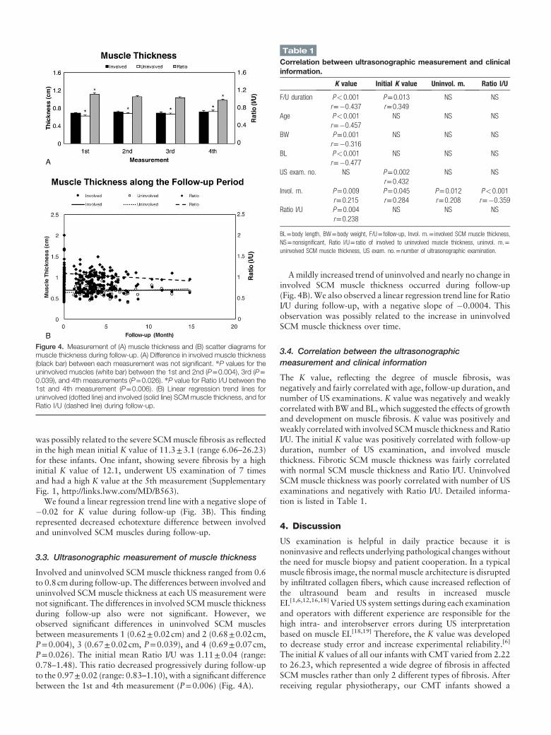
was possibly related to the severe SCMmuscle fibrosis as reflectedin the high mean initial K value of 11.3±3.1 (range 6.06–26.23)for these infants. One infant, showing severe fibrosis by a highinitial K value of 12.1, underwent US examination of 7 timesand had a high K value at the 5th measurement (SupplementaryFig. 1, http://links.lww.com/MD/B563).We found a linear regression trend line with a negative slope of
�0.02 for K value during follow-up (Fig. 3B). This findingrepresented decreased echotexture difference between involvedand uninvolved SCM muscles during follow-up.
3.3. Ultrasonographic measurement of muscle thickness
Involved and uninvolved SCMmuscle thickness ranged from 0.6to 0.8cm during follow-up. The differences between involved anduninvolved SCMmuscle thickness at each US measurement werenot significant. The differences in involved SCMmuscle thicknessduring follow-up also were not significant. However, weobserved significant differences in uninvolved SCM musclesbetween measurements 1 (0.62±0.02cm) and 2 (0.68±0.02cm,P=0.004), 3 (0.67±0.02cm, P=0.039), and 4 (0.69±0.07cm,P=0.026). The initial mean Ratio I/U was 1.11±0.04 (range:0.78–1.48). This ratio decreased progressively during follow-upto the 0.97±0.02 (range: 0.83–1.10), with a significant differencebetween the 1st and 4th measurement (P=0.006) (Fig. 4A).
Amildly increased trend of uninvolved and nearly no change ininvolved SCM muscle thickness occurred during follow-up(Fig. 4B).We also observed a linear regression trend line for RatioI/U during follow-up, with a negative slope of �0.0004. Thisobservation was possibly related to the increase in uninvolvedSCM muscle thickness over time.
3.4. Correlation between the ultrasonographicmeasurement and clinical information
The K value, reflecting the degree of muscle fibrosis, wasnegatively and fairly correlated with age, follow-up duration, andnumber of US examinations. K value was negatively and weaklycorrelatedwith BWand BL, which suggested the effects of growthand development on muscle fibrosis. K value was positively andweakly correlated with involved SCMmuscle thickness andRatioI/U. The initial K value was positively correlated with follow-upduration, number of US examination, and involved musclethickness. Fibrotic SCM muscle thickness was fairly correlatedwith normal SCM muscle thickness and Ratio I/U. UninvolvedSCM muscle thickness was poorly correlated with number of USexaminations and negatively with Ratio I/U. Detailed informa-tion is listed in Table 1.
4. Discussion
US examination is helpful in daily practice because it isnoninvasive and reflects underlying pathological changes withoutthe need for muscle biopsy and patient cooperation. In a typicalmuscle fibrosis image, the normal muscle architecture is disruptedby infiltrated collagen fibers, which cause increased reflection ofthe ultrasound beam and results in increased muscleEI.[1,6,12,16,18] Varied US system settings during each examinationand operators with different experience are responsible for thehigh intra- and interobserver errors during US interpretationbased on muscle EI.[18,19] Therefore, the K value was developedto decrease study error and increase experimental reliability.[6]
The initial K values of all our infants with CMT varied from 2.22to 26.23, which represented a wide degree of fibrosis in affectedSCM muscles rather than only 2 different types of fibrosis. Afterreceiving regular physiotherapy, our CMT infants showed a
Figure 4. Measurement of (A) muscle thickness and (B) scatter diagrams formuscle thickness during follow-up. (A) Difference in involved muscle thickness(black bar) between each measurement was not significant. ∗P values for theuninvolved muscles (white bar) between the 1st and 2nd (P=0.004), 3rd (P=0.039), and 4th measurements (P=0.026). ∗P value for Ratio I/U between the1st and 4th measurement (P=0.006). (B) Linear regression trend lines foruninvolved (dotted line) and involved (solid line) SCM muscle thickness, and forRatio I/U (dashed line) during follow-up.
Table 1
Correlation between ultrasonographic measurement and clinicalinformation.
K value Initial K value Uninvol. m. Ratio I/U
F/U duration P<0.001 P=0.013 NS NSr=�0.437 r=0.349
Age P<0.001 NS NS NSr=�0.457
BW P=0.001 NS NS NSr=�0.316
BL P<0.001 NS NS NSr=�0.477
US exam. no. NS P=0.002 NS NSr=0.432
Invol. m. P=0.009 P=0.045 P=0.012 P<0.001r=0.215 r=0.284 r=0.208 r=�0.359
Ratio I/U P=0.004 NS NS NSr=0.238
BL=body length, BW=body weight, F/U= follow-up, Invol. m.= involved SCM muscle thickness,NS=nonsignificant, Ratio I/U= ratio of involved to uninvolved muscle thickness, uninvol. m.=uninvolved SCM muscle thickness, US exam. no.=number of ultrasonographic examination.

muscles during physiotherapy. From our observations, infantswithhigh initialKvalue, implying severefibrosis,will receive a longtreatment course and frequentUS examinations.More frequentUSexaminationsand longerphysiotherapycoursemaybe required forinfants with greater degree of SCM muscle fibrosis.
4.1. Study limitations
US is a valuable workup tool for following the clinical status ofCMT infants but cannot elucidate the pathogenesis of the musclefibrosis. Echotexture analysis and thickness measurement ofinvolved SCM muscles are end-clinical presentations of CMTinfants. Study design only correlated the K value with theexistence of clinical symptoms of head tilting, facial asymmetry,and neck range of motion, instead of the severity of clinicalpresentation.Wewill stress on this field in the near future.Musclehistopathological results were not available, because no subjectsin the study underwent surgical intervention. Therefore, it is hardto differentiate the effects of maturation, therapy, or growth oninvolved muscle in the study.
5. Conclusions
In addition to decreasing fibrosis, both growth and developmentand stretch-induced muscle hypertrophy affect the musclearchitecture in affected SCM muscles in infants with CMT.The muscle EI reflects muscle tissue characterization and iscorrelated with muscle fibrosis in different diseaseentities.[12,21–23] The K value, derived from the differencebetween the involved and uninvolved muscle MEI, can aid infollowing the progression of muscle fibrosis in CMT infantsduring treatment. Quantification of muscle fibrosis is a digitizedand sensitive indicator reflecting a wide degree of SCM musclefibrosis in CMT and can be of great help in clinical practice.
References
[1] Hsu TC, Wang CL, Wong MK, et al. Correlation of clinical andultrasonographic features in congenital muscular torticollis. Arch PhysMed Rehabil 1999;80:637–41.
[2] Kaplan SL, Coulter C, Fetters L. Physical therapy management ofcongenital muscular torticollis: an evidence-based clinical practiceguideline: from the Section on Pediatrics of the American PhysicalTherapy Association. Pediatr Phys Ther 2013;25:348–94.
[3] Hong SK, Song JW, Woo SB, et al. Clinical usefulness of sonoelastog-raphy in infants with congenital muscular torticollis. Ann Rehabil Med2016;40:28–33.
[4] Lee SY, Park HJ, Choi YJ, et al. Value of adding sonoelastography toconventional ultrasound in patients with congenital muscular torticollis.Pediatr Radiol 2013;43:1566–72.
[5] Wang L, Zhang L, Tang Y, et al. The value of high-frequency and colorDoppler ultrasonography in diagnosing congenital muscular torticollis.BMC Musculoskelet Disord 2012;13:209.
[6] Hu CF, Chen CPC, Tsai WC, et al. Quantification of skeletal musclefibrosis at different healing stages using sonography: a morphologic andhistologica study in an animal model. J Ultrasound Med 2012;31:43–8.
[7] Tang SFT, Hsu KH, Wong AMK, et al. Longitudinal followup study ofultrasonography in congenital muscular torticollis. Clin Orthop RelatRes 2002;403:179–85.
[8] Cheng JC,Metreweli C, Chen TM, et al. Correlation of ultrasonographicimaging of congenital muscular torticollis with clinical assessment ininfants. Ultrasound Med Biol 2000;26:1237–41.
[9] Chen MM, Chang HC, Hsieh CF, et al. Predictive model for congenitalmuscular torticollis: analysis of 1021 infants with sonography. Arch PhysMed Rehabil 2005;86:2199–203.
[10] Park HJ, Kim SS, Lee SY, et al. Assessment of follow-up sonography andclinical improvement among infants with congenital muscular torticollis.AJNR Am J Neuroradiol 2013;34:890–4.
significant decrease in K values during follow-up on serial sonograms of affected SCM muscles (Fig. 3A). The decrease in K value indicates that the MEI for involved muscle approaches that for uninvolved muscle, for overall decreased muscle fibrosis.[6]
Muscle biopsy specimens from patients with neuromuscular disease show common histopathologic findings of atrophied muscle fibers and increased perimysial fibrosis.[20] Muscle EI has been clinically used to quantify severity of muscle fibrosis in patients with Duchenne muscular dystrophy (DMD),[21] inflam-matory myopathy,[12] metabolic myopathy,[22] and lumbar radiculopathy.[23] The muscle EI of serial muscle images was used in the follow-up of boys 4 to 12 years old with DMD and was inversely related to ambulatory status, functional grading, muscle strength, and motor ability.[21] A study used the Heckmatt scale to follow the spasticity of involved gastrocnemius muscles in stroke patients before and after botulinum toxin-A injection.[24]
The EI is difficult to differentiate in different muscle pathologies, but it seemed a good candidate for reflecting the clinical status during the course of treatment from the above reports.Besides the muscle fibrosis, growth and development may
affect muscle components.[25,26] The uninvolved muscle thickness increased along the follow-up period representing muscle growth effects in the study. Thickness changes of involved muscles, reflecting the complex of decreased fibrosis after physiotherapy and increased normal muscle component with growth, were not distinctive. Selective activation of protein kinase B caused by the release of insulin growth factor-1 from muscle fibers during passive stretch promotes myosatellite cell proliferation and induces skeletal muscle hypertrophy.[27,28] Stretch-induced antifibrotic effects, as much as the well-known antifibrotic agent decorin, has also been reported in injured gastrocnemius muscles of rats.[29] Therefore, both normal growth and development and physiotherapy may affect the improvement of SCM muscle fibrosis as reflected in the change in K value during follow-up. The K value is useful for continuously observing severity of SCM muscle fibrosis and may be a competent indicator in mirroring the progression of muscle fibrosis during treatment.With pure imaging, sonograms can tell us only the change in
fibrosis type or sustained decrease in muscle fibrosis in the involved SCM muscle during the treatment course for CMT infants. However, the image presentation lacks a digitized indicator to show the amelioration of fibrosis in CMT infants after physiotherapy, which can be demonstrated by the K value (Supplementary Fig. 1, http://links.lww.com/MD/B563). In contrast, changes in sequential measurements of the involved SCM muscle thickness and differences between bilateral SCM muscles during the treatment course were not significant. We found an increasing trend of uninvolved muscle thickness during follow-up, with a significant increase at about 3 months (2.9 ± 0.2 months) (Fig. 4A). Uninvolved SCM muscle thickness in all infants slowly increased with follow-up duration (Fig. 4B). Ratio I/U tended to decreased during treatment, with no decrease in involved SCM muscle thickness during follow-up (Fig. 4B). The significant negative correlation between uninvolved muscle thickness and Ratio I/U was largely due to the increase in uninvolved SCM muscle thickness.As compared with changes in muscle thickness and thickness
ratio during treatment, the improvement in echotexture was noticeable. Nevertheless, our results showed that involved SCM muscle thickness was not related to treatment period. Both these findings suggest that the K value is a better candidate than muscle thickness in describing the therapeutic effect on affected SCM

[11] Pillen S, Scholten RR, Zwarts MJ, et al. Quantitative skeletal muscleultrasonography in children with suspected neuromuscular disease.Muscle Nerve 2003;27:699–705.
[12] Reimers CD, Fleckenstein JL, Witt TN, et al. Muscular ultrasound inidiopathic inflammatory myopathies of adults. J Neurol Sci 1993;116:82–92.
[13] Peetrons P. Ultrasound of muscles. Eur Radiol 2002;12:35–43.[14] Walker FO, Cartwright MS, Wiesler ER, et al. Ultrasound of nerve and
muscles. Clin Neurophysiol 2004;115:495–507.[15] Young HJ, Jenkins NT, Zhao Q, et al. Measurement of intramuscular fat
by muscle echo intensity. Muscle Nerve 2015;52:963–71.[16] Kim HJ, Ryu KN, Sung DW, et al. Correlation between sonographic and
pathologic findings in muscle injury: experimental study in the rabbit. JUltrasound Med 2002;21:1113–9.
[17] Cheng JC, Au AW. Infantile torticollis: a review of 624 cases. J PediatrOrthop 1994;14:802–8.
[18] Pillen S, Keimpema M, Nievelstein RA, et al. Skeletal muscleultrasonography: visual versus quantitative evaluation. UltrasoundMed Biol 2006;32:1315–21.
[19] Jacobson JA. Musculoskeletal ultrasound: focused impact onMRI. Am JRoentgenol 2009;193:619–27.
[20] Joyce NC, Oskarsson B, Jin LW. Muscle biopsy evaluation inneuromuscular disorders. Phys Med Rehabil Clin N Am 2012;23:609–31.
[21] JansenM, van Alfen N, Nijhuis van der SandenMWG, et al. Quantitativemuscle ultrasound is a promising longitudinal follow-up tool in Duchennemuscular dystrophy. Neuromuscul Disord 2012;22:306–17.
[22] Pillen S, Verrips A, van Alfen N, et al. Quantitative skeletal muscleultrasound: diagnosetic value in childhood neuromuscular disease.Neuromuscul Disord 2007;17:509–16.
[23] Frost LR, Brown SHM. Neuromuscular ultrasound imaging in low backpain patients with radiculopathy. Man Ther 2016;21:83–8.
[24] Picelli A, Bonetti P, Fontana C, et al. Is spastic muscle echo intensityrelated to the response to botulinum toxin type a in patients with stroke?A cohort study. Arch Phys Med Rehabil 2012;93:1253–8.
[25] Kriketos AD, Baur LA, O’Connor J, et al. Muscle fibre type compositionin infant and adult populations and relationships with obesity. Int J ObesRelat Metab Disord 1997;21:796–801.
[26] Sanjurjo P, Aldamiz-Echevarria L, Prado C, et al. Fatty acid compositionof skeletal muscle and adipose tissue in Spanish infants and children. Br JNutr 2006;95:168–73.
[27] Sandri M. Signaling in muscle atrophy and hypertrophy. Physiology2008;23:160–70.
[28] De Deyne PG. Application of passive stretch and its implications formuscle fibers. Phys Ther 2001;81:819–27.
[29] Hwang JH,RaYJ,LeeKM,et al. Therapeutic effect of passivemobilizationexercise on improvement ofmuscle regeneration and prevention of fibrosisafter laceration injury of rat. Arch Phys Med Rehabil 2006;87:20–6.

FLEX THERAPIST CEUs
811 El Capitan Way Ste 110
San Luis Obispo, Ca 93401
Ph (800) 413-9636
www.flextherapistceus.com
References: Torticollis
Reference Articles:
Hardgrib, N., Rahbek, O., Møller-Madsen, B., & Maimburg, R.D. (2017). Do obstetric risk factors truly
influence the etiopathogenesis of congenital muscular torticollis? J Orthop Traumatol; 18:359–364 DOI
10.1007/s10195-017-0461-z
Driehuis F, Hoogeboom TJ, Nijhuis-van der Sanden MWG, de Bie RA, Staal JB (2019) Spinal manual
therapy in infants, children and adolescents: A systematic review and meta- analysis on treatment indication,
technique and outcomes. PLoS ONE 14(6): e0218940. https://doi. org/10.1371/journal.pone.0218940
Ching-Fang, Hu, et al. (2017). Longitudinal follow-up of muscle echotexture in infants with congenital
muscular torticollis. Medicine; 96:6. http://dx.doi.org/10.1097/MD.0000000000006068
Additional References:
Paige NM, Miake-Lye IM, Booth MS, Beroes JM, Mardian AS, Dougherty P, et al. Association of Spinal
Manipulative Therapy With Clinical Benefit and Harm for Acute Low Back Pain: Systematic Review and
Meta-analysis. Jama. 2017; 317(14):1451–60. https://doi.org/10.1001/jama.2017.3086 PMID: 28399251
Cochrane Effective Practice and Organisation of Care (EPOC). Taking account of adverse effects in EPOC
reviews. EPOC Resources for review authors; 2017.

“This course was developed and edited from the open access article: Do obstetric risk factors truly influence the etiopathogenesis of congenital muscular torticollis? - J Orthop Traumatol (2017) 18:359–364 (DOI
10.1007/s10195-017-0461-z), used under the Creative Commons Attribution License.”
“This course was developed and edited from the open access article: Driehuis F, Hoogeboom TJ, Nijhuis-van der Sanden MWG, de Bie RA, Staal JB (2019) Spinal manual therapy in infants, children and adolescents: A systematic review and meta-analysis on treatment indication, technique and outcomes. PLoS ONE 14(6):
e0218940. (https://doi. org/10.1371/journal.pone.0218940), used under the Creative Commons Attribution License.”
“This course was developed and edited from the open access article: Longitudinal follow-up of muscle echotexture in infants with congenital muscular torticollis - Hu et al. Medicine (2017) 96:6,
(http://dx.doi.org/10.1097/MD.0000000000006068), used under the Creative Commons Attribution License.”


![EXERCISES FOR RIGHT SIDED TORTICOLLIS (SHORT RIGHT … · EXERCISES FOR RIGHT SIDED TORTICOLLIS (SHORT RIGHT SCM) SIDE BENDING [for RIGHT Torticollis] Place your RIGHT hand …](https://img.pdfslide.us/doc/110x75/5b6dfb927f8b9aed178dfe37/exercises-for-right-sided-torticollis-short-right-exercises-for-right-sided.jpg)