Embed Size (px)
Citation preview
Torins are potent antimalarials that blockreplenishment of Plasmodium liver stageparasitophorous vacuole membrane proteinsKirsten K. Hansona,1, Ana S. Ressurreiçãob, Kathrin Buchholzc, Miguel Prudêncioa, Jonathan D. Herman-Ornelasc,Maria Rebeloa, Wandy L. Beattyd, Dyann F. Wirthc, Thomas Hänscheida, Rui Moreirab, Matthias Martic,and Maria M. Motaa,1
aInstituto de Medicina Molecular, Faculdade de Medicina, Universidade de Lisboa, 1649-028 Lisboa, Portugal; biMed.UL, Faculdade de Farmácia, Universidadede Lisboa, 1649-003 Lisboa, Portugal; cDepartment of Immunology and Infectious Diseases, Harvard School of Public Health, Boston, MA 02115;and dWashington University School of Medicine, St. Louis, MO 63110
Edited* by Louis H. Miller, National Institutes of Health, Rockville, MD, and approved May 23, 2013 (received for review April 2, 2013)
Residence within a customized vacuole is a highly successfulstrategy used by diverse intracellular microorganisms. The para-sitophorous vacuole membrane (PVM) is the critical interfacebetween Plasmodium parasites and their possibly hostile, yetultimately sustaining, host cell environment. We show that tor-ins, developed as ATP-competitive mammalian target of rapamy-cin (mTOR) kinase inhibitors, are fast-acting antiplasmodial com-pounds that unexpectedly target the parasite directly, blockingthe dynamic trafficking of the Plasmodium proteins exportedprotein 1 (EXP1) and upregulated in sporozoites 4 (UIS4) to theliver stage PVM and leading to efficient parasite elimination bythe hepatocyte. Torin2 has single-digit, or lower, nanomolar po-tency in both liver and blood stages of infection in vitro and islikewise effective against both stages in vivo, with a single oraldose sufficient to clear liver stage infection. Parasite eliminationand perturbed trafficking of liver stage PVM-resident proteins areboth specific aspects of torin-mediated Plasmodium liver stage in-hibition, indicating that torins have a distinct mode of action com-pared with currently used antimalarials.
host-parasite interactions | malaria | protein trafficking | P. falciparum
The population at risk for developing malaria is vast, com-prising some 3.3 billion people particularly in sub-Saharan
Africa and Southeast Asia, with mortality estimates ranging from655,000 to 1,200,000 (1). Widespread resistance has limited thetherapeutic utility of most existing antimalarial drugs, and artemisi-nin, the highly efficacious cornerstone of artemisinin combinationtherapies, appears to be at risk for the same fate (2). The need fornew antimalarial chemotherapeutic strategies is thus acute.Plasmodium spp., the causative agents of malaria, have a complex
life cycle with alternating motile-nonreplicative and sessile-replica-tive forms in both mammal and mosquito. In the mammalian host,Plasmodium invades and replicates inside two very distinct celltypes: hepatocytes and red blood cells (RBCs). In mammals, thePlasmodium life cycle is initiated by a motile sporozoite that invadesa hepatocyte, where it resides for 2–14 d, multiplying into >10,000merozoites in a single cycle (3). Once released into the bloodstream,each of these motile merozoites will infect an RBC and, within 1–3d, generate 10–30 new merozoites, which will contribute to thecontinuous cycle of blood stage infection that causes the symptoms,morbidity, and mortality of malaria.These two stages of mammalian infection, despite taking place
in distinct cell types and having an orders-of-magnitude differencein parasite replication, do share common features. In both, themotile “zoite” invades the host cell through formation of a para-sitophorous vacuole (PV). Both stages grow and replicate exclu-sively within the confines of the PV, and the parasitophorousvacuole membrane (PVM), which is populated with parasite pro-teins, constitutes the physical host–parasite interface throughoutdevelopment. Unlike the vacuoles of many intracellular pathogens
including Leishmania, Chlamydia, Mycobacteria, and Legionella(4, 5), the Plasmodium vacuole, like that of Toxoplasma gondii,does not fuse with host lysosomes and is not acidified (6). This isnot unsurprising in the context of Plasmodium development in anRBC, which lacks endomembrane system trafficking and, indeed,lysosomes. The highly polarized hepatocyte, however, has exten-sive vesicular transport networks (7) and can target intracellularpathogens residing in a vacuole (8), suggesting that the exo-erythrocytic form (EEF) may need to resist host cell attack.Although the PVM is thought to be critical for Plasmodium
growth in both the hepatocyte and the RBC contexts, its cellularroles remain elusive. The importance of several PlasmodiumPVM-resident proteins, however, has been conclusively demon-strated in both blood and liver stages. Attempts to generateexported (exp)1 and Plasmodium translocon of exported protein(ptex)150 knockout parasites in Plasmodium falciparum failed (9,10), revealing that these are both essential proteins for the bloodstage, whereas Plasmodium berghei and Plasmodium yoeliimutants lacking up-regulated in sporozoites (uis)3 or uis4 fail tocomplete liver stage development (11, 12). These PVM-residentproteins, and thus the PVM itself, are performing functions thatare crucial for Plasmodium growth, but delineating the functionsof individual PVM-resident proteins has proven as difficult asidentifying the cellular processes mediated by the PVM.
Significance
Plasmodium parasites have two distinct intracellular growthstages inside the mammalian host—the first stage, which isclinically silent, in liver hepatocytes, and the second, whichcauses the symptoms of malaria, in red blood cells. This studyreports the discovery of a class of antimalarial compoundscalled torins, which are extremely potent inhibitors of bothintracellular stages of Plasmodium. We show that torins blocktrafficking of liver stage parasite proteins to the physical host–parasite interface, called the parasitophorous vacuole mem-brane (PVM), and that without continuous trafficking of PVM-resident proteins, the parasite is subject to elimination by itshost hepatocyte.
Author contributions: K.K.H., D.F.W., and M.M.M. designed research; K.K.H., K.B., M.P.,J.D.H-O., M.R., and W.L.B. performed research; A.S.R. and R.M. contributed new reagents/analytic tools; K.K.H., K.B., M.P., J.D.H-O., M.R., W.L.B., D.F.W., T.H., M.M., and M.M.M.analyzed data; and K.K.H. and M.M.M. wrote the paper.
The authors declare no conflict of interest.
*This Direct Submission article had a prearranged editor.
Freely available online through the PNAS open access option.1To whom correspondence may be addressed. E-mail: [email protected] or [email protected].
This article contains supporting information online at www.pnas.org/lookup/suppl/doi:10.1073/pnas.1306097110/-/DCSupplemental.
E2838–E2847 | PNAS | Published online July 8, 2013 www.pnas.org/cgi/doi/10.1073/pnas.1306097110
The one process in which both the centrality of the PVM isknown and evidence for the participation of specific PVM pro-teins exists is the export of parasite proteins to the RBC. A cohortof parasite proteins that are involved in extensive physiologicaland structural modifications of the infected RBC (iRBC) isexported into the iRBC cytoplasm and beyond (13). Five proteinshave been identified as components of PTEX, the proposed ex-port machinery at the iRBC PVM (9). Although liver stageprotein export has been shown for the Circumsporozite (CS)protein (14) and PTEX components are expressed in P. falcipa-rum EEFs (15), a role for parasite protein export into the he-patocyte remains speculative; the host hepatocyte may notrequire the extensive structural remodeling that the iRBC does.Conversely, however, the hepatocyte, with its extensive vesic-
ular transport network, intuitively constitutes a more hostile hostenvironment than the RBC, and there is evidence that the liverstage PVM may play a crucial role in preventing host cell-mediated parasite killing, as it does in Toxoplasma gondii (16).Support for a protective role for the liver stage PVM comes fromknockout parasites that fail in the earliest steps of PVM for-mation and remodeling. Sporozoites lacking the p52/p36 genepair invade hepatocytes successfully, but fail in PVM formation(17, 18) and are severely reduced in abundance midway throughliver stage development. Parasites lacking slarp/sap (19, 20), aregulator of early liver stage development, fail to express UIS4and exported protein 1 (EXP-1), along with other parasite pro-teins, and are also eliminated at the beginning of infection.Acquisition of resources from the host-cell environment, an
unambiguous requirement for an obligate intracellular parasitelike Plasmodium, is a function ascribed to the PVM in bothmammalian stages. The PVM allows the free passage of molecules(21, 22), presumably through proteinaceous pores, which maycontribute to acquisition of host nutrients and disposal of parasitewaste products. Members of the early transcribed membraneprotein (ETRAMP) family, single-pass transmembrane proteinsconserved among Plasmodium spp., which are highly expressedand developmentally regulated in both blood and liver stage par-asites (23, 24), could be candidates for mediating uptake of hostresources. Such a role in lipid uptake has indeed been proposed forthe P. berghei ETRAMP UIS3 on the basis of its interaction withhost-cell L-FABP (liver fatty acid binding protein) (25).Although Plasmodium parasites must use host resources to
support their own growth in both mammalian stages, the singlecycle replicative output of the liver stage parasite is vastly greaterthan that of the blood stage, which may reflect a similarly increasedneed for host resources. In this respect, the hepatocyte constitutesfar superior “raw material” compared with the RBC; hepatocytesare not onlymetabolically active, but also highly versatile cells, whichare capable of altering uptake, storage, production, and degradationof a wide array of macromolecules in response to cellular and or-ganismal requirements. The presence of a growing Plasmodiumparasite is sensed by the host hepatocyte, which responds with acti-vation of cellular stress responses and altered metabolism (26, 27).The mammalian target of rapamycin (mTOR) kinase integratessignals from amino acids, stress, oxygen, energy, and growth factorsand responds by altering cellular protein and lipid synthesis, as wellas autophagy (28). As such, we sought to determine how inhibitionof host mTOR signaling would affect Plasmodium liver stage de-velopment. Here we show that torins, a single structural class ofmTOR inhibitors, are highly potent antiplasmodial compoundstargeting both mammalian stages in vitro and in vivo. Independentof host-cell mTOR, torins impair trafficking of Plasmodium liverstage PVM-resident proteins, revealing the fast turnover of theseproteins at the liver stage PVM, and provoke elimination of liverstage parasites.
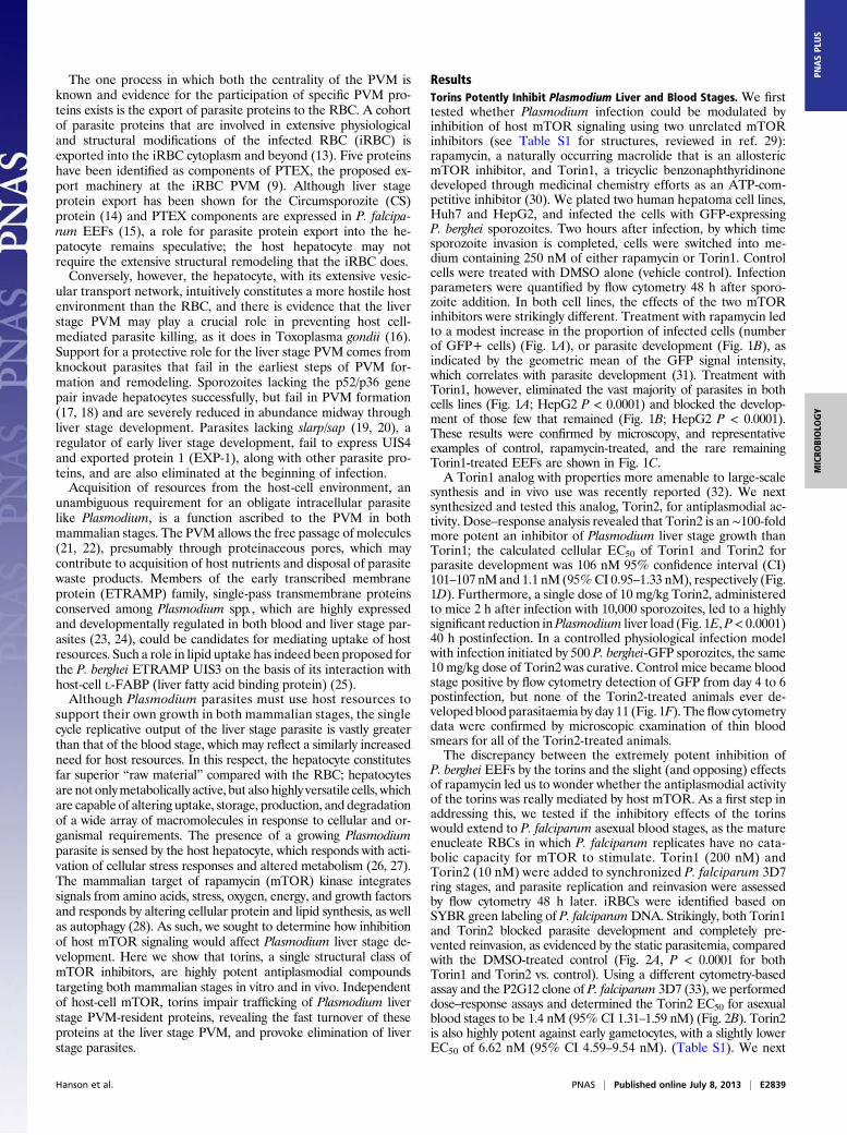
ResultsTorins Potently Inhibit Plasmodium Liver and Blood Stages. We firsttested whether Plasmodium infection could be modulated byinhibition of host mTOR signaling using two unrelated mTORinhibitors (see Table S1 for structures, reviewed in ref. 29):rapamycin, a naturally occurring macrolide that is an allostericmTOR inhibitor, and Torin1, a tricyclic benzonaphthyridinonedeveloped through medicinal chemistry efforts as an ATP-com-petitive inhibitor (30). We plated two human hepatoma cell lines,Huh7 and HepG2, and infected the cells with GFP-expressingP. berghei sporozoites. Two hours after infection, by which timesporozoite invasion is completed, cells were switched into me-dium containing 250 nM of either rapamycin or Torin1. Controlcells were treated with DMSO alone (vehicle control). Infectionparameters were quantified by flow cytometry 48 h after sporo-zoite addition. In both cell lines, the effects of the two mTORinhibitors were strikingly different. Treatment with rapamycin ledto a modest increase in the proportion of infected cells (numberof GFP+ cells) (Fig. 1A), or parasite development (Fig. 1B), asindicated by the geometric mean of the GFP signal intensity,which correlates with parasite development (31). Treatment withTorin1, however, eliminated the vast majority of parasites in bothcells lines (Fig. 1A; HepG2 P < 0.0001) and blocked the develop-ment of those few that remained (Fig. 1B; HepG2 P < 0.0001).These results were confirmed by microscopy, and representativeexamples of control, rapamycin-treated, and the rare remainingTorin1-treated EEFs are shown in Fig. 1C.A Torin1 analog with properties more amenable to large-scale
synthesis and in vivo use was recently reported (32). We nextsynthesized and tested this analog, Torin2, for antiplasmodial ac-tivity. Dose–response analysis revealed that Torin2 is an∼100-foldmore potent an inhibitor of Plasmodium liver stage growth thanTorin1; the calculated cellular EC50 of Torin1 and Torin2 forparasite development was 106 nM 95% confidence interval (CI)101–107 nMand 1.1 nM (95%CI 0.95–1.33 nM), respectively (Fig.1D). Furthermore, a single dose of 10 mg/kg Torin2, administeredto mice 2 h after infection with 10,000 sporozoites, led to a highlysignificant reduction inPlasmodium liver load (Fig. 1E,P< 0.0001)40 h postinfection. In a controlled physiological infection modelwith infection initiated by 500 P. berghei-GFP sporozites, the same10 mg/kg dose of Torin2 was curative. Control mice became bloodstage positive by flow cytometry detection of GFP from day 4 to 6postinfection, but none of the Torin2-treated animals ever de-veloped blood parasitaemia by day 11 (Fig. 1F). The flow cytometrydata were confirmed by microscopic examination of thin bloodsmears for all of the Torin2-treated animals.The discrepancy between the extremely potent inhibition of
P. berghei EEFs by the torins and the slight (and opposing) effectsof rapamycin led us to wonder whether the antiplasmodial activityof the torins was really mediated by host mTOR. As a first step inaddressing this, we tested if the inhibitory effects of the torinswould extend to P. falciparum asexual blood stages, as the matureenucleate RBCs in which P. falciparum replicates have no cata-bolic capacity for mTOR to stimulate. Torin1 (200 nM) andTorin2 (10 nM) were added to synchronized P. falciparum 3D7ring stages, and parasite replication and reinvasion were assessedby flow cytometry 48 h later. iRBCs were identified based onSYBR green labeling of P. falciparumDNA. Strikingly, both Torin1and Torin2 blocked parasite development and completely pre-vented reinvasion, as evidenced by the static parasitemia, comparedwith the DMSO-treated control (Fig. 2A, P < 0.0001 for bothTorin1 and Torin2 vs. control). Using a different cytometry-basedassay and the P2G12 clone of P. falciparum 3D7 (33), we performeddose–response assays and determined the Torin2 EC50 for asexualblood stages to be 1.4 nM (95% CI 1.31–1.59 nM) (Fig. 2B). Torin2is also highly potent against early gametocytes, with a slightly lowerEC50 of 6.62 nM (95% CI 4.59–9.54 nM). (Table S1). We next
Hanson et al. PNAS | Published online July 8, 2013 | E2839
MICRO
BIOLO
GY
PNASPL
US
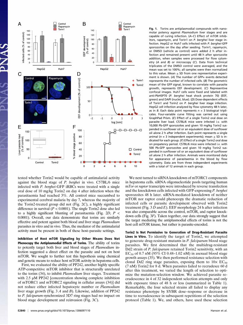
tested whether Torin2 would be capable of antimalarial activityagainst the blood stage of P. berghei in vivo. C57BL/6 miceinfected with P. berghei-GFP iRBCs were treated with a singleoral dose of 10 mg/kg Torin2 on day 4 after infection when theparasitaemia had reached 3%. All control mice succumbed toexperimental cerebral malaria by day 7, whereas the majority ofthe Torin2-treated group did not (Fig. 2C), a highly significantdifference in survival (P < 0.0001). The single Torin2 dose also ledto a highly significant blunting of parasitaemia (Fig. 2D, P <0.0001). Overall, our data demonstrate that torins are similarlyeffective and potent against both blood and liver stage Plasmodiumparasites in vitro and in vivo. Thus, the mediator of the antimalarialactivity must be present in both of these host–parasite settings.
Inhibition of Host mTOR Signaling by Other Means Does NotPhenocopy the Antiplasmodial Effects of Torins. The ability of torinsto potently target both liver and blood stages of Plasmodium in-fection suggested a direct effect on the parasite and not on hostmTOR. We sought to further test this hypothesis using chemicaland genetic means to reduce host mTOR activity in hepatoma cells.First, we evaluated the ability of PP242, another highly specific
ATP-competitive mTOR inhibitor that is structurally unrelatedto the torins (34), to inhibit Plasmodium liver stages. Treatmentwith 2.5 μM PP242 [concentration showing complete inhibitionof mTORC1 and mTORC2 signaling in cellular assays (34)] didnot reduce either infected hepatocyte number or Plasmodiumliver stage growth (Fig. 3 A and B). Likewise, addition of PP242to P. falciparum-synchronized 3D7 ring stages had no impact onblood stage development and reinvasion (Fig. 3C).
We next turned to siRNA knockdown of mTORC1 componentsin hepatoma cells. siRNA oligonucleotide pools targeting humanmTor or raptor transcripts were introduced by reverse transfectionand the knockdown cells infected with GFP-expressing P. bergheisporozoites 48 h later. siRNA-mediated knockdown of neithermTOR nor raptor could phenocopy the dramatic reduction ofinfected cells or parasite development observed with Torin2treatment (Fig. 3 D and E). EEF morphology 24 h after infectionwas also comparable across the control, mTOR, and raptor knock-down cells (Fig. 3F). Taken together, our data strongly suggest thatthe target mediating the antiplasmodial effects of torins is not thehost cell mTOR kinase, but rather is parasite-encoded.
Torin2 Is Not Permissive to Generation of Drug-Resistant ParasiteLines in Vitro. To identify the target of Torin2, we attemptedto generate drug-resistant mutants in P. falciparum blood stageparasites. We first determined that the multidrug-resistantDd2 strain of P. falciparum retained Torin2 sensitivity, with anEC50 of 0.7 nM (95% CI 0.48–1.02 nM) in asexual blood stagegrowth assays (35). We then performed resistance selection withclonal Dd2 ring stage parasites, exposing them to 10× EC50(7 nM) Torin2 for 8 d. When parasites failed to recrudesce 60 dafter this treatment, we varied the length of selection to opti-mize the mutation-selection window. We achieved parasite re-crudescence in 4 of 32 independent selection attempts and onlywith exposure times of 48 h or less (summarized in Table 1).Remarkably, the four selected strains all failed to display anyresistance phenotype by SYBR dose–response or decrease intime to recrudescence in subsequent repetitions of the selectionprotocol (Table 1). We, and others, have used these selection
Fig. 1. Torins are antiplasmodial compounds with nano-molar potency against Plasmodium liver stages and arecapable of curing infection. (A–C) Effect of mTOR inhib-itors, rapamycin, and Torin1 on P. berghei liver stage in-fection. HepG2 or Huh7 cells infected with P. berghei-GFPsporozoites on the day after seeding. Torin1, rapamycin,or DMSO (vehicle as control) were added 2 h after in-fection and remained present until 48 h after sporozoiteaddition, when samples were processed for flow cytom-etry (A and B) or microscopy (C). Data from technicaltriplicates of the DMSO control were averaged, and themean was set to 100%; all samples were then normalizedto this value. Mean ± SD from one representative experi-ment is shown. (A) The number of GFP+ events detectedrepresents the number of infected cells. (B) The geometricmean of the GFP signal, known to correlate with parasitegrowth, represents EEF development. (C) Representiveconfocal images. Huh7 cells were fixed and labeled withanti-PbHSP70 (P. berghei heat shock protein 70) (EEF,green) and DAPI (nuclei, blue). (D) Dose-dependent effectsof Torin1 and Torin2 on P. berghei liver stage infection.HepG2 cell infection analyzed by flow cytometry 48 h later,as in B. Each data point represents n = 3 biological tripli-cates. Four-variable curve fitting was carried out usingGraphPad Prism. (E) Effect of a single Torin2 oral dose onparasite liver load. C57BL/6 mice were infected i.v. with10,000 Pb-GFP sporozoites and given 10 mg/kg Torin2 sus-pended in sunflower oil or an equivalent dose of sunfloweroil alone 2 h after infection. Each point represents a singleanimal (n = 3 independent experiments); mean ± SD is in-dicated for each group. (F) Effect of a single Torin2 oral doseon prepatency period. C57BL/6 mice were infected i.v. with500 Pb-GFP sporozoites and given 10 mg/kg Torin2 sus-pended in sunflower oil or an equivalent dose of sunfloweroil alone 2 h after infection. Animals were monitored dailyfor appearance of parasitaemia in the blood by flowcytometry. Data are from three independent experimentswith a total of 12 animals in each group.
E2840 | www.pnas.org/cgi/doi/10.1073/pnas.1306097110 Hanson et al.
methodologies to raise resistance to antimalarials that act ona variety of targets within the parasite’s cytosol and mitochon-dria (36, 37). In contrast, Torin2 pressure has thus far failed toinduce a stable resistance phenotype after varied, repeatedattempts, suggesting that additional unbiased approaches will benecessary to elucidate the drug target(s).
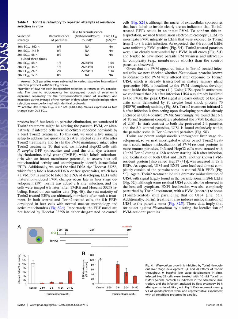
Plasmodium Growth Is Inhibited by Torin2 Throughout Liver StageDevelopment. With the evidence leaning toward torins acting di-rectly on Plasmodium spp., we sought to gain insight into the killingmechanism by asking when the target(s) of Torin2 antiplasmodialactivity are present in liver stages. We first varied compoundtreatment windows to target different stages of parasite invasionand development during liver stage infection. Initially, wechecked whether Torin2 treatment of HepG2 cells 2 h beforesporozoite addition could impact either sporozoite invasion orEEF development. Torin2 pretreatment did not alter eitherparasite numbers or development, as assayed 24 h after infection(Fig. S1A). We next tested if Torin2 treatment concomitantwith sporozoite addition would affect parasite invasion and againfound no effect; comparable amounts of cells were infected(GFP+) after 2 h, demonstrating that sporozoite invasion occursnormally in the presence of Torin2 (Fig. S1B). Next, sporozoiteswere allowed to complete invasion of HepG2 cells, and then 10nM Torin2 was added for varying time periods (as schematized inFig. 4A) corresponding roughly to “early PVM remodeling” (2 h),trophozoite (6 h), and schizont (24 h) stages of intrahepatocytedevelopment. Infection was analyzed 50 h after sporozoite addi-
tion by flow cytometry, and none of the Torin2 treatment periodswas found to significantly increase HepG2 cell death (Fig. S1C).As previously shown, continuous incubation of infected cells with10 nM Torin2 after sporozoite invasion results in near-completeparasite elimination (Fig. 4A). Remarkably, a mere 4-h in-cubation with 10 nM Torin2 from 2 to 6 h after infection was alsocapable of eliminating more than 90% of all parasites (Fig. 4A);Torin2 treatment from 6 to 24 h postinfection was similarly ef-fective (Fig. 4A). The very few developing EEFs under the 2- to 6-or 6- to 24-h conditions showed slightly reduced developmentcompared with the control (Fig. 4B). Interestingly, when Torin2treatment was initiated only after the start of schizogony (24–50 h),fewer parasites were eliminated, with parasite numbers about60% of the control (Fig. 4B). However, in this treatment group,EEF development showed the strongest inhibition—to 30% ofcontrol levels (Fig. 4B); on an individual level, these parasitesshow aberrant development and fail to form merozoites (Fig.S1D). Our data demonstrate that Torin2 is a potent inhibitor ofall phases of Plasmodium EEF development through late schi-zogony. Torin2-treated parasite elimination, however, occurredefficiently only when EEFs were exposed to Torin2 before theonset of schizogony.
Torin2 Treatment Leaves the PVM Structurally Intact, but LackingPVM-Resident Proteins. A number of gene-knockout parasitelines that successfully invade hepatocytes exist, but fail duringPVM establishment or remodeling and are rapidly eliminated(11, 12, 17–20, 38). Given this phenotypic parallel to the effectsof Torin treatment, which also does not affect the invasion
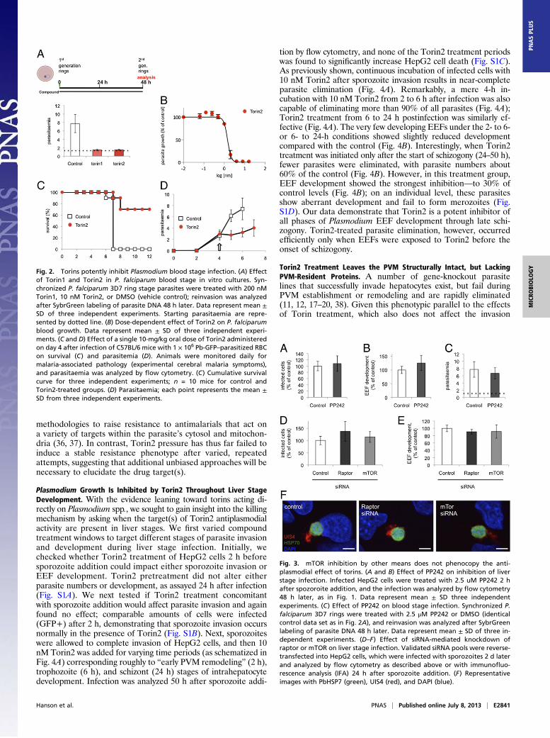
Fig. 2. Torins potently inhibit Plasmodium blood stage infection. (A) Effectof Torin1 and Torin2 in P. falciparum blood stage in vitro cultures. Syn-chronized P. falciparum 3D7 ring stage parasites were treated with 200 nMTorin1, 10 nM Torin2, or DMSO (vehicle control); reinvasion was analyzedafter SybrGreen labeling of parasite DNA 48 h later. Data represent mean ±SD of three independent experiments. Starting parasitaemia are repre-sented by dotted line. (B) Dose-dependent effect of Torin2 on P. falciparumblood growth. Data represent mean ± SD of three independent experi-ments. (C and D) Effect of a single 10-mg/kg oral dose of Torin2 administeredon day 4 after infection of C57BL/6 mice with 1 × 10⁶ Pb-GFP–parasitized RBCon survival (C) and parasitemia (D). Animals were monitored daily formalaria-associated pathology (experimental cerebral malaria symptoms),and parasitaemia was analyzed by flow cytometry. (C) Cumulative survivalcurve for three independent experiments; n = 10 mice for control andTorin2-treated groups. (D) Parasitaemia; each point represents the mean ±SD from three independent experiments.
Fig. 3. mTOR inhibition by other means does not phenocopy the anti-plasmodial effect of torins. (A and B) Effect of PP242 on inhibition of liverstage infection. Infected HepG2 cells were treated with 2.5 uM PP242 2 hafter spozoroite addition, and the infection was analyzed by flow cytometry48 h later, as in Fig. 1. Data represent mean ± SD three independentexperiments. (C) Effect of PP242 on blood stage infection. Synchronized P.falciparum 3D7 rings were treated with 2.5 μM PP242 or DMSO (identicalcontrol data set as in Fig. 2A), and reinvasion was analyzed after SybrGreenlabeling of parasite DNA 48 h later. Data represent mean ± SD of three in-dependent experiments. (D–F) Effect of siRNA-mediated knockdown ofraptor or mTOR on liver stage infection. Validated siRNA pools were reverse-transfected into HepG2 cells, which were infected with sporozoites 2 d laterand analyzed by flow cytometry as described above or with immunofluo-rescence analysis (IFA) 24 h after sporozoite addition. (F) Representativeimages with PbHSP7 (green), UIS4 (red), and DAPI (blue).
Hanson et al. PNAS | Published online July 8, 2013 | E2841
MICRO
BIOLO
GY
PNASPL
US
process itself, but leads to parasite elimination, we wondered ifTorin2 treatment might be altering the parasite PVM, or alter-natively, if infected cells were selectively rendered nonviable bya brief Torin2 treatment. To this end, we used a live imagingsetup to address two questions: (i) Are infected cells viable afterTorin2 treatment? and (ii) Is the PVM maintained intact afterTorin2 treatment? To that end, we infected HepG2 cells withP. berghei-GFP sporozoites and used the vital dye tetrame-thylrhodamine, ethyl ester (TMRE), which labels mitochon-dria with an intact membrane potential, to assess host-cellmitochondrial activity and unambiguously identify intracellularEEFs. Additionally, we used the vital DNA dye Hoechst 33258,which freely labels host-cell DNA or free sporozoites, which lacka PVM, but is unable to label the DNA of developing EEFs untilmaturation-induced PVM changes occur late in liver stage de-velopment (39). Torin2 was added 2 h after infection, and thecells were imaged 6 h later, after TMRE and Hoechst 33258 la-beling. Based on our earlier data (Fig. 4B), the vast majority ofTorin2-treated EEFs are ultimately nonviable after such a treat-ment. In both control and Torin2-treated cells, the 8-h EEFsdeveloped in host cells with normal nuclear morphology andactive mitochondria (Fig. S2A). Importantly, the EEF nuclei arenot labeled by Hoechst 33258 in either drug-treated or control
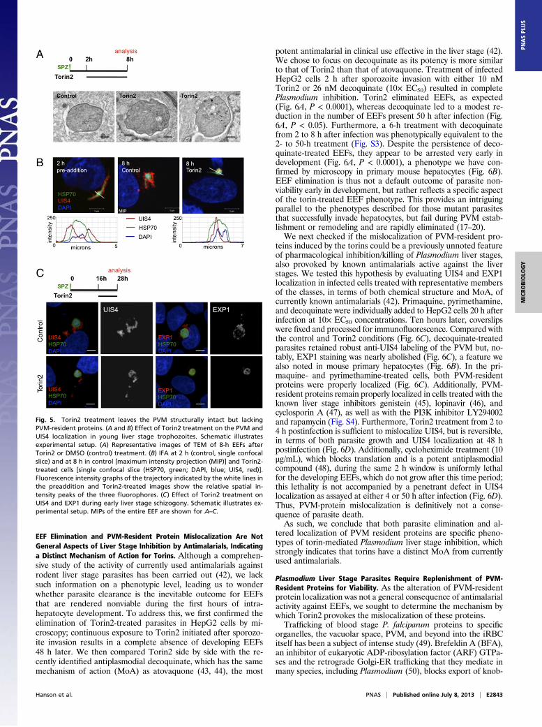
cells (Fig. S2A), although the nuclei of extracellular sporozoitesthat have failed to invade clearly are an indication that Torin2-treated EEFs reside in an intact PVM. To confirm this in-terpretation, we used transmission electron microscopy (TEM) toinvestigate PVM integrity in EEFs that were exposed to Torin2from 2 to 8 h after infection. As expected, the 8-h control EEFswere uniformly PVM-positive (Fig. 5A). Torin2-treated parasiteswere also clearly surrounded by a PVM in all cases (Fig. 5A)and tended to have more parasite PM waviness and intracellu-lar complexity (e.g., membranous whorls) than the controlparasites observed.Given that the PVM appeared intact in Torin2-treated infec-
ted cells, we next checked whether Plasmodium proteins knownto localize to the PVM were altered after exposure to Torin2.UIS4, which is already transcribed in mature salivary glandsporozoites (40), is localized to the PVM throughout develop-ment inside the hepatocyte (11). Using UIS4-specific antiserum,we confirmed that 2 h after infection UIS4 was already localizedto the PVM; the peak UIS4 signal is clearly outside of the par-asite soma delineated by P. berghei heat shock protein 70(HSP70) antibody staining (Fig. 5B). Torin2 treatment initiated 2h after infection is thus acting upon developing parasites alreadyenclosed in UIS4-positive PVMs. Surprisingly, we found that 6 hof Torin2 treatment completely abolished the PVM localizationof UIS4. In stark contrast to both the pretreated 2-h parasitesand the 8-h control parasites, UIS4 is found exclusively withinthe parasite soma in Torin2-treated parasites (Fig. 5B).Torins are potent antiplasmodials throughout liver stage de-
velopment, so we next investigated whether or not Torin2 treat-ment could induce mislocalization of PVM-resident proteins inmore mature parasites. Infected HepG2 cells were treated with10 nM Torin2 during a 12-h window starting 16 h after infection,and localization of both UIS4 and EXP1, another known PVM-resident protein [also called Hep17 (41)], was assessed in 28 hEEFs. As expected, UIS4 and EXP1 were localized almost com-pletely outside of the parasite soma in control 28-h EEFs (Fig.5C). Again, Torin2 treatment led to a dramatic mislocalization ofUIS4, with signal largely found in the parasite soma in 28-h EEFs(Fig. 5C), although some residual UIS4 could also be observed inthe host-cell cytoplasm. EXP1 localization was also completelyperturbed by Torin2 treatment, with a PVM (control) to soma(Torin2-treated) shift paralleling that of UIS4 (Fig. 5C).Additionally, Torin1 treatment also induces mislocalization ofUIS4 to the parasite soma (Fig. S2B). These data imply thattorins act against Plasmodium by altering the localization ofPVM-resident proteins.
Table 1. Torin2 is refractory to repeated attempts at resistanceselection in vitro
Selectionstrategy
Recrudescenceof parasites
Days to recrudescence(first/second/third
round)*Fold EC50
(nM)**
10× EC50, 192 h 0/8 NA NA10x EC50, 144 h 0/4 NA NA20x EC50, 48 h
pulsed three times0/4 NA NA
20x EC50, 48 h 1/7 26/24/30 1.0420x EC50, 36 h 1/3 26/23/30 0.9320x EC50, 24 h 2/4 22/24/31 1.1420x EC50, 12 h 0/2 NA NA
Asexual Dd2 parasites were subjected to varied step-wise intermittentselection protocol with10x EC50 Torin2.*Number of days for each independent selection to return to 1% parasite-mia. The time to recrudescence for subsequent rounds of selection isexpressed separately for the first/second/third round of selections andexpressed as the average of the number of days when multiple independentselections were performed with identical protocols.**Parental Dd2 strain EC50 is 0.7 nM (0.48,1.02). Values expressed as foldchange over Dd2 EC50.
Fig. 4. Plasmodium growth is inhibited by Torin2 through-out liver stage development. (A and B) Effects of Torin2throughout P. berghei liver stage development in vitro.Infected HepG2 cells were treated with 10 nM Torin2 orDMSO (vehicle control) as indicated in the schematic illus-tration, and the infection analyzed by flow cytometry 50 hafter sporozoite addition, as in Fig. 1. Data represent mean ±SD of quadruplicates from one representative experimentwith all conditions processed in parallel.
E2842 | www.pnas.org/cgi/doi/10.1073/pnas.1306097110 Hanson et al.
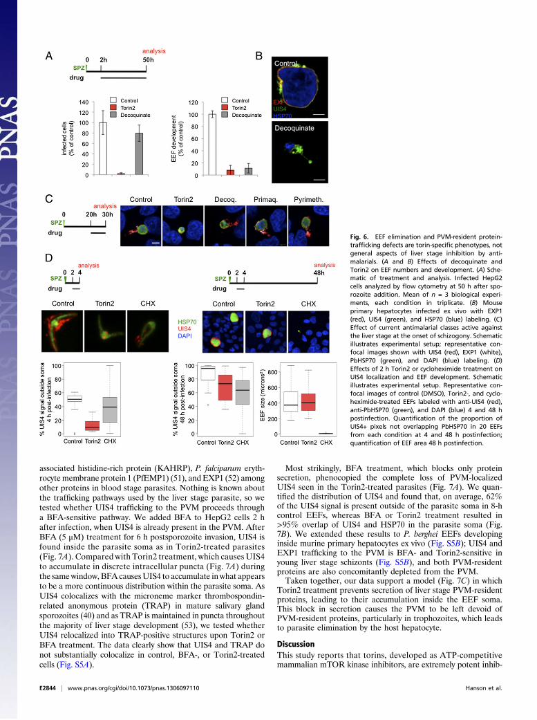
EEF Elimination and PVM-Resident Protein Mislocalization Are NotGeneral Aspects of Liver Stage Inhibition by Antimalarials, Indicatinga Distinct Mechanism of Action for Torins. Although a comprehen-sive study of the activity of currently used antimalarials againstrodent liver stage parasites has been carried out (42), we lacksuch information on a phenotypic level, leading us to wonderwhether parasite clearance is the inevitable outcome for EEFsthat are rendered nonviable during the first hours of intra-hepatocyte development. To address this, we first confirmed theelimination of Torin2-treated parasites in HepG2 cells by mi-croscopy; continuous exposure to Torin2 initiated after sporozo-ite invasion results in a complete absence of developing EEFs48 h later. We then compared Torin2 side by side with the re-cently identified antiplasmodial decoquinate, which has the samemechanism of action (MoA) as atovaquone (43, 44), the most
potent antimalarial in clinical use effective in the liver stage (42).We chose to focus on decoquinate as its potency is more similarto that of Torin2 than that of atovaquone. Treatment of infectedHepG2 cells 2 h after sporozoite invasion with either 10 nMTorin2 or 26 nM decoquinate (10× EC50) resulted in completePlasmodium inhibition. Torin2 eliminated EEFs, as expected(Fig. 6A, P < 0.0001), whereas decoquinate led to a modest re-duction in the number of EEFs present 50 h after infection (Fig.6A, P < 0.05). Furthermore, a 6-h treatment with decoquinatefrom 2 to 8 h after infection was phenotypically equivalent to the2- to 50-h treatment (Fig. S3). Despite the persistence of deco-quinate-treated EEFs, they appear to be arrested very early indevelopment (Fig. 6A, P < 0.0001), a phenotype we have con-firmed by microscopy in primary mouse hepatocytes (Fig. 6B).EEF elimination is thus not a default outcome of parasite non-viability early in development, but rather reflects a specific aspectof the torin-treated EEF phenotype. This provides an intriguingparallel to the phenotypes described for those mutant parasitesthat successfully invade hepatocytes, but fail during PVM estab-lishment or remodeling and are rapidly eliminated (17–20).We next checked if the mislocalization of PVM-resident pro-
teins induced by the torins could be a previously unnoted featureof pharmacological inhibition/killing of Plasmodium liver stages,also provoked by known antimalarials active against the liverstages. We tested this hypothesis by evaluating UIS4 and EXP1localization in infected cells treated with representative membersof the classes, in terms of both chemical structure and MoA, ofcurrently known antimalarials (42). Primaquine, pyrimethamine,and decoquinate were individually added to HepG2 cells 20 h afterinfection at 10× EC50 concentrations. Ten hours later, coverslipswere fixed and processed for immunofluorescence. Compared withthe control and Torin2 conditions (Fig. 6C), decoquinate-treatedparasites retained robust anti-UIS4 labeling of the PVM but, no-tably, EXP1 staining was nearly abolished (Fig. 6C), a feature wealso noted in mouse primary hepatocytes (Fig. 6B). In the pri-maquine- and pyrimethamine-treated cells, both PVM-residentproteins were properly localized (Fig. 6C). Additionally, PVM-resident proteins remain properly localized in cells treated with theknown liver stage inhibitors genistein (45), lopinavir (46), andcyclosporin A (47), as well as with the PI3K inhibitor LY294002and rapamycin (Fig. S4). Furthermore, Torin2 treatment from 2 to4 h postinfection is sufficient to mislocalize UIS4, but is reversible,in terms of both parasite growth and UIS4 localization at 48 hpostinfection (Fig. 6D). Additionally, cycloheximide treatment (10μg/mL), which blocks translation and is a potent antiplasmodialcompound (48), during the same 2 h window is uniformly lethalfor the developing EEFs, which do not grow after this time period;this lethality is not accompanied by a penetrant defect in UIS4localization as assayed at either 4 or 50 h after infection (Fig. 6D).Thus, PVM-protein mislocalization is definitively not a conse-quence of parasite death.As such, we conclude that both parasite elimination and al-
tered localization of PVM resident proteins are specific pheno-types of torin-mediated Plasmodium liver stage inhibition, whichstrongly indicates that torins have a distinct MoA from currentlyused antimalarials.
Plasmodium Liver Stage Parasites Require Replenishment of PVM-Resident Proteins for Viability. As the alteration of PVM-residentprotein localization was not a general consequence of antimalarialactivity against EEFs, we sought to determine the mechanism bywhich Torin2 provokes the mislocalization of these proteins.Trafficking of blood stage P. falciparum proteins to specific
organelles, the vacuolar space, PVM, and beyond into the iRBCitself has been a subject of intense study (49). Brefeldin A (BFA),an inhibitor of eukaryotic ADP-ribosylation factor (ARF) GTPa-ses and the retrograde Golgi-ER trafficking that they mediate inmany species, including Plasmodium (50), blocks export of knob-
Fig. 5. Torin2 treatment leaves the PVM structurally intact but lackingPVM-resident proteins. (A and B) Effect of Torin2 treatment on the PVM andUIS4 localization in young liver stage trophozoites. Schematic illustratesexperimental setup. (A) Representative images of TEM of 8-h EEFs afterTorin2 or DMSO (control) treatment. (B) IFA at 2 h (control, single confocalslice) and at 8 h in control [maximum intensity projection (MIP)] and Torin2-treated cells [single confocal slice (HSP70, green; DAPI, blue; UIS4, red)].Fluorescence intensity graphs of the trajectory indicated by the white lines inthe preaddition and Torin2-treated images show the relative spatial in-tensity peaks of the three fluorophores. (C) Effect of Torin2 treatment onUIS4 and EXP1 during early liver stage schizogony. Schematic illustrates ex-perimental setup. MIPs of the entire EEF are shown for A–C.
Hanson et al. PNAS | Published online July 8, 2013 | E2843
MICRO
BIOLO
GY
PNASPL
US
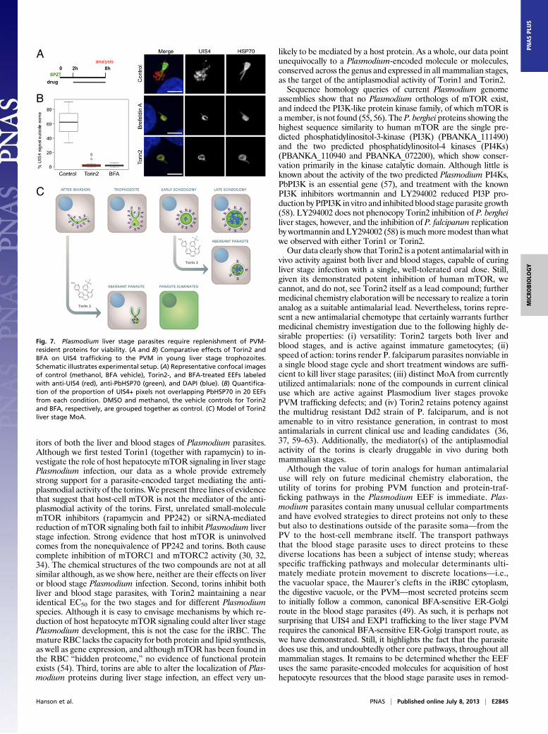
associated histidine-rich protein (KAHRP), P. falciparum eryth-rocytemembrane protein 1 (PfEMP1) (51), and EXP1 (52) amongother proteins in blood stage parasites. Nothing is known aboutthe trafficking pathways used by the liver stage parasite, so wetested whether UIS4 trafficking to the PVM proceeds througha BFA-sensitive pathway. We added BFA to HepG2 cells 2 hafter infection, when UIS4 is already present in the PVM. AfterBFA (5 μM) treatment for 6 h postsporozoite invasion, UIS4 isfound inside the parasite soma as in Torin2-treated parasites(Fig. 7A). Compared with Torin2 treatment, which causes UIS4to accumulate in discrete intracellular puncta (Fig. 7A) duringthe samewindow, BFA causesUIS4 to accumulate in what appearsto be a more continuous distribution within the parasite soma. AsUIS4 colocalizes with the microneme marker thrombospondin-related anonymous protein (TRAP) in mature salivary glandsporozoites (40) and as TRAP is maintained in puncta throughoutthe majority of liver stage development (53), we tested whetherUIS4 relocalized into TRAP-positive structures upon Torin2 orBFA treatment. The data clearly show that UIS4 and TRAP donot substantially colocalize in control, BFA-, or Torin2-treatedcells (Fig. S5A).
Most strikingly, BFA treatment, which blocks only proteinsecretion, phenocopied the complete loss of PVM-localizedUIS4 seen in the Torin2-treated parasites (Fig. 7A). We quan-tified the distribution of UIS4 and found that, on average, 62%of the UIS4 signal is present outside of the parasite soma in 8-hcontrol EEFs, whereas BFA or Torin2 treatment resulted in>95% overlap of UIS4 and HSP70 in the parasite soma (Fig.7B). We extended these results to P. berghei EEFs developinginside murine primary hepatocytes ex vivo (Fig. S5B); UIS4 andEXP1 trafficking to the PVM is BFA- and Torin2-sensitive inyoung liver stage schizonts (Fig. S5B), and both PVM-residentproteins are also concomitantly depleted from the PVM.Taken together, our data support a model (Fig. 7C) in which
Torin2 treatment prevents secretion of liver stage PVM-residentproteins, leading to their accumulation inside the EEF soma.This block in secretion causes the PVM to be left devoid ofPVM-resident proteins, particularly in trophozoites, which leadsto parasite elimination by the host hepatocyte.
DiscussionThis study reports that torins, developed as ATP-competitivemammalian mTOR kinase inhibitors, are extremely potent inhib-
Fig. 6. EEF elimination and PVM-resident protein-trafficking defects are torin-specific phenotypes, notgeneral aspects of liver stage inhibition by anti-malarials. (A and B) Effects of decoquinate andTorin2 on EEF numbers and development. (A) Sche-matic of treatment and analysis. Infected HepG2cells analyzed by flow cytometry at 50 h after spo-rozoite addition. Mean of n = 3 biological experi-ments, each condition in triplicate. (B) Mouseprimary hepatocytes infected ex vivo with EXP1(red), UIS4 (green), and HSP70 (blue) labeling. (C)Effect of current antimalarial classes active againstthe liver stage at the onset of schizogony. Schematicillustrates experimental setup; representative con-focal images shown with UIS4 (red), EXP1 (white),PbHSP70 (green), and DAPI (blue) labeling. (D)Effects of 2 h Torin2 or cycloheximide treatment onUIS4 localization and EEF development. Schematicillustrates experimental setup. Representative con-focal images of control (DMSO), Torin2-, and cyclo-heximide-treated EEFs labeled with anti-UIS4 (red),anti-PbHSP70 (green), and DAPI (blue) 4 and 48 hpostinfection. Quantification of the proportion ofUIS4+ pixels not overlapping PbHSP70 in 20 EEFsfrom each condition at 4 and 48 h postinfection;quantification of EEF area 48 h postinfection.
E2844 | www.pnas.org/cgi/doi/10.1073/pnas.1306097110 Hanson et al.
itors of both the liver and blood stages of Plasmodium parasites.Although we first tested Torin1 (together with rapamycin) to in-vestigate the role of host hepatocyte mTOR signaling in liver stagePlasmodium infection, our data as a whole provide extremelystrong support for a parasite-encoded target mediating the anti-plasmodial activity of the torins.We present three lines of evidencethat suggest that host-cell mTOR is not the mediator of the anti-plasmodial activity of the torins. First, unrelated small-moleculemTOR inhibitors (rapamycin and PP242) or siRNA-mediatedreduction of mTOR signaling both fail to inhibit Plasmodium liverstage infection. Strong evidence that host mTOR is uninvolvedcomes from the nonequivalence of PP242 and torins. Both causecomplete inhibition of mTORC1 and mTORC2 activity (30, 32,34). The chemical structures of the two compounds are not at allsimilar although, as we show here, neither are their effects on liveror blood stage Plasmodium infection. Second, torins inhibit bothliver and blood stage parasites, with Torin2 maintaining a nearidentical EC50 for the two stages and for different Plasmodiumspecies. Although it is easy to envisage mechanisms by which re-duction of host hepatocyte mTOR signaling could alter liver stagePlasmodium development, this is not the case for the iRBC. ThematureRBC lacks the capacity for both protein and lipid synthesis,as well as gene expression, and although mTOR has been found inthe RBC “hidden proteome,” no evidence of functional proteinexists (54). Third, torins are able to alter the localization of Plas-modium proteins during liver stage infection, an effect very un-
likely to be mediated by a host protein. As a whole, our data pointunequivocally to a Plasmodium-encoded molecule or molecules,conserved across the genus and expressed in all mammalian stages,as the target of the antiplasmodial activity of Torin1 and Torin2.Sequence homology queries of current Plasmodium genome
assemblies show that no Plasmodium orthologs of mTOR exist,and indeed the PI3K-like protein kinase family, of which mTOR isamember, is not found (55, 56). The P. berghei proteins showing thehighest sequence similarity to human mTOR are the single pre-dicted phosphatidylinositol-3-kinase (PI3K) (PBANKA_111490)and the two predicted phosphatidylinositol-4 kinases (PI4Ks)(PBANKA_110940 and PBANKA_072200), which show conser-vation primarily in the kinase catalytic domain. Although little isknown about the activity of the two predicted Plasmodium PI4Ks,PbPI3K is an essential gene (57), and treatment with the knownPI3K inhibitors wortmannin and LY294002 reduced PI3P pro-duction by PfPI3K in vitro and inhibited blood stage parasite growth(58). LY294002 does not phenocopy Torin2 inhibition of P. bergheiliver stages, however, and the inhibition of P. falciparum replicationby wortmannin and LY294002 (58) is muchmoremodest than whatwe observed with either Torin1 or Torin2.Our data clearly show that Torin2 is a potent antimalarial with in
vivo activity against both liver and blood stages, capable of curingliver stage infection with a single, well-tolerated oral dose. Still,given its demonstrated potent inhibition of human mTOR, wecannot, and do not, see Torin2 itself as a lead compound; furthermedicinal chemistry elaboration will be necessary to realize a torinanalog as a suitable antimalarial lead. Nevertheless, torins repre-sent a new antimalarial chemotype that certainly warrants furthermedicinal chemistry investigation due to the following highly de-sirable properties: (i) versatility: Torin2 targets both liver andblood stages, and is active against immature gametocytes; (ii)speed of action: torins render P. falciparum parasites nonviable ina single blood stage cycle and short treatment windows are suffi-cient to kill liver stage parasites; (iii) distinct MoA from currentlyutilized antimalarials: none of the compounds in current clinicaluse which are active against Plasmodium liver stages provokePVM trafficking defects; and (iv) Torin2 retains potency againstthe multidrug resistant Dd2 strain of P. falciparum, and is notamenable to in vitro resistance generation, in contrast to mostantimalarials in current clinical use and leading candidates (36,37, 59–63). Additionally, the mediator(s) of the antiplasmodialactivity of the torins is clearly druggable in vivo during bothmammalian stages.Although the value of torin analogs for human antimalarial
use will rely on future medicinal chemistry elaboration, theutility of torins for probing PVM function and protein-traf-ficking pathways in the Plasmodium EEF is immediate. Plas-modium parasites contain many unusual cellular compartmentsand have evolved strategies to direct proteins not only to thesebut also to destinations outside of the parasite soma—from thePV to the host-cell membrane itself. The transport pathwaysthat the blood stage parasite uses to direct proteins to thesediverse locations has been a subject of intense study; whereasspecific trafficking pathways and molecular determinants ulti-mately mediate protein movement to discrete locations—i.e.,the vacuolar space, the Maurer’s clefts in the iRBC cytoplasm,the digestive vacuole, or the PVM—most secreted proteins seemto initially follow a common, canonical BFA-sensitive ER-Golgiroute in the blood stage parasites (49). As such, it is perhaps notsurprising that UIS4 and EXP1 trafficking to the liver stage PVMrequires the canonical BFA-sensitive ER-Golgi transport route, aswe have demonstrated. Still, it highlights the fact that the parasitedoes use this, and undoubtedly other core pathways, throughout allmammalian stages. It remains to be determined whether the EEFuses the same parasite-encoded molecules for acquisition of hosthepatocyte resources that the blood stage parasite uses in remod-
Fig. 7. Plasmodium liver stage parasites require replenishment of PVM-resident proteins for viability. (A and B) Comparative effects of Torin2 andBFA on UIS4 trafficking to the PVM in young liver stage trophozoites.Schematic illustrates experimental setup. (A) Representative confocal imagesof control (methanol, BFA vehicle), Torin2-, and BFA-treated EEFs labeledwith anti-UIS4 (red), anti-PbHSP70 (green), and DAPI (blue). (B) Quantifica-tion of the proportion of UIS4+ pixels not overlapping PbHSP70 in 20 EEFsfrom each condition. DMSO and methanol, the vehicle controls for Torin2and BFA, respectively, are grouped together as control. (C) Model of Torin2liver stage MoA.
Hanson et al. PNAS | Published online July 8, 2013 | E2845
MICRO
BIOLO
GY
PNASPL
US
elling the “inert” RBC or whether components of the host hepa-tocyte vesicular trafficking networks are co-opted by the parasite.Comparing BFA- and Torin2-treated EEFs, we find clear
commonalities and subtle differences in phenotype. When eitherdrug is added after sporozoite invasion and intial UIS4-positivePVM establishment, both treatments result in complete loss ofUIS4 at the PVM 6 h later, as well as intracellular accumulationof UIS4. BFA blocks ER to Golgi trafficking by inhibiting the P.falciparum orthologue of Arf1 (64). BFA-treated blood stageparasites form a hybrid ER- Golgi compartment (65) thataccumulates proteins utilizing the canonical secretory pathway.We interpret the similarity of BFA- and Torin2-treated EEFphenotypes as highly suggestive that Torin2, like BFA, causesa failure in anterograde protein trafficking. The compartment inwhich UIS4 is retained appears qualitatively different, however,with Torin2 treatment leading to the appearance of small puncta ofUIS4 and BFA treatment leading to a more continuous distribu-tion of UIS4 inside the parasite soma. Future characterization ofthe intracellular location in which PVM-resident proteins accu-mulate after Torin2 treatment may help shed light on which traf-ficking step is inhibited by Torin2.Localization of a membrane protein to a specific compartment
can be achieved by either its retention at, or continuous transportto, the compartment. Most intriguingly, our data illustrate thatUIS4 and EXP1 must be continuously transported to the PVMthroughout at least the first 30 h of liver stage development.Several fascinating questions arise from this: Are UIS4 and EXP1“lost” to the host cell during EEF development? Are UIS4 andEXP1 subject to retrograde trafficking back into the PV or par-asite soma? Are these dynamics generalizable to all liver stagePVM-resident proteins, or do they reflect the specific functions ofEXP1 and UIS4? Whether or not torin treatment alters proteintrafficking in Plasmodium asexual and sexual blood stages alsoremains to be established.Proteins that are known to populate the nascent PVM of the
invading P. falciparum merozoite are synthesized during the pre-ceding schizont stage and stored in the apical organelles of themerozoite before release in the invasion process (66). The trans-locon components HSP101, PTEX150, and EXP2 are examples ofthis; they are found in the dense granules of merozoites, but will beassociated with the PVM throughout the subsequent cycle post-invasion (67). The sporozoite invasion process is assumed to beanalogous to that of the merozoite. During the transit of thePlasmodium sporozoite from the bite site to the hepatocytes, UIS4is stored inside the sporozoite where it colocalizes with TRAP (40),apparently in the micronemes, and is discharged only once thesporozoite is in the process of hepatocyte invasion. We clearlydemonstrate that the pool of UIS4 that initially populates theyoung EEF PVM is gone after 6 h of either Torin2 or BFA treat-ment. As BFA is not thought to affect retrograde trafficking, it isvery unlikely that this indicates a block in UIS4 recycling from theparasite soma to the PVM, suggesting that this pool of UIS4 hasbeen degraded either by the host cell or the parasite itself. How-ever, this is not the case with the translocon components in theiRBC, in which the initial pool of proteins released from the densegranules appears to be stably retained at the PVM throughoutblood stage development, with further synthesis and traffickingnot required (67).Although it is clear that the blood stage parasite is actively
secreting proteins, such as the stage-specific ETRAMPS (24), to
the PVM throughout development, more investigation will berequired to determine if some blood PVM proteins show dy-namics like those of UIS4, and indeed whether EXP1 itself issimilarly dynamically localized to the blood stage PVM. Thehypothesis that PVM protein turnover would be related to pro-tein function is attractive, but as the liver stage PVM also mustcontend with extensive interactions with hepatocyte compo-nents (22, 68), it remains possible that the host cell itselfdictates the turnover of the liver stage PVM-resident proteinsthat we have examined.Regardless of whether host or parasite ultimately drives the
turnover of the liver stage PVM-resident proteins that we havestudied, their replenishment at the PVM via continued expres-sion and secretion through the early schizont stage, at the least,is crucial for parasite viability. Clearly, the trafficking route tothe liver stage PVM and the molecular players that mediate it,as well as the protective role of the PVM, constitute fascinatingavenues for future research into Plasmodium biology, as well asantimalarial drug development.
Experimental ProceduresSee detailed version in SI Experimental Procedures.
Plasmodium Liver Stage Assays. GFP-expressing P. berghei sporozoites wereadded to HepG2 or Huh7 cells cultured in 24-well plates. Infected cells wereprocessed and analyzed by flow cytometry as described in ref. 31.
A total of 10,000 P. berghei-GFP sporozoites were injected i.v. into C57BL6mice; 2 h later, a 10-mg/kg dose of Torin2 was given orally as a sunflower oilslurry. Control animals received an equal dose of oil. Livers were harvested44 h after infection, mRNA was extracted, and liver parasite load was de-termined by quantitative RT-PCR of P. berghei 18s rRNA.
Plasmodium Blood Stage Assays. P. falciparum strains were cultured in vitro,and parasite proliferation was determined by flow cytometry.
To test the antimalarial properties of Torin2 in vivo, 1 × 10⁶ P. berghei-GFPiRBCs were injected intraperitoneally into C57BL/6 mice, and parasitaemiawas monitored by flow cytometry. Torin2 (10 mM) in DMSO was diluted inPBS, and 10 mg/kg was given orally on day 4 postinfection when parasitaemiawas above 3%. Control animals received equal doses of DMSO in PBS.
For resistance selection, ∼109 parasites were subject to a stepwise in-termittent selection protocol beginning with 10× EC50 for various exposure-time windows. Optimal resistance-selection conditions were obtained thatselected against the majority of parasites, but allowed recrudescence ofresistant parasites within 60 d. Upon recrudescence, additional selectionrounds were conducted to optimally obtain clones uninhibited by Torin2.
Immunofluorescence and Microscopy. Infected cells were fixed in 4% para-formaldehyde (wt/vol) for 10 min at room temperature, permeabilized,blocked in 2% BSA (wt/vol), and incubated with 1° antibodies. After wash-ing, appropriate 2° antibodies were added, and coverslips were mounted inFluoromount. All images were acquired on Zeiss confocal microscopes.
ACKNOWLEDGMENTS. We thank Ana Parreira for mosquito production andinfection; Fernanda Baptista for laboratory support; and Liliana Mancio,Vanessa Luis, and Ghislain Cabal for advice and reagents. Additionally, weare grateful to Volker Heussler, Stefan Kappe, and Miguel Seabra forproviding antisera, and to David Sabatini for providing Torin1. This work wassupported by Fundação para a Ciência e Tecnologia (FCT, Portugal) GrantsPTDC/SAU-GMG/100313/2008 and EXCL/IMI-MIC/0056/2012, and EuropeanResearch Council funding (to M.M.M.). K.K.H. was supported by funds fromthe European Community’s Seventh Framework Programme (FP7/2007-2013)Marie Curie IntraEuropean Fellowship Grant PIEF-GA-2008-221854 and FCTGrant SFRH/BPD/40989/2007.
1. Murray CJL, et al. (2012) Global malaria mortality between 1980 and 2010: A
systematic analysis. Lancet 379(9814):413–431.2. O’Brien C, Henrich PP, Passi N, Fidock DA (2011) Recent clinical and molecular insights
into emerging artemisinin resistance in Plasmodium falciparum. Curr Opin Infect Dis
24(6):570–577.3. Prudêncio M, Rodriguez A, Mota MM (2006) The silent path to thousands of
merozoites: The Plasmodium liver stage. Nat Rev Microbiol 4(11):849–856.
4. Moradin N, Descoteaux A (2012) Leishmania promastigotes: Building a safe niche
within macrophages. Front Cell Infect Microbiol 2:121.5. Kumar Y, Valdivia RH (2009) Leading a sheltered life: Intracellular pathogens and
maintenance of vacuolar compartments. Cell Host Microbe 5(6):593–601.6. Lingelbach K, Joiner KA (1998) The parasitophorous vacuole membrane surrounding
Plasmodium and Toxoplasma: An unusual compartment in infected cells. J Cell Sci 111
(Pt 11):1467–1475.
E2846 | www.pnas.org/cgi/doi/10.1073/pnas.1306097110 Hanson et al.
7. Wang L, Boyer JL (2004) The maintenance and generation of membrane polarity inhepatocytes. Hepatology 39(4):892–899.
8. Bast A, et al. (2011) Defense mechanisms of hepatocytes against Burkholderiapseudomallei. Front Microbiol 2:277.
9. de Koning-Ward TF, et al. (2009) A newly discovered protein export machine inmalaria parasites. Nature 459(7249):945–949.
10. Maier AG, et al. (2008) Exported proteins required for virulence and rigidity ofPlasmodium falciparum-infected human erythrocytes. Cell 134(1):48–61.
11. Mueller AK, et al. (2005) Plasmodium liver stage developmental arrest by depletion ofa protein at the parasite-host interface. Proc Natl Acad Sci USA 102(8):3022–3027.
12. MuellerAK, LabaiedM,KappeSH,Matuschewski K (2005)GeneticallymodifiedPlasmodiumparasites as a protective experimental malaria vaccine. Nature 433(7022):164–167.
13. Maier AG, Cooke BM, Cowman AF, Tilley L (2009) Malaria parasite proteins thatremodel the host erythrocyte. Nat Rev Microbiol 7(5):341–354.
14. Singh AP, et al. (2007) Plasmodium circumsporozoite protein promotes thedevelopment of the liver stages of the parasite. Cell 131(3):492–504.
15. Vaughan AM, et al. (2012) Complete Plasmodium falciparum liver-stage developmentin liver-chimeric mice. J Clin Invest 122(10):3618–3628.
16. Hunn JP, Feng CG, Sher A, Howard JC (2011) The immunity-related GTPases inmammals: A fast-evolving cell-autonomous resistance system against intracellularpathogens. Mamm Genome 22(1–2):43–54.
17. Labaied M, et al. (2007) Plasmodium yoelii sporozoites with simultaneous deletion ofP52 and P36 are completely attenuated and confer sterile immunity against infection.Infect Immun 75(8):3758–3768.
18. van Dijk MR, et al. (2005) Genetically attenuated, P36p-deficient malarial sporozoitesinduce protective immunity and apoptosis of infected liver cells. Proc Natl Acad SciUSA 102(34):12194–12199.
19. Silvie O, Goetz K, Matuschewski K (2008) A sporozoite asparagine-rich proteincontrols initiation of Plasmodium liver stage development. PLoS Pathog 4(6):e1000086.
20. Aly AS, et al. (2008) Targeted deletion of SAP1 abolishes the expression of infectivityfactors necessary for successful malaria parasite liver infection. Mol Microbiol 69(1):152–163.
21. Desai SA, Rosenberg RL (1997) Pore size of the malaria parasite’s nutrient channel.Proc Natl Acad Sci USA 94(5):2045–2049.
22. Bano N, Romano JD, Jayabalasingham B, Coppens I (2007) Cellular interactions ofPlasmodium liver stage with its host mammalian cell. Int J Parasitol 37(12):1329–1341.
23. MacKellar DC, Vaughan AM, Aly AS, DeLeon S, Kappe SH (2011) A systematic analysisof the early transcribed membrane protein family throughout the life cycle ofPlasmodium yoelii. Cell Microbiol 13(11):1755–1767.
24. Spielmann T, Fergusen DJ, Beck HP (2003) etramps, a new Plasmodium falciparumgene family coding for developmentally regulated and highly charged membraneproteins located at the parasite-host cell interface. Mol Biol Cell 14(4):1529–1544.
25. Mikolajczak SA, Jacobs-Lorena V, MacKellar DC, Camargo N, Kappe SH (2007) L-FABPis a critical host factor for successful malaria liver stage development. Int J Parasitol 37(5):483–489.
26. Albuquerque SS, et al. (2009) Host cell transcriptional profiling during malaria liverstage infection reveals a coordinated and sequential set of biological events. BMCGenomics 10:270.
27. Chattopadhyay R, et al. (2011) Early transcriptional responses of HepG2-A16 liver cellsto infection by Plasmodium falciparum sporozoites. J Biol Chem 286(30):26396–26405.
28. Laplante M, Sabatini DM (2012) mTOR signaling in growth control and disease. Cell149(2):274–293.
29. Guertin DA, Sabatini DM (2009) The pharmacology of mTOR inhibition. Sci Signal 2(67):pe24.
30. Thoreen CC, et al. (2009) An ATP-competitive mammalian target of rapamycininhibitor reveals rapamycin-resistant functions of mTORC1. J Biol Chem 284(12):8023–8032.
31. Prudêncio M, Rodrigues CD, Ataíde R, Mota MM (2008) Dissecting in vitro host cellinfection by Plasmodium sporozoites using flow cytometry. Cell Microbiol 10(1):218–224.
32. Liu Q, et al. (2011) Discovery of 9-(6-aminopyridin-3-yl)-1-(3-(trifluoromethyl)phenyl)benzo[h][1,6]naphthyridin-2(1H)-one (Torin2) as a potent, selective, and orallyavailable mammalian target of rapamycin (mTOR) inhibitor for treatment of cancer. JMed Chem 54(5):1473–1480.
33. Buchholz K, et al. (2011) A high-throughput screen targeting malaria transmissionstages opens new avenues for drug development. J Infect Dis 203(10):1445–1453.
34. Feldman ME, et al. (2009) Active-site inhibitors of mTOR target rapamycin-resistantoutputs of mTORC1 and mTORC2. PLoS Biol 7(2):e38.
35. Van Tyne D, et al. (2011) Identification and functional validation of the novelantimalarial resistance locus PF10_0355 in Plasmodium falciparum. PLoS Genet 7(4):e1001383.
36. Dharia NV, et al. (2009) Use of high-density tiling microarrays to identify mutationsglobally and elucidate mechanisms of drug resistance in Plasmodium falciparum.Genome Biol 10(2):R21.
37. Rottmann M, et al. (2010) Spiroindolones, a potent compound class for the treatmentof malaria. Science 329(5996):1175–1180.
38. Vera IM, Beatty WL, Sinnis P, Kim K (2011) Plasmodium protease ROM1 is importantfor proper formation of the parasitophorous vacuole. PLoS Pathog 7(9):e1002197.
39. Sturm A, et al. (2009) Alteration of the parasite plasma membrane and theparasitophorous vacuole membrane during exo-erythrocytic development of malariaparasites. Protist 160(1):51–63.
40. Kaiser K, Matuschewski K, Camargo N, Ross J, Kappe SH (2004) Differentialtranscriptome profiling identifies Plasmodium genes encoding pre-erythrocytic stage-specific proteins. Mol Microbiol 51(5):1221–1232.
41. Doolan DL, et al. (1996) Identification and characterization of the protectivehepatocyte erythrocyte protein 17 kDa gene of Plasmodium yoelii, homolog ofPlasmodium falciparum exported protein 1. J Biol Chem 271(30):17861–17868.
42. Delves M, et al. (2012) The activities of current antimalarial drugs on the life cyclestages of Plasmodium: A comparative study with human and rodent parasites. PLoSMed 9(2):e1001169.
43. Nam TG, et al. (2011) A chemical genomic analysis of decoquinate, a Plasmodiumfalciparum cytochrome b inhibitor. ACS Chem Biol 6(11):1214–1222.
44. da Cruz FP, et al. (2012) Drug screen targeted at Plasmodium liver stages identifiesa potent multistage antimalarial drug. J Infect Dis 205(8):1278–1286.
45. Cunha-Rodrigues M, et al. (2008) Genistein-supplemented diet decreases malaria liverinfection in mice and constitutes a potential prophylactic strategy. PLoS ONE 3(7):e2732.
46. Hobbs CV, et al. (2009) HIV protease inhibitors inhibit the development ofpreerythrocytic-stage plasmodium parasites. J Infect Dis 199(1):134–141.
47. Meister S, et al. (2011) Imaging of Plasmodium liver stages to drive next-generationantimalarial drug discovery. Science 334(6061):1372–1377.
48. Gershon PD, Howells RE (1986) Mitochondrial protein synthesis in Plasmodiumfalciparum. Mol Biochem Parasitol 18(1):37–43.
49. Deponte M, et al. (2012) Wherever I may roam: Protein and membrane trafficking inP. falciparum-infected red blood cells. Mol Biochem Parasitol 186(2):95–116.
50. Crary JL, Haldar K (1992) Brefeldin A inhibits protein secretion and parasitematuration in the ring stage of Plasmodium falciparum. Mol Biochem Parasitol 53(1–2):185–192.
51. Wickham ME, et al. (2001) Trafficking and assembly of the cytoadherence complex inPlasmodium falciparum-infected human erythrocytes. EMBO J 20(20):5636–5649.
52. Nacer A, Berry L, Slomianny C, Mattei D (2001) Plasmodium falciparum signalsequences: Simply sequences or special signals? Int J Parasitol 31(12):1371–1379.
53. Jayabalasingham B, Bano N, Coppens I (2010) Metamorphosis of the malaria parasitein the liver is associated with organelle clearance. Cell Res 20(9):1043–1059.
54. D’Alessandro A, Righetti PG, Zolla L (2010) The red blood cell proteome andinteractome: An update. J Proteome Res 9(1):144–163.
55. Brown JR, Auger KR (2011) Phylogenomics of phosphoinositide lipid kinases:Perspectives on the evolution of second messenger signaling and drug discovery. BMCEvol Biol 11:4.
56. Ward P, Equinet L, Packer J, Doerig C (2004) Protein kinases of the human malariaparasite Plasmodium falciparum: The kinome of a divergent eukaryote. BMCGenomics 5:79.
57. Tawk L, et al. (2010) Phosphatidylinositol 3-phosphate, an essential lipid inPlasmodium, localizes to the food vacuole membrane and the apicoplast. EukaryotCell 9(10):1519–1530.
58. Vaid A, Ranjan R, Smythe WA, Hoppe HC, Sharma P (2010) PfPI3K,a phosphatidylinositol-3 kinase from Plasmodium falciparum, is exported to the hosterythrocyte and is involved in hemoglobin trafficking. Blood 115(12):2500–2507.
59. Oduola AM, Milhous WK, Weatherly NF, Bowdre JH, Desjardins RE (1988) Plasmodiumfalciparum: Induction of resistance to mefloquine in cloned strains by continuousdrug exposure in vitro. Exp Parasitol 67(2):354–360.
60. Ritchie GY, et al. (1996) In vitro selection of halofantrine resistance in Plasmodiumfalciparum is not associated with increased expression of Pgh1.Mol Biochem Parasitol83(1):35–46.
61. Eastman RT, Dharia NV, Winzeler EA, Fidock DA (2011) Piperaquine resistance isassociated with a copy number variation on chromosome 5 in drug-pressuredPlasmodium falciparum parasites. Antimicrob Agents Chemother 55(8):3908–3916.
62. Korsinczky M, et al. (2000) Mutations in Plasmodium falciparum cytochrome b thatare associated with atovaquone resistance are located at a putative drug-binding site.Antimicrob Agents Chemother 44(8):2100–2108.
63. Barnes DA, Foote SJ, Galatis D, Kemp DJ, Cowman AF (1992) Selection for high-levelchloroquine resistance results in deamplification of the pfmdr1 gene and increasedsensitivity to mefloquine in Plasmodium falciparum. EMBO J 11(8):3067–3075.
64. Baumgartner F, Wiek S, Paprotka K, Zauner S, Lingelbach K (2001) A point mutationin an unusual Sec7 domain is linked to brefeldin A resistance in a Plasmodiumfalciparum line generated by drug selection. Mol Microbiol 41(5):1151–1158.
65. Elmendorf HG, Haldar K (1993) Identification and localization of ERD2 in the malariaparasite Plasmodium falciparum: Separation from sites of sphingomyelin synthesisand implications for organization of the Golgi. EMBO J 12(12):4763–4773.
66. Cowman AF, Berry D, Baum J (2012) The cellular and molecular basis for malariaparasite invasion of the human red blood cell. J Cell Biol 198(6):961–971.
67. Bullen HE, et al. (2012) Biosynthesis, localization, and macromolecular arrangementof the Plasmodium falciparum translocon of exported proteins (PTEX). J Biol Chem287(11):7871–7884.
68. Gomes-Santos CS, et al. (2012) Highly dynamic host actin reorganization arounddeveloping Plasmodium inside hepatocytes. PLoS ONE 7(1):e29408.
Hanson et al. PNAS | Published online July 8, 2013 | E2847
MICRO
BIOLO
GY
PNASPL
US