Embed Size (px)
Citation preview
29
Topoisomerase I and II Expression in Recurrent Colorectal Cancer Cells: A Dubious Matter
Panagiotis Gouveris, Elias Skopelitis and Nicolas Tsavaris University of Athens, School of Medicine, Department of Pathophysiology, Athens
Greece
1. Introduction
DNA topoisomerases are enzymes which alter and modify the topology of double-stranded DNA, without changing the sequence of the structural units of DNA, namely the nucleotides. They act by transiently breaking and then religating the DNA helix. Therefore, they unwind the double helix, relaxing the supercoiled DNA, and they allow DNA strands or double helices to pass through each other. Topoisomerases are essential for replication, transcription, translation and recombination of DNA because, relaxing the double helix, they facilitate the function of other enzymes, like DNA and RNA polymerases. Topoisomerases are the magicians of the DNA world, as they practically solve all the topological problems occurring during every aspect of DNA metabolism (Gupta et al., 1995; Pommier et al., 1998; Stewart et al, 1998; Champoux et al., 2001; Burden et al., 1998; Kellner et al., 2003; Wang et al., 2002). The first topoisomerase discovered, in 1971, was E. coli topoisomerase I, originally known as ω protein (Wang, 1971). Next year there was the discovery of eukaryotic topoisomerase I in nuclei extract from secondary mouse-embryo cells (Champoux et al., 1972) In 1976, DNA gyrase (topoisomerse II) was purified from Escherichia coli cells (Gellert et al., 1976a). Topoisomerases have been categorized in two basic families, according to their structure and mechanism of action: type I topoisomerases and type II topoisomerases. Type I topoisomerases mediate transient breaks in one of the DNA strands. They are further classified in the subfamilies IA and IB. Type IA topoisomerases form a covalent intermediate with the 5’-phosphoryl end of DNA, while type IB topoisomerases form a covalent intermediate with the 3’-phosphoryl end of DNA (Champoux et al., 2001). On the other hand, type II topoisomersases transiently cleave both of the two DNA strands. They are also further classified in the subfamilies IIA and IIB on the basis of differences in their protein structure. In human and higher eukaryotic organisms, three groups of topoisomerases have been described. The first group includes topoisomerase I and mitochondrial DNA topoisomerase, which are type IB enzymes. The second group includes topoisomerases II┙ and II┚, which are type II enzymes. The third group, which was later discovered, includes topoisomerases III┙ and III┚, which are type ΙΑ enzymes (Lodge et al., 2000; Kwan et al., 2001; Hanai et al., 1996). Topoisomerase IV has also been described, a bacterial type II topoisomerase (Kato et al., 1990). More recently, topoisomerase V has been described, which is a prokaryotic counterpart to the eukaryotic topoisomerase I (Slesarev et al., 1994).
www.intechopen.com

DNA Replication - Current Advances 640
Topoisomerase I is the well known Scl-70 antigen, against which high autoantibodies titters are developed in diffuse cutaneous systemic scleroderma (Shero et al., 1986; Guldner et al., 1986; Shero et al., 1987). Antibodies against topoisomerase II┙ are detected in localized scleroderma (Takehara et al., 2005). Topoisomerases III┙ and III┚ are possibly involved in the pathogenesis of Bloom, Werner and Rothmund-Thomson syndromes, which are associated with genetic instability (Kwan et al., 2001; Raynard et al., 2006). Similar enzymes, as mentioned above, do exist in the prokaryotic cell (bacterial topoisomerases-gyrases) (Wang, 1971; Gellert et al., 1976a; Brown et al., 1979), and in viruses (e.g Vaccinia virus topoisomerase I) (Shaffer et al, 1987; Shuman, 1998). The clinical significance of bacterial gyrases is great, as they constitute targets for antibiotic drugs like novobiocin, nalidixic acid and new quinolones (Gellert et al., 1976b; Sugino et al., 1977; Smith, 1986; Maxwell, 1992). Topoisomerase II┙ and topoisomerase I are the best studied human topoisomerases, and constitute molecular targets for well known and widely used antineoplastic drugs (Pommier et al., 1998; Burden et al., 1998).
2. Topoisomerase I
Human DNA topoisomerase I is essential for vital cellular processes, namely DNA replication, transcription, translation, recombination and repair. It is a 91 kDa monomeric polypeptide which consists of 765 amino acids. It is encoded by a single copy gene which is located on chromosome 20q12-13.2 (Champoux et al., 2001). It catalyzes the relaxation of both positively and negatively supercoiled DNA, while bacterial topoisomerase I catalyzes the relaxation of negatively supercoiled DNA. Superhelix density for most natural DNA molecules ranges from -0.03 to -0.09 (Bauer et al., 1980). The negative sign means that DNA superhelices are left-handed. In other words, superhelices are derived from reverse winding of the helix. During the process of transcription, RNA polymerase follows the helical path along DNA double strand, having the tendency to generate positive supercoils in front of it and negative supercoils behind it. The accumulation of such supercoils could block the process of transcription, harming cell viability. Therefore, toposomerase I acts as a swivel to relieve the torsional strain caused by the generation of positive supercoils upstream and negative supercoils downstream of the moving RNA polymerase. It gives the solution, removing any undesirable supercoil (Liu et al., 1987; Wang et al., 1993). During the process of replication, topoisomerase I has a similar function. There is a tendency of positive supercoils accumulation. Topoisomerase I relaxes the supercoils, relieving the torsional strain of the DNA molecule (Avemann et al., 1998; Hsiang et al., 1989). Topoisomerase I has also a role in the process of DNA recombination, DNA repair, and mitotic chromosome condensation (Pommier et al., 1998; Bullock et al., 1985; Wang et al., 1991; Shuman et al., 1989; Castano et al., 1996; Subramanian et al., 1998). Topoisomerase I does not require ATP for its action, in contrast to topoisomerase II, which does need ATP (Wang et al., 1969). In the active centre of the enzyme there is the active site tyrosine (Tyr-723) of which the hydroxyl group performs a nucleophilic attack on the phosphodiesteric bond of one of the two DNA strands. The phosphodiesteric bond is broken now. The enzyme remains covalently attached to the 3’ end of the broken strand, creating a transient covalent complex, known as cleavable complex. The 5’ hydroxyl group of the broken strand remains free. The DNA molecule can now rotate around the intact strand. This rotation leads to the relaxation of the abovementioned positive and negative supercoils.
www.intechopen.com
Topoisomerase I and II Expression in Recurrent Colorectal Cancer Cells: A Dubious Matter 641
The whole process is completed as the exposed 5’ hydroxyl group performs a nucleophilic attack on the transient phosphotyrosine linkage. The active site tyrosin is detached from the 3’ end. The cleaved strand is resealed (Pommier et al., 1998; Sari et al., 2005). In summary, topoisomrasre I unwinds and uncoils the DNA supercoiled double helix by transiently cleaving one of the two strands and allowing rotation over the other. In the end it reseals the cleaved strand. Topoisomerase I is not essential for viability in yeast (Thrash et al., 1984; Uemura et al.,
1984). On the contrary, it is essential for embryonic development in Drosophila melanogaster
and mouse (Lee et al., 1993; Morham et al., 1996).
By contrast to topoisomerase II, topoisomerase I levels are not cycle-specific and remain
relatively constant throughout the cell cycle (Heck et al., 1988). Topoisomerase I seems to be
expressed in all cells, even those which do not divide. However, the enzyme levels are
higher in cancer tissues, compared to the adjacent normal tissues. This fact renders
topoisomerase I an attractive target for chemotherapy drugs (Bronstein et al., 1996; Husain
et al., 1994).
Topoisomerase I catalytic activity was the first parameter of topoisomerase I evaluable in
biomedical studies and it was detected in all normal tissues at a fairly constant level. Two
more parameters were added in the investigational process: topoisomerase I immunoprotein
levels, estimated by Western Blotting analysis, and topoisomerase I gene expression
(toposomerase I mRNA), evaluated by Northern Blotting analysis. There seems to be a good
correlation between topoisomerase expression and the catalytic activity of the enzyme
(Bronstein et al., 1996; Husain et al., 1994). Using the abovementioned parameters and
methods, elevated topoisoomerase I levels were detected in ovarian cancer, cervical cancer,
colorectal cancer, prostate cancer, malignant melanoma, lymphoma (Bronstein et al., 1996;
Husain et al., 1994; Van der Zee et al., 1991; Van der Zee et al., 1994; Perego et al., 1994;
McLeod et al., 1994; Goldwasser et al., 1995; Guichard et al., 1999; Giovanella et al., 1989;
Florell et al., 1996). On the other hand, there was no elevation in topoisomerase I levels
detected in lung cancer, breast cancer, renal cancer, and rhabdomyosarcoma (Bronstein et
al., 1996; Husain et al., 1994; McLeod et al., 1994). With regard to colorectal tumors,
topoisomerase I levels have been found to demonstrate 5-35-fold increases in the cancer
tissue, compared to the adjacent normal colonic mucosa (Bronstein et al., 1996; Husain et al.,
1994). In 1997, a new evaluable parameter of topo I appeared in biomedical studies: topo I
expression, evaluated by immunohistochemistry in paraffin embedded human tissues. Since
then, topo I expression has been estimated in several neoplastic tissues. In fact, elevation of
topo I was found in ovarian carcinomas (43% of tumors examined); colorectal carcinomas
(ranging from 43% to 86% in different studies) ; testicular tumors (30-38% of seminomas,
30% of embryonal carcinomas, but 100% of teratomas and yolk sac tumours); urinary
bladder carcinomas (77%); renal cell carcinomas (ranging from 36-100% according to
histological grade); malignant melanomas (41,6%); gastric carcinomas (68%); sarcomas
(13%); breast carcinomas (41%); oral dysplasias (79%) and squamous cell carcinomas (92%)
(Holden et al., 1997; Boonsong et al., 2002; Staley et al., 1999; Paradiso et al., 2004; Monnin et
al., 1999; Coleman et al., 2001; Coleman et al., 2000; Berney et al., 2002; Lynch et al., 2001;
Gupta et al., 2000; Lynch et al., 1998; Hafian et al., 2004; Coleman et al., 2002). As far as
topoisomerase I immunoreactivity in normal tissues is concerned, this appeared to be
strongest in the germinal centres of the tonsil and in the lymphocytes of colonic mucosa,
while it was also detected in the glandular colonic epithelium (Holden et al., 1997).
www.intechopen.com

DNA Replication - Current Advances 642
3. Topoisomerase II
Topoisomerase II is a ubiquitous enzyme which is essential for the viability of all eukaryotic organisms and plays a crucial role in literally every aspect of DNA metabolism and chromosome organization. In contrast to topoisomerase I, which is actually monomeric, topoisomerase II exists in two homologous but different isoforms, designated IΙ┙ and II┚, which are closely related. The two isoforms share homology in the amino acid sequence (up to 70%), but they are encoded by separate genes. Topoisomerse II┙ is the isoform originally described and characterized in mammalian species. It has a molecular mass of 170 kDa and is encoded by a gene which is located on chromosome 17q21-22. Topoisomerse II┚ has a molecular mass of 180 kDa and is encoded by a gene which is located on chromosome 3q24. Both of the proteins exist as homodimers (Burden et al., 1998; Kellner et al., 2003; Pommier et al., 2001; Jenkins et al., 1992; Austin et al., 1993). Topoisomerase II┚ concentrations are relatively constant during the cell cycle. On the contrary, topoisomerase II┙ levels are tightly associated with the proliferative state of the cell, and increase 2-3 fold during G2/M phase. This increase takes place in rapidly proliferating tissues, while quiescent populations demonstrate low enzyme levels (Heck et al., 1988). Therefore, it is believed that the ┚ isoform is responsible for the “housekeeping” functions of the enzyme, while the ┙ isoform seems to be the type II enzyme which unlinks daughter chromosomes following replication. A small percentage of the total topoisomerase II pool in mammalian cells exists as ┙/┚ heterodimers (Biersack et al., 1996). The enzymological characteristics of all eukaryotic type II appear to be similar. Each topoisomerase II monomer can be divided in three domains. The N-terminal domain, which includes the first 660 amino acids of the enzyme, is homologous to the B subunit of DNA gyrase and contains sequences for ATP binding. The central domain of the enzyme, which extends to amino acid 1200, is homologous to the A subunit of DNA gyrase and contains the active site tyrosine that forms the covalent bond with DNA during scission. The C-terminal domain of the enzyme varies from species to species and doesn’t seem to have homology with DNA gyrase. The physiological function of this domain remains unclear. It possibly has a role in modulating the DNA cleavage/ligation reaction of the enzyme (Wang et al., 1996; Berger et al., 1998; Dickey et al., 2005). Topoisomerase II works in a way similar to topoisomerase I, with the difference that it cleaves both strands of the nucleic acid substrate, allowing the passage of an intact double helix through the break. The enzyme Topoisomerase II is able to catalyze the relaxation of both positively and negatively supercoiled DNA. Its catalytic action begins with the simultaneous cleavage of both of the two DNA strands. The active site tyrosins (one for each monomer of the homodimer) covalently bind to the 5’ end of the broken strands and create the transient complex which is known as cleavable complex. Till this step, energy from ATP hydrolysis is not required. The protein dimer is stabilized with disulfide bridges that create a gate through which we have the passage of another intact double-stranded DNA helix. This passage takes place at the expense of ATP hydrolysis. The whole procedure is completed with the resealing of the cleaved double strand (Roca et al., 1994; Schultz et al., 1996; Lindsley et al., 1996). As a result of this DNA passage mechanism, topoisomerase II is able not only to remove negative or positive DNA supercoils, but also to unlink intertwined pairs of newly replicated chromosomes. This extra feature of topoisomerase II is important for chromosome organization and segregation. Topoisomerase I doesn’t share this feature (Roca et al., 1996).
www.intechopen.com
Topoisomerase I and II Expression in Recurrent Colorectal Cancer Cells: A Dubious Matter 643
As mentioned above, topoisomerase II┙ levels are cell cycle-dependent, with maximum concentrations measured during the G2/M phase. That’s why the percentage of positive of positive what? for topoisomerase II┙ cells practically represents the percentage of dividing cells. Thus, several studies have displayed that topoisomerase II┙ levels may act as a reliable marker of cell proliferation in tumors. Topoisomerase II┙ has been used as a proliferation marker in several studies, including patients with colorectal cancer (Monnin et al., 1999; Holden et al., 1995; Nakopoulou et al., 2001; Gibbons et al., 1997). Concerning normal tissues, topoisomerase II┙ levels are higher in tissues with proliferating cells (eg. spermatocytes, germinal cells, and proliferative endometrium) references?. In contrast, no detectable topoismerase II┙ was detected in terminally differentiated tissues, e.g cerebral cortex, skeletal muscle, and nerve (Bauman et al., 1997). Concerning neoplastic tissues, high levels of topoisomerase II┙ are observed in biologically aggressive or rapidly proliferating tumors, like high-grade lymphomas or seminomas. Topoisomerase II┙ is detectable in both the cell nucleus and cytoplasm. Topoisomerase II┚ is localized both in the nucleoli and the nucleoplasm. It is ubiquitously expressed in vivo and it is present in quiescent cell populations (Turley et al., 1997). Concerning normal colon mucosa, topoisomerase II┙ is expressed only in the lower crypt zone. In adenomas, the topoisomerase II┙ expression is expanded in the upper crypt region, while it is diffuse in carcinomas ( Fogt et al., 1997). Topoisomerase II constitutes the molecular target of a great number of antineoplastic drugs, which are widely used in cancer chemotherapy, including anthracyclins and epipodophyllotoxins. These drugs constitute substrates of the classic MDR proteins. It is interesting that topoisomerase II is implicated in a type of multiple drug resistance which is called atypical MDR. There is actually a type of multiple drug resistance only to anthracyclins and epipodophyllotoxins, not the vinca alkaloids and colchicine. This type has no relation with classical MDR (Borst et al, 1995). This atypical MDR is attributed to mutations associated with topoisomerase II. There are two basic types of such mutations: 1. Mutations which lead to low levels of the enzyme. Drugs targeting topoisomerase II
stabilize the cleavable complexes between the enzyme and DNA, creating permanent breaks of the double strand. Reduced levels of topoisomerase II lead to reduced DNA strand breaks, therefore reduced drug activity (Zijlstra et al., 1990; Cole et al., 1991; Sullivan et al., 1987).
2. Mutations leading to a qualitatively modified topoisomerase II, less sensitive to the chemotherapy drugs (Beck et al., 1993; Glisson et al., 1987).
4. Topoisomerase I inhibitors
There are a lot of inhibiting topoisomerase I (Pommier et al., 1998; Wang et al., 1997). In clinical practice only camptothecin derivatives are used. Camptothecin (CPT) is an alkaloid found in the wood and bark of the Chinese tree Camptotheca acuminata (Nyssaceae) (Wall et al., 1966). Wall and Perdue isolated CPT from Camptotheca acuminate during the ‘60s. CPT and its derivatives were also found in other plant families. In the early phase-I and II clinical studies regarding CPT, the water soluble sodium salt NSC-100880 was used. Despite the antineoplastic activity, the studies were suspended due to unacceptable hematologic (myelosupression) and non hematologic (hemorrhagic cystitis) toxicity (Gottlieb et al., 1970; Muggia et al., 1972; Moertel et al., 1972; Gottlieb et al., 1972;
www.intechopen.com
DNA Replication - Current Advances 644
Schaeppi et al., 1974). Later, it was shown that the cytotoxic activity of camptothecin, that has pentacyclic structure, was attributed to its E-ring lactone. When the E-ring opens to form hydroxyacid, the drug becomes inactive. In water solutions the inactive open form is favored (Slichenmyer et al., 1993). On the other hand, the low water solubility of the drug was the main reason for its toxicity. Therefore, an effort began having to do with the synthesis of CPT derivatives that combine the maximum possible water solubility with the best cytotoxic activity. Semisynthetic water soluble derivatives were produced. The most important of them were topotecan (TPT) and irinotecan (CPT-11). These new drugs were developed with modification of the Α-ring of the camptothecin molecule (Kerrigan et al., 2001). Camptothecin derivatives act causing irreversible breaks on the DNA strands. They actually
act stabilizing the cleavable complexes formed by topoisomerase I, which normally have
short half life. This stabilization takes place bringing a guanine at the 5’ end of the cleaved
DNA. While the stabilization is irreversible, it causes irreversible break of the double strand,
when the replication fork meets a cleavable complex (Covey et al., 1989; Hsiang et al., 1989;
Pommier et al., 1996; Jaxel et al., 1991). When topoisomerase I levels are higher, the cleavable
complexes are more frequent, and the DNA strand breaks are more frequent, too. These
breaks lead to the cell cycle arrest at S/G2 phase, activation of apoptotic mechanisms and
cell death (Hsiang et al., 1988). That’s why camptothecin derivatives are cytotoxic in
presence of active DNA replication or RNA transcription. Cells synchronized in S phase are
1000-fold more sensitive than in phases G1 and G2/ M. Despite the fact that topoisomerase
levels remain stable during the cell cycle, camptothecin derivatives constitute cycle-specific
drugs (Liu et al., 1983; D'Incalci et al., 1993).
The term “inhibitors” when we refer to topoisomerase I inhibitors is somewhat catachrestic.
Camptothecin derivatives actually do not inhibit topoisomerase I but they use the enzyme
function in order to transform the enzyme into a cell poison.
Irinotecan is bioactivated in liver by carboxylesterase to the active metabolite SN-38. SN-38
is 1000-fold more active (Slichenmyer et al., 1993). Both irinotecan and SN-38 are susceptible
to pH – dependent reversible hydrolysis and transformation of the active closed ring
(lactone) to the open form of hydroxyacid. Acidic pH favors the active form of closed ring.
The open form not only lacks the ability of cleavable complexes stabilization, but also the
ability of entrance into the cell via the cell membrane.
The active metabolite SN-38 is responsible not only for the antineoplastic activity of
irinotecan, but also for the side-effects. Late diarrhea is the most important side effect and it
often does not respond to common anti-diarrheic drugs. SN38 is further metabolized by the
UDP-glucuronosyltransferase 1A1 (UGT1A1) enzyme to the inactive form SN38 glucuronide
(SN38-Glu), which is excreted in the gastrointestinal lumen via bile. It is believed that
intestinal bacteria produce ┚-glucuronidase, that hydrolizes SN38-Glu to the active form
SN38. SN38 causes intestinal mucosa injury, that leads to late diarrhea. At the same time, the
non-metabolized irinotecan constitutes a weak acetylcholinestarase inhibitor and may cause
acute cholinergic symptoms, among which is the early diarrhea in some patients (Abigerges
et al., 1994; Kehrer et al., 2001; Lokiec et al., 1995; Takasuna et al., 1996; Gupta et al., 1994).
Irinotecan received in 1996 and 1998 FDA approval for treatment of metastatic colorectal
cancer after failure of treatment with fluorouracil. The importance of the drug was proved in
two European randomized studies (Cunningham et al., 1998; Rougier et al., 1998). In 2000,
www.intechopen.com
Topoisomerase I and II Expression in Recurrent Colorectal Cancer Cells: A Dubious Matter 645
irinotecan received FDA approval for first line treatment in metastatic colorectal cancer,
combined with fluorouracil/leucovorin (Saltz et al., 2000; Douillard et al., 2000).
Topotecan is the second camptothecin derivative. It is itself an active drug. A low percentage of the drug is metabolized by microsomal enzymes to N-demethyltopotecan, which is also an active metabolite. In contrast to irinotecan, which is metabolized in liver, topotecan is mainly excreted in urine. Topotecan received FDA approval as second line therapy in metastatic ovarian cancer (1996) and in SCLC (1998).
5. Topoisomerase II inhibitors
Topoisomerase II inhibitors are cytostatic drugs widely used in clinical practice since decades (Hande et al., 1998). According to their mechanism of action, they are divided in two broad categories. The first category includes DNA intercalators, which intercalate between the base pairs of DNA, disrupting DNA function. This category includes cytotoxic antibiotics like anthracyclines (eg. doxorubicin, epirubicin, mitoxantrone) and aminoacridines, among which amsacrine is the main drug (Bailly, 2000). The second category includes substances which do not act with an intercalation mechanism. Epipodophyllotoxins and some isoflavones, like genistein belong to this category. Genistein is included in soy, and it is possibly responsible for the low incidence of breast cancer, prostate cancer and colorectal cancer in Asian populations (Barnes et al., 1995; Stoll et al., 1997). Quinolones, the well known family of broad spectrum antibiotic, are also topisomerase II inhibitors. They are the only group of drugs with activity against both eukaryotic topoisomerase II┙ and its prokaryotic homologue, bacterial gyrase. Quinolones are not used as antineoplasic drugs, till now, but they include widely used antibiotics like ciprofloxacin and norfloxacin (Burden et al., 1998; Maxwell et al., 1992; Robinson et al., 1991; Robinson et al., 1992). Epipodophyllotoxins were synthesized in an effort of amelioration of podophyllotoxin activity. Podophyllotoxin is a substance which constitutes extract of the plant Podophyllum peltatum, known by the American Indians for its emetical, cathartic, and anthelminthic activity (Mantle et al., 2000). Two semisynthetic glucosides were synthesized from podophyllotoxin: etoposide and tenoposide. Etoposide is produced with the attachment of podophyllotoxin to a glucopyranoside with a methyl group, while tenoposide is produced with the attachment of podophyllotoxin to a glucopyranoside with a thenyliden group. This simple modification causes an important change in the way of action of the drug. Therefore, while podophyllotoxin acts on microtubules that form mitotic spindle, etoposide and teniposide act as topoisomerase II┙ inhibitors (Schilstra et al., 1989). Etoposide and teniposide act on topoisomerase II┙, in a way similar to which irinotecan and topotecan act on topoisomerase I. They actually take advantage of the normal topoisomerase II┙ action, in order to transform it into a cell poison. The whole process takes place during stabilization of DNA- topoisomerase II┙ complexes, which induces double strand breaks (Burden et al., 1998). Cells in S and G2 phases of the cell cycle are more sensitive to epipodophyllotoxins. However, in contrast to the camptothecins derivatives, which are characterized by high cycle specificity, DNA synthesis inhibition only partially affects epipodophyllotoxin induced cytotoxicity (Holm et al., 1989). Etoposide is a widely used antineoplastic drug highly active in germ cell tumors, ovarian cancer, SCLC, NSCLC, non-Hodgkin lymphoma, acute leukemia, Ewing sarcoma, Kaposi sarcoma, neuroblastoma. Tenoposide is less used in clinical practice.
www.intechopen.com

DNA Replication - Current Advances 646
6. Original research
6.1. Pilot study 6.1.1 Materials and methods
6.1.1.1 Patients’ characteristics – study and control groups
In the study we are describing in this chapter, a total of twenty-five patients were included. Those patients had colorectal cancer which had recurred following surgery and chemotherapy. Specifically, patients had undergone complete surgical resection of the primary tumour and subsequently were submitted to a 5-FU-based adjuvant chemotherapy regimen, postoperatively. Patients were followed-up until recurrence. When recurrences occurred, patients underwent a second surgical resection. Biopsy specimens from both surgical procedures for each patient were, therefore, collected, so that at the end of the study we evaluated two histological specimens from each patient: one from the primary tumour location (i.e. before the administration of 5-FU-based adjuvant chemotherapy) and a second one from the neoplastic tissue at the recurrence site (i.e. following the 5-FU-based chemotherapy regimen). In order to be able to make comparisons we needed a control group, so we selected a group of twelve patients with colorectal cancer who underwent resection after initial diagnosis, but received no 5-FU-based adjuvant chemotherapy. Patients in the control group were age and gender mached and had also similar tumour characteristics with patients included in the main study group. For each patient in the control group two biopsy specimens were available for evaluation since they were submitted to surgery following recurrence.
6.1.1.2 Histologic evaluation – immunochemistry
For each biopsy specimen, the expression of topoismerase-I was quantified by means of standard three-step immunohistochemistry on paraffin embedded sections. For this purpose the Topogen Topo I Monoclonal Antibody (2012-3) was used; this is a monoclonal mouse antibody (IgG2b isotype). The epitope of the antibody has not been mapped. Normal human tonsil tissues served as positive control. Histological sections were examined by a single investigator with no previous knowledge of the clinical status of the examined specimen. During histologic examination, immunostaining for topoisomerase I was graded according to the percentage of tumour cells with positive staining (- and ± for <5%, + for 5%-50% and ++ for 50% to 75%) and according to the intensity of staining (weakly positive, moderately positive, or strongly positive). Only specimens with strongly immunoreactive nuclei were considered as a positive biopsy for topoisomerase I; weak and moderate intensity of staining was considered as negative for the expression of topoisomerase I.
6.1.1.3 Statistical Analysis
Following this initial evaluation, the sections were examined in pairs (one section for the first surgery and one section for the second surgery for each patient) during the statistical analysis. For the latter, we used McNemar's paired chi-square test to assess the possible modification of the levels of topoisomerase I following chemotherapy with 5-FU. Fisher’s exact test was performed in order to assess the possible relationship of the topoisomerase I increase with gender, Duke’s stage, grade of differentiation and tumour localization. Mann-Whitney U-Test was performed in order to assess possible relationship between the age of patients and changes of topoisomerase I levels as well as to investigate possible correlations between the relapse free interval (RFI) and alteration of topoisomerase I levels.
www.intechopen.com
Topoisomerase I and II Expression in Recurrent Colorectal Cancer Cells: A Dubious Matter 647
6.1.2 Results
6.1.2.1 Topoisomerase I level increase
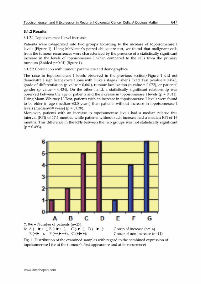
Patients were categorized into two groups according to the increase of topoisomerase I levels (Figure 1). Using McNemar’s paired chi-square test, we found that malignant cells from the tumour recurrences were characterized by the presence of a statistically significant increase in the levels of topoisomerase I when compared to the cells from the primary tumours (2-sided p=0.01) (figure 1).
6.1.2.2 Correlation with tumour parameters and demographics
The raise in topoisomerase I levels observed in the previous section/Figure 1 did not demonstrate significant correlations with Duke΄s stage (Fisher’s Exact Test p-value = 0.496), grade of differentiation (p value = 0.661), tumour localization (p value = 0.072), or patients’ gender (p value = 0.434). On the other hand, a statistically significant relationship was observed between the age of patients and the increase in topoisomerase I levels (p = 0.011). Using Mann-Whitney U-Test, patients with an increase in topoisomerase I levels were found to be older in age (median=62.5 years) than patients without increase in topoisomerase I levels (median=50 years) (p = 0.038). Moreover, patients with an increase in topoisomerase levels had a median relapse free interval (RFI) of 17.5 months, while patients without such increase had a median RFI of 16 months. This difference in the RFIs between the two groups was not statistically significant (p = 0.493).
Y: 0-6 = Number of patients (n=25) X: A (� ►++), B (+►++), C (-►+), D (�►+): Group of increase (n=14) E (+►�), F (++►++), G (+►+): Group of non-increase (n=11)
Fig. 1. Distribution of the examined samples with regard to the combined expression of topoisomerase I (i.e at the tumour’s first appearance and at its recurrence)
www.intechopen.com

DNA Replication - Current Advances 648
6.1.2.3 Comparison with control group
No statistically significant differences were found concerning topoisomerase I expression in malignant cells from the primary tumour between patients in the study and the control group.
6.1.2.4 Association with other morphologic characteristics
With regard to tumour-adjacent morphologically non-dysplastic mucosa, when colonic crypts were cut longitudinally, some topoisomerase I positive colonic cells were detectable in the proliferation zone (lower 1/3 of the colonic crypts). In totally normal colonic mucosa, obtained from tumour-free surgical margins, no such expression was detectable.
6.1.3 Discussion
In the current study, 13 out of the 25 patients with colorectal cancer (52%) stained positive for topoisomerase I (Figure 2). In respect with the percentage of tumour cells staining positive for topoisomerase I, 9 out of the 25 patients (36%) presented with 5% - 50% positive staining cells and 4 out of the 25 (16%) with >50% positive staining cells. These data are in line with Boonsong et al. (Boonsong et al., 2002), while Staley et al. (Staley et al., 1999) demonstrated a higher percentage (86%) of positive staining in samples of 29 patients. Topoisomerase I immunoreactivity was confined to be present in the nucleus of all biopsy samples, a finding which is consistent with the role of topoisomerase I as a nuclear protein. Importantly, we displayed a significant increase of topoisomerase I expression by means of immunohistochemistry in recurrences of the initial neoplasia, thus reinforcing the notion that topoisomerase I expression is likely to be part of the malignant cells’ phenotype in recurrent colorectal carcinomas. This is in agreement with previous experiments (Ichikawa et al., 1999; Paradiso et al., 2004), which demonstrated a direct correlation between thymidylate synthase (TS) and topoisomerase I expression in tumours and hypothesized, similarly to TS (Leichman et al., 1997), that high topoisomerase I expression is related to a more aggressive biological phenotype. However, our finding is in contrast with other studies (Boonsong et al., 2002; Paradiso et al., 2004), which have postulated the absence of a role for topoisomerase I in the acquisition of a metastatic phenotype. Such disperse results in the literature may reflect an interlesion heterogeneity concerning topoisomerase I expression. Since the increased expression of topoisomerase I immunostain in neoplastic tissues from recurrences was demonstrated following 5-FU-based adjuvant chemotherapy regimen, it would be tempting to attribute this particular alteration to 5-FU itself. Additionally, given the fact that high levels of topoisomerase I expression has been shown to correlate with sensitivity to camptothecin chemotherapy (Monnin et al., 1999), patients with advanced colorectal cancer are likely to benefit from topoisomerase I-targeted anticancer drug therapy. This also might explain why patients with metastatic colorectal cancer appear to benefit more when they are treated with a combination chemotherapy regimen. Of note, strong synergism between 5-FU and irinotecan (CTP-11) has been reported (Guichard et al., 1997, 1998) after sequential exposure to both agents, whereas additivity or antagonism has been reported only after simultaneous exposure. The above facts are suggestive that the mechanisms of interaction between these two drugs might be multifactorial and the specific schedule of administration represents a critical parameter of their chemotherapeutic efficacy. In fact, the combination of CTP-11 and 5-FU+leucovorin (LV) has been approved (Saltz et al., 2000, 2001; Vanhoefer et al., 2001) as reference first-line chemotherapy for
www.intechopen.com
Topoisomerase I and II Expression in Recurrent Colorectal Cancer Cells: A Dubious Matter 649
A
B
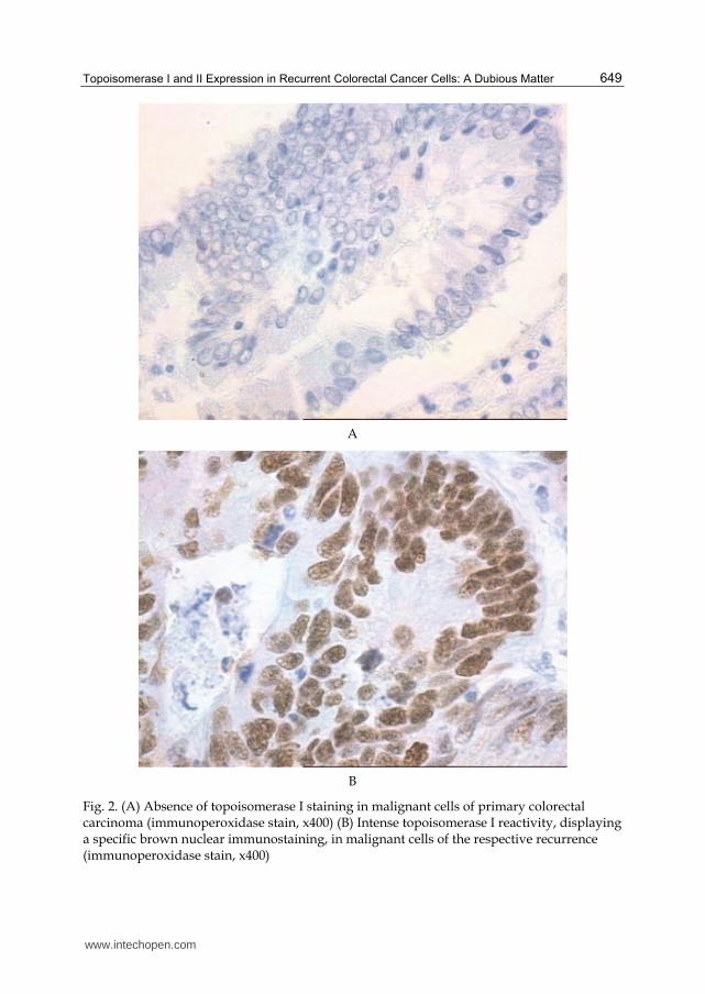
Fig. 2. (A) Absence of topoisomerase I staining in malignant cells of primary colorectal carcinoma (immunoperoxidase stain, x400) (B) Intense topoisomerase I reactivity, displaying a specific brown nuclear immunostaining, in malignant cells of the respective recurrence (immunoperoxidase stain, x400)
www.intechopen.com
DNA Replication - Current Advances 650
patients with metastatic colorectal cancer, a regimen being superior to 5-FU+LV alone
which has been demonstrated to offer consistently improved tumour control and
prolonged survival, albeit, only approximately 40% of advanced colorectal cancer patients
seem to be potentially responsive to the aforementioned combination regimen (Waters et
al., 2001).
One method of increasing the above described percentage of responsiveness to combination
chemotherapy is relied on the availability of biomarkers, capable for identifying patients
who might potentially respond to topoisomerase I inhibitor-based chemotherapy. With
regard to CPT-11 efficacy, in vitro data have suggested that topoisomerase I expression
could be considered as an important cellular sensitivity determinant (Paradiso et al., 2004).
In particular, decreased DNA topoisomerase I expression has been shown to correlate with
camptothecin resistant cell lines (Sanghani et al., 2003), a finding being in agreement with
Jansen et al. (Jansen et al., 1997), who conversely reported a positive correlation between
CPT-11 sensitivity and increased topoisomerase I activity. Other indicators for
topoisomerase I drug response of colorectal cancer cell lines, possibly include a high growth
fraction and a functional apoptotic pathway (Hafian et al., 2004), while Saltz et al. (Saltz et
al., 1998) have suggested an association between CPT-11 efficacy and topoisomerase
expression in colorectal neoplastic tissue; the latter being a potential predictor of 5-FU
resistance (Leichman et al., 1997).
Moreover, in our study, the percentage or intensity of increase of topoisomerase I protein
expression in recurrences did not demonstrate any significant correlations with Duke΄s
stage, grade of tumour differentiation, localization, and patients’ gender. These data suggest
that there might be no obvious benefit from evaluating histologicaly the tumour-cell
sensitivity to topoisomerase I-targeted drugs; subsequently, such drugs appear to be
effective across a various spectrum of pathologies, stage and gender. However, the
aforementioned increase of topoisomerase I expression was correlated with age, a fact which
might be suggestive that treatment with topoisomerase I inhibitors would be more
beneficial in older colorectal cancer patients. Of note, these data should be interpreted with
caution due to the limited number of evaluated patients.
6.2 Main research on topoisomerase I and II expression in colorectal cancer cells 6.2.1 Patients and methods
6.2.1.1 Patients
Forty patients with colorectal cancer that had recurred following surgery and adjuvant
chemotherapy who underwent a second operation were included in this study. All had
undergone surgical resection of the primary tumour and had received post-operatively 5-
FU-based [5FU and Leucovorin (LV), Mayo Clinic regimen] adjuvant chemotherapy
(Heidelberger et al., 1957). Patients’ characteristics are described in Table 1.
6.2.1.2 Study plan
The first tumour tissue was collected from the primary tumour during the initial operation,
before the administration of any adjuvant chemotherapy. The second tissue sample was
obtained at the time of recurrence, during the second operation and following
chemotherapy. Both tissue samples for each patient, were analyzed for the expression of
both topoisomerase-I and topoisomerase-IIa proteins.
www.intechopen.com
Topoisomerase I and II Expression in Recurrent Colorectal Cancer Cells: A Dubious Matter 651
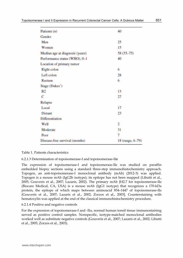
Table 1. Patients characteristics
6.2.1.3 Determination of topoisomerase-I and topoisomerase-IIα
The expression of topoisomerase-I and topoisomerase-IIa was studied on paraffin embedded biopsy sections using a standard three-step immunohistochemistry approach. Topogen, an anti-topoisomerase-I monoclonal antibody (mAb) (2012-3) was applied. Topogen is a mouse mAb (IgG2b isotype); its epitope has not been mapped (Libutti et al., 2005; Gouveris et al., 2007; Lazaris, 2002). The primary mAb JH2.7 for topoisomerase-IIa (Biocare Medical, CA, USA) is a mouse mAb (IgG1 isotype) that recognizes a 170-kDa protein, the epitope of which maps between aminoacid 854–1447 of topoisomerase-IIa (Gouveris et al., 2007; Lazaris et al., 2002; Zorzos et al., 2003]. Counterstaining with hematoxylin was applied at the end of the classical immunohistochemistry procedure.
6.2.1.4 Positive and negative controls
For the expression of topoisomerase-I and -II, normal human tonsil tissue immunostaining served as positive control samples. Nonspecific, isotype-matched monoclonal antibodies worked well as substitute negative controls (Gouveris et al., 2007; Lazaris et al., 2002; Libutti et al., 2005; Zorzos et al., 2003).
www.intechopen.com

DNA Replication - Current Advances 652
First step: Sections were examined for quantified immunoreactivity by two independent investigators blinded to any relevant patient clinical data. They evaluated more than 1,000 malignant cells in consecutive sections of neoplastic tissue specimens. The numbers of positive cells were then expressed as a percentage of labeled tumour cells with respect to the total number of tumour cells that could be idendified. Immunostaining for topoisomerase-I and -IIa was then graded according to the percentage of tumour cells staining positively (- and ± for <5%, + for 5–50% and ++ for 50 to 75% of cells with positive staining). A characterization was given additionally for the intensity of topoisomerase immunostaining (weakly positive, moderately positive, or strongly positive). To simplify the scoring, we graded them as 0 (negative cells), 1 (± and/or <5%), 2 (+ and/or 5–50%), 3 (++ and/or 50–75%). Then, the biopsy specimens were analysed as paired samples: one section from the first surgery and one section from the second surgery for each patient. Second step: The percentages of immunoreactive malignant cells were calculated using an image analysis system with an appropriate software package [Sigma Scan Pro, Version 5.0 (SPSS Science, Erkrath, Germany)]. The ratio was calculated as a percentage of immunohistochemically positive neoplastic cells over the total number (stained and unstained) of neoplastic cells. The membrane, cytoplasmic or nuclear intensity of the specific immunohistochemical stain, was also assessed. All the positively stained cells were classified for the purpose of simplicity into two groups: weakly stained and strongly stained. DNA ploidy of neoplastic cells was evaluated by image DNA flow-cytometry performed on
Feulgen-stained sections. Images were acquired using a Zeiss Axiolab microscope (Carl
Zeiss Jena GmbH, Jena, Germany) with a mechanical stage, fitted with a SONY-iris CCD
video camera (SONY Corporation, Tokyo, Japan). The latter was connected to a Pentium II
personal computer, which included the relevant software. Slides were examined at low
power magnification (40x) to identify the areas with the highest cellularity. In each case, a
total number of ≥200 Feulgen-stained nuclei was selected at high power magnification
(400x) and stored as JPEG file [1,550 x 1,070 pixels, 16.7 million colors (24-bit)]. Then the
images were converted to gray scale and the staining intensity of the Feulgen-stained nuclei
was measured semi-automatically. A classification of the nuclei in pairs according to their
staining intensity followed. Finally, a graphic presentation of the nuclei, demonstrating their
distribution according to their DNA content, was also performed.
6.2.1.5 Statistical analysis
All analyses were performed with SPSS version 10.1 (Statistical Product and Service Solutions; SPSS Inc., Chicago, IL, USA). We used McNemar’s paired Chi-square test to assess the possible alteration of the levels of topoisomerases after chemotherapy with 5-FU. Fisher’s exact test was performed in order to assess the possible relationship of topoisomerase increase with gender, Dukes’ stage, tumour grade and localization. Mann–Whitney U Test was performed to assess a possible relationship between patient age and alteration in topoisomerases levels.
6.2.2 Results
6.2.2.1 Patients
Forty patients were included in the present study. Of these, 25 were males and 15 were females. The median age was 58 years (range 35 – 75). All patients had a performance status
www.intechopen.com
Topoisomerase I and II Expression in Recurrent Colorectal Cancer Cells: A Dubious Matter 653
(PS) of 0 – 1. Tumour localization at the time of diagnosis was: right colon in six patients, left colon in 28 patients, and rectum in the remaining six patients. In respect with pathological classification, 13 patients had Dukes B2 tumours, and the remaining 27 had Dukes C tumours. With regard to differentiation, two patients had well differentiated tumours, 31 patients had moderately differentiated tumours and seven patients had poorly differentiated tumours. Of all forty patients, 17 relapsed locally and 23 manifested with distant metastases at the time of recurrence. Overall, the median relapse-free interval was 18 (range 6 – 79) months (Table 1). All patients who entered the trial were finally evaluable for analysis.
6.2.2.2 Ploidy
Out of the 40 primary tumours, 12 were highly aneuploid and the remaining 28 were moderately aneuploid. There was no association between the degree of DNA aneuploidy and the expression of any of the analyzed markers.
6.2.2.3 Topoisomerase I
Immunohistochemical analysis revealed that levels of topoisomerase-I expression were
higher in malignant cells from tumour recurrences compared to cells from the primary
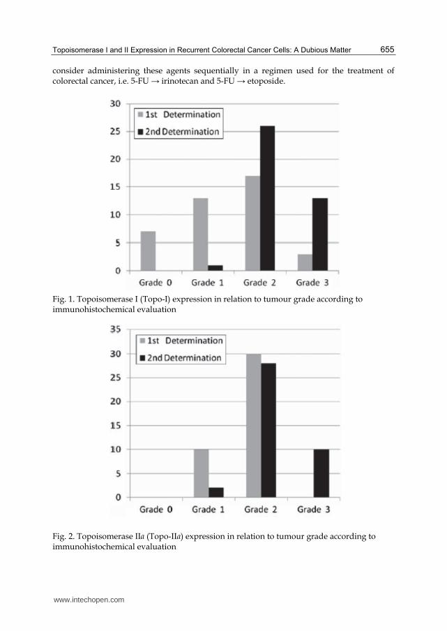
tumours (2 sided paired Chi square test, P = 0.0001) (Table 2; Fig. 1). Topoisomerase-I
expression was also decreased in grade 1 (P = 0.007) and increased in grade 3 tumours (2
sided Fischer’s exact test, P = 0.003) (Table 2). By image analysis evaluation, a significant
raise in malignant cells from the tumour recurrences could be recognised (2 sided paired
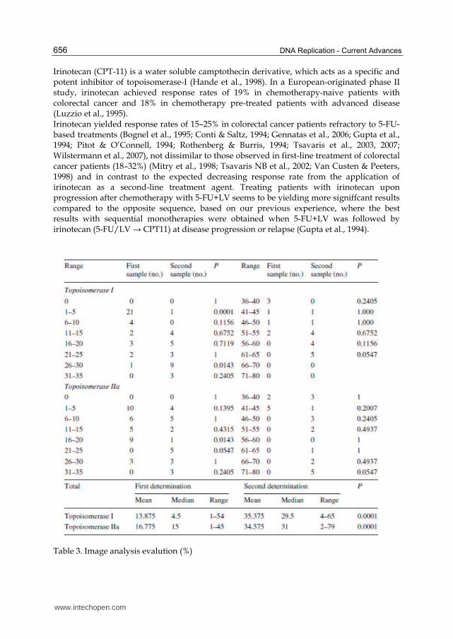
Chi square test, P = 0.0001) (Table 3); low expression of topoisomerase-I was noticed in
range 1–5 (P = 0.0001) and 26–30 (2 sided Fischer’s exact test, P = 0.0143) (Table 3; Fig. 3).
The increase in topoisomerase-I levels was not significantly correlated with gender,
performance status (WHO), location of the primary tumour, Dukes’ stage, grade of
differentiation, and localization of relapse. There was a statistically significant correlation
between the age of patients and the expression of topoisomerase-I (Mann–Whitney U Test, P
= 0.011). Patients with an increased expression of topoisomerase-I levels were older in age
(median=62.5 years) than patients without increased expression (median=50 years).
6.2.2.4 Topoisomerase IIα
Malignant cells from tumour recurrences showed a statistically significant increase of the
levels of topoisomerase-II, compared to those of the primary tumours (2 sided paired Chi
square test, P = 0.0001) (Table 2; Fig. 2). The topoisomerase-II levels were decreased in grade
3 lesions (2 sided Fischer’s exact test, P = 0.0001) (Table 2; Fig. 4). In image analysis, a
significant increase in malignant cells from tumour recurrences could be identified (2 sided
Fischer’s exact test, P = 0.0001) (Table 3); low expression of topoisomerase-II was noticed in
the range of 16–20 (2 sided Fischer’s exact test, P = 0.0143) (Table 3). Levels of
topoisomerase-IIa expression were also higher in malignant cells from tumour recurrences
compared to cells from primary tumours (Chi square test, P = 0.0001). There was a
statistically significant positive correlation between the age of patients and increased levels
of expression of topoisomerase-IIa (Mann–Whitney U Test, P = 0.011). The increase in
topoisomerase-IIa levels did not demonstrate any significant correlation with gender,
performance status (WHO), localization of primary tumour, Dukes’ stage, tumour grade,
and location of relapse, nor were such correlations found between the above parameters and
the differences of topoisomerase-IIa levels in the primary tumours and relapses.
www.intechopen.com
DNA Replication - Current Advances 654
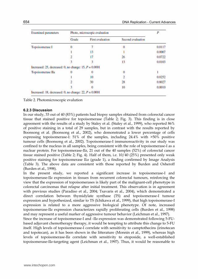
Table 2. Photomicroscopic evalution
6.2.3 Discussion
In our study, 33 out of 40 (83%) patients had biopsy samples obtained from colorectal cancer tissue that stained positive for topoisomerase (Table 2; Fig. 3). This finding is in close agreement with the results of a study by Staley et al. (Staley et al., 1999), who reported 86% of positive staining in a total of 29 samples, but in contrast with the results reported by Boonsong et al. (Boonsong et al., 2002), who demonstrated a lower percentage of cells expressing topoisomerase-I: 51% of the samples, including 24.4% with >50% positive tumour cells (Boonsong et al., 2002). Topoisomerase-I immunoreactivity in our study was confined to the nucleus in all samples, being consistent with the role of topoisomerase-I as a nuclear protein. For topoisomerase-IIa, 21 out of the 40 samples (52%) of colorectal cancer tissue stained positive (Table 2; Fig. 4). Half of them, i.e. 10/40 (25%) presented only weak
positive staining for topoisomerase II (grade 1), a finding confirmed by Image Analysis (Table 3). The above data are consistent with those reported by Burden and Osheroff (Burden et al., 1998). In the present study, we reported a significant increase in topoisomerase-I and topoisomerase-IIa expression in tissues from recurrent colorectal tumours, reinforcing the view that the expression of topoisomerases is likely part of the malignant-cell phenotype in colorectal carcinomas that relapse after initial treatment. This observation is in agreement with previous studies (Paradiso et al., 2004; Tsavaris et al., 2004), which demonstrated a direct correlation between thymidylate synthase (TS) and topoisomerase-I tumour expression and hypothesized, similar to TS (Ichikawa et al., 1999), that high topoisomerase-I expression is related to a more aggressive biological phenotype. Of note, increased topoisomerase-IIa expression characterizes rapidly proliferating cells (Burden et al., 1998) and may represent a useful marker of aggressive tumour behavior (Leichman et al., 1997). Since the increase of topoisomerase-I and -IIa expression was demonstrated following 5-FU-based adjuvant chemotherapy therapy, it would be tempting to attribute this change to 5-FU itself. High levels of topoisomerase-I correlate with sensitivity to camptothecins (irinotecan and topotecan), as it has been shown in the litterature (Monnin et al., 1999), whereas high levels of topoisomerase-IIa correlate with sensitivity to etoposide, a well-established topoisomerase-IIa-targeting agent (Leichman et al., 1997). Thus, it would be reasonable to
www.intechopen.com
Topoisomerase I and II Expression in Recurrent Colorectal Cancer Cells: A Dubious Matter 655
consider administering these agents sequentially in a regimen used for the treatment of colorectal cancer, i.e. 5-FU → irinotecan and 5-FU → etoposide.
Fig. 1. Topoisomerase I (Topo-I) expression in relation to tumour grade according to immunohistochemical evaluation
Fig. 2. Topoisomerase IIa (Topo-IIa) expression in relation to tumour grade according to immunohistochemical evaluation
www.intechopen.com
DNA Replication - Current Advances 656
Irinotecan (CPT-11) is a water soluble camptothecin derivative, which acts as a specific and potent inhibitor of topoisomerase-I (Hande et al., 1998). In a European-originated phase II study, irinotecan achieved response rates of 19% in chemotherapy-naive patients with colorectal cancer and 18% in chemotherapy pre-treated patients with advanced disease (Luzzio et al., 1995). Irinotecan yielded response rates of 15–25% in colorectal cancer patients refractory to 5-FU-based treatments (Bognel et al., 1995; Conti & Saltz, 1994; Gennatas et al., 2006; Gupta et al., 1994; Pitot & O’Connell, 1994; Rothenberg & Burris, 1994; Tsavaris et al., 2003, 2007; Wilstermann et al., 2007), not dissimilar to those observed in first-line treatment of colorectal cancer patients (18–32%) (Mitry et al., 1998; Tsavaris NB et al., 2002; Van Custen & Peeters, 1998) and in contrast to the expected decreasing response rate from the application of irinotecan as a second-line treatment agent. Treating patients with irinotecan upon progression after chemotherapy with 5-FU+LV seems to be yielding more signiffcant results compared to the opposite sequence, based on our previous experience, where the best results with sequential monotherapies were obtained when 5-FU+LV was followed by irinotecan (5-FU/LV → CPT11) at disease progression or relapse (Gupta et al., 1994).
Table 3. Image analysis evalution (%)
www.intechopen.com
Topoisomerase I and II Expression in Recurrent Colorectal Cancer Cells: A Dubious Matter 657
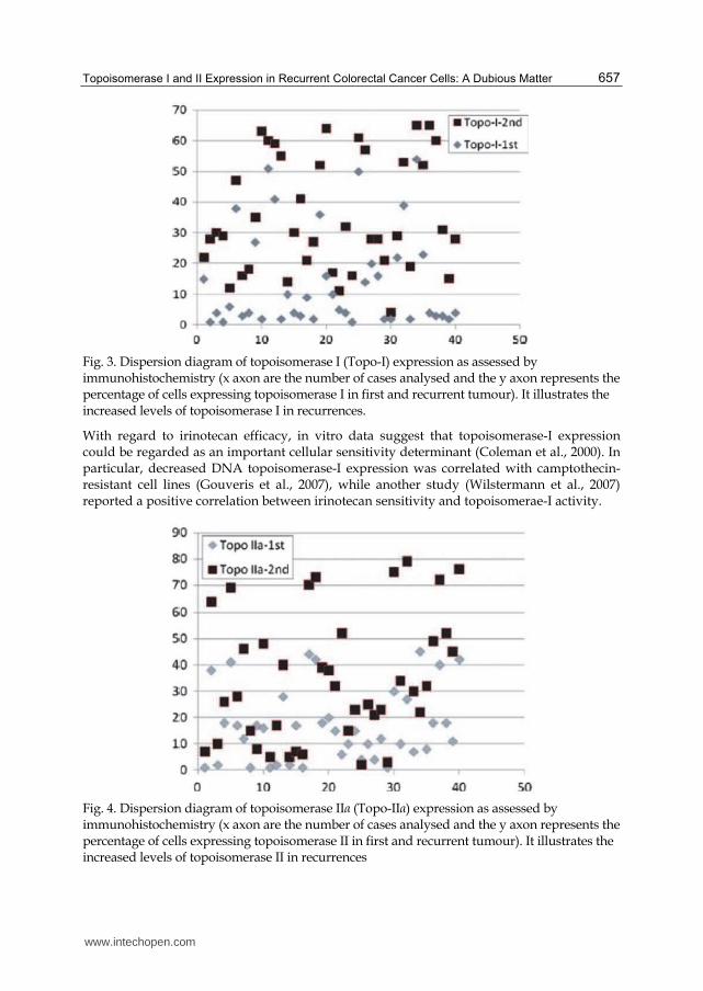
Fig. 3. Dispersion diagram of topoisomerase I (Topo-I) expression as assessed by immunohistochemistry (x axon are the number of cases analysed and the y axon represents the percentage of cells expressing topoisomerase I in first and recurrent tumour). It illustrates the increased levels of topoisomerase I in recurrences.
With regard to irinotecan efficacy, in vitro data suggest that topoisomerase-I expression could be regarded as an important cellular sensitivity determinant (Coleman et al., 2000). In particular, decreased DNA topoisomerase-I expression was correlated with camptothecin-resistant cell lines (Gouveris et al., 2007), while another study (Wilstermann et al., 2007) reported a positive correlation between irinotecan sensitivity and topoisomerae-I activity.
Fig. 4. Dispersion diagram of topoisomerase IIα (Topo-IIα) expression as assessed by immunohistochemistry (x axon are the number of cases analysed and the y axon represents the percentage of cells expressing topoisomerase II in first and recurrent tumour). It illustrates the increased levels of topoisomerase II in recurrences
www.intechopen.com

DNA Replication - Current Advances 658
Drugs that target topoisomerase-II, such as the epipodophylotoxins, etoposide (VP-16) and teniposide (VM-26) (Leichman et al., 1997) (VP-16), doxorubicin, and mitoxantrone, are among the most effective anticancer drugs still in clinical use. Especially, etoposide acts by destroying cells via inhibition of the ability of topoisomerase-II to ligate nucleic acids which are cleaved during the double-stranded DNA passage reaction (Bleiberg, 1998). Studies including previously untreated patients with advanced colorectal carcinoma evaluating the combination of etoposide with cisplatin or 5-FU demonstrated minimal activity in metastatic colorectal cancer (Boige et al., 1998; Passalacqua et al., 1991; Planting et al., 1996; Posner et al., 1990). There have been no clinical data supporting the in vitro synergy observed between these cytotoxic agents (Colucci et al., 1995; Zelkowitz et al., 1989). Other studies failed to prove any benefit with the combination of etoposide with 5-FU or cisplatin/carboplatin (Inaba et al., 1994). However, the combination of etoposide with 5-FU + LV had demonstrated some activity when administered as second-line treatment after failure of weekly 5-FU + LV in patients with metastatic colorectal cancer (Stuart et al., 1995; Tsavaris et al., 2002). The results reported herein underscore the role of topoisomerases (topoisomerase-I and topoisomerase-IIa) expression in colorectal cancer. It is believed that translational studies of molecular targets for currently applied cytotoxic and biological agents (like bevacizumab and cetuximab) might form the basis of shaping current and future drug combinations and of rationalizing the optimal chemotherapeutic drug schedule and sequence, which will eventually translate in improved tumour eradication and prolongation of survival. This work is unique since it presents the first two of a series of studies that demonstrate an increase of topoisomerase expression following chemotherapy with 5FU. This may denote that such tumours are sensitized through 5FU chemotherapy regimens to topoisomerase inhibitors, providing a pathophysiologic mechanism to explain the described effectiveness of such agents in recurrent colorectal cancer after the first adjuvant chemotherapy.
7. References
Abigerges D, Armand JP, Chabot GG, Da Costa L, Fadel E, Cote C, Herait P, Gandia D. Irinotecan (CPT-11) high-dose escalation using intensive high-dose loperamide to control diarrhea. J Natl Cancer Inst. 1994 Mar 16;86(6):446-9.
Austin CA, Sng JH, Patel S, Fisher LM. Novel HeLa topoisomerase II is the II beta isoform: complete coding sequence and homology with other type II topoisomerases. Biochim Biophys Acta. 1993 Mar 20;1172(3):283-91.
Avemann K, Knippers R, Koller T, Sogo JM. Camptothecin, a specific inhibitor of type I DNA topoisomerase, induces DNA breakage at replication forks. Mol Cell Biol.1988 Aug;8(8):3026-34.
Bailly C. Topoisomerase I poisons and suppressors as anticancer drugs. Curr Med Chem. 2000 Jan;7(1):39-58. Review.
Barnes S, Peterson TG, Coward L. Rationale for the use of genistein-containing soy matrices in chemoprevention trials for breast and prostate cancer. J Cell Biochem Suppl. 1995;22:181-7.
Bauer WR, Crick FH, White JH. Supercoiled DNA. Sci Am. 1980 Jul;243(1):100-13. Bauman ME, Holden JA, Brown KA, Harker WG, Perkins SL. Differential
immunohistochemical staining for DNA topoisomerase II alpha and beta in human
www.intechopen.com
Topoisomerase I and II Expression in Recurrent Colorectal Cancer Cells: A Dubious Matter 659
tissues and for DNA topoisomerase II beta in non-Hodgkin's lymphomas. Mod Pathol. 1997 Mar;10(3):168-75.
Beck WT, Danks MK, Wolverton JS, Kim R, Chen M. Drug resistance associated with altered DNA topoisomerase II. Adv Enzyme Regul. 1993;33:113-27. Review.
Berger JM. Type II DNA topoisomerases. Curr Opin Struct Biol. 1998 Feb;8(1):26-32. Review. Berney DM, Shamash J, Gaffney J, Jordan S, Oliver RT. DNA topoisomerase I and II
expression in drug resistant germ cell tumours. Br J Cancer. 2002 Sep 9;87(6):624-9. Biersack H, Jensen S, Gromova I, Nielsen IS, Westergaard O, Andersen AH. Active
heterodimers are formed from human DNA topoisomerase II alpha and II beta isoforms. Proc Natl Acad Sci U S A. 1996 Aug 6;93(16):8288-93.
Bleiberg H (1998) Continuing the fight against advanced colorectal cancer: new and future treatment options. Anticancer Drugs 9:18–28
Bognel C, Rekacewicz C, Mankarios H, Pignon JP, Elias D, Duvillard P, Prade M, Ducreux M, Kac J, Rougier P, Eschwège F, Lasser P (1995) Prognostic value of neural invasion in rectal carcinoma: a multivariate analysis on 339 patients with curative resection. Eur J Cancer 31A:894–898
Boige V, Raymond E, Armand JP (1998) Irinotecan: various administration schedules, study of drug combinations, phase I experience. Bull Cancer 26-32
Boonsong A, Curran S, McKay JA, Cassidy J, Murray GI, McLeod HL. Topoisomerase I protein expression in primary colorectal cancer and lymph node metastases. Hum Pathol. 2002 Nov;33(11):1114-9.
Bronstein IB, Vorobyev S, Timofeev A, Jolles CJ, Alder SL, Holden JA. Elevations of DNA topoisomerase I catalytic activity and immunoprotein in human malignancies. Oncol Res. 1996;8(1):17-25.
Brown PO, Peebles CL, Cozzarelli NR. A topoisomerase from Escherichia coli related to DNA gyrase. Proc Natl Acad Sci U S A. 1979 Dec;76(12):6110-4.
Bullock P, Champoux JJ, Botchan M. Association of crossover points with topoisomerase I cleavage sites: a model for nonhomologous recombination.Science. 985 Nov 22;230(4728):954-8.
Burden DA, Osheroff N: Mechanism of action of eukaryotic topoisomerase II and drugs targeted to the enzyme. Biochim Biophys Acta 1400:139-154, 1998.
Castano IB, Brzoska PM, Sadoff BU, Chen H, Christman MF. Mitotic chromosome condensation in the rDNA requires TRF4 and DNA topoisomerase I in Saccharomyces cerevisiae. Genes Dev. 1996 Oct 15;10(20):2564-76.
Champoux JJ, Dulbecco R. An activity from mammalian cells that untwists superhelical DNA--a possible swivel for DNA replication (polyoma-ethidium bromide-mouse-embryo cells-dye binding assay). Proc Natl Acad Sci U S A. 1972 Jan;69(1):143-6.
Champoux JJ. DNA topoisomerases: structure, function, and mechanism. Annu Rev Biochem 2001;70:369.
Cole SP, Chanda ER, Dicke FP, Gerlach JH, Mirski SE. Non-P-glycoprotein-mediated multidrug resistance in a small cell lung cancer cell line: evidence for decreased susceptibility to drug-induced DNA damage and reduced levels of topoisomerase II. Cancer Res. 1991 Jul 1;51(13):3345-52.
Coleman LW, Perkins SL, Bronstein IB, Holden JA (2000) Expression of DNA toposiomerase I and DNA topoisomerase IIalpha in testicular seminomas. Hum Pathol 31:728–733
www.intechopen.com
DNA Replication - Current Advances 660
Coleman LW, Bronstein IB, Holden JA. Immunohistochemical staining for DNA topoisomerase I, DNA topoisomerase II-alpha and p53 in gastric carcinomas. Anticancer Res. 2001 Mar-Apr;21(2A):1167-72.
Coleman LW, Rohr LR, Bronstein IB, Holden JA. Human DNA topoisomerase I: An anticancer drug target present in human sarcomas. Hum Pathol. 2002 Jun;33(6):599-607.
Colucci G, Maiello E, Giuliani F, Cifarelli RA, Giotta F (1995) Carboplatin and etoposide in previously treated colorectal cancer patients. Tumori 81:36–38
Conti JA, Saltz L (1994) Irinotecan is an active agent in untreated patients with metastatic colorectal cancer. Proc Am Soc Clin Oncol 13:195 (abstr)
Covey JM, Jaxel C, Kohn KW, Pommier Y. Protein-linked DNA strand breaks induced in mammalian cells by camptothecin, an inhibitor of topoisomerase I. Cancer Res. 1989 Sep 15;49(18):5016–5022.
Cunningham D, Pyrhonen S, James RD, Punt CJ, Hickish TF, Heikkila R, Johannesen TB, Starkhammar H, Topham CA, Awad L, Jacques C, Herait P. Randomised trial of irinotecan plus supportive care versus supportive care alone after fluorouracil failure for patients with metastatic colorectal cancer. Lancet. 1998 Oct 31;352(9138):1413-8.
Dickey JS, Osheroff N. Impact of the C-terminal domain of topoisomerase IIalpha on the DNA cleavage activity of the human enzyme. Biochemistry. 2005 Aug 30;44(34):11546-54.
D'Incalci Μ. DNA-topoisomerase inhibitors.Curr Opin Oncol. 1993 Nov;5(6):1023-8. Douillard JY, Cunningham D, Roth AD, Navarro M, James RD, Karasek P, Jandik P, Iveson
T, Carmichael J, Alakl M, Gruia G, Awad L, Rougier P: Irinotecan combined with fluorouracil compared with fluorouracil alone as first-line treatment for metastatic colorectal cancer: a multicentre randomised trial. Lancet 2000, 355:1041-1047.
Florell SR, Martinchick JF, Holden JA. Purification of DNA topoisomerase I from the spleen of a patient with non-Hodgkin's lymphoma. Anticancer Res. 1996 Nov-Dec;16(6B):3467-74.
Fogt F, Nikulasson ST, Holden JA, Alder SA, Hallgrimsson J, Jessup MJ, O'Brien MJ, Lavin PT, Goldman H. Topoisomerase II alpha expression in normal, inflammatory, and neoplastic conditions of the gastric and colonic mucosa. Mod Pathol. 1997 Apr;10(4):296-302.
Gellert M, Mizuuchi K, O'Dea MH, Nash HA. DNA gyrase: an enzyme that introduces superhelical turns into DNA. Proc Natl Acad Sci U S A. 1976 Nov;73(11):3872-6.
Gellert M, O'Dea MH, Itoh T, Tomizawa J. Novobiocin and coumermycin inhibit DNA supercoiling catalyzed by DNA gyrase. Proc Natl Acad Sci U S A. 1976 Dec;73(12):4474-8.
Gennatas C, Papaxoinis G, Michalaki V, Mouratidou D, Andreadis C, Tsavaris N, Paffiti A (2006) A prospective randomized study of irinotecan (CPT-11), leucovorin (LV) and 5-Xuorouracil (5FU) versus leucovorin and 5-Fluorouracil in patients with advanced colorectal carcinoma. J Chemother 18:538–544
Gibbons D, Fogt F, Kasznica J, Holden J, Nikulasson S. Comparison of topoisomerase II alpha and MIB-1 expression in uterine cervical squamous lesions. Mod Pathol. 1997 May;10(5):409-13.
www.intechopen.com
Topoisomerase I and II Expression in Recurrent Colorectal Cancer Cells: A Dubious Matter 661
Giovanella BC, Stehlin JS, Wall ME, Wani MC, Nicholas AW, Liu LF, Silber R, Potmesil M. DNA topoisomerase I-targeted chemotherapy of human colon cancer in xenografts. Science. 1989 Nov 24;246(4933):1046-8.
Glisson BS, Sullivan DM, Gupta R, Ross WE. Mediation of multi-drug resistance in a Chinese hamster ovary cell line by a mutant type II topoisomerase. NCI Monogr. 1987;(4):89-93.
Goldwasser F, Bae I, Valenti M, et al: Topoisomerase I–related parameters and camptothecin activity in the colon carcinoma cell lines from the National Cancer Institute and anticancer screen. Cancer Res 55:2116-2121, 1995.
Gottlieb JA, Guarino AM, Call JB, Oliverio VT, Block JB. Preliminary pharmacologic and clinical evaluation of camptothecin sodium (NSC-100880). Cancer Chemother Rep Part 1 1970; 54: 461-70.
Gottlieb JA, Luce JK: Treatment of malignant melanoma with camptothecin (NSC-100880). Cancer Chemother Rep 1972;56:103–105.
Gouveris P, Lazaris AC, Papathomas TG, Nonni A, Kyriakou V, Delladetsima J, Patsouris ES, Tsavaris N (2007) Topoisomerase I protein expression in primary colorectal cancer and recurrences after 5-FU-based adjuvant chemotherapy. J Cancer Res Clin Oncol 133:1011–1015
Guichard S, Cussac D, Hennebelle I, Bugat R, Canal P (1997) Sequence-dependent activity of the irinotecan-5FU combination in human colon-cancer model HT-29 in vitro and in vivo. Int J Cancer 73:729-734
Guichard S, Hennebelle I, Bugat R, Canal P (1998) Cellular interactions of 5- fluorouracil and the camptothecin analogue CPT-11 (irinotecan) in a human colorectal carcinoma cell line. Biochem Pharmacol 55:667-676
Guichard S, Terret C, Hennebelle I, Lochon I, Chevreau P, Fretigny E, Selves J, Chatelut E, Bugat R, Canal P. CPT-11 converting carboxylesterase and topoisomerase activities in tumour and normal colon and liver tissues. Br J Cancer. 1999 May; 80(3-4):364-70.
Guldner HH, Szostecki C, Vosberg HP, Lakomek HJ, Penner E, Bautz FA. Scl 70 autoantibodies from scleroderma patients recognize a 95 kDa protein identified as DNA topoisomerase I. Chromosoma. 1986;94(2):132-8.
Gupta D, Bronstein IB, Holden JA: Expression of DNA topoisomerase I in neoplasms of the kidney: Correlation with histological grade, proliferation, and patient survival. Hum Pathol 31:214-219, 2000.
Gupta E, Lestingi TM, Mick R, Ramirez J, Vokes EE, Ratain MJ. Metabolic fate of irinotecan in humans: correlation of glucuronidation with diarrhea. Cancer Res. 1994 Jul 15;54(14):3723-5.
Gupta M, Fujimori A, Pommier Y. Eukaryotic DNA topoisomerases I. Biochim Biophys Acta. 1995 May 17;1262(1):1-14.
Hafian H, Venteo L, Sukhanova A, Nabiev I, Lefevre B, Pluot M. Immunohistochemical study of DNA topoisomerase I, DNA topoisomerase II alpha, p53, and Ki-67 in oral preneoplastic lesions and oral squamous cell carcinomas. Hum Pathol. 2004 Jun;35(6):745-51.
Hanai R, Caron PR, Wang JC. Human TOP3: a single-copy gene encoding DNA topoisomerase III. Proc Natl Acad Sci U S A. 1996 Apr 16;93(8):3653-7.
www.intechopen.com
DNA Replication - Current Advances 662
Hande KR. Clinical applications of anticancer drugs targeted to topoisomerase II. Biochim Biophys Acta. 1998 Oct 1;1400(1-3):173-84.
Heck MM, Hittelman WN, Earnshaw WC. Differential expression of DNA topoisomerases I and II during the eukaryotic cell cycle. Proc Natl Acad Sci U S A. 1988 Feb;85(4):1086-90.
Heidelberger C, Chaudhuri NK, Danneberg P, Mooren D, Griesbach L, Duschinsky R, Schnitzer RJ, Pleven E, Scheiner J (1957) Fluorinated pyrimidines: a new class of tumor inhibitory compounds. Nature 179:663–666
Holden JA, Perkins SL, Snow GW, Kjeldsberg CR. Immunohistochemical staining for DNA topoisomerase II in non-Hodgkin's lymphomas. Am J Clin Pathol. 1995 Jul;104(1):54-9.
Holden JA, Rahn MP, Jolles CJ, Vorobyev SV, Bronstein IB. Immunohistochemical detection of DNA topoisomerase I in formalin fixed, paraffin wax embedded normal tissues and in ovarian carcinomas. Mol Pathol. 1997 Oct;50(5):247-53.
Holm C, Covey JM, Kerrigan D, Pommier Y. Differential requirement of DNA replication for the cytotoxicity of DNA topoisomerase I and II inhibitors in Chinese hamster DC3F cells. Cancer Res. 1989 Nov 15;49(22):6365-8.
Hsiang YH, Liu LF. Identification of mammalian DNA topoisomerase I as an intracellular target of the anticancer drug camptothecin. Cancer Res. 1988 Apr 1;48(7):1722-1726.
Hsiang YH, Lihou MG, Liu LF. Arrest of replication forks by drug-stabilized topoisomerase I-DNA cleavable complexes as a mechanism of cell killing by camptothecin. Cancer Res. 1989 Sep 15;49(18):5077-82.
Husain I, Mohler JL, Seigler HF, Besterman JM. Elevation of topoisomerase I messenger RNA, protein, and catalytic activity in human tumors: demonstration of tumor-type specificity and implications for cancer chemotherapy. Cancer Res. 1994 Jan 15;54(2):539-46.
Ichikawa W, Uetake H, Nihei Z, Mastuo K, Fujita H, Yamada Y (1999) Topoisomerase I (Topo-I) expression correlates to thymidylate synthase (TS) expression in colorectal cancer (CRC). Proc Am Soc Clin Oncol 18:946 (abstract)
Inaba M, Mitsuhashi J, Kawada S, Nakano H (1994) Different modes of cell-killing action between DNA topoisomerase I and II inhibitors revealed by kinetic analysis. Jpn J Cancer Res 85:187–193
Jansen WJ, Zwart B, Hulscher ST, Giaccone G, Pinedo HM, Boven E (1997) CPT-11 in human colon-cancer cell lines and xenografts: characterization of cellular sensitivity determinants. Int J Cancer 70:335–340
Jaxel C, Capranico G, Kerrigan D, Kohn KW, Pommier Y. Effect of local DNA sequence on topoisomerase I cleavage in the presence or absence of camptothecin. J Biol Chem. 1991 Oct 25;266(30):20418–20423.
Jenkins JR, Ayton P, Jones T, Davies SL, Simmons DL, Harris AL, Sheer D, Hickson ID. Isolation of cDNA clones encoding the beta isozyme of human DNA topoisomerase II and localisation of the gene to chromosome 3p24. Nucleic Acids Res. 1992 Nov 11;20(21):5587-92.
Kato J, Nishimura Y, Imamura R, Niki H, Hiraga S, Suzuki H. New topoisomerase essential for chromosome segregation in E. coli. Cell. 1990 Oct 19;63(2):393-404.
www.intechopen.com
Topoisomerase I and II Expression in Recurrent Colorectal Cancer Cells: A Dubious Matter 663
Kehrer DF, Sparreboom A, Verweij J, de Bruijn P, Nierop CA, van de Schraaf J, Ruijgrok EJ, de Jonge MJ. Modulation of irinotecan-induced diarrhea by cotreatment with neomycin in cancer patients. Clin Cancer Res. 2001 May;7(5):1136-41.
Kellner U, Sehested M, Jensen PB, et al. Culprit and victim—DNA topoisomerase II. Lancet Oncol 2003;3:235.
Kerrigan JE, Pilch DS. A structural model for the ternary cleavable complex formed between human topoisomerase I, DNA, and camptothecin. Biochemistry. 2001 Aug 21;40(33):9792-8.
Kwan KY, Wang JC. Mice lacking DNA topoisomerase IIIbeta develop to maturity but show a reduced mean lifespan.Proc Natl Acad Sci U S A. 2001 May 8;98(10):5717-21. Epub 2001 May 1.
Lazaris AC, Kavantzas NG, Zorzos HS, Tsavaris NV, Davaris PS (2002) Markers of drug resistance in relapsing colon cancer. J Cancer Res Clin Oncol 128:114–118
Lee MP, Brown SD, Chen A, Hsieh TS. DNA topoisomerase I is essential in Drosophila melanogaster. Proc Natl Acad Sci U S A. 1993 Jul 15;90(14):6656-60.
Leichman CG, Lenz HJ, Leichman L, Danenberg K, Baranda J, Groshen S, Boswell W, Metzger R, Tan M, Danenberg PV (1997) Quantitation of intratumoral thymidylate synthase expression predicts for disseminated colorectal cancer response and resistance to protracted-infusion fluorouracil and weekly leucovorin. J Clin Oncol 15:3223-3229
Libutti SK, Salz LB, Rustgi AK, Tepper JE (2005) Cancer of the colon. In: Devita VT, Hellman S, Rosenberg S (eds) Cancer: principles and practice of oncology, Chap 29, Sect 8. Lippincott Williams and Wilkins, Philadelphia
Lindsley JE. Intradimerically tethered DNA topoisomerase II is catalytically active in DNA transport. Proc Natl Acad Sci U S A. 1996 Apr 2;93(7):2975-80.
Liu, L. F. DNA topoisomerases: enzymes that catalyze the breaking and rejoining of DNA. CRC Critical Review of Biochemistry, 1983 15, pp. 1-24.
Liu LF, Wang JC. Supercoiling of the DNA template during transcription. Proc Natl Acad Sci U S A. 1987 Oct;84(20):7024-7.
Lodge AJ, Anderson JJ, Ng SW, Fenwick F, Steward M, Haugk B, Horne CH, Angus B. Expression of topoisomerase IIIalpha in normal and neoplastic tissues determined by immunohistochemistry using a novel monoclonal antibody. Br J Cancer. 2000 Aug;83(4):498-505.
Lokiec F, Canal P, Gay C, Chatelut E, Armand JP, Roche H, Bugat R, Goncalves E, Mathieu-Boue A. Pharmacokinetics of irinotecan and its metabolites in human blood, bile, and urine. Cancer Chemother Pharmacol. 1995;36(1):79-82.
Luzzio MJ, Besterman JM, Emerson DL, Evans MG, Lackey K, Leitner PL, McIntyre G, Morton B, Myers PL, Peel M, Sisco JM, Sternbach DD et al (1995) Synthesis and antitumor activity of novel water soluble derivatives of camptothecin as specific inhibitors of topoisomerase I. J Med Chem 38:395–401
Lynch BJ, Komaromy-Hiller G, Bronstein IB, Holden JA. Expression of DNA topoisomerase I, DNA topoisomerase II-alpha, and p53 in metastatic malignant melanoma. Hum Pathol. 1998 Nov;29(11):1240-5.
Lynch BJ, Bronstein IB, Holden JA: Elevations of DNA topoisomerase I in invasive carcinoma of the breast. Breast J 7:176-180, 2001.
www.intechopen.com
DNA Replication - Current Advances 664
Mantle D, Lennard TW, Pickering AT. Therapeutic applications of medicinal plants in the treatment of breast cancer: a review of their pharmacology, efficacy and tolerability.Adverse Drug React Toxicol Rev. 2000 Aug;19(3):223-40. Review.
Maxwell A. The molecular basis of quinolone action. J Antimicrob Chemother. 1992 Oct;30(4):409-14. Review.
McLeod HL, Douglas F, Oates M, Symonds RP, Prakash D, van der Zee AG, Kaye SB, Brown R, Keith WN. Topoisomerase I and II activity in human breast, cervix, lung and colon cancer. Int J Cancer. 1994 Dec 1;59(5):607-11.
Mitry E, Ducreux M, Rougier P (1998) Second-line irinotecan chemotherapy in the treatment of metastatic colorectal cancers: phase III trials. Bull Cancer 38-42
Moertel CG, Schutt AJ, Reitemeir RJ, et al: Phase II study of camptothecin (NSC-100880) in the treatment of advanced gastrointenstinal cancer. Cancer Chemother Rep 1972;56:95–101.
Monnin KA, Bronstein IB, Gaffney DK, Holden JA. Elevations of DNA topoisomerase I in transitional cell carcinoma of the urinary bladder: correlation with DNA topoisomerase II-alpha and p53 expression. Hum Pathol. 1999 Apr;30(4):384-91.
Morham SG, Kluckman KD, Voulomanos N, Smithies O. Targeted disruption of the mouse topoisomerase I gene by camptothecin selection. Mol Cell Biol. 1996 Dec;16(12):6804-9.
Muggia FM, Creaven PJ, Hansen HH, et al: Phase I clinical trial of weekly and daily treatment with camptothecin (NSC-100880): Correlation with preclinical studies. Cancer Chemother Rep 1972;56:515–521.
Nakopoulou L, Zervas A, Lazaris AC, Constantinides C, Stravodimos C, Davaris P, Dimopoulos C. Predictive value of topoisomerase II alpha immunostaining in urothelial bladder carcinoma. J Clin Pathol. 2001 Apr;54(4):309-13.
P. Borst and H.M. Pinedo. Drug Resistance. Oxford textbook of oncology, Oxford Press, 1995, Chapter 4.13.
Paradiso A, Xu J, Mangia A, Chiriatti A, Simone G, Zito A, Montemurro S, Giuliani F, Maiello E, Colucci G. Topoisomerase-I, thymidylate synthase primary tumour expression and clinical efficacy of 5-FU/CPT-11 chemotherapy in advanced colorectal cancer patients. Int J Cancer. 2004 Aug 20;111(2):252-8.
Passalacqua R, Bisagni G, Cocconi G, Boni C, Di Blasio B, Ceci G (1991) Cisplatin and etoposide in advanced colorectal carcinoma.Ann Oncol 2:687–688
Perego P, Capranico G, Supino R, et al: Topoisomerase I gene expression and cell sensitivity to camptothecin in human cell lines of different tumor types. Anticancer Drugs 5:645-649, 1994.
Pitot HC, O’Connell MJ (1994) A phase II trial of CPT-11 in patients with metastatic colorectal carcinoma: a North Central Cancer Treatment Group Study. Proc Am Soc Clin Oncol 13:197 (abstr)
Planting AS, van der Burg ME, van den Bent MJ, de Boer-Dennert M, Stoter G, Verweij J (1996) Phase II study of a short course of weekly high-dose cisplatin combined with long-term oral etoposide in metastatic colorectal cancer. Br J Cancer 73:1265–1267
Pommier Y. Eukaryotic DNA topoisomerase I: genome gate keeper and its intruders, camptothecins. Semin Oncol. 1996 23: 1-10.
www.intechopen.com

Topoisomerase I and II Expression in Recurrent Colorectal Cancer Cells: A Dubious Matter 665
Pommier Y, Pourquier P, Fan Y, Strumberg D. Mechanism of action of eukaryotic DNA topoisomerase I and drugs targeted to the enzyme. Biochim Biophys Acta. 1998 Oct 1;1400(1-3):83-105. Review.
Pommier YG, Goldwasser F, Strumberg D. Topoisomerase II inhibitors: epipodophyllotoxins, acridines, ellipticines, and bisdioxopiperazines. In: Chabner BA, Longo DL, eds., Cancer chemotherapy & biotherapy: principles and practice, 3rd ed. Philadelphia: Lippincott Williams & Wilkins, 2001:538.
Posner M, Slapak CA, Browne MJ, Clark JW, Curt G, Weitberg A, Calabresi P, Cummings FJ, Wiemann M, Urba S (1990) A phase I-II trial of continuous-infusion cisplatin, continuous-infusion 5-Fluorouracil, and VP-16 in colorectal cancer. Am J Clin Oncol 13:455–458
Raynard S, Bussen W, Sung P. A double holliday junction dissolvasome comprising BLM, topoisomerase IIIalpha, and BLAP75. J Biol Chem.2006 Apr 4.
Robinson MJ, Martin BA, Gootz TD, McGuirk PR, Moynihan M, Sutcliffe JA, Osheroff N. Effects of quinolone derivatives on eukaryotic topoisomerase II. A novel mechanism for enhancement of enzyme-mediated DNA cleavage. J Biol Chem. 1991 Aug 5;266(22):14585-92.
Robinson MJ, Martin BA, Gootz TD, McGuirk PR, Osheroff N. Effects of novel fluoroquinolones on the catalytic activities of eukaryotic topoisomerase II: Influence of the C-8 fluorine group. Antimicrob Agents Chemother. 1992 Apr;36(4):751-6.
Roca J, Wang JC. DNA transport by a type II DNA topoisomerase: evidence in favor of a two-gate mechanism. Cell. 1994 May 20;77(4):609-16.
Roca J, Wang JC. The probabilities of supercoil removal and decatenation by yeast DNA topoisomerase II. Genes Cells. 1996 Jan;1(1):17-27.
Rothenberg ML, Burris HA III (1994) Irinotecan as second line therapy for patients with 5-FU refractory colorectal cancer. Proc Am Soc Clin Oncol 13:198 (abstr)
Rougier P, Van Cutsem E, Bajetta E, Niederle N, Possinger K, Labianca R, Navarro M, Morant R, Bleiberg H, Wils J, Awad L, Herait P, Jacques C. Randomised trial of irinotecan versus fluorouracil by continuous infusion after fluorouracil failure in patients with metastatic colorectal cancer. Lancet. 1998 Oct 31;352(9138):1407-12.
Saltz L, Danenberg K, Paty P, Kelsen D, Salonga D, Park JM, Danemberg P (1998) High thymidylate synthase (TS) expression does not preclude activity of CTP-11 in colorectal cancer (CRC). Proc Am Soc Clin Oncol 17:991 (abstract)
Saltz LB, Cox JV, Blanke C, Rosen LS, Fehrenbacher L, Moore MJ, Maroun JA, Ackland SP, Locker PK, Pirotta N, Elfring GL, Miller LL. Irinotecan plus fluorouracil and leucovorin for metastatic colorectal cancer. Irinotecan Study Group. N Engl J Med. 2000 Sep 28;343(13):905-14.
Saltz LB, Douillard JY, Pirotta N, Alakl M, Gruia G, Awad L, Elfring GL, Locker PK, Miller LL (2001) Irinotecan plus fluorouracil/leucovorin for metastatic colorectal cancer: a new survival standard. Oncologist 6:81-91
Sanghani SP, Quinney SK, Fredenburg TB, Sun Z, Davis WI, Murry DJ, Cummings OW, Seitz DE, Bosron WF (2003) Carboxylesterases expressed in human colon tumor tissue and their role in CPT-11 hydrolysis. Clin Cancer Res 9:4983-4991
www.intechopen.com

DNA Replication - Current Advances 666
Sari L, Andricioaei I. Rotation of DNA around intact strand in human topoisomerase I implies distinct mechanisms for positive and negative supercoil relaxation. Nucleic Acids Res. 2005 Nov 27;33(20):6621-34.
Schaeppi V, Fleischman RW, Cooney DA: Toxicity of camptothecin (NSC-100880). Cancer Chemother Rep 1974;58: 25-36.
Schilstra MJ, Martin SR, Bayley PM. The effect of podophyllotoxin on microtubule dynamics. J Biol Chem. 1989 May 25;264(15):8827-34.
Schultz P, Olland S, Oudet P, Hancock R. Structure and conformational changes of DNA topoisomerase II visualized by electron microscopy. Proc Natl Acad Sci U S A. 1996 Jun 11;93(12):5936-40.
Shaffer R, Traktman P. Vaccinia virus encapsidates a novel topoisomerase with the properties of a eucaryotic type I enzyme. J Biol Chem. 1987 Jul 5;262(19):9309-15.
Shero JH, Bordwell B, Rothfield NF, Earnshaw WC. High titers of autoantibodies to topoisomerase I (Scl-70)in sera from scleroderma patients.Science.1986 Feb14;231(4739):737-40.
Shero JH, Bordwell B, Rothfield NF, Earnshaw WC. Antibodies to topoisomerase I in sera from patients with scleroderma. J Rheumatol Suppl. 1987 Jun;14 Suppl 13:138-40.
Shuman S. Vaccinia DNA topoisomerase I promotes illegitimate recombination in Escherichia coli. Proc Natl Acad Sci U S A. 1989 May;86(10):3489-93.
Shuman S. Vaccinia virus DNA topoisomerase: a model eukaryotic type IB enzyme. Biochim Biophys Acta. 1998 Oct 1;1400(1-3):321-37. Review.
Slesarev AI, Stetter KO, Lake JA, Gellert M, Krah R, Kozyavkin SA. DNA topoisomerase V is a relative of eukaryotic topoisomerase I from a hyperthermophilic prokaryote. Nature. 1993 Aug 19;364(6439):735-7.
Slichenmyer WJ, Rowinsky EK, Donehower RC, Kaufmann SH. The current status of camptothecin analogues as antitumor agents. J Natl Cancer Inst. 1993 Feb 17;85(4):271-91. Review.
Smith JT. [Mechanism of action of quinolones] Infection. 1986;14 Suppl 1:S3-15. German. Staley BE, Samowitz WS, Bronstein IB, Holden JA (1999) Expression of DNA topoisomerase
I and DNA topoisomerase II-┙lpha in carcinoma of colon. Mod Pathol 12:356-361 Staley BE, Samowitz WS, Bronstein IB, Holden JA. Expression of DNA topoisomerase I and
DNA topoisomerase II-┙ in carcinoma of colon. Mod Pathol 12:356-361, 1999. Stewart L, Redinbo MR, Qiu X, Hol WG, Champoux JJ. A model for the mechanism of
human topoisomerase I. Science. 1998 Mar 6;279(5356):1534-41. Stoll BA. Eating to beat breast cancer: potential role for soy supplements. Ann Oncol. 1997
Mar;8(3):223-5. Stuart K, Posner M, Campbell K, Huberman M (1995) Cisplatin and chronic oral etoposide
as salvage therapy for advanced colorectal carcinoma. Am J Clin Oncol 18:300–302 Subramanian D, Rosenstein BS, Muller MT. Ultraviolet-induced DNA damage stimulates
topoisomerase I-DNA complex formation in vivo: possible relationship with DNA repair. Cancer Res. 1998 Mar 1;58(5):976-84.
Sugino A, Peebles CL, Kreuzer KN, Cozzarelli NR. Mechanism of action of nalidixic acid: purification of Escherichia coli nalA gene product and its relationship to DNA gyrase and a novel nicking-closing enzyme. Proc Natl Acad Sci U S A. 1977 Nov;74(11):4767-71.
www.intechopen.com

Topoisomerase I and II Expression in Recurrent Colorectal Cancer Cells: A Dubious Matter 667
Sullivan DM, Chow KC, Glisson BS, Ross WE. Role of proliferation in determining sensitivity to topoisomerase II-active chemotherapy agents. NCI Monogr. 1987;(4):73-8.
Takasuna K, Hagiwara T, Hirohashi M, Kato M, Nomura M, Nagai E, Yokoi T, Kamataki T. Involvement of beta-glucuronidase in intestinal microflora in the intestinal toxicity of the antitumor camptothecin derivative irinotecan hydrochloride (CPT-11) in rats. Cancer Res. 1996 Aug 15;56(16):3752-7.
Takehara K, Sato S. Localized scleroderma is an autoimmune disorder. Rheumatology (Oxford). 2005 Mar;44(3):274-9. Epub 2004 Nov 23. Review. Erratum in: Rheumatology (Oxford). 2005 Apr;44(4):569.
Thrash C, Voelkel K, DiNardo S, Sternglanz R. Identification of Saccharomyces cerevisiae mutants deficient in DNA topoisomerase I activity. J Biol Chem. 1984 Feb 10;259(3):1375-7.
Tsavaris N, Kosmas C, Gennatas K, Vadiaka M, Skopelitis E, Xila V, Rokana S, Margaris E, Zografos G, Papastratis G, Kouraklis G (2002) Etoposide, leucovorin (LV) and 5-FLuorouracil (5-FU) in 5-FU + LV pre-treated patients with advanced colorectal cancer. J Chemother 14:406–411
Tsavaris N, Ziras N, Kosmas C, Giannakakis T, Gouveris P, Vadiaka M, Dimitrakopoulos A, Karadima D, Rokana S, Papalambros E, Papastratis G, Margaris H et al (2003) Two diVerent schedules of irinotecan (CPT-11) in patients with advanced colorectal carcinoma relapsing after a 5-Fluorouracil and leucovorin combination. A randomized study. Cancer Chemother Pharmacol 52:514–519
Tsavaris N, Kosmas C, Zorzos H, Lazaris A, Vadiaka M, Dimitrakopoulos A, Siakantaris MP, Rokana S, Papalambros E, Pangalis GA, Davaris P (2004) Breast cancer after curative chemotherapy in non-Hodgkin’s lymphoma: examination of the role of drug resistance and retrospective comparison to the outcome of de novo breast cancer. Oncol Rep 11:899–903
Tsavaris N, Kosmas C, Skopelitis H, Papadoniou N, Polyzos A, Zografos G, Adoniou E, Gryniatsos J, Felekouras E, Zacharakis M, Sigala F, Bacoyiannis C et al (2007) Sequential administration of 5-Fluorouracil (5FU)/leucovorin (LV) followed by irinotecan (CPT-11) at relapse versus CPT-11 followed by 5-FU/LV in advanced colorectal carcinoma. A phase III randomized study. Chemotherapy 53:282–291
Tsavaris NB, Polyzos A, Gennatas K, Kosmas Ch, Vadiaka M, Dimitrakopoulos A, Macheras A, Papastratis G, Tsipras H, Margaris H, Papalambros E, Giannopoulos A et al (2002) Irinotecan (CPT-11) in patients with advanced colon carcinoma relapsing after 5-Fluorouracil-leucovorin combination. Chemotherapy 48:94–99
Turley H, Comley M, Houlbrook S, Nozaki N, Kikuchi A, Hickson ID, Gatter K, Harris AL. The distribution and expression and expression of the two isoforms of DNA topoisomerase II in normal and neoplastic human tissues. Br J Cancer. 1997;75(9):1340-6.
Uemura T, Yanagida M. Isolation of type I and II DNA topoisomerase mutants from fission yeast: single and double mutants show different phenotypes in cell growth and chromatin organization. EMBO J. 1984 Aug;3(8):1737-44.
Van Cutsem E, Peeters M (1998) Irinotecan monotherapy in the treatment of colorectal cancers: results of phase II trials. Bull Cancer 33-7
www.intechopen.com
DNA Replication - Current Advances 668
Van der Zee AG, Hollema H, de Jong S, Boonstra H, Gouw A, Willemse PH, Zijlstra JG, de Vries EG .P-glycoprotein Expression and DNA Topoisomerase I and II Activity in Benign Tumors of the Ovary and in Malignant Tumors of the Ovary, Before and After Platinum/Cyclophosphamide Chemotherapy. CancerRes. 1991, 51, 5915-5920.
Van der Zee AG, de Jong S, Keith WN, Hollema H, Boonstra H, de Vries EG. Quantitative and qualitative aspects of topoisomerase I and II alpha and beta in untreated and platinum/cyclophosphamide treated malignant ovarian tumors.CancerRes.1994 Feb1;54(3):749-55.
Vanhoefer U, Harstrick A, Achterrath W, Cao S, Seeber S, Rustum YM (2001) Irinotecan in the treatment of colorectal cancer: clinical overview. J Clin Oncol 19:1501-1518
Wall ME, Wani MC, Cook CE, Palmer KH, McPhail AT, Sim GA. Plant antitumor agents. I. The isolation and structure of camptothecin, a novel alkaloidal leukemia and tumor inhibitor from camptotheca acuminata. J Am Chem Soc 1966; 88: 3888-90.
Wang HK, Morris-Natschke SL, Lee KH. Recent advances in the discovery and development of topoisomerase inhibitors as antitumor agents. Med Res Rev. 1997 Jul;17(4):367-425. Review.
Wang HP, Rogler CE. Topoisomerase I-mediated integration of hepadnavirus DNA in vitro. J Virol. 1991 May;65(5):2381-92.
Wang JC. Cellular roles of DNA topoisomerases: a molecular perspective. Nat Rev Mol Cell Biol. 2002 Jun;3(6):430-40. Review.
Wang JC. Degree of superhelicity of covalently closed cyclic DNA's from Escherichia coli. J Mol Biol. 1969 Jul 28;43(2):263-72.
Wang JC. DNA topoisomerases.Annu Rev Biochem. 1996;65:635-92. Review. Wang JC. Interaction between DNA and an Escherichia coli protein omega.
J Mol Biol. 1971 Feb 14;55(3):523-33. Wang, J. C. and Lynch, A. S., "Transcription and DNA supercoiling," Curr. Opin. in Genes
and Dev. 3, 764-768 (1993). Waters J, Cunningham D (2001) The changing face of chemotherapy in colorectal cancer. Br J
Cancer 84:1-7 Wilstermann AM, Bender RP, Godfrey M, Choi S, Anklin C, Berkowitz DB, OsheroV N,
Graves DE (2007) Topoisomerase II-drug interaction domains: identification of substituents on etoposide that interact with the enzyme. Biochemistry 46:8217–8225
Zelkowitz RS, Posner MR, Cummings F, Beitz J, Weitberg AB (1989) A phase I/II trial of 5-Fluorouracil and etoposide in metastatic colorectal carcinoma. Am J Clin Oncol 12:492–493
Zijlstra JG, de Jong S, de Vries EG, Mulder NH. Topoisomerases, new targets in cancer chemotherapy. Med Oncol Tumor Pharmacother. 1990;7(1):11-8. Review.
Zorzos HS, Lazaris AC, Korkolopoulou PA, Kavantzas NG, Tseleni-Balafouta S, Patsouris ES, Tsavaris NV, Davaris PS (2003) Multidrug resistance proteins and topoisomerase II alpha expression in colon cancer: association with metastatic potential. Pathology 35:315–318
www.intechopen.com

DNA Replication-Current AdvancesEdited by Dr Herve Seligmann
ISBN 978-953-307-593-8Hard cover, 694 pagesPublisher InTechPublished online 01, August, 2011Published in print edition August, 2011
InTech EuropeUniversity Campus STeP Ri Slavka Krautzeka 83/A 51000 Rijeka, Croatia Phone: +385 (51) 770 447 Fax: +385 (51) 686 166www.intechopen.com
InTech ChinaUnit 405, Office Block, Hotel Equatorial Shanghai No.65, Yan An Road (West), Shanghai, 200040, China
Phone: +86-21-62489820 Fax: +86-21-62489821
The study of DNA advanced human knowledge in a way comparable to the major theories in physics,surpassed only by discoveries such as fire or the number zero. However, it also created conceptual shortcuts,beliefs and misunderstandings that obscure the natural phenomena, hindering its better understanding. Thedeep conviction that no human knowledge is perfect, but only perfectible, should function as a fair safeguardagainst scientific dogmatism and enable open discussion. With this aim, this book will offer to its readers 30chapters on current trends in the field of DNA replication. As several contributions in this book show, the studyof DNA will continue for a while to be a leading front of scientific activities.
How to referenceIn order to correctly reference this scholarly work, feel free to copy and paste the following:
Panagiotis Gouveris, Elias Skopelitis and Nicolas Tsavaris (2011). Topoisomerase I and II Expression inRecurrent Colorectal Cancer Cells: A Dubious Matter, DNA Replication-Current Advances, Dr Herve Seligmann(Ed.), ISBN: 978-953-307-593-8, InTech, Available from: http://www.intechopen.com/books/dna-replication-current-advances/topoisomerase-i-and-ii-expression-in-recurrent-colorectal-cancer-cells-a-dubious-matter

© 2011 The Author(s). Licensee IntechOpen. This chapter is distributedunder the terms of the Creative Commons Attribution-NonCommercial-ShareAlike-3.0 License, which permits use, distribution and reproduction fornon-commercial purposes, provided the original is properly cited andderivative works building on this content are distributed under the samelicense.