Embed Size (px)
Citation preview
The Journal of Neuroscience August 1986, 6(8). 2371-2383
Topography of NPY-, Somatostatin-, and VIP-lmmunoreactive, Neuronal Subpopulations in the Guinea Pig Celiac-Superior Mesenteric Ganglion and Their Projection to the Pylorus
B. Lindh, T. Hiikfelt, L.-G. Elfvin, L. Terenius,* J. Fahrenkrug,? R. Elde,* and M. Goldsteing
Departments of Anatomy and Histology, Karolinska Institutet, Stockholm, Sweden, *Department of Pharmacology, Uppsala University, Uppsala, Sweden, TDepartment of Clinical Chemistry, Bispebjerg Hospital, Copenhagen, Denmark, *Department of Anatomy, University of Minnesota, Minneapolis, Minnesota, and the #Department of Psychiatry, New York University Medical Center, New York, New York
The topography of the peptidergic neuronal subpopulations in the guinea pig celiac-superior mesenteric ganglion was studied analyzing the distribution of immunoreactivity to neuropeptide Y (NPY), somatostatin (SOM), and vasoactive intestinal poly- peptide (VIP)/polypeptide HI (PHI). For comparison, the gan- glion was also studied using antisera against the 2 catechol- amine-synthesizing enzymes tyrosine hydroxylase (TH) and dopamine &hydroxylase (DBH). Approximately 65% of the neuronal cell bodies contained NPY-like immunoreactivity (NPY-LI), whereas 25% of the principal ganglion cells con- tained SOM-like immunoreactivity (SOM-LI). Though occa- sional cells were found to contain both NPY-LI and SOM-LI, these peptides had a complementary distribution in the gan- glion, with NPY cells in the celiac poles and SOM cells in the superior mesenteric pole. The vast majority of both the NPY- and SOM-positive cells also contained TH-like immunoreactiv- ity (TH-LI), confirming their catecholaminergic, presumably noradrenergic, nature. Some noradrenergic neurons seemed to lack NPY- and SOM-LI. Small numbers of VIP/PHI-contain- ing cell bodies were found in areas where the NPY-immuno- reactive neurons predominated. Many of the VIP/PHI-positive cells contained NPY-LI and occasionally also TH-LI.
The immunohistochemical markers were also observed in fi- bers. Thus, a comparatively weak NPY-LI was seen in smooth fibers, probably representing axons and axon bundles. SOM- LI was seen in a similar type of fiber but also in more strongly fluorescent fibers with a varicose appearance. The latter fibers were observed only in the SOM-dominated part of the ganglion, often surrounding the ganglion cells. Varicose fibers with a sim- ilar distribution containing DBH-like immunoreactivity (DBH- LI) were also seen. In addition, DBH- and TH-LI were seen in smooth axonlike processes. VIP-positive fibers exhibited a very dense fiber network, almost exclusively related to the SOM cell- dominated part of the ganglion.
The projection of the postganglionic sympathetic neurons was studied with special reference to the pylorus using a combina- tion of retrograde axonal tracing and indirect immunofluores- cence techniques. Seventy-two hours after injection of the flu- orescent tracer Fast Blue into the pyloric sphincter, labeled
Received Oct. 28, 1985: revised Jan. 23, 1986; accepted Jan. 24, 1986. The skillful technical assistance of Ms. A.-S. Hijijer and Ms. W. Hiort and the
excellent secretarial help of Ms. M. Gottfridsson, Ms. M. Jepsen, and Ms. M. Rapp are gratefully acnowledged. This study was supported by grants from the Karolinska Institutet and the Swedish Medical Research Council (12x-5 189,04X- 2887), Ruth and Richard Julins Stiftelse and Alice and Knut Wallenbergs Stiftelse.
Correspondence should be addressed to Bjiim Lindh, Department of Anatomy, Karolinska Institutet, Box 60 400, S- 104 0 1 Stockholm, Sweden.
Copyright 0 1986 Society for Neuroscience 0270-6474/86/082371-13$02.00/O
neurons were found in the ganglion. By comparing the Fast Blue-labeled cells with the immunoreactive cell bodies, neurons containing both dye and NPY- or SOM-LI were observed. In elution-restaining experiments, it was established that the ma- jority of these cells were also immunoreactive to TH, indicating that they produce noradrenaline. Occasional cells that con- tained the fluorescent tracer and NPY-LI seemed to lack TH- LI. Some Fast Blue-containing cells with TH-LI apparently lacked both NPY- and SOM-LI.
The guinea pig pylorus has extrinsic and intrinsic innervation. The extrinsic innervation is extensive and consists ofboth motor and sensory components. The motor fibers originate from cell bodies in the dorsal motor nucleus of the vagus and the celiao superior mesenteric ganglion. In a recent detailed study of the origin of the extrinsic innervation of the guinea pig pylorus using the HRP axonal tracing technique, postganglionic sympathetic neurons located in the celiac-superior mesenteric ganglion were found to be retrogradely labeled after application of HRP into the pylorus (Elfvin and Lindh, 1982).
In recent years a number of small biologically active peptides with a presumed neuromodulator-neurotransmitter function have been identified within the sympathetic nervous system. In the celiac-superior mesenteric ganglion of the guinea pig, cell bodies containing immunoreactivity to somatostatin (SOM) (Hokfelt et al., 1977a) and vasoactive intestinal polypeptide (VIP) (Hokfelt et al., 1977b)-as well as a pancreatic polypeptide (PP)-like peptide, presumably neuropeptide Y (NPY) (Lundberg et al., 1982a, 1983)-have been demonstrated. There is strong evidence that SOM-like immunoreactivity (LI) and NPY-LI coexist with noradrenaline in some of these postganglionic sym- pathetic neurons (Hokfelt et al., 1977a; Lundberg et al., 1982a, 1983). SOM is the growth hormone release-inhibiting factor, which originally was discovered by Krulich and collaborators (1968) and which was then isolated and characterized as a tetra- decapeptide by Brazeau et al. (1973). Further investigations revealed that SOM is not exclusively localized to the hypo- thalamus but has a wide distribution in both CNS and PNS (Hiikfelt et al., 1978). VIP was characterized by Said and Mutt (1970; Mutt and Said, 1974) and recently it has been demon- strated that the peptide HI (PHI) (Tatemoto and Mutt, 1980, 198 l), which is structurally similar to VIP, is produced from the same precursor as VIP (Itoh et al., 1983). NPY was isolated from porcine brain and characterized as a 36 amino acid residue long peptide (Tatemoto, 1982; Tatemoto et al., 1982). Further investigations have revealed the presence of NPY-LI not only in peripheral noradrenergic (NA) neurons (Lundberg et al., 1983), but also in central catecholaminergic neurons (Eve&t et al., 1984).
2371
2372 Lindh et al. Vol. 6, No. 8, Aug. 1986
The terminal areas of the above-mentioned peptide-contain- ing neurons in the celiac-superior mesenteric ganglion are not completely known, but among others a projection to the pylorus seems plausible, since it is known that the pylorus receives a prominent innervation of postganglionic sympathetic neurons (Costa and Gabella, 197 1; Elfvin and Lindh, 1982; Gabella, 1979) and that peptide-containing nerves are abundant in this region (Schultzberg et al., 1980). The aim of the present study was to identify the peptide content in the neurons of the celiac- superior mesenteric ganglion and to determine which of these neurons project to the guinea pig pylorus using a combination of retrograde axonal tracing and indirect immunofluorescence techniques (Hiikfelt et al., 1983; Skirboll et al., 1984). In an earlier study using this technique on the sensory innervation of the guinea pig pylorus, it was found that a substantial population (about 60%) of dorsal root ganglion cells projecting to the py- lorus contain substance P-L1 (Lindh et al., 1983). Some sensory vagal nerves projecting to the pylorus were also found to be substance P-immunoreactive.
Materials and Methods All experiments were performed on male guinea pigs (body weight 200- 300 pm) under Stesolid (Dumex, Copenhagen, Denmark) and Hypnorm Vet (Leo, Helsingborg, Sweden) anesthesia. Between 10 and 20 pl (in most cases 10 ~1) of a 5% (wt/vol) suspension of the fluorescent tracer Fast Blue (Dr. Illing, Polyloy, Gross-Umstadt, FRG) was injected with a Hamilton microsyringe at multiple sites into the ventral wall of the pyloric sphincter. Possible leakage was prevented by applying pieces of gel foam (Ferrosan, Denmark) onto the injection sites. Before the ab- domen was closed, the pieces oi‘gel foam were removed and the injection sites were inspected. Seventy-two hours later, the animals were re- anesthetized and perfused through the ascending aorta with 100 ml of Tyrode’s solution followed by ice-cold 10% formalin prepared according to Pease (1962). In some cases, picric acid-containing formalin was used as described by Zamboni and de Martin0 (1967). The celiaosuperior mesenteric ganglion was rapidly dissected and immersed in the same fixative for 90 min at 4°C. The trigeminal ganglia were also dissected and served as controls for possible leakage and subsequent vascular transport (Dalsgaard, unpublished observation, 1985). The specimens were transferred to a phosphate-buffered 15% sucrose solution id stored overnight at 4°C. They were cut on a cryostat (Dittes. Heidelbera. FRG) _ . -I I at 14 irn section thickness, and the sections were examined in a Zeiss fluorescence microscope equipped with a dark-field oil condenser, a Schott UG 1 excitation filter, and a Zeiss 41 stop filter. After photog- raphy, the sections of the celiac-superior mesenteric ganglion were pro- cessed for indirect immunohistochemistry according to Coons and col- laborators (see Coons, 1958). The sections were incubated in a humid atmosphere at 4°C for 24-48 hr with antisera raised in rabbits toward SOM (dilution 1:200, 1:400; Elde et al., 1978), NPY (dilution 1:400; Lundberg et al., 1984b), VIP and polypeptide HI (PHI; dilution 1:400; Fahrenkrug and Schaffalitzky de Muckadell, 1977, 1978; Fahrenkrua and Pedersen, 1984; Fahrenkrug et al., 1985), tyrosine hydroxylase (THY dilution 1:400: Goldstein et al., 1973: Markev et al.. 1980). and do- pamine @-hydroxylase (DBH; dilution 1:400; Goldstein et al., 1973). After rinsing, the sections were incubated at 37°C for 30 min with green fluorescent fluorescein isothiocyanate (FITC) or red fluorescent tetra- methyl rhodamine isothiocyanate (TRITC) conjugated swine antirabbit antibodies (Dakopatts, Copenhagen, Denmark, dilution 1: 10). All sera contained 0.3% T&on-X 100 (Hartman et al., 1972). To visualize the FITC-induced fluorescence, a Schott KP 500 excitation filter and an LP 520 stop filter were used. For the TRITC-induced fluorescence, a Schott BP 546 excitation filter and an LP 590 stop filter were used.
In order to show concomitant localization of 2 or more antigens, the elution-restaining technique of Tramu et al. (1978) was used. Briefly, the slides were immersed in PBS. The coverslips were then carefully removed, and, after rinsing, the sections were exposed for 30-90 set to a solution of acid potassium permanganate containing 2 ml 2.5% KMnO, plus 2 ml 5% H,SO, added to 50 ml distilled water. The elution oro- cedure was tested by incubating the sections with the secondary anti- body, as above, followed by microscopic examination. If the sections were devoid of fluorescence, they were incubated with a new primary antibody followed by the secondary antibody, as above. The following sequences of primary antisera were analyzed: (1) DBH antiserum fol-
lowed by SOM antiserum; (2) TH antiserum followed by SOM anti- serum, occasionally followed by NPY antiserum; (3) SOM antiserum followed by TH antiserum, occasionally followed by NPY antiserum; (4) NPY antiserum followed by TH antiserum; (5) NPY antiserum followed by SOM antiserum, occasionally followed by TH antiserum; (6) VIP/PHI antiserum followed by NPY antiserum, followed by TH antiserum; (7) VIP/PHI antiserum followed by TH antiserum, followed by NPY antiserum. As control, sections were incubated with SOM, NPY, and VIP/PHI antisera pretreated with an excess of SOM. NPY. and VIP (10 nmoVm1 diluted antisera), respectively. Normal rabbit serum served as control for TH and DBH antisera. For nhotoeranhv. Scopix G (Gevaert, Belgium), and Kodak Tri-X black-and-whyte ‘film were used. It has recently been established in studies on a human cell line that the precursor for VIP contains a polypeptide with a marked structural homology to VIP, PHM (Itoh et al., 1983). PHM in all prob- ability represents the human counterpart to PHI, which has been isolated from porcine intestine by Tatemoto and Mutt (198 1). Since antisera to PHI and VIP in almost all cases label the same neurons (see Lundberg et al., 1984a, 1985), we will here refer to such structures as VIP/PHI- immunoreactive.
To make a quantitative evaluation, serial sections were cut through a ganglion. Three consecutive sections from 4 different levels within the ganglion were collected and incubated with antisera to SOM, NPY, and PHI, respectively, as above. The peptide-containing neurons were counted in the fluorescence microscope. The sections were then counterstained with 0.0001% ethidium bromide for 1 min (Schmued et al., 1982), which produces a bright-red Nissl stain when excited by green light. The sec- tions were reexamined in the fluorescence microscope, and the total number of principal ganglion cells on each section was counted. From these results the percentage of NPY-. SOM-. and VIP/PHI-containine cells of total principal ga&ion cells could bk calculated.
-Y
The exact identity of the proteins and peptides analyzed in this study cannot be established with the present techniques. Therefore, we will use expressions such as “NPY-like immunoreactivity” (NPY-LI), “NPY- immunoreactive,” and “NPY-positive.”
Results
Distribution of catecholamine-synthesizing enwmes and peptides
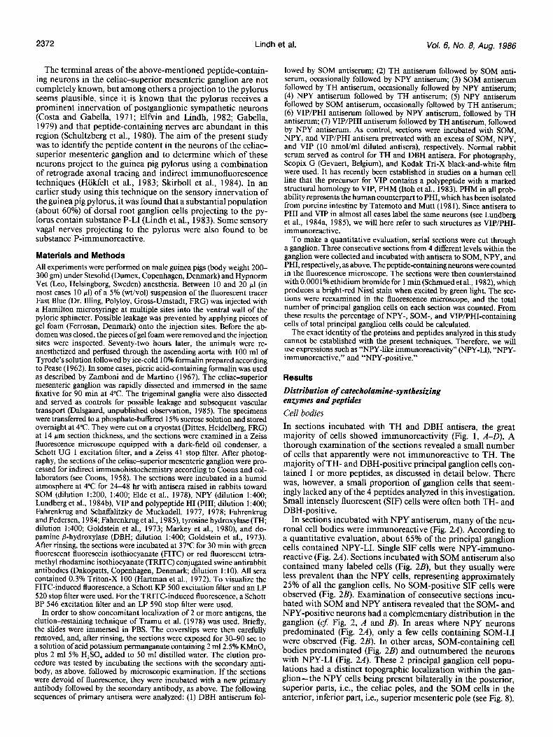
Cell bodies In sections incubated with TH and DBH antisera, the great majority of cells showed immunoreactivity (Fig. 1, A-D). A thorough examination of the sections revealed a small number of cells that apparently were not immunoreactive to TH. The majority of TH- and DBH-positive principal ganglion cells con- tained 1 or more peptides, as discussed in detail below. There was, however, a small proportion of ganglion cells that seem- ingly lacked any of the 4 peptides analyzed in this investigation. Small intensely fluorescent (SIF) cells were often both TH- and DBH-positive.
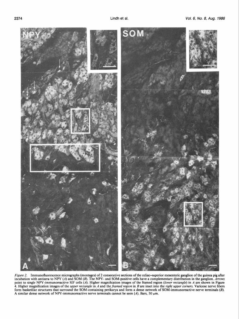
In sections incubated with NPY antiserum, many of the neu- ronal cell bodies were immunoreactive (Fig. 2A). According to a quantitative evaluation, about 65% of the principal ganglion cells contained NPY-LI. Single SIF cells were NPY-immuno- reactive (Fig. 2A). Sections incubated with SOM antiserum also contained many labeled cells (Fig. 2B), but they usually were less prevalent than the NPY cells, representing approximately 25% of all the ganglion cells. No SOM-positive SIF cells were observed (Fig. 2B). Examination of consecutive sections incu- bated with SOM and NPY antisera revealed that the SOM- and NPY-positive neurons had a complementary distribution in the ganglion (cJ: Fig. 2, A and B). In areas where NPY neurons predominated (Fig. 2A), only a few cells containing SOM-LI were observed (Fig. 2B). In other areas, SOM-containing cell bodies predominated (Fig. 2B) and outnumbered the neurons with NPY-LI (Fig. 2A). These 2 principal ganglion cell popu- lations had a distinct topographic localization within the gan- glion-the NPY cells being present bilaterally in the posterior, superior parts, i.e., the celiac poles, and the SOM cells in the anterior, inferior part, i.e., superior mesenteric pole (see Fig. 8).
The Journal of Neuroscience Celiac Neuronal Subpopulations and Projections to Pylorus 2373
Figure 1. Immunofluorescence micrographs (D is a montage) of the celiac-superior mesenteric ganglion after incubation with antisera to TH (A) and DBH (B-D). Section incubated with antisera to DBH is semiconsecutive to the sections shown in Figure 2, A and B. Virtually all cell bodies are TH-positive (A) and DBH-positive (C-D). A network of DBH-positive varicose nerve fibers can be seen (B). A similar varicose network cannot be seen in a corresponding region incubated with TH antiserum (A). In C the DBH-positive varicose nerve fibers from the framed region in B are shown at higher magnification. The DBH-positive varicose fiber network can be seen in the lower part of the montage in D. By comparing the semiconsecutive sections (Fig. 2, A, B), it could be established that the varicose fiber network is found in a region where the SOM cells predominate. Bars, 50 pm.
2374 Lindh et al. Vol. 6, No. 8, Aug. 1986
Figure 2. Immunofluorescence micrographs (montages) of 2 consecutive sections of the celiaosuperior mesenteric ganglion of the guinea pig after incubation with antisera to NPY (A) and SOM (B). The NPY- and SOM-positive cells have a complementary distribution in the ganglion. Arrows point to single NPY-immunoreactive SIF cells (A). Higher magnification images of the framed region (lower rectangle) in A are shown in Figure 4. Higher magnification images of the upper rectangle in A and the framed region in B are inset into the right upper corners. Varicose nerve fibers form basketlike structures that surround the SOM-containing perikruya and form a dense network of SOM-immunoreactive nerve terminals (B). A similar dense network of NPY-immunoreactive nerve terminals cannot be seen (A). Bars, 50 pm.
The Journal of Neuroscience Celiac Neuronal Subpopulations and Projections to Pylorus 2375
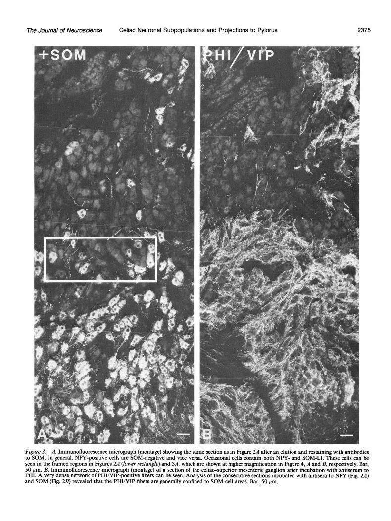
Figure 3. A, Immunofluorescence micrograph (montage) showing the same section as in Figure 24 after an elution and restaining with antibodies to SOM. In general, NPY-positive cells are SOM-negative and vice versa. Occasional cells contain both NPY- and SOM-LI. These cells can be seen in the framed regions in Figures 2A (lower rectungk) and 3A, which are shown at higher magnification in Figure 4, A and B, respectively. Bar, 50 pm. B, Immunofluorescence micrograph (montage) of a section of the celiac-superior mesenteric ganglion after incubation with antiserum to PHI. A very dense network of PHI/VIP-positive fibers can be seen. Analysis of the consecutive sections incubated with antisera to NPY (Fig. 24) and SOM (Fig. 2B) revealed that the PHI/VIP fibers are generally confined to SOM-cell areas. Bar, 50 pm.
2376 Lindh et al. Vol. 6, No. 8, Aug. 1986
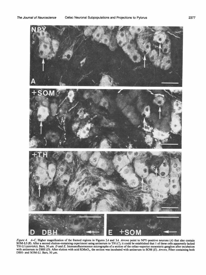
In elution-restaining experiments, it could be established that, in general, NPY-positive cells were SOM-negative and vice ver- sa (Figs. 2A; 3A; 4, A, B). However, occasional cells contained both NPY- and SOM-LI (Figs. 2A; 3A; 4, A, B). After a second elution and restaining with TH antiserum, it could be confirmed that the vast majority of NPY- and SOM-positive cells also contained this catecholamine-synthesizing enzyme (Fig. 4C). A small number of principal ganglion cells containing both NPY- and SOM-LI were, however, apparently TH-negative (Fig. 4, A-C).
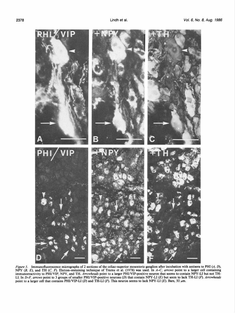
A third population of principal ganglion cells was also en- countered reacting with both VIP and PHI antiserum (Fig. 5, A and D). Their number was low compared with the SOM- and NPY-immunoreactive cell populations, comprising less than 1% of the principal ganglion cells. They were mainly located in the celiac poles (see Fig. 8). Analysis of thin adjacent sections re- vealed that the same cells were both VIP- and PHI-immuno- reactive. Two populations could be distinguished with regard to size, single large cells with a diameter of about 50 pm (Fig. 5A), and a larger number of small cells with a diameter of about 25 pm (Fig. 5D). In restaining experiments it could be estab- lished that the population of small VIP/PHI-positive cells also contained NPY-LI (Fig. 5E). These cells seemed to lack TH-LI (Fig. 5F). Some of the larger cells seemed to contain immu- noreactivity both to the enzyme and the 2 peptides, VIP/PHI plus NPY (Fig. 5, B, C). The SIF cells did not appear VIP/PHI- immunoreactive (Fig. 3B).
Fiber networks A dense network of SOM-immunoreactive, varicose nerve fibers was observed in the part of the ganglion containing SOM-im- munoreactive cells, forming basketlike structures around the SOM-containing perikarya, as well as running between the gan- glion cells (Fig. 2B). Single SOM-positive fibers were also en- countered among the NPY-containing cells. It was not possible to establish definitely whether the varicose SOM fibers origi- nated from SOM-containing principal ganglion cells, being either axons or dendrites, possibly partly accessory dendrites, or orig- inated extrinsically. NPY-positive fibers were seen either in bun- dles or as single fibers, mainly around the NPY-positive cells and in lower numbers among the SOM-positive cells (Fig. W). However, they did not exhibit the characteristic, distinctly var- icose network formed by SOM terminals, either in the NPY- or in the SOM-dominated part of the ganglion.
In sections incubated with DBH antiserum, there was a net- work of varicose fibers that was fairly similar to the one seen with SOM antiserum (Fig. 1, B-D). By comparing consecutive sections it could be established that this fiber network was con- fined to the region predominated by SOM-containing neurons. Elution-restaining experiments revealed a few cases in which these 2 markers (DBH and SOM) occurred in the same fibers (Fig. 4, D, E). A corresponding varicose fiber network could not be found in sections incubated with TH antiserum (Fig. 1A). Dense networks of TH-positive fibers were, however, also seen, but they had a smooth appearance without apparent varicosities and were distributed fairly evenly within the ganglion. TH-, DBH-, NPY-, and SOM-positive fibers were also seen in intra- ganglionic nerve bundles, as well as in small nerves leaving the ganglion.
In sections incubated with antisera raised toward VIP or PHI, a very dense network of VIP/PHI-positive fibers was observed (Fig. 3B). Analysis of consecutive sections showed that the VIP/ PHI-positive fibers were generally confmed to areas where SOM- containing cells predominated (cf: Figs. 3B and 2B), and only a few fibers were seen where the NPY- and the VIP/PHI-im- munoreactive cell bodies were located.
Retrograde tracing experiments Seventy-two hours after injection of Fast Blue into the pyloric sphincter, retrogradely labeled, bright-blue fluorescent neuronal cell bodies were observed in the celiaosuperior mesenteric gan- glion (Figs. 6, A-C and 7A). The labeled cell bodies were found in all parts of the ganglion, with a possible predominance in the celiac poles. No clearly labeled cells were found in the trigeminal ganglia.
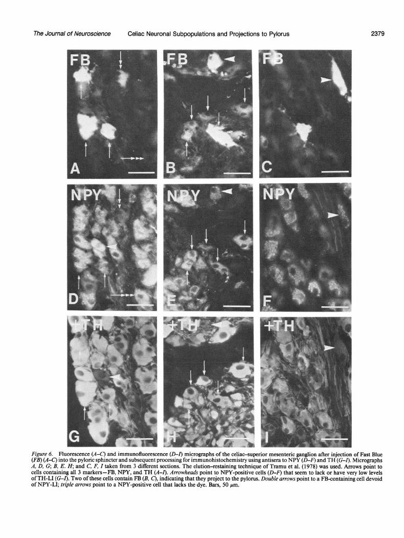
Examination of micrographs taken from sections containing Fast Blue-labeled cell bodies, subsequently stained for TH, showed that the overwhelming majority of cell bodies that con- tained the fluorescent tracer also contained the enzyme. Com- parison of micrographs taken from the same sections analyzed consecutively for retrogradely transported Fast Blue and NPY- and SOM-LI showed neurons containing both the fluorescent tracer and immunoreactivity to NPY (Figs. 6, D-F; 7B) or SOM (Fig. 7C). So far, no cell bodies have been observed to contain all 3 markers (Fast Blue, NPY, and SOM). The Fast Blue-labeled cells containing NPY-LI clearly outnumbered the Fast Blue- labeled cells containing SOM-LI. In addition, there were cell bodies that contained only the fluorescent tracer or only 1 of the peptides. Elution-restaining experiments established that neurons containing both Fast Blue and a peptide were also im- munoreactive to TH (Figs. 6, G-Zand 70). Occasional cells that contained both Fast Blue and NPY-LI and that seemed to lack TH-LI were, however, also observed (Fig. 6, C, F, and I). Fur- thermore, it could be established that some Fast Blue-labeled cells containing TH-LI seemed to lack immunoreactivity toward SOM and NPY (Fig. 7, A-D).
After incubation with control sera, none of the immunoreac- tive staining patterns described above could be observed.
Discussion
Transmitter enzymes and peptides in cell bodies and fibers The present results confirm and extend earlier information on the localization and distribution of various peptides within the principal ganglion and SIF cells of the celiac-superior mesenteric ganglion. A summary of the results obtained is presented in Figure 8. Thus, SOM-positive principal ganglion cells were first demonstrated in high numbers (Hbkfelt et al., 1977a) and, sub- sequently, it was recognized that a different population of neu- rons contains a peptide that can be demonstrated with antise- rum raised to avian pancreatic polypeptide (APP) (Lundberg et al., 1982a). Subsequent analyses have revealed that this peptide in all probability represents an NPY-like peptide (Lundberg et al., 1983, 1984b). The present findings clearly demonstrate that the 2 peptides are in separate cell populations that occupy spe- cific domains within the ganglion (Fig. 8). Thus, the SOM cells are mainly found in the anterior-inferior part of the ganglion, representing the superior mesenteric pole, whereas the NPY cells are found bilaterally in the celiac poles, which occupy the pos- terior-superior parts of the ganglion. A similar regional pre- ponderance of the SOM-positive cells in the anterior-inferior part of the ganglion was described in the earlier paper by Hokfelt et al. (1977a). A certain degree of overlap and intermingling exists, and there are, in fact, a few cells that are immunoreactive to both peptides. The present quantitative evaluation demon- strates that NPY-positive cells are most numerous, representing about two-thirds of all ganglion cells, whereas the SOM cells make up about 25%.
A third population of ganglion cells was observed, character- ized by its content of a VIP-like peptide, in agreement with earlier studies (Hiikfelt et al., 1977b). This cell population also contains a second peptide, peptide histidine isoleucine (PHI), which has recently been discovered by Tatemoto and Mutt (1980,
The Journal of Neuroscience Celiac Neuronal Subpopulations and Projections to Pylorus 2377
Figure 4. A-C, Higher magnification of the framed regions in Figures 24 and 3A. Arrows point to NPY-positive neurons (A) that also contain SOM-LI (B). After a second elution-restaining experiment using antiserum to TH (C), it could be established that 1 of these cells apparently lacked TH-LI (asterisks). Bars, 50 pm. D and E, Immunofluorescence micrographs of a section of the celiac-supetior mesenteric ganglion after incubation with antiserum to DBH (0). After elution with acid KMnO,, the section was incubated with antiserum to SOM (E). Arrows, Fiber containing both DBH- and SOM-LI. Bars, 50 pm.
2378 Lindh et al. Vol. 6, No. 8, Aug. 1986
Figure 5. Immunofluorescence micrographs of 2 sections of the celiac-superior mesenteric ganglion after incubation with antisera to PHI (A, D), NPY (B. El. and TH (C. F). Elution-restainine. techniaue of Tramu et al. (1978) was used. In A-C, arrows ooint to a lamer cell containinn immuhoreactivity to PI?IINIP, NPY, and TH. zrowheahs point to a larger PI?I/VIP-positive neuron that seems-to contain NPY-LI but not THI LI. In D-F, arrows point to 3 groups of smaller PHWIP-positive neurons (0) that contain NPY-LI Q but seem to lack TH-LI Q. Arrowheads point to a larger cell that contains PHI/VIP-L1 (0) and TH-LI (F). This neuron seems to lack NPY-LI (E). Bars, 50 pm.
The Journal of Neuroscience Celiac Neuronal Subpopulations and Projections to Pylorus
Figure 6. Fluorescence (A-C) and immunofluorescence (D-Z) micrographs of the celiac-superior mesenteric ganglion after injection of Fast Blue (FB) (A-C’) into the pyloric sphincter and subsequent processing for immunohistochemistry using antisera to NPY (0-Z) and TH (G-Z). Micrographs A, D, G, B, E. H; and C, F, Z taken from 3 different sections. The elution-restaining technique of Tramu et al. (1978) was used. Arrows point to cells containing all 3 markers-FB, NPY, and TH (A-Z). Arrowheads point to NPY-positive cells (D-F) that seem to lack or have very low levels of TH-LI (G-Z). Two of these cells contain FB (B, C), indicating that they project to the pylorus. Double arrows point to a FB-containing cell devoid of NPY-LI; triple arrows point to a NPY-positive cell that lacks the dye. Bars, 50 pm.

2380 Lindh et al. Vol. 6, No. 8, Aug. 1986
Figure 7. Fluorescence (A) and im- munofluorescence (B-D) micro- graphs of a section of the celiac-su- perior mesenteric ganglion after injection of Fast Blue (A) into the py- 101% sphincter and subsequent pro- cessing for immunohistochemistry using antisera in the sequence SOM (C), TH (D), and NPY (B). The elu- tion-restaining technique of Tramu et al. (1978) was used. Arrows point to 3 cells that contain the dye (A), one of which is SOM-positive (C, aster- isk) and a second one that is NPY- positive (B, asterisk). All 3 cells con- tain TH-LI (0). One of these cells, which also contains the dye, lacks peptide immunoreactivity (cJ A-D). Bars, 50 pm.
CELL TYPES:
SYMBOLS :
O=NA 0 =NPY 0 =sobA + =vIP L
J
o= NPY+NA
@ = SOM+NA
@ = VIP+NPY
O-NA @= NPY+SOM+NA
q =NPY @ = VIP+NA
Ss = NPY*SOM @ = VIP+NPY+NA
Figure 8. Schematic drawing illustrating the celiaosuperior mesen- teric ganglion and the various cell types found in this study. A, Topo- graphic localization of the 3 main populations of peptidergic cells in the ganglion. For each of the 3 peptides, the most frequent coexistence situation is indicated. B, Other, more infrequently occurring cell types defined by their transmitter content as found in this study. The drawings give only a rough quantitative account of the different cell types.
198 1). Subsequent studies using recombinant DNA technology have demonstrated that PHI is part of the precursor for VIP, which thus contains a copy of both VIP and PHI (Itoh et al., 1983). These findings are in complete agreement with the pres- ent demonstration of PHI and VIP in the same prevertebral ganglion cells, as well as with other immunohistochemical stud- ies demonstrating colocalization of these 2 peptides (Bishop et al., 1984; Fahrenkrug et al., 1985; Lundberg et al., 1984a; Yanai- hara et al., 1983). Two populations of VIP/PHI neurons could be distinguished by their size: small cells, which were more numerous, and single large ones. Also, the VIP/PHI neurons occupied a distinct domain in the ganglion within the celiac poles, intermingling with the NPY-immunoreactive cell bodies (Fig. 8).
The vast majority of the NPY- and SOM-positive cells con- tained TH- and DBH-LI, suggesting that they are noradrenergic. A few NPY-immunoreactive cells did, however, lack TH, in- dicating that a small population of principal ganglion cells is nonadrenergic. The occurrence of non-adrenergic NPY-positive autonomic neurons is discussed further below. The majority of the VIP/PHI-positive cells were also TH-negative, and these non-adrenergic cells were of the small type, whereas single large VIP/PHI-positive cells also contained TH-LI. Whether these differences reflect the actual situation or whether an insensitivity in our immunohistochemical technique is involved is not known.
The present study confirms earlier findings of a lack of SOM- and VIP-L1 in SIF cells. It adds the information that a few SIF cells are NPY-immunoreactive.
The present findings demonstrate differences in appearances of SOM- and NPY-immunoreactive fibers. NPY-immunoreac- tive fibers have a smooth appearance and can be found in all parts of the ganglion. They are only weakly immunoreactive
The Journal of Neuroscience Celiac Neuronal Subpopulations and Projections to Pylorus 2381
and could represent axonal processes and fiber bundles. Similar SOM-positive fibers can also be seen, but, in addition, there is a distinct and fairly dense network of strongly fluorescent, var- icose SOM-positive fibers, in most cases closely surrounding SOM-positive cells but also found in the fiber strands in between the ganglion cells. The nature of these fibers is uncertain. They may represent dendritic processes emanating from the gangli- onic cell bodies or collaterals from the axons leaving the ganglion cells. Alternatively, they may have an extrinsic origin. Experi- ments are now in progress to try to define these fibers in more detail. Interestingly, a fiber network of a similar morphology and apparent density is seen after incubation with DBH anti- serum. Surprisingly, this DBH-positive network was mainly confined to the SOM-dominated part of the ganglion, thus show- ing a close overlap with the varicose SOM fibers. We attempted to establish or exclude identity between these 2 fiber networks by carrying out restaining experiments. In a few cases, DBH- and SOM-LI could be seen in the same fibers, but difficulties in restaining such thin fibers precluded a conclusive answer.
Ifthe SOM-positive varicose network emanates from the SOM ganglion cells, these cells would have a distinctly different mor- phology compared to the NPY-immunoreactive neurons, which apparently lack processes of this type. Whether this distinct difference between NPY and SOM neurons is real or represents an artifact related to the immunohistochemical technique is at present being analyzed by studies at the EM level. Incubation with TH antiserum did not reveal a fiber network corresponding to the DBH-positive varicosities. Instead, there was a dense network of smooth TH-positive fibers. This may be because the enzyme TH is cytoplasmic and fills out the entire axon, whereas DBH is largely membrane-bound (vesicles) and therefore stains varicosities preferentially.
Projections to the pylorus The present investigation gives strong evidence for a projection to the guinea pig pylorus from the celiac-superior mesenteric ganglion, in agreement with an earlier study by Elfvin and Lindh (1982). Here, we describe at least 4 subpopulations of postgan- glionic sympathetic neurons in the celiac-superior mesenteric ganglion projecting to the pylorus. These are neurons apparently containing only TH-LI, neurons containing immunoreactivity to both TH and NPY, neurons immunoreactive to both TH and SOM, and neurons containing immunoreactivity only to NPY. It should be pointed out that the definition of 4 categories is based on presence or absence of the enzyme TH and 2 peptides as revealed with immunohistochemistry. It is quite possible that our technique is not sensitive enough to reveal low levels of these compounds, and the present subdivision may therefore have to be revised in the future.
The description of these pathways is based on a technique that involves a combination of fluorescent retrograde tracing with immunohistochemistry (Hiikfelt et al., 1983; Skirboll et al., 1984). The method has previously been used in our labo- ratory in, for example, studies of peptidergic pathways project- ing to the inferior mesenteric ganglion in the guinea pig (Dals- gaard et al., 1982a, b) and of the origin of the sensory substance P innervation of the pylorus (Lindh et al., 1983). Since the fluorescent tracer Fast Blue is not stable during the immuno- cytochemical procedures, there is a risk that the tracer diffuses out of the cells during the incubation period. Therefore, the labeled cells are photographed before the sections are incubated with the different primary antisera. Fast Blue has a tendency to “shine through” the FITC filters, and we have therefore used a TRITC conjugated secondary antibody. Cells retrogradely la- beled with Fast Blue were found in all parts of the ganglion, but they seemed to be more prevalent in the celiac poles, suggesting a somatotopic organization within the ganglion.
Since peripherally injected fluorescent dyes can reach gangli-
onic cell bodies via the vascular system (Dalsgaard, unpublished observations), we also analyzed possible labeling of cell bodies in the trigeminal ganglion. Since no Fast Blue cell bodies were observed in this ganglion in our study, it seems likely that the Fast Blue-positive cells in the celiac-superior mesenteric gan- glion have been labeled by retrograde axonal transport and not by blood-borne dye.
The retrogradely labeled cells were practically all TH-con- taining, which shows that the pylorus receives a heavy NA pro- jection from the ganglion. In earlier studies, it has been shown by means of the formaldehyde-induced (Falck-Hillarp) fluores- cence technique that the pyloric sphincter muscles of the guinea pig have a dense network of NA nerve terminals (Costa and Gabella, 197 1; Gabella, 1979). The NA innervation of the guinea pig pylorus has also been demonstrated with immunohisto- chemical techniques with antisera raised toward DBH (Schultz- berg et al., 1980). The adrenergic innervation to the pylorus exerts a contractile action on the sphincter, demonstrated by splanchnic nerve stimulation and local intraarterial infusion of noradrenaline (Edin et al., 1979).
A substantial number of the Fast Blue-labeled cells containing TH were also immunoreactive for SOM and NPY. The present observations show that some of these cells with multiple mes- sengers also project to the guinea pig pylorus. The results of the present study agree with the findings of Fumess et al. (1983) and Costa and Fumess (1984), who, using a combination of immunohistochemistry and nerve lesion techniques, demon- strated a projection to the guinea pig small intestine of NA neurons containing SOM- or NPY-LI (see below).
There were more Fast Blue-labeled cells immunoreactive to NPY than to SOM in the ganglion, which, in part, may reflect the higher proportion of NPY- as compared to SOM-immu- noreactive principal ganglion cells and the fact that most retro- gradely labeled cells were found in the celiac poles, i.e., the region containing predominantly NPY-positive ganglion cells. This indicates that the pyloric region receives a somewhat more prominent projection of NA/NPY neurons than of NA/SOM neurons. Two more subgroups of neurons projecting to the guinea pig pylorus comprise cells containing only a catecholamine and a small group of cells that appear to contain only NPY-LI. In this context it should be mentioned that Hassal and Bumstock (1984) and Dalsgaard et al. (1985) recently provided evidence for NPY-immunoreactive cell bodies in atria1 ganglia that seemed to lack TH-LI. Such NPY-positive, apparently noncat- echolaminergic neurons have also been observed in the rat vas deferens (Fried et al., 1985) and in the human fallopian tube (Samuelson and Dalsgaard, 1985). The latter results must be interpreted with caution, since they may reflect a difficulty in detecting the enzyme, but the tissue culture studies of Hassal and Bumstock (1984) strongly support the presence of noncat- echolaminergic NPY neurons. Furthermore, it is known that immunoreactive NPY cell bodies that are apparently non-NA occur in the enteric ganglia in the gastrointestinal tract of the guinea pig and the rat (Fumess et al., 1983; Sundler et al., 1983).
The exact target areas in the pylorus of the subclasses of neurons demonstrated in the present study are unknown and can only be clearly determined by a systematic analysis of the terminal network of the nerves in combination with denervation procedures.
Functional aspects Although the classification of the functional roles of the various nerves projecting to the guinea pig pylorus has to await further study, some comparison can be made with earlier studies in which the specific functions of the NA/NPY, NASOM, and NA neurons have been investigated. Thus, in their elegant ex- periments, Fumess et al. (1983) and Costa and Fumess (1984) showed that the different subgroups of NA neurons in the celiac-
2382 Lindh et al. Vol. 6, No. 8, Aug. 1986
superior mesenteric ganglion of the guinea pig seem to supply separate tissue components in the wall of the small intestine. The major targets for the NA/NPY neurons were the intestinal blood vessels, whereas the NA/SOM neurons seemed to inner- vate the submucosal ganglia and the mucosa. The NA nerves lacking NPY- and SOM-LI projected to the myenteric ganglia. The different target areas for the neurochemical groups of NA nerves suggest that these groups also have different functional properties. The NA/NPY neurons would be involved in regu- lation of intestinal blood flow, the NABOM neurons in control of mucosal function, and the NA nerves lacking NPY- and SOM-LI primarily in regulation of intestinal motility. A pref- erential role of NA sympathetic neurons containing NPY-LI in control of blood flow has also been suggested in several studies by Lundberg et al. (1982b, 1985).
The analysis of the distribution patterns of peptide terminals in the celiac-superior mesenteric ganglion confirms earlier find- ings of a dense, heterogeneously distributed VIP-immunoreac- tive fiber network (HGkfelt et al., 1977b). The present results confirm the previous demonstration that these terminals mainly innervate NA cell bodies containing SOM-LI (Lundberg et al., 1982a). These VIP fibers presumably originate in the intestinal wall, as has been shown for the guinea pig inferior mesenteric ganglion (Dalsgaard et al., 1983). The results suggest chemical coding within the celiac-superior mesenteric ganglion with re- gard to content of messenger molecules both in the principal ganglion cells and in fibers of extrinsic origin. The strict orga- nizational principles may point to a possible functional differ- entiation on the basis of chemical specificity. In this respect, the studies of Costa and Furness (Costa and Fumess, 1984; Fumess et al., 1983) discussed above are interesting. This would suggest that VIP-containing afferents are mainly important in controling mucosal function and may not participate in regu- lating blood flow (see above). Related studies show that several other peptide-containing afferents to the celiac-superior mes- enteric ganglion-including dynorphin-, cholecystokinin-, and bombesin-immunoreactive fibers- have a distribution parallel to that of the VIP/PHI-immunoreactive fibers (unpublished ob- servations). Since they may all originate in the gastrointestinal wall, they may participate in the reflex arc between intestine and prevertebral ganglia. In contrast, strongly enkephalin- as well as choline acetyltransferase-immunoreactive fibers also in- nervate the NPY domains of the ganglia, and these fibers may have a central origin. Such findings suggest that control of in- testinal blood flow (via NA/NPY prevertebral neurons) is main- ly exerted via the CNS. Only further experiments can verify this hypothesis.
References Bishop, A. E., J. M. Polak, Y. Yangou, N. D. Christofides, and S. R.
Bloom (1984) The distribution of PHI and VIP in porcine gut and their co-localisation to a proportion of intrinsic ganglion cells. Pep- tides 5: 255-259.
Brazeau, P., W. Vale, R. Burgus, N. Ling, M. Butcher, J. Rivier, and R. Guillemin (1973) Hvoothalamic DolvDeDtide that inhibits the secretion of immunoieacti;e pituitary &o$h hormone. Science 179: 77-79.
Coons, A. H. (1958) Fluorescent antibody methods. In General Cy- tochemicalMethods, J. F. Danielli, ed., pp. 399-422, Academic, New York.
Costa, M., and J. B. Fumess (1984) Somatostatin is present in a subpopulation of noradrenergic nerve fibres supplying the intestine. Neuroscience 13: 9 1 l-9 19.
Costa, M., and G. Gabella (197 1) Adrenergic innervation of the ali- mentary canal. Z. Zellforsch. 122: 357-377.
Dalsgaard, C.-J., A. France-Cereceda, A. Saria, J. M. Lundberg, E. Theodorsson-Norheim, and T. Hijkfelt (1986) Distribution and or-
igin of substance P- and neuropeptide Y-immunoreactive nerves in the guinea pig heart. Cell Tissue Res. 243: 477-485.
Dalsaaard. C.-J.. T. H&felt. L.-G. Elfvin. L. Skirboll. and P. Emson (1582a)’ Substance P-containing prima& sensory neirons projecting to the inferior mesenteric ganglion: Evidence from combined retro- grade tracing and immunohistochemistry. Neuroscience 7: 647-654.
Dalsgaard, C.-J., T. Hakfelt, L.-G. Elfvin, and L. Terenius (1982b) Enkephalin-containing sympathetic preganglionic neurons projecting to the inferior mesenteric ganglion: Evidence from combined retro- grade tracing and immunohistochemistry. Neuroscience 7: 2039-2050.
Dalsgaard, C.-J., T. Hijkfelt, M. Schultzberg, J. M. Lundberg, L. Teren- ius, G. J. Dockray, and M. Goldstein (1983) Origin of peptide con- taining fibers in the inferior mesenteric ganglion of the guinea-pig: Immunohistochemical studies with antisera to substance P, enke- phalin, vasoactive intestinal polypeptide, cholecystokinin and bom- besin. Neuroscience 9: 19 l-2 11.
Edin, R., H. Ahlman, and J. Kewenter (1979) The vagal control of the feline pyloric sphincter. Acta Physiol. S&d. 107: 169-l 74.
Elde. R.. T. H&felt. 0. Johansson. M. Schultzbera. S. Efendic. and R. L;ft i1978) Cellular localizatidn of somatosta%n. Clin. Eip. Met- abol. (Suppl. 1) 27: 115 l-l 159.
Elfvin, L.-G., and B. Lindh (1982) A study ofthe extrinsic innervation of the guinea pig pylorus with the horseradish peroxidase tracing technique. J. Comp. Neurol. 208: 3 17-324.
Eve&t. B. J.. T. Hijkfelt. L. Terenius. K. Tatemoto. V. Mutt. and M. Gol&tein 11984) Difl&ential co-existence of neur&ptide ? (NPY)- like immunoreactivity with catecholamines in the central nervous system of the rat. Neuroscience 1 I: 443-462.
Fahrenkrug, J., and J. H. Pedersen (1984) Development and validation of a specific radioimmunoassay for PHI in plasma. Clin. Chim. Acta 143: 183-192.
Fahrenkrug, J., and 0. B. Schaffalitzky de Muckadell (1977) Radioim- munoassay of vasoactive intestinal polypeptide (VIP) in plasma. J. Lab. Clin. Med. 89: 1379-1388.
Fahrenkrug, J., and 0. B. Schaffalitzky de Muckadell (1978) Distri- bution of vasoactive intestinal polypeptide (VIP) in the porcine central nervous system. J. Neurochem. 31: 1445-1451.
Fahrenkrug, J., T. Bek, J. M. Lundberg, and T. HGkfelt (1985) VIP and PHI in cat neurons: Colocalization but variable tissue content possibly due to differential processing. Regul. Pept. 12: 21-34.
Fried. G.. L. Terenius. T. Hakfelt. and M. Goldstein (1985) Evidence for’the’differential lbcalization bf noradrenaline ad neuiopeptide Y (NPY) in neuronal storage vesicles isolated from rat vas deferens. J. Neurosci. 5: 450-458.
Fumess, J. B., M. Costa, P. C. Emson, R. Hakanson, E. Moghimzadeh, F. Sundler, I. L. Taylor, and R. E. Chance (1983) Distribution, pathways and reactions to drug treatment of nerves with neuropeptide Y- and pancreatic polypeptide-like immunoreactivity in the guinea pig digestive tract. Cell Tissue Res. 234: 7 l-92.
Gabella, G. (1979) Innervation of the gastrointestinal tract. Int. Rev. Cytol. 59: 129-193.
Goldstein, M., B. Anagnoste, L. S. Freedman, M. Roffman, R. P. Eb- stein, D. H. Park, K. Fuxe, and T. Hijkfelt (1973) Characterization, localization and regulation of catecholamine synthesizing enzymes. In Frontiers in Catecholamine Research, E. Usdin and S. Snyder, eds., pp. 69-78, Pergamon, New York.
Hartman, B. K., D. Zide, and S. Udenfriend (1972) The use of do- pamine fi-hydroxylase as a marker for the noradrenergic pathways of the central nervous system in the rat. Proc. Natl. Acad. Sci. USA 69: 2722-2726.
Hassal, C. J. S., and G. Bumstock (1984) Neuropeptide Y-like im- munoreactivity in cultured intrinsic neurons of the heart. Neurosci. L&t. 52: 11 l-l 15.
HGkfelt, T., L.-G. Elftin, R. Elde, M. Schultzberg, M. Goldstein, and R. Luft (1977a) Occurrence of somatostatin-like immunoreactivity in some peripheral sympathetic noradrenergic neurons. Proc. Natl. Acad. Sci. USA 74: 3587-3591.
Hiikfelt, T., L.-G. Elfvin, M. Schultzberg, K. Fuxe, S. I. Said, V. Mutt, and M. Goldstein ( 1977b) Immunohistochemical evidence of vas- oactive intestinal pdlypeptide-containing neurons and nerve fibers in sympathetic ganglia. Neuroscience 2: 885-896.
Hakfelt, T., M. Schultzberg, 0. Johansson, A. Ljungdahl, L.-G. Elfvin, R. Elde, L. Terenius, G. Nilsson, S. Said, and M. Goldstein (1978) Central and peripheral peptide producing neurons. In Gut Hormones, S. R. Bloom, ed., pp. 423-433, Churchill Livingstone, Edinburgh.
The Journal of Neuroscience Celiac Neuronal Subpopulations and Projections to Pylorus 2383
Hijkfelt, T., G. Skagerberg, L. Skirboll, and A. Bjijrklund (1983) Com- bination of retrograde tracing and neurotransmitter histochemistry.
Pease, D. C. (1962) Buffered formaldehyde as a killing agent and
In Handbook of Chemical Neuroanatomy, Vol. 1, A. Bjijrklund and primary fixative for electron microscopy. Anat. Rec. 142: 342.
T. Hiikfelt, eds., pp. 228-285, Elsevier, Amsterdam. Said, S. I., and V. Mutt (1970) Polypeptide with broad biological
activity. Isolation from small intestine. Science 169: 12 17-l 2 18. Itoh, N., K. Obata, N. Yanaihara, and H. Okamoto (1983) Human Samuelson, U. E., and C. J. Dalsgaard (1985) Effects and localization
preprovasoactive intestinal polypeptide contains a novel PHI-27-like of neuropeptide Y in human fallopian tube. Neurosci. Lett. 58: 49- peptide, PHM-27. Nature 304: 547-549. 54.
Krulich, L., A. P. S. Dhariwal, and S. M. McCann (1968) Stimulatory and inhibitory effects of purified hypothalamic extracts on growth hormone release from pituitary in vitro. Endocrinology 83: 783-790.
Lindh, B., C.-J. Dalsgaard, L.-G. Elfvin, T. H&felt, and A. C. Cue110 (1983) Evidence of substance P immunoreactive neurons in dorsal root ganglia and vagal ganglia projecting to the guinea pig pylorus. Brain Res. 269: 365-369.
Schmued, L. C., L. W. Swanson, and P. E. Sawchenko (1982) Some fluorescent counterstains for neuroanatomical studies. J. Histochem. Cytochem. 30: 123-128.
Lundberg, J. M., T. HBkfelt, A. i&g&d, L. Terenius, R. Elde, K. Markey, M. Goldstein, and J. Kimmel (1982a) Organizational prin- ciples in the peripheral sympathetic nervous system: Subdivision by coexisting peptides (somatostatin-, avian pancreatic polypeptide-, and vasoactive intestinal polypeptide-like- immunorea&e^mate- rials). Proc. Natl. Acad. Sci. USA 79: 1303-1307.
Schultzberg, M., T. Hiikfelt, G. Nilsson, L. Terenius, J. F. Rehfeld, M. Brown. R. Elde. M. Goldstein. and S. Said (1980) Distribution of peptide- and catkcholamine-containing neurons in the gastrointestinal tract of rat and guinea-pig: Immunohistochemical studies with anti- sera to substance P, vasoactive intestinal polypeptide, enkephalins, somatostatin, gastrin/cholecystokinin, neurotensin and dopamine fi-hydroxylase. Neuroscience 5: 689-744.
Skirboll, L., T. HBkfelt, G. NorelJ, 0. Phillipson, H. G. J. M. Kuypers, M. Bentivoglio, C. E. Catsman-Berrevoets, T. J. Visser, H. Stein- busch, A. Verhofstad, A. C. Cuello, M. Goldstein, and M. Brownstein (1984) A method for specific transmitter identification ofretrogradely labeled neurons: Immunofluorescence combined with fluorescence tracing. Brain Res. Rev. 8: 99-l 27,
Lundberg, J. M., L. Terenius, T. Hiikfelt, C. R. Martling, K. Tatemoto, V. Mutt, J. Polak, S. Bloom, and M. Goldstein (1982b) Neuropep- tide Y (NPY)-like immunoreactivity in peripheral noradrenergic neu- rons and effects ofNPY on sympathetic function. Acta Physiol. Stand. 116: 477-480.
Lundberg, J. M., L. Terenius, T. Hokfelt, and M. Goldstein (1983) High levels of neuropeptide Y in peripheral noradrenergic neurons in various mammals including man. Neurosci. Lett. 42: 167-172.
Lundberg, J. M., J. Fahrenkrug, T. HBkfelt, C.-R. Marding, 0. hsson, K. Tatemoto, and A. Ana&d (1984a) Coexistence of nentide HI (PHI) and VIP in neuronsregulating blood flow and bronchial smooth muscle tone in various mammals including man. Peptides 5: 593- 606.
Lundberg, J. M., L. Terenius, T. Hbkfelt, and K. Tatemoto (1984b) Comparative immunohistochemical and biochemical analysis of pan- creatic polypeptide-like peptides with special reference to presence of neuropeptide Y in central and peripheral neurons. J. Neurosci. 4: 2376-2386.
Lundberg, J. M., A. Am&d, J. Pernow, and T. H&felt (1985) Neu- ropeptide Y-, substance P- and VIP-immunoreactive nerves in cat spleen in relation to autonomic vascular and volume control. Cell Tissue Res. 239: 9-l 8.
Markey, K. A., S. Kondo, L. Shenkman, and M. Goldstein (1980) Purification and characterization of tyrosine hydroxylase from a clon- al pheochromocytoma cell line. Mol. Phannacol. 17: 79-85.
Mutt, V., and S. I. Said (1974) Structure of the porcine vasoactive intestinal octacosapeptide: The amino-acid sequence. Use of kalli- krein in its determination. Eur. J. Biochem. 42: 581-589.
Sundler, F., E. Moghimzadeh, R. Hakanson, M. Ekelund,, and P. Emson (1983) Nerve fibers in the gut and pancreas of rat displaying neuropeptide-Y immunoreactivity. Cell Tissue Res. 230: 487-493.
Tatemoto, K. (1982) Neuropeptide Y: Complete amino-acid sequence of the brain nentide. Proc. Natl. Acad. Sci. USA 79: 5485-5489.
Tatemoto, K., and V. Mutt (1980) Isolation of two novel candidate hormones using a chemical method for finding naturally occurring polypeptides. Nature 285: 4 17-4 18.
Tatemoto, K., and V. Mutt (198 1) Isolation and characterization of the intestinal peptide porcine PHI (PHI-27), a new member of the glucagon-secretin family. Proc. Natl. Acad. Sci. USA 78: 6603-6607.
Tatemoto, K., M. Carlquist, and V. Mutt (1982) Neuropeptide Y- A novel brain peptide with structural similarities to peptide YY and pancreatic polypeptide. Nature 296: 659-660.
Tramu, G., A. Pillez, and J. Leonardelli (1978) An efficient method of antibody elution for the successive or simultaneous localization of two antigens by immunocytochemistry. J. Histochem. Cytochem. 26: 322-324.
Yanaihara, N., K. Nokihara, C. Yanaihara, T. Iwanaga, and T. Fujita (1983) Immunocytochemical demonstration of PHI and its co-ex- istence with VIP in intestinal nerves of the rat and pig. Arch. Histol. Jpn. 46: 575-58 1.
Zamboni, L., and C. de Martin0 (1967) Buffered pi&c acid formal- dehyde: A new rapid fixative for electron microscopy. J. Cell Biol. 35: 148A.


![NPY Rule Book [constitution] catsi act approved at 14.11.08](https://img.pdfslide.us/doc/110x75/554c05deb4c9058e098b51ad/npy-rule-book-constitution-catsi-act-approved-at-141108.jpg)