Embed Size (px)
Citation preview
Topic Number Eleven
The Immune System
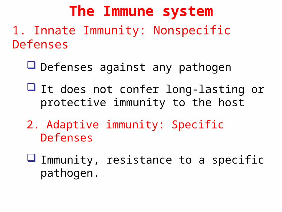
The Immune system1. Innate Immunity: Nonspecific Defenses
Defenses against any pathogen
It does not confer long-lasting or protective immunity to the host
2. Adaptive immunity: Specific Defenses
Immunity, resistance to a specific pathogen.
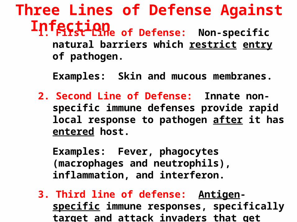
1. First Line of Defense: Non-specific natural barriers which restrict entry of pathogen.
Examples: Skin and mucous membranes.
2. Second Line of Defense: Innate non-specific immune defenses provide rapid local response to pathogen after it has entered host.
Examples: Fever, phagocytes (macrophages and neutrophils), inflammation, and interferon.
3. Third line of defense: Antigen-specific immune responses, specifically target and attack invaders that get past first two lines of defense.
Examples: Antibodies and lymphocytes.
Three Lines of Defense Against Infection
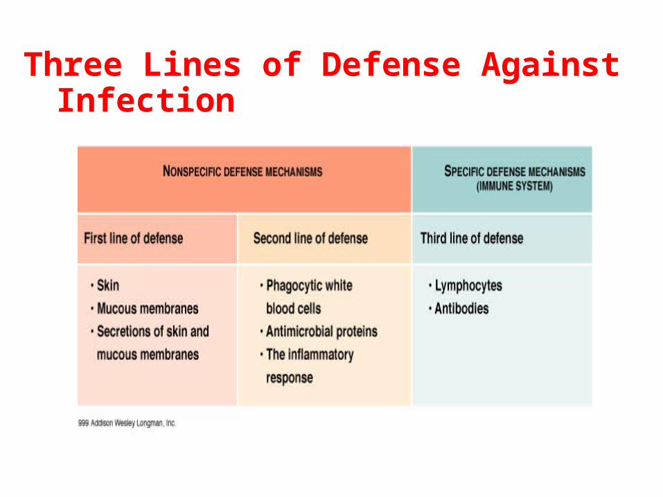
Three Lines of Defense Against Infection
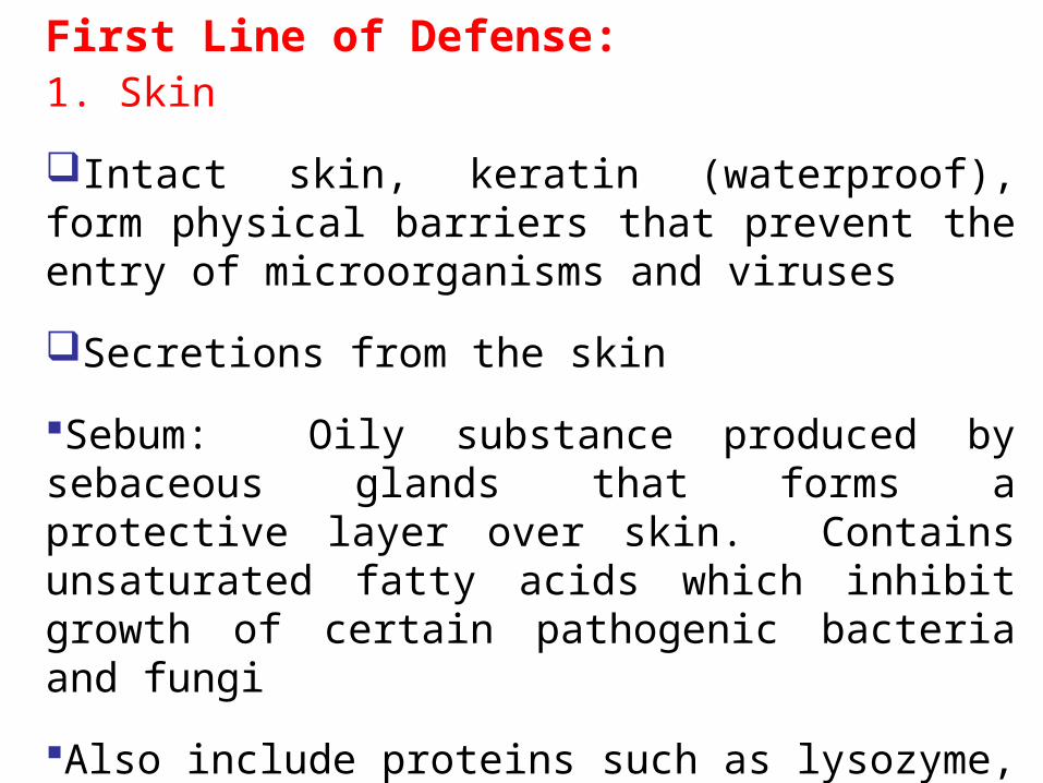
First Line of Defense:1. Skin
Intact skin, keratin (waterproof), form physical barriers that prevent the entry of microorganisms and viruses
Secretions from the skin
Sebum: Oily substance produced by sebaceous glands that forms a protective layer over skin. Contains unsaturated fatty acids which inhibit growth of certain pathogenic bacteria and fungi
Also include proteins such as lysozyme, an enzyme that digests the cell walls of many bacteria
Infections are rare in intact skin. Exceptions:
Hookworms can penetrate intact skin
Dermatophytes: “Skin loving” fungi
Normal microbiota compete with pathogens.
2. Mucous membranes
Saliva: Washes microbes from teeth and mouth mucous membranes.
Mucus: Thick secretion that traps many microbes.
Urination: Cleanses urethra.
Vaginal Secretions: Remove microbes from genital tract.
II. Second Line of Defense
1. Phagocytosis
Phagocytosis is carried out by white blood cells: macrophages, neutrophils, and occasionally eosinophils.
Wandering macrophages: Originate from monocytes that leave blood and enter infected tissue, and develop into phagocytic cells.
Fixed Macrophages (Histiocytes): Located in liver, nervous system, lungs, lymph nodes, bone marrow, and several other tissues.
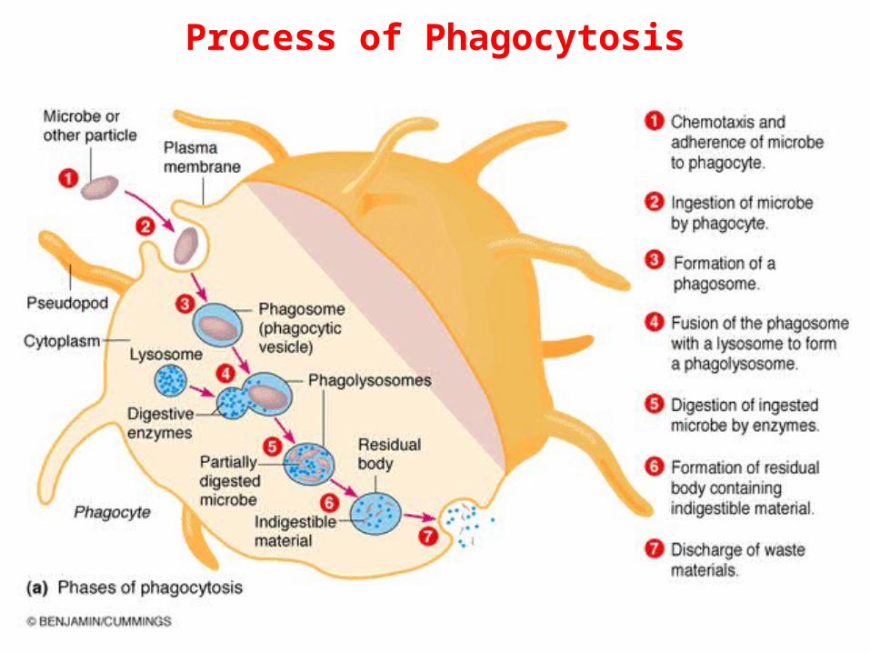
Process of Phagocytosis
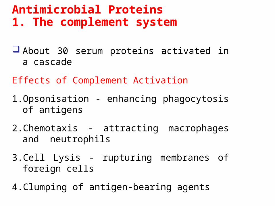
Antimicrobial Proteins1. The complement system
About 30 serum proteins activated in a cascade
Effects of Complement Activation
1.Opsonisation - enhancing phagocytosis of antigens
2.Chemotaxis - attracting macrophages and neutrophils
3.Cell Lysis - rupturing membranes of foreign cells
4.Clumping of antigen-bearing agents
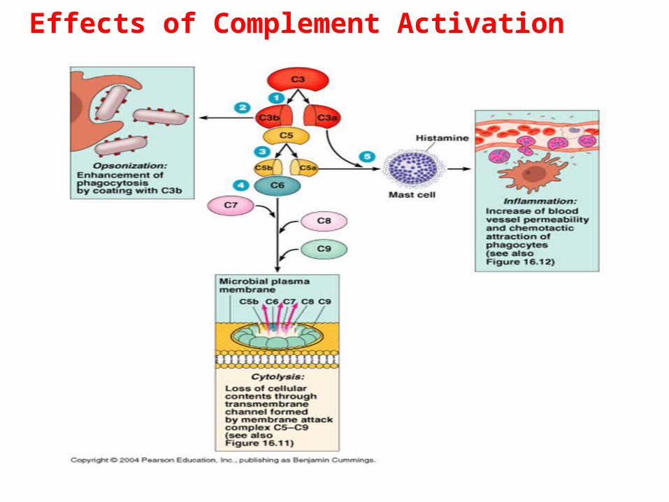
Effects of Complement Activation
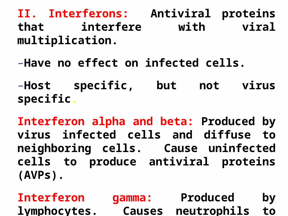
II. Interferons: Antiviral proteins that interfere with viral multiplication.
–Have no effect on infected cells.
–Host specific, but not virus specific.
Interferon alpha and beta: Produced by virus infected cells and diffuse to neighboring cells. Cause uninfected cells to produce antiviral proteins (AVPs).
Interferon gamma: Produced by lymphocytes. Causes neutrophils to kill bacteria.
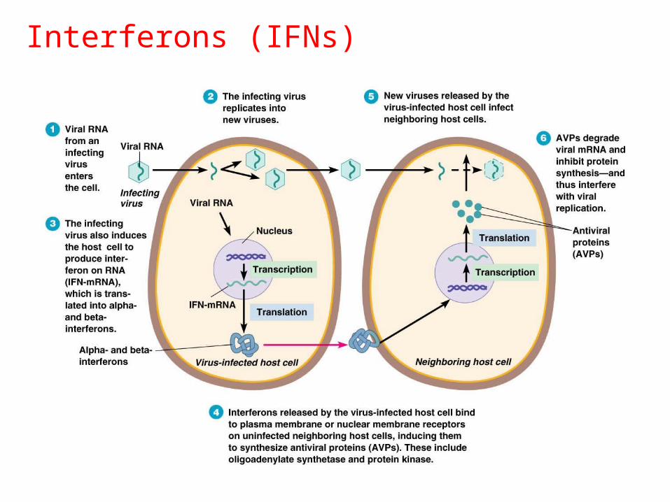
Interferons (IFNs)
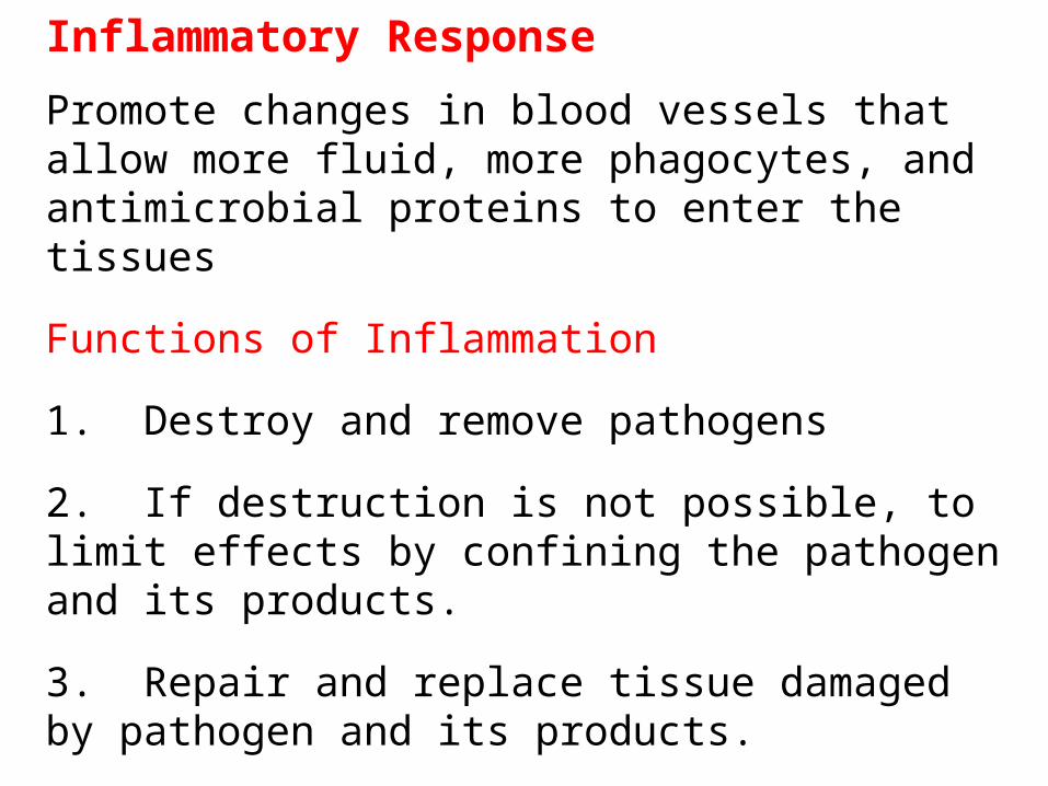
Inflammatory Response
Promote changes in blood vessels that allow more fluid, more phagocytes, and antimicrobial proteins to enter the tissues
Functions of Inflammation
1. Destroy and remove pathogens
2. If destruction is not possible, to limit effects by confining the pathogen and its products.
3. Repair and replace tissue damaged by pathogen and its products.
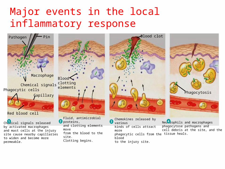
Major events in the local inflammatory responsePathogen Pin
Macrophage
Chemical signals
Capillary
Phagocytic cells
Red blood cell
Bloodclottingelements
Blood clot
Phagocytosis
Fluid, antimicrobial proteins, and clotting elements move from the blood to the site.Clotting begins.
2Chemical signals released by activated macrophages and mast cells at the injury site cause nearby capillaries to widen and become more permeable.
1Chemokines released by various kinds of cells attract more phagocytic cells from the bloodto the injury site.
3 Neutrophils and macrophagesphagocytose pathogens and cell debris at the site, and the tissue heals.
4
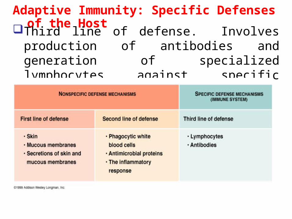
Adaptive Immunity: Specific Defenses of the Host
Third line of defense. Involves production of antibodies and generation of specialized lymphocytes against specific antigens
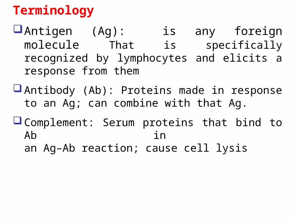
Terminology
Antigen (Ag): is any foreign molecule That is specifically recognized by lymphocytes and elicits a response from them
Antibody (Ab): Proteins made in response to an Ag; can combine with that Ag.
Complement: Serum proteins that bind to Ab in an Ag–Ab reaction; cause cell lysis
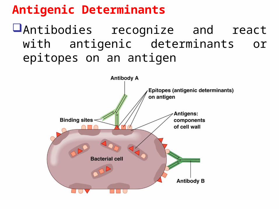
Antigenic Determinants
Antibodies recognize and react with antigenic determinants or epitopes on an antigen
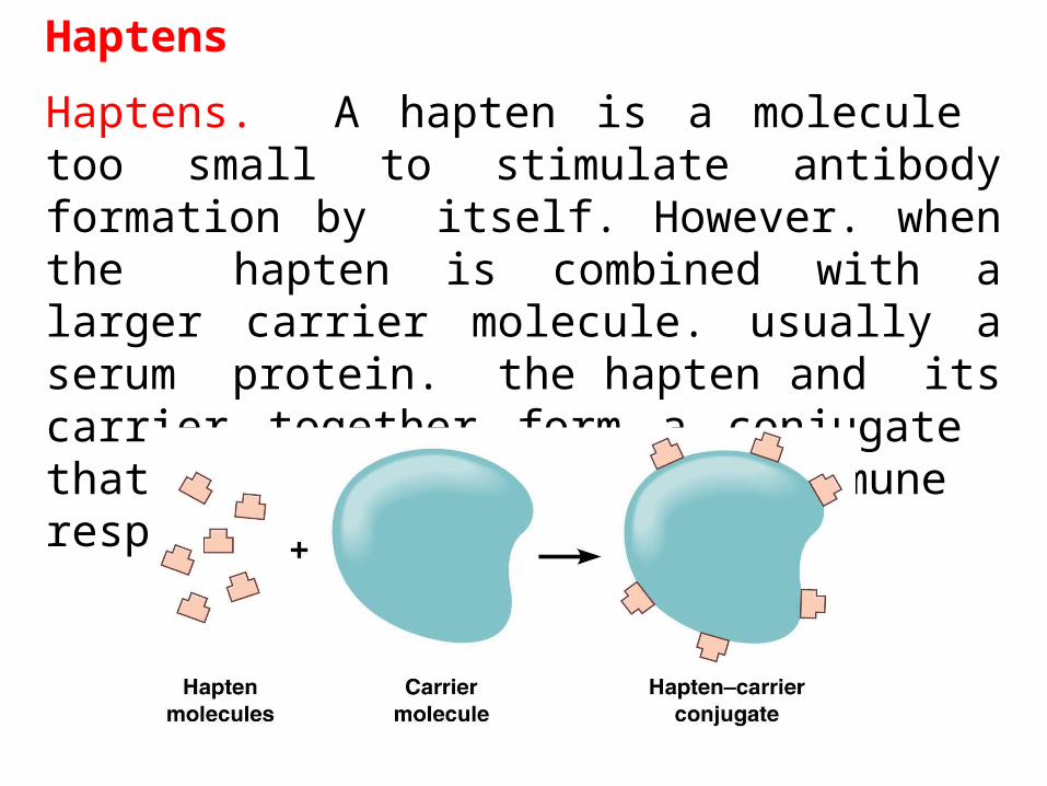
Haptens
Haptens. A hapten is a molecule too small to stimulate antibody formation by itself. However. when the hapten is combined with a larger carrier molecule. usually a serum protein. the hapten and its carrier together form a conjugate that can stimulate an immune response.
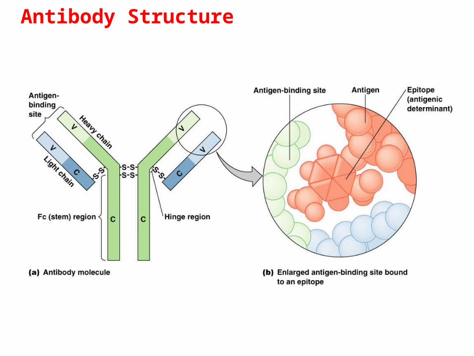
Antibody Structure
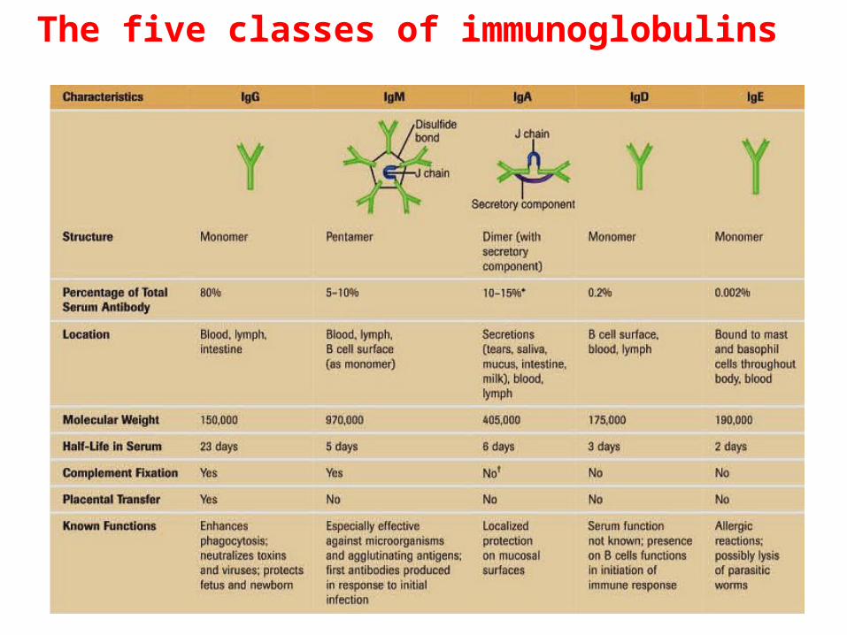
The five classes of immunoglobulins
Lymphocytes
The vertebrate body is populated by two main types of lymphocytes: B lymphocytes (B cells) and T lymphocytes (T cells)
The plasma membranes of both B cells and T cells have about 100,000 antigen receptor that all recognize the same epitope
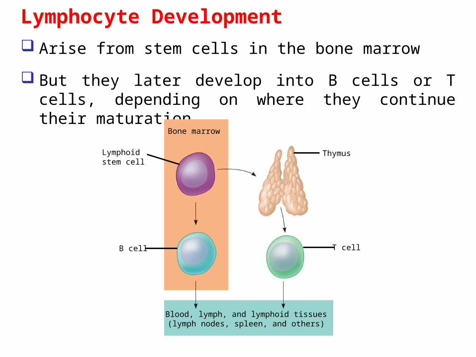
Lymphocyte Development
Arise from stem cells in the bone marrow
But they later develop into B cells or T cells, depending on where they continue their maturation
Bone marrow
Lymphoidstem cell
B cell
Blood, lymph, and lymphoid tissues(lymph nodes, spleen, and others)
T cell
Thymus
T Cell Receptors
V V
C C
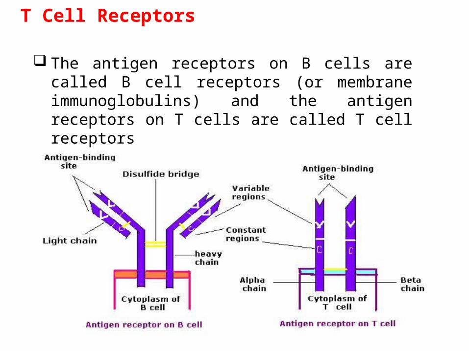
The antigen receptors on B cells are called B cell receptors (or membrane immunoglobulins) and the antigen receptors on T cells are called T cell receptors
MHC
MHC molecules : Are encoded by a family of genes called the major histocompatibility complex and function in signaling between lymphocytes and cells expressing antigen.
Infected cells produce MHC molecules which bind to antigen fragments and then are transported to the cell surface in a process called antigen presentation
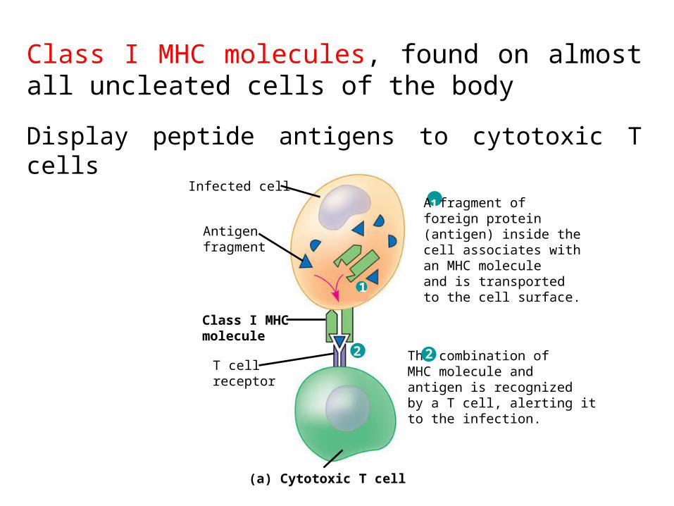
Infected cell
Antigenfragment
Class I MHCmolecule
T cellreceptor
(a) Cytotoxic T cell
A fragment offoreign protein(antigen) inside thecell associates withan MHC moleculeand is transportedto the cell surface.
1
The combination ofMHC molecule andantigen is recognizedby a T cell, alerting itto the infection.
2
1
2
Class I MHC molecules, found on almost all uncleated cells of the body
Display peptide antigens to cytotoxic T cells
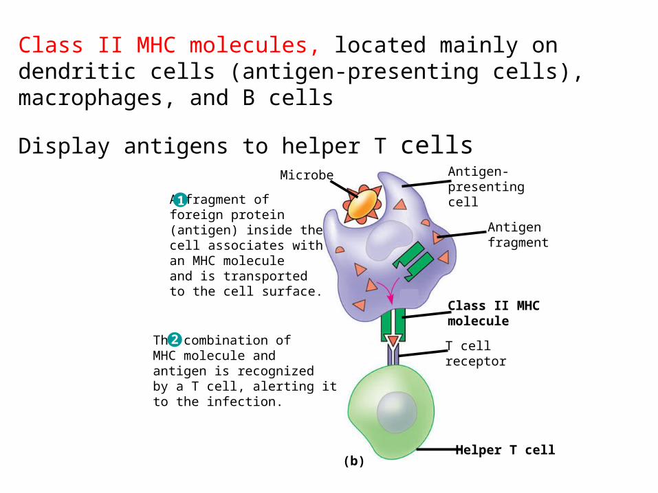
Class II MHC molecules, located mainly on dendritic cells (antigen-presenting cells), macrophages, and B cells
Display antigens to helper T cells
1
2
Microbe Antigen-presentingcell
Antigenfragment
Class II MHCmolecule
T cellreceptor
Helper T cell
A fragment offoreign protein(antigen) inside thecell associates withan MHC moleculeand is transportedto the cell surface.
1
The combination ofMHC molecule andantigen is recognizedby a T cell, alerting itto the infection.
2
(b)
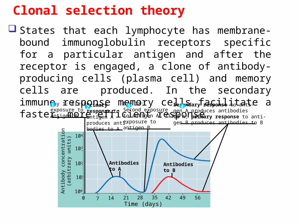
Clonal selection theory
States that each lymphocyte has membrane-bound immunoglobulin receptors specific for a particular antigen and after the receptor is engaged, a clone of antibody-producing cells (plasma cell) and memory cells are produced. In the secondary immune response memory cells facilitate a faster, more efficient response
An
tibo
dy
con
cen
tra
tion
(arb
itra
ry u
nits
)
104
103
102
101
100
0 7 14 21 28 35 42 49 56Time (days)
Antibodiesto A
Antibodiesto B
Primaryresponse toantigen Aproduces anti-bodies to A
2Day 1: First exposure toantigen A
1 Day 28: Second exposureto antigen A; firstexposure to antigen B
3 Secondary response to anti-gen A produces antibodiesto A; primary response to anti-gen B produces antibodies to B
4
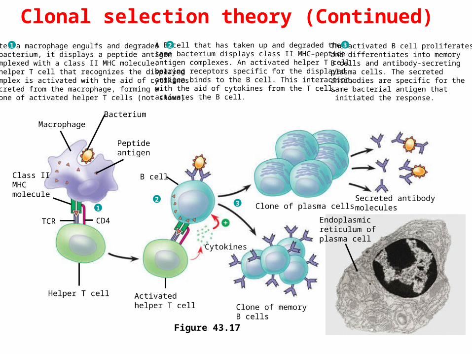
Clonal selection theory (Continued)
21
3
B cell
Bacterium
Peptide antigen
Class II MHCmolecule
TCR
Helper T cell
CD4
Activated helper T cell Clone of memory
B cells
Cytokines
Clone of plasma cellsSecreted antibodymolecules
Endoplasmicreticulum of plasma cell
Macrophage
After a macrophage engulfs and degradesa bacterium, it displays a peptide antigencomplexed with a class II MHC molecule.A helper T cell that recognizes the displayed complex is activated with the aid of cytokines secreted from the macrophage, forming a clone of activated helper T cells (not shown).
1 A B cell that has taken up and degraded the same bacterium displays class II MHC–peptide antigen complexes. An activated helper T cellbearing receptors specific for the displayedantigen binds to the B cell. This interaction,with the aid of cytokines from the T cell,activates the B cell.
2 The activated B cell proliferatesand differentiates into memoryB cells and antibody-secreting plasma cells. The secreted antibodies are specific for the same bacterial antigen that initiated the response.
3
Figure 43.17
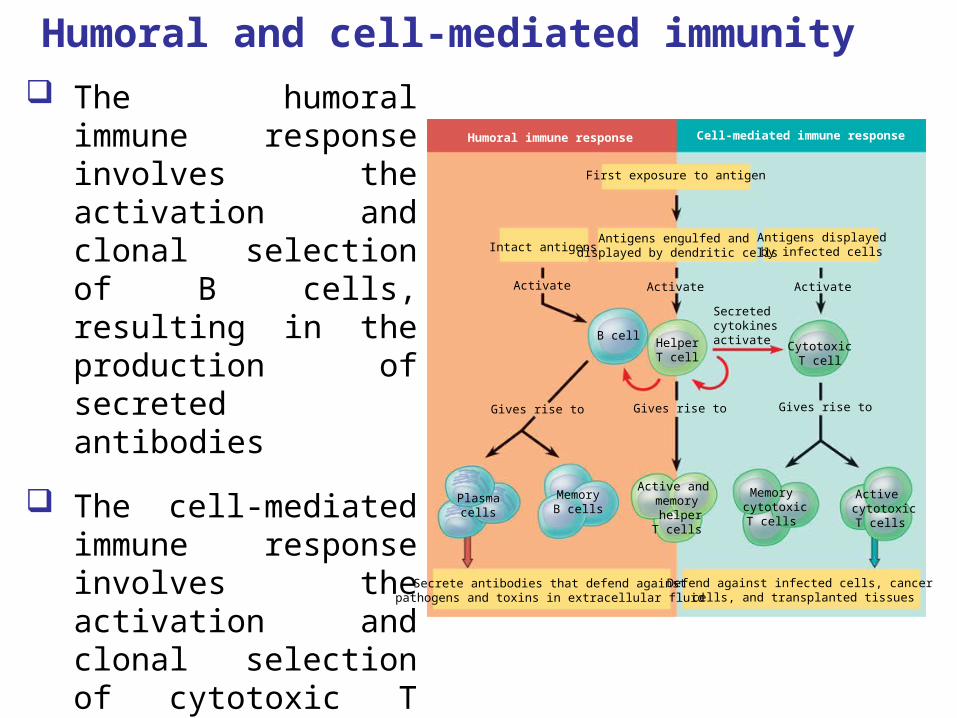
Humoral and cell-mediated immunity
The humoral immune response involves the activation and clonal selection of B cells, resulting in the production of secreted antibodies
The cell-mediated immune response involves the activation and clonal selection of cytotoxic T cells
Humoral immune response Cell-mediated immune response
First exposure to antigen
Intact antigensAntigens engulfed and
displayed by dendritic cellsAntigens displayed
by infected cells
Activate Activate Activate
Gives rise to Gives rise to Gives rise to
B cellHelperT cell
CytotoxicT cell
Plasmacells
MemoryB cells
Active and memory helperT cells
Memory cytotoxic
T cells
Active cytotoxic
T cells
Secrete antibodies that defend againstpathogens and toxins in extracellular fluid
Defend against infected cells, cancer cells, and transplanted tissues
Secretedcytokinesactivate
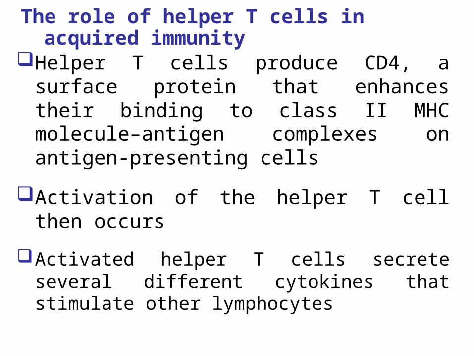
The role of helper T cells in acquired immunity
Helper T cells produce CD4, a surface protein that enhances their binding to class II MHC molecule–antigen complexes on antigen-presenting cells
Activation of the helper T cell then occurs
Activated helper T cells secrete several different cytokines that stimulate other lymphocytes
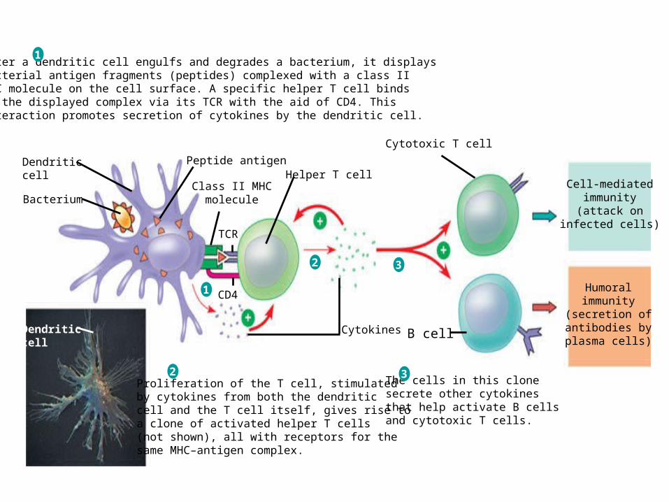
After a dendritic cell engulfs and degrades a bacterium, it displays bacterial antigen fragments (peptides) complexed with a class II MHC molecule on the cell surface. A specific helper T cell binds to the displayed complex via its TCR with the aid of CD4. This interaction promotes secretion of cytokines by the dendritic cell.
Proliferation of the T cell, stimulatedby cytokines from both the dendritic cell and the T cell itself, gives rise toa clone of activated helper T cells(not shown), all with receptors for thesame MHC–antigen complex.
The cells in this clonesecrete other cytokines that help activate B cellsand cytotoxic T cells.
Cell-mediatedimmunity(attack on
infected cells)
Humoralimmunity
(secretion ofantibodies byplasma cells)
Dendriticcell
Dendriticcell
Bacterium
Peptide antigen
Class II MHCmolecule
TCR
CD4
Helper T cell
Cytokines
Cytotoxic T cell
B cell
1
2 3
1
2 3
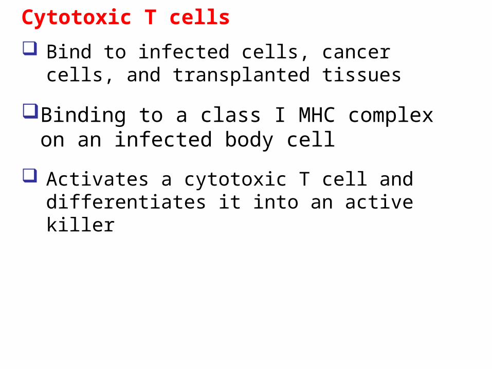
Cytotoxic T cells
Bind to infected cells, cancer cells, and transplanted tissues
Binding to a class I MHC complex on an infected body cell
Activates a cytotoxic T cell and differentiates it into an active killer
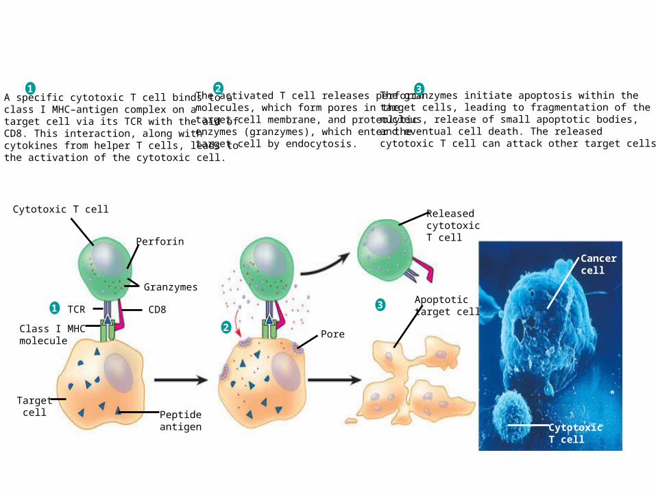
Cytotoxic T cell
Perforin
Granzymes
CD8TCR
Class I MHCmolecule
Targetcell Peptide
antigen
Pore
ReleasedcytotoxicT cell
Apoptotictarget cell
Cancercell
CytotoxicT cell
A specific cytotoxic T cell binds to a class I MHC–antigen complex on a target cell via its TCR with the aid of CD8. This interaction, along with cytokines from helper T cells, leads to the activation of the cytotoxic cell.
1The activated T cell releases perforin molecules, which form pores in the target cell membrane, and proteolytic enzymes (granzymes), which enter the target cell by endocytosis.
2The granzymes initiate apoptosis within the target cells, leading to fragmentation of thenucleus, release of small apoptotic bodies, and eventual cell death. The released cytotoxic T cell can attack other target cells.
3
1
2
3
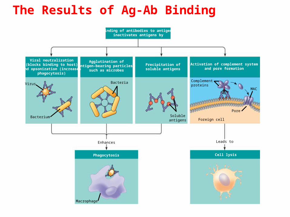
The Results of Ag-Ab Binding
Binding of antibodies to antigensinactivates antigens by
Viral neutralization(blocks binding to host)
and opsonization (increasesphagocytosis)
Agglutination ofantigen-bearing particles,
such as microbes
Precipitation ofsoluble antigens
Activation of complement systemand pore formation
Bacterium
Virus Bacteria
Solubleantigens Foreign cell
Complementproteins
MAC
Pore
Enhances
Phagocytosis
Leads to
Cell lysis
Macrophage
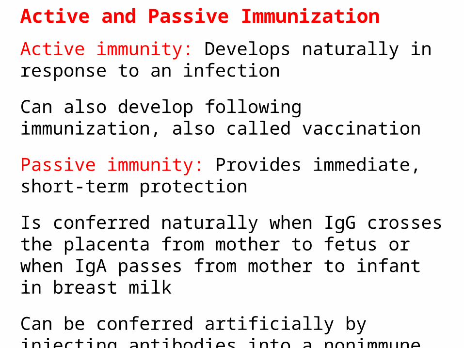
Active and Passive Immunization
Active immunity: Develops naturally in response to an infection
Can also develop following immunization, also called vaccination
Passive immunity: Provides immediate, short-term protection
Is conferred naturally when IgG crosses the placenta from mother to fetus or when IgA passes from mother to infant in breast milk
Can be conferred artificially by injecting antibodies into a nonimmune person
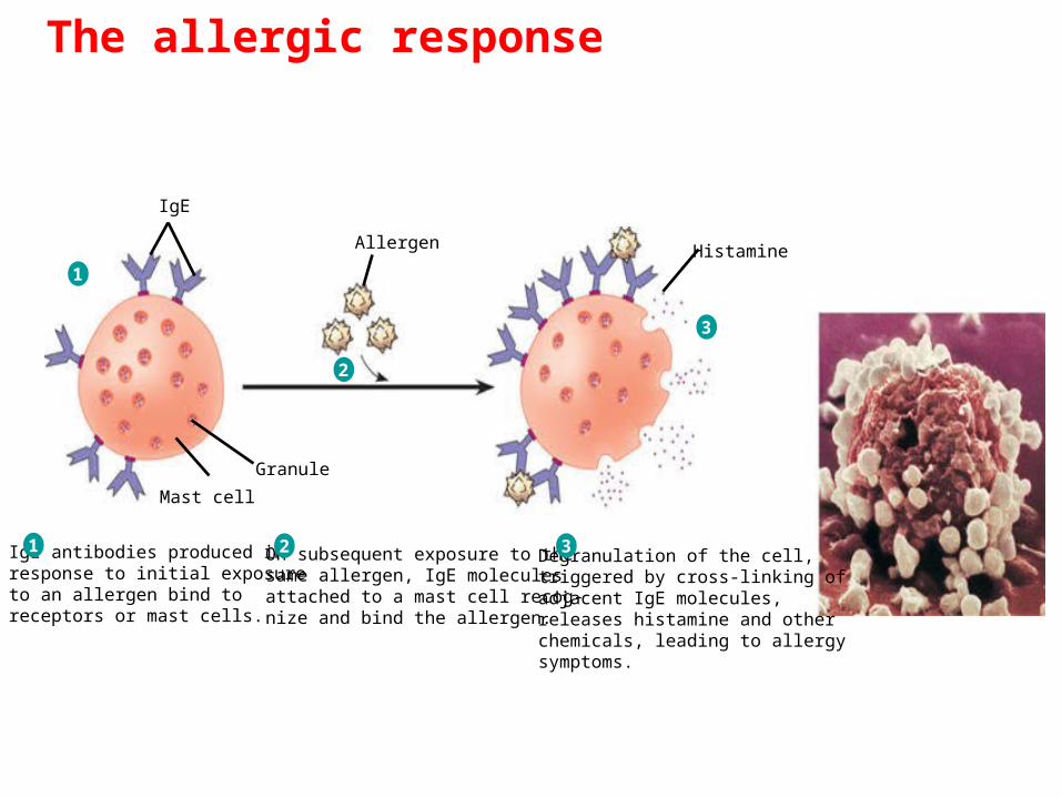
The allergic response
IgE antibodies produced in response to initial exposure to an allergen bind to receptors or mast cells.
1 On subsequent exposure to the same allergen, IgE molecules attached to a mast cell recog-nize and bind the allergen.
2 Degranulation of the cell,triggered by cross-linking of adjacent IgE molecules, releases histamine and other chemicals, leading to allergysymptoms.
3
1
2
3
Allergen
IgE
Histamine
Granule
Mast cell