Embed Size (px)
Citation preview
Top 30 Skin Diseases
Feb 12 2003
S Radhakrishna

Top 30 Skin Diseases
• Scaly Red Rashes (5)
• Pigment Changes (2)
• Nodules (2)
• Purpura (1)
• Blisters (4)
• Systemic (3)
• Benign Growths (3)
• Premalignant Growths (2)
• Malignant Growths (3)
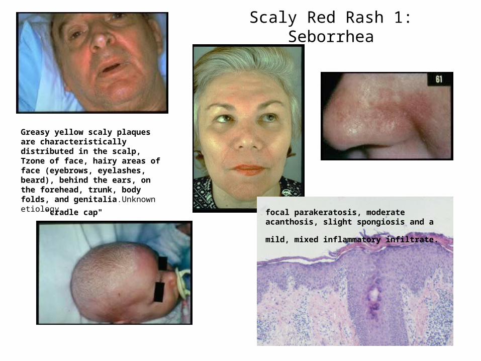
Scaly Red Rash 1: Seborrhea
focal parakeratosis, moderate acanthosis, slight
spongiosis and a mild, mixed inflammatory infiltrate. "cradle cap"
Greasy yellow scaly plaques are characteristically distributed in the scalp, Tzone of face, hairy areas of face (eyebrows, eyelashes, beard), behind the ears, on the forehead, trunk, body folds, and genitalia.Unknown etiology.
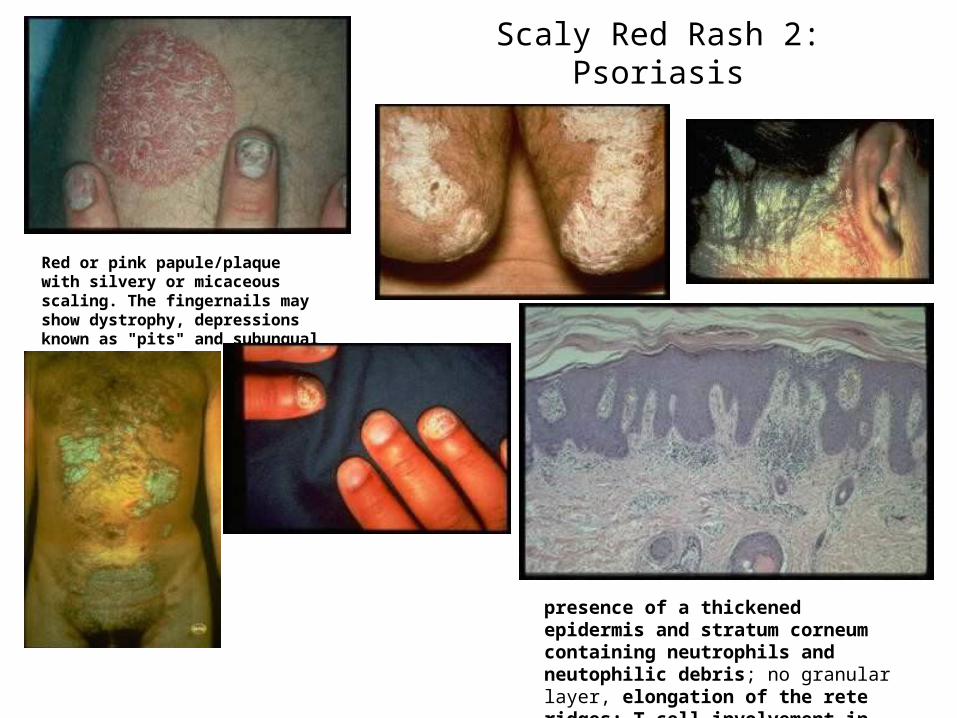
Scaly Red Rash 2: Psoriasis
Red or pink papule/plaque with silvery or micaceous scaling. The fingernails may show dystrophy, depressions known as "pits" and subungual debis
presence of a thickened epidermis and stratum corneum containing neutrophils and neutophilic debris; no granular layer, elongation of the rete ridges; T cell involvement in etiology
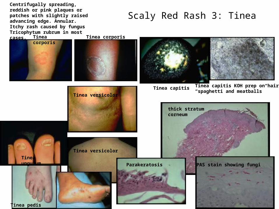
Scaly Red Rash 3: TineaCentrifugally spreading, reddish or pink plaques or patches with slightly raised advancing edge. Annular. Itchy rash caused by fungus Tricophytum rubrum in most cases.
Tinea corporis Tinea corporis
Tinea capitis
Tinea pedis
Tinea unguium
Tinea capitis KOH prep on hair“spaghetti and meatballs”
Tinea versicolor
Tinea versicolor
Parakeratosis PAS stain showing fungi
thick stratum corneum
Scaly Red Rash 4: Eczema
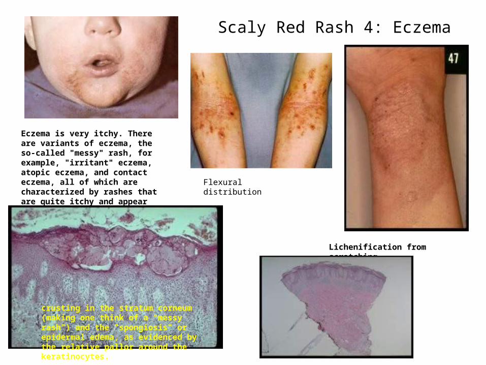
Eczema is very itchy. There are variants of eczema, the so-called "messy" rash, for example, "irritant" eczema, atopic eczema, and contact eczema, all of which are characterized by rashes that are quite itchy and appear "messy" because they are often scratched.TH2 mediated DTH
Flexural distribution
Lichenification from scratching
crusting in the stratum corneum (making one think of a "messy rash") and the "spongiosis" or epidermal edema, as evidenced by the relative pallor around the keratinocytes.
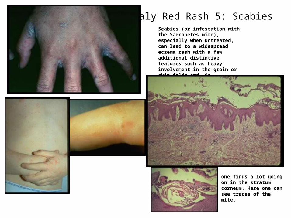
Scaly Red Rash 5: ScabiesScabies (or infestation with the Sarcopetes mite), especially when untreated, can lead to a widespread eczema rash with a few additional distintive features such as heavy involvement in the groin or skin folds and, in particular, involvement of the interdigital web spaces with crusting.
one finds a lot going on in the stratum corneum. Here one can see traces of the mite.
Pigment Changes 1: Vitiligo
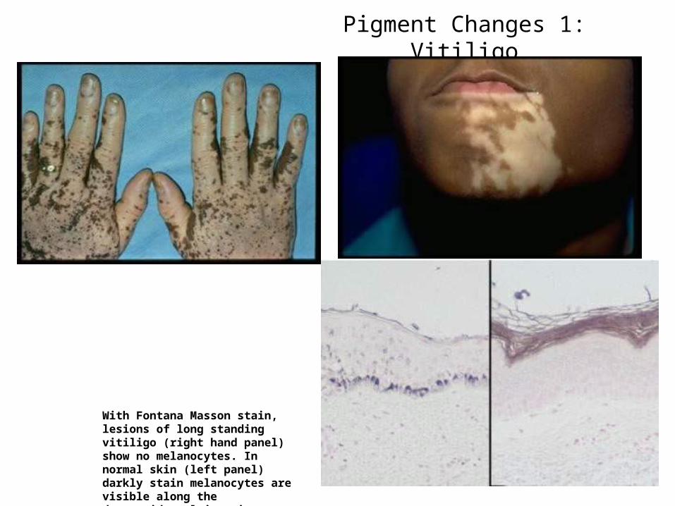
With Fontana Masson stain, lesions of long standing vitiligo (right hand panel) show no melanocytes. In normal skin (left panel) darkly stain melanocytes are visible along the dermoepidermal junction.
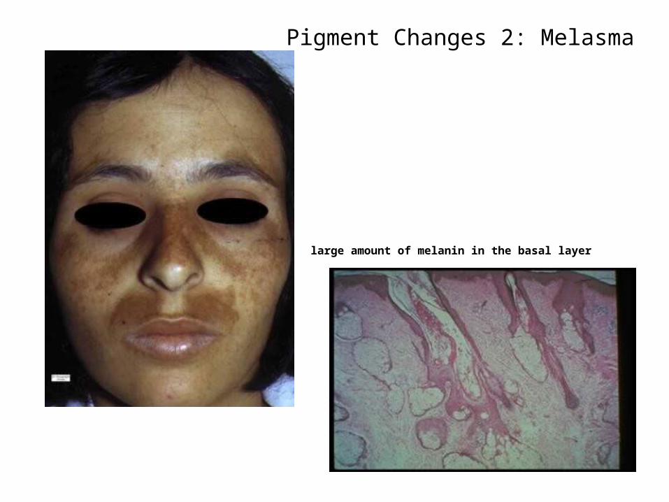
Pigment Changes 2: Melasma
large amount of melanin in the basal layer
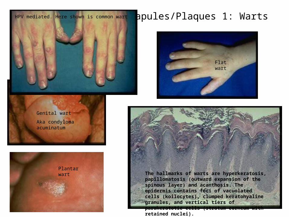
Papules/Plaques 1: Warts
The hallmarks of warts are hyperkeratosis, papillomatosis (outward expansion of the spinous layer) and acanthosis. The epidermis contains foci of vacuolated cells (koilocytes), clumped keratohyaline granules, and vertical tiers of parakeratotic cells (stratum corneum with retained nuclei).
Flat wart
Genital wart
Aka condyloma acuminatum
Plantar wart
HPV mediated. Here shown is common wart
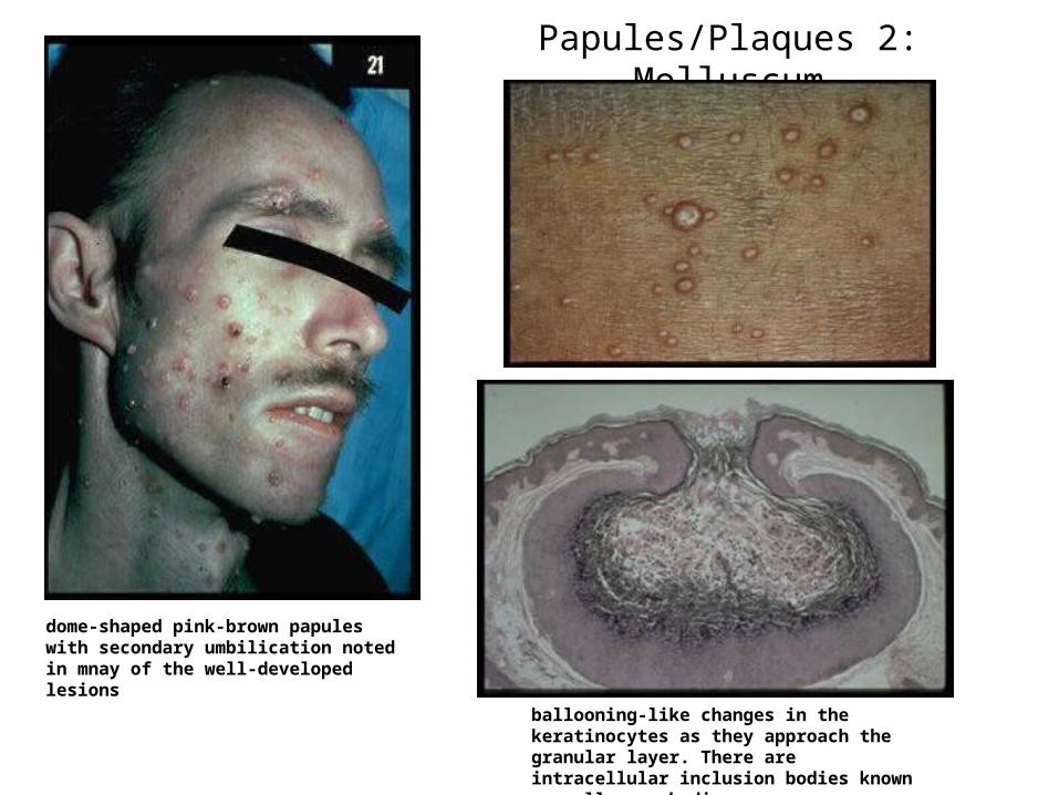
Papules/Plaques 2: Molluscum
dome-shaped pink-brown papules with secondary umbilication noted in mnay of the well-developed lesions
ballooning-like changes in the keratinocytes as they approach the granular layer. There are intracellular inclusion bodies known as molluscum bodies.
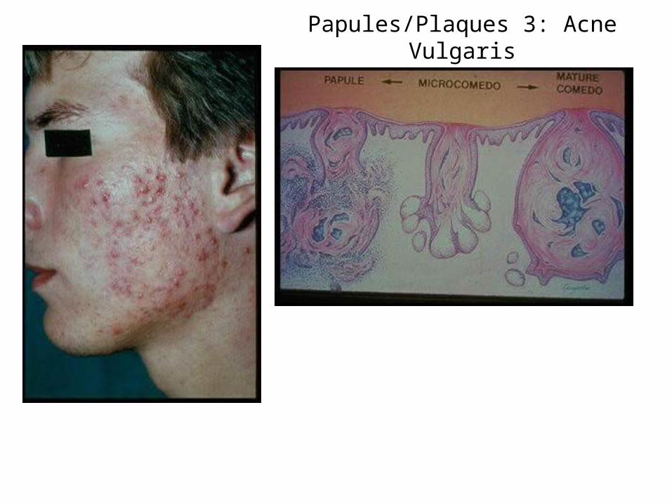
Papules/Plaques 3: Acne Vulgaris
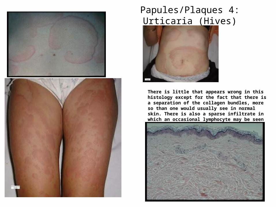
Papules/Plaques 4: Urticaria (Hives)
There is little that appears wrong in this histology except for the fact that there is a separation of the collagen bundles, more so than one would usually see in normal skin. There is also a sparse infiltrate in which an occasional lymphocyte may be seen
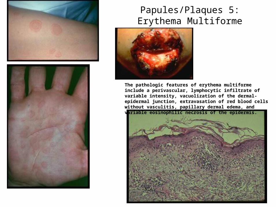
Papules/Plaques 5: Erythema Multiforme
The pathologic features of erythema multiforme include a perivascular, lymphocytic infiltrate of variable intensity, vacuolization of the dermal-epidermal junction, extravasation of red blood cells without vasculitis, papillary dermal edema, and variable eosinophilic necrosis of the epidermis.
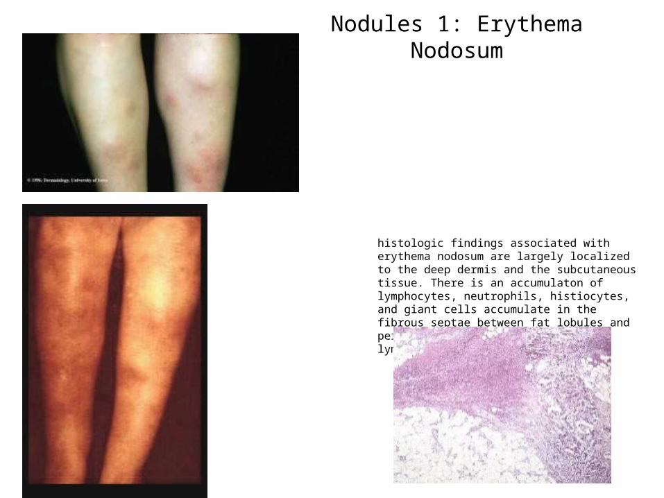
Nodules 1: Erythema Nodosum
histologic findings associated with erythema nodosum are largely localized to the deep dermis and the subcutaneous tissue. There is an accumulaton of lymphocytes, neutrophils, histiocytes, and giant cells accumulate in the fibrous septae between fat lobules and perivascular infiltration of lymphocytes in the dermis.
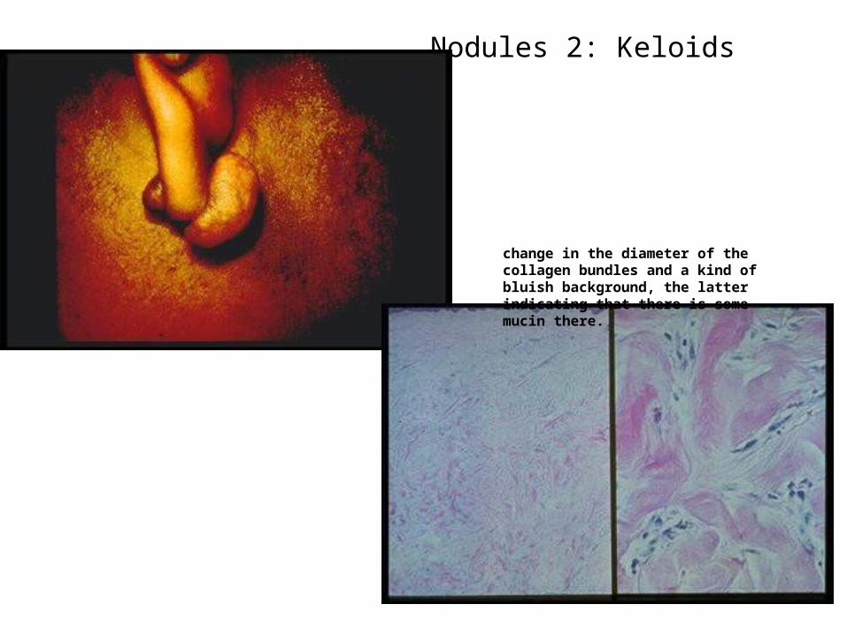
Nodules 2: Keloids
change in the diameter of the collagen bundles and a kind of bluish background, the latter indicating that there is some mucin there.
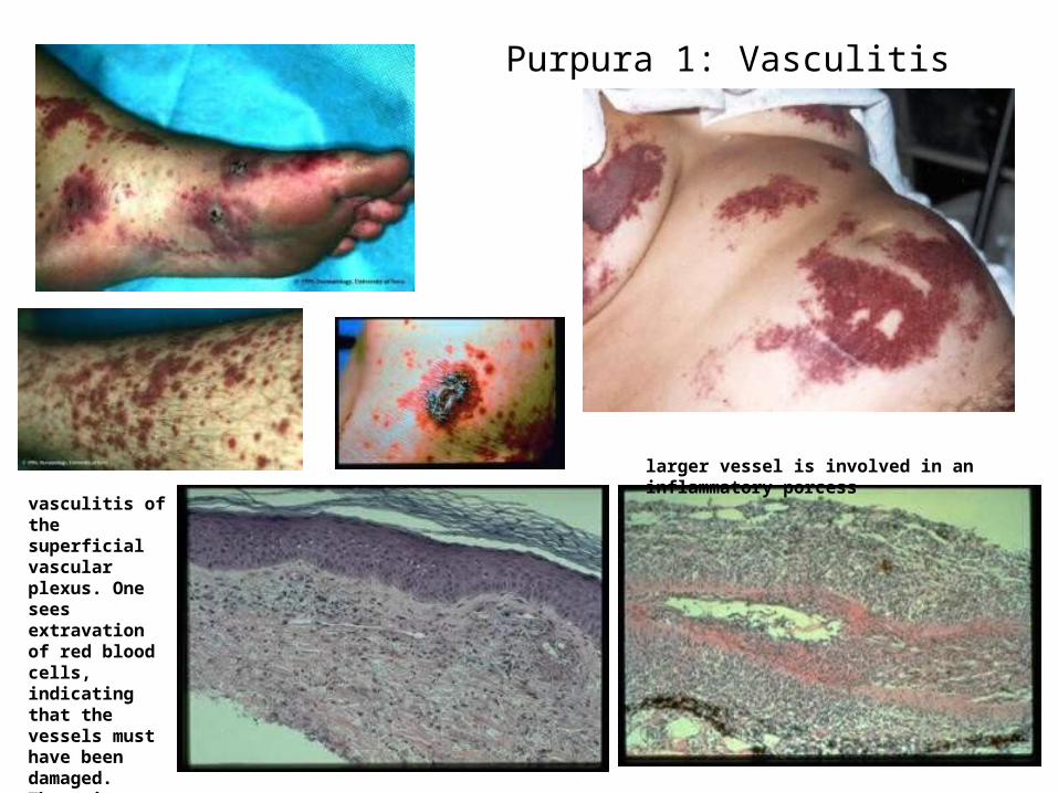
Purpura 1: Vasculitis
larger vessel is involved in an inflammatory porcess
vasculitis of the superficial vascular plexus. One sees extravation of red blood cells, indicating that the vessels must have been damaged. There is a lot of neutrophilic debris.
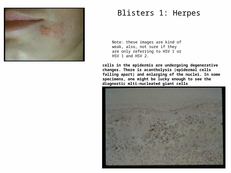
Blisters 1: Herpes
cells in the epidermis are undergoing degenerative changes. There is acantholysis (epidermal cells falling apart) and enlarging of the nuclei. In some specimens, one might be lucky enough to see the diagnostic mlti-nucleated giant cells
Note: these images are kind of weak, also, not sure if they are only referring to HSV 1 or HSV 1 and HSV 2.
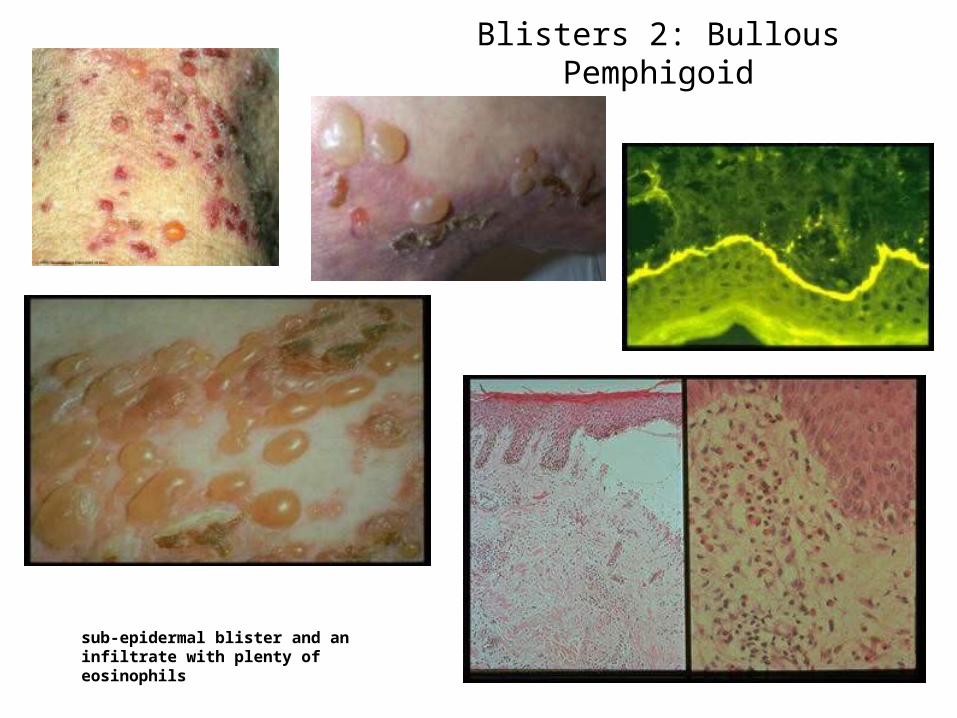
Blisters 2: Bullous Pemphigoid
sub-epidermal blister and an infiltrate with plenty of eosinophils
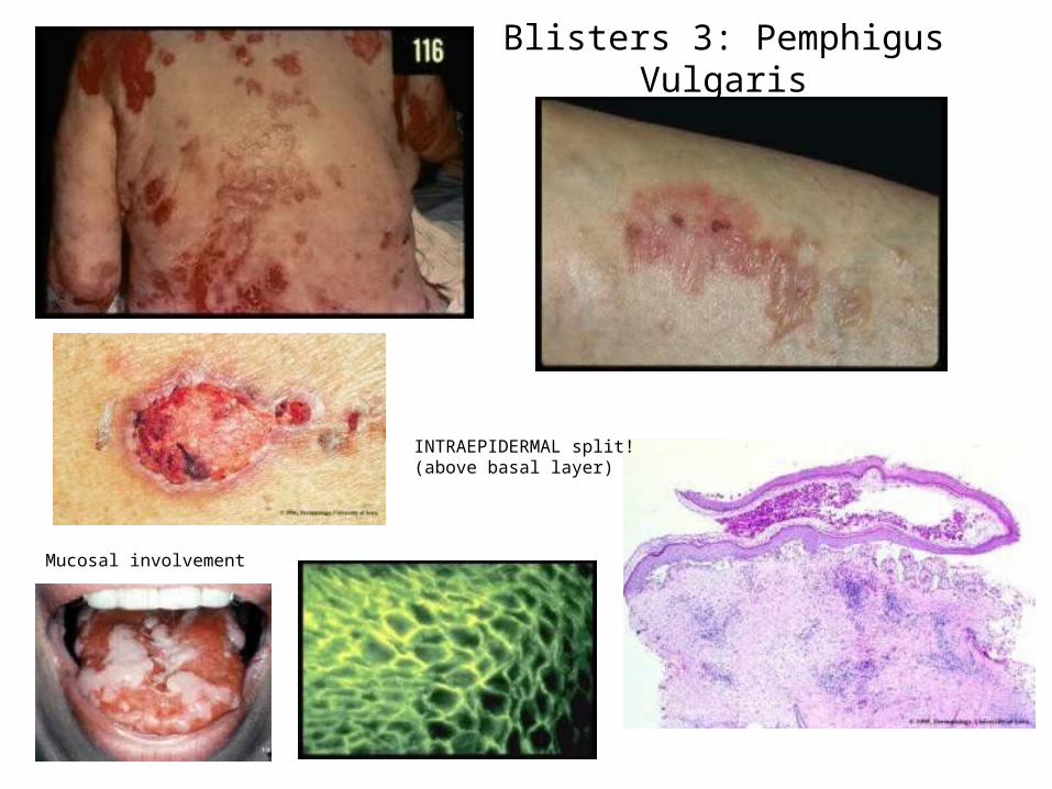
Blisters 3: Pemphigus Vulgaris
INTRAEPIDERMAL split! (above basal layer)
Mucosal involvement
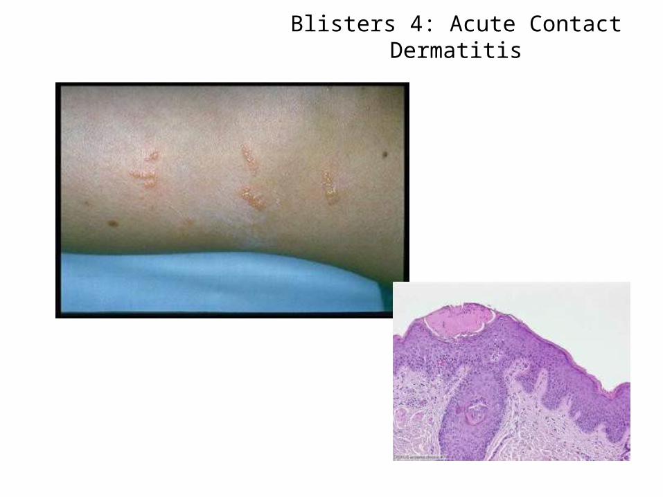
Blisters 4: Acute Contact Dermatitis
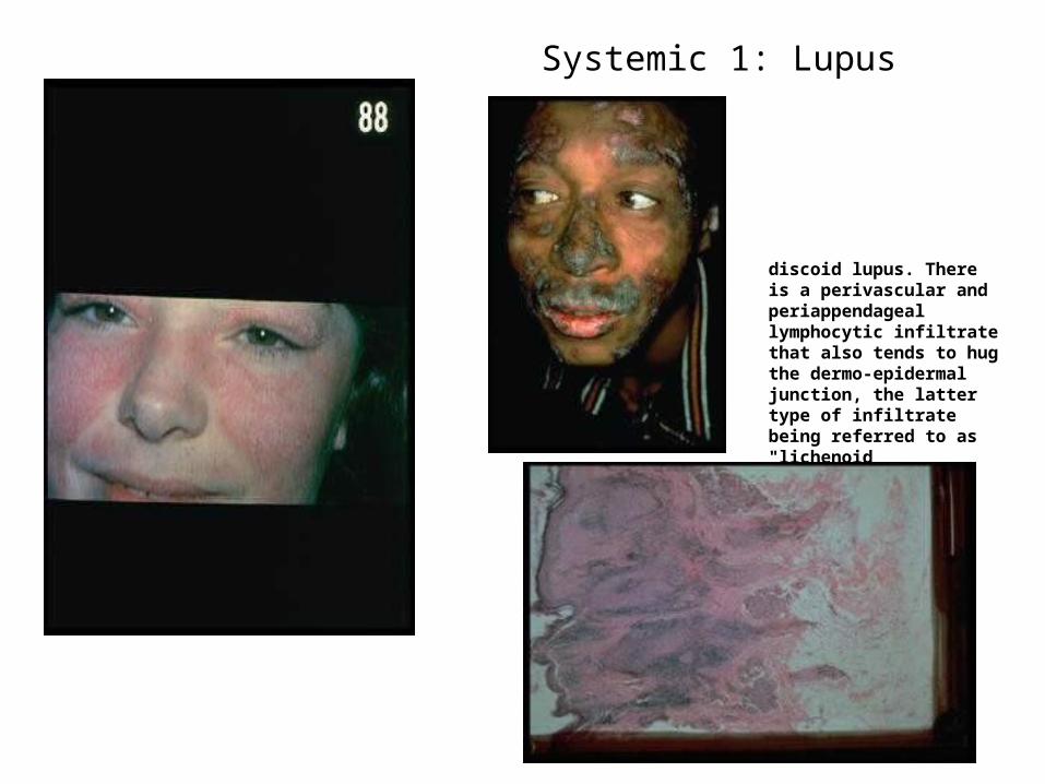
Systemic 1: Lupus
discoid lupus. There is a perivascular and periappendageal lymphocytic infiltrate that also tends to hug the dermo-epidermal junction, the latter type of infiltrate being referred to as "lichenoid
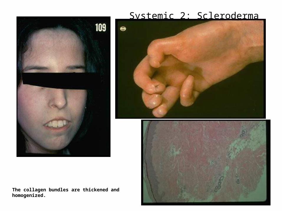
Systemic 2: Scleroderma
The collagen bundles are thickened and homogenized.
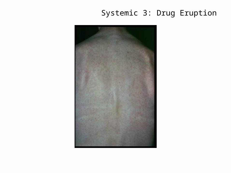
Systemic 3: Drug Eruption
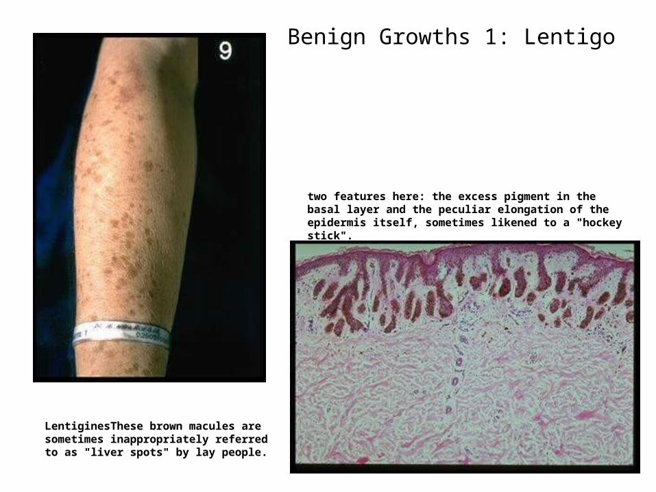
Benign Growths 1: Lentigo
LentiginesThese brown macules are sometimes inappropriately referred to as "liver spots" by lay people.
two features here: the excess pigment in the basal layer and the peculiar elongation of the epidermis itself, sometimes likened to a "hockey stick".
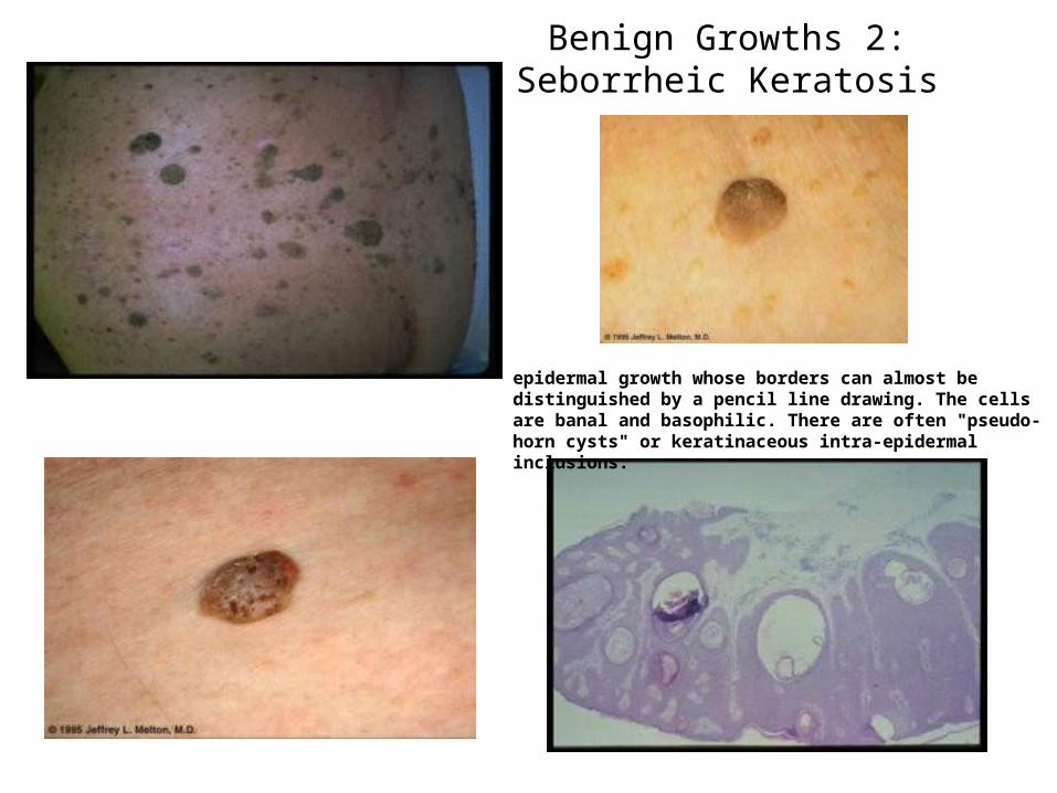
Benign Growths 2: Seborrheic Keratosis
epidermal growth whose borders can almost be distinguished by a pencil line drawing. The cells are banal and basophilic. There are often "pseudo-horn cysts" or keratinaceous intra-epidermal inclusions.
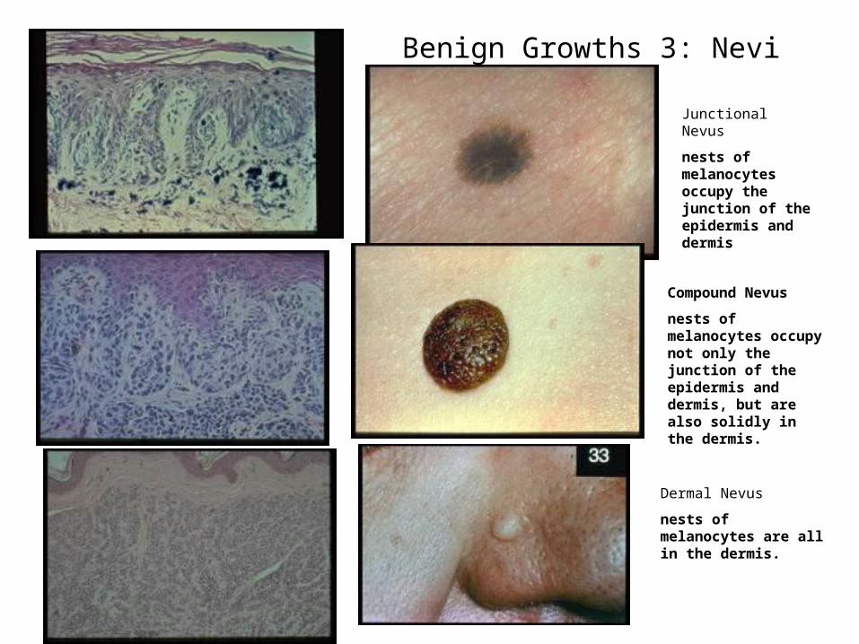
Benign Growths 3: Nevi
Compound Nevus
nests of melanocytes occupy not only the junction of the epidermis and dermis, but are also solidly in the dermis.
Junctional Nevus
nests of melanocytes occupy the junction of the epidermis and dermis
Dermal Nevus
nests of melanocytes are all in the dermis.
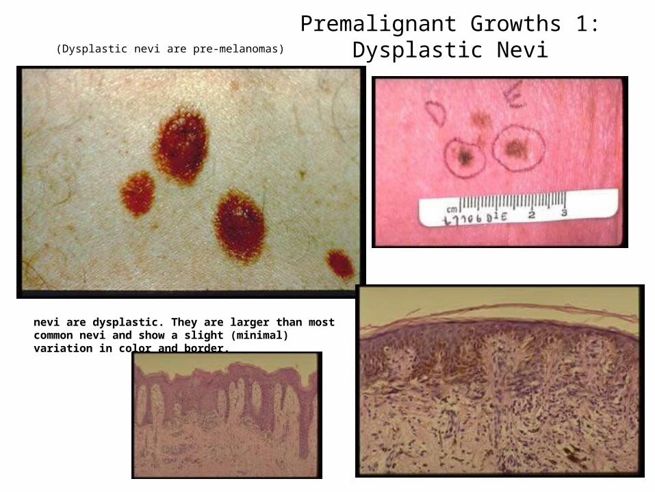
Premalignant Growths 1: Dysplastic Nevi
nevi are dysplastic. They are larger than most common nevi and show a slight (minimal) variation in color and border.
(Dysplastic nevi are pre-melanomas)
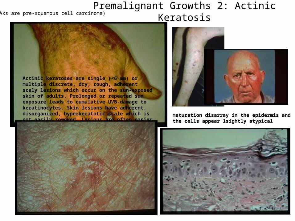
Premalignant Growths 2: Actinic Keratosis
maturation disarray in the epidermis and the cells appear lsightly atypical
Actinic keratoses are single (<6 mm) or multiple discrete, dry, rough, adherent scaly lesions which occur on the sun-exposed skin of adults. Prolonged or repeated sun exposure leads to cumulative UVB-damage to keratinocytes. Skin lesions have adherent, disorganized, hyperkeratotic scale which is not easily removed. Lesions are often easier to feel (they fill like sandpaper) than to see. They are typically distributed on the face, ears, neck, forearms and dorsa of hands.
(Aks are pre-squamous cell carcinoma)
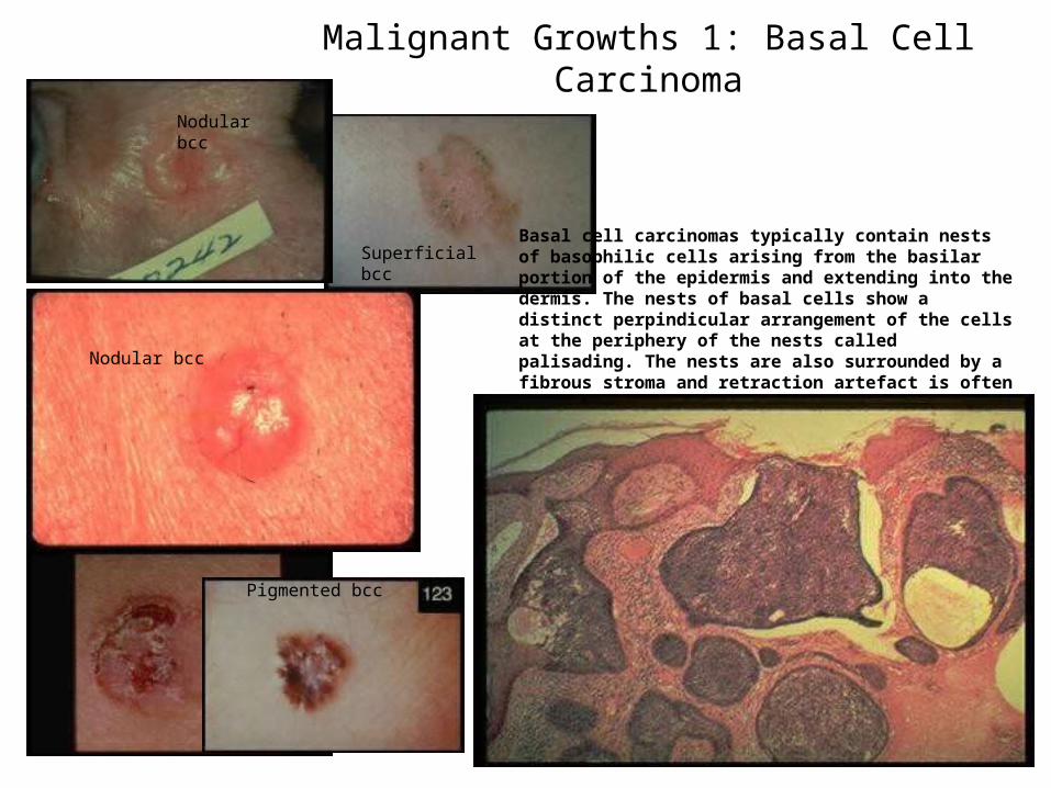
Malignant Growths 1: Basal Cell Carcinoma
Basal cell carcinomas typically contain nests of basophilic cells arising from the basilar portion of the epidermis and extending into the dermis. The nests of basal cells show a distinct perpindicular arrangement of the cells at the periphery of the nests called palisading. The nests are also surrounded by a fibrous stroma and retraction artefact is often observed at the edges of many nest.Nodular bcc
Superficial bcc
Nodular bcc
Pigmented bcc
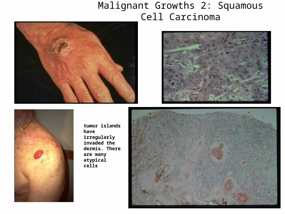
Malignant Growths 2: Squamous Cell Carcinoma
tumor islands have irregularly invaded the dermis. There are many atypical cells
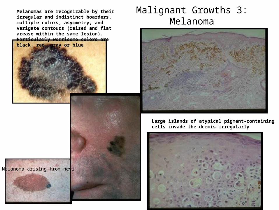
Malignant Growths 3: Melanoma
Large islands of atypical pigment-containing cells invade the dermis irregularly
Melanomas are recognizable by their irregular and indistinct boarders, multiple colors, asymmetry, and varigate contours (raised and flat arease within the same lesion). Particularly worrisome colors are black, red, gray or blue
Melanoma arising from nevi


![Top 30 Skin Diseases[1]](https://img.pdfslide.us/doc/110x75/563dbb10550346aa9aa9f83b/top-30-skin-diseases1-566ef53399edb.jpg)