Embed Size (px)
Citation preview

Tooth-Preserving Surgery

Edited by
Andreas Filippi, Prof Dr med dentClinic for Oral Surgery, Oral Radiology, and Oral Medicine
University Center for Dentistry BaselBasel, Switzerland
Sebastian Kühl, Prof Dr med dentClinic for Oral Surgery, Oral Radiology, and Oral Medicine
University Center for Dentistry BaselBasel, Switzerland
TOOTH-PRESERVING
SURGERY
This book was originally published in German in 2018 under the title Atlas der modernen zahnerhaltenden Chirurgie (Quintessenz Verlags).
Library of Congress Cataloging-in-Publication Data
Names: Filippi, Andreas, editor. | Kühl, Sebastian, editor. Title: Tooth-preserving surgery / edited by Andreas Filippi, Sebastian Kühl; with contributions from J. Thomas Lambrecht, Frank P. Strietzel, Georg Damerau, Adrian Kasaj, and Hermann Derks. Other titles: Atlas der modernen zahnerhaltenden Chirurgie. English Description: Batavia, IL : Quintessence Publishing Co, Inc, [2020] | Includes bibliographical references and index. | Originally published in German under the title Atlas der modernen zahnerhaltenden Chirurgie. | Summary: “Illustrated reference of surgical methods of tooth preservation, including indications and contraindications, step-by-step surgical procedure featuring case examples, and prognosis and potential complications of exposure and alignment, apicoectomy, intentional replan-tation and transreplantation, hemisection and root amputation, and transplantation”-- Provided by publisher. dentifiers 2 1 4 4 4 (print) 2 1 4 4 (ebook) 1 (hardcover) 1 (ebook)
Subjects: MESH: Oral Surgical Procedures--methods | Atlas lassification 1 (print) 1 (ebook) U . DD 1 . --dc23 record available at https lccn.loc.gov 2 1 4 4 4 ebook record available at https lccn.loc.gov 2 1 4 4
© 2020 Quintessence Publishing Co, Inc
Quintessence Publishing Co, Inc411 addant oad
atavia, 1www.quintpub.com
4 3 2 1
All rights reserved. This book or any part thereof may not be reproduced, stored in a retrieval system, or transmitted in any form or by any means, electronic, mechanical, photocopying, or otherwise, without prior written permission of the publisher.
ditor arieke affronDesign: Sue ZubekProduction: Angelina Schmelter
rinted in the U A
Atlas der Atlas der Atlas der Atlas der Atlas der Atlas der
Preface viiContributors viii
Introduction 1
History of Tooth-Preserving Surgery 5TransplantationReplantation
ApicoectomyHemisection and Root AmputationExposure and Alignment
Exposure and Alignment 17IndicationsContraindicationsSurgical ProcedurePossible Complications and Recall
Prognosis
Apicoectomy 39IndicationsContraindicationsSurgical ProcedurePossible Complications and RecallPrognosis
CONTENTS
12
3
4
Intentional Replantation and Transreplantation 55
Intentional Replantation Transreplantation
Resective Furcation Therapy, Hemisection, and Root Amputation 79 Diagnosis and Classification of Furcation Involvement TreatmentofFurcationInvolvement IndicationsandContraindicationsforResectiveFurcationTherapy ResectiveMethodsforFurcationTherapy Possible Complications and Recall Prognosis
Transplantation 95Indications
Contraindications Surgical Procedure Possible Complications and Recall Prognosis
Success with Tooth-Preserving Surgery 117Success Rates
CriteriaforEvaluatingSuccess SuccessFactors
Index 126
5
6
7
8

vii
Tooth-preserving surgery is probably only performed occasionally in dental practices, but it is done several times per day in oral surgery or oral and maxillofacial surgery practices and clinics. The authors have many years of experience in tooth-preserving surgery. It is an integral part of dentistry that—despite all the advances made in oral implantology—seeks to preserve teeth whenever possible. For this reason, the possibilities of tooth-preserving surgery should be considered before any dental extraction. As patients age, this often satisfies their wish to hold on to their own teeth for as long as is possible and practical.
A lot of the techniques of tooth-preserving surgery are classic methods (eg, apicoec-tomy, exposure and alignment of teeth, hemisection, root amputation). hile many have enjoyed a renaissance in recent years (eg, tooth transplantation), many, unfortunately, are still little known (eg, intentional replantation) or even totally unknown (eg, transre-plantation). hat these procedures all have in common is the fact that considerable knowledge about them has been gained in the past 1 to 1 years. This is evident from ever-improving techniques, higher success rates, and better predictability, which all provide great benefits for the patient.
The aim of this book is to present modern tooth-preserving surgery to expand the range of treatments offered in daily practice or to bring them up to date. This volume is not intended as a textbook but rather as an illustrated atlas and reference work. A further aim is to communicate the latest knowledge clearly to students of dentistry for the benefit of their eventual patients.
The products and medications used and recommended by the authors are listed at the end of each chapter. The references in the clinical chapters (ie, chapters 3 to ) have been kept to a minimum to avoid redundancies with chapter 8, which presents an up-to-date analysis of the success rates and influencing factors based on the existing literature.
Acknowledgments
Our special thanks go to everyone who has been involved in the creation of this book: our coauthors Georg Damerau, Hermann Derks, Adrian Kasaj, J. Thomas Lambrecht, and Frank triet el; all the dentists at the linic for Dental urgery, adiology, and Oral Medicine who produced many of the clinical illustrations; Nicolas Lienert and Nicola Feola, for the excellent cover image; Anita Hattenbach from Quintessence Berlin for her helpful and highly professional editing; ohannes olters from uintessence erlin for many years of loyal and always agreeable cooperation; and—last but not least—our families, without whose patience this book would not have been possible alongside all our professional commitments.
PREFACE
viii
Georg Damerau, Dr med dent
Department of Oral Surgery Clinic for Oral and Maxillofacial Surgery Center for Dental MedicineUniversity Hospital urichZurich, Switzerland
Hermann Derks, Dr med dent
Private Practicemmerich am hein, ermany
AndreasFilippi,Prof Dr med dent
linic for Oral urgery, Oral adiology, and Oral MedicineUniversity enter for Dentistry aselBasel, Switzerland
Adrian Kasaj, MSc, Prof Dr med dent
Outpatient Department of estorative Dentistry and PeriodontologyUniversity edicine ohannes utenberg University ainMainz, Germany
SebastianKühl,Prof Dr med dent
linic for Oral urgery, Oral adiology, and Oral MedicineUniversity enter for Dentistry aselBasel, Switzerland
J.ThomasLambrecht,Prof Dr med dent
linic for Oral urgery, Oral adiology, and Oral MedicineUniversity enter for Dentistry aselBasel, Switzerland
FrankP.Strietzel, Dr med dent
Department of Oral Medicine, Dental adiology, and urgery
harit - Universit tsmedi in erlinBerlin, Germany
CONTRIBUTORSCONTRIBUTORS
1
IN D I NAndreas Filippi, Prof Dr med dent1
Over 20 years ago, as a young assistant dental surgeon, I had the opportunity of contributing
to the second edition of Atlas der Chiru-rgischen Zahnerhaltung or Atlas of Surgi-cal Tooth Preservation (Hanser Fachbuch, 1996) under Professor Horst Kirschner. This experience and related activities made an enormous impression on me, with the result that this subject area has always been a focal point in my clinical work.
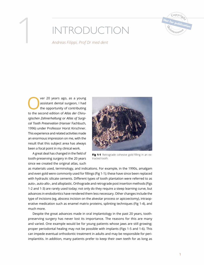
A great deal has changed in the field of tooth-preserving surgery in the 20 years since we created the original atlas, such as materials used, terminology, and indications. For example, in the 1990s, amalgam and even gold were commonly used for fillings (Fig 1-1); these have since been replaced with hydraulic silicate cements. Different types of tooth plantation were referred to as auto-, auto-allo-, and alloplastic. Orthograde and retrograde post insertion methods (Figs 1-2 and 1-3) are rarely used today; not only do they require a steep learning curve, butadvances in endodontics have rendered them less necessary. Other changes include the type of incisions (eg, abscess incision on the alveolar process or apicoectomy), intraop-erative medication such as enamel matrix proteins, splinting techniques (Fig 1-4), andmuch more.
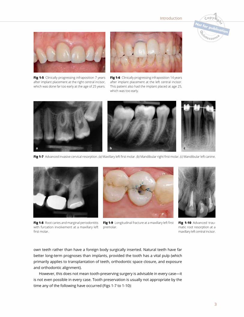
Despite the great advances made in oral implantology in the past 20 years, tooth- preserving surgery has never lost its importance. The reasons for this are many and varied. One example would be for young patients whose jaws are still growing; proper periodontal healing may not be possible with implants (Figs 1-5 and 1-6). This can impede eventual orthodontic treatment in adults and may be responsible for peri- implantitis. In addition, many patients prefer to keep their own teeth for as long as
Fig 1-1 Retrograde cohesive gold filling in an ex-tracted tooth.

IN D I N1
2
possible. The risk of implant treatment for patients taking certain medications may be too great. There are also financial considerations—implants may be too expensive for many patients.
Tooth-preserving surgery has also developed considerably over the years, and its current success rates need not be overshadowed by those of oral implantology. Tooth-preserving surgery is not only more convenient; it is a far more natural, biologic therapy that more closely fulfills the wishes of many patients who prefer to keep their
Fig 1-2 Retrograde post insertion. (a) ituation after wide lumen rotary preparation. (b) ealer fixation of a smooth cylindric tita-nium post. (c) Resulting radiograph.
Fig 1-4 (a) ld wire and brac et splint. (b) onphysiologic rigid bonded ring splint. (c) Modern Titanium Trauma plint TT Medartis . (d) ince 2017, the TT has also been available in a far less conspicuous silver color.
Fig 1-3 efore the introduc-tion of titanium posts, ceram-ic posts were used.
a b c
a
c
b
d
1
3
Fig 1-5 linically progressing infraposition 7 years after implant placement at the right central incisor, which was done far too early at the age of 25 years.
Fig 1-6 linically progressing infraposition 14 years after implant placement at the left central incisor. This patient also had the implant placed at age 25, which was too early.
own teeth rather than have a foreign body surgically inserted. Natural teeth have far better long-term prognoses than implants, provided the tooth has a vital pulp (which primarily applies to transplantation of teeth, orthodontic space closure, and exposure and orthodontic alignment).
However, this does not mean tooth-preserving surgery is advisable in every case—it is not even possible in every case. Tooth preservation is usually not appropriate by the time any of the following have occurred (Figs 1-7 to 1-10):
Fig 1-7 Advanced invasive cervical resorption. (a) Maxillary left first molar. (b) Mandibular right first molar. (c) Mandibular left canine.
a b c
Fig 1-8 Root caries and marginal periodontitis with furcation involvement at a maxillary left first molar.
Fig 1-9 ongitudinal fracture at a maxillary left first premolar.
Fig 1-10 Advanced trau-matic root resorption at a maxillary left central incisor.
Introduction 1
IN D I N1
4
• Extensive root caries• Advanced root resorption (eg, invasive cervical resorption, replacement resorption,
resorption due to infection)• “Final” marginal periodontitis• Deep crown and root or longitudinal fractures
The indications for tooth extraction are much stronger (and options for tooth preser-vation limited) in the presence of general medical problems, such as planned heart valve replacement, necessary antiresorptive therapies (eg, bisphosphonates, denosumab), immunosuppression, radiotherapy in the head and neck area, and serious psychiatric or degenerative central nervous system diseases such as dementia. Depending on the individual case, teeth that may need to be removed include teeth with periodontal furcations that are not already loose, teeth with apical periodontitis, teeth with untreated pulpal necrosis, or partially impacted teeth. In these situations, other options such as apicoectomy, intentional replantation, transplantation, transreplantation, or exposure and alignment may not even be considered.
For many patients, however, both options may be considered or at least discussed: (1) tooth-preserving surgery or (2) dental extraction followed by the placement of a tooth-, implant- or mucosa-supported prosthesis. Additional factors that will play a role include local anatomical factors, the visibility of the teeth when smiling, the situation of the adjacent teeth, and the residual dentition as well as patient compliance or treatability.
Unfortunately, this discussion for or against tooth preservation is also influenced by the attending dentist. Many of the options in tooth-preserving surgery, such as transre-plantation, are unfamiliar to many dentists. They may not be performed in the dental practice or even in other local oral surgery practices. In that situation, well-informed and motivated patients must often travel long distances to specialist centers, which is a shame. This book is intended to inform dental surgeons about the current possibilities of tooth-preserving surgery and encourage them to offer these treatment options to their patients.
resorption (eg, invasive cervical resorption, replacement resorption,
126
INDEXAAcid-etch technique
bracket attachment using, 14–15development of, 15preparation for, 29, 29f–30f
Advanced periodontitis, 68, 69f, 96Aggressive periodontitis, 86fAllogeneic transplantation, 8Aluminum chloride paste, 51, 51fAmalgam fillings, 1Anesthesia, local. See Local anesthesia.Ankylosis
after transplantation, 114of impacted teeth, 19, 35intentional replantation for, 55, 57trauma-induced, 57–58, 57f–58f
Apical advancement flap, 21, 22fApical osteitis, 39Apical periodontitis, 55, 56f, 58f, 60–63, 61f–62f, 67Apicoectomy
apical periodontitis after, 56f, 58fbleeding after, 51, 51fcomplications of, 51–52, 51f–52fcontraindications for, 40, 40f, 56crown-to-root ratio and, 40, 40fextraoral, 49, 49f–51f, 60, 61f–62f, 75factors that affect, 39fistula as contraindication for, 40, 40f, 42healing classifications after, 120history of, 9–13indications for, 39, 39finstruments for, 41fintentional replantation versus, 55–56, 67intraoral
closure of, 47, 47f, 49fhydraulic silicate cements used in, 45, 46f, 48f, 50fincisions for, 42, 42flocal anesthesia for, 41, 42fmarginal incision for, 42, 42f, 52micro aspirators used in, 45, 46fretrograde cavity preparation for, 44, 44fretrograde filling in, 45–46, 46f, 48f–50f, 121smear layer, 44f, 44–45tunneling defect in, 47, 47f
postoperative care for, 51prognosis of, 47, 52recall after, 52–53root canal filling in, 40, 40fscarring after, 52, 52fsuccess/success rates for, 117–121wound dehiscence after, 52, 52f
Autogenous transplantation, 8Avulsed teeth, transreplantation for, 68
CCanine(s)
maxillary. See Maxillary canines.primary, transplantation of, 110, 110t, 111f–113f, 119
CBCT. See Cone beam computed tomography.Cementoenamel junction, 28, 29f
Ceramic posts, 2fCervical resorption, 3f, 99fChronic periodontitis, 87fCone beam computed tomography
furcation-involved teeth on, 82impacted teeth on, 17, 18f, 24f, 26f, 36fmaxillary canine impaction on, 24f, 26fmaxillary molar impaction on, 36f
Crestal incision, 26fCrown-root fractures, 56–57, 57f, 59, 59f, 63–67, 64f–65fCrown-to-root ratio, 40, 40f, 57, 99f
DDental follicle, 28–29, 29f, 35
EEmdogain, 74f, 75, 97Exposure and alignment
history of, 14–15of impacted teeth. See Impacted teeth, exposure
and alignment of.Extraction, tooth
after resective furcation therapy, 90, 92from animals, 5historical description of, 5indications for, 4molars, 103f
Extraoral apicoectomy, 49, 49f–51f, 60, 61f–62f, 75Extraoral trephination, 71f
FFistula, 40, 40f, 42Fistulous tract, 43, 43fFurcation involvement
anatomy of, 79, 80fClass II, 82, 89fClass III, 82, 86f–87fclassification of, 80–82cone beam computed tomography of, 82diagnosis of, 80–82overview of, 79in periodontal disease, 79prevalence of, 81prognosis for, 91–92resective furcation therapies for
complications of, 90–91contraindications for, 83extraction after, 90, 92hemisection, 13–14, 14f, 85, 86f–88f, 118, 120indications for, 83premolarization, 88–89prognosis after, 91–92purpose of, 91recall after, 90–91root amputation, 84–85, 84f–85fsurvival rates after, 91trisection, 85, 87ftunneling, 89f, 89–90
treatment of, 82
Page references followed by “f” denote figures, “t” denote tables, and “b” denote boxes.

INDEX
127
GGBR. See Guided bone regeneration.Gold chain attachment, for impacted teeth, 18, 18f,
32, 32f–33fGold fillings, 1, 1fGuided bone regeneration, 47, 47f“Guided endodontics,” 58, 59f
HHemisection
description of, 85, 86f–88fhistory of, 13–14illustration of, 14fsuccess/success rates for, 118, 120, 122
HSCs. See Hydraulic silicate cements.Hydraulic silicate cements, 45, 46f, 48f, 50f, 61f, 74fHydrogen peroxide-soaked pellets, 51, 51f
IImpacted teeth
ankylosis of, 19, 35cone beam computed tomography of, 17, 18f,
26f, 36fdiagnosis of, 17, 18fexposure and alignment of
apical advancement flap in, 21, 22fcementoenamel junction considerations, 28, 29fclosed exposure technique, 22–25, 23f–26fcomplications of, 35contraindications for, 19, 19fgold chain attachment, 32, 32f–33f, 35indications for, 19instrumentation used in, 20, 20flight curing of adhesive, 30–31, 31flocal anesthesia for, 25–26, 27fopen exposure technique, 21–22, 21f–22fprognosis after, 35–37, 36frecall after, 35step-by-step procedure for, 25–34, 26f–34fsurgical procedure, 20–34, 21f–34f
gold chain attachment for, 18, 18f, 32, 32f–33fpanoramic radiograph of, 17, 18f, 27fprimary failure of eruption of, 19, 19froot resorption of, 19, 19f, 35surgical exposure of, 18, 18fwire ligature for, 18, 18f
Implantsafter extraction, 92infraposition of teeth affected by, 1, 3fperiodontal healing affected by, 1
Incisionsabscess, 1crestal, 26fintentional replantation, 59–60, 60fmarginal. See Marginal incision.paramarginal, 24, 24f, 52, 52fPartsch, 12vertical relieving, 42, 42f
Infiltration anesthesia, 41, 42fIntentional replantation
apical periodontitis treated with, 55, 56f, 58f, 60–63, 61f–62f, 67
apicoectomy versus, 55–56, 67complications of, 66–67contraindications for, 59, 59fcrown-root fracture treated with, 56–57, 57f,
63–67, 64f–65fdescription of, 55history of, 8–9incision for, 59–60, 60findications for, 55–58, 56f–58f
prognosis for, 67recall after, 66–67success/success rates for, 118–120, 122surgical procedure for, 59–66, 60f–66ftooth transplantation versus, 8
Intraalveolar transverse fractures, 59Intraoral apicoectomy. See Apicoectomy, intraoral.
LLocal anesthesia
for apicoectomy, 41, 42ffor exposure and alignment of impacted teeth,
25–26, 27fLongitudinal fracture, 3f, 59, 59f
MMandibular molars
crown-root fracture of, 64fimpaction of, 19fpremolarization of, 88–89root anatomy of, 80ftunneling of, 89
Mandibular premolar impaction, 34fMarginal incision
for apicoectomy, 42, 42f, 52for exposure and alignment of impacted teeth,
25, 25f, 28fwound dehiscence after, 52
Maxillary caninesimpaction of
closed exposure technique for, 23fcone beam computed tomography of, 24fdescription of, 17illustration of, 18fpanoramic radiograph of, 27fvestibular, 22f
transplantation of, 113fMaxillary incisors, 111fMaxillary molars
furcation involvement of, 80f, 81, 82f–83fimpaction of, 19f
Mental nerve, 25, 26fMicro aspirators, 45, 46fMolars
extraction of, 103fmandibular. See Mandibular molars.maxillary. See Maxillary molars.transplantation of, 97–98, 99f–104f, 110t, 119
Mucoperiosteal flap, 22, 25–26, 42
OOral implantology, 1–2
PParamarginal incisions, 24, 24f, 52, 52fPartsch incision, 12Periodontal disease, furcation defects in, 79Periodontitis
advanced, 68, 69f, 96apical, 55, 56f, 58f, 60–63, 61f–62f, 67
Periotest, 57Pfaff, Philipp, 5–6PFE. See Primary failure of eruption.Piezoelectric surgery, 28, 28fPortland cement, 45, 46f, 48f, 50fPost insertion, 1, 2fPremolar(s)
agenesis of, 102f, 104f, 107f–108fimpaction of, 34flongitudinal fracture of, 3f

INDEX
128
transplantation of, 98, 104, 106f–109f, 110t, 119Premolarization, 88–89Primary canine transplantation, 110, 110t,
111f–113f, 119Primary failure of eruption, 19, 19fPulp necrosis, 114–115Pulpotomy, 101f
RReplantation. See Intentional replantation;
Transreplantation.Resective furcation therapy
complications of, 90–91contraindications for, 83extraction after, 90, 92hemisection, 13–14, 14f, 85, 86f–88f, 118, 120indications for, 83premolarization, 88–89prognosis after, 91–92purpose of, 91recall after, 90–91root amputation, 84–85, 84f–85fsurvival rates after, 91trisection, 85, 87ftunneling, 89f, 89–90
Retrograde fillingdescription of, 12in intraoral apicoectomy, 45–46, 46f, 48f–50f, 121
Root amputationfurcation-involved teeth treated with, 84–85,
84f–85fhistory of, 13–14in maxillary first molar, 84fsuccess/success rates for, 118, 122
Root canal fillingin apicoectomy, 40, 40fintentional replantation for, 55–56
Root canal obliteration, 58fRoot caries, 3fRoot fractures, 59, 59f. See also Crown-root
fractures.Root resorption
Hunter’s description of, 6of impacted teeth, 19, 19f, 35instruments for, 85fmaxillary canine impaction as cause of, 17traumatic, 3f
Root tip resection, 9, 11–12Root-end resection. See Apicoectomy.
SSmear layer, 44f, 44–45Snaring, 14–15, 15fSplinting
changes in, 1, 2fTitanium Trauma Splint, 62, 62f, 66f, 72f, 76, 98in transreplantation, 75–76
Success/success rates. See also for specific procedure.
definition of, 117
TTitanium Trauma Splint, 62, 62f, 66f, 72f, 76, 98Tooth extraction. See Extraction, tooth.Tooth impaction. See Impacted teeth.Tooth-preserving surgery. See also specific method.
benefits of, 2–3changes in, 1contraindications for, 3f, 3–4, 59, 96factors that affect, 4history of, 5–15importance of, 1success with, 117–122
Transdental fixation, 14, 14fTransplantation
allogeneic, 8in animals, 6fankylosis after, 114autogenous, 8complications of, 114contraindications for, 96criticisms of, 6failure of, 95history of, 5–7, 6findications for, 95, 96bmolars, 97–98, 99f–104f, 110t, 119orthodontic movements after, 114premolars, 98, 104, 106f–109f, 110tprerequisites for, 96primary canine, 110, 110t, 111f–113f, 119prognosis after, 114–115pulp necrosis after, 114–115recall after, 114recipient regions for, 110trisks associated with, 115success/success rates for, 119, 121–122surgical procedure for, 96–113, 99f–113ftooth replantation versus, 8
Transreplantationadvanced periodontitis treated with, 68avulsed teeth treated with, 68in children, 68, 77complications of, 76contraindications for, 68description of, 55, 67–68indications for, 68prognosis for, 77recall after, 76splinting in, 75–76surgical procedure for, 68–76, 69f–75f
Trauma-induced ankylosis, 57–58, 57f–58fTrisection, 85, 87fTTS. See Titanium Trauma Splint.Tunneling, 89f, 89–90Tunneling defect, 47, 47f
VVertical relieving incisions, 42, 42f
WWire ligature, for impacted teeth, 18, 18fWound dehiscence, after apicoectomy, 52, 52f
Titanium Trauma Splint, 62, 62f, 66f, 72f, 76, 98