Embed Size (px)
Citation preview

Tools of the
Laboratory:
The Methods for
Studying
MicroorganismsMicroorganismsChapter 2
Copyright © The McGraw-Hill Companies, Inc. Permission required for reproduction or display.
1. Macronutrientes : requeridos en grandes cantidades
C, H, O, N, P, SCarbono: •elemento más abundante en todas las macromoléculas
Nitrógeno:• necesario para síntesis de proteínas y ácidos nucleicos• necesario para síntesis de proteínas y ácidos nucleicos
Oxígeno e hidrógeno:•presentes en macromoléculas y compuestos orgánicos que sirven de fuente de energía
Fósforo: • necesario para síntesis de fosfolípidos y ácidos nucleicos
Azufre:•necesario para síntesis de ciertos amino ácidos (cisteína y metionina) y vitaminas
Otros Macronutrientes: K, Mg, Ca, Na
Potasio: requerido para la actividad de ciertas enzimas, en particular aquellas envueltas en síntesis de proteínas
Magnesio: estabiliza ribosomas, ácidos nucléicos, requerido para la actividad de varias enzimas
Calcio : estabiliza la pared celular, confiere resistencia al calor en endoesporas
Sodio: necesario para el crecimiento de microorganismos adaptados a Presiones osmóticas asociadas ambientes marinos o hipersalinos
2. Micronutrientes : compuestos inorgánicos(metales) requeridos en pequeñas cantidades (elementos trazas)
Hierro : requerido en proteínas asociadas al transporte de electrones
necesarios como cofactores de enzimasFe, Mn, Cr, Ni, Zn, Se, Cu, Co
Hierro : requerido en proteínas asociadas al transporte de electronesdurante el proceso de respiración celular (citocromos, proteínas dehierro -azufre ). Hierro esta presente en cantidades muy bajas en ambientes naturales
Sideroforos : Agente quelante producido por células, capaz de fijar o secuestrar iones metálicos en el ambiente para translocarlos al interior
3. Factores de crecimiento : compuestos Orgánicos requeridos en pequeñas cantidades
vitaminas, amino ácidos, purinas, pirimidinas,
deben ser suplidos a ciertos microorganismos que no pueden sintetizarlos ej. bacterias productoras de ácido láctico•Streptococcus•Streptococcus•Lactobacillus•Leuconostoc
vitaminas : factores de crecimiento más requeridos, se utilizan como cofactores de enzimas (componente necesario para el funcionamiento de una enzima)
Otros grupos basados en su requerimiento de oxigeno
Anaerobios facultativos = estos son organismos aeróbicos que puedenrespirar anaeróbicamente o fermentar.
Ejemplos: Escherichia coli, Enterobacter, Salmonella
Anaerobios aerotolerantes = estos son organismos que no respiran oxigenosino que solo fermentan pero el oxigeno no los afecta o limita.
Ejemplos: LactobacillusEjemplos: Lactobacillus
Microaerofilicos = estos requieren oxigeno exclusivamente pero en concentraciones bajas 2% - 10% mas de esto seria toxico.
Ejemplo: Helicobacter pylori
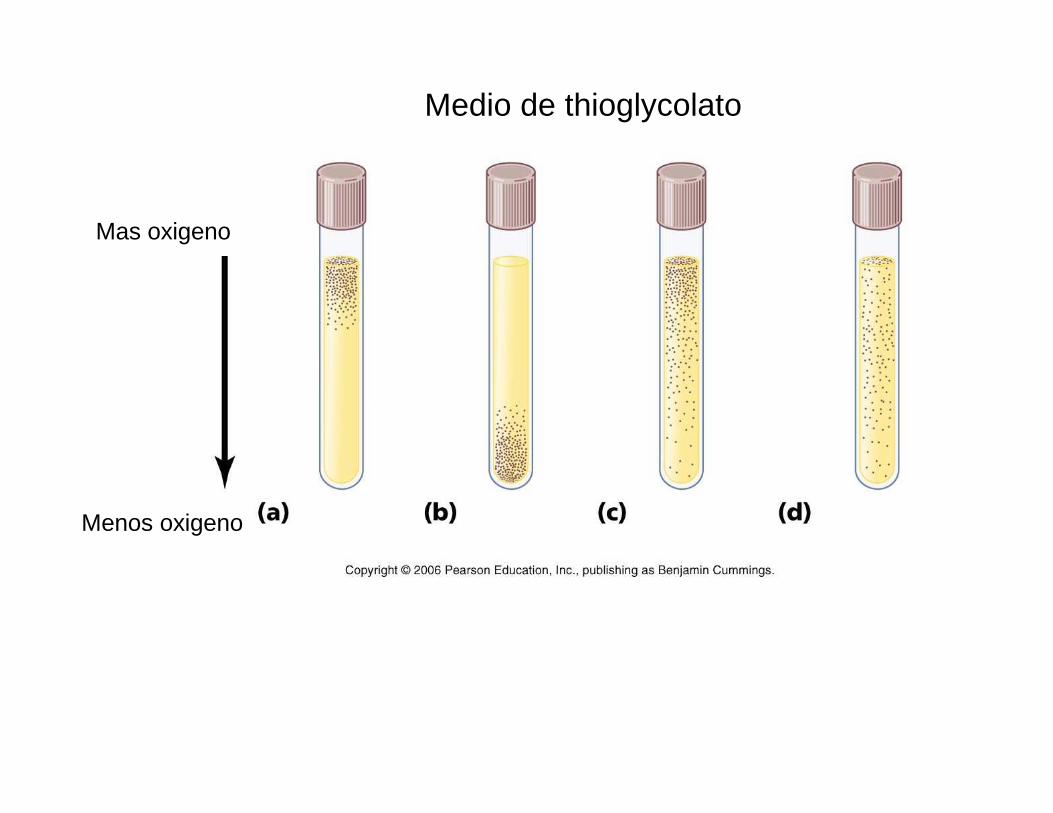
Mas oxigeno
Medio de thioglycolato
Menos oxigeno
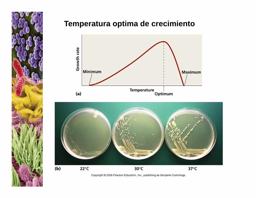
Temperatura optima de crecimiento
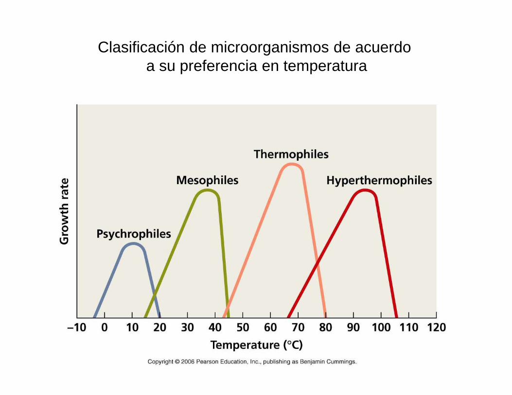
Clasificación de microorganismos de acuerdo a su preferencia en temperatura
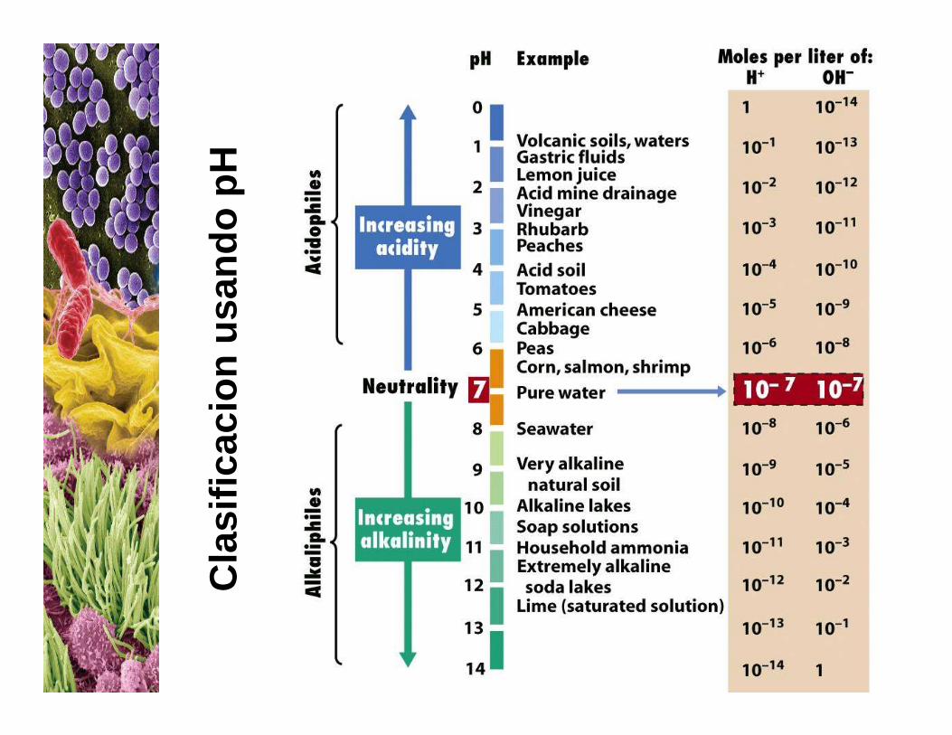
Cla
sific
acio
nus
ando
pHC
lasi
ficac
ion
Cultivo de microorganismos
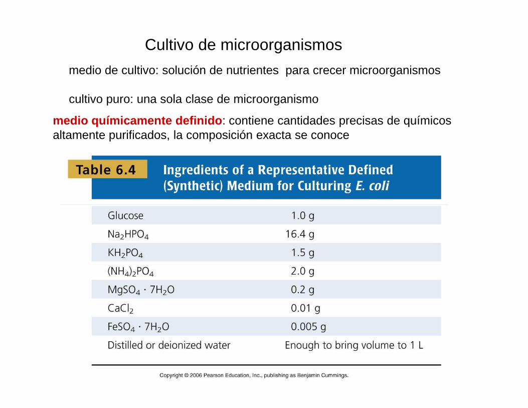
medio de cultivo: solución de nutrientes para crecer microorganismos
cultivo puro: una sola clase de microorganismo
medio químicamente definido : contiene cantidades precisas de químicos altamente purificados, la composición exacta se conoce
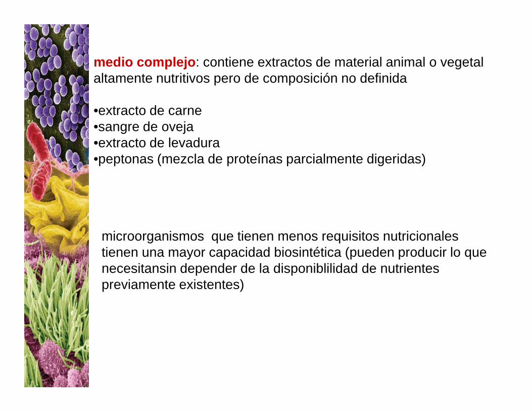
medio complejo : contiene extractos de material animal o vegetal altamente nutritivos pero de composición no definida
•extracto de carne•sangre de oveja•extracto de levadura•peptonas (mezcla de proteínas parcialmente digeridas)
microorganismos que tienen menos requisitos nutricionales tienen una mayor capacidad biosintética (pueden producir lo que necesitansin depender de la disponiblilidad de nutrientes previamente existentes)
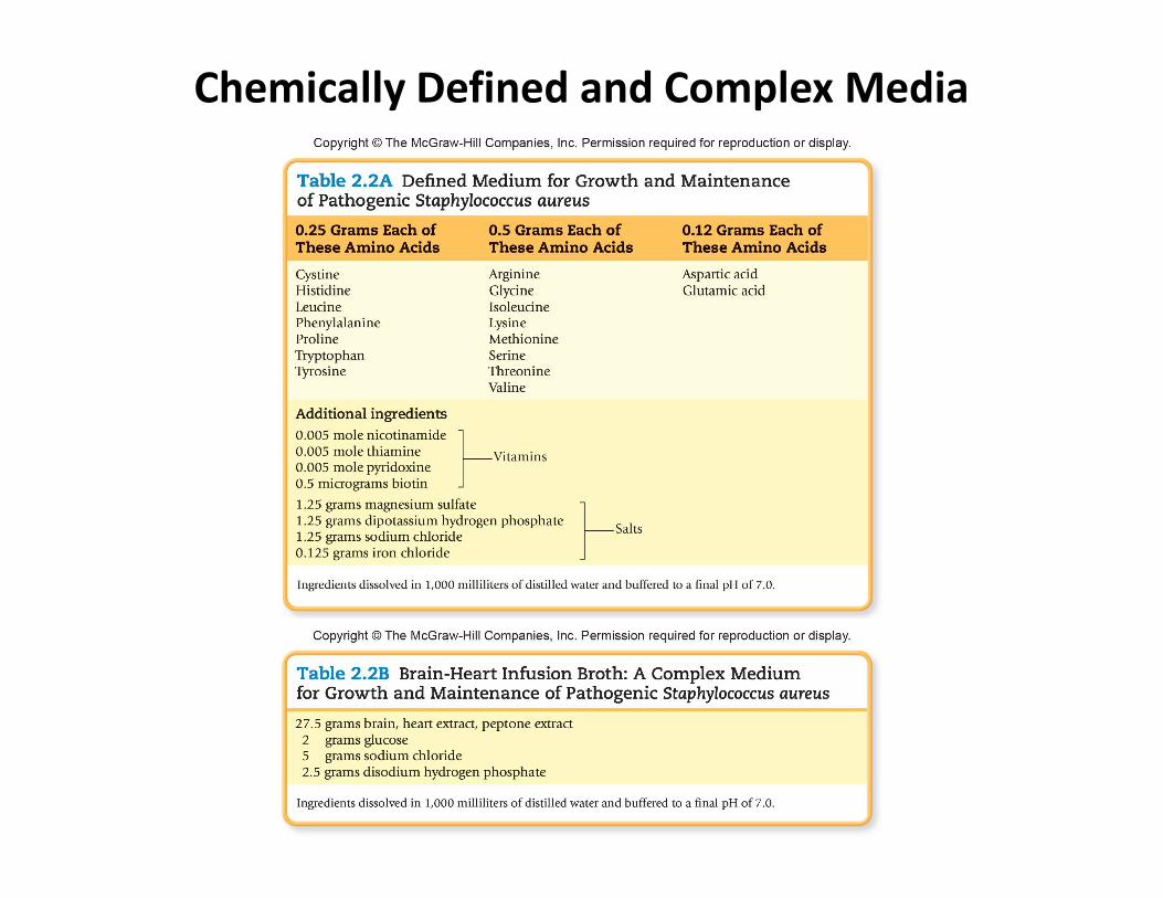
Chemically Defined and Complex Media
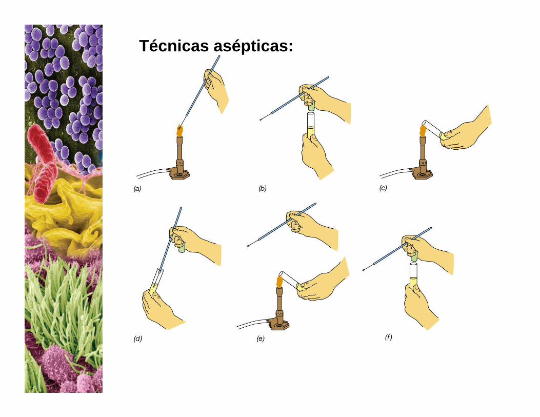
Técnicas asépticas:
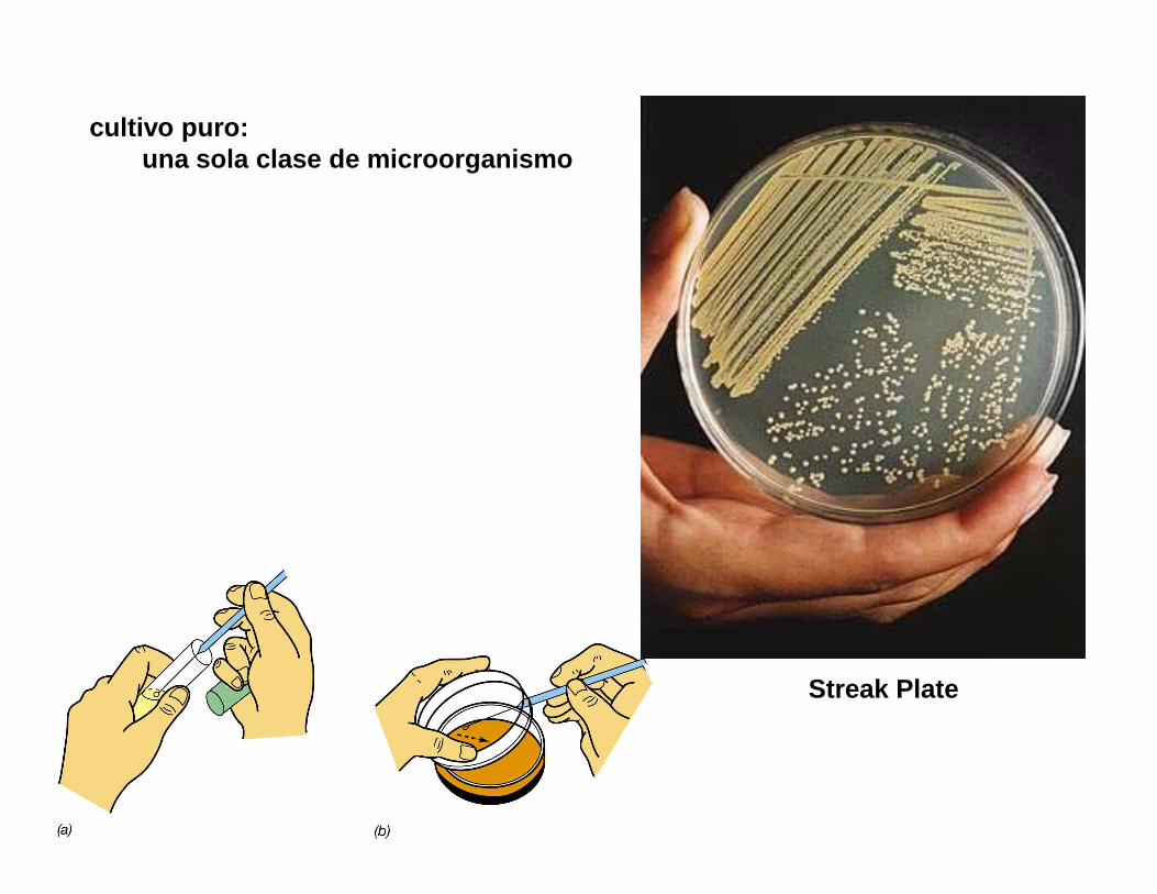
cultivo puro: una sola clase de microorganismo
Streak Plate
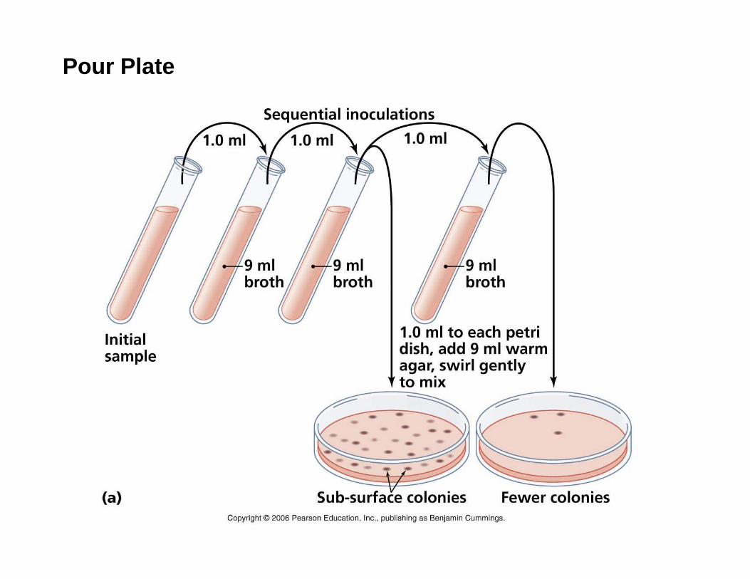
Pour Plate
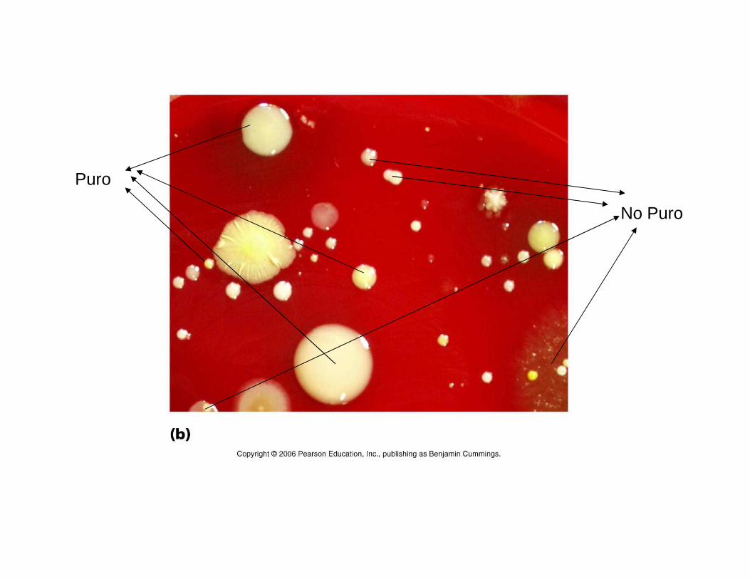
Puro
No Puro
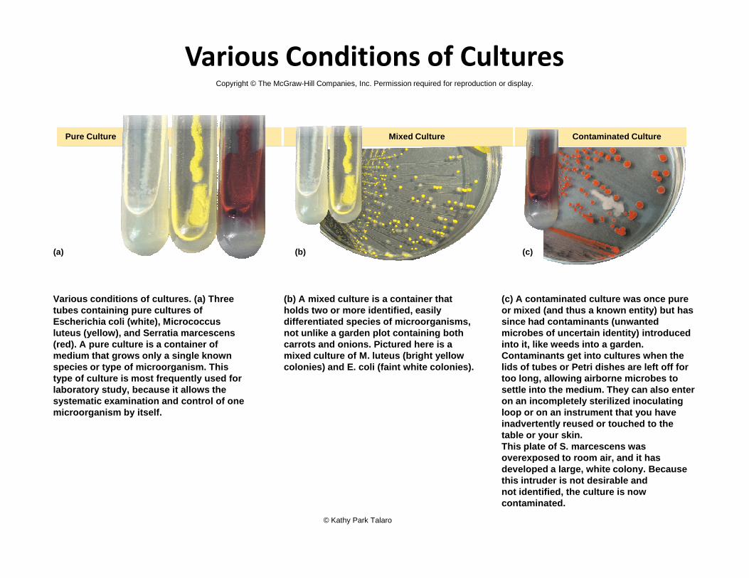
Various Conditions of Cultures
Pure Culture Mixed Culture Contaminated Culture
(c)(b)(a)
Copyright © The McGraw-Hill Companies, Inc. Permission required for reproduction or display.
Various conditions of cultures. (a) Three tubes containing pure cultures of Escherichia coli (white), Micrococcus luteus (yellow), and Serratia marcescens (red). A pure culture is a container of medium that grows only a single known species or type of microorganism. This type of culture is most frequently used for laboratory study, because it allows the systematic examination and control of one microorganism by itself.
(b) A mixed culture is a container that holds two or more identified, easily differentiated species of microorganisms, not unlike a garden plot containing both carrots and onions. Pictured here is a mixed culture of M. luteus (bright yellow colonies) and E. coli (faint white colonies).
(c) A contaminated culture was once pure or mixed (and thus a known entity) but has since had contaminants (unwanted microbes of uncertain identity) introduced into it, like weeds into a garden. Contaminants get into cultures when the lids of tubes or Petri dishes are left off for too long, allowing airborne microbes tosettle into the medium. They can also enter on an incompletely sterilized inoculating loop or on an instrument that you have inadvertently reused or touched to the table or your skin. This plate of S. marcescens was overexposed to room air, and it has developed a large, white colony. Because this intruder is not desirable andnot identified, the culture is now contaminated.
© Kathy Park Talaro
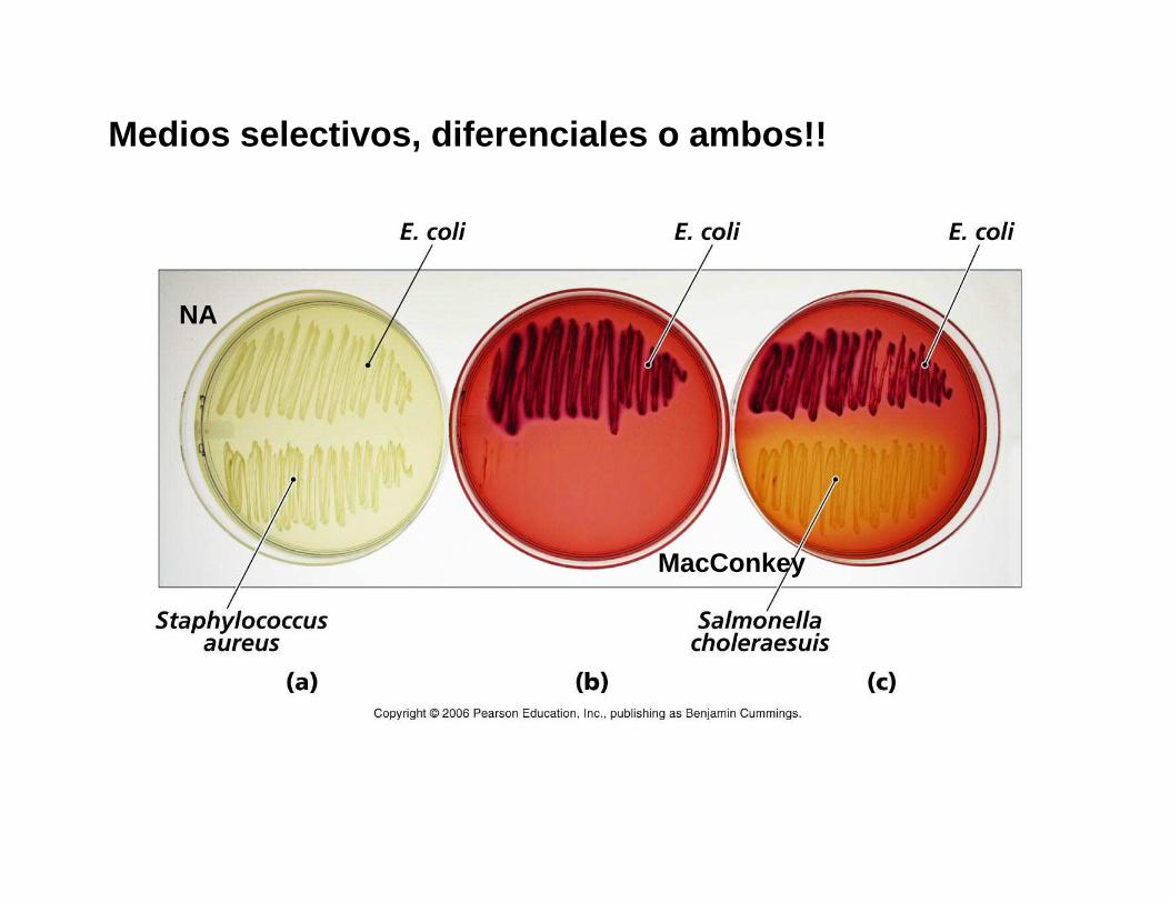
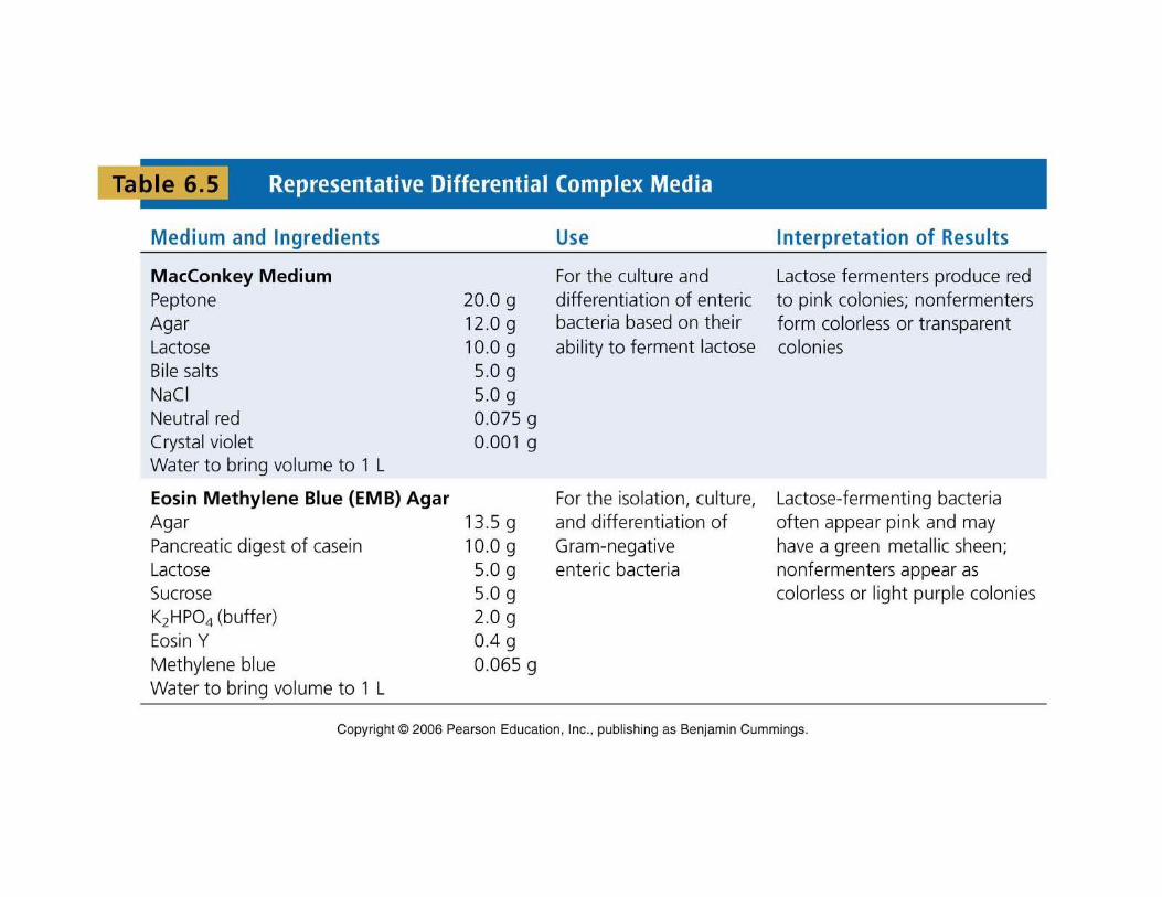
Medios selectivos, diferenciales o ambos!!
NA
MacConkey
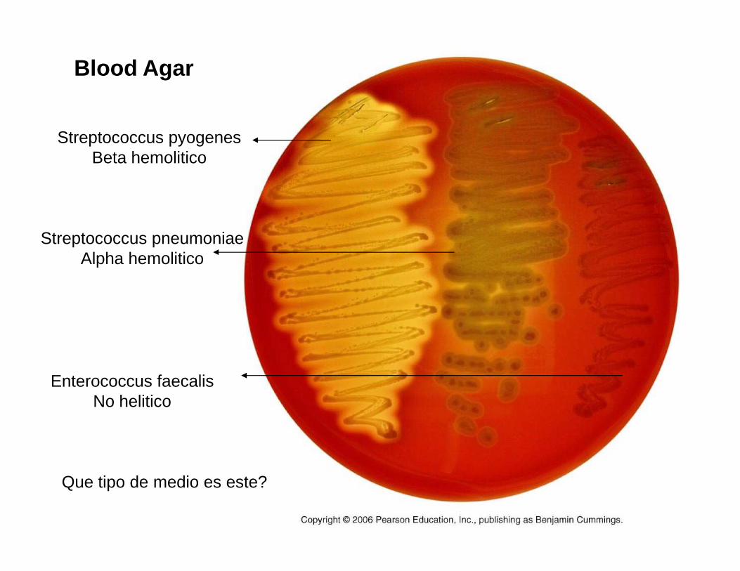
Blood Agar
Streptococcus pyogenesBeta hemolitico
Streptococcus pneumoniaeAlpha hemolitico
Enterococcus faecalisNo helitico
Que tipo de medio es este?
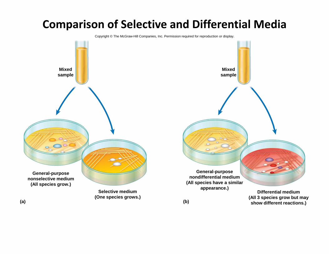
Comparison of Selective and Differential Media
Mixedsample
Mixedsample
Copyright © The McGraw-Hill Companies, Inc. Permission required for reproduction or display.
Differential medium(All 3 species grow but mayshow different reactions.)
General-purposenondifferential medium
(All species have a similarappearance.)
Selective medium(One species grows.)
(b)(a)
General-purposenonselective medium
(All species grow.)
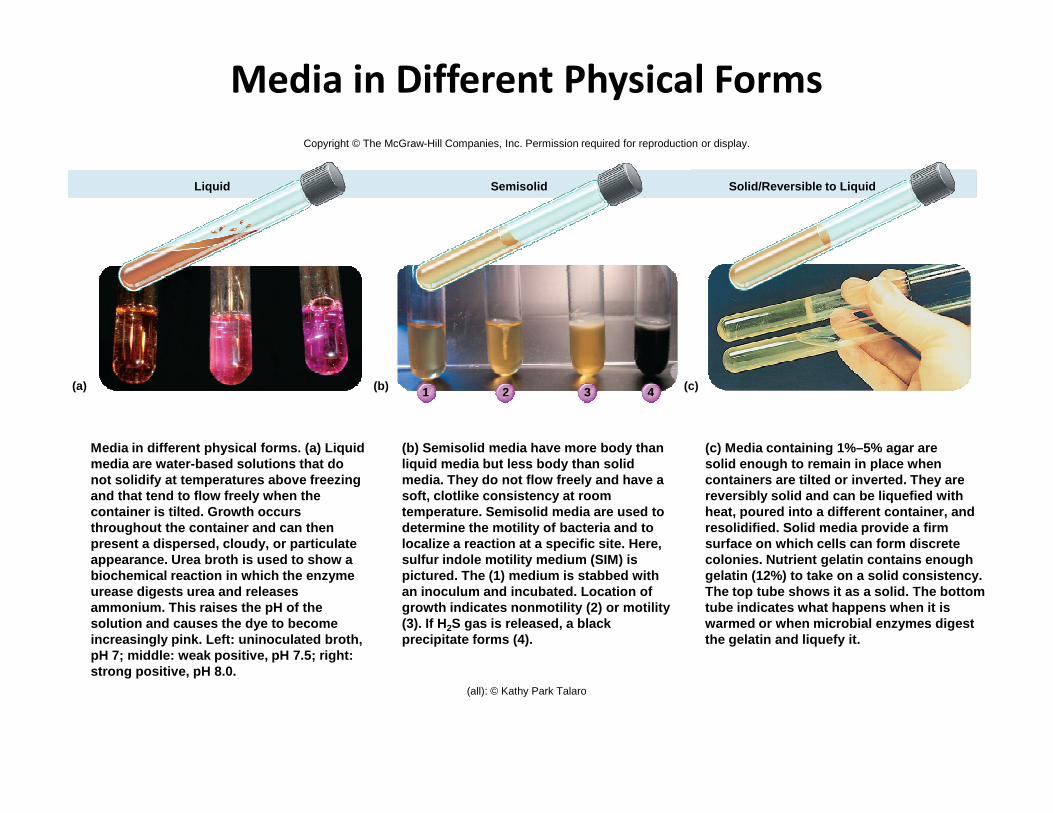
Media in Different Physical Forms
1 2 3 4 (c)(b)(a)
Liquid Semisolid Solid/Reversible to Liquid
Copyright © The McGraw-Hill Companies, Inc. Permission required for reproduction or display.
Media in different physical forms. (a) Liquid media are water-based solutions that do not solidify at temperatures above freezing and that tend to flow freely when the container is tilted. Growth occurs throughout the container and can then present a dispersed, cloudy, or particulate appearance. Urea broth is used to show a biochemical reaction in which the enzyme urease digests urea and releases ammonium. This raises the pH of the solution and causes the dye to become increasingly pink. Left: uninoculated broth, pH 7; middle: weak positive, pH 7.5; right: strong positive, pH 8.0.
(b) Semisolid media have more body thanliquid media but less body than solid media. They do not flow freely and have a soft, clotlike consistency at room temperature. Semisolid media are used to determine the motility of bacteria and to localize a reaction at a specific site. Here, sulfur indole motility medium (SIM) is pictured. The (1) medium is stabbed with an inoculum and incubated. Location of growth indicates nonmotility (2) or motility (3). If H2S gas is released, a black precipitate forms (4).
(c) Media containing 1%–5% agar aresolid enough to remain in place whencontainers are tilted or inverted. They arereversibly solid and can be liquefied withheat, poured into a different container, andresolidified. Solid media provide a firmsurface on which cells can form discretecolonies. Nutrient gelatin contains enoughgelatin (12%) to take on a solid consistency.The top tube shows it as a solid. The bottom tube indicates what happens when it is warmed or when microbial enzymes digest the gelatin and liquefy it.
(all): © Kathy Park Talaro
The Five I’s of Microbiology
•Inoculation
•Incubation
•Isolation
•Inspection
•Identification
Miscellaneous Media
•Reducing medium
- contains a substance (thioglycolic acid or
cystine) that absorbs oxygen or slows the
penetration of oxygen
- important for growing anaerobic bacteria- important for growing anaerobic bacteria
•Carbohydrate fermentation media
- contain sugars that can be fermented and a pH
indicator that shows this reaction
- can contain a Durham tube to collect gas
bubbles
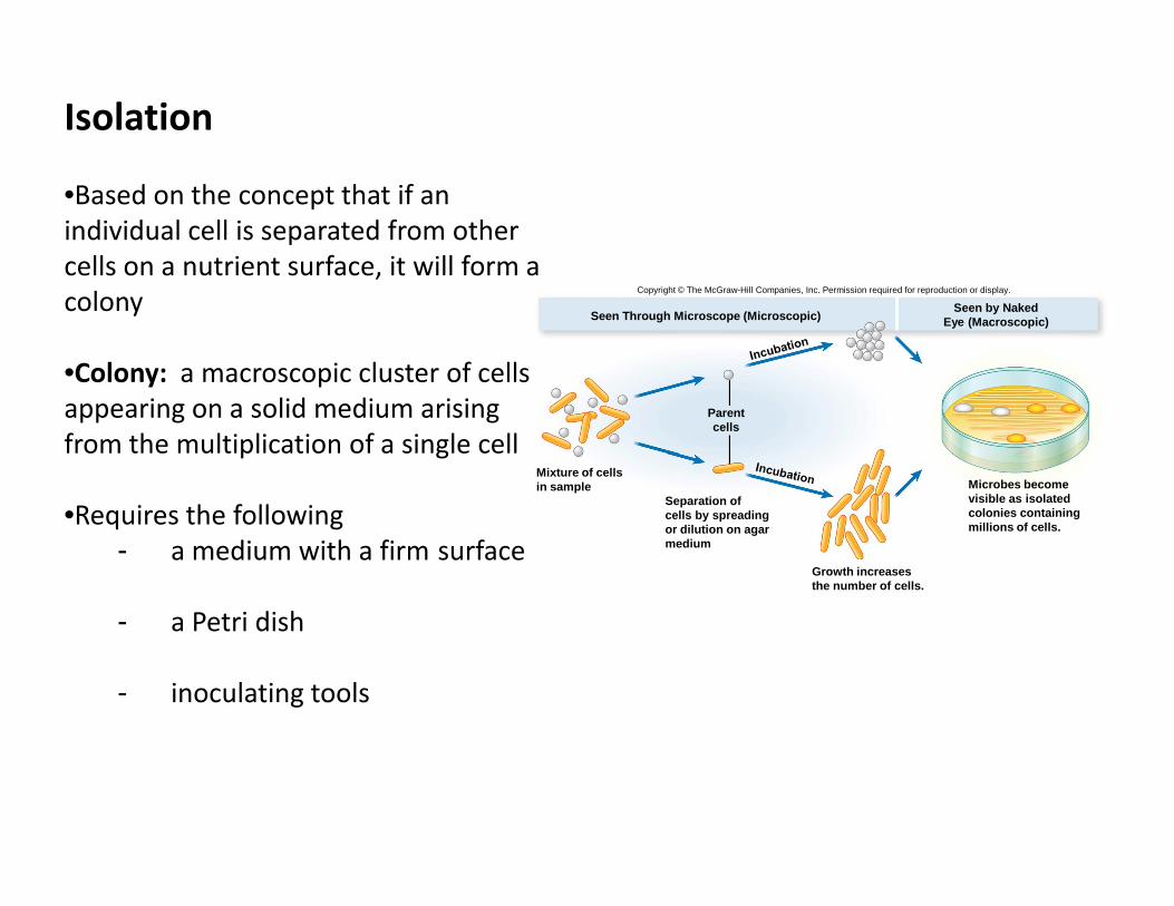
Isolation
•Based on the concept that if an
individual cell is separated from other
cells on a nutrient surface, it will form a
colony
•Colony: a macroscopic cluster of cells
appearing on a solid medium arising
from the multiplication of a single cell
Seen Through Microscope (Microscopic)Seen by Naked
Eye (Macroscopic)
Parentcells
Copyright © The McGraw-Hill Companies, Inc. Permission required for reproduction or display.
from the multiplication of a single cell
•Requires the following
- a medium with a firm surface
- a Petri dish
- inoculating tools
Microbes becomevisible as isolatedcolonies containingmillions of cells.
Growth increasesthe number of cells.
Separation ofcells by spreadingor dilution on agarmedium
Mixture of cellsin sample
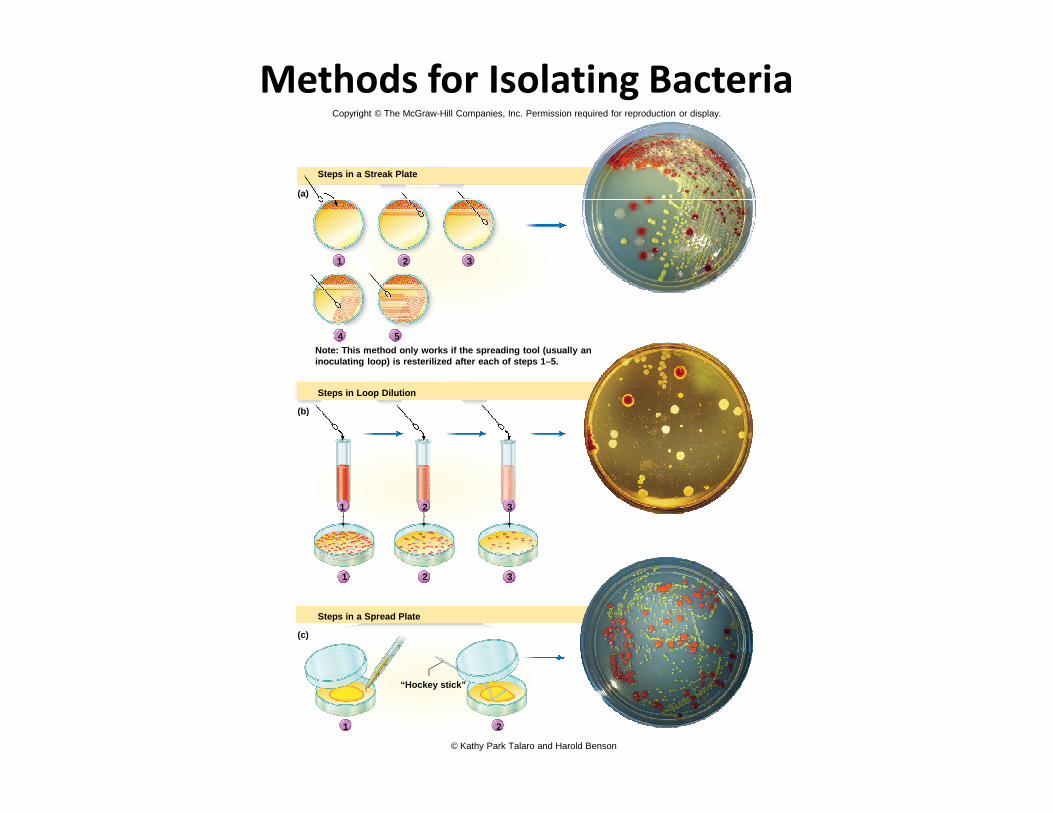
Methods for Isolating Bacteria
1 2 3
4 5
Steps in a Streak Plate
Note: This method only works if the spreading tool (usually aninoculating loop) is resterilized after each of step s 1–5.
Steps in Loop Dilution
(b)
(a)
Copyright © The McGraw-Hill Companies, Inc. Permission required for reproduction or display.
1 2
1 2
3
1 2 3
(b)
Steps in a Spread Plate
(c)
“Hockey stick”
© Kathy Park Talaro and Harold Benson
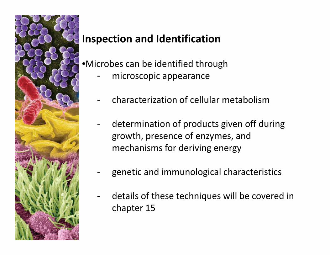
Inspection and Identification
•Microbes can be identified through
- microscopic appearance
- characterization of cellular metabolism
- determination of products given off during - determination of products given off during
growth, presence of enzymes, and
mechanisms for deriving energy
- genetic and immunological characteristics
- details of these techniques will be covered in
chapter 15
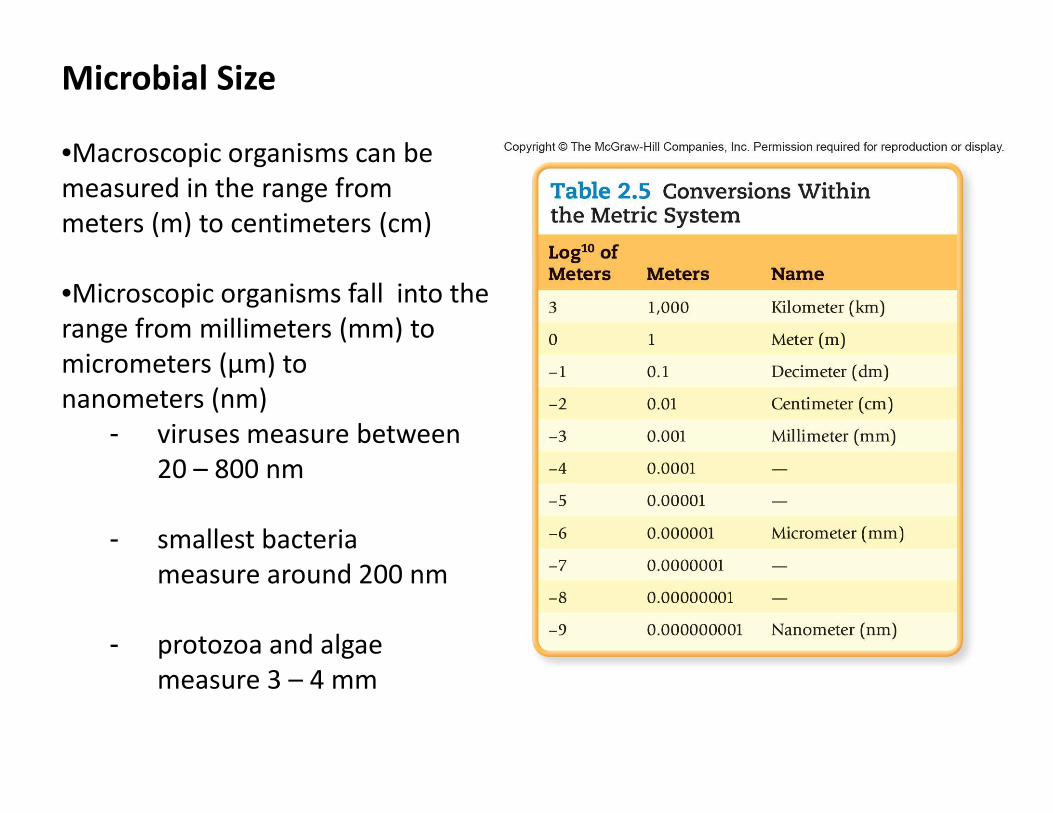
Microbial Size
•Macroscopic organisms can be
measured in the range from
meters (m) to centimeters (cm)
•Microscopic organisms fall into the
range from millimeters (mm) to
micrometers (μm) to
nanometers (nm)nanometers (nm)
- viruses measure between
20 – 800 nm
- smallest bacteria
measure around 200 nm
- protozoa and algae
measure 3 – 4 mm
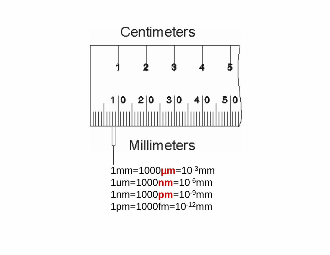
1mm=1000µµµµm=10-3mm1um=1000nm=10-6mm1nm=1000pm=10-9mm1pm=1000fm=10-12mm
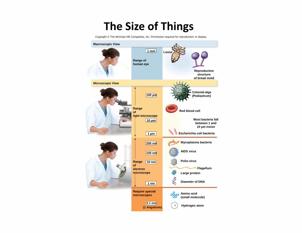
The Size of Things
Red blood cell
Colonial alga(Pediastrum)
Reproductivestructure
of bread mold
Louse
Macroscopic View
Microscopic View
100 µm
Range ofhuman eye
Rangeoflight microscope
1 mm
Copyright © The McGraw-Hill Companies, Inc. Permission required for reproduction or display.
Hydrogen atom
Amino acid(small molecule)
Diameter of DNA
Large protein
Flagellum
Polio virus
AIDS virus
Mycoplasma bacteria
Escherichia coli bacteria
Most bacteria fallbetween 1 and10 µm insize
light microscope
10 µm
1 µm
200 nm
100 nm
Rangeofelectronmicroscope
10 nm
1 nm
Require specialmicroscopes
0.1 nm(1 Angstrom)
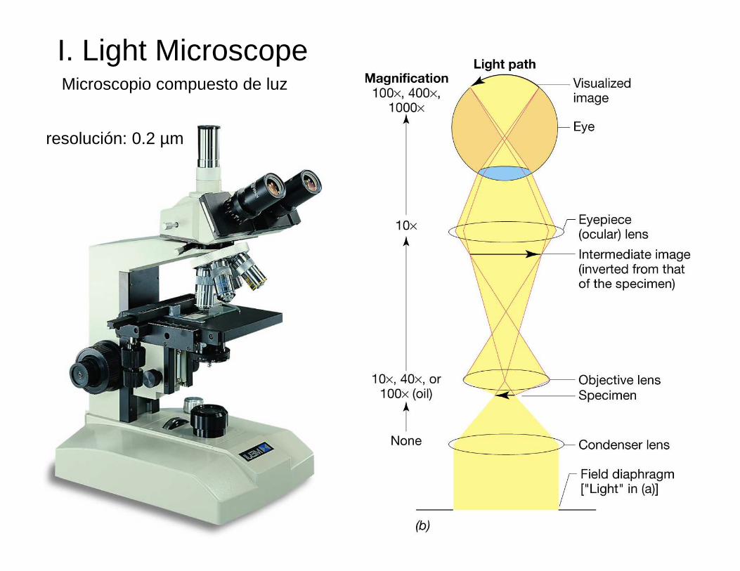
I. Light Microscope
resolución: 0.2 µm
Microscopio compuesto de luz
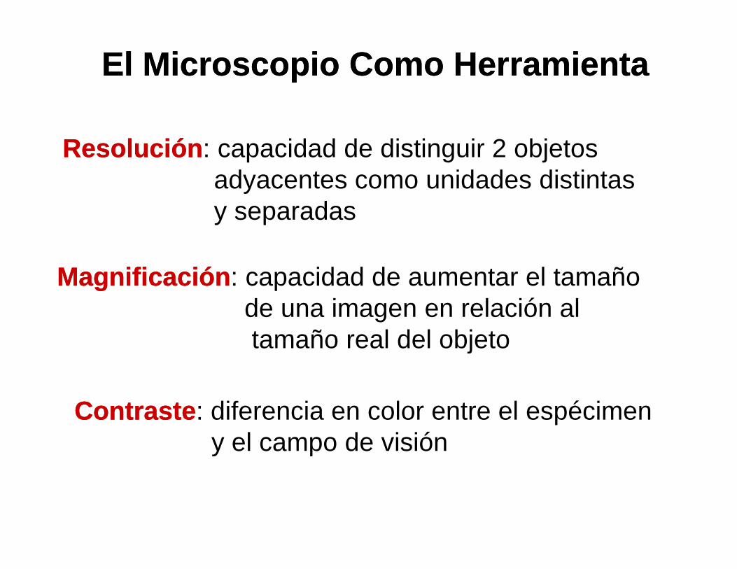
ResoluciónResolución : capacidad de distinguir 2 objetos adyacentes como unidades distintasy separadas
El Microscopio Como HerramientaEl Microscopio Como Herramienta
MagnificaciónMagnificación : capacidad de aumentar el tamaño
ContrasteContraste : diferencia en color entre el espécimeny el campo de visión
MagnificaciónMagnificación : capacidad de aumentar el tamaño de una imagen en relación altamaño real del objeto
Principles of Light Microscopy (cont’d)
•Resolution (resolving power)
- the capacity of an optical system to distinguish
or separate two adjacent points or objects from
one another
- the human eye can resolve two objects that are
no closer than 0.2 mm apart
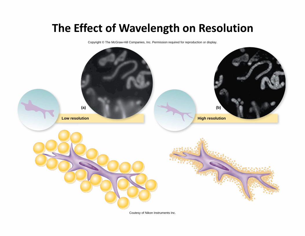
The Effect of Wavelength on Resolution
High resolutionLow resolution
(a) (b)
Copyright © The McGraw-Hill Companies, Inc. Permission required for reproduction or display.
High resolutionLow resolution
Coutesy of Nikon Instruments Inc.
Principles of Light Microscopy
(cont’d)
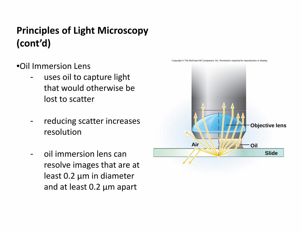
•Oil Immersion Lens
- uses oil to capture light
that would otherwise be
lost to scatter
- reducing scatter increases
Copyright © The McGraw-Hill Companies, Inc. Permission required for reproduction or display.
- reducing scatter increases
resolution
- oil immersion lens can
resolve images that are at
least 0.2 μm in diameter
and at least 0.2 μm apart
Objective lens
SlideOilAir
Principles of Microscopy (cont’d)
•Contrast
- refractive index: a measurement of the degree
of bending that light undergoes as it passes from
one medium to another
- the higher the difference in refractive indexes,
the greater the contrast the greater the contrast
- the iris diaphragm can control the amount of
light entering the condenser and increase
contrast
- special lenses and dyes are also used to increase
contrast
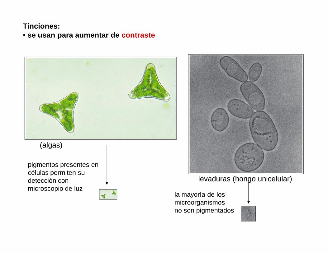
Tinciones: • se usan para aumentar de contraste
(algas)
pigmentos presentes en células permiten su detección con microscopio de luz
levaduras (hongo unicelular)
la mayoría de los microorganismosno son pigmentados
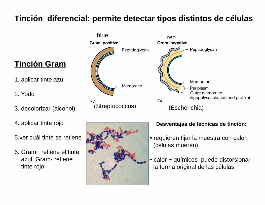
Tinción diferencial: permite detectar tipos distintos de cé lulas
Tinción Gram
1. aplicar tinte azul
2. Yodo
blue red
3. decolorizar (alcohol)
4. aplicar tinte rojo
5.ver cuál tinte se retiene
6. Gram+ retiene el tinte azul, Gram- retienetinte rojo
(Streptococcus) (Escherichia)
Desventajas de técnicas de tinción:
• requieren fijar la muestra con calor:(células mueren)
• calor + químicos puede distorsionarla forma original de las células
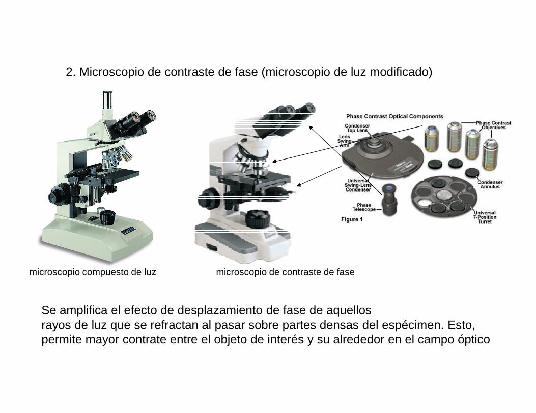
2. Microscopio de contraste de fase (microscopio de luz modificado)
microscopio compuesto de luz microscopio de contraste de fase
Se amplifica el efecto de desplazamiento de fase de aquellos rayos de luz que se refractan al pasar sobre partes densas del espécimen. Esto,permite mayor contrate entre el objeto de interés y su alrededor en el campo óptico
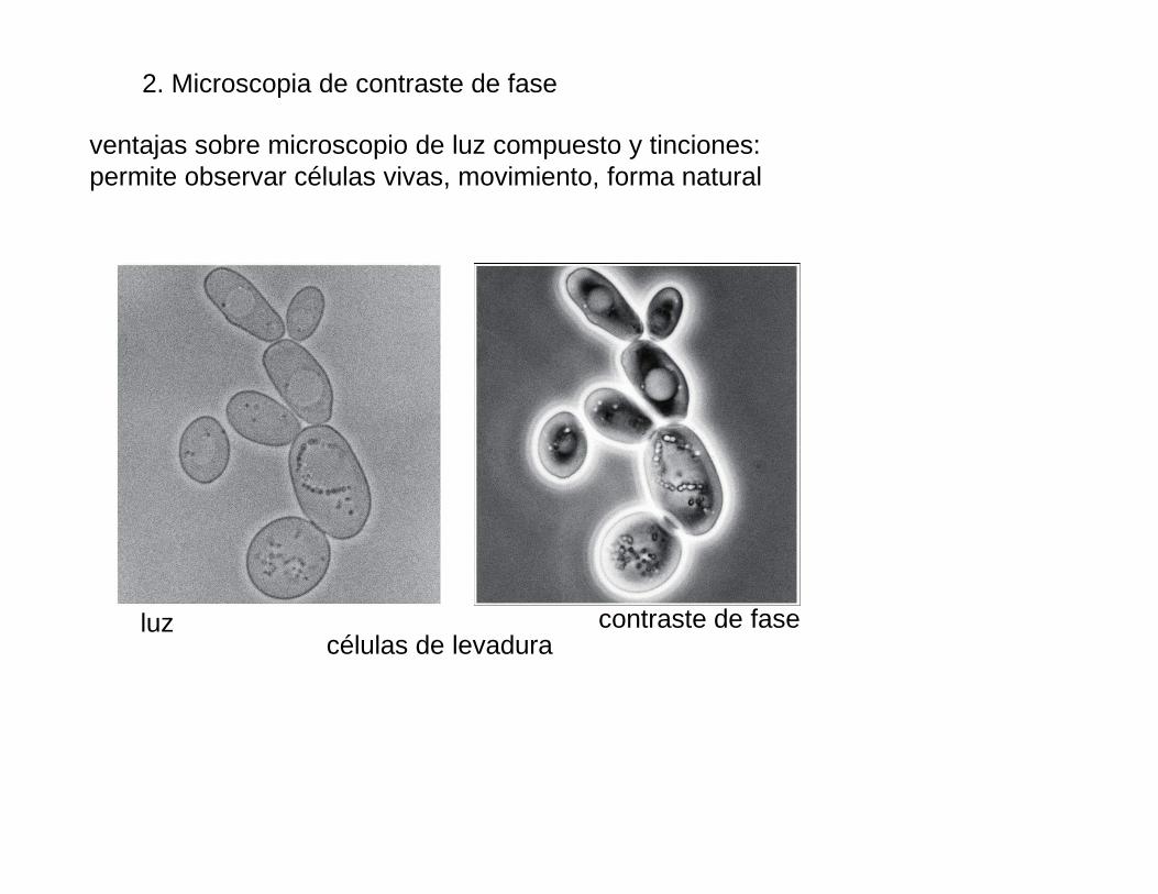
2. Microscopia de contraste de fase
ventajas sobre microscopio de luz compuesto y tinciones:permite observar células vivas, movimiento, forma natural
células de levaduraluz contraste de fase
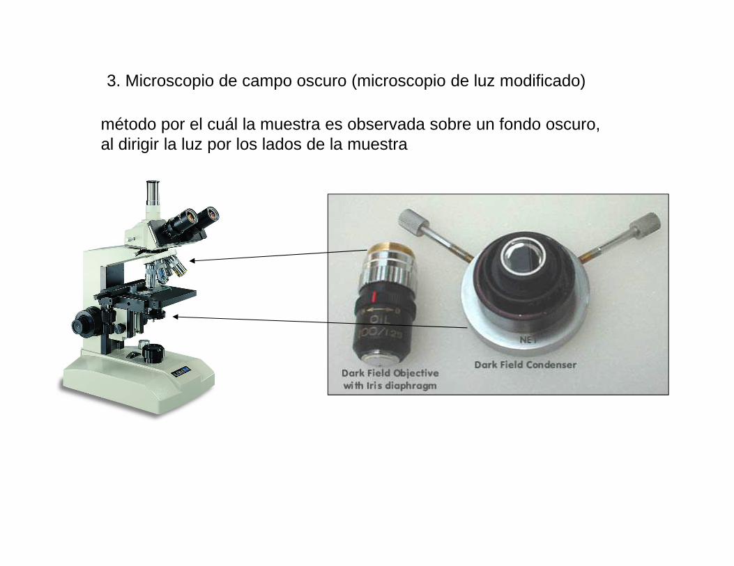
3. Microscopio de campo oscuro (microscopio de luz modificado)
método por el cuál la muestra es observada sobre un fondo oscuro, al dirigir la luz por los lados de la muestra
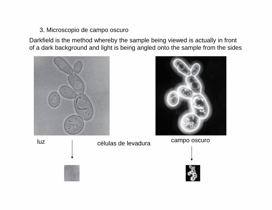
3. Microscopio de campo oscuro
Darkfield is the method whereby the sample being viewed is actually in front of a dark background and light is being angled onto the sample from the sides
células de levaduraluz campo oscuro
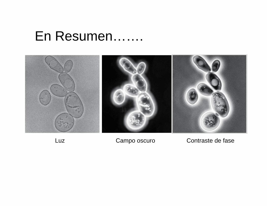
En Resumen…….
Luz Campo oscuro Contraste de fase
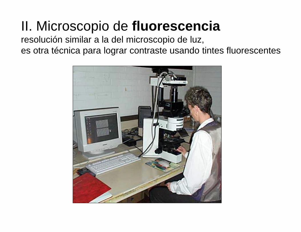
II. Microscopio de fluorescencia resolución similar a la del microscopio de luz, es otra técnica para lograr contraste usando tintes fluorescentes
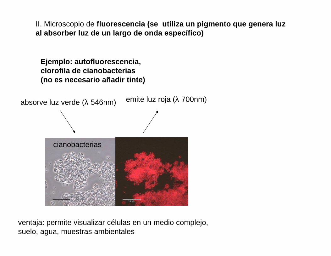
II. Microscopio de fluorescencia (se utiliza un pigmento que genera lu z al absorber luz de un largo de onda específico)
Ejemplo: autofluorescencia, clorofila de cianobacterias (no es necesario añadir tinte)
absorve luz verde (λ 546nm) emite luz roja (λ 700nm)
ventaja: permite visualizar células en un medio complejo,suelo, agua, muestras ambientales
cianobacterias
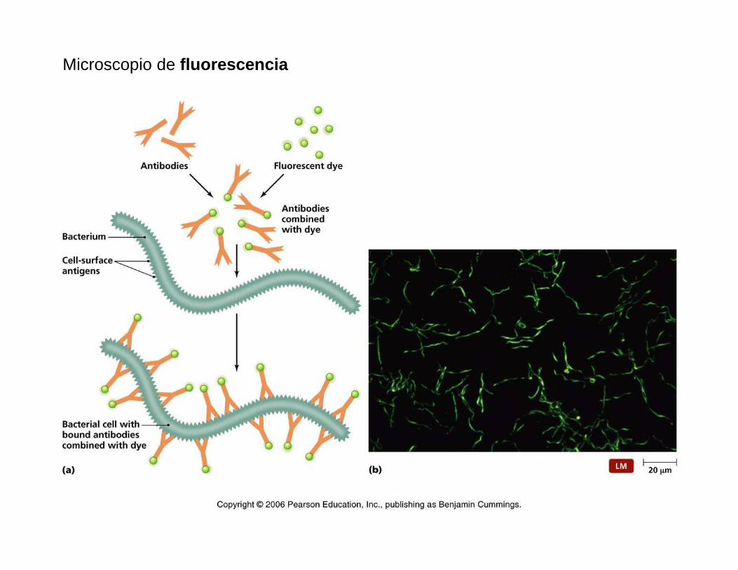
Microscopio de fluorescencia
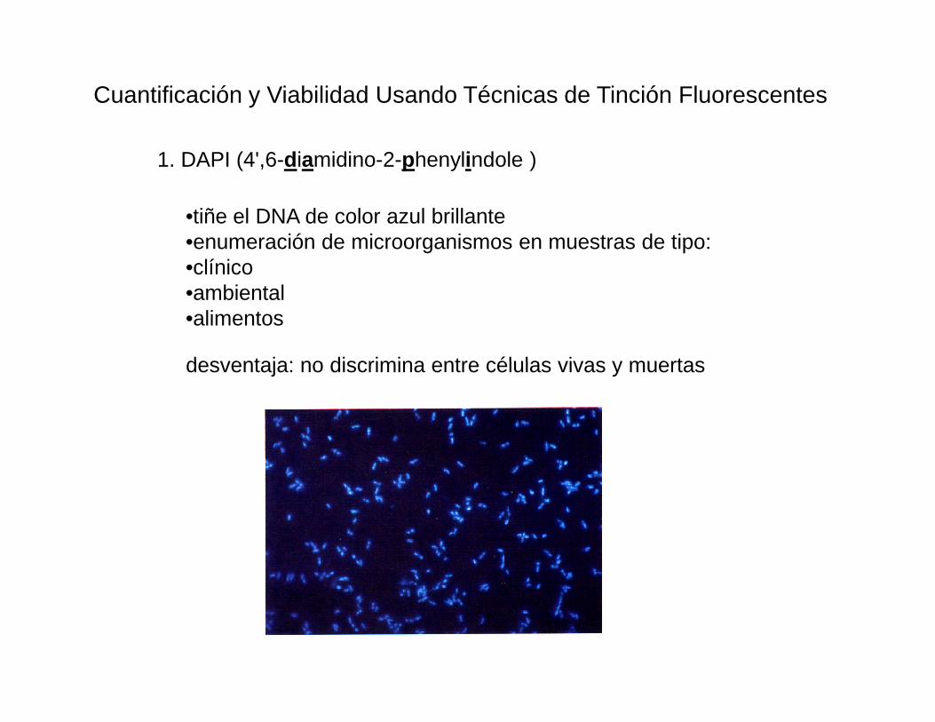
Cuantificación y Viabilidad Usando Técnicas de Tinción Fluorescentes
•tiñe el DNA de color azul brillante •enumeración de microorganismos en muestras de tipo:•clínico•ambiental•alimentos
desventaja: no discrimina entre células vivas y muertas
1. DAPI (4',6-diamidino-2-phenylindole )
desventaja: no discrimina entre células vivas y muertas
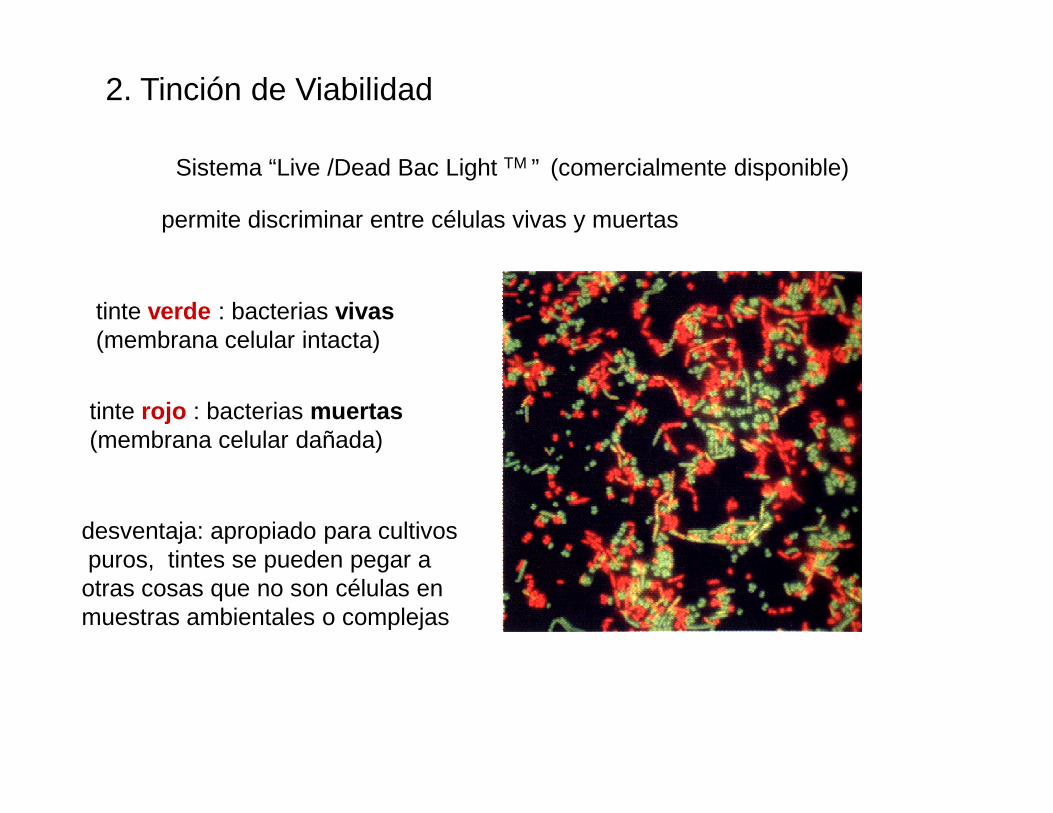
2. Tinción de Viabilidad
Sistema “Live /Dead Bac Light TM ” (comercialmente disponible)
permite discriminar entre células vivas y muertas
tinte verde : bacterias vivas(membrana celular intacta)
tinte rojo : bacterias muertastinte rojo : bacterias muertas(membrana celular dañada)
desventaja: apropiado para cultivospuros, tintes se pueden pegar a otras cosas que no son células en muestras ambientales o complejas
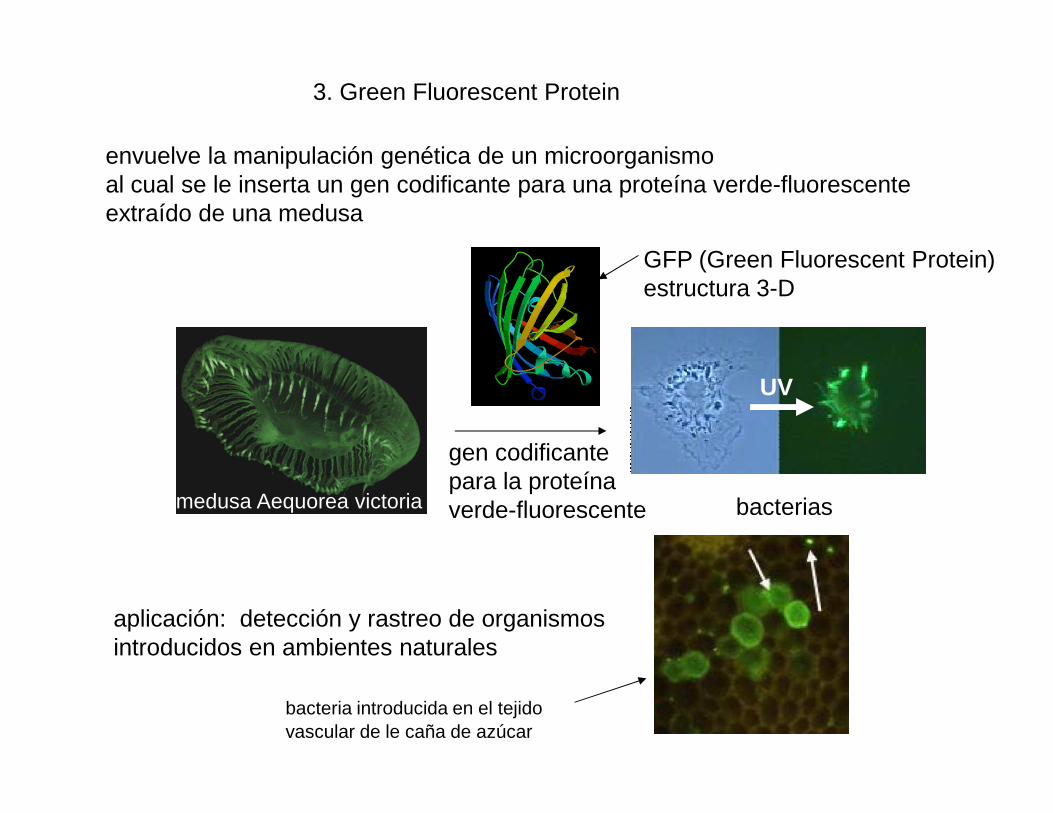
GFP (Green Fluorescent Protein)estructura 3-D
3. Green Fluorescent Protein
envuelve la manipulación genética de un microorganismo al cual se le inserta un gen codificante para una proteína verde-fluorescente extraído de una medusa
UV
medusa Aequorea victoria bacterias
gen codificantepara la proteínaverde-fluorescente
aplicación: detección y rastreo de organismos introducidos en ambientes naturales
bacteria introducida en el tejidovascular de le caña de azúcar
III. Microscopía en tres dimensiones de alta resolución
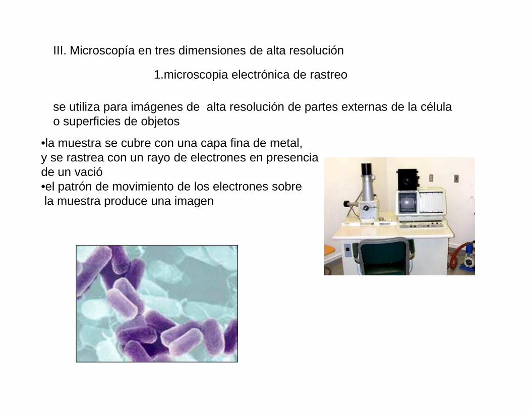
1.microscopia electrónica de rastreo
se utiliza para imágenes de alta resolución de partes externas de la célulao superficies de objetos
•la muestra se cubre con una capa fina de metal, y se rastrea con un rayo de electrones en presencia de un vació•el patrón de movimiento de los electrones sobrela muestra produce una imagenla muestra produce una imagen
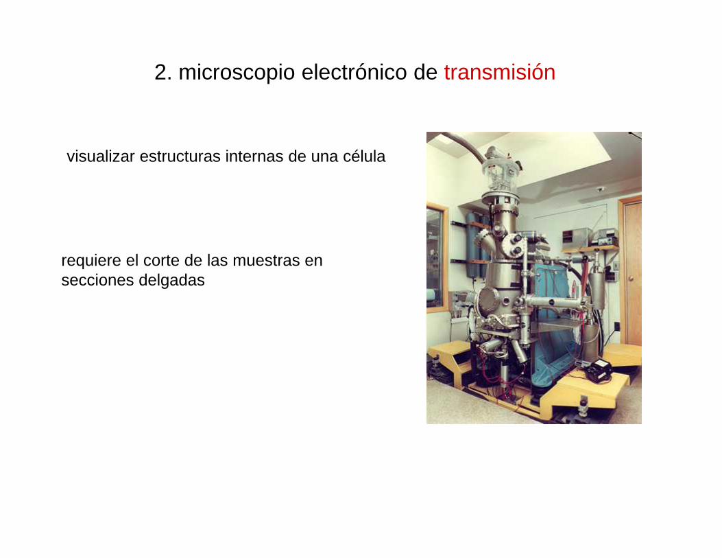
2. microscopio electrónico de transmisión
visualizar estructuras internas de una célula
requiere el corte de las muestras en secciones delgadassecciones delgadas
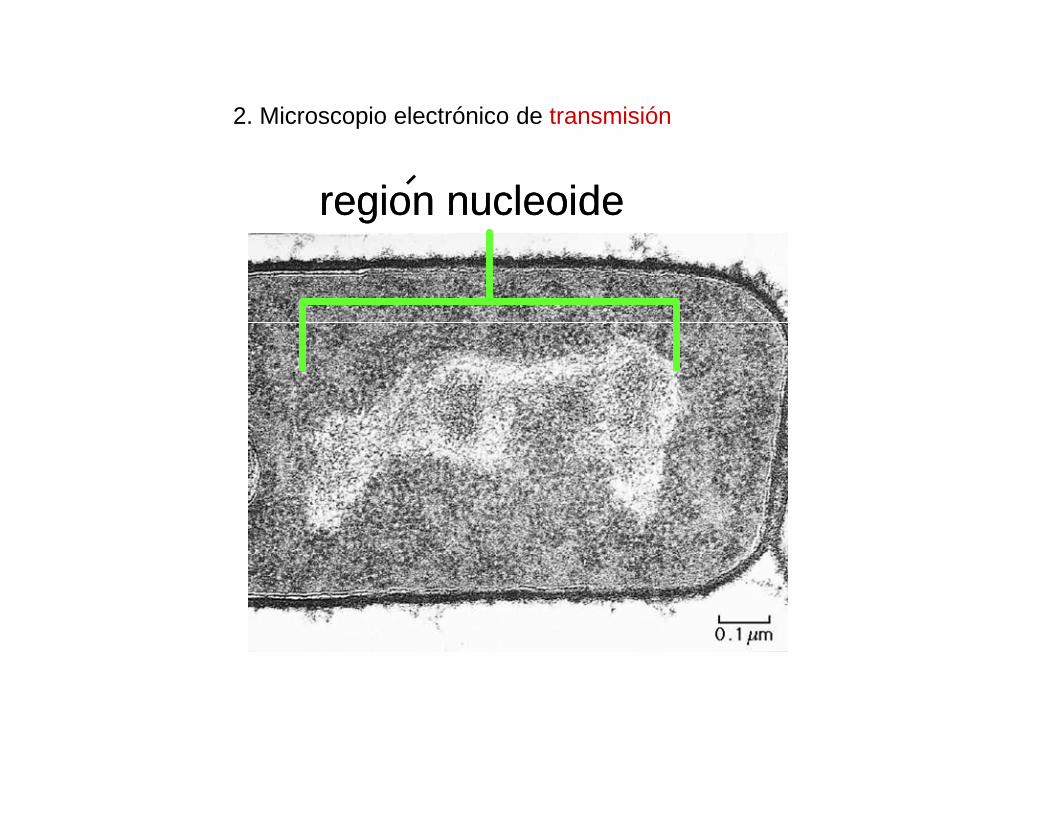
2. Microscopio electrónico de transmisión
region nucleoideregion nucleoide
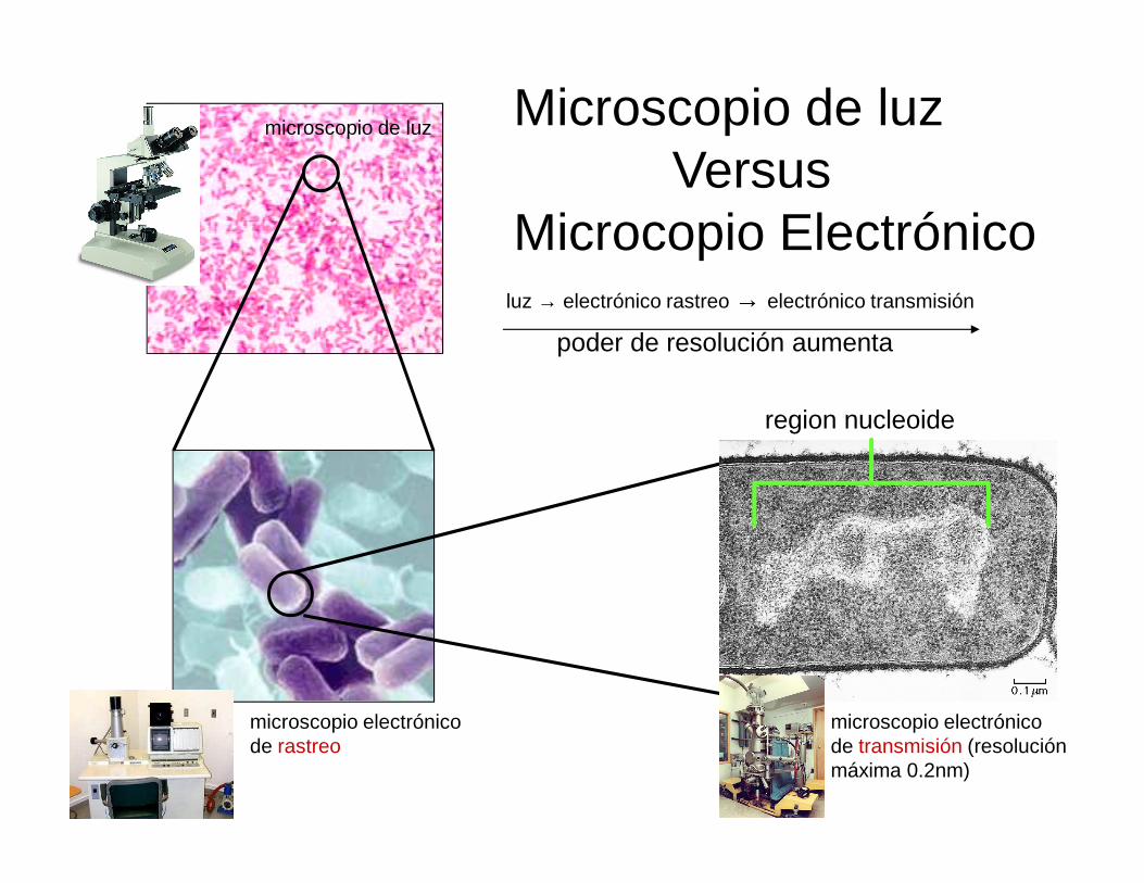
microscopio de luz Microscopio de luzVersus
Microcopio Electrónico
region nucleoide
luz → electrónico rastreo → electrónico transmisión
poder de resolución aumenta
microscopio electrónico de rastreo
microscopio electrónico de transmisión (resoluciónmáxima 0.2nm)

Cuanto hemos mejorado?
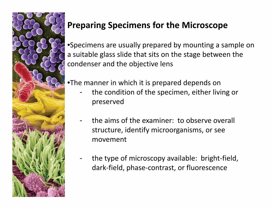
Preparing Specimens for the Microscope
•Specimens are usually prepared by mounting a sample on
a suitable glass slide that sits on the stage between the
condenser and the objective lens
•The manner in which it is prepared depends on
- the condition of the specimen, either living or
preservedpreserved
- the aims of the examiner: to observe overall
structure, identify microorganisms, or see
movement
- the type of microscopy available: bright-field,
dark-field, phase-contrast, or fluorescence
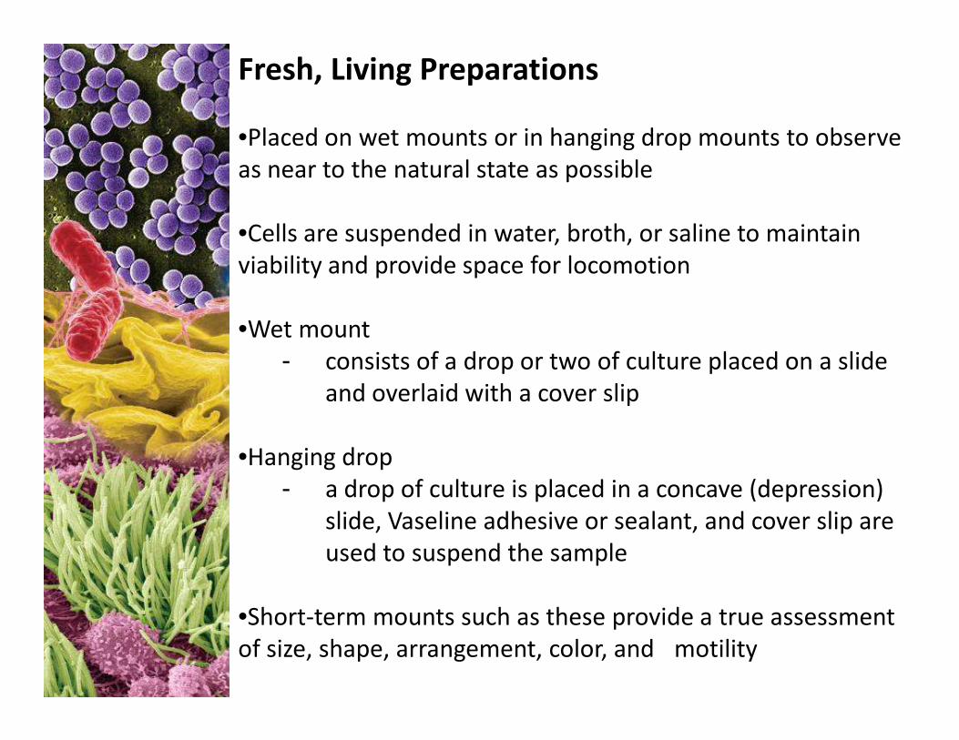
Fresh, Living Preparations
•Placed on wet mounts or in hanging drop mounts to observe
as near to the natural state as possible
•Cells are suspended in water, broth, or saline to maintain
viability and provide space for locomotion
•Wet mount
- consists of a drop or two of culture placed on a slide - consists of a drop or two of culture placed on a slide
and overlaid with a cover slip
•Hanging drop
- a drop of culture is placed in a concave (depression)
slide, Vaseline adhesive or sealant, and cover slip are
used to suspend the sample
•Short-term mounts such as these provide a true assessment
of size, shape, arrangement, color, and motility
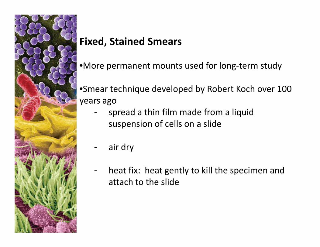
Fixed, Stained Smears
•More permanent mounts used for long-term study
•Smear technique developed by Robert Koch over 100
years ago
- spread a thin film made from a liquid
suspension of cells on a slidesuspension of cells on a slide
- air dry
- heat fix: heat gently to kill the specimen and
attach to the slide
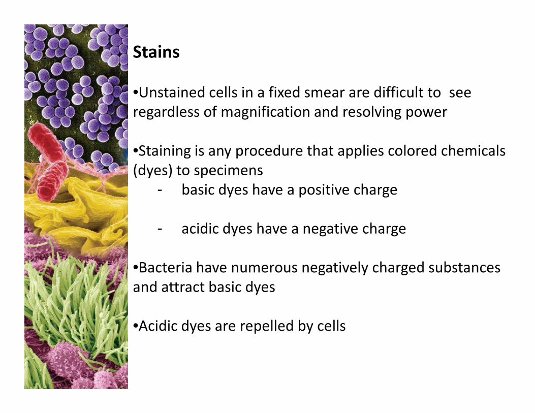
Stains
•Unstained cells in a fixed smear are difficult to see
regardless of magnification and resolving power
•Staining is any procedure that applies colored chemicals
(dyes) to specimens
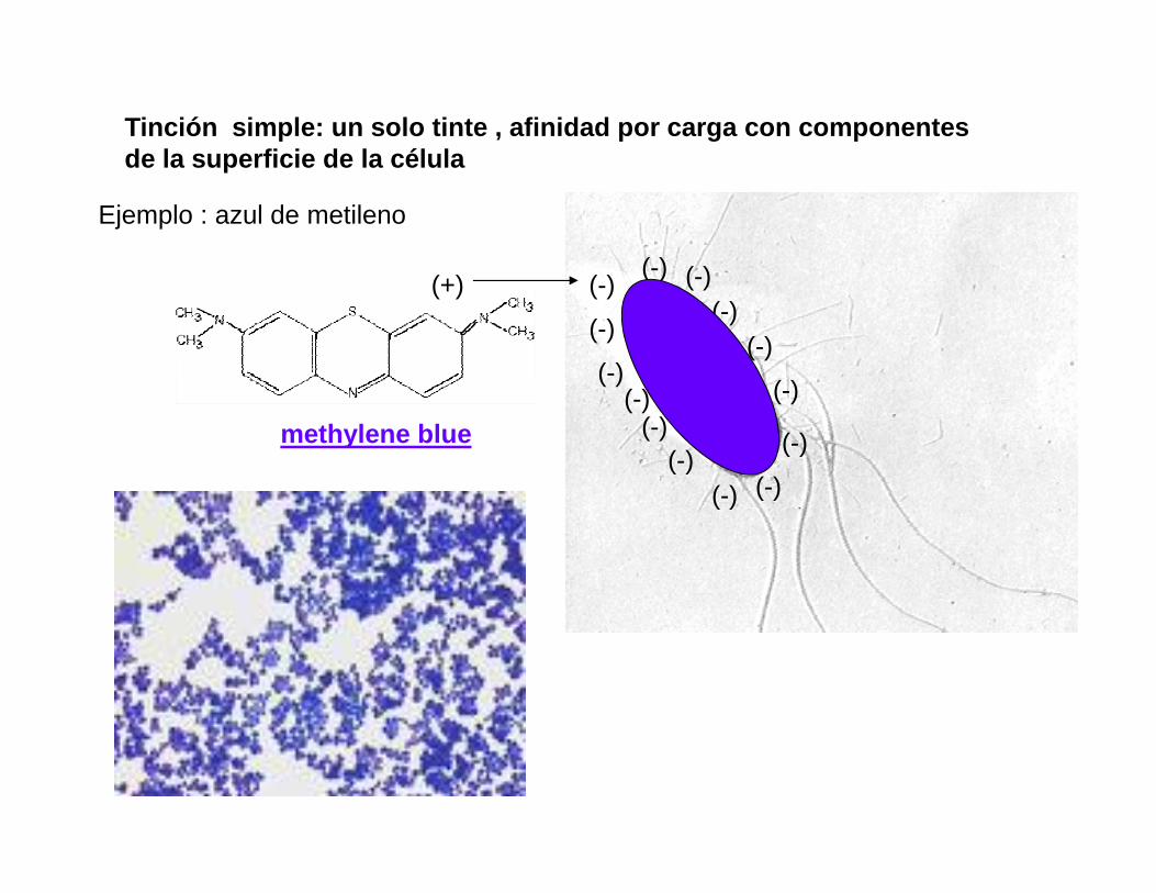
- basic dyes have a positive charge
- acidic dyes have a negative charge
•Bacteria have numerous negatively charged substances
and attract basic dyes
•Acidic dyes are repelled by cells
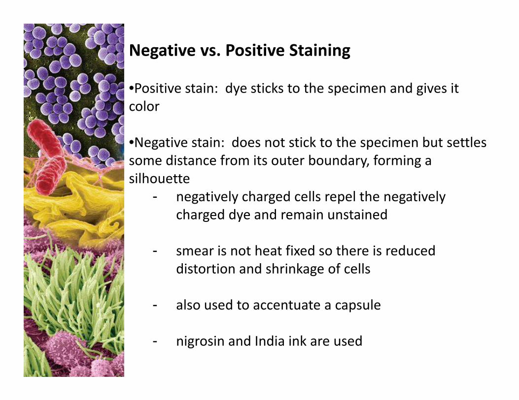
Negative vs. Positive Staining
•Positive stain: dye sticks to the specimen and gives it
color
•Negative stain: does not stick to the specimen but settles
some distance from its outer boundary, forming a
silhouette
- negatively charged cells repel the negatively - negatively charged cells repel the negatively
charged dye and remain unstained
- smear is not heat fixed so there is reduced
distortion and shrinkage of cells
- also used to accentuate a capsule
- nigrosin and India ink are used
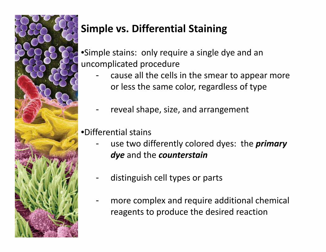
Simple vs. Differential Staining
•Simple stains: only require a single dye and an
uncomplicated procedure
- cause all the cells in the smear to appear more
or less the same color, regardless of type
- reveal shape, size, and arrangement
Differential stains •Differential stains
- use two differently colored dyes: the primary
dye and the counterstain
- distinguish cell types or parts
- more complex and require additional chemical
reagents to produce the desired reaction
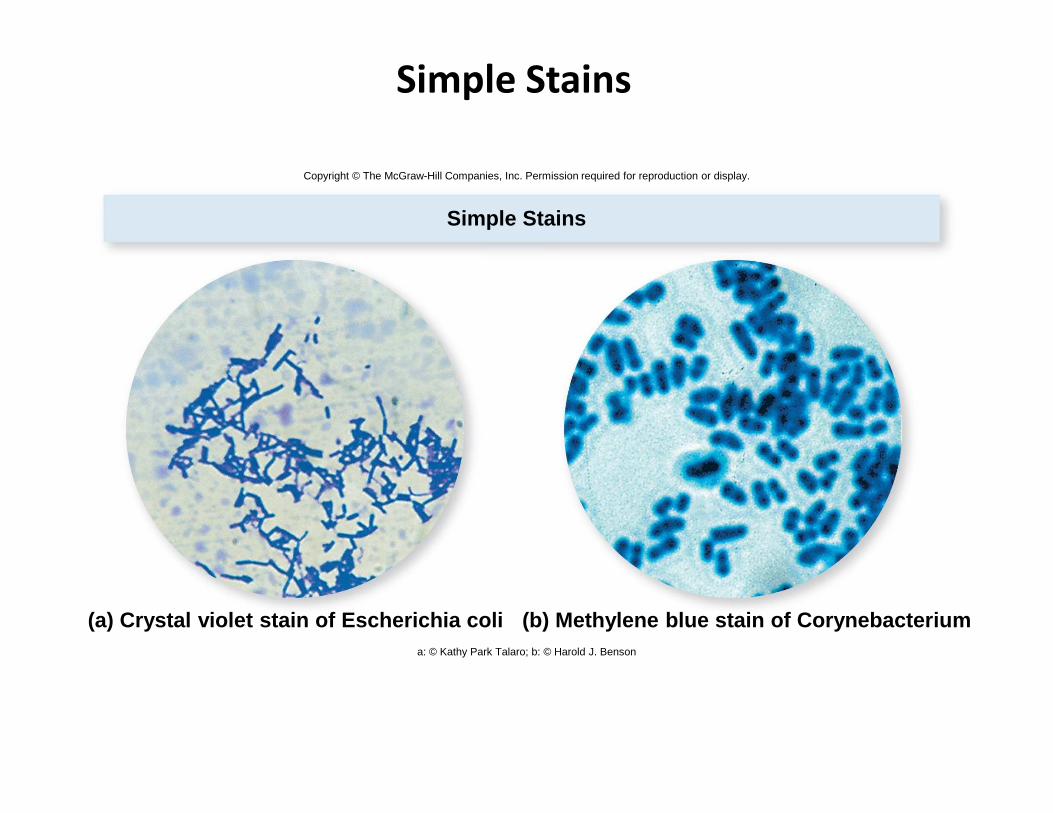
Simple Stains
Simple Stains
Copyright © The McGraw-Hill Companies, Inc. Permission required for reproduction or display.
(b) Methylene blue stain of Corynebacterium(a) Crystal violet stain of Escherichia colia: © Kathy Park Talaro; b: © Harold J. Benson
(-)
(-)
(-)(-)
(-)(-)
(-)
(-)
(-)(-)
(-)
(-)
(+)
methylene blue
Tinción simple: un solo tinte , afinidad por carga c on componentes de la superficie de la célula
Ejemplo : azul de metileno
(-)
(-)(-)
(-)
methylene blue
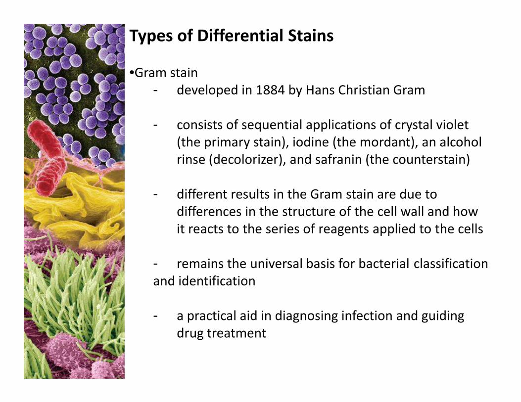
Types of Differential Stains
•Gram stain
- developed in 1884 by Hans Christian Gram
- consists of sequential applications of crystal violet
(the primary stain), iodine (the mordant), an alcohol
rinse (decolorizer), and safranin (the counterstain)
- different results in the Gram stain are due to - different results in the Gram stain are due to
differences in the structure of the cell wall and how
it reacts to the series of reagents applied to the cells
- remains the universal basis for bacterial classification
and identification
- a practical aid in diagnosing infection and guiding
drug treatment
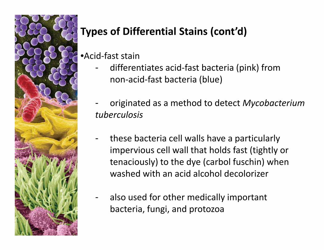
Types of Differential Stains (cont’d)
•Acid-fast stain
- differentiates acid-fast bacteria (pink) from
non-acid-fast bacteria (blue)
- originated as a method to detect Mycobacterium
tuberculosis
- these bacteria cell walls have a particularly
impervious cell wall that holds fast (tightly or
tenaciously) to the dye (carbol fuschin) when
washed with an acid alcohol decolorizer
- also used for other medically important
bacteria, fungi, and protozoa
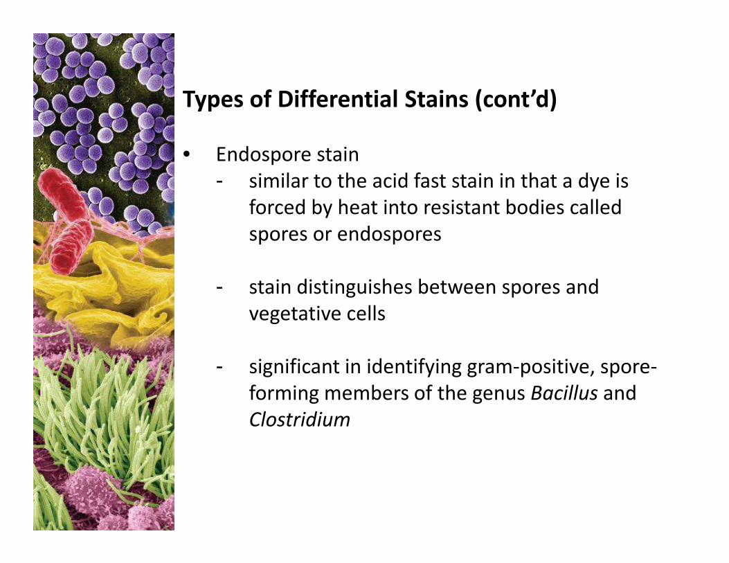
Types of Differential Stains (cont’d)
• Endospore stain
- similar to the acid fast stain in that a dye is
forced by heat into resistant bodies called
spores or endospores
- stain distinguishes between spores and - stain distinguishes between spores and
vegetative cells
- significant in identifying gram-positive, spore-
forming members of the genus Bacillus and
Clostridium
Differential Stains
Differential Stains
Copyright © The McGraw-Hill Companies, Inc. Permission required for reproduction or display.
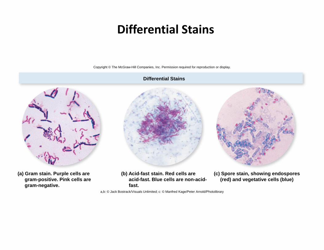
(c) Spore stain, showing endospores(red) and vegetative cells (blue)
(b) Acid-fast stain. Red cells areacid-fast. Blue cells are non-acid-fast.
(a) Gram stain. Purple cells aregram-positive. Pink cells aregram-negative.
a,b: © Jack Bostrack/Visuals Unlimited; c: © Manfred Kage/Peter Arnold/Photolibrary
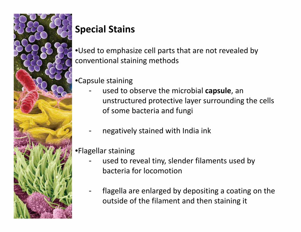
Special Stains
•Used to emphasize cell parts that are not revealed by
conventional staining methods
•Capsule staining
- used to observe the microbial capsule, an
unstructured protective layer surrounding the cells
of some bacteria and fungi
- negatively stained with India ink
•Flagellar staining
- used to reveal tiny, slender filaments used by
bacteria for locomotion
- flagella are enlarged by depositing a coating on the
outside of the filament and then staining it


![Trends and barriers to lateral gene transfer in prokaryotesacademic.uprm.edu/~lrios/4368/Popa2011_HGT_defense.pdf · into their genome in a process called lateral gene (LGT) [1]](https://img.pdfslide.us/doc/110x75/5ed66f096ff22a66535f499d/trends-and-barriers-to-lateral-gene-transfer-in-lrios4368popa2011hgtdefensepdf.jpg)