Embed Size (px)
Citation preview
L E O M A R T I N E Z , M . D .
U N I V E R S I T Y O F T E X A S M E D I C A L B R A N C H ( U T M B H E A L T H )
D E P A R T M E N T O F O T O L A R Y N G O L O G Y
G R A N D R O U N D S P R E S E N T A T I O N
N O V E M B E R 3 0 , 2 0 1 1
Tongue Anatomy and Glossectomy: 101
Outline
Relevant anatomy
History of glossectomy
Disease overview of the tongue
Glossectomy procedures
Reconstruction options
Conclusion and coding
Embryology
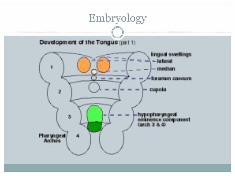
The tongue appears in the fourth week of life
It develops with the turberculum impar, which is a mesenchyme swelling in the floor of the primitive pharynx, cranially to the foremen cecum.
The anterior two thirds form from the first pharyngeal arches, lateral to the turberculum impar.
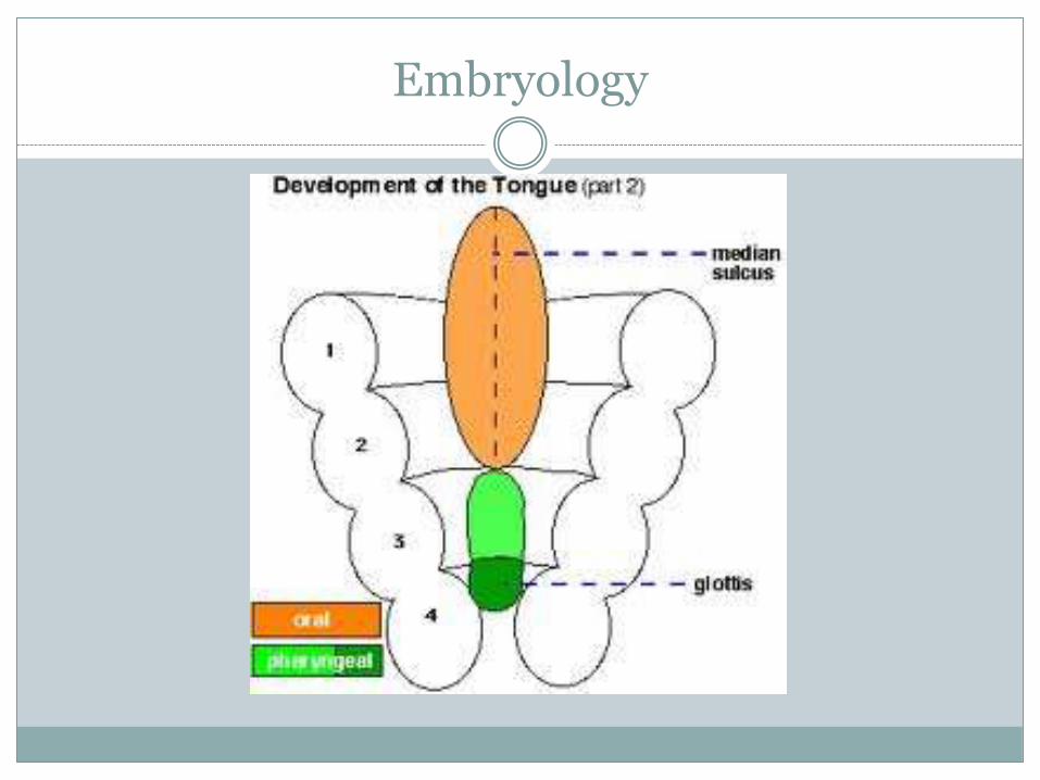
They fuse to form the medial sulcus of the tongue
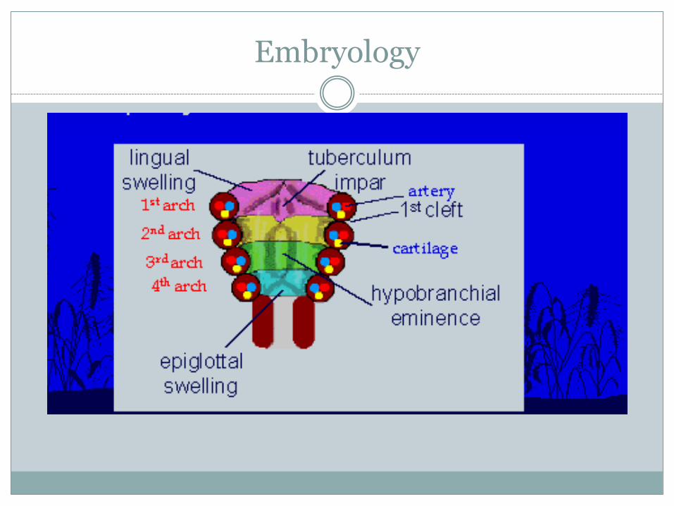
Embryology
Embryology
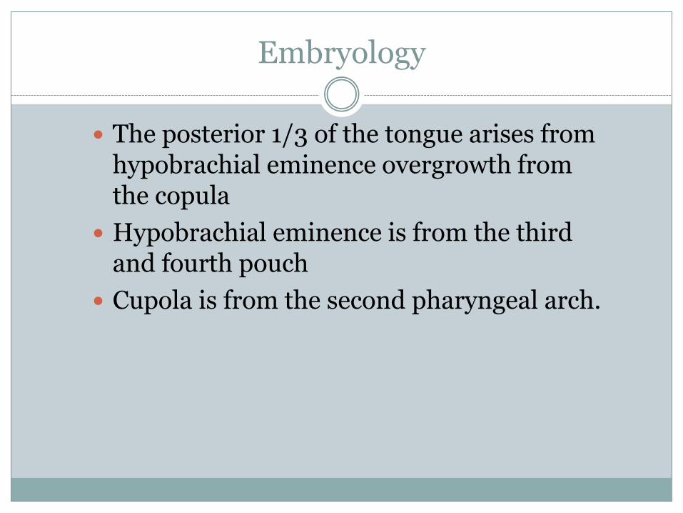
The posterior 1/3 of the tongue arises from hypobrachial eminence overgrowth from the copula
Hypobrachial eminence is from the third and fourth pouch
Cupola is from the second pharyngeal arch.
Embryology
Embryology
Embryology
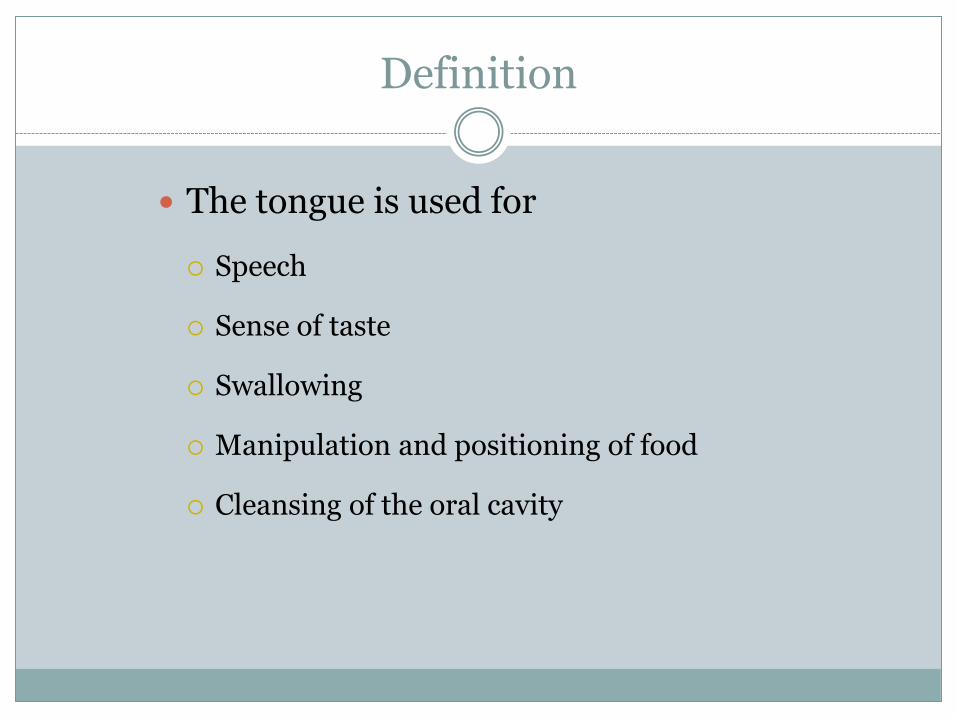
Definition
The tongue is used for
Speech
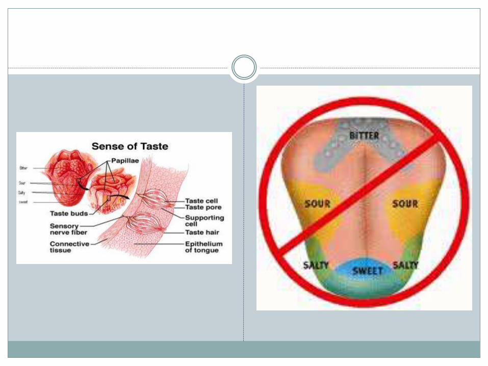
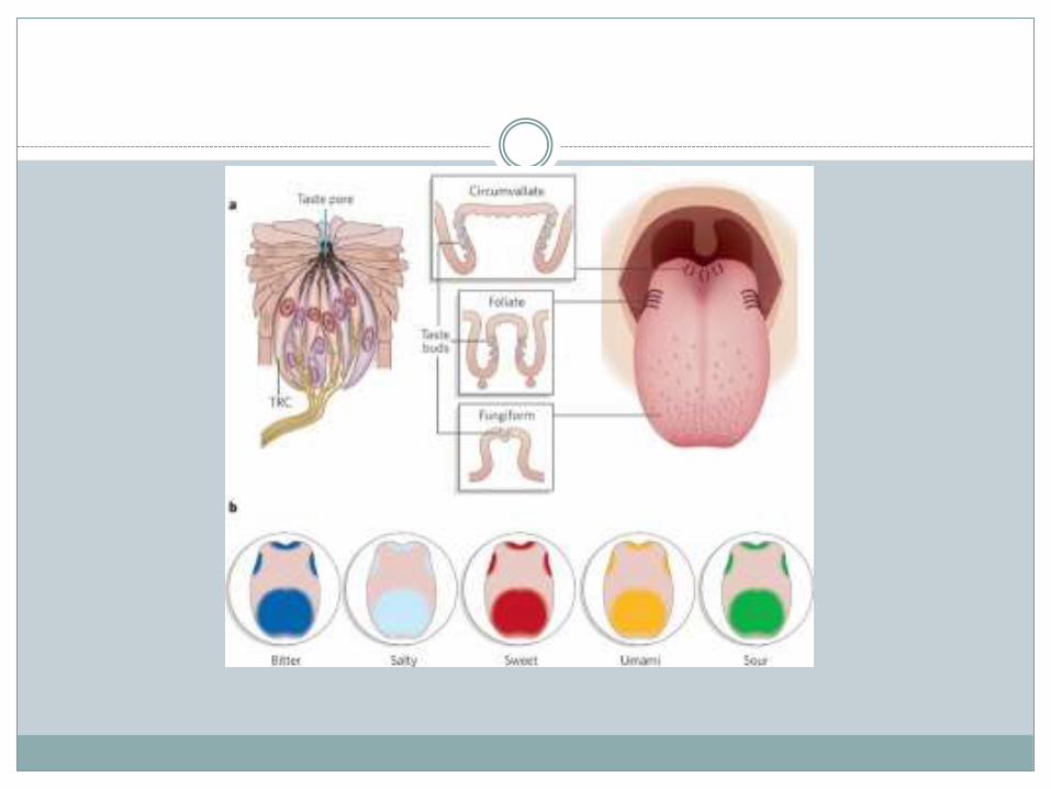
Sense of taste
Swallowing
Manipulation and positioning of food
Cleansing of the oral cavity
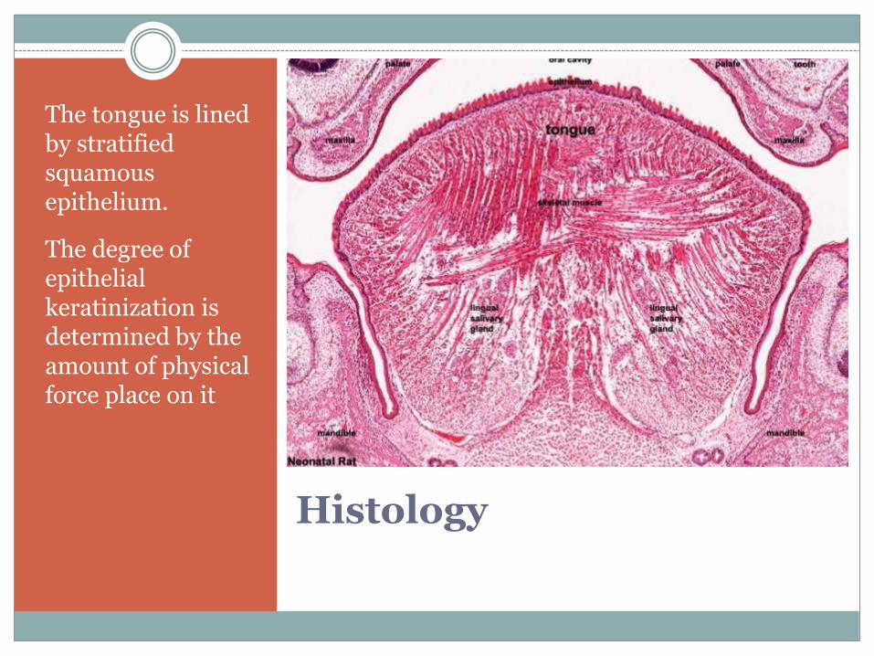
Histology
The tongue is lined by stratified squamous epithelium.
The degree of epithelial keratinization is determined by the amount of physical force place on it
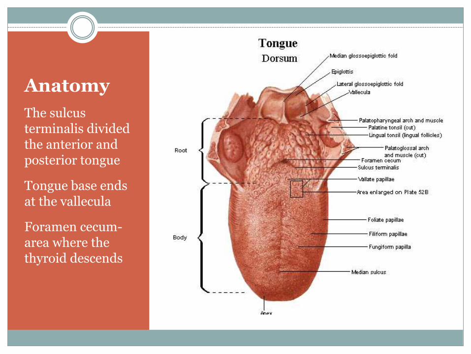
Anatomy
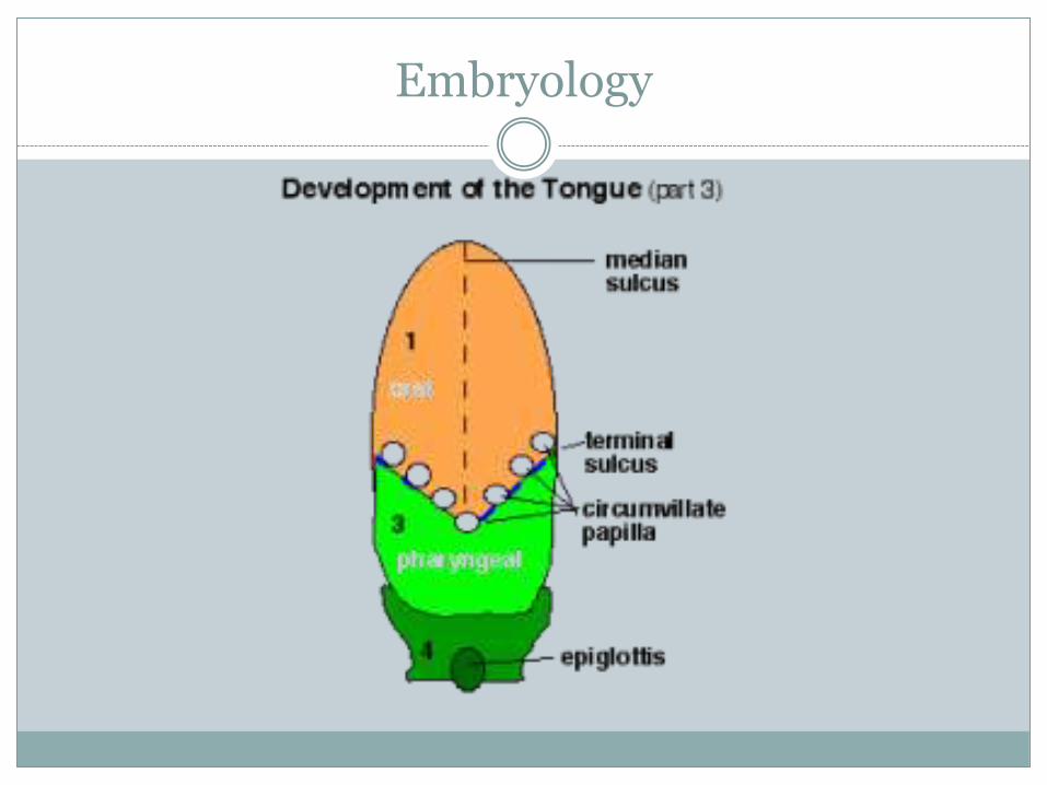
The sulcus terminalis divided the anterior and posterior tongue
Tongue base ends at the vallecula
Foramen cecum-area where the thyroid descends
Anatomy
There are 8 muscles of the tongue
They are classified as intrinsic and extrinsic muscles
Anatomy
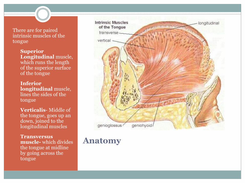
There are for paired intrinsic muscles of the tongue
Superior Longitudinal muscle, which runs the length of the superior surface of the tongue
Inferior longitudinal muscle, lines the sides of the tongue
Verticalis- Middle of the tongue, goes up an down, joined to the longitudinal muscles
Transversus muscle- which divides the tongue at midline by going across the tongue
Anatomy
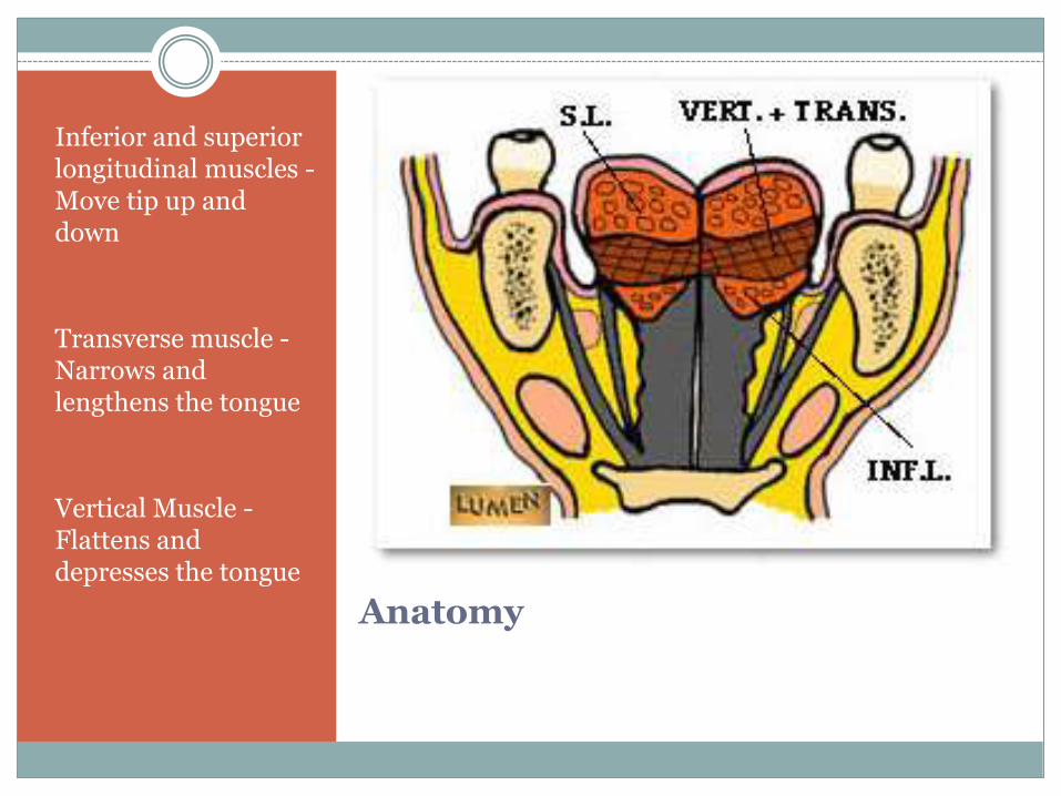
Inferior and superior longitudinal muscles - Move tip up and down
Transverse muscle -Narrows and lengthens the tongue
Vertical Muscle - Flattens and depresses the tongue
Anatomy
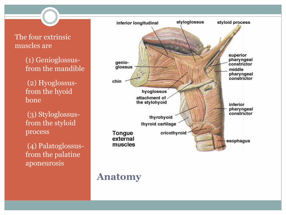
The four extrinsic muscles are
1) (1) Genioglossus-from the mandible
2) (2) Hyoglossus- from the hyoid bone
3) (3) Styloglossus- from the styloid process
4) (4) Palatoglossus- from the palatine aponeurosis
Anatomy
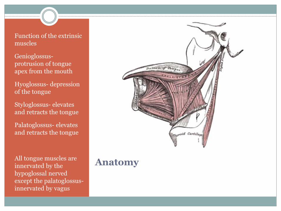
Function of the extrinsic muscles
Genioglossus- protrusion of tongue apex from the mouth
Hyoglossus- depression of the tongue
Styloglossus- elevates and retracts the tongue
Palatoglossus- elevates and retracts the tongue
All tongue muscles are innervated by the hypoglossal nerved except the palatoglossus- innervated by vagus

Anatomy
The main artery of the tongue is the lingual branch of the external carotid artery
Other contributors include the ascending palatine and tonsillar branch of the facial artery
There is an extensive submucosal plexus which is responsible for the vast blood supply of the tongue
Anatomy
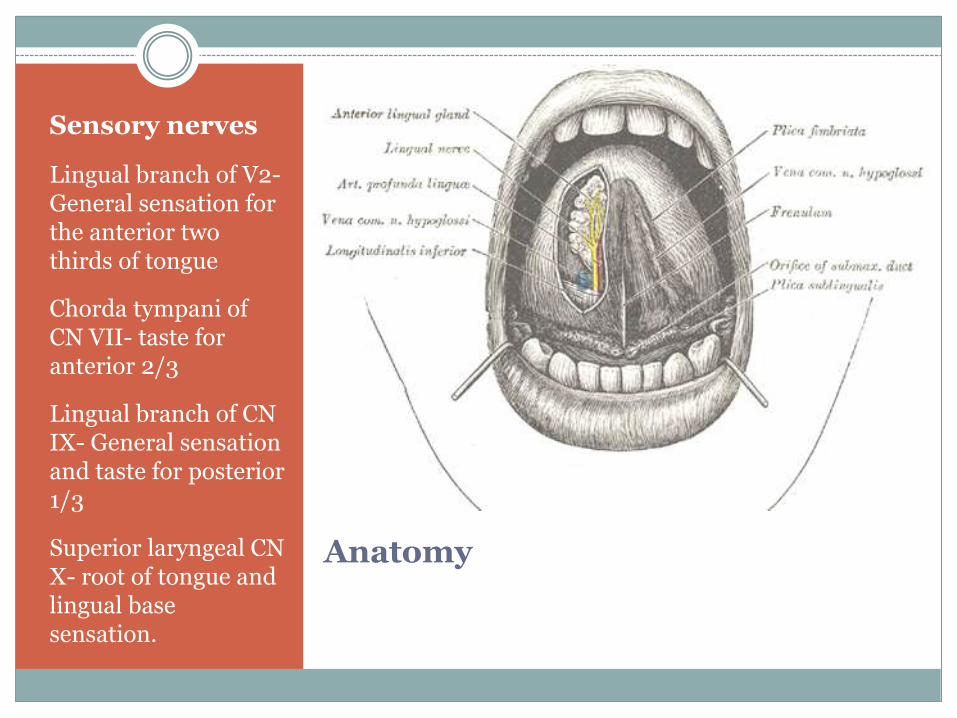
Sensory nerves
Lingual branch of V2-General sensation for the anterior two thirds of tongue
Chorda tympani of CN VII- taste for anterior 2/3
Lingual branch of CN IX- General sensation and taste for posterior 1/3
Superior laryngeal CN X- root of tongue and lingual base sensation.
History
The earliest description of tongue lesions came from the Egyptian Papyrus Ebers, originally thought around 1550 BC
The Egyptians tried milk gargles and various chewed concoction to treat tongue lesions
Other treatments over the years included sclerosing agents, leeches, surgical interventions
History
600-300 BC Sushruta produced surgical text describing macroglossia and procedure of rupturing the cyst of the tongue
Dark ages had Albucasis (1013-1106), which described different oral and tongue conditions, but the only surgical procedure described draining of ranula by forcing the fluid out.
History
Early surgical interventions included bleeding of the tongue, to surgically mauling of the tongue to cause scaring and contraction.
Case reports 1600- Sweden patient with macroglossia underwent surgical
resection of part of tongue that protruded from mouth, without cautery. Patient survived despite severe hemorrhage
1865- Ligation of external carotid on one side, and when that did not help, tied off common carotid on the other side. Tongue shrunk in size, and patient survived, although not sure quality of life.
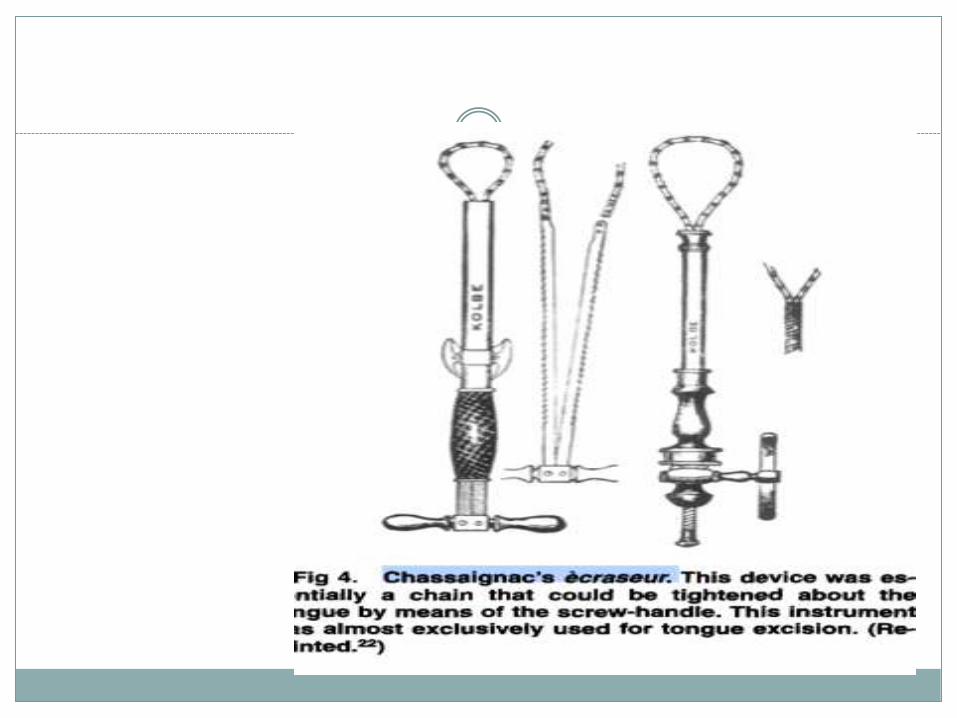
1900- French used an écraseur which was like a snare, but instead of wire, had chain links, with a handle to tighten the chain
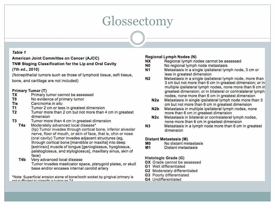
Glossectomy
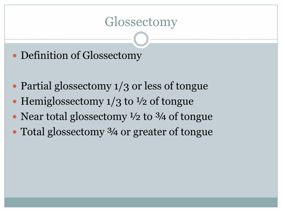
Definition of Glossectomy
Partial glossectomy 1/3 or less of tongue
Hemiglossectomy 1/3 to ½ of tongue
Near total glossectomy ½ to ¾ of tongue
Total glossectomy ¾ or greater of tongue
Glossectomy
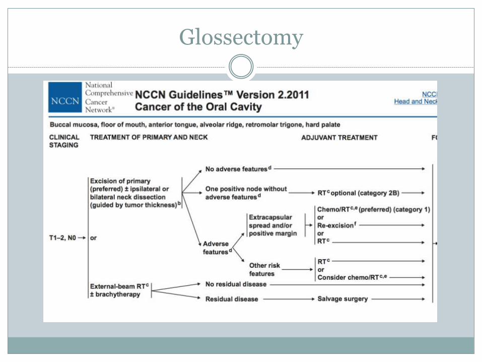
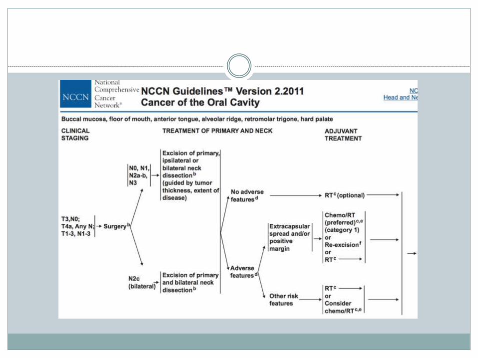
Treatment planning in patients with cancer of the tongue depends on involvement of the floor of mouth, mandible and other surrounding landmarks, the size of the cancer, and the presence of lymph node disease.
Oral Cancer may be treated with radiation or surgery, but surgery still remained the favored treatment of primary choice.
Glossectomy
For T1/T2 lesion, a transoral partial glossectomy provided adequate margins of resection.
This maintains articulation and swallowing function
However, even early stage cancer may be associated with rates of nodal metastasis of 30%.
Also, an increase in loco-regional and disease free survival after elective neck dissections for clinically tumor negative necks mandates aggressive treatment.
Glossectomy
For T3/T4 cancers, a hemiglossectomy or total glossectomy is usually necessary.
This is because they usually involve adjacent structures such as the floor of the mouth, tonsillar pillar, and or mandible
Glossectomy
A comprehensive treatment strategy has been developed by O’brien and associates which includes
Initial surgery for primary cancer
Preservation of mandible whenever possible
Selective neck dissection for level 1-4 negative neck, and modified radical (or radical) neck dissection for clinically positive neck
Tracheostomy for advance cancer
Glossectomy
Speech therapy after treatment
Postoperative radiation therapy for : T3/T4 primary cancer
Positive surgical margins (however ideally treated with resection)
Poor differentiation
Perineural invasion
Involvement of multiple nodes
Extracapsular spread of nodal disease
This applies to carcinoma of the tongue, but also can be used with other primary cancers of the oral cavity.
Glossectomy
Plan on use of partial glossectomy with primary closure or skin graft.
Selective neck dissection with N0/N1 and modified with N2 or greater neck disease
History of alcohol consumption is critical for prevention of delirium tremens in the postoperative period. Also concern over malnutrition.
Glossectomy
Patients should be examined for tongue motility and otalgia
Otalgia suggests perineural or deep invasion
Deviation of tongue suggests deep infiltration of muscularity of the tongue.
Exam should include evaluation of endophytic vs exophytic lesions
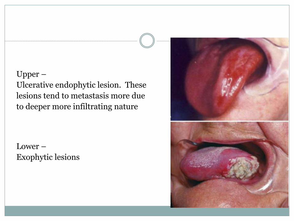
Upper –
Ulcerative endophytic lesion. These
lesions tend to metastasis more due
to deeper more infiltrating nature
Lower –
Exophytic lesions
Glossectomy
Sparana et al (2004) has suggested that tumor thickness is a factor in regional recurrence and survivorship.
Tumor of ≥ 4mm in thickness have been shown to have 59% chance of neck metastasis
Tumors of ≥ 3-4mm have shown to have worse regional recurrence and survivorship.
Glossectomy
Therefore, bimanual palpation of tumor size is essential.
A surface measurement should also be performed to properly stage the disease
As always, a complete examination of the oral cavity with indirect laryngoscopy should be performed.
Glossectomy
Complete dental evaluation is necessary.
Periodontal disease needs a dental consultation
Full mouth extractions are needed prior to radiation treatment to prevent osteoradionecrosis
Also, extraction of poor dentition will help with wound healing, as increased bacterial count is associated with periodontal
Glossectomy
Patients should have complete neck examination and a CT scan of the neck with contrast to evaluate for neck disease.
Sometimes, an MRI may help with depth determination and extend of involvement of the muscle fibers of the tongue. This information can be especially helpful with reconstruction planning.
Glossectomy
Glossectomy
Glossectomy
With T1/T2 lesions that are not biopsied previously, but show characteristic of carcinoma, an excisional biopsy as the partial glossectomy may be performed in the OR with a frozen section performed. This can done instead of subjecting the patient to outpatient biopsies. A neck dissection can then performed after the frozen biopsy confirms the disease
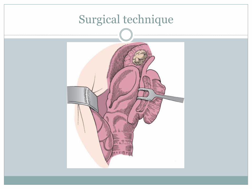
Surgical Technique
Patients should be placed under nasal tracheal intubation is no tracheostomy is planned. This will provide better exposure.
A tracheostomy should be performed with patients with extensive disease or patients who will require a skin graft with bolus.
Before any surgical intervention, a direct laryngoscopy and esophagoscopy should be performed
Surgical technique-Partial glossectomy
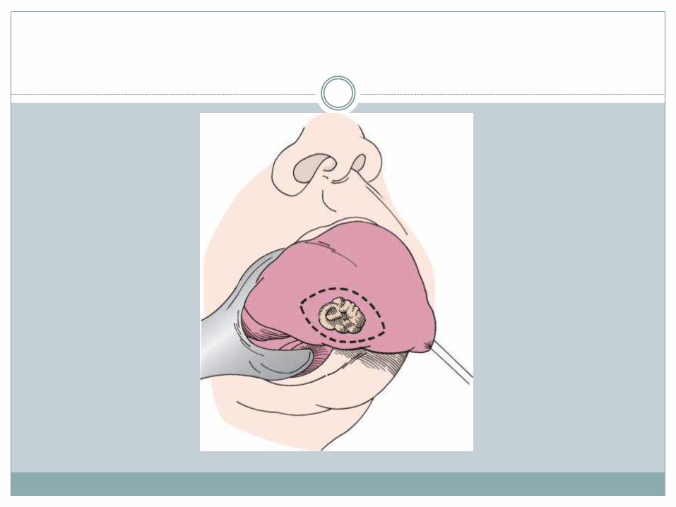
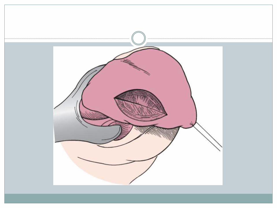
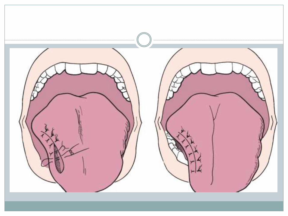
A mouth gag or bite block should be placed.
A silk suture is placed 1 cm behind the tip in the avascular midline and used to extract the tongue out.
At least a 1 cm margin of normal tissue is usually taken.
The specimen is then marked with sutures and ink margins.
Appropriate frozen section margins are taken as well
Surgical Technique
The hemiglossectomy is carried out the same way, except the incision is started at the midline raphe and carried down. The posterior and lateral cuts are then made to complete the resection, with appropriate margins.
This more extensive resection may not be able to closed primarily or if closed primarily, would cause significant restriction of the tongue.
Therefore a split thickness skin graft may be performed
Surgical Technique
Skin graft size of 0.016-0.018 are appropriate
Use of interrupted or quilting suture technique
Every other suture should be left long to provide adequate suture for the bolus.
Surgical Technique
With lesions that are T3/T4, management is more complex.
These may infiltrate the floor, more posteriorly, and involve the mandible
Also, tongue base lesions may be more difficult to manage.
They tend to be more infiltrated and poorly differentiated.
Usually a laryngectomy is needed if the lesion extends inferiorly to the hyoid
However, a larynx preserving procedure may be used if adequate margins is made posteriorly
Surgical technique
Successful rehabilitation after total glossectomy without laryngectomy is possible
Care should incorporate speech pathology, dietitian, and social workers
Surgical technique
When evaluating a patient with an extensive lesion, again CT scan is choice of radiograph
Soft tissue replacement becomes more of a concern.
The surgical approach depends on the following
Size
Location
Invasions of mandible
Need for neck dissection
Surgical technique
The main types of approaches to the primary tumor
Transoral
Midline mandibulectomy/Mandibulotomy
Neck approaches
Surgical technique
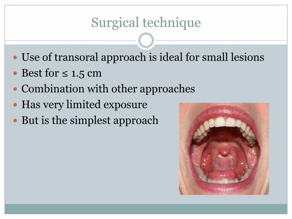
Use of transoral approach is ideal for small lesions
Best for ≤ 1.5 cm
Combination with other approaches
Has very limited exposure
But is the simplest approach
Surgical technique
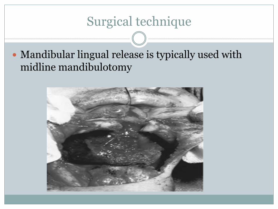
Mandibular lingual release is typically used with midline mandibulotomy
Surgical technique
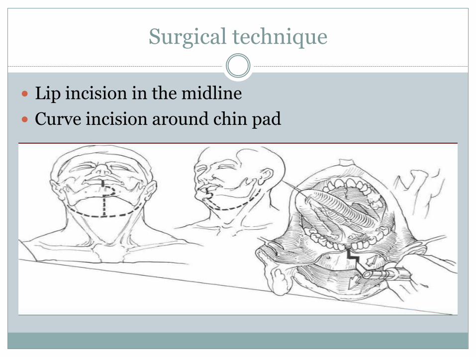
Lip incision in the midline
Curve incision around chin pad
Surgical technique
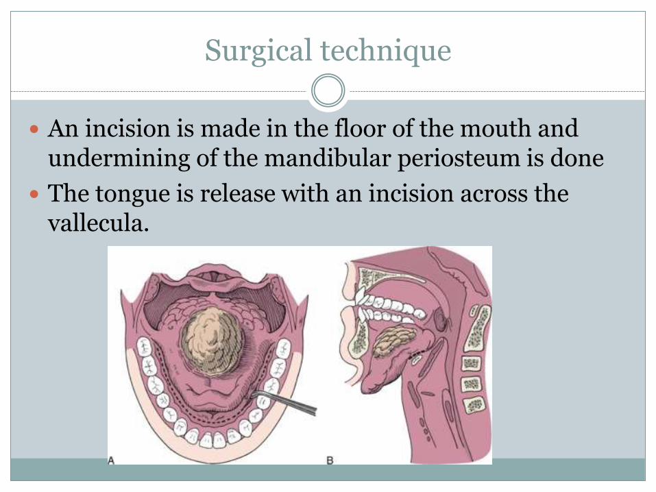
An incision is made in the floor of the mouth and undermining of the mandibular periosteum is done
The tongue is release with an incision across the vallecula.
Surgical technique
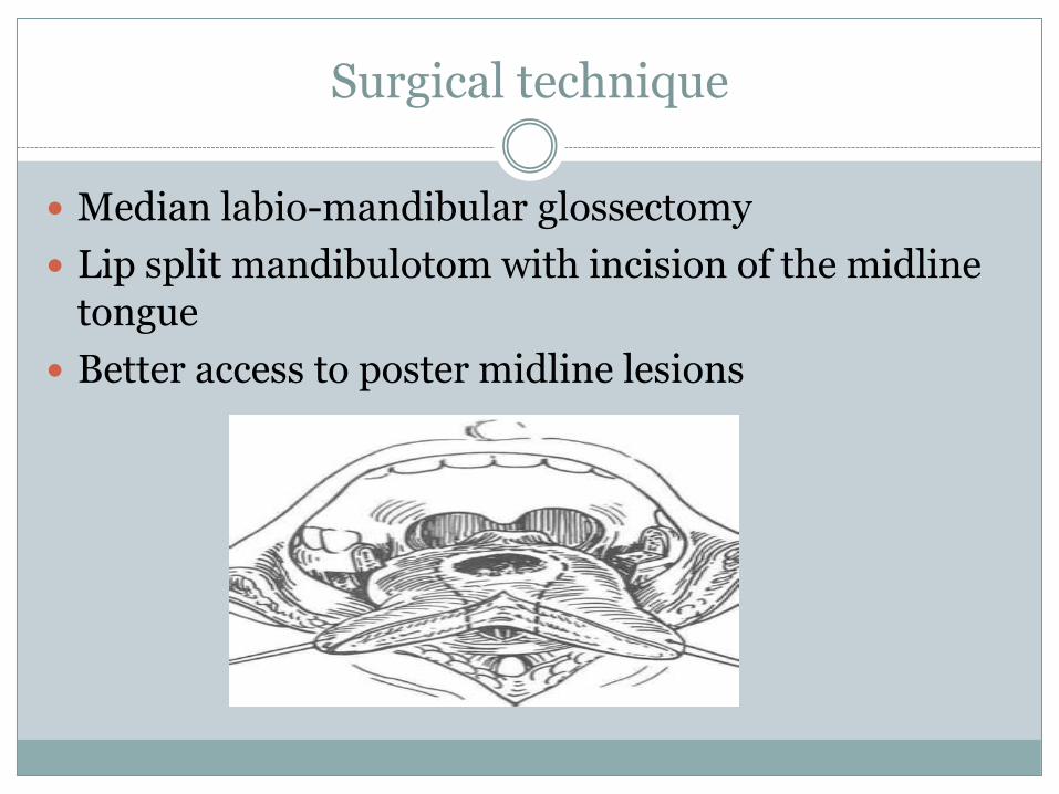
Median labio-mandibular glossectomy
Lip split mandibulotom with incision of the midline tongue
Better access to poster midline lesions
Surgical technique
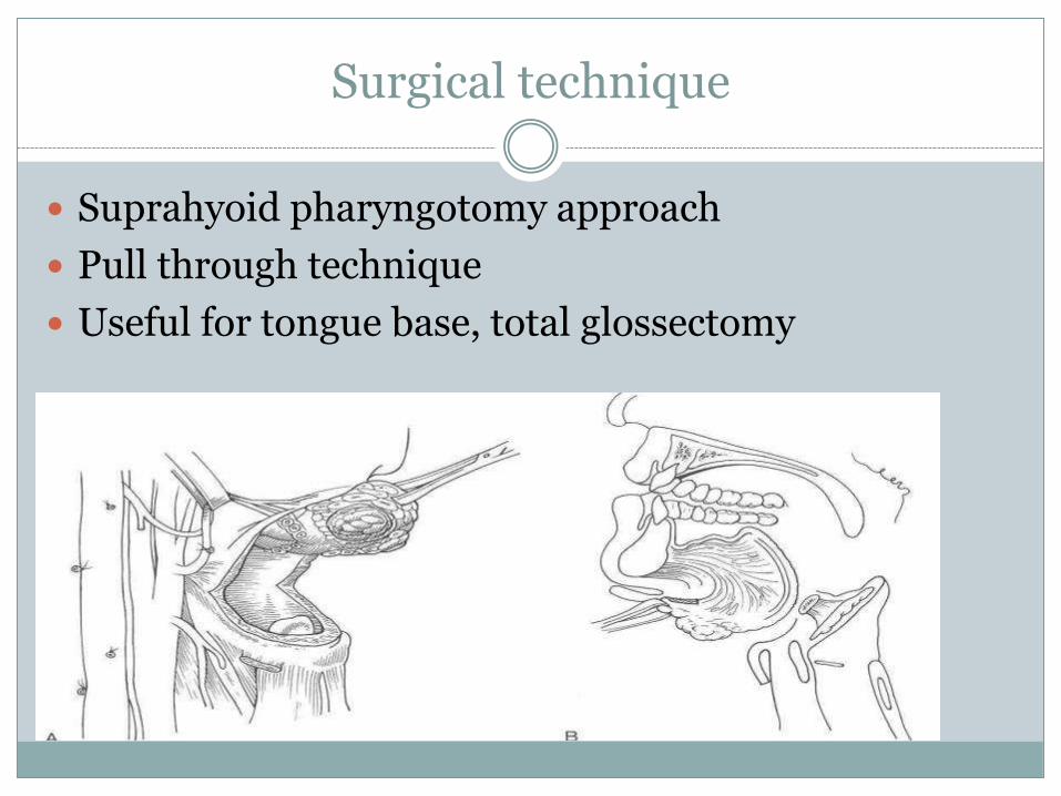
Suprahyoid pharyngotomy approach
Pull through technique
Useful for tongue base, total glossectomy
Surgical technique
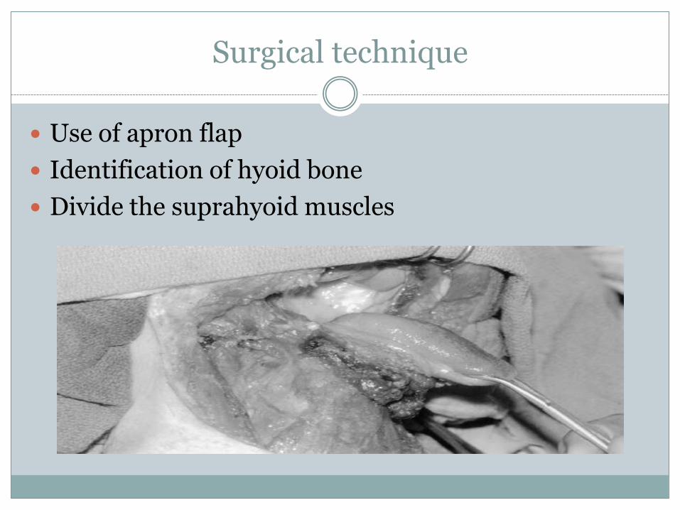
Use of apron flap
Identification of hyoid bone
Divide the suprahyoid muscles
Surgical technique
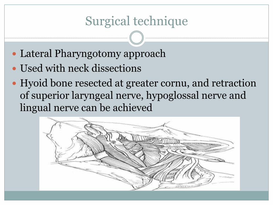
Lateral Pharyngotomy approach
Used with neck dissections
Hyoid bone resected at greater cornu, and retraction of superior laryngeal nerve, hypoglossal nerve and lingual nerve can be achieved
Surgical technique
Surgical technique
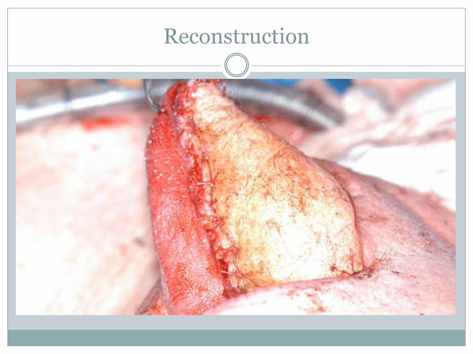
Reconstruction
As stated before, small defects can be closed primarily
Defect size of 1/3 the volume can be closed.
Resection of ½ the tongue results in loss of tongue bulk and scar contracture if limited reconstruction option are pursued
The decrease in lingual contact with the palate, lip, and teeth can result in impaired articulation and propulsion of food bolus.
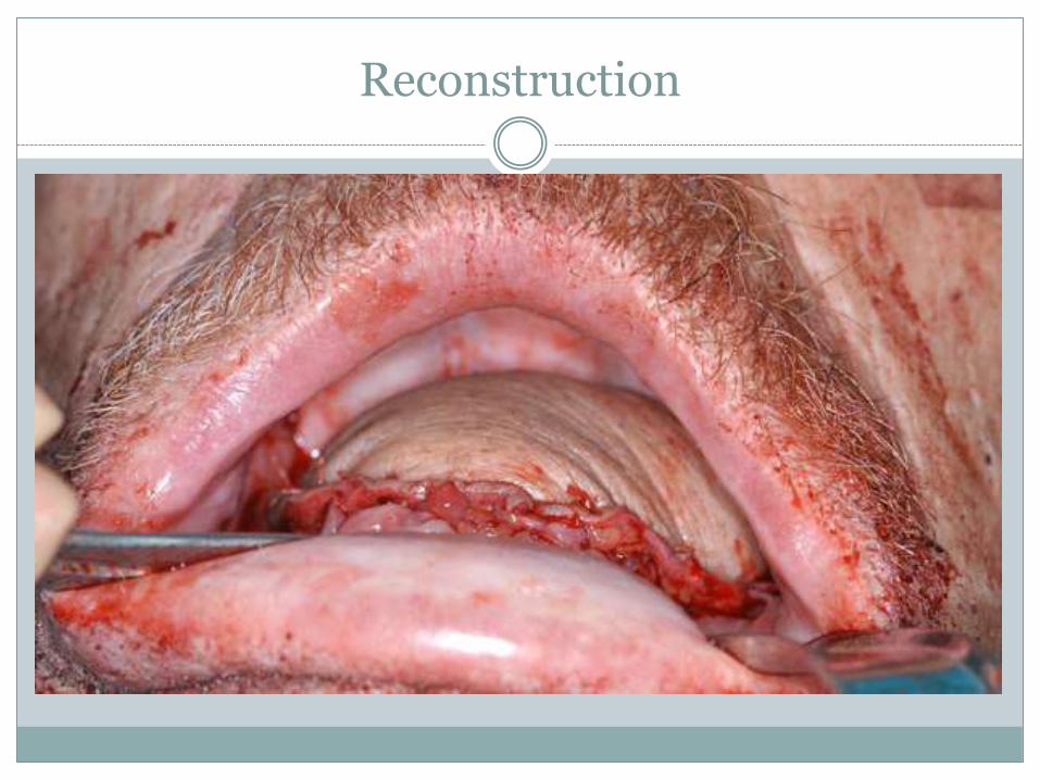
Reconstruction
Therefore larger than 1/3 defects required some form of reconstruction
Local flap vs regional flap can be used for this purpose
Goal is to achieve mastication, speech, and acceptable aesthetic results
Reconstruction
Local flaps
Limited amount of tissue
Inferior functional results
Not useful for tongue defects- can cause limited tongue motions
Tongue flap – divide tongue anteriorly and rotate posteriorly
Reconstruction
Regional flaps are well vascularized
Need single state reconstruction
Harvest not too difficult
However, they have limited reach,
Can be bulky
Necrosis at distal end
Reconstruction
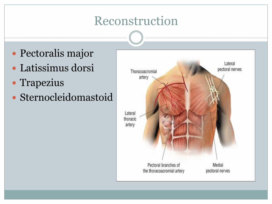
Pectoralis major
Latissimus dorsi
Trapezius
Sternocleidomastoid
Reconstruction
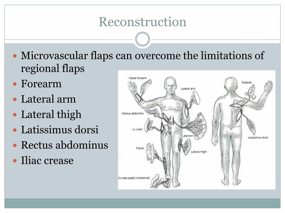
Microvascular flaps can overcome the limitations of regional flaps
Forearm
Lateral arm
Lateral thigh
Latissimus dorsi
Rectus abdominus
Iliac crease
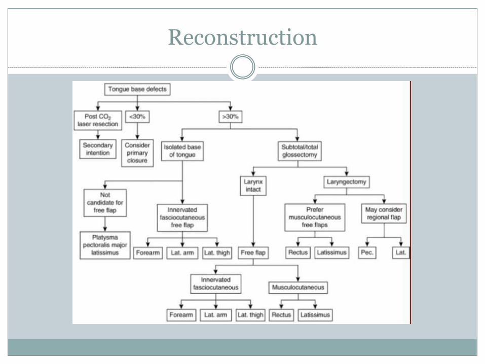
Reconstruction
The preferred method of reconstruction of hemiglossectomy to near total is free tissue flaps.
Able to select donor site to match requirements of defect
Also, donor site may provide tissue that is no subject to locoregion therapy such as radiation.
Reconstruction
Reconstruction
Reconstruction
Reconstruction
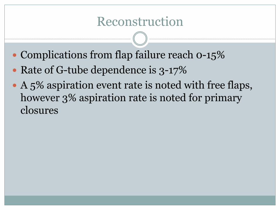
Complications from flap failure reach 0-15%
Rate of G-tube dependence is 3-17%
A 5% aspiration event rate is noted with free flaps, however 3% aspiration rate is noted for primary closures
Reconstruction
Regardless of treatment of tongue SCCA, surveillance is needed
1st year 1-3 months interval
2nd year 2-4 months intervals
3rd year 3-6 month intervals
4th and 5th year 4-6 month intervals
Yearly intervals after that
Reconstruction
Survival rates have been reported for five year as 82% with stage I or II disease, and 49% for stage III/IV
However, with recurrence, despite the type of salvage therapy offered, survival of five years is 15-35%
Coding
CHANGE IN KEY INDICATOR PROCEDURES
The Review Committee for Otolaryngology has made a change to one of the Key Indicator Procedures.
The Laryngectomy key indicator procedure category will be replaced with Glossectomy [Oral Cavity Resection]. This change will be incorporated into the summary data provided for the upcoming 2011–2012 otolaryngology residency graduates.
Coding
Glossectomy Procedures
When a significant portion of the tongue is removed, the procedure should be reported as a glossectomy codes. These codes vary according to
1) the amount of tongue that is removed,
2) whether a neck dissection was performed,
3) whether a resection of the floor of the mouth was also performed and
4) whether the mandible was resected.

Coding
Glossectomy; less than one-half tongue: 41120 Glossectomy; hemiglossectomy: 41130 Glossectomy; partial, with unilateral radical neck dissection: 41135 Glossectomy; complete or total, with or without tracheostomy, without radical neck
dissection: 41140 Glossectomy; complete or total, with or without tracheostomy, with unilateral
radical neck dissection: 41145 Excision of lesion of tongue with closure; posterior one-third: 41113\ Excision of lesion of tongue with closure; with local tongue flap: 41114 Excision of malignant tumor of mandible: 21044 Excision of malignant tumor of mandible; radical resection: 21045 Excision of benign tumor or cyst of mandible; requiring extra-oral osteotomy and
partial mandibulectomy (e.g., locally aggressive or destructive lesion(s)): 21047 Glossectomy; composite procedure with resection floor of mouth and mandibular
resection, without radical neck dissection: 41150 Glossectomy; composite procedure with resection floor of mouth, with suprahyoid neck dissection: 41153
Glossectomy; composite procedure with resection floor of mouth, mandibular resection, and radical neck dissection (Commando type): 4115
Coding
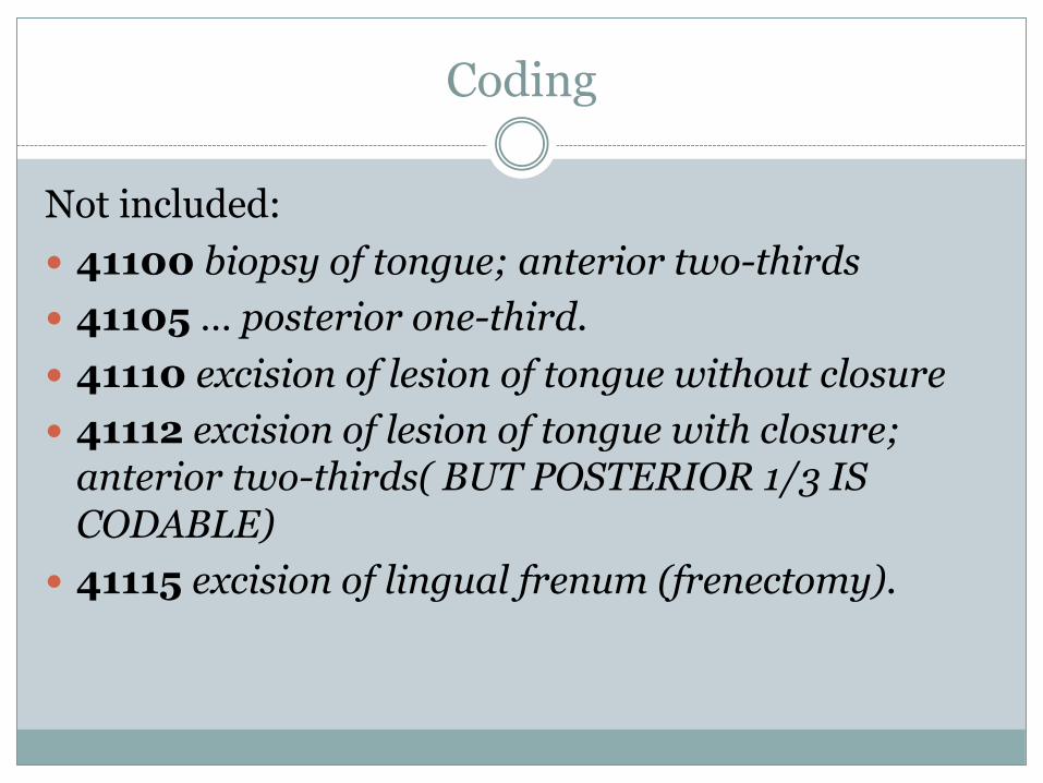
Not included:
41100 biopsy of tongue; anterior two-thirds
41105 … posterior one-third.
41110 excision of lesion of tongue without closure
41112 excision of lesion of tongue with closure; anterior two-thirds( BUT POSTERIOR 1/3 IS CODABLE)
41115 excision of lingual frenum (frenectomy).

Conclusion
Tongue anatomy and embryology provide useful information for surgical treatment
Varying degrees of surgical treatment options are available, depending on size of lesion
Careful and extensive workup is key before any surgical intervention
Reconstruction is challenging for tongue defects

Questions?
References
Bailey, B.J., et al. Surgery of the Oral Cavity Year Book Medical Publishers, Inc., Chicago, IL c. 1989.
Bailey, B.J., et al. Head & Neck Surgery—Otolaryngology Lippincott Williams & Wilkins, Philadelphia, PA, c. 2006.
Ferner, H. Eduard Pernkopf Atlas of Topographical and Applied Human Anatomy, Volume I, Urban & Schwarzenberg, Inc., Baltimore, MA, c.1980.
Lore, J.M., et al. An Atlas of Head and Neck Surgery W.B. Saunders Co., Philadelphia, PA, c.2006.
Myers, E.N. Operative Otolaryngology Head and Neck Surgery W.B. Saunders Co., Philadelphia, PA, c. 2008.
Warwick R., Williams, P.L. Gray’s Anatomy W.B. Saunders, Co., Philadelphia, PA, c. 2010.
Morton DA, Foreman KB, Albertine KH. Chapter 24. Oral Cavity. In: Morton DA, Foreman KB, Albertine KH, eds. The Big Picture: Gross Anatomy. New York: McGraw-Hill; 2011. http://www.accessmedicine.com/content.aspx?aID=8667503. Accessed November 22, 2011
Dhillon N. Chapter 1. Anatomy. In: Lalwani AK, ed. CURRENT Diagnosis & Treatment in Otolaryngology—Head & Neck Surgery. 2nd ed. New York: McGraw-Hill; 2008. http://www.accessmedicine.com/content.aspx?aID=2823000. Accessed November 22, 2011.
http://emedicine.medscape.com/article/1890823-overview#a15
http://www.supercoder.com/articles/articles-alerts/otc/glossectomy-location-size-and-procedures-are-key-to-coding-accuracy/
Duvvuri U, Simental Jr AA, D'Angelo G, et al: Elective neck dissection and survival in patients with squamous cell carcinoma of the oral cavity and oropharynx. Laryngoscope 2004; 114:2228-2234.
O'Brien CJ, Lee KK, Castle GK, Hughes CJ: Comprehensive treatment strategy for oral and oropharyngeal cancer. Am J Surg 1992; 164:582-586.
Sparano A, Weinstein G, Chalian A, et al: Multivariate predictors of occult neck metastasis in early oral tongue cancer. Otolaryngol Head Neck Surg 2004; 131:472-476.